Progressive Increase in Telomerase Activity From Benign Melanocytic Conditions to Malignant Melanoma

Pharmacological Research 51 (2005) 437–443
Induction of apoptosis and inhibition of telomerase activity by aqueousextract fromPlatycodon grandiflorumin human lung carcinoma cells
Dong Il Parka, Jae Hun Leea, Sung-Kwon Moonb, Cheorl-Ho Kimc, Yong Tae Leea,JaeHun Cheongd, Byung Tae Choia, Yung Hyun Choia,∗
a Department of Oriental Medicine, Dongeui University College of Oriental Medicine and Research Institute of Oriental Medicine,Busan 614-052, Republic of Korea
b Department of Food and Biotechnology, Chungju National University, Chungju 380-702, Republic of Koreac Department of Oriental Medicine, Dongguk University College of Oriental Medicine, Kyungju 780-714, Republic of Korea
d Department of Molecular Biology, Pusan National University, Busan 609-735, Republic of Korea
Accepted 4 November 2004
Abstract
t ition andi w cytometrya of caspase-3.A telomeraser treatment.T egulation ofB©
K
1
pttnappod
c
o the
kary-rma-nomer-
rsible
nsateacti-heirggestlularssionlengthnts;
1d
The objective of the present study was to investigate the effect of aqueous extract from the root ofPlatycodon grandiflorum(AEPG) onhe cell growth and apoptosis in human lung carcinoma cell line A549. Exposure of A549 cells to AEPG resulted in growth inhibnduction of apoptosis in a dose-dependent manner as measured by hemocytometer counts, fluorescence microscopy and flonalysis. This increase in apoptosis was associated with a decrease in Bcl-2 expression, an increase of Bax and an activationEPG treatment markedly inhibited the activity of telomerase in a dose-dependent fashion. Additionally, the expression of human
everse transcriptase (hTERT), a main determinant of the telomerase enzymatic activity, was progressively down-regulated by AEPGhese findings suggest that the apoptotic events by AEPG were associated with the diminished telomerase activity and down-rcl-2 expression.2004 Elsevier Ltd. All rights reserved.
eywords: Platycodon grandiflorum; Apoptosis; Bcl-2; Telomerase; hTERT
. Introduction
It is now well accepted that apoptosis is a physiologicalhenomenon that plays an important role in the regulation of
issue development and homeostasis. Deregulation of apop-osis has been shown to contribute to the pathogenesis of aumber of human diseases including cancer[1,2]. There arepparently many factors, including Bcl-2 family and caspaseroteases, involved in the apoptotic process through the ex-ression of genes, and the characterization of the functionf these gene products will help to define the process of celleath at the molecular levels[3–5].
In addition to deregulation of apoptosis, it is increasinglylear that the process of neoplasia is characterized by the
∗ Corresponding author. Tel.: +82 51 850 7413; fax: +82 51 853 3578.E-mail address:[email protected] (Y.H. Choi).
activation of telomerase that adds telomeric repeats tends of replicating chromosomes, telomeres[6,7]. Telom-eres are essential units that stabilize the ends of euotic chromosome and prevent the loss of genetic infotion. In normal human somatic cells, which show little ortelomerase activity to synthesize new telomeres, the teloicDNAs progressively shorten with each cell division[6,8].Critically short telomeres are suggested to cause irrevecell growth arrest and cellular senescence[6,7,9,10]. Con-versely, most tumor cells have mechanisms that compefor telomere shortening, most commonly through thevation of telomerase, allowing them to stably maintain ttelomeres and grow indefinitely. These observations suthat telomerase reactivation is a rate-limiting step in celimmortality and carcinogenesis, and telomerase reprecan act as a tumor-suppressive mechanism. Telomerein human is primarily controlled by three major compone
043-6618/$ – see front matter © 2004 Elsevier Ltd. All rights reserved.oi:10.1016/j.phrs.2004.11.003
438 D.I. Park et al. / Pharmacological Research 51 (2005) 437–443
the human telomerase RNA (hTR), telomerase-associatedprotein-1 (TEP-1) and the human telomerase reverse tran-scriptase (hTERT). There is a good correlation between ex-pression of hTERT mRNA and the presence of telomeraseactivity in extracts from tissue culture cells and normaland cancer tissues[7,11,12]. Concomitant up-regulation ordown-regulation of the hTERT mRNA expression and telom-erase activity during cell immortalization or differentiationis observed, suggesting that control of the hTERT expres-sion at the mRNA level mainly contributes to the regula-tion of telomerase enzymatic activity[13–15]. However, themolecular mechanisms by which telomerase activity is regu-lated in concordance with cell growth properties remain un-clear.
Platycodi Radix, the root ofPlatycodon grandiflorumA.DC (Campanulaceae), commonly known as Doraji in Korea(Chinese name, ‘Jiegeng’, and Japanese name, ‘Kikyo’) hasbeen used as an expectorant in traditional Oriental medicine.In Korea, Platycodi Radix is also used as a food and em-ployed as a folk remedy for adult diseases, such as, bronchi-tis, asthma and pulmonary tuberculosis, hyperlipidemia, di-abetes, and inflammatory diseases, and as a sedative[16,17].Several studies on its chemical and immunopharmacologicaleffects including immunostimulation and antitumor activityhave been performed[18–23]. However, the relevant molecu-l y wasp eousea lungc 549c hibi-t wasa andd
2
2
i-v ractf -p t9 aturem omt llowe
2
inedf )a rs-b BS)a ith
5% CO2. For growth-inhibition analysis, cells were seededand exposed to various concentrations of AEPG for 48 h.The cells were trypsinized, washed with phosphate-bufferedsaline (PBS) and the viable cells were scored with a hemo-cytometer through exclusion of trypan blue.
2.3. Flow cytometry analysis
After treatment with AEPG the cells were collected bytrypsinization, washed with cold PBS, and resuspended inPBS. DNA contents of cells were measured using a DNAstaining kit (CycleTESTTM PLUS kit, Becton Dickinson,Heidelberg, Germany). Propidium iodide-stained nuclearfractions were obtained by following the kit protocol. Flu-orescence intensity was determined using a FACScan flowcytometer and analyzed by CellQuest software (Becton Dick-inson).
2.4. Nuclear staining with DAPI
Cells were washed with PBS and fixed with 3.7%paraformaldehyde in PBS for 10 min at room temperature.Fixed cells were washed with PBS, and stained with 4,6-diamidino-2-phenylindole (DAPI, Sigma Chemical Co., St.Louis, MO) solution for 10 min at room temperature. Thec yzedv
2
re-v er-m er-c epa-r thene icher& ctedt byt shamC toc anda Inc.( odiesw
2
en,L yn-t rip-t truc-t t in aM s ofd at7 ersw
ar mechanisms are poorly understood. The present studerformed to investigate the apoptotic pathway by aquxtract from the root ofPlatycodon grandiflorum(AEPG)nd AEPG’s effect on telomerase activity in a humanancer cell line A549. We report here that exposure of Aells to AEPG resulted in a dose-dependent growth inion and apoptosis. This increase in apoptosis by AEPGccompanied by down-regulation of Bcl-2 expressioniminished telomerase activity.
. Materials and methods
.1. Plant material
Platycodon grandiflorumwas supplied by Dongeui Unersity Oriental Hospital, Busan, Korea. Aqueous extrom the root ofPlatycodon grandiflorum(AEPG) was preared as previously described[21]. In brief, distilled water a5 CircC was added to powdered roots and the temperaintained for 10 h. The mixture was allowed to cool to ro
emperature, then filtered and lyophilized. The pale–yextract was dissolved directly in distilled water.
.2. Cell culture and growth-inhibition study
The human lung carcinoma cell line A549 was obtarom American Type Culture Collection (Rockville, MDnd cultured in RPMI 1640 medium (Gibco BRL, Gaitheurg, MD) supplemented with 10% fetal bovine serum (Fnd 1% penicillin–streptomycin in a 37 CircC incubator w
ells were washed two more times with PBS and analia a fluorescence microscope.
.5. Protein extraction and Western blot analysis
Cell lysates were lysed in extraction buffer as piously described[24]. Protein concentration was detined with a Bio-Rad protein assay kit (Bio-Rad, H
ules, CA). For Western blot analysis, proteins were sated by SDS–polyacrylamide gel electrophoresis andlectrotransferred to a nitrocellulose membrane (Schle
Schuell, Keene, NH). The membranes were subjeo immunoblot analysis and proteins were visualizedhe enhanced chemiluminescence (ECL) method (Amerorp., Arlington Heights, IL). Monoclonal antibodiesaspase-3 and Bcl-2, and polyclonal antibodies to Baxctin were purchased from Santa Cruz BiotechnologySanta Cruz, CA). Peroxidase-labeled secondary antibere purchased from Amersham Corp.
.6. RNA extraction and RT-PCR
Total RNA was prepared using an RNeasy kit (Qiaga Jolla, CA) and primed with random hexamers to s
hesize complementary DNA using AMV reverse transcase (Amersham) according to the manufacturer’s insions. Polymerase chain reaction (PCR) was carried ouastercycler (Eppendorf, Hamburg, Germany) with cycleenaturation at 94◦C, annealing at 58◦C, and extension2◦C for 30 s, respectively. The following sets of primere used in PCR amplification: Bax (sense: 5′-ATG GAC
D.I. Park et al. / Pharmacological Research 51 (2005) 437–443 439
GGG TCC GGG GAG-3′, antisense: 5′-TGG AAG AAGATG GGC TGA-3′), Bcl-2 (sense: 5′-CAG CTG CAC CTGACG-3′, antisense: 5′-GCT GGG TAG GTG CAT-3′), hTERT(sense: 5′-TGA ACT TGC GGA AGA CAG TGG-3′, anti-sense: 5′-ATG CGT GAA ACC TGT ACG CCT-3′), TEP-1 (sense: 5′-TCA AGC CAA ACC TGA ATC TGA G-3′,antisense: 5′-CCC CGA GTG AAT CTT TCT ACG C-3′),hTR (sense: 5′-GAA GGG CGT AGG CGC CCG TGC TTTTGC-3′, antisense: 5′-GTT TGC TCT AGA ATG AAC GGTGGA AGG-3′) and glyceraldehyde-3-phosphate dehydroge-nase (GAPDH, sense: 5′-CGG AGT CAA CGG ATT TGGTCG TAT-3′; antisense: 5′-AGC CTT CTC CAT GGT GGTGAA GAC-3′). Amplification products obtained by PCRwere electrophoretically separated on a 1% agarose gel andvisualized by ethidium bromide (EtBr, Sigma) staining.
2.7. Caspase-3 activity assay
Caspase-3 activity was assayed by cleavage of Asp–Glu–Val–Asp (DEVD)-pNA using a commercially available kit(Clontech, Palo Alto, CA). Briefly, cells were cultured in thepresence or absence of AEPG, and then 2× 10⁶ cells wereincubated with DEVD-pNA in the presence of dithiothreitolfor 60 min at 37◦C. The reaction was measured by changesin absorbance at 405 nm using the VERSAmax tunable mi-c
2
asedt e-l gerM fac-t G,h n2 the
TRAP reaction, 2�l of cell extract (containing 2�g protein)was added to 25�l of reaction mixture with the appropriateamount of sterile water to make a final volume of 50 ml. PCRwas performed in a Mastercycler as follows: primer elonga-tion (30 min, 25◦C), telomerase inactivation (5 min, 94◦C),product amplification by the repeat of 30 cycles (94◦C for30 s, 50◦C for 30 s, 72◦C for 90 s). Hybridization and theELISA reaction were carried out following the manufac-turer’s instructions. The extract of HeLa cells was used aspositive controls. 10�l of HeLa cell extract was incubatedwith DNase-free RNase (1�g/�l) for 20 min at 37◦C, and2�l of the RNase-treated extract was used as negative con-trols.
3. Results
3.1. Growth inhibition by AEPG
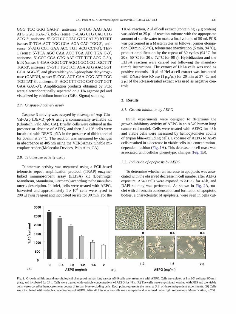
Initial experiments were designed to determine thegrowth-inhibitory activity of AEPG in an A549 human lungcancer cell model. Cells were treated with AEPG for 48 hand viable cells were measured by hemocytometer countsof trypan blue-excluding cells. Exposure of AEPG to A549cells resulted in a decrease in viable cells in a concentration-d asa
3
asso-c EPGe andDc oticb cul-
F ncer Ap rations d the vc cells. E ellsw bation ion,
roplate reader (Molecular Devices, Palo Alto, CA).
.8. Telomerase activity assay
Telomerase activity was measured using a PCR-belomeric repeat amplification protocol (TRAP) enzyminked immunosorbent assay (ELISA) kit (Boehrin
annheim, Mannheim, Germany) according to the manuurer’s description. In brief, cells were treated with AEParvested and approximately 1× 10⁶ cells were lysed i00�l lysis reagent and incubated on ice for 30 min. For
ig. 1. Growth inhibition and morphological changes of human lung calate, and incubated for 24 h. Cells were treated with variable concentells were scored by hemocytometer counts of trypan blue-excludingere incubated with variable concentrations of AEPG. After 48 h incu
ependent fashion (Fig. 1A). This decrease in cell mass wssociated with cellular phenotypic changes (Fig. 1B).
.2. Induction of apoptosis by AEPG
To determine whether an increase in apoptosis wasiated with the observed decrease in cell number after Axposure, A549 cells were exposed to AEPG for 48 h,API staining was performed. As shown inFig. 2A, nu-lei with chromatin condensation and formation of apoptodies, a characteristic of apoptosis, were seen in cells
549 cells after treatment with AEPG. Cells were plated at 1× 10³ cells per 60-mmof AEPG for 48 h. (A) The cells were trypsinized, washed with PBS aniableach point represents the mean± S.E. of three independent experiments. (B) C
cells were sampled and examined under light microscopy. Magnificat×200.

440 D.I. Park et al. / Pharmacological Research 51 (2005) 437–443
tured with AEPG in a dose-dependent manner, but very fewin control cells. Thus we quantified the extent of apopto-sis by measuring the fraction of nuclei that contained sub-diploid DNA content using flow cytometry. As shown bydata inFig. 2B, while the induction of apoptosis was almostnegligible (3.2 and 4.3% compared to 2.6% of control) atthe low concentrations (0.4 and 0.8 mg/ml), the high con-centrations resulted in an increase in apoptosis (14.5, 18.2and 24.9% at 1.2, 1.6 and 2.0 mg/ml) as determined by flowcytometry.
3.3. Down-regulation of Bcl-2 and activation ofcaspase-3 by AEPG
To investigate the apoptotic cascades involved by AEPG,cells were exposed to AEPG and the levels of Bax, Bcl-2,caspase-3 and cytochromecexpression, and in vitro caspase-3 activity levels were measured. Western immunoblottingand RT-PCR indicated that treatments of cells with AEPGmarkedly resulted in a down-regulation of Bcl-2, however,Bax expression was up-regulated (Fig. 3A and B). As shownin Fig. 3C, AEPG-induced apoptosis was also associatedwith a decreased expression of the pro-caspase-3 proteinand a concomitant increase of the active subunit of caspase-3. Additionally, lysates equalized for protein from cellst 3 ac-t in-c rsedb HO( osisi ea cellsu ria.T tot t man-
ner (Fig. 3C), suggesting that AEPG-induced apoptosis inA549 cells is mediated, at least in part, by the mitochondrial-signaling pathway.
3.4. Down-regulation of telomerase activity and hTERTexpression by AEPG
To examine the effect of AEPG on telomerase activity,cells were cultured in the absence or presence of AEPG for48 h, and telomerase activity was measured by TRAP-ELISAkit. As shown inFig. 4A, a reduction of telomerase activ-ity in A549 cells was observed with AEPG treatment. Ofthe components comprising telomerase, hTERT is a criti-cal determinant of the enzyme activity of telomerase. Wetherefore examined changes in hTERT expression on treat-ment with AEPG using the RT-PCR assay. As indicated inFig. 4B, hTERT mRNA expression but not hTR and TEP-1 was markedly decreased by treatment with AEPF in aconcentration-dependent fashion suggesting that the repres-sion of telomerase activity by AEPG was associated withdown-regulation of hTERT mRNA.
4. Discussion
andi andB mul-t cl-2r at ap -h o-p f thea ther and
F ere in ncubationa ed with df dent in y. The profir nts the±
reated with AEPG were assayed for in vitro caspase-ivity using DEVD-pNA as a substrate. AEPG exposurereased caspase-3 activity, which could be partially revey the addition of the caspase-3 inhibitor, Ac-DEVD-CFig. 3D). To examine whether the AEPG-induced apopts mediated by the release of mitochondrial cytochromc,
cytosolic fraction was prepared from AEPG treatednder conditions that maintain the integrity of mitochondhe amount of cytochromec released from mitochondria in
he cytosol was markedly increased in a dose-dependen
ig. 2. Exposure of A549 cells to AEPG induces apoptosis. (A) Cells wt room temperature, the cells were washed with PBS and photograph
or 48 h with increasing concentration of AEPG showed a dose-depenepresents the increase of sub-G1 population and each point represe
The regulation of apoptosis is a complex processnvolves a number of gene products including Bcl-2ax. It has been reported that Bcl-2 protects against
iple signals that lead to cell death, indicating that Begulates a common cell death pathway and functionsoint where various signals converge[4,5]. Bcl-2 acts to inibit cytochromec translocation from mitochodria to cytlasm, thereby blocking the caspase activation step opoptotic process[3,4]. Thus, it has been suggested thatatio between the level of pro-apoptotic Bax protein
cubated with AEPG for 48 h and then stained with DAPI. After 10 min ia fluorescence microscope using blue filter. Magnification,×400. (B) Cells treate
crease in the number of apoptotic cells as measured by flow cytometrlemeanS.E. of three independent experiments.

D.I. Park et al. / Pharmacological Research 51 (2005) 437–443 441
Fig. 3. AFPG exposure inhibits Bcl-2 expression and increases caspase-3 activity in A549 cells. (A) Western blot analysis of Bax and Bcl-2 after AEPGtreatment for 48 h. Whole-cell extracts were prepared, and the expression levels of Bax and Bcl-2 protein were examined by Western blotting with anti-Baxand Bcl-2 antibodies, and ECL detection system. Actin was used as an internal control. (B) After 48 h incubation with AEPG, total RNA was isolated usinganRNA Zol B reagent and RT-PCR was performed using Bax and Bcl-2 primers. The amplified PCR products were run in a 1% agarose gel and visualized by EtBrstaining. GAPDH was used as a house-keeping control gene. (C) Following treatment with different concentrations of AEPG for 48 h, the cytosolic fractionswere prepared from cells and then subjected to immunoblot analysis with antibodies to cytochromec and ECL detection. (D) Cell lysates from cells treatedwith AEPG for 48 h were assayed for in vitro caspase-3 activity using DEVD-pNA as a substrate. AEPG exposure markedly increased caspase-3 activity, whichcould be partially reversed by the addition of the caspase-3 inhibitor, Ac-DEVD-CHO. Each point represents the mean± S.E. of three independent experiments.
that of the anti-apoptotic factor Bcl-2 determines whethera cell responds to an apoptotic signal. The present dataclearly demonstrated that AEPG induces apoptosis in humanlung carcinoma A549 cells, which appears to account for itsanti-proliferating activity. In the present study, the levels ofBcl-2 mRNA and protein expression were down-regulatedby AEPG treatment in a concentration-dependent fashion,whereas Bax expression was up-regulated (Fig. 3A and B).Additionally, the amount of cytochromec released from mi-tochondria into the cytosol was markedly increased (Fig. 4C).Further studies showed that exposure of A549 cells to AEPGcaused a dose-dependent proteolytic cleavage and activationof caspase-3, a main executioner of apoptosis (Fig. 3C andD), which plays a central role in the biological processingof apoptosis[3,4]. Therefore, our data indicate that the path-way for apoptosis by AEPG in A549 cells is in part througha down-regulation of Bcl-2 expression, a release of cy-tochromec form mitochondria to cytosol, and an activation ofcaspase-3.
Recently, many studies suggested that telomerase mightbe an important factor in suppressing apoptotic signaling cas-cades[25,26]. Thus, it is believed that the modification oftelomerase activity may be a potential therapeutic modalityfor the treatment of human cancers. Moreover, recent studieshave suggested that the expression of the telomerase catalyticsubunit gene, hTERT, mainly regulates the expression of hu-man telomerase enzymatic activity[7,8,11,12]. Thus, investi-gation of the molecular mechanisms that regulate the hTERTgene expression could lead to a better understanding of telom-erase regulation, cellular senescence and immortalization,and human carcinogenesis. Our present results demonstratedthat an increased apoptotic population of A549 cells causedby AEPG was accompanied by an inhibition of the telomeraseactivity and hTERT mRNA expression (Fig. 4A).
Although both activation of telomerase activity and Bcl-2 deregulation have been widely detected in human cancercells, it remains unclear whether there is any linkage betweenthe deregulation of Bcl-2 and telomerase activity. Mandal

442 D.I. Park et al. / Pharmacological Research 51 (2005) 437–443
Fig. 4. Inhibition of telomerase activity and down-regulation of hTERT byAEPG treatment in A549 cells. (A) After 48 h incubation with AEPG, telom-erase activity of A549 cells was measured using a TRAP-ELISA kit. For onesample, 2× 10⁵ cells were lysed, and 1/100 was used in the assay. Inactivatedlysate (RNase-treated lysate) was analyzed as negative control (NC) and 293cell extracts as positive control (PC). The experiments were performed intriplicate and data were presented as percent compared with relative telom-erase activity (RTA) of PC. (B) After 48 h incubation with AEPG, total RNAwas isolated using an RNA Zol B reagent and RT-PCR was performed us-ing indicated primers described in Section2. The amplified PCR productswere run in a 1% agarose gel and visualized by EtBr staining. GAPDH wasused as a house-keeping control gene. The relative fold-increase (FI) afternormalization to GAPDH internal control was indicated.
and Kumar[27] reported that the overexpression of Bcl-2in human cervical and colorectal carcinoma cells resulted inan increased telomerase activity and a resistance to apop-tosis, indicating a link between Bcl-2 expression and thetelomerase activity in human cancer cells. Additionally, inpheochromocytoma cells, overexpression of Bcl-2 and thecaspase inhibitor protected cells against apoptosis by telom-erase inhibitors, suggesting telomerase is a site of action be-fore caspase is activated and mitochondrial become dysfunc-tional [28]. Recently, Ramirez et al.[29] demonstrated thatthe massive loss of telomere length was an early event dur-ing DNA damage-induced apoptosis, which was not relatedwith activation of caspase proteases. Our present findings in-dicated that AEPG potently suppresses proliferation of A549
human lung cancer cells by induction of apoptosis through adecrease of Bcl-2/Bax ratio, and concomitantly causes a lossof telomerase activity by decreasing the hTERT mRNA level.Although further studies on the isolation and characterizationof the active chemical constituents of AEPG are needed, thepresent work suggests that loss of telomerase activity may bea good surrogate biomarker for assessing anti-tumor activityof AEPG.
References
[1] McDonald 3rd ER, El-Deiry WS. Cell cycle control as a basis forcancer drug development. Int J Oncol 2000;16:871–86.
[2] Schultz DR, Harrington Jr WJ. Apoptosis: programmed cell deathat a molecular level. Semin Arthritis Rheu 2003;32:345–69.
[3] Decaudin D, Marzo I, Brenner C, Kroemer G. Mitochondria inchemotherapy-induced apoptosis: a prospective novel target of cancertherapy. Int J Oncol 1998;12:141–52.
[4] Earnshaw WC, Martins LM, Kaufmann SH. Mammalian caspases:structure, activation, substrates, and functions during apoptosis. AnnuRev Biochem 1999;6:383–424.
[5] Adams JM, Cory S. Life-or-death decisions by the Bcl-2 proteinfamily. Trends Biochem Sci 2001;26:61–6.
[6] Oulton R, Harrington L. Telomeres, telomerase, and cancer: life onthe edge of genomic stability. Curr Opin Oncol 2000;12:74–81.
[7] Hahn WC, Meyerson M. Telomerase activation, cellular immortal-
ero-macol
the
[ rtal-Med
[ n ofctivity
[ , etnant,
[ x-devel-723–
[ i V,inederve
ffer
[ n oftiated–61.
[ -ndaku-
[s ofku-
[ onins-
ization and cancer. Ann Med 2001;33:123–9.[8] Odago FO, Gerson SL. Telomerase inhibition and telomere
sion: a two-pronged strategy in cancer therapy. Trends PharSci 2003;24:328–31.
[9] Autexier C, Greider CW. Telomerase and cancer: revisitingtelomere hypothesis. Trends Biochem Sci 1996;21:387–91.
10] Chiu C-P, Harley CB. Replicative senescence and cell immoity: the role of telomeres and telomerase. P Soc Exp Biol1997;214:99–106.
11] Takakura M, Kyo S, Kanaya T, Tanaka M, Inoue M. Expressiohuman telomerase subunits and correlation with telomerase ain cervical cancer. Cancer Res 1998;58:1558–61.
12] Kyo S, Kanaya T, Takakura M, Tanaka M, Yamashita A, Inoue Hal. Expression of human telomerase subunits in ovarian maligborderline and benign tumors. Int J Cancer 1999;80:804–9.
13] Greenberg RA, Allsopp RC, Chin L, Morin GB, DePinho RA. Epression of mouse telomerase reverse transcriptase duringopment, differentiation and proliferation. Oncogene 1998;16:130.
14] Darimont C, Zbinden I, Avanti O, Leone-Vautravers P, GiustBurckhardt P, et al. Reconstitution of telomerase activity combwith HPV-E7 expression allow human preadipocytes to prestheir differentiation capacity after immortalization. Cell Death Di2003;10:1025–31.
15] Xu D, Gruber A, Bjorkholm M, Peterson C, Pisa P. Suppressiotelomerase reverse transcriptase (hTERT) expression in differenHL-60 cells: regulatory mechanisms. Br J Cancer 1999;80:1156
16] Takagi K, Lee EB. Pharmacological studies onPlatycodon grandiflorum A. DC. Activities of crude platycodin on respiratory acirculatory systems and its other pharmacological activities. Ygaku Zasshi 1972;92:969–73.
17] Lee EB. Pharmacological studies onPlatycodon grandiflorumA.DC. IV. A comparison of experimental pharmacological effectcrude platycodin with clinical indications of platycodi radix. Yagaku Zasshi 1973;93:1188–94.
18] Tada A, Kaneiwa Y, Shoji J, Shibata S. Studies on the sapof the root of Platycodon grandiflorum. A. De Candolle I: isola

D.I. Park et al. / Pharmacological Research 51 (2005) 437–443 443
tion and the structure of platycodin-d. Chem Pharm Bull (Tokyo)1975;23:2965–72.
[19] Kubo M, Nagao T, Matsuda H, Namba K. Immune pharmacologicalstudies on platycodi radix I: effect on the phagocytosis in mouse.Shoyakugaku Zasshi 1986;40:367–74.
[20] Nagao T, Matsuda H, Namba K, Kubo M. Immune pharmacologi-cal studies on platycodi radix II: antitumor activity of inulin fromplatycodi radix. Shoyakugaku Zasshi 1986;40:375–80.
[21] Choi CY, Kim JY, Kim YS, Chung YC, Hahm KS, Jeong HG. Aug-mentation of macrophage functions by an aqueous extract isolatedfrom Platycodon grandiflorum. Cancer Lett 2001;166:17–25.
[22] Han SB, Park SH, Lee KH, Lee CW, Lee SH, Kim HC, et al.Polysaccharide isolated from the radix ofPlatycodon grandiflorumselectively activates B cells and macrophages but not T cells. IntImmunopharmacol 2001;1:1969–78.
[23] Yoon YD, Han SB, Kang JS, Lee CW, Park SK, Lee HS, et al.Toll-like receptor 4-dependent activation of macrophages by polysac-charide isolated from the radix ofPlatycodon grandiflorum. Int Im-munopharmacol 2003;3:1873–82.
[24] Choi YH, Lee SJ, Nguyen P, Jang JS, Lee J, Wu ML, etal. Regulation of cyclin D1 by calpain protease. J Biol Chem1997;272:28479–84.
[25] Boklan J, Nanjangud G, MacKenzie KL, May C, Sadelain M,Moore MA. Limited proliferation and telomere dysfunction follow-ing telomerase inhibition in immortal murine fibroblasts. Cancer Res2002;62:2104–14.
[26] Haendeler J, Hoffmann J, Rahman S, Zeiher AM, Dimmeler S. Reg-ulation of telomerase activity and anti-apoptotic function by protein-protein interaction and phosphorylation. FEBS Lett 2003;536:180–6.
[27] Mandal M, Kumar R. Bcl-2 modulates telomerase activity. J BiolChem 1997;72:14183–7.
[28] Fu W, Begley JG, Killen MW, Mattson MP. Anti-apoptoticrole of telomerase in pheochromocytoma cells. J Biol Chem1999;274:7264–71.
[29] Ramirez R, Carracedo J, Jimenez R, Canela A, Herrera E, AljamaP, et al. Massive telomere loss is an early event of DNA damage-induced apoptosis. J Biol Chem 2003;278:836–42.
Copyright © 2022 FDOKUMEN