Computing Stability of Differential Equations with Bounded Distributed Delays
Upload
independentCategory
view
4download
0
Telomerase Reverse Transcriptase DelaysAging in Cancer-Resistant MiceAntonia Tomas-Loba,1,5 Ignacio Flores,1,5 Pablo J. Fernandez-Marcos,2 Marıa L. Cayuela,1,6 Antonio Maraver,2
Agueda Tejera,1 Consuelo Borras,3 Ander Matheu,2 Peter Klatt,1,2 Juana M. Flores,4 Jose Vina,3 Manuel Serrano,2
and Maria A. Blasco1,*1Telomeres and Telomerase Group2Tumor Suppression GroupMolecular Oncology Program, Spanish National Cancer Centre (CNIO), Madrid 28029, Spain3Department of Physiology, University of Valencia, Valencia 46010, Spain4Department of Animal Surgery and Medicine, Complutense University of Madrid, Madrid 28040, Spain5These authors contributed equally to this work6Present address: Hospital Virgen de la Arrixaca, Murcia 30120, Spain
*Correspondence: [email protected]
DOI 10.1016/j.cell.2008.09.034
SUMMARY
Telomerase confers limitless proliferative potentialto most human cells through its ability to elongatetelomeres, the natural ends of chromosomes, whichotherwise would undergo progressive attrition andeventually compromise cell viability. However, therole of telomerase in organismal aging has remainedunaddressed, in part because of the cancer-promot-ing activity of telomerase. To circumvent this problem,we have constitutively expressed telomerase reversetranscriptase (TERT), one of the components of telo-merase, in mice engineered to be cancer resistant bymeans of enhanced expression of the tumor suppres-sors p53,p16,andp19ARF. In thiscontext,TERTover-expression improves the fitness of epithelial barriers,particularly the skin and the intestine, and producesa systemic delay in aging accompanied by extensionof the median life span. These results demonstratethat constitutive expression of Tert provides antiagingactivity in the context of a mammalian organism.
INTRODUCTION
Aging is a multifactorial process that has been adjusted by
nature to a wide spectrum of life spans, even in closely related
species, thus suggesting that aging is a flexible trait susceptible
to the influence of a number of molecular pathways (Brown-Borg
et al., 1996; Haigis and Guarente, 2006; Kenyon, 2005). One
such process is the progressive attrition of telomeres that occurs
in association with organismal aging in humans (Harley et al.,
1990) and in other mammals, such as mice (Flores et al., 2008).
Telomeres are specialized structures at the ends of chromo-
somes that have a role in protecting the chromosome ends
from DNA repair and degrading activities (Blackburn, 2001; de
Lange, 2005). Mammalian telomeres consist of TTAGGG repeats
bound by a multiprotein complex known as shelterin (de Lange,
2005). A minimum length of TTAGGG repeats and the integrity of
the shelterin complex are necessary for telomere protection
(Blackburn, 2001; de Lange, 2005). Telomerase is a cellular
reverse transcriptase (TERT, telomerase reverse transcriptase)
capable of compensating telomere attrition through de novo ad-
dition of TTAGGG repeats onto the chromosome ends by using
an associated RNA component as template (Terc, telomerase
RNA component) (Greider and Blackburn, 1985). Telomerase is
expressed in most adult stem cell compartments; however,
this is not sufficient to maintain telomere length, as evidenced
by the fact that telomere shortening occurs with age in most
human and mouse tissues (Harley et al., 1990; Blasco, 2007;
Flores et al., 2008). Furthermore, some diseases characterized
by premature loss of tissue renewal and premature death,
such as dyskeratosis congenita, anemia, and idiopathic pulmo-
nary fibrosis, are linked to germline mutations in Tert and Terc
genes, which result in decreased telomerase activity and
accelerated telomere shortening (Mitchell et al., 1999; Vulliamy
et al., 2001; Yamaguchi et al., 2005; Tsakiri et al., 2007; Armanios
et al., 2007). A role for telomerase in tissue renewal and organis-
mal life span is also supported by telomerase-deficient (Terc�/�)
mice (Blasco et al., 1997). Longevity is progressively shortened
upon successive intercrossing of telomerase-deficient mice
(Blasco, 2005), an effect already noticeable at the first generation
of Terc null mice where both the median and maximum life span
are reduced (Garcia-Cao et al., 2006). Finally, telomerase overex-
pression is sufficient to extend the life span of most human cells in
culture (Bodnar et al., 1998). Together, the evidence strongly
suggests that telomerase activity and telomere length are rate
limiting for mammalian life span and supports a model in which
short telomeres actively contribute to aging by limiting tissue
renewal. An important prediction of this model is that slowing
the rate of telomere shortening should delay aging. However, to
address experimentally this prediction, it is necessary to take
into account the role of telomere biology in cancer.
Telomere shortening is an important barrier for the uncontrolled
proliferation of tumor cells (Feldser and Greider, 2007; Blasco,
2005). In this context, it is not surprising that constitutive telome-
rase expression in several independent Tert-transgenic mouse
Cell 135, 609–622, November 14, 2008 ª2008 Elsevier Inc. 609

610 Cell 135, 609–622, November 14, 2008 ª2008 Elsevier Inc.

models resulted in increased incidence of spontaneous tumors
(Gonzalez-Suarez et al., 2001; Gonzalez-Suarez et al., 2002;
Artandi et al., 2002; Canela et al., 2004). Therefore, in normal
laboratory mice, the advantageous impact of telomerase on can-
cer cells obscures the potentially beneficial effects of telomerase
in the context of normal cells and tissues; for this reason, we have
resorted to cancer-resistant mice previously generated by us
(Garcia-Cao et al., 2002; Matheu et al., 2004; 2007). In particular,
by transgenesis using large, nonmanipulated, genomicsegments,
we have generated mice carrying a single transgenic copy of p53
ora single transgenic copy of the locus encoding Arf and p16 (Gar-
cia-Cao et al., 2002; Matheu et al., 2004). These three tumor sup-
pressors are involved in protection against a large variety of can-
cers (Collado et al., 2007). The above-mentioned transgenic mice,
which we call super-p53 (Sp53) or super-p16/Arf (Sp16/SArf),
possess three genedoses of each of the indicated tumor suppres-
sors instead of the normal diploid gene dose; this increase in
tumor suppression activity is sufficient to significantly increase
cancer resistance (Garcia-Cao et al., 2002; Matheu et al., 2004).
Of relevance, single transgenic Sp53 or Sp16/SArf mice have
a normal aging and life span (Garcia-Cao et al., 2002; Matheu
et al., 2004), but the combination of both transgenes in doubly
transgenic Sp53/Sp16/SArf mice results in delayed aging, proba-
bly because of the ability of these tumor suppressors to eliminate
cellular damage and damaged cells (Matheu et al., 2007).
Here, we address the role of telomerase in mouse fitness and
aging by generating Tert-transgenic mice in a tumor-resistant
genetic background, in an effort to dissociate the effects of telo-
merase on cancer and aging. In this context, we demonstrate
that Tert overexpression has antiaging activity.
RESULTS
Generation of Cancer-Resistant Mice with IncreasedTERT LevelsMice that constitutively express the telomerase Tert gene in the
stem and proliferative compartments of a wide range of epithelial
tissues (K5-Tert mice, Gonzalez-Suarez et al., 2001, 2002; re-
ferred hereafter as TgTert), were crossed with cancer-resistant
Sp53 and Sp16/SArf mice (Garcia-Cao et al., 2002; Matheu
et al., 2004; 2007). The resulting Tert-transgenic mice with in-
creased tumor resistance were named Sp53/Sp16/SArf/TgTert
mice. TgTert tissues show %10-fold increase in telomerase
activity compared to wild-type tissues (Gonzalez-Suarez et al.,
2001, 2002), which is within the range of telomerase activation
upon various stimuli, such as T cell activation (Buchkovich and
Greider, 1996) or myc expression (Flores et al., 2006). TgTert-
associated phenotypes include increased wound healing,
improved epidermal stem cell mobilization/proliferation, and
increased tumorigenesis (Gonzalez-Suarez et al., 2001, 2002;
Flores et al., 2005; 2006). These effects of Tert overexpression
are noticeable already at young ages and require the telomerase
Terc component (Flores et al., 2005, 2006; Cayuela et al., 2005),
thus suggesting that they are the consequence of increased
telomerase activity and longer telomeres in these mice. Never-
theless, it is relevant to note that other authors have suggested
additional roles of Tert overexpression on stem cell proliferation,
which are independent of telomerase activity (Sarin et al., 2005)
and could also have a positive impact on aging and longevity.
We used a single set of parental mice (TgTert 3 Sp53/Sp16/
SArf) to obtain two pairs of cancer-resistant strains—namely,
Sp53 and Sp53/TgTert strains, and Sp53/Sp16/SArf and Sp53/
Sp16/SArf/TgTert strains (see Experimental Procedures). We first
analyzed whether the tumorigenic effects of the Tert transgene
(Figure 1A) had been canceled in the Sp53 and Sp53/Sp16/SArf
backgrounds. Importantly, both the incidence and time of onset
of malignant tumors (lymphomas, sarcomas, and carcinomas)
did not differ significantly between Sp53 and Sp53/TgTert co-
horts, or between Sp53/Sp16/SArf and Sp53/Sp16/SArf/TgTert
mice (Figure 1A), thus indicating that the tumorigenic effects of
TgTert have been effectively canceled in these backgrounds.
TERT Expression Decreases Aging-AssociatedPathologiesDegenerative inflammatory pathologies of the skin (dermatitis)
and of the gastrointestinal (GI) tract (gastritis, enteritis, and
peritonitis) are common in aged mice and are often associated
with increased susceptibility to pathogens or external antigens.
Analysis of these lesions in aged mice indicated that TgTert-
expressing mice have a decreased incidence of degenerative in-
flammatory pathologies in both cancer-resistant contexts (Sp53
background and Sp53/Sp16/SArf background; p = 0.02 and
p = 0.08, respectively; Figure 1B). Although a direct effect of
Tert expression on the long-term function of the immune system
cannot be dismissed, these results also suggest that telomerase
may contribute to a more robust maintenance of the skin and GI
epithelia. To further support this, we analyzed morphological cri-
teria of aging in these tissues. The thinning of the subcutaneous
adipose layer is a known aging-associated morphological
feature, which, in turn, is associated with increased risk of skin in-
juries and infections. Aged Sp53/TgTert and Sp53/Sp16/SArf/
TgTert mice showed a better preservation of both the thickness
of the epidermis and of the subcutaneous fat layer compared
to their corresponding Sp53 and Sp53/Sp16/SArf controls
Figure 1. Delayed Cancer and Aging in Sp53/Sp16/SArf/TgTert Mice
(A) Mice showing malignant tumors at the time of death (i.e., moribund mice sacrificed hours before death). n, number of mice. Data are given as mean ± standard
error of the mean (SEM).
(B) Percentage of mice showing the indicated degenerative lesions at the time of death. Representative images are also shown.
(C) Thickness of the outer skin layer (epidermis) in mice of the indicated genotypes. n, number of independent measurements from at least three mice per
genotype. Data are given as mean ± SEM.
(D) Thickness of the subcutaneous fat layer. Note thinning of the subcutaneous fat layer with increasing mouse age. In contrast, Sp53Sp53/Sp16/SArf/TgTert
mice preserve a normal subcutaneous fat layer at old ages. Data are given as mean ± SEM.
(E) Representative images of skin sections. Yellow arrows, interfollicular epidermis. Black arrows, subcutaneous sebaceous layer.
(F) Length of the intestinal villi. n, number of independent measurements from at least three mice per genotype. Data are given as mean ± SEM. Representative
small intestine images are shown in the bottom panel.
Cell 135, 609–622, November 14, 2008 ª2008 Elsevier Inc. 611

(p < 0.05 for all comparisons; Figures 1C and 1E). Similarly, we
also detected a better preservation of the GI tract epithelia (length
of the villi) in the TgTert-expressing genotypes at old age
compared with their non-TgTert controls (p < 0.001 for both
comparisons; Figure 1F). To address whether these mice had
an improved epithelial barrier function in the GI tract, we treated
mice with dextran sodium sulfate (DSS), a well-established in-
ducer of intestinal ulcers (Rakoff-Nahoum et al., 2004). Interest-
ingly, Sp53/Sp16/SArf/TgTert mice were more resistant to DSS
than were wild-type and Sp53/Sp16/SArf controls, as indicated
by decreased intestinal bleeding and a lower number and grade
of ulcers following DSS treatment (Figures 2A–2D) (Experimental
Procedures). A similar protective effect of TgTert was observed
when Sp53/TgTert mice were compared to Sp53 mice (Figures
2A–2D). Finally, the onset of a large variety of age-related pathol-
ogies, mostly atrophies and inflammatory processes, was also
significantly delayed in Sp53/Sp16/SArf/TgTert mice, compared
with the Sp53 and Sp53/Sp16/SArf controls (p < 0.05 in both
cases; see Figure S1 and the Supplemental Experimental Proce-
dures available online). Altogether, these results indicate that, in
two independent cancer-resistant backgrounds (Sp53 and Sp53/
Sp16/SArf mice), TgTert expression provides improved fitness
to epithelial barriers and protection against aging-associated
degenerative and inflammatory processes.
Sp53/Sp16/SArf/TgTert Mice Exhibit ImprovedNeuromuscular Coordination and GlucoseTolerance Compared to Age-Matched ControlsAntiaging manipulations targeted at a single tissue can result in
systemic antiaging effects, as illustrated by mice lacking the
Figure 2. Improved GI Tract Epithelial
Barrier Function in Sp53/Sp16/SArf/TgTert
Mice
(A) Intestinal sensitivity to dextrane (DSS). Per-
centage of blood-positive feces in 21–51-week-
old mice at days 3, 5, and 7 after treatment with
DSS 2%. The total number of fecal pellets (n = 21)
analyzed from a total of 5 mice per genotype is
indicated. Data are given as mean ± SEM.
(B) Total number of ulcers at day 7 posttreatment.
Ulcers are classified according to their grade
(Experimental Procedures and panel D).
(C) Histological score of the ulcers obtained by
multiplying the score and the size of the ulcers
(Experimental Procedures). Data are given as
mean ± SEM.
(D) Representative examples of ulcer grades.
insulin receptor exclusively in the adipose
tissue and showing an extended longev-
ity (Bluher et al., 2003), or by systemic
antiaging effects mediated by soluble
factors in the blood (Conboy et al., 2005).
Along these lines, we wondered whether
targeted Tert expression to epithelial
tissues, as performed here, in cancer-re-
sistant mice also had systemic antiaging
effects. To this end, we analyzed various
additional well-established biomarkers of aging, such as the
progressive loss of neuromuscular coordination (Ingram and
Reynolds, 1986), which can be measured with the so-called
tightrope test (see Experimental Procedures). Interestingly,
tightrope test performance was significantly improved in Sp53/
Sp16/SArf/TgTert mice at R1 years of age, compared with age-
matched wild-type and Sp53/Sp16/SArf controls (p < 0.05 for
both comparisons; Figures 3A and 3B), and the contribution of
TgTert was also significant in the Sp53/TgTert mouse cohort,
compared with both wild-type and Sp53 controls (p % 0.04;
Figures 3A and 3B).
Metabolic disorders, such as glucose intolerance, have been
also associated with aging (Goren et al., 2004). Interestingly,
both Sp53/TgTert and Sp53/Sp16/SArf/TgTert mice showed
a significantly improved glucose tolerance, compared with the
other genotypes, as indicated by a better glucose uptake follow-
ing glucose injection (p < 0.05 in both cases, Figure 3C; see
Figure S2 for complete glucose curves). Finally, using metabolic
cages, we observed no gross differences in food or water intake,
or in feces and urine output, between the different genotypes
(see Experimental Procedures) (Figure S3). In addition, body
weights were similar in both males and females at young and
old ages in the different mouse cohorts (Figure S4), in agreement
with the fact that these mice were not calorie restricted and had
ad libitum access to food.
Higher Serum IGF1 Levels and Decreased g-H2AXFoci in Sp53/Sp16/SArf/TgTert MiceAlthough lifelong exposure to low IGF1 extends life span (Brown-
Borg et al., 1996; Murphy et al., 2003), it is also known that IGF1
612 Cell 135, 609–622, November 14, 2008 ª2008 Elsevier Inc.

levels decrease with aging (Hammerman, 1987). In addition,
some mouse models of premature aging present an accelerated
decrease in IGF1 (Niedernhofer et al., 2006), suggesting that
decreased IGF1 signaling in aged mice could be secondary to
organismal decay. Interestingly, old Sp53/Sp16/SArf/TgTert
mice showed significantly higher serum IGF1 levels compared
with age-matched wild-type and Sp53/Sp16/SArf controls
(p = 0.02 in both cases; Figure 4); similar differences were ob-
served in young mice from both Sp53/TgTert and Sp53/Sp16/
SArf/TgTert genotypes (Figure 4A). These observations suggest
that the delayed aging of these mice is not the consequence of
decreased serum IGF1 levels. High serum IGF1 levels in Sp53/
TgTert and Sp53/Sp16/SArf/TgTert mice may explain their
improved glucose uptake and their improved neuromuscular
Figure 3. Decreased Biomarkers of Aging in Sp53/Sp16/
SArf/TgTert Mice
(A) Neuromuscular coordination was quantified as the percentage of
mice that successfully passed the tightrope test. n, number of mice.
(B) Representative images show the design of the test.
(C) Glucose tolerance measured as the area under the curve (AUC)
representing glucose concentration in blood at different times post-
glucose i.p. injection (Figure S2 for glucose curves). n = number of
mice (30–76 weeks old). Data are given as mean ± SEM.
coordination, because IGF1 has been described to act as
a insulin mimetic (Yuen and Dunger, 2007) and to have
beneficial effects on neuromuscular fitness (Delbono,
2003), respectively.
We previously described that Sp53/Sp16/SArf mice
show decreased levels of oxidized proteins and oxidized
lipids both at young and old ages as the result of a higher
expression of the antioxidant p53-target genes Sesn1
and Sesn2 (Matheu et al., 2007), in agreement with the
recently proposed antioxidant role for p53 (Sablina et al.,
2005). However, analysis of protein oxidation in liver
(Figure S5 and Supplemental Experimental Procedures),
and expression of antioxidant p53 targets (Figure S6), in-
dicated that they were not specifically affected by TgTert
and therefore could not explain its longevity effects. Sim-
ilarly, activation of the p16/Arf locus, which is one of the
most dramatic molecular changes associated with organ-
ismal aging (Matheu et al., 2007; Krishnamurthy et al.,
2004; Zindy et al., 1997), was not significantly affected
by telomerase (Figure S7), thus indicating that the delayed
aging of Sp53/Sp16/SArf/TgTert mice is not due to a lower
activation of the p16/Arf locus in these mice.
A recently described molecular marker of aging is the
accumulation of phosphorylated histone H2AX (g-H2AX)
foci in aged tissues, which is thought to reflect the accu-
mulation of DNA double strand breaks, including those
derived from critically short/dysfunctional telomeres (Se-
delnikova et al., 2004; Herbig et al., 2006; d’Adda di Faga-
gna et al., 2003). As shown in Figures 4C–4F, g-H2AX-
positive cells were increased both in the skin and the
esophagus of old wild-type and Sp53 mice compared
with younger counterparts, but this aging-dependent
increase in g-H2AX foci was significantly attenuated by
the presence of TgTert in the Sp53/TgTert and Sp53/Sp16/
SArf/TgTert mouse cohorts compared with both wild-type
mice and to their corresponding Sp53 and Sp53/Sp16/SArf
controls (Figures 4C–4F). Of note, old Sp53/Sp16/SArf mice
also showed significantly lower frequencies of gH2AX-positive
cells, in agreement with the reported synergic effect of p19ARF
and p53 in decreasing aging-associated DNA damage (Matheu
et al., 2007). Importantly, the presence of TgTert completely
eliminated the telomeric g-H2AX signals in the skin of Sp53/
TgTert and Sp53/Sp16/SArf/TgTert mice, as indicated by
a lack of colocalization of g-H2AX foci with the TRF1 telomeric
protein (Figures 4H and 4I). These results suggest that TgTert
expression is particularly efficient at eliminating DNA damage
associated with dysfunctional telomeres.
Cell 135, 609–622, November 14, 2008 ª2008 Elsevier Inc. 613

Figure 4. Decreased Molecular Markers of Aging in Sp53/Sp16/SArf/TgTert Mice
(A and B) IGF1 serum levels in renal and retro-orbital cavity blood from young (A) and aged (B) mice of the indicated genotypes. Data are mean ± SEM; n, number
of mice. Skin (C and D) and esophagus (E and F) sections from young and aged mice were analyzed for the percentage (C and D) of g-H2AX positive nuclei by
confocal microscopy. g-irradiated wild-type skin was included as positive control. Representative images (D and F) of g-H2AX positive cells (indicated by white
arrows) are shown. The c2 test was used for statistical comparisons. A cell was considered positive for g-H2AX when it showed more than 2 signals. n, number of
cells from a total of three mice per genotype.
(G and H) Colocalization of g-H2AX and TRF1 foci (TIF) by confocal microscopy. Representative images (H) are shown for Sp53/Sp16/SArf (1 colocalization event)
and Sp53/TgTert (no colocalization events) genotypes. The Fisher’s exact test was used for statistical comparisons. A cell was considered positive when one or
more g-H2AX foci colocalized with TRF1 signals.
614 Cell 135, 609–622, November 14, 2008 ª2008 Elsevier Inc.

All together, the analysis of a variety of aging-associated pa-
thologies (degenerative and inflammatory lesions), biomarkers
of aging (including subcutaneous fat layer, epithelial barrier
function, neuromuscular coordination, and glucose tolerance),
and molecular markers of aging (IGF1, DNA damage, telo-
mere-associated DNA damage) strongly indicate that the TgTert
allele improves organismal fitness and delays aging.
Increased Median Life Span in Sp53/Sp16/SArf/TgTert
MiceGiven the above-described antiaging activity of telomerase, we
next addressed whether the TgTert allele had a detectable effect
on the life span of cancer-resistant Sp53 and Sp53/Sp16/SArf
mice. To this end, we obtained survival curves covering the
entire life span of Sp53/TgTert and Sp53/Sp16/SArf/TgTert
mice, compared with Sp53 and Sp53/Sp16/SArf controls; all
these mice have the same genetic background composition of
75%:25% C57BL6/DBA (see Experimental Procedures). Sp53
mice were used as a reference for normal longevity in our mouse
cohorts because they have been previously shown to have the
same longevity as wild-type mice in two independent studies
(Garcia-Cao et al., 2002; Matheu et al., 2007). Of note, we did
not observe a significant difference in the survival of Sp53
mice or Sp53/Sp16/SArf control mice, whether in a pure 100%
C57BL6 background or in a mixed 75%:25% C57BL6/DBA
background, indicating that the 25% DBA contribution does
not affect longevity in our mouse cohorts (Figure S8). Analysis
of the survival curves indicated a significant extension of median
life span of 9% and 26% by TgTert expression in the context of
cancer-resistant Sp53 and Sp53/Sp16/SArf mice, respectively
(p = 0.05; Figure 5A). To further dissociate the effects of TgTert
expression on cancer and aging, we considered separately the
life span of cancer-free mice (i.e., those mice that died without
malignant tumors; Figure S9). In this subgroup of mice, whose
life spans are determined by aging and not by cancer, the impact
of TgTert expression was even more evident, resulting in a me-
dian life span extension of 18% and 38% in Sp53/TgTert and
Sp53/Sp16/SArf/TgTert mice, respectively, compared with the
Sp53 and Sp53/Sp16/SArf controls (Figure S9). Furthermore,
combined TgTert and Sp53/Sp16/SArf transgenes resulted in
a 40.2% extension of the median life span when compared to
single Sp53 mice (our reference for normal longevity), which
was further increased to 50% when considering cancer-free
mice (Figure S9). To estimate whether TgTert expression had
an effect on maximum longevity, we studied the group of lon-
gest-lived mice of each genotype. First, the percentage of
mice that reached the extremely old age of 3 years is significantly
larger for Sp53/Sp16/SArf/TgTert mice than for their Sp53/Sp16/
SArf controls (42% versus 8%; Figure 5B), and this extreme old
survival is further increased when considering cancer-free mice
(up to 50%; Figure S9). Second, the mean age of the upper lon-
gevity quartile is significantly higher in Sp53/Sp16/SArf/TgTert
mice than in their Sp53/Sp16/SArf controls (163 weeks versus
146 weeks; p < 0.01; Figure 5C). These observations indicate
that TgTert expression changes the longevity curve of mice, sig-
nificantly extending the median life span and significantly in-
creasing the percentage of mice that reach extremely old
ages. In addition to its well-established telomere-maintenance
activity, there is evidence for nontelomeric activities of TERT
that can be exerted in the absence of its catalytic activity (Sarin
et al., 2005). We wondered whether the observed effects of tel-
omerase on longevity required its telomere-maintenance activ-
ity; for this, we examined the longevity curve of TgTert mice lack-
ing the RNA component of telomerase (Terc), which is essential
for telomerase catalytic activity. We observed that TgTert does
not have an effect on the longevity curve of Terc-deficient mice
across different generations up to the fourth generation (G2–
G4) (Figure 5D). Similarly, TgTert does not have a significant ef-
fect on the incidence of cancer and aging pathologies in G1–G4
Terc-deficient mice (Figures 5E–5G), in marked contrast to
TgTert mice in a wild-type Terc background (see Figure 1A).
These results indicate that the antiaging activity of TERT requires
telomerase catalytic activity, strongly implicating telomere main-
tenance as the main mechanism underlying the longevity effect
of TgTert expression.
TERT Expression Delays Telomere Loss with AgeOne of the best known molecular mechanisms of aging is the
progressive attrition of telomeres with age both in humans and
mice (Harley et al., 1990; Flores et al., 2008). We have recently
shown that mouse telomeres suffer significant shortening at
old ages (>2 years old), in both the stem cell and differentiated
compartments of a wide range of tissues, which is concomitant
with decreased telomerase activity (Flores et al., 2008). These re-
sults suggest that failure to proficiently maintain telomeres at old
ages may significantly contribute to mouse aging (Flores et al.,
2008). Here, we address whether the beneficial effects of the
TgTert allele on mouse fitness and aging, even at young ages,
could be related to the biology of telomeres. Telomerase activity
was significantly increased in skin keratinocytes derived from old
Sp53/TgTert and Sp53/Sp16/SArf/TgTert mice compared with
age-matched wild-type mice and with their respective Sp53
and Sp53/Sp16/SArf controls (p < 0.05 for all comparisons, Fig-
ures 6A and 6B). Coincidental with higher telomerase activity in
TgTert mice, we observed significantly longer telomeres and
a lower percentage of short telomeres in both the skin and kera-
tinocyte metaphases derived newborn mice (Figures S10A–
S10E), suggesting that TgTert expression decreases telomere
erosion associated with normal skin homeostasis already at
very young ages, in agreement with TgTert promoter expression
(keratin 5) from day 11.5 of embryonic development (Ramırez
et al., 1994). Next, we analyzed telomere length in both the
stem cell (hair bulge) and differentiated (interfollicular epidermis)
compartments of the skin using a high-resolution telomere
Q-FISH technique based on confocal microscopy, which allows
generating telomere length maps within tissues (‘‘telomapping’’)
(Flores et al., 2008). Telomapping confirmed telomere shortening
with age in wild-type mice, as well as the presence of longer
telomeres in the stem cell compartment compared with the
more differentiated compartments (Figures 6C–6E) (Flores et al.,
2008). Importantly, telomapping showed that old Sp53S/Sp16/
SArf/TgTert mice had the highest average telomere length com-
pared with the other genotypes both in the stem cell (hair bulge)
and differentiated compartments (interfollicular epidermis) of the
skin (Figures 6C–6E and S11). Concomitant with this, old Sp53/
Sp16/SArf/TgTert mice had the lowest percentage of cells with
Cell 135, 609–622, November 14, 2008 ª2008 Elsevier Inc. 615

616 Cell 135, 609–622, November 14, 2008 ª2008 Elsevier Inc.

very short telomeres (<10,000 arbitrary units of fluorescence;
Figures 6C and 6D), suggesting a decreased rate of telomere
shortening. These results were confirmed using conventional
quantitative telomere FISH (Q-FISH) (Munoz et al., 2005) on
both skin and esophagus sections (Figure S12), which, in turn,
allows for the determination of the percentage of very short telo-
meres within each nucleus. All together, these findings indicate
that TgTert-expressing mice have a better preservation of telo-
meres in epithelial tissues both at young and old ages compared
with their respective controls. Interestingly, we noticed that the
Sp53 and Sp16/SArf transgenes also had a positive effect on
the preservation of telomeres, an effect that could be secondary
to a general reduction in proliferation and a better elimination of
cells with critically short telomeres.
Increased Clonogenic Activity of Sp53/Sp16/SArf/
TgTert Epidermal Stem CellsTelomerase has been reported to improve the regenerative
potential of the epidermal stem cells (ESCs), an effect that is
probably due to a more robust and resilient maintenance of telo-
meres but that could also involve non-telomere-dependent
mechanisms (Flores et al., 2005; Sarin et al., 2005). To directly
address a possible contribution of stem cells to the increased
fitness and delayed aging activity of TERT, we performed skin
clonogenic assays in both young and old mice expressing or
not expressing the TgTert allele (Figures 7A and 7B) (see Exper-
imental Procedures). Colonies in this assay have been proposed
to derive from individual epidermal stem cells, therefore involving
multiple rounds of cell division (Barrandon and Green, 1987;
Flores et al., 2005). In agreement with increased telomerase
activity and longer telomeres, the TgTert genotypes showed in-
creased skin clonogenic potential at both young and old ages,
and in both an Sp53 background and an Sp53/Sp16/SArf back-
ground (Figures 7A and 7B). Of notice, TgTert expression was
able to overcome the negative impact of increased p53/p16/
p19ARF on ESC clonogenic activity (Figure 7A) (Molofsky
et al., 2006). In summary, our results indicate that Tert expres-
sion preserves the aging-associated loss of telomeres and
improves epithelial stem cell function, and both effects could
explain its beneficial effects on physiology and aging.
DISCUSSION
The results shown here demonstrate that Tert expression can
significantly delay aging in mice, although this requires counter-
acting the protumorigenic effects of telomerase by increasing
tumor suppression. The beneficial effects of telomerase are par-
ticularly notable in epithelial tissues, which show an improved
fitness and a more efficient epithelial barrier function at old
ages, resulting in decreased aging-associated inflammatory pro-
cesses. This is accompanied by an increased longevity of Tert-
transgenic mice, which present a different longevity curve char-
acterized by a significant increase in median survival and an
increase in the percentage of mice reaching extremely old ages.
Analysis of a number of antiaging mechanisms suggests that
telomerase exerts its antiaging activity mainly by slowing telo-
mere attrition and by preserving the proliferative potential of
stem cells. In particular, TgTert mice have longer telomeres in
different epithelia at both young and old ages. As telomere length
influences the ability of epidermal stem cells to mobilize and to
regenerate tissues (Flores et al., 2005), it is conceivable that lon-
ger telomeres in young individuals may result in an increased re-
generative capacity of tissues. In particular, a better preservation
of telomeres at young ages may affect the proliferative potential
and regenerative capacity of tissues, in agreement with the fact
that young TgTert mice present a better fitness in a number of
assays, such as improved GI tract barrier function, increased
clonogenic activity, and improved glucose tolerance. Also in
agreement with this notion, we have previously shown that
TgTert mice have augmented proliferative responses already at
very young ages (Gonzalez-Suarez et al., 2001; Flores et al.,
2005) and that these proliferative phenotypes require an active
telomerase complex (i.e., the Terc RNA component) (Cayuela
et al., 2005; Flores et al., 2005). These observations suggest
that telomere loss, even at young ages, may limit the proliferation
of a fraction of cells, including the stem cells and, therefore, telo-
mere maintenance is relevant not only for aging but also for the
optimal fitness of young organisms. Importantly, we demon-
strate here that the longevity effects of TgTert expression require
its telomere-maintenance activity, as TgTert expression did not
modify the longevity curve of Terc-deficient mice across four
mouse generations. These observations strongly implicate
telomere-maintenance as the main mechanism underlying the
antiaging activity of TERT. There are, however, nontelomeric
activities of TERT (Sarin et al., 2005; Choi et al., 2008), which
could also conceivably contribute to the antiaging phenotypes
reported here. Finally, it is worth highlighting that we have ob-
served an improved fitness in organs that do not express TgTert,
such as the brain or muscle (i.e., neuromuscular coordination as-
say), suggesting that TgTert expression has systemic antiaging
effects. It will be of great interest to study the impact of ubiqui-
tous TgTert expression on mouse fitness and longevity.
Figure 5. Increased Longevity of Sp53/Sp16/SArf/TgTert Mice
(A) Survival curves (Kaplan-Meier representation) of the indicated mouse cohorts. Only mice that reached at least up to 50 weeks of age were included.
n = number of mice per genotype. Mice alive at the time of the quantification are indicated in the curves with small bars. Statistical significance was assessed
using the log rank test.
(B) Percentage of mice living for more than three years (bar graph) of the indicated mouse cohorts. Only mice that reached their maximum life span are included
(n). Statistical significance was assessed using the c2 test.
(C) Average life span of mice belonging to the upper longevity quartile in the indicated genotypes. Each dot represents an individual mouse. Statistical significance
comparisons are indicated.
(D) Survival curves (Kaplan-Meier representation) of the indicated mouse cohorts. Only mice that reached their maximum life span are included (n).
(E) Percentage of mice of the indicated genotypes showing the indicated malignant tumors at the time of death. Only mice that reached their maximum life span
are included (n). Percentage of mice showing degenerative lesions of the skin (F) or in the GI tract (G), which are associated with loss of epithelial barrier. Statistical
comparisons using the c2 test are shown. Only mice that reached their maximum life span are included (n).
Cell 135, 609–622, November 14, 2008 ª2008 Elsevier Inc. 617

618 Cell 135, 609–622, November 14, 2008 ª2008 Elsevier Inc.
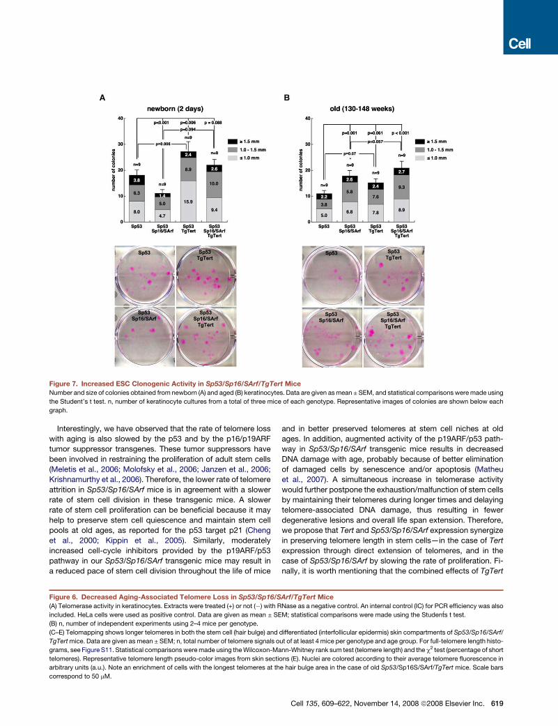
Figure 7. Increased ESC Clonogenic Activity in Sp53/Sp16/SArf/TgTert Mice
Number and size of colonies obtained from newborn (A) and aged (B) keratinocytes. Data are given as mean ± SEM, and statistical comparisons were made using
the Student’s t test. n, number of keratinocyte cultures from a total of three mice of each genotype. Representative images of colonies are shown below each
graph.
Interestingly, we have observed that the rate of telomere loss
with aging is also slowed by the p53 and by the p16/p19ARF
tumor suppressor transgenes. These tumor suppressors have
been involved in restraining the proliferation of adult stem cells
(Meletis et al., 2006; Molofsky et al., 2006; Janzen et al., 2006;
Krishnamurthy et al., 2006). Therefore, the lower rate of telomere
attrition in Sp53/Sp16/SArf mice is in agreement with a slower
rate of stem cell division in these transgenic mice. A slower
rate of stem cell proliferation can be beneficial because it may
help to preserve stem cell quiescence and maintain stem cell
pools at old ages, as reported for the p53 target p21 (Cheng
et al., 2000; Kippin et al., 2005). Similarly, moderately
increased cell-cycle inhibitors provided by the p19ARF/p53
pathway in our Sp53/Sp16/SArf transgenic mice may result in
a reduced pace of stem cell division throughout the life of mice
and in better preserved telomeres at stem cell niches at old
ages. In addition, augmented activity of the p19ARF/p53 path-
way in Sp53/Sp16/SArf transgenic mice results in decreased
DNA damage with age, probably because of better elimination
of damaged cells by senescence and/or apoptosis (Matheu
et al., 2007). A simultaneous increase in telomerase activity
would further postpone the exhaustion/malfunction of stem cells
by maintaining their telomeres during longer times and delaying
telomere-associated DNA damage, thus resulting in fewer
degenerative lesions and overall life span extension. Therefore,
we propose that Tert and Sp53/Sp16/SArf expression synergize
in preserving telomere length in stem cells—in the case of Tert
expression through direct extension of telomeres, and in the
case of Sp53/Sp16/SArf by slowing the rate of proliferation. Fi-
nally, it is worth mentioning that the combined effects of TgTert
Figure 6. Decreased Aging-Associated Telomere Loss in Sp53/Sp16/SArf/TgTert Mice
(A) Telomerase activity in keratinocytes. Extracts were treated (+) or not (�) with RNase as a negative control. An internal control (IC) for PCR efficiency was also
included. HeLa cells were used as positive control. Data are given as mean ± SEM; statistical comparisons were made using the Students t test.
(B) n, number of independent experiments using 2–4 mice per genotype.
(C–E) Telomapping shows longer telomeres in both the stem cell (hair bulge) and differentiated (interfollicular epidermis) skin compartments of Sp53/Sp16/SArf/
TgTert mice. Data are given as mean ± SEM; n, total number of telomere signals out of at least 4 mice per genotype and age group. For full-telomere length histo-
grams, see Figure S11. Statistical comparisons were made using the Wilcoxon-Mann-Whitney rank sum test (telomere length) and the c2 test (percentage of short
telomeres). Representative telomere length pseudo-color images from skin sections (E). Nuclei are colored according to their average telomere fluorescence in
arbitrary units (a.u.). Note an enrichment of cells with the longest telomeres at the hair bulge area in the case of old Sp53/Sp16S/SArf/TgTert mice. Scale bars
correspond to 50 mM.
Cell 135, 609–622, November 14, 2008 ª2008 Elsevier Inc. 619

and Sp53/Sp16/SArf transgenes on median mouse longevity are
of similar magnitude to those produced by calorie restriction, by
decreased growth hormone (GH)/IGF1 signaling, or by abroga-
tion of the type 5 adenylyl cyclase (Weindruch et al., 1986;
Brown-Borg et al., 1996; Yan et al., 2007).
Together, these observations demonstrate the antiaging
effects of telomerase in the context of living organisms and
provide further support to the important contribution of telomere
biology to aging.
EXPERIMENTAL PROCEDURES
Mice
Mice were housed at the pathogen-free barrier area of the CNIO, in accor-
dance with the recommendations of the Federation of European Laboratory
Animal Science Associations. Mice were observed daily and sacrificed when
they showed signs of morbidity or tumors in accordance to the Guidelines
for Humane Endpoints for Animals Used in Biomedical Research. Sp53,
Sp53/Sp16/SArf, Sp53/TgTert, and Sp53/Sp16/SArf/TgTert mice, as well as
their wild-type controls, have a 75% C57BL6, 25% DBA/2 genetic background
and were derived from the same parents by crossing K5-Tert females
(C57BL6-DBA/2 background) (Gonzalez-Suarez et al., 2001) with Sp53/
Sp16/SArf males (100% C57BL6 background) (Matheu et al., 2007). In parallel,
Sp53 and Sp53/Sp16/SArf mice were maintained in the original 100% C57BL6
background (Figure S8).
Mice shown in Figures 5D–5G have a 100% C57Bl6 genetic background
(Cayuela et al., 2005). Mice within each Terc�/� mouse generation are
littermates.
Skin and Small Intestine Epithelia Measurements
Epidermal and subcutaneous fat layer thickness and intestinal villi length were
determined using 10–74 random measurements along the length of skin and
small intestine from at least 3 mice per age group and genotype. For skin
sections, the skin was cut parallel to the spine and sections were cut perpen-
dicular to the skin surface. For villi sections, intestinal tracts were flushed with
PBS and rolled up in a compact circle using longitudinally oriented jejunal
sections for analysis; 5 mM sections were used for hematoxylin-eosin staining,
and Image J software was used for length measurements.
Induction of Intestinal Ulcers
Ulcers were induced as described elsewhere (Rakoff-Nahoum et al., 2004).
Briefly, 5 mice per genotype received 2% (wt/vol) DSS (36,000 kDa; MP
Biomedicals) in their drinking water for 7 days. Feces (21 fecal pellets per ge-
notype) were analyzed at days 3, 5, and 7 for the presence of blood using the
ColoScreen test (Helena Laboratories). At day 7, large intestines were excised,
fixed in formalin, and embedded in paraffin. Hematoxylin-eosin slides,
separating 200 mm and spanning the whole paraffin block, were analyzed to
score the number, size, and grade of ulcers. Histological scoring was made
as follows: Grade 0, normal colon; Grade 1, mild inflammation in lamina propria
with mixed component (neutrophils and plasma cells), hyperplasia of goblet
cells, and disorganized crypts, with frequent fusions between them; Grade
2, moderate inflammation in lamina propria and signs of regenerative dysplasia
(frequent mitosis, loss of crypt polarization, and decreased number of goblet
cells); Grade 3, severe inflammation in lamina propria, with a high proportion
of neutrophils, single-cell layer of epithelium without crypts, and frequent
edema; and Grade 4, severe inflammation in lamina propria, with high propor-
tion of neutrophils, lamina propria directly exposed to the light of the colon,
with no epithelium lining it, and frequent edema. Final histological score was
calculated as the sum of the factors obtained by multiplying size and histolog-
ical score for every ulcer.
Tightrope Test (Neuromuscular Coordination Assay)
The tightrope test was performed as described elsewhere (Ingram and
Reynolds, 1986; Matheu et al., 2007). Briefly, mice were placed on a bar of cir-
cular section (60 cm long and 1.5 cm diameter) and the test was considered
620 Cell 135, 609–622, November 14, 2008 ª2008 Elsevier Inc.
successful when a mouse did not fall during a period of 60 s in at least one trial
out of 5 consecutive trials.
Intraperitoneal Glucose Tolerance Tests
Glucose curves were performed as described elsewhere (Moynihan et al.,
2005). Briefly, males were fasted for 15 hr, and then i.p.-injected with 50%
dextrose (2 g/kg body weight). Tail blood glucose levels were measured with
a glucometer (Glucocard Memory 2, Arkray, Japan) at the indicated times after
injection. Standard glucose curves were represented, and area under the
curve (AUC) was calculated.
Measurements of IGF1 Levels
Renal or retro-orbital cavity blood was extracted, and after 5 min at room
temperature, samples were centrifuged at 1,600 3 g for 5 min at 4�C. Super-
natants (serum) were collected and stored at �80�C. Serum levels of IGF1
were measured using commercial kits (Rat/Mouse IGF1 EIA DSL-10-2900
from Diagnostic Systems Laboratorie or Rat/Mouse IGF1 ELISA from GroPep).
Quantification of Phosphorylated H2AX Foci
and of Telomere-Induced Foci
Phosphorylated H2AX foci were detected using a mouse monoclonal anti-
phospho-histone H2AX antibody (1:500; from Upstate Biotechnology) as
described elsewhere (Munoz et al., 2005). Sections from g-irradiated mice
were used as positive controls. Mice were irradiated with a dose of 10 Gy using
a 137Cs source (MARK 1-30 irradiator; Shepherd & Associates) at a rate of
2.11 Gy/min. Images were obtained with a confocal ultra-spectral microscope
(Leica TCS-SP2-A-OBS-UV) and a Leica CTR MIC microscope equipped
with a COHU High-Performance CCD camera. A cell was positive for
g-H2AX staining when it showed two or more foci.
For TRF1-g-H2AX colocalization (TIF), after incubation with the g-H2AX
antibody described above, a second staining was performed with rabbit
anti-mTRF1 (1:100) (Munoz et al., 2005) overnight at 4�C. Then tissues were
rinsed 3 times with PBS and incubated with goat anti-rabbit Alexa 488
(1:400) (Molecular Probes). Skin sections were mounted with Vectashield
with 40,6-diamino-2 phenylindole (DAPI). Images were obtained as described
above. A positive colocalization was considered when one or more TRF1
foci could be superimposed to g-H2AX foci.
Isolation of Keratinocytes
Keratinocytes from newborn mice were isolated as described previously
(Flores et al., 2005). Adult skin keratinocyes were isolated as described
elsewhere (Munoz et al., 2005).
Telomeric Repeat Amplification Protocol
Telomeric repeat amplification (TRAP) reactions of freshly isolated adult kera-
tinocytes were performed as described elsewhere (Blasco et al., 1997). As
negative control, extracts were preincubated with 5 mg RNase for 10 min at
30�C before the extension reaction. As a positive control, telomerase-positive
HeLa cells were included. An internal control for PCR efficiency was included
(IC) (TRAPeze kit Oncor, Gaithersburg, MD).
Generation of Telomere Length Maps on Histological Sections
or ‘‘Telomapping’’
Quantitative image analysis was performed on confocal images using the
Metamorph platform (version 6.3r6; Molecular Devices, Union City, CA) as
described elsewhere (Flores et al., 2008).
Colony Forming Assay
One thousand mouse keratinocytes obtained from newborn mice and
ten thousand mouse keratinocytes obtained from old mice were used in
clonogenic assays as described elsewhere (Flores et al., 2005).
Statistical Analyses
To calculate statistical significance of pathological analyses (malignant
tumors, lesions associated with the loss of epithelial barrier, and degenerative
lesions of the skin and the GI tract) as well as for calculation of the time of onset
of malignant tumors and the age-related pathologies the c2 test was used. The

two-sided p values were obtained from a 2 3 2 contingency table analyzed by
the c2 test (including Yate’s continuity correction). A t-student test with two
tails, two samples of unequal variance (or Welch’s correction), was used to
calculate statistical significance of thickness of the epidermal and subcutane-
ous fat layers, histological score of ulcers, glucose tolerance, IGF-1 ELISA
analysis, TRAP, colony forming assay, food and water intake controls, and
the measurement of the oxidative damage. The Fisher’s exact test was used
to calculate statistical differences in g-H2AX foci and TIFs, as well as for
calculation of survival at three years, analysis of blood in feces, and the neuro-
muscular coordination assay. The Wilcoxon-Mann-Whitney rank sum test was
used to calculate the statistical significance of the observed differences in
telomere length. Finally, a log rank test was used to calculate the statistical
differences in the survival of the different mouse cohorts. Microsoft Excel
version 2003 and GraphPad Instat version 2.03 were used for calculations.
SUPPLEMENTAL DATA
Supplemental Data include Supplemental Experimental Procedures, Supple-
mental References, and 12 figures and can be found with this article online
at http://www.cell.com/supplemental/S0092-8674(08)01191-4.
ACKNOWLEDGMENTS
A.T. is a predoctoral fellow of the Regional Government of Madrid. I.F. is a Ra-
mon y Cajal scientist. M.A.B.’s laboratory is funded by the Ministry of Science
and Innovation of Spain, by the European Union, by the Korber European
Science Award, and by the Spanish Association Against Cancer. M.S.’s
laboratory is funded by the Ministry of Science and Innovation of Spain, by
the European Union, and by the Fundacıon Marcelino Botin.
Received: February 15, 2008
Revised: June 18, 2008
Accepted: September 15, 2008
Published: November 13, 2008
REFERENCES
Armanios, M.Y., Chen, J.J., Cogan, J.D., Alder, J.K., Ingersoll, R.G., Markin,
C., Lawson, W.E., Xie, M., Vulto, I., Phillips, J.A., 3rd, Lansdorp, P.M., Greider,
C.W., and Loyd, J.E. (2007). Telomerase mutations in families with idiopathic
pulmonary fibrosis. N. Engl. J. Med. 356, 1317–1326.
Artandi, S.E., Alson, S., Tietze, M.K., Sharpless, N.E., Ye, S., Greenberg, R.A.,
Castrillon, D.H., Horner, J.W., Weiler, S.R., Carrasco, R.D., and DePinho, R.A.
(2002). Constitutive telomerase expression promotes mammary carcinomas in
aging mice. Proc. Natl. Acad. Sci. USA 99, 8191–8196.
Barrandon, Y., and Green, H. (1987). Three clonal types of keratinocyte
with different capacities for multiplication. Proc. Natl. Acad. Sci. USA 84,
2302–2306.
Blackburn, E.H. (2001). Switching and signaling at the telomere. Cell 106,
661–673.
Blasco, M.A., Lee, H.W., Hande, M.P., Samper, E., Lansdorp, P.M., DePinho,
R.A., and Greider, C.W. (1997). Telomere shortening and tumor formation by
mouse cells lacking telomerase RNA. Cell 91, 25–34.
Blasco, M.A. (2005). Telomeres and human disease: ageing, cancer and
beyond. Nat. Rev. Genet. Nat. Rev. Genet. 6, 611–622.
Blasco, M.A. (2007). Telomere length, stem cells and aging. Nat. Chem. Biol. 3,
640–649.
Bodnar, A.G., Ouellette, M., Frolkis, M., Holt, S.E., Chiu, C.P., Morin, G.B.,
Harley, C.B., Shay, J.W., Lichtsteiner, S., and Wright, W.E. (1998). Extension
of life-span by introduction of telomerase into normal human cells. Science
279, 349–352.
Brown-Borg, H.M., Borg, K.E., Meliska, C.J., and Bartke, A. (1996). Dwarf mice
and the ageing process. Nature 384, 33.
Bluher, M., Kahn, B.B., and Kahn, C.R. (2003). Extended longevity in mice
lacking the insulin receptor in adipose tissue. Science 299, 572–574.
Buchkovich, K.J., and Greider, C.W. (1996). Telomerase regulation during
entry into the cell cycle in normal human T cells. Mol. Biol. Cell 7, 1443–1454.
Canela, A., Martin-Caballero, J., Flores, J.M., and Blasco, M.A. (2004).
Constitutive expression of tert in thymocytes leads to increased incidence
and dissemination of T-cell lymphoma in Lck-Tert mice. Mol. Cell. Biol. 24,
4275–4293.
Cayuela, M.L., Flores, J.M., and Blasco, M.A. (2005). The telomerase RNA
component, Terc, is required for the telomere-length independent tumor
promoting effects of Tert over-expression. EMBO Rep. 6, 268–274.
Cheng, T., Rodrigues, N., Shen, H., Yang, Y., Dombkowski, D., Sykes, M., and
Scadden, D.T. (2000). Hematopoietic stem cell quiescence maintained by
p21cip1/waf1. Science 287, 1804–1808.
Choi, J., Southworth, L.K., Sarin, K.Y., Venteicher, A.S., Ma, W., Chang, W.,
Cheung, P., Jun, S., Artandi, M.K., Shah, N., Kim, S.K., and Artandi, S.E.
(2008). TERT promotes epithelial proliferation through transcriptional control
of a Myc- and Wnt-related developmental program. PLoS Genet. 4, e10.
Collado, M., Blasco, M.A., and Serrano, M. (2007). Cellular senescence in
cancer and aging. Cell 130, 223–233.
Conboy, I.M., Conboy, M.J., Wagers, A.J., Girma, E.R., Weissman, I.L., and
Rando, T.A. (2005). Rejuvenation of aged progenitor cells by exposure to
a young systemic environment. Nature 433, 760–764.
d’Adda di Fagagna, F., Reaper, P.M., Clay-Farrace, L., Fiegler, H., Carr, P.,
Von Zglinicki, T., Saretzki, G., Carter, N.P., and Jackson, S.P. (2003). A DNA
damage checkpoint response in telomere-initiated senescence. Nature 426,
194–198.
de Lange, T. (2005). Shelterin: the protein complex that shapes and safeguards
human telomeres. Genes Dev. 19, 2100–2110.
Delbono, O. (2003). Neural control of aging skeletal muscle. Aging Cell 2,
21–29.
Feldser, D.M., and Greider, C.W. (2007). Short telomeres limit tumor progres-
sion in vivo by inducing senescence. Cancer Cell 11, 461–469.
Flores, I., Cayuela, M.L., and Blasco, M.A. (2005). Effects of telomerase and
telomere length on epidermal stem cell behavior. Science 309, 1253–1256.
Flores, I., Evan, G., and Blasco, M.A. (2006). Genetic analysis of Myc and
telomerase interactions in vivo. Mol. Cell. Biol. 26, 6130–6138.
Flores, I., Canela, A., Vera, E., Tejera, A., Cotsarelis, G., and Blasco, M.A.
(2008). The longest telomeres: a general signature of adult stem cell compart-
ments. Genes Dev. 22, 654–667.
Garcia-Cao, I., Garcia-Cao, M., Martin-Caballero, J., Criado, L.M., Klatt, P.,
Flores, J.M., Weill, J.C., Blasco, M.A., and Serrano, M. (2002). ‘‘Super p53’’
mice exhibit enhanced DNA damage response, are tumor resistant and age
normally. EMBO J. 21, 6225–6235.
Garcia-Cao, I., Garcia-Cao, M., Tomas-Loba, A., Martin-Caballero, J., Flores,
J.M., Klatt, P., Blasco, M.A., and Serrano, M. (2006). Increased p53 activity
does not accelerate telomere-driven ageing. EMBO Rep. 7, 546–552.
Gonzalez-Suarez, E., Samper, E., Ramirez, A., Flores, J.M., Martin-Caballero,
J., Jorcano, J.L., and Blasco, M.A. (2001). Increased epidermal tumors and
increased skin wound healing in transgenic mice overexpressing the
catalytic subunit of telomerase, mTERT, in basal keratinocytes. EMBO J. 20,
2619–2630.
Gonzalez-Suarez, E., Flores, J.M., and Blasco, M.A. (2002). Cooperation
between p53 mutation and high telomerase transgenic expression in sponta-
neous cancer development. Mol. Cell. Biol. 22, 7291–7301.
Goren, H.J., Kulkarni, R.N., and Kahn, C.R. (2004). Glucose homeostasis
and tissue transcript content of insulin signaling intermediates in four inbred
strains of mice: C57BL/6, C57BLKS/6, DBA/2, and 129X1. Endocrinology
145, 3307–3323.
Greider, C.W., and Blackburn, E.H. (1985). Identification of a specific telomere
terminal transferase activity in Tetrahymena extracts. Cell 43, 405–413.
Haigis, M.C., and Guarente, L.P. (2006). Mammalian sirtuins—emerging roles
in physiology, aging, and calorie restriction. Genes Dev. 20, 2913–2921.
Cell 135, 609–622, November 14, 2008 ª2008 Elsevier Inc. 621

Hammerman, M.R. (1987). Insulin-like growth factors and aging. Endocrinol.
Metab. Clin. North Am. 16, 995–1011.
Harley, C.B., Futcher, A.B., and Greider, C.W. (1990). Telomeres shorten dur-
ing ageing of human fibroblasts. Nature 345, 458–460.
Herbig, U., Ferreira, M., Condel, L., Carey, D., and Sedivy, J.M. (2006). Cellular
senescence in aging primates. Science 311, 1257.
Ingram, D.K., and Reynolds, M.A. (1986). Assessing the predictive validity of
psychomotor tests as measures of biological age in mice. Exp. Aging Res.
12, 155–162.
Janzen, V., Forkert, R., Fleming, H.E., Saito, Y., Waring, M.T., Dombkowski,
D.M., Cheng, T., DePinho, R.A., Sharpless, N.E., and Scadden, D.T. (2006).
Stem-cell ageing modified by the cyclin-dependent kinase inhibitor
p16INK4a. Nature 443, 421–426.
Kenyon, C. (2005). The plasticity of aging: insights from long-lived mutants.
Cell 120, 449–460.
Kippin, T.E., Martens, D.J., and van der Kooy, D. (2005). p21 loss compro-
mises the relative quiescence of forebrain stem cell proliferation leading to
exhaustion of their proliferation capacity. Genes Dev. 19, 756–767.
Krishnamurthy, J., Torrice, C., Ramsey, M.R., Kovalev, G.I., Al-Regaiey, K., Su,
L., and Sharpless, N.E. (2004). Ink4a/Arf expression is a biomarker of aging.
J. Clin. Invest. 114, 1299–1307.
Krishnamurthy, J., Ramsey, M.R., Ligon, K.L., Torrice, C., Koh, A., Bonner-
Weir, S., and Sharpless, N.E. (2006). p16INK4a induces an age-dependent
decline in islet regenerative potential. Nature 443, 453–457.
Matheu, A., Pantoja, C., Efeyan, A., Criado, L.M., Martin-Caballero, J., Flores,
J.M., Klatt, P., and Serrano, M. (2004). Increased gene dosage of Ink4a/Arf
results in cancer resistance and normal aging. Genes Dev. 18, 2736–2746.
Matheu, A., Maraver, A., Klatt, P., Flores, I., Garcia-Cao, I., Borras, C., Flores,
J.M., Vina, J., Blasco, M.A., and Serrano, M. (2007). Delayed aging through
damage protection by the Arf/p53 pathway. Nature 448, 375–379.
Meletis, K., Wirta, V., Hede, S.M., Nister, M., Lundeberg, J., and Frisen, J.
(2006). p53 suppresses the self-renewal of adult neural stem cells. Develop-
ment 133, 363–369.
Mitchell, J.R., Wood, E., and Collins, K. (1999). A telomerase component is
defective in the human disease dyskeratosis congenita. Nature 402, 551–555.
Molofsky, A.V., Slutsky, S.G., Joseph, N.M., He, S., Pardal, R., Krishnamurthy,
J., Sharpless, N.E., and Morrison, S.J. (2006). Increasing p16INK4a expression
decreases forebrain progenitors and neurogenesis during ageing. Nature 443,
448–452.
Moynihan, K.A., Grimm, A.A., Plueger, M.M., Bernal-Mizrachi, E., Ford, E.,
Cras-Meneur, C., Permutt, M.A., and Imai, S. (2005). Increased dosage of
mammalian Sir2 in pancreatic beta cells enhances glucose-stimulated insulin
secretion in mice. Cell Metab. 2, 105–117.
Murphy, C.T., McCarroll, S.A., Bargmann, C.I., Fraser, A., Kamath, R.S.,
Ahringer, J., Li, H., and Kenyon, C. (2003). Genes that act downstream of
DAF-16 to influence the lifespan of Caenorhabditis elegans. Nature 424,
277–283.
622 Cell 135, 609–622, November 14, 2008 ª2008 Elsevier Inc.
Munoz, P., Blanco, R., Flores, J.M., and Blasco, M.A. (2005). XPF nuclease-
dependent telomere loss and increased DNA damage in mice overexpressing
TRF2 result in premature aging and cancer. Nat. Genet. 10, 1063–1071.
Niedernhofer, L.J., Garinis, G.A., Raams, A., Lalai, A.S., Robinson, A.R.,
Appeldoorn, E., Odijk, H., Oostendorp, R., Ahmad, A., van Leeuwen, W.,
et al. (2006). A new progeroid syndrome reveals that genotoxic stress
suppresses the somatotroph axis. Nature 444, 1038–1043.
Rakoff-Nahoum, S., Paglino, J., Eslami-Varzaneh, F., Edberg, S., and Medzhi-
tov, R. (2004). Recognition of commensal microflora by toll-like receptors is
required for intestinal homeostasis. Cell 118, 229–241.
Ramırez, A., Bravo, A., Jorcano, J.L., and Vidal, M. (1994). Sequences 50 of the
bovine keratin 5 gene direct tissue- and cell-type-specific expression of a lacZ
gene in the adult and during development. Differentiation 58, 53–64.
Sablina, A.A., Budanov, A.V., Ilyinskaya, G.V., Agapova, L.S., Kravchenko,
J.E., and Chumakov, P.M. (2005). The antioxidant function of the p53 tumor
suppressor. Nat. Med. 11, 1306–1313.
Sarin, K.Y., Cheung, P., Gilison, D., Lee, E., Tennen, R.I., Wang, E., Artandi,
M.K., Oro, A.E., and Artandi, S.E. (2005). Conditional telomerase induction
causes proliferation of hair follicle stem cells. Nature 436, 1048–1052.
Sedelnikova, O.A., Horikawa, I., Zimonjic, D.B., Popescu, N.C., Bonner, W.M.,
and Barrett, J.C. (2004). Senescing human cells and ageing mice accumulate
DNA lesions with unrepairable double-strand breaks. Nat. Cell Biol. 6,
168–170.
Tsakiri, K.D., Cronkhite, J.T., Kuan, P.J., Xing, C., Raghu, G., Weissler, J.C.,
Rosenblatt, R.L., Shay, J.W., and Garcia, C.K. (2007). Adult-onset pulmonary
fibrosis caused by mutations in telomerase. Proc. Natl. Acad. Sci. USA 104,
7552–7557.
Vulliamy, T., Marrone, A., Goldman, F., Dearlove, A., Bessler, M., Mason, P.J.,
and Dokal, I. (2001). The RNA component of telomerase is mutated in autoso-
mal dominant dyskeratosis congenita. Nature 413, 432–435.
Weindruch, R., Walford, R.L., Fligiel, S., and Guthrie, D. (1986). The retardation
of aging in mice by dietary restriction: longevity, cancer, immunity and lifetime
energy intake. J. Nutr. 116, 641–654.
Yan, L., Vatner, D.E., O’Connor, J.P., Ivessa, A., Ge, H., Chen, W., Hirotani, S.,
Ishikawa, Y., Sadoshima, J., and Vatner, S.F. (2007). Type 5 adenylyl cyclase
disruption increases longevity and protects against stress. Cell 130, 247–258.
Yamaguchi, H., Calado, R.T., Ly, H., Kajigaya, S., Baerlocher, G.M., Chanock,
S.J., Lansdorp, P.M., and Young, N.S. (2005). Mutations in TERT, the gene for
telomerase reverse transcriptase, in aplastic anemia. N. Engl. J. Med. 352,
1413–1424.
Yuen, K.C., and Dunger, D.B. (2007). Therapeutic aspects of growth hormone
and insulin-like growth factor-I treatment on visceral fat and insulin sensitivity
in adults. Diabetes Obes. Metab. 9, 11–22.
Zindy, F., Quelle, D.E., Roussel, M.F., and Sherr, C.J. (1997). Expression of the
p16INK4a tumor suppressor versus other INK4 family members during mouse
development and aging. Oncogene 15, 203–211.
Copyright © 2022 FDOKUMEN