Progressive Increase in Telomerase Activity From Benign Melanocytic Conditions to Malignant Melanoma
8
Neoplasia v Vol. 1, No. 1, April 1999, pp. 42 –49 42 Available On-line at http:// www.neoplasia.org Progressive Increase in Telomerase Activity From Benign Melanocytic Conditions to Malignant Melanoma Ruben D. Ramirez ) , Stefania D’Atri ‡ , Elena Pagani ‡ , Tullio Faraggiana ‡ , Pedro Miguel Lacal ‡ , R. Stan Taylor † and Jerry W. Shay ) ) Department of Cell Biology and Neuroscience, † Department of Dermatology, The University of Texas Southwestern Medical Center, Dallas, TX; ‡ Laboratory of Clinical Pharmacology, Istituto Dermatopatico Dell’Immacolata, Rome, Italy Abstract The expression of telomerase activity and the in situ localization of the human telomerase RNA component ( ) hTR in melanocytic skin lesions was evaluated in specimens from sixty-three patients. Specimens of melanocytic nevi, primary melanomas and subcuta- neous metastases of melanoma were obtained from fifty-eight patients, whereas metastasized lymph nodes were obtained from five patients. Telomerase activity was determined in these specimens by using a Poly- ( ) merase Chain Reaction–based assay TRAP . High rela- tive mean telomerase activity levels were detected in ( metastatic melanoma subcutaneous metastases s54.5, ) lymph node metastases s56.5 . Much lower levels were detected in primary melanomas, which increased with ( advancing levels of tumor cell penetration Clark II s ) 0.02, Clark III s1.1, and Clark IV s1.9 . Twenty-six for- malin-fixed, paraffin-embedded melanocytic lesions were sectioned and analyzed for telomerase RNA with a radioactive in situ hybridization assay. In situ hy- bridization studies with a probe to the template RNA component of telomerase confirmed that expression was almost exclusively confined to tumor cells and not infiltrating lymphocytes. These results indicate that lev- els of telomerase activity and telomerase RNA in melanocytic lesions correlate well with clinical stage and could potentially assist in the diagnosis of border- line lesions. Keywords: skin cancer, immortality, telomeres, metastases, prognosis. Introduction Telomeres, the chromosomal end regions composed of [] tandem repeats of the sequence TTAGGG in vertebrates 1 are believed to be important in stabilizing the chromosomes [] during replication 2 . Because DNA polymerase is unable [ ] to completely replicate telomeric sequences 3,4 , it has been proposed that progressive loss of telomere length during cell division could act as a mechanism that limits the [ ] replicative capacity of normal somatic cells 5,6 . Telom- erase is a ribonucleoprotein enzyme that can use its RNA [] 7 component as a template and the telomerase catalytic [ ] protein reverse transcriptase 8–10 for the addition of telomeric repeats to the 3 X ends of the chromosomes. Thus, telomerase is thought to partially or completely compensate for the progressive telomere erosion that would occur in its absence. Because most normal somatic cells in vertebrates do not express telomerase, it is thought that their telomeres progressively shorten, undergo genomic instability, and [ ] cease proliferating once a critical length is reached 11 . It has been suggested that upregulation or re-expression of telomerase may result in chromosomal stability leading to [] continuous cell proliferation 6 . Cancer is generally believed to be caused by accumula- tion of multiple somatic mutations that subvert the normal [ ] growth control mechanisms 12 . It has been suggested that in most cancers cellular immortality is among the changes [ ] required for sustained long-term growth 6,13,14 . Telom- erase activity has been detected in 85% of tumors repre- senting 15 cancer types, whereas most benign tumors and somatic tissues have very low levels or lack detectable [ ] telomerase activity 15,16 . Furthermore, in special metastatic tumor types such as neuroblastoma stage IVs, in which there is a high rate of spontaneous regression, telom- erase activity is very low or not detectable, and telomere [ ] lengths are considerably shorter than adjacent tissue 17 . Thus, although telomerase is not necessarily required for the initiation of a tumor, it is likely to be important in the sustained growth of the tumor. The diagnosis of melanoma can be made clinically in [ ] 80% to 90% of cases 18 , whereas the remainder require histological examination for diagnosis. However, a certain degree of ambiguity and thus indecision remains in the [ ] histological diagnosis of melanoma 19 . To accurately pin- point tumor cells and improve diagnosis, pathologists often require the use of neoplastic markers. Telomerase has been touted as such a marker. It has been shown previ- ( ) ously that the human telomerase template RNA hTR can be used in situ to distinguish between cancer cells and [ ] normal cells in humans 20,21 and that levels of telomerase Address reprint requests to: Jerry W. Shay, Ellison Medical Foundation Senior Scholar, Department of Cell Biology and Neuroscience, The University of Texas Southwestern Medical Center, 5323 Harry Hines Blvd., Dallas, TX, 75235-9039. E-mail: [email protected] Received 11 December 1998; Accepted 5 January 1999. Copyright 1999 Stockton Press. All rights reserved 1522-8002/99/$12.00
-
Upload
independent -
Category
Documents
-
view
1 -
download
0
Transcript of Progressive Increase in Telomerase Activity From Benign Melanocytic Conditions to Malignant Melanoma

Neoplasia v Vol. 1, No. 1, April 1999, pp. 42–49 42
Available On-line at http://www.neoplasia.org
Progressive Increase in Telomerase Activity From BenignMelanocytic Conditions to Malignant Melanoma
Ruben D. Ramirez ) , Stefania D’Atri ‡, Elena Pagani ‡, Tullio Faraggiana ‡, Pedro Miguel Lacal ‡,R. Stan Taylor † and Jerry W. Shay )
)Department of Cell Biology and Neuroscience, †Department of Dermatology, The University of TexasSouthwestern Medical Center, Dallas, TX; ‡Laboratory of Clinical Pharmacology, Istituto DermatopaticoDell’Immacolata, Rome, Italy
AbstractThe expression of telomerase activity and the in situlocalization of the human telomerase RNA component( )hTR in melanocytic skin lesions was evaluated inspecimens from sixty-three patients. Specimens ofmelanocytic nevi, primary melanomas and subcuta-neous metastases of melanoma were obtained fromfifty-eight patients, whereas metastasized lymph nodeswere obtained from five patients. Telomerase activitywas determined in these specimens by using a Poly-
( )merase Chain Reaction–based assay TRAP . High rela-tive mean telomerase activity levels were detected in
(metastatic melanoma subcutaneous metastasess54.5,)lymph node metastasess56.5 . Much lower levels were
detected in primary melanomas, which increased with(advancing levels of tumor cell penetration Clark IIs
)0.02, Clark IIIs1.1, and Clark IVs1.9 . Twenty-six for-malin-fixed, paraffin-embedded melanocytic lesionswere sectioned and analyzed for telomerase RNA with aradioactive in situ hybridization assay. In situ hy-bridization studies with a probe to the template RNAcomponent of telomerase confirmed that expressionwas almost exclusively confined to tumor cells and notinfiltrating lymphocytes. These results indicate that lev-els of telomerase activity and telomerase RNA inmelanocytic lesions correlate well with clinical stageand could potentially assist in the diagnosis of border-line lesions.
Keywords: skin cancer, immortality, telomeres, metastases, prognosis.
IntroductionTelomeres, the chromosomal end regions composed of
[ ]tandem repeats of the sequence TTAGGG in vertebrates 1are believed to be important in stabilizing the chromosomes
[ ]during replication 2 . Because DNA polymerase is unable[ ]to completely replicate telomeric sequences 3,4 , it has
been proposed that progressive loss of telomere lengthduring cell division could act as a mechanism that limits the
[ ]replicative capacity of normal somatic cells 5,6 . Telom-erase is a ribonucleoprotein enzyme that can use its RNA[ ]7 component as a template and the telomerase catalytic
[ ]protein reverse transcriptase 8–10 for the addition of
telomeric repeats to the 3X ends of the chromosomes. Thus,telomerase is thought to partially or completely compensatefor the progressive telomere erosion that would occur in itsabsence. Because most normal somatic cells in vertebratesdo not express telomerase, it is thought that their telomeresprogressively shorten, undergo genomic instability, and
[ ]cease proliferating once a critical length is reached 11 . Ithas been suggested that upregulation or re-expression oftelomerase may result in chromosomal stability leading to
[ ]continuous cell proliferation 6 .Cancer is generally believed to be caused by accumula-
tion of multiple somatic mutations that subvert the normal[ ]growth control mechanisms 12 . It has been suggested that
in most cancers cellular immortality is among the changes[ ]required for sustained long-term growth 6,13,14 . Telom-
erase activity has been detected in 85% of tumors repre-senting 15 cancer types, whereas most benign tumors andsomatic tissues have very low levels or lack detectable
[ ]telomerase activity 15,16 . Furthermore, in specialmetastatic tumor types such as neuroblastoma stage IVs, inwhich there is a high rate of spontaneous regression, telom-erase activity is very low or not detectable, and telomere
[ ]lengths are considerably shorter than adjacent tissue 17 .Thus, although telomerase is not necessarily required forthe initiation of a tumor, it is likely to be important in thesustained growth of the tumor.
The diagnosis of melanoma can be made clinically in[ ]80% to 90% of cases 18 , whereas the remainder require
histological examination for diagnosis. However, a certaindegree of ambiguity and thus indecision remains in the
[ ]histological diagnosis of melanoma 19 . To accurately pin-point tumor cells and improve diagnosis, pathologists oftenrequire the use of neoplastic markers. Telomerase hasbeen touted as such a marker. It has been shown previ-
( )ously that the human telomerase template RNA hTR canbe used in situ to distinguish between cancer cells and
[ ]normal cells in humans 20,21 and that levels of telomerase
Address reprint requests to: Jerry W. Shay, Ellison Medical Foundation SeniorScholar, Department of Cell Biology and Neuroscience, The University of TexasSouthwestern Medical Center, 5323 Harry Hines Blvd., Dallas, TX, 75235-9039.E-mail: [email protected] 11 December 1998; Accepted 5 January 1999.
Copyright 1999 Stockton Press. All rights reserved 1522-8002/99/$12.00
Telomerase in Melanoma Ramirez et al. 43
activity correlate well with the degree of hTR. Thus, in situtechniques applied to tissue samples can now be used tolocalize the presence of telomerase in melanoma cellsthereby, potentially aiding in diagnosis.
Traditionally, once the diagnosis is confirmed, tumors arestaged based on the clinical and/or radiographic evidenceof spread through the body. Clinical staging has been criti-cal to determining treatment and prognosis. If there is no
(indication that melanoma has spread beyond the skin clini-)cal stage I and II , treatment decisions are based on the
[ ]anatomical level of invasion 22,23 of tumor cells into theskin. A number of histological parameters have been con-sidered in an attempt to identify the greatest predictive
[ ]value for the development of metastases 24–26 and over-all survival. Tumor thickness has consistently been shown
[ ]to have the most predictive value 27,28 . Two classificationschemes are used by pathologists, including the Breslow
( )thickness penetration depth measured in millimeters and(the Clark classification five levels of penetration based on
) [ ]anatomic location of tumor cells in the skin 22,23,27,28 .Because telomerase activity levels and RNA levels correlate
[ ]well with degree of malignancy 20,21 , it is possible thatdetermination of telomerase RNA expression or telomeraseactivity levels in primary melanomas could help to deter-mine more precisely the aggressive nature of individualtumors, thus enabling more appropriate therapies and im-proved prognosis.
In our present study, we analyzed and compared thelevels of telomerase activity with a variety of clinical andhistological parameters of a large group of cutaneousmelanocytic conditions, including compound or junctionalmelanocytic nevi, malignant primary melanomas, subcuta-neous metastasis of melanoma, and lymph nodes contain-ing metastatic melanoma. We also used a telomerase RNAin situ hybridization assay to determine the pattern of ex-pression of hTR on formalin-fixed paraffin –embeddedmelanocytic specimens.
Materials and Methods
Tissue AcquisitionAll tissue samples were collected at the Istituto Der-
matopatico dell’Immacolata, Rome, Italy. Specimens ofmelanocytic nevi, primary melanomas, and subcutaneousmetastases of melanoma were obtained under local anes-
(thesia from a total of fifty-eight patients with informed)consent who underwent surgical removal of the lesion for
( )histological examination. Only relatively large )1 cm pri-mary lesions were used for this study. Metastasized lymphnodes were obtained from five patients who underwenttherapeutic regional lymph node dissection.
Specimens were received fresh in the surgical pathologyroom. Those in which a reasonably obvious diagnosis ofbenign or malignant nature was not possible were submittedfor standard histological evaluations. Obvious subcutaneousmetastases and grossly involved lymph nodes were bi-sected, half of which were submitted for routine histological
evaluation and half stored for telomerase assays. Primarylesions were serially sectioned, and at least three sectionsdisplaying maximal thickness, as judged by gross inspec-tion, were submitted for routine histological examination.One section from the periphery of the pigmented lesion wastaken for in situ telomerase RNA analysis. Subcutaneousand lateral portions of grossly uninvolved skin were thenremoved with a scalpel so that only the clear-cut lesion andthe overlaying epidermis remained. Samples of normal skinadjacent to primary lesions were also collected and testedfor telomerase activity.
Tissue samples were rapidly frozen and stored at y808Cuntil they were tested for telomerase activity. The entireprocedure from the time of surgery to the freezing step wasperformed under sterile conditions and always completedwithin 30 minutes.
Histopathologic EvaluationSections from each paraffin block were cut at 3-mm
thickness and stained with hematoxylin and eosin. Thediagnosis of melanoma or melanocytic nevus was estab-lished according to the standard accepted criteria. Breslowthickness was measured with a Leitz micrometer eyepiece( ) (E. Leitz, Ruckleigh, NJ . Mitotic rate was rated as high 6
2) (mitotic figures per mm or low fewer than 6 mitotic figures2)per mm . Melanoma patients were staged according to the
( )American Joint Commission on Cancer AJCC .
Preparation of Tissue ExtractsTissue extracts were prepared as previously described
[ ]29 with the following modifications. Frozen specimens con-taining dissected samples were thawed and subsequently
(transferred to Kontes homogenization tubes VWR Scientific)Inc, Sugarland, TX containing 100 to 200 mL of ice-cold
( [( )lysis buffer 0.5% 3- 3-cholamidopropyl -dimethyl-am-] ( )monio -1-propanesulfonate CHAPS , 10 mmol/L Tris–HCl
[ ]pH 7.5 , 1 mmol/L MgCl , 1 mmol/L ethyleneglycol-bis-2
( X X ( )-aminoethyl ether-N,N,N ,N -tetraacetic acid EGTA , 5[ ( )mmol/L mercaptoethanol, 0.1 mmol/L 4- 2-aminoethyl -
] )benzene-sulfonyl fluoride hydrochlorine , 10% glycerol . Ho-(mogenization was carried out with disposable pestles VWR
) (Scientific, Dallas, TX attached to a cordless drill Black and)Decker, Baltimore, MD and rotated at 450 rpm. Ho-
mogenates were left on ice for 30 minutes, followed bycentrifugation of lysates at 14,000 rpm at 48C for 20 min-
( )utes. Supernatants 80–160 mL were collected in fresh(Eppendorf 1.5 micro tubes United Scientific Products, San
)Leandro, CA flash frozen in liquid nitrogen, and stored aty808C. Before freezing, a 10-mL aliquot was transferredinto a separate Eppendorf 1.5 cc micro tube for protein
( )concentration analysis by using the BCA bicinchoninic acid( )protein assay kit Pierce Chemical Co, Rockford, IL .
Telomeric Repeat Amplification ProtocolTo evaluate the approximate levels of telomerase activity
in the different dissected compartments, telomeric repeat( )amplification protocol TRAP assays were performed as
[ ]previously described 30,31 , including the use of an internal
Neoplasia v Vol. 1, No. 1, April 1999
Telomerase in Melanoma Ramirez et al.44
( ) [ ]telomerase assay standard ITAS 31 . Telomerase canadd TTAGGG repeats to a nontelomeric oligonucleotide TS( X )5 -AATCCGTCGAGCAGAGTT that serves as the forwardprimer. These elongation products can then be amplified by
( )polymerase chain reaction PCR with the reverse primer( X [ ] )CX 5 - CCCTTA CCCTAA , resulting in a ladder of 6 base3
( )pair bp addition products.Tissue extracts were normalized to 6.0 mg total protein
(per assay. The PCR-based TRAP Boehringer Mannheim,)Indianapolis, IN assay was carried out in previously pre-
( )pared HotStart tubes 50 GIBCO BRL, Gaithersburg, MDcontaining 100 ng of CX oligonucleotide beneath a waxbarrier and stored at 48C until needed. Aliquots containing6.0 mg of total protein were assayed in 50 mL of reaction
( [ ]mixture 20 mmol/L Tris–HCl pH 8.3 , 68 mmol/L KCl, 1.5mmol/L MgCl , 1 mmol/L EGTA, 0.05% Tween 20, 0.1 mg2
( )of TS primer Midland Certified Reagent Co , 0.5 mmol/L T4
gene 32 protein, 50 mmol/L of each deoxynucleotidetriphosphate, 2 U of Taq DNA polymerase, 4 mCi of[y3 2 ] [ ] y18 ( )P dCTP 10 mCi/mmol , and 5=10 g 5 attograms
[ ]of ITAS 31 . The reaction mixture was incubated at roomtemperature for 30 minutes to allow telomerase to extend
(TS primer followed by a 31-cycle PCR amplification ther-mocycling block with heated lid, HB-60, MJ Research, Wa-
)tertown, MA of the telomeric products. Analysis of 20 mL ofthe PCR mixtures was performed on 10% nondenaturingacrylamide gels. Gels were fixed in 0.5 mol /L NaCl, 50%
( )ethanol, and 40 mmol/L sodium acetate pH 4.2 on arocking table for 30 minutes at room temperature. Excessfixing solution was removed, and gels were exposed atroom temperature to phosphor screens overnight and visu-alized on a PhosphorImager with ImageQuant software( )Molecular Dynamics, Sunnyvale, CA .
Quantitation of Telomerase ActivityThe telomerase assay can be made semiquantitative by
y18 ( )the inclusion of 5=10 g 5 attograms of an ITAS, a150-bp fragment containing TS and CX sequences at theends. That is co-amplified along with the telomerase ladder[ ]30,31 . The levels of telomerase activity were normalized tothat of the ITAS. The inclusion of ITAS not only permitsrelative quantitation between samples, it also aids in theidentification of false-negative samples that contain Taqpolymerase or other PCR inhibitors. The relative telomeraseactivity was determined by calculating the ratio of the inten-sity of the area under peaks representing telomeric repeats( )TRAP ladder to the area under the peak representingamplified ITAS. When the levels of telomerase activity weretoo high to allow amplification of ITAS, ten-fold dilutions ofthese samples were assayed for telomerase activity. Tripli-cate aliquots from selected samples were analyzed, andconsistent results were obtained. Pooled ratio results fromeach sample group were then compared for statistical signif-icance by using nonparametric analyses and rank method( )JMP software, Cary, NC . The Wilcoxon/Kruskal Wallistests combined with the Tukey-Kramer multiple mean com-parisons procedure and the ANOVA/t test procedures, wereused for all independent groups. All image operations and
calculations were performed by using ImageQuant software( ) (Molecular Dynamics and Microsoft Excel 5.0 Microsoft
)Corporation, Redmond, WA .
Preparation and Prehybridization of Tissue Sections for InSitu Hybridization
( )Formalin-fixed paraffin–embedded 3-mm tissue sec-(tions were placed onto Superfrost/Plus slides Fisher Scien-
)tific, Pittsburgh, PA . The sections were then deparaffinized,( )rehydrated in phosphate buffered saline PBS , and treated
( ) (with proteinase K 20 mg/mL in 50 mmol/L Tris-HCl pH)7.5 , 5 mmol/L EDTA, for 20 minutes at room temperature.
After rinsing for 5 minutes in PBS, sections were postfixedin 4% paraformaldehyde/PBS, rinsed in water, and acety-lated in freshly prepared 0.25% acetic anhydride/0.1 mol/Ltriethanolamine for 10 minutes. The slides were then dehy-drated in gradually increasing concentrations of ethanolbefore hybridization.
Probe Preparation for In Situ Hybridization( )The plasmid pGEM-5Z Promega Corp, Madison, WI ,
(containing a full-length hTR complementary DNA 560 nu-)cleotides , obtained from the Geron Corp, Menlo Park, CA,
( )was used as a template to generate sense control and[35 ]antisense probe. S UTP-labeled single-stranded RNA
(probes were synthesized according to manufacturer’s Amb-)ion, Inc, Austin, TX conditions. Transcripts were alkali hy-
drolyzed to generate probes, with an average length of 200nucleotides for efficient hybridization, purified with G-50
( )column Boehringer Mannheim Corp, Indianapolis, IN , andprecipitated in ethanol. The probes were then resuspendedin 30 mL of 100 mmol/L dithiothreitol. The specific activity ofthe radiolabeled probes, aliquoted and stored at y808Cuntil use, was approximately 3 to 5=106 cpm/mg templateDNA.
Hybridization and Washing ProceduresSections were hybridized overnight at 528C in 50% deion-
(ized formamide, 0.3 mol/L NaCl, 20 mmol/L Tris–HCl pH) ( )7.5 , 5 mmol/L EDTA, 10 mmol/L NaH PO pH 8.0 , 10%2 4
dextran sulfate, 1= Denhardt’s, 500 mg/mL total yeastRNA, 10 mmol / L dithiothreitol, and 50,000 cpm / mL35S-labeled cRNA probe. The slides were then subjected tostringent washing at 508C in 5= SSC, 10 mmol/L dithio-threitol for 30 minutes, at 658C in 50% formamide, 2= SSC,10 mmol/L DTT for 30 minutes and washed twice at 378C in
( )0.4 mol/L NaCl, 10 mmol/L Tris-HCl pH 7.5 , 5 mmol/LEDTA for 10 minutes before treatment with 20 mg/mLRNase A at 378C for 30 minutes. Following washes in 2=SSC and 0.1= SSC for 1 hour at 378C, the slides were
(dehydrated and dipped in Kodak Eastman Kodak Co.,)Rochester, NY NTB-2 nuclear track emulsion and exposed
for 3 to 4 weeks in light tight boxes with desiccant at 48C.(The slides were developed in Kodak Dektol developer 3.5
) ( ) ( )min , washed in water 30 sec , fixed in Kodak fixer 6 min ,rinsed in water, counterstained in hematoxylin, dehydrated,
( )coverslips mounted with Paramount Sigma, St. Louis, MO ,(and finally observed under light microscope Olympus
Neoplasia v Vol. 1, No. 1, April 1999
Telomerase in Melanoma Ramirez et al. 45
)America Inc., Lake Success, NY in both bright and darkfields, followed by photography. In the present study sam-ples were considered hTR positive if uniformly dense silvergrains were overlying tumor nuclei similar to those observedin the normal adult testis. Specimens were considered neg-ative if there was complete absence of background hy-bridization similar to sense control.
Results
Telomerase Activity Is Detected at Highest Levels in Metas-tases of Melanoma
Sixty-six samples from a total of sixty-three patients withseveral different types of melanocytic lesions and adjacent
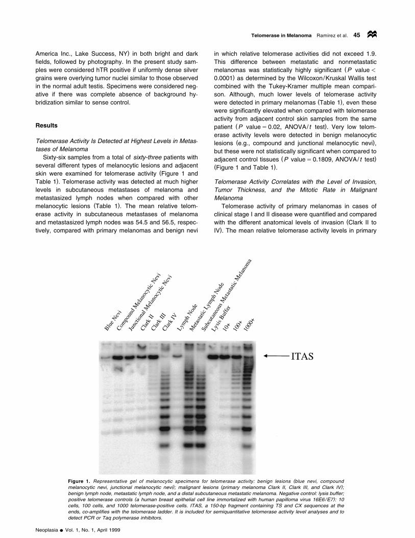
(skin were examined for telomerase activity Figure 1 and)Table 1 . Telomerase activity was detected at much higher
levels in subcutaneous metastases of melanoma andmetastasized lymph nodes when compared with other
( )melanocytic lesions Table 1 . The mean relative telom-erase activity in subcutaneous metastases of melanomaand metastasized lymph nodes was 54.5 and 56.5, respec-tively, compared with primary melanomas and benign nevi
in which relative telomerase activities did not exceed 1.9.This difference between metastatic and nonmetastatic
(melanomas was statistically highly significant P value-)0.0001 as determined by the Wilcoxon/Kruskal Wallis test
combined with the Tukey-Kramer multiple mean compari-son. Although, much lower levels of telomerase activity
( )were detected in primary melanomas Table 1 , even thesewere significantly elevated when compared with telomeraseactivity from adjacent control skin samples from the same
( )patient P values0.02, ANOVA/t test . Very low telom-erase activity levels were detected in benign melanocytic
( )lesions e.g., compound and junctional melanocytic nevi ,but these were not statistically significant when compared to
( )adjacent control tissues P values0.1809, ANOVA/t test( )Figure 1 and Table 1 .
Telomerase Activity Correlates with the Level of Invasion,Tumor Thickness, and the Mitotic Rate in MalignantMelanoma
Telomerase activity of primary melanomas in cases ofclinical stage I and II disease were quantified and compared
(with the different anatomical levels of invasion Clark II to)IV . The mean relative telomerase activity levels in primary
(Figure 1. Representative gel of melanocytic specimens for telomerase activity: benign lesions blue nevi, compound) ( )melanocytic nevi, junctional melanocytic nevi ; malignant lesions primary melanoma Clark II, Clark III, and Clark IV ;
benign lymph node, metastatic lymph node, and a distal subcutaneous metastatic melanoma. Negative control: lysis buffer;( )positive telomerase controls a human breast epithelial cell line immortalized with human papilloma virus 16E6/E7 : 10
cells, 100 cells, and 1000 telomerase-positive cells. ITAS, a 150-bp fragment containing TS and CX sequences at theends, co-amplifies with the telomerase ladder. It is included for semiquantitative telomerase activity level analyses and todetect PCR or Taq polymerase inhibitors.
Neoplasia v Vol. 1, No. 1, April 1999
Telomerase in Melanoma Ramirez et al.46
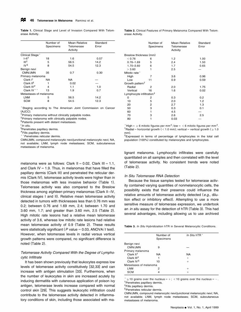
Table 1. Clinical Stage and Level of Invasion Compared With Telom-erase Activity.
Number of Mean Relative StandardSpecimens Telomerase Error
Activity
)Clinical Stage†I–II 18 1.6 0.57
‡III 5 56.5 14.2§IV 8 54.5 12.3
Benign neviCMN/JMN 35 0.7 0.30
Primary melanoma5Clark I NA NA —¶Clark II 1 0.02 —aClark III 4 1.1 1.0))Clark IV 13 1.9 0.7
Metastases of melanomaLNM 5 56.5 14.2SCM 8 54.5 12.3
)Staging according to The American Joint Commission on Cancer( )AJCC .†Primary melanoma without clinically palpable nodes.‡Primary melanoma with clinically palpable nodes.§Patients present with distant metastases.5 In situ.¶Penetrates papillary dermis.aFills papillary dermis.))Penetrates reticular dermis.CMN/JMN, compound melanocytic nevi/junctional melanocytic nevi; NA,not available; LNM, lymph node metastases; SCM, subcutaneousmetastases of melanoma.
melanoma were as follows: Clark IIs0.02, Clark IIIs1.1,and Clark IVs1.9. Thus, in melanomas that have filled the
( )papillary dermis Clark III and penetrated the reticular der-( )mis Clark IV , telomerase activity levels were higher than in
( )those melanomas with less invasive behavior Table 1 .Telomerase activity was also compared to the Breslow
(thickness among eighteen primary melanomas Clark II–IV,)clinical stages I and II . Relative mean telomerase activity
detected in tumors with thicknesses less than 0.76 mm was0.2; between 0.76 and 1.69 mm, 2.4; between 1.70 and
( )3.60 mm, 1.7; and greater than 3.60 mm, 2.5 Table 2 .High mitotic rate lesions had a relative mean telomeraseactivity of 3.6, whereas low mitotic rate lesions had relative
( )mean telomerase activity of 0.9 Table 2 . These results( )were statistically significant P values0.03, ANOVA/t test .
However, when telomerase levels in radial versus verticalgrowth patterns were compared, no significant difference is
( )noted Table 2 .
Telomerase Activity Compared With the Degree of Lympho-cytic Infiltrate
It has been shown previously that leukocytes express low[ ]levels of telomerase activity constitutively 32,33 and can
[ ]increase with antigen stimulation 33 . Furthermore, whenthe number of leukocytes in skin are increased acutely byinducing dermatitis with cutaneous application of poison ivyantigen, telomerase levels increase compared with normal
[ ]control skin 29 . This suggests leukocytic infiltration couldcontribute to the telomerase activity detected in inflamma-tory conditions of skin, including those associated with ma-
Table 2. Clinical Features of Primary Melanoma Compared With Telom-erase Activity.
Number of Mean Relative StandardSpecimens Telomerase Error
Activity
( )Breslow thickness mm- 0.76 6 1.2 1.000.76–1.69 5 2.4 1.501.70–3.60 6 1.7 0.65)3.60 1 2.5 —
)Mitotic rateHigh 7 3.6 0.96Low 11 0.9 0.59
†Growth patternRadial 2 2.0 1.75Vertical 16 1.6 0.02
‡Lymphocyte infiltration0 2 0.3 0.210 5 2.0 1.220 2 2.7 1.350 3 0.3 0.160 1 4.5 —70 3 2.6 2.580 1 0.03 —
)HighsG 6 mitotic figures per mm 2; lows- 6 mitotic figures per mm 2.† ( ) (Radialshorizontal growth -1.0 mm ; verticals vertical growth G1.0
)mm .‡Expressed in terms of percentage of lymphocytes in the total cell
( )population 100% constituted by melanocytes and lymphocytes.
lignant melanoma. Lymphocytic infiltrates were carefullyquantitated on all samples and then correlated with the levelof telomerase activity. No consistent trends were noted( )Table 2 .
In Situ Telomerase RNA DetectionBecause the tissue samples tested for telomerase activ-
ity contained varying quantities of nonmelanocytic cells, thepossibility exists that their presence could influence the
(relative amounts of telomerase activity detected e.g., dilu-)tion effect or inhibitory effect . Attempting to use a more
sensitive measure of telomerase expression, we undertook( )an in situ assay for the detection of hTR Table 3 . This had
several advantages, including allowing us to use archived
Table 3. In Situ Hybridization hTR in Several Melanocytic Conditions.
)Number of In Situ hTRSpecimens
Benign neviCMN/JMN 9 —
Primary melanoma†Clark II NA NA‡Clark III 1 q§Clark IV 10 q
Metastases of melanomaLNM 2 qSCM 4 q
)G10 grains over the nucleussq; -10 grains over the nucleussy.†Penetrates papillary dermis.‡Fills papillary dermis.§Penetrates reticular dermis.CMN/JMN, compound melanocytic nevi/junctional melanocytic nevi; NA,not available; LNM, lymph node metastases; SCM, subcutaneousmetastases of melanoma.
Neoplasia v Vol. 1, No. 1, April 1999

Telomerase in Melanoma Ramirez et al. 47
Fig
ure
2.In
situ
hyb
rid
iza
tion
of
pri
ma
rym
ela
no
ma
Cla
rkIV
.A
,D
an
dG
are
sta
ine
dw
ithh
em
ato
xylin
an
de
osi
n;
B,
Ea
nd
Ha
rela
be
led
with
rad
ioa
ctiv
eh
TR
sta
ine
dw
ithh
em
ato
xylin
an
dvi
sua
lize
d(
under
brig
ht
field
mic
rosc
opy;
C,
Fand
Iar
ela
bele
dw
ithra
dio
act
ive
hT
Rand
visu
aliz
ed
under
dark
field
mic
rosc
opy.
A–
Car
eat
100=
fina
lm
ag
nifi
catio
n;
D–
F2
00=
fina
lm
ag
nifi
catio
n;
an
dG
–I
)40
0=
fina
lm
ag
nifi
catio
n;
Sca
leb
ars
:As
80m
m,
Ds
85m
m,
an
dGs
75m
m.
Neoplasia v Vol. 1, No. 1, April 1999
Telomerase in Melanoma Ramirez et al.48
formalin fixed paraffin-embedded specimens, and to identifythe exact cell type with increased hTR. Previous studieshave shown that in situ hTR could easily distinguish cancercells from normal cells and that the levels of hTR signal
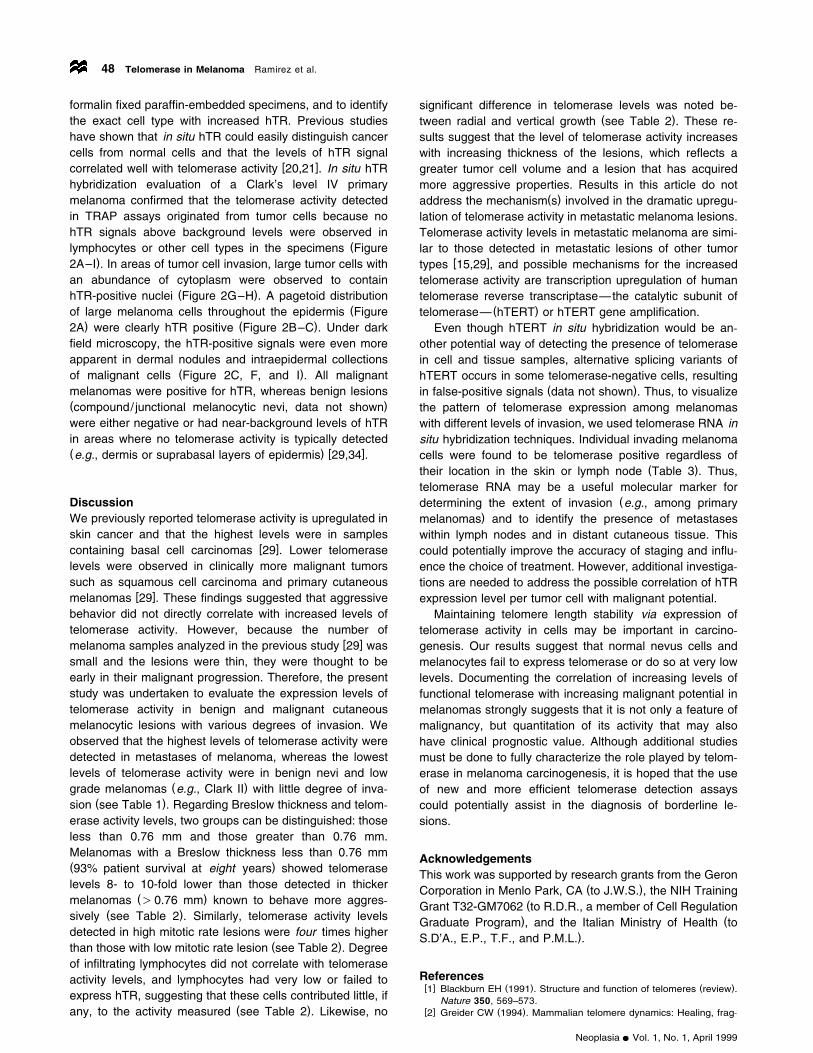
[ ]correlated well with telomerase activity 20,21 . In situ hTRhybridization evaluation of a Clark’s level IV primarymelanoma confirmed that the telomerase activity detectedin TRAP assays originated from tumor cells because nohTR signals above background levels were observed in
(lymphocytes or other cell types in the specimens Figure)2A–I . In areas of tumor cell invasion, large tumor cells with
an abundance of cytoplasm were observed to contain( )hTR-positive nuclei Figure 2G–H . A pagetoid distribution
(of large melanoma cells throughout the epidermis Figure) ( )2A were clearly hTR positive Figure 2B–C . Under dark
field microscopy, the hTR-positive signals were even moreapparent in dermal nodules and intraepidermal collections
( )of malignant cells Figure 2C, F, and I . All malignantmelanomas were positive for hTR, whereas benign lesions( )compound/ junctional melanocytic nevi, data not shownwere either negative or had near-background levels of hTRin areas where no telomerase activity is typically detected( ) [ ]e.g., dermis or suprabasal layers of epidermis 29,34 .
DiscussionWe previously reported telomerase activity is upregulated inskin cancer and that the highest levels were in samples
[ ]containing basal cell carcinomas 29 . Lower telomeraselevels were observed in clinically more malignant tumorssuch as squamous cell carcinoma and primary cutaneous
[ ]melanomas 29 . These findings suggested that aggressivebehavior did not directly correlate with increased levels oftelomerase activity. However, because the number of
[ ]melanoma samples analyzed in the previous study 29 wassmall and the lesions were thin, they were thought to beearly in their malignant progression. Therefore, the presentstudy was undertaken to evaluate the expression levels oftelomerase activity in benign and malignant cutaneousmelanocytic lesions with various degrees of invasion. Weobserved that the highest levels of telomerase activity weredetected in metastases of melanoma, whereas the lowestlevels of telomerase activity were in benign nevi and low
( )grade melanomas e.g., Clark II with little degree of inva-( )sion see Table 1 . Regarding Breslow thickness and telom-
erase activity levels, two groups can be distinguished: thoseless than 0.76 mm and those greater than 0.76 mm.Melanomas with a Breslow thickness less than 0.76 mm( )93% patient survival at eight years showed telomeraselevels 8- to 10-fold lower than those detected in thicker
( )melanomas )0.76 mm known to behave more aggres-( )sively see Table 2 . Similarly, telomerase activity levels
detected in high mitotic rate lesions were four times higher( )than those with low mitotic rate lesion see Table 2 . Degree
of infiltrating lymphocytes did not correlate with telomeraseactivity levels, and lymphocytes had very low or failed toexpress hTR, suggesting that these cells contributed little, if
( )any, to the activity measured see Table 2 . Likewise, no
significant difference in telomerase levels was noted be-( )tween radial and vertical growth see Table 2 . These re-
sults suggest that the level of telomerase activity increaseswith increasing thickness of the lesions, which reflects agreater tumor cell volume and a lesion that has acquiredmore aggressive properties. Results in this article do not
( )address the mechanism s involved in the dramatic upregu-lation of telomerase activity in metastatic melanoma lesions.Telomerase activity levels in metastatic melanoma are simi-lar to those detected in metastatic lesions of other tumor
[ ]types 15,29 , and possible mechanisms for the increasedtelomerase activity are transcription upregulation of humantelomerase reverse transcriptase—the catalytic subunit of
( )telomerase— hTERT or hTERT gene amplification.Even though hTERT in situ hybridization would be an-
other potential way of detecting the presence of telomerasein cell and tissue samples, alternative splicing variants ofhTERT occurs in some telomerase-negative cells, resulting
( )in false-positive signals data not shown . Thus, to visualizethe pattern of telomerase expression among melanomaswith different levels of invasion, we used telomerase RNA insitu hybridization techniques. Individual invading melanomacells were found to be telomerase positive regardless of
( )their location in the skin or lymph node Table 3 . Thus,telomerase RNA may be a useful molecular marker for
(determining the extent of invasion e.g., among primary)melanomas and to identify the presence of metastases
within lymph nodes and in distant cutaneous tissue. Thiscould potentially improve the accuracy of staging and influ-ence the choice of treatment. However, additional investiga-tions are needed to address the possible correlation of hTRexpression level per tumor cell with malignant potential.
Maintaining telomere length stability via expression oftelomerase activity in cells may be important in carcino-genesis. Our results suggest that normal nevus cells andmelanocytes fail to express telomerase or do so at very lowlevels. Documenting the correlation of increasing levels offunctional telomerase with increasing malignant potential inmelanomas strongly suggests that it is not only a feature ofmalignancy, but quantitation of its activity that may alsohave clinical prognostic value. Although additional studiesmust be done to fully characterize the role played by telom-erase in melanoma carcinogenesis, it is hoped that the useof new and more efficient telomerase detection assayscould potentially assist in the diagnosis of borderline le-sions.
AcknowledgementsThis work was supported by research grants from the Geron
( )Corporation in Menlo Park, CA to J.W.S. , the NIH Training(Grant T32-GM7062 to R.D.R., a member of Cell Regulation
) (Graduate Program , and the Italian Ministry of Health to)S.D’A., E.P., T.F., and P.M.L. .
References[ ] ( ) ( )1 Blackburn EH 1991 . Structure and function of telomeres review .
Nature 350, 569–573.[ ] ( )2 Greider CW 1994 . Mammalian telomere dynamics: Healing, frag-
Neoplasia v Vol. 1, No. 1, April 1999

Telomerase in Melanoma Ramirez et al. 49
mentation shortening and stabilization. Curr Opin Genet Dev 4,203–211.
[ ] ( )3 Olovnikov AM 1971 . Principle of marginotomy in template synthe-sis of polynucleotides. Dokl Akad Nauk SSSR 201, 1496–1499.
[ ] ( )4 Watson JD 1972 . Origin of concatemeric T7 DNA. Nature NewBiology 239, 197–201.
[ ] ( )5 Harley CB, Futcher AB, and Greider CW 1990 . Telomeres shortenduring aging of human fibroblasts. Nature 345, 458–460.
[ ] ( )6 Shay JW, Werbin H, and Wright WE 1994 . Telomere shorteningmay contribute to aging and cancer: A perspective. Mol Cell Diff 2,1–18.
[ ]7 Feng J, Funk WD, Wang SS, Weinrich SL, Avilion AA, Adams RR,Chang E, Allsopp RC, Yu J, Le S, West MD, Andrews WH, Greider
( )CW, and Villeponteau B 1995 . The RNA component of humantelomerase. Science 269, 1236–1241.
[ ] ( )8 Counter CM, Meyerson M, Eaton EN, and Weinberg RA 1997 .The catalytic subunit of yeast telomerase. Proc Natl Acad Sci USA94, 9202–9207.
[ ]9 Meyerson M, Counter CM, Eaton EN, Ellisen LW, Steiner P, Cad-dle SD, Ziaugra L, Beijersbergen RL, Davidoff MJ, Liu Q, Bacchetti
( )S, Haber DA, and Weinberg RA 1997 . hEST2, the putative hu-man telomerase catalytic subunit gene, is up-regulated in tumorcells and during immortalization. Cell 90, 785–795.
[ ]10 Nakamara TM, Morin GB, Chapman KB, Weinrich SL, Andrews( )WH, Lingner J, Harley CB, and Cech TR 1997 . Telomerase
catalytic subunit homologs from fission yeast and human. Science277, 955–959.
[ ] ( )11 Allsopp RC and Harley CB 1995 . Evidence for a critical telomerelength in senescent human fibroblasts. Exp Cell Res 219, 130–136.
[ ] ( )12 Alberts B, Brays D, Lewis J, et al 1994 . In: B Alberts, D Bray, JLewis, M Raff, K Roberts, and JD Watson, Eds. Molecular Biologyof the Cell, 3rd ed. New York & London. Garland Publishing, Inc,pp. 1255–1294.
[ ] ( )13 Shay JW and Wright WE 1996 . Telomerase activity in humancancer. Curr Opin Oncol 8, 66–71.
[ ] ( )14 Shay JW 1995 . Aging and cancer: Are telomeres and telomerasethe connection? Mol Med Today 1, 378–384.
[ ]15 Kim NW, Piatyszek MA, Prowse KR, Harley CB, West MD, Ho PL,( )Coviello GM, Wright WE, Weinrich SL, and Shay JW 1994 . Spe-
cific association of human telomerase activity with immortal cellsand cancer. Science 266, 2011–2015.
[ ] ( )16 Shay JW and Bacchetti S 1997 . A survey of telomerase activity inhuman cancer. Eur J Cancer 33, 787–791.
[ ]17 Hiyama E, Hiyama K, Yokoyama T, Matsuura Y, Piatyszek MA,( )and Shay JW 1995 . Correlating telomerase activity levels with
human neuroblastoma outcomes. Nat Med 1, 249–255.[ ] ( )18 Whited JD, Hall RP, Simel DL, and Horner RD 1997 . Primary care
clinicians’ performance for detecting actinic keratoses and skincancer. Arch Intern Med 157, 985–990.
[ ]19 Corona R, Mele A, Amini M, De Rosa G, Coppola G, Piccardi P,( )Fucci M, Pasquini P, and Faraggiana T 1996 . Interobserver vari-
ability on the histopathologic diagnosis of cutaneous melanomaand other pigmented skin lesions. J Clin Oncol 14, 1218–1223.
[ ]20 Yashima K, Piatyszek MA, Saboorian HM, Virmani AK, Brown D,( )Shay JW, and Gazdar AF 1997 . Expression of telomerase activity
and in situ telomerase RNA exression in malignant and non-malig-nant lymph nodes. J Clin Pathol 50, 110–117.
[ ]21 Yashima K, Litzky LA, Kaiser L, Rogers T, Lam S, Wistuba I,( )Milchgrub S, Srivastava S, Piatyszek MA, Shay JW et al 1997 .
Telomerase expression in bronchial epithelium during multistagepathogenesis of lung carcinomas. Cancer Res 57, 2373–2377.
[ ]22 Clark WHJ, Elder DE, Guerry IV D, Braitman LE, Trock BJ, Schultz( )D, Synnestvedt M, and Halpern AC 1989 . Model predicting sur-
vival in stage I melanoma based on tumor progression. J NatlCancer Inst 81, 1893–904.
[ ] ( )23 Clark WHJ, From L, Bernardino EA, and Mihm MC 1969 . Thehistogenesis and biologic behavior of primary human malignantmelanomas of the skin. Cancer Res 29, 705–726.
[ ]24 Balch CM, Soong S, Shaw HM, Urist MM, and McCarthy WH( )1992 In: CM Balch, AN Houghton, GW Milton, AJ Sober, and SSoong, Eds. Cutaneous Melanoma, 2nd ed. JB Lippincott, Philadel-phia. An Analysis of Prognostic Factors in 8500 Patients withCutaneous Melanoma pp. 165–187.
[ ]25 Ketcham AS, Moffat FL, Balch CM, In: CM Balch, AN Houghton,GW Milton, AJ Sober, and S Soong, Eds. Cutaneous Melanoma,2nd ed. JB Lippincott, Philadelphia. Classification and Staging, pp.213–222.
[ ] ( )26 Vollmer RT 1989 . Malignant melanoma: A multivariate analysis ofprognostic factors. Pathol Annu 24, 383.
[ ] ( )27 Breslow A 1975 . Tumor thickness, level of invasion and nodedissection in stage I cutaneous melanoma. Ann Surg 182, 572–575.
[ ] ( )28 Breslow A 1970 . Thickness, cross-sectional areas and depth ofinvasion in the prognosis of cutaneous melanoma. Ann Surg 172,902–908.
[ ]29 Taylor RS, Ramirez RD, Ogoshi M, Chaffins M, Piatyszek MA, and( )Shay JW 1996 . Detection of telomerase activity in malignant and
nonmalignant skin conditions. J Invest Dermatol 106, 759–765.[ ]30 Piatyszek MA, Kim NW, Weinrich SL, Hiyama K, Hiyama E, Wright
( )WE, and Shay JW 1995 . Detection of telomerase activity inhuman cells and tumors by a telomeric repeat amplification proto-
( )col TRAP . Methods Cell Sci 17, 1–15.[ ] ( )31 Wright WE, Shay JW, and Piatyszek MA 1995 . Modifications of a
( )telomeric repeat amplification protocol TRAP result in increasedreliability, linearity and sensitivity. Nucleic Acids Res 23, 3794–3795.
[ ] ( )32 Counter CM, Gupta L, Harley CB, Leber B, and Bacchetti S 1995 .Telomerase activity in normal leukocytes and in hematologic malig-nancies. Blood 85, 2315–2320.
[ ]33 Hiyama K, Hirai Y, Kyoizumi S, Akiyama M, Hiyama E, Piatyszek( )MA, Shay JW, Ishioka S, and Yamakido M 1995 . Activation of
telomerase in human lymphocytes and hematopoietic progenitorcells. J Immunol 155, 3711–3715.
[ ] ( )34 Harle-Bachor C and Boukamp P 1996 . Telomerase activity in theregenerative basal layer of the epidermis in human skin and inimmortal and carcinoma-derived skin keratinocytes. Proc Natl AcadSci USA 93, 6476–6481.
Neoplasia v Vol. 1, No. 1, April 1999