Cell Cycle-regulated Trafficking of Human Telomerase to Telomeres
39
1 Cell cycle regulated trafficking of human telomerase to telomeres Rebecca L. Tomlinson, Tania D. Ziegler, Teerawit Supakorndej, Rebecca M. Terns 2 and Michael P. Terns 1 Departments of Biochemistry and Molecular Biology, and Genetics University of Georgia, Athens, GA30602, USA Running head: Telomerase moves to telomeres at S phase Corresponding authors: 1 e-mail: [email protected] 2 e-mail: [email protected] Keywords: telomerase, telomeres, cell cycle, S phase, Cajal bodies
Transcript of Cell Cycle-regulated Trafficking of Human Telomerase to Telomeres

1
Cell cycle regulated trafficking of human telomerase to telomeres
Rebecca L. Tomlinson, Tania D. Ziegler, Teerawit Supakorndej, Rebecca M. Terns2 and Michael P. Terns1
Departments of Biochemistry and Molecular Biology, and Genetics
University of Georgia, Athens, GA30602, USA
Running head: Telomerase moves to telomeres at S phase Corresponding authors: 1e-mail: [email protected] 2e-mail: [email protected] Keywords: telomerase, telomeres, cell cycle, S phase, Cajal bodies

2
Abstract
Telomerase synthesizes telomeres at the ends of human chromosomes during
S phase. The results presented here suggest that telomerase activity may be
regulated by intranuclear trafficking of the key components of the enzyme in human
cells. We examined the subcellular localization of endogenous human telomerase
RNA (hTR) and telomerase reverse transcriptase (hTERT) in HeLa cervical
carcinoma cells. Throughout most of the cell cycle, we found that the two essential
components of telomerase accumulate at intranuclear sites separate from telomeres.
However, during S phase, both hTR and hTERT are specifically recruited to subsets
of telomeres. The localization of telomerase to telomeres is dynamic, peaking at
mid-S phase. We also found complex associations of both hTR and hTERT with
nucleoli and Cajal bodies during S phase, implicating both structures in the
biogenesis and trafficking of telomerase. Our results mark the first observation of
human telomerase at telomeres, and provide a mechanism for the cell cycle-
dependent regulation of telomere synthesis in human cells.

3
Introduction
The ends of linear eukaryotic chromosomes are capped by nucleoprotein
structures termed telomeres. In vertebrates, telomeres consist of simple DNA repeats of
TTAGGG bound by several proteins (Colgin and Reddel, 2004; Smogorzewska and de
Lange, 2004). Telomeres serve to maintain chromosome integrity, preventing
illegitimate recombination and end-to-end joining (de Lange, 2002; Harrington, 2004;
Blasco, 2005). However, due to the unidirectional nature of DNA polymerases and DNA
processing events, some telomere sequence is lost with each round of DNA replication.
If this loss is not compensated, the telomeres will reach a critically short length,
triggering the cell to enter a state of replicative senescence or apoptosis (McEachern et
al., 2000; de Lange, 2002).
Telomerase is the ribonucleoprotein (RNP) enzyme that synthesizes telomeres.
The telomerase reverse transcriptase (hTERT) catalyzes de novo repeat addition using a
short motif within the integral telomerase RNA (hTR) as a template (Greider and
Blackburn, 1989). These two components are essential for the activity of the enzyme.
Human telomere synthesis occurs early in development (Collins and Mitchell, 2002;
Cong et al., 2002). The majority of adult somatic cells do not have appreciable
telomerase activity and telomeres gradually shorten, limiting cell division capacity
(Harley et al., 1990). In the majority of human cancers, however, telomerase is re-
activated and provides the sustained proliferative capacity of these cells (Shay and
Bacchetti, 1997). An understanding of telomerase biology thus has important
implications for both cancer and aging.

4
Telomeres are synthesized during S phase in human cells (Ten Hagen et al., 1990;
Wright et al., 1999), however, it is unclear how telomerase is restricted to function
specifically during this stage of the cell cycle. Some existing evidence is consistent with
the idea that the cell cycle-dependent regulation of telomerase could occur at the level of
subcellular trafficking. Redistribution of components of telomerase has been observed
during S phase, when telomere synthesis occurs. Wong et al. showed that the subnuclear
distribution of GFP-hTERT fusion proteins changed from predominantly nucleolar to
nucleoplasmic as cells progressed through S phase (Wong et al., 2002). In similar
studies, Yang et al. also reported movement of GFP-hTERT protein during S phase, in
this case into nucleoli (Yang et al., 2002b). Regarding the other essential component of
the enzyme, Jady et al. reported a possible influx of telomerase RNA through Cajal
bodies during S phase (Jady et al., 2004). However, while there is some evidence that
both hTR and hTERT move during S phase, movement to telomeres, and thus a direct
link to regulation of function, has not been found in vertebrate cells. More persuasive
evidence is available from ciliates, where telomerase RNA localizes to discrete nuclear
foci throughout most of the cell cycle, and a fraction of the RNA is mobilized to the
replication band, the site of DNA (and telomere) synthesis, during S phase (Fang and
Cech, 1995).
In this work, we have investigated the subnuclear distribution of endogenous
human TR and TERT over the course of the cell cycle, and found compelling evidence
that regulation of telomerase activity occurs via trafficking of hTR and hTERT in human
cells. Our results indicate that hTR and hTERT move to telomeres from separate sites
specifically during S phase. hTR is found in Cajal bodies, as we and others have reported

5
previously (Jady et al., 2004; Zhu et al., 2004), throughout most of the cell cycle. Here
we provide the first clear evidence that hTERT resides in subnuclear foci that do not
correspond to nucleoli, Cajal bodies or telomeres during most of the cell cycle. The
movement of hTR and hTERT to telomeres during S phase is preceded and accompanied
by other changes in localization that may relate to biogenesis and/or transport of the
components. Our results suggest that hTERT moves to nucleoli, and that Cajal bodies
containing hTR accumulate at the periphery of nucleoli early in S phase. In addition, we
find that both hTR and hTERT localize to foci adjacent to Cajal bodies during S phase,
marking a potential site outside of telomeres where both endogenous telomerase
components are detected. The implications of our findings with regard to telomerase
biogenesis and telomere length regulation are discussed.
6
Materials and Methods
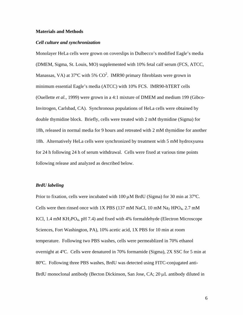
Cell culture and synchronization
Monolayer HeLa cells were grown on coverslips in Dulbecco’s modified Eagle’s media
(DMEM, Sigma, St. Louis, MO) supplemented with 10% fetal calf serum (FCS, ATCC,
Manassas, VA) at 37ºC with 5% CO2. IMR90 primary fibroblasts were grown in
minimum essential Eagle’s media (ATCC) with 10% FCS. IMR90-hTERT cells
(Ouellette et al., 1999) were grown in a 4:1 mixture of DMEM and medium 199 (Gibco-
Invitrogen, Carlsbad, CA). Synchronous populations of HeLa cells were obtained by
double thymidine block. Briefly, cells were treated with 2 mM thymidine (Sigma) for
18h, released in normal media for 9 hours and retreated with 2 mM thymidine for another
18h. Alternatively HeLa cells were synchronized by treatment with 5 mM hydroxyurea
for 24 h following 24 h of serum withdrawal. Cells were fixed at various time points
following release and analyzed as described below.
BrdU labeling
Prior to fixation, cells were incubated with 100 μM BrdU (Sigma) for 30 min at 37ºC.
Cells were then rinsed once with 1X PBS (137 mM NaCl, 10 mM Na2 HPO4, 2.7 mM
KCl, 1.4 mM KH2PO4, pH 7.4) and fixed with 4% formaldehyde (Electron Microscope
Sciences, Fort Washington, PA), 10% acetic acid, 1X PBS for 10 min at room
temperature. Following two PBS washes, cells were permeablized in 70% ethanol
overnight at 4ºC. Cells were denatured in 70% formamide (Sigma), 2X SSC for 5 min at
80ºC. Following three PBS washes, BrdU was detected using FITC-conjugated anti-
BrdU monoclonal antibody (Becton Dickinson, San Jose, CA; 20 μL antibody diluted in

7
70 μL PBST (0.05% Tween-20 in PBS)) for 2 hours at room temperature. After three
PBS washes, coverslips were mounted in 90% glycerol, 1 mg/mL p-Phenylenediamine,
1X PBS, and 0.1 μg/mL DAPI. If FISH was to be performed, cells were fixed again in
4% formaldehyde in 1X PBS for 10 minutes at room temperature and washed twice with
PBS.
hTR and telomere FISH
Probes complementary to different regions of telomerase RNA (nucleotides indicated) or
telomere repeats were as follows:
hTR 128-183 (probe 1)
GCT*GACATTTTT*TGTTTGCTCT*AGAATGAACGGT*GGAAGGCGGCAGGCC
GAGGCT*T;
hTR 331-383 (probe 2)
CT*CCGTTCCTCTTCCT*GCGGCCTGAAAGGCCT*GAACCTCGCCCT*CGCCCC
CGAGT*G;
hTR 393-449 (probe 3)
AT*GTGTGAGCCGAGT*CCTGGGTGCACGT*CCCACAGCTCAGGGAAT*CGCG
CCGCGCT*C;
and telomere repeats (probe 4)
CT*AACCCTAACCCT*AACCCTAACCCT*AACCCTAACCCT*AACCCTAACCCT
*A. T* indicate aminoallyl-modified thymidines. All probes were synthesized by
Qiagen (Valencia, CA). Probes were conjugated with either cy3 or cy5 monofunctional
reactive dye according to manufacturer’s protocol (Amersham Pharmacia, Piscataway,
8
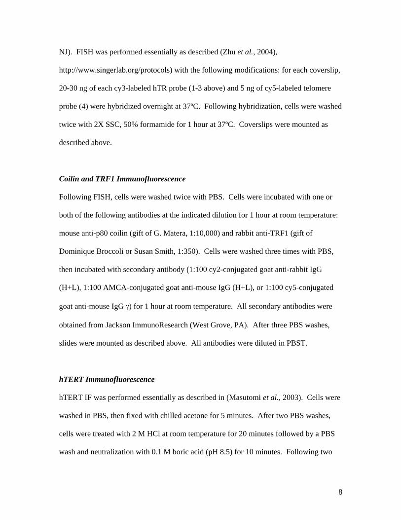
NJ). FISH was performed essentially as described (Zhu et al., 2004),
http://www.singerlab.org/protocols) with the following modifications: for each coverslip,
20-30 ng of each cy3-labeled hTR probe (1-3 above) and 5 ng of cy5-labeled telomere
probe (4) were hybridized overnight at 37ºC. Following hybridization, cells were washed
twice with 2X SSC, 50% formamide for 1 hour at 37ºC. Coverslips were mounted as
described above.
Coilin and TRF1 Immunofluorescence
Following FISH, cells were washed twice with PBS. Cells were incubated with one or
both of the following antibodies at the indicated dilution for 1 hour at room temperature:
mouse anti-p80 coilin (gift of G. Matera, 1:10,000) and rabbit anti-TRF1 (gift of
Dominique Broccoli or Susan Smith, 1:350). Cells were washed three times with PBS,
then incubated with secondary antibody (1:100 cy2-conjugated goat anti-rabbit IgG
(H+L), 1:100 AMCA-conjugated goat anti-mouse IgG (H+L), or 1:100 cy5-conjugated
goat anti-mouse IgG γ) for 1 hour at room temperature. All secondary antibodies were
obtained from Jackson ImmunoResearch (West Grove, PA). After three PBS washes,
slides were mounted as described above. All antibodies were diluted in PBST.
hTERT Immunofluorescence
hTERT IF was performed essentially as described in (Masutomi et al., 2003). Cells were
washed in PBS, then fixed with chilled acetone for 5 minutes. After two PBS washes,
cells were treated with 2 M HCl at room temperature for 20 minutes followed by a PBS
wash and neutralization with 0.1 M boric acid (pH 8.5) for 10 minutes. Following two

9
additional PBS washes, cells were blocked with 1% BSA in PBS at 4°C overnight. Cells
were incubated with mouse anti-hTERT 2C4 (Abcam, Cambridge, MA; 1:2000-1:5000 in
1% BSA), for 2 hours at room temperature, washed three times with PBS, and incubated
with cy2-conjugated goat anti-mouse IgM (Jackson ImmunoResearch; 1:100 in PBST)
for 2 hours at room temperature. After three PBS washes, the cells were mounted as
described above. When hTERT staining was combined with TRF1 or coilin, the hTERT
IF protocol was followed using the above described antibodies at the indicated dilutions
except that cy3-conjugated goat anti-rabbit IgG (H+L) was used to recognize TRF1 and
mouse anti-coilin was recognized with cy5-conjugated goat anti-mouse IgG γ antibody.
Both secondary antibodies were obtained from Jackson ImmunoResearch.
Microscopy
Analysis was performed on a Zeiss Axioskop 2 Mot Plus fluorescence microscope
(Thornwood, NY). Images were acquired at 63x or 100x magnification (Plan
Apochromat objectives, NA=1.4) using a cooled charge-coupled device Retiga Exi Fast
1394 camera (Qimaging, Burnaby, BC Canada) and IP Lab Spectrum software.

10
Results
Novel patterns of telomerase RNA localization during S phase
Our laboratory developed a fluorescence in situ hybridization (FISH) procedure
specific for the detection of endogenous hTR in human cells and was the first to describe
its intranuclear localization (Zhu et al., 2004). Several controls established the specificity
of our FISH procedure for hTR (Zhu et al., 2004). Using this technique, we found that
hTR localized to Cajal bodies in telomerase-positive human cancer cell lines (Zhu et al.,
2004). In the course of that study (performed with asynchronous populations of cells),
we noticed that a small percentage of cells displayed different patterns of hTR
localization. For example, we observed additional hTR foci that did not stain with
antibodies against coilin (the marker protein of Cajal bodies).
To investigate whether the secondary hTR localization patterns observed at low
frequency in asynchronous populations reflected cells in particular stages of the cell
cycle, we performed hTR FISH and coilin immunofluorescence (IF) on cell cycle-
synchronized HeLa cervical carcinoma cells. HeLa cells were synchronized with a
double thymidine block (or hydroxyurea, data not shown) and monitored for
synchronization efficiency at the indicated time points by 5-bromodeoxyuridine (BrdU)
analysis (to distinguish S phase cells) and 4’,6-diamidino-2-phenylindole (DAPI) staining
(to distinguish mitotic cells). (FACS (fluorescence activated cell sorting) analysis with
propidium iodide-stained cells (to examine DNA content) was also performed to confirm
the synchronization efficiency in some cases.) The BrdU staining patterns also allowed
us to assess the S sub-phase (i.e. early, mid and late) status of populations of cells as well
as individual cells (O'Keefe et al., 1992).

11
As expected, throughout the majority of the cell cycle, hTR localizes exclusively
to Cajal bodies (G1 and G2, Figure 1). However, during S phase, a dynamic change in
the subcellular distribution of hTR was observed in a significant number of cells. hTR
remained associated with Cajal bodies in most cells; however, several novel hTR
localization patterns were observed, which peaked at distinct points in S phase.
Beginning in G1/S and peaking in early S, hTR foci were found in ring-like patterns
within the nucleus (Figure 1, row 2). In most cells, coilin co-localized with hTR in the
rings, as shown (Figure 1, row 2). By mid-S phase, the distribution of hTR foci in rings
had declined, and hTR appeared in small, nucleoplasmic foci that did not stain with anti-
coilin antibodies (Figure 1, row 3, denoted by arrowheads). In addition, hTR was
sometimes observed in foci immediately adjacent to Cajal bodies (detailed below). These
patterns were specific to S phase; they were not observed in cells in G1 or G2 phases of
the cell cycle.
During mitosis, previous research has shown that Cajal bodies dissociate, and
then re-form during early G1 of the cell cycle (Andrade et al., 1993). We found that hTR
localization parallels that of the Cajal body marker protein coilin during cell division,
with only a slight temporal delay in re-association after mitosis. Like coilin, hTR
displayed a diffuse localization pattern throughout mitosis (Figure 1, row 5 shows hTR
diffusely localized in interchromatin region). No association with discrete foci was
observed. Following mitosis, Cajal body formation precedes hTR accumulation in Cajal
bodies. In early G1, Cajal bodies had re-formed in nearly all (approximately 90%) of
cells examined, but no hTR foci were found in most (approximately 65%) cells (Figure 1,

12
row 6). By mid-G1 (15 hours post release) hTR had re-accumulated in all Cajal bodies
(Figure 1, row 1).
Taken together, these results demonstrate that the cell cycle has dramatic effects
on the localization of hTR and that there is a dynamic, S phase-specific rearrangement of
hTR in human cancer cell nuclei.
Telomerase RNA localizes to the periphery of nucleoli during early S phase
We found that the ring-like pattern of hTR (and coilin) foci observed during early
S phase appears to correspond to localization to the periphery of nucleoli (Figure 2A). In
addition to the ring pattern, we also observed hTR foci that appeared to be distributed
across the surface of a nucleolus in some focal planes (Figure 2A). At early S phase,
approximately 17% of cells (370 cells analyzed from 3 separate experiments) contained
hTR foci localized around the periphery of a nucleolus. In the majority of cases, the hTR
ring was observed around only one nucleolus within a given cell, suggesting a previously
undescribed heterogeneity among individual nucleoli. We did not find telomeres at these
rings; telomeres co-localized with hTR/coilin rings in less than 1% of 1197 cells
examined in 2 experiments. The peripheral nucleolar hTR pattern was less frequent at
G1/S and mid-S than at early S phase, and was not observed in cells outside S phase.
As shown in Figure 1, coilin was found with hTR in the nucleolar rings in the
majority (approximately 80%) of cells. The localization of Cajal bodies to the periphery
of nucleoli is well documented (indeed Cajal bodies were initially termed nucleolar
accessory bodies (Gall, 2003; Cioce and Lamond, 2004)), though an S phase-specific
association has not been previously described. We cannot exclude the possibility that the

13
Cajal bodies and hTR are internal to nucleoli based on the studies presented here, but the
distribution of the foci is suggestive of localization on the surface of the nucleolus.
Elegant studies in HeLa cells have demonstrated the movement of Cajal bodies to and
from nucleoli (Platani et al., 2000). Our results suggest that Cajal bodies containing hTR
move to the periphery of nucleoli during early S phase in HeLa cells.
Telomerase RNA localizes to subsets of telomeres during mid-S phase
To ascertain whether the coilin-negative, S phase-specific hTR foci described
above (Figure 1, row 3) were at telomeres, we co-analyzed hTR and telomeres in S phase
cells. We performed hTR FISH in conjunction with either telomere FISH (probe directed
against telomere repeat sequences) or immunofluorescence with antibodies against the
double-stranded telomere binding protein TRF1 (Figure 2B). Cells were examined 0
(G1/S), 2 (early S), 4 (mid S), 6 (late S), and 8 (S/G2) hours post release from a double
thymidine block. At G1/S and early S, we found that hTR co-localized with a few
telomeres (1-2 per cell) in approximately 3% and 9% of cells, respectively (562 and 370
cells analyzed from 6 and 3 separate experiments, respectively). During mid-S, there was
an increase in both the number of cells that had hTR-telomere associations and in the
number of associations per cell. In approximately 19% of mid-S phase cells (698 cells
from 5 experiments), hTR was found at telomeres – typically 1-5 per cell with a
maximum of 11 co-localizations observed in one cell. The telomere associations
declined in late S phase, to 11% of cells (336 cells from 3 experiments) and 1-2 per cell.
hTR was also still found in or near Cajal bodies in most of these cells. hTR was not
14
found at telomeres in G2 phase (data not shown). These results indicate that human
telomerase RNA moves to telomeres during mid-S phase.
hTERT also associates with telomeres and nucleoli during S phase
Telomerase requires both hTR and hTERT for function. Previous studies found
that ectopically expressed GFP-hTERT fusion protein shows striking S phase-specific
intranuclear rearrangements (Wong et al., 2002; Yang et al., 2002b), but was not found to
localize to telomeres. Here we have investigated the localization of endogenous hTERT
protein.
We performed immunofluorescence using the 2C4 monoclonal antibody against
hTERT (Masutomi et al., 2003) and observed a punctate nuclear staining pattern in HeLa
cells. The staining pattern previously described for this antibody in HeLa cells was more
generally nucleoplasmic (Masutomi et al., 2003), so we examined the specificity of the
pattern observed in our experiments (Figure 3A). We found that the more restricted
staining pattern that we obtained was specific to hTERT. We knocked down hTERT
expression in HeLa cells by RNA interference (Masutomi et al., 2003) and observed a
marked decrease in the fluorescence signal intensity (including complete elimination of
signal in some cells), indicating that the signal in the intranuclear foci corresponded to
hTERT (Figure 3A). As an additional test, we compared staining in IMR90 primary lung
fibroblasts to IMR90 cells stably expressing hTERT from an exogenous construct
(Ouellette et al., 1999). We observed some staining in the primary fibroblasts (Figure
3A), consistent with previous observations that normal cells express a low level of
hTERT protein (Masutomi et al., 2003). The number and intensity of the foci was

15
significantly greater in the cells expressing exogenous hTERT. Similar results were seen
when hTERT was stably expressed in BJ cells (data not shown). Taken together, these
results indicate that the 2C4 staining observed under these conditions is specific to
hTERT and that hTERT is found in nucleoplasmic foci.
In synchronized cells, we found that hTERT (like hTR) localizes to nucleoli
during the early stages of S phase (Figure 3B). Diffuse nucleolar localization of hTERT
is detectable in 23% of cells at G1/S (429 cells analyzed from 2 separate experiments),
and intensifies as cells enter early S, when the localization is apparent in 35% of cells
(282 cells from 2 experiments) (Figure 3B). Like hTR, hTERT was observed in a subset
of nucleoli (typically one) within a cell. Nucleolar association of hTERT declined as
cells progressed through S phase (9% of 236 cells in late S phase in 2 experiments).
Later in S phase, additional hTERT foci appeared, which corresponded to a subset
of telomeres (Figure 3C). Co-localization of hTERT and telomeres (assessed by TRF1
(or TRF2, data not shown) antibody staining) occurred in a few (less than 5%) cells at
G1/S (391 cells from 2 experiments). Localization of hTERT to telomeres peaked in
mid-S phase when 24% of cells (463 cells from 2 experiments) were found to contain 1-5
co-localizations (Figure 3C).
Unfortunately, to date we have been unsuccessful in simultaneously analyzing
hTERT and hTR (due to incompatibility of the IF and FISH protocols). Independently,
hTR and hTERT display remarkably similar temporal patterns of localization to
telomeres and nucleoli during S phase (Figure 4). The results suggest an S phase-specific
mobilization of the components of the telomerase RNP to its functional destination, the
telomere.

16
hTR and hTERT are found in foci adjacent to Cajal bodies during S phase
Our results indicate that hTR normally accumulates in Cajal bodies in HeLa cells,
and hTERT does not. (Previously, we reported localization of YFP-hTERT to Cajal
bodies in HeLa cells (Zhu, 2004). However in the current work, we did not detect
significant accumulation of endogenous hTERT in Cajal bodies in either asynchronous or
S phase cells; hTERT co-localized with coilin in less than 5% of cells (Figure 4).) While
we did not observe endogenous hTERT in Cajal bodies, we did detect a specific
accumulation of both hTERT and hTR in foci immediately adjacent to Cajal bodies
during S phase. Figure 5 shows examples of the associations of hTR and hTERT with
Cajal bodies observed in S phase HeLa cells. The associations appear to represent one or
two structures containing hTR or hTERT in direct contact with a Cajal body. In these
cases, there is relatively little hTR (or hTERT) found in the core of the Cajal bodies.
(The pattern does not represent simple pixel shifting relative to coilin data since the
offsets occurred along multiple vectors within a single field and did not affect co-
localizations in adjacent cells.) In some cases, particularly for hTR, it appears that the
foci may be present at poles of the Cajal body (rather than in distinct adjacent structures)
(e.g. Figure 5, row 2). In other cases, it is clear that the hTR or hTERT foci are distinct
from the Cajal body (e.g. Figure 5, row 4). The timing and frequency with which hTR or
hTERT is observed closely associated with Cajal bodies are similar to telomere
associations (Figure 4). At mid-S phase, localization to foci associated with Cajal bodies
was found in 31% cells for hTR (698 cells analyzed from 5 separate experiments) and in
38% cells for hTERT (280 cells from 2 experiments). At least in the case of hTERT,

17
there is also significant association in early S phase, suggesting that the peak of
localization to Cajal body-associated foci occurs just before mid-S phase (Figure 4). It is
not currently known whether the hTR or hTERT foci correspond with other, previously
described Cajal body-associated nuclear bodies (Liu and Dreyfuss, 1996; Yannoni and
White, 1997; Schul et al., 1999; Zhao et al., 2000; Miele et al., 2005).

18
Discussion
Telomere synthesis is restricted to S phase in human cells. In this study, we show
that the two key components of telomerase, telomerase RNA and the catalytic protein
subunit hTERT, are targeted to telomeres specifically in S phase. Our study marks the
first time human telomerase has been visualized at telomeres and suggests that the
restriction of telomere elongation to S phase of the cell cycle is achieved by subnuclear
trafficking of the telomerase RNP. Furthermore, our results indicate that multiple nuclear
structures play roles in the regulated transport and biogenesis of telomerase.
Telomere synthesis
Our finding that the recruitment of telomerase to telomeres is restricted to S phase
(peaking in mid-S, Figures 2B, 3C and 4) correlates well with the known timing of
telomere elongation in human cells (Ten Hagen et al., 1990; Wright et al., 1999).
Strikingly, our data suggest that telomerase accumulates at only a subset of telomeres in a
given cell (and in only a fraction of cells in a population) at any given time (Figures 2B,
3C and 4). The lack of detection of telomerase at some telomeres could certainly reflect
further limitations of the experimental approach. However, this finding is also consistent
with the idea that telomerase may not act on every telomere during every cell cycle, as
has been demonstrated in yeast where only a small fraction of telomeres (~7%) are
extended within a given cycle (Teixeira et al., 2004). Studies in both yeast and
mammalian cells indicate that telomerase preferentially elongates the shortest telomeres
in a population (Ouellette et al., 2000; Teixeira et al., 2004). Alternatively, all telomeres
may be extended during each cell cycle, but not simultaneously, such that telomerase is

19
active at only a subset of telomeres at any given time point. It is known that
chromosomes replicate at different rates during S phase (Woodfine et al., 2004; Zou et
al., 2004), and thus the timing of telomere synthesis could vary, for example, with the
timing of replication-induced changes in chromatin structure and telomere accessibility at
individual chromosomes.
Telomerase trafficking pathway
By following the subcellular localization of endogenous hTR and hTERT
molecules throughout the cell cycle, we have obtained important insight into the pathway
that telomerase may follow on its way to its site of action, the telomere (Figure 6).
However, it is important to note that while the successful detection of hTR and hTERT at
telomeres suggests excellent sensitivity, the results of our experiments do not preclude
the presence of lower concentrations of hTR or hTERT in cellular compartments other
than those identified, including the nucleoplasm. In addition, as discussed above, the
various localization patterns described here are not observed in every cell in a
synchronized population at a given time point, which may reflect technical limitations or
real differences in the timing or extent of telomere synthesis within and among individual
cells.
Prior to S phase, during G1 (and also following S phase, in G2), hTR and hTERT
are observed in separate intranuclear structures (Figure 6A). Telomerase RNA is present
in Cajal bodies (Figure 1), consistent with previous findings (Jady et al., 2004; Zhu et al.,
2004). In contrast, hTERT accumulates in distinct nucleoplasmic foci, which may
represent previously unrecognized nuclear bodies or identified structures not previously
20
known to contain hTERT (Figure 3). These findings suggest that the two key subunits of
telomerase may be sequestered away from one another throughout most of the cell cycle.
Specifically during S phase, hTR and hTERT exhibit a dynamic redistribution and
become targeted to common intranuclear sites. In early S phase, both hTR and hTERT
can be found associated with nucleoli, although apparently not within a shared
compartment (Figure 6B). hTR is present in Cajal bodies that appear to reside around the
periphery of the nucleolus (Figures 1 and 2), while hTERT appears to be distributed
throughout the interior of the nucleolus (Figure 3). Movement of Cajal bodies to and
from nucleoli has been documented previously (Platani et al., 2000) and may account for
the appearance of hTR at nucleoli in S phase. Intriguingly, we often find hTR or hTERT
at a single nucleolus within a cell. While it is well known that the nucleolus supports a
number of functions beyond its conventional role in ribosome biogenesis (Pederson,
1998; Olson et al., 2000; Lam et al., 2005), our results suggest a previously undescribed
division of labor among nucleoli.
Beginning in early S phase and peaking at mid-S (Figure 6C), a novel pattern of
hTR and hTERT localization emerges in foci that appear to be physically associated with
Cajal bodies (Figure 5). The hTERT found in Cajal body-associated foci may originate
in nucleoli, or come directly from the nucleoplasmic hTERT foci. The hTR foci may
arise by segregation of hTR to one pole of a Cajal body, as is suggested by some of our
data (e.g Figure 5, row 2). While we cannot demonstrate co-localization of hTR and
hTERT in the Cajal body-associated foci (or at telomeres) for technical reasons, it seems
possible (based on the similarity of the spatial and temporal patterns, and frequency of
occurrence) that both components of telomerase are found together here (Figure 6C).

21
Our analysis of hTERT suggests that localization to the Cajal body-associated foci
precedes localization to telomeres, which peaks more distinctly in mid-S phase (Figure 4,
see occurrence of Cajal body association at early S). We envision that Cajal bodies with
a compartmentalized cargo of hTR and hTERT deliver telomerase to individual telomeres
throughout the cell (Figure 6C).
In support of a role for Cajal bodies in the delivery of telomerase to telomeres, we
have observed occasional co-localization of the Cajal body-associated hTR and hTERT
foci with telomeres (data not shown), but the very low frequency of these associations
(observed in ~2-3% of S phase cells) suggests that the interactions with telomeres would
be either transient or not preserved under our experimental conditions. In addition, live
cell imaging has revealed that Cajal bodies undergo dramatic movements within the
nucleus, including journeys across the diameter of the nucleus, fusion with other Cajal
bodies, fragmentation into smaller bodies, and transient associations with nucleoli and
specific chromosomal loci (Gall, 2000; Platani et al., 2000; Sleeman et al., 2003; Cioce
and Lamond, 2004). (In contrast, the majority of telomeres appear to be anchored to the
nuclear matrix with limited capacity for migration (Luderus et al., 1996; Molenaar et al.,
2003).) Finally, an intriguingly similar cell cycle-regulated delivery of transcription and
processing factors to histone gene loci appears also to involve Cajal bodies and closely
associated foci. Emerging evidence suggests that HiNF-P, a histone gene transcription
factor, and p220/NPAT, an associated protein, co-localize at or near Cajal bodies in S
phase, which localize to histone gene loci resulting in activation of histone gene
transcription (Frey and Matera, 1995; Ma et al., 2000; Zhao et al., 2000; Shopland et al.,
2001; Miele et al., 2005). Similarly, the RNA processing factors CstF and CPSF appear
22
to move out of Cajal bodies into adjacent structures (termed cleavage bodies) that co-
localize with histone gene loci in S phase (Schul et al., 1999).
While neither the detailed localization of endogenous hTR and hTERT through
the cell cycle nor the visualization of hTR and hTERT at telomeres has been described
previously, some aspects of the pathway described in this work are supported by previous
reports. The association of hTR with Cajal bodies in human cells throughout most of the
cell cycle is consistent with the previous observations of our lab and others (Jady et al.,
2004; Zhu et al., 2004). However, Jady et al. reported an increase in the brightness of
hTR FISH signal in Cajal bodies (relative to nucleoplasm) in cells in S phase (Jady et al.,
2004), and it is not immediately clear how this observation relates to the pathway defined
here. Consistent with our findings, a small percentage of cellular hTR has been detected
in biochemical fractions containing nucleoli (Mitchell et al., 1999), and hTR localizes to
nucleoli (as well as Cajal bodies) when injected into Xenopus oocytes (Lukowiak et al.,
2001). In addition, ectopically expressed GFP-hTERT fusion proteins have been found
to localize to nucleoli, and Yang et al. reported increased nucleolar association during S
phase (Wong et al., 2002; Yang et al., 2002b). Our results place the previous
observations firmly in the context of a cell cycle-regulated pathway.
Regulation of telomerase trafficking
Our results indicate that the transport of telomerase to telomeres is exquisitely
regulated in the context of the cell cycle. On the basis of these findings, it will now be
interesting to determine how movement of the RNA and protein subunits are linked to the
cell cycle. One logical possibility is that some of the same kinases and phosphatases that

23
drive other S phase events (e.g. cdk2/cyclin A and cdc25) also modify telomerase
subunits (and telomere binding proteins) to regulate molecular interactions and thereby
influence telomerase trafficking (and telomere accessibility). In hTR, RNA elements
termed the CAB box and H/ACA motif have been found to be important for localization
of the RNA to Cajal bodies and nucleoli, respectively (Lukowiak et al., 2001; Jady et al.,
2004), and the domains of hTERT that mediate nucleolar localization have been defined
(Etheridge et al., 2002; Yang et al., 2002b). One would predict that these domains and
proteins that interact with these domains could be modified to effectively regulate
telomerase trafficking.
Telomerase biogenesis
It is clear from our results that the trafficking of hTR and hTERT is regulated by
the cell cycle. Interestingly, our findings suggest the possibility that the assembly of the
telomerase enzyme may also be regulated to restrict telomere synthesis to S phase (i.e.
the essential subunits may be compartmentalized away from each other as well as from
their substrate). The detectable pools of hTR and hTERT are not found in common
structures outside of S phase (Figures 1 and 3), suggesting that human telomerase is
assembled specifically during S phase and disassembled (or destroyed in the case of
hTERT (Masutomi et al., 2003; Kim et al., 2005)) after each cell cycle, perhaps during M
phase when the telomerase subunits do not appear to be associated with structures (Figure
1 and data not shown).
If the biogenesis of telomerase is regulated by the cell cycle, our results suggest
two likely sites for the assembly of the enzyme during S phase. hTR and hTERT are both

24
found in foci associated with Cajal bodies and at telomeres in mid-S phase. The Cajal
body-associated foci hold the potential for interaction with the SMN complex, a known
RNP assembly factor that resides in Cajal bodies (Terns and Terns, 2001; Gubitz et al.,
2004). SMN is known to interact with telomerase, and specifically to interact with GAR1
(an hTR-associated protein) and hTERT, suggesting that SMN may function in the
biogenesis of telomerase (Bachand et al., 2002; Whitehead et al., 2002).
Our data are also consistent with the possibility that telomerase may assemble at
the telomere. There is solid evidence that yeast telomerase is assembled at telomeres. In
this case, the core components of telomerase, TLC1 (telomerase RNA) and Est2p (TERT)
are constitutively present at telomeres, and assembly of active telomerase is regulated by
an S phase-specific recruitment of an essential telomerase subunit, Est1 to telomeres
(Taggart et al., 2002).
However, while telomerase activity is restricted to S phase in intact cells (Ten
Hagen et al., 1990; Wright et al., 1999), catalytically active telomerase enzyme (assessed
by TRAP assay) can be extracted from both human and yeast cells at any stage of the cell
cycle ((Holt et al., 1997) but see (Zhu et al., 1996; Yang et al., 2002a)). It is not clear if
this extracted telomerase activity reflects enzyme present in cells or assembled from
individual components after cell lysis and extract preparation (as has been demonstrated
to occur in the case of another RNA-protein complex (Mili and Steitz, 2004)). The
potential regulation of telomerase biogenesis by the cell cycle will require further
investigation.

25
Coupling telomerase trafficking to the cell cycle may have evolved in eukaroytes
as an efficient mechanism to restrict the activity of telomerase to the period when
chromosomes are replicated, and limit potentially deleterious activity of telomerase at
non-telomeric sites (i.e. chromosome healing at double-stranded breaks) during the
remainder of the cell cycle (Sprung et al., 1999). Future research in the field will further
delineate the intranuclear trafficking patterns of telomerase, define the molecular
mechanisms of telomerase biogenesis and telomere recruitment, and determine how these
processes are regulated by the cell cycle.

26
Acknowledgements
We are grateful to the following people for providing cell lines and antibodies:
Gregory Matera, Case Western Reserve University (HeLa cell line and anti-coilin
antibody); William Hahn, Dana Farber Cancer Institute, Harvard Medical School (HeLa
cell line expressing hTERT shRNA); Jerry Shay, UT Southwestern (IMR90-TERT cells);
Dominique Broccoli, Fox Chase Cancer Center and Susan Smith, Skirball Institute (anti-
TRF1 antibodies). This work was supported by grants from the American Cancer Society
and National Cancer Institute to MPT and RMT. RLT was supported by an NIH training
grant to the Department of Genetics at University of Georgia (GM07103).
27
References
Andrade, L.E., Tan, E.M., and Chan, E.K. (1993). Immunocytochemical analysis of the coiled body in the cell cycle and during cell proliferation. Proc Natl Acad Sci U S A 90, 1947-1951. Bachand, F., Boisvert, F.M., Cote, J., Richard, S., and Autexier, C. (2002). The product of the survival of motor neuron (SMN) gene is a human telomerase-associated protein. Mol Biol Cell 13, 3192-3202. Blasco, M.A. (2005). Mice with bad ends: mouse models for the study of telomeres and telomerase in cancer and aging. Embo J 24, 1095-1103. Cioce, M., and Lamond, A.I. (2004). Cajal Bodies: A Long History of Discovery. Annu Rev Cell Dev Biol. Colgin, L., and Reddel, R. (2004). Telomere biology: a new player in the end zone. Curr Biol 14, R901-902. Collins, K., and Mitchell, J.R. (2002). Telomerase in the human organism. Oncogene 21, 564-579. Cong, Y.S., Wright, W.E., and Shay, J.W. (2002). Human telomerase and its regulation. Microbiol Mol Biol Rev 66, 407-425, table of contents. de Lange, T. (2002). Protection of mammalian telomeres. Oncogene 21, 532-540. Etheridge, K.T., Banik, S.S., Armbruster, B.N., Zhu, Y., Terns, R.M., Terns, M.P., and Counter, C.M. (2002). The nucleolar localization domain of the catalytic subunit of human telomerase. J Biol Chem 277, 24764-24770. Fang, G., and Cech, T.R. (1995). Telomerase RNA localized in the replication band and spherical subnuclear organelles in hypotrichous ciliates. J Cell Biol 130, 243-253. Frey, M.R., and Matera, A.G. (1995). Coiled bodies contain U7 small nuclear RNA and associate with specific DNA sequences in interphase human cells. Proc Natl Acad Sci U S A 92, 5915-5919. Gall, J.G. (2000). Cajal bodies: the first 100 years. Annu Rev Cell Dev Biol 16, 273-300. Gall, J.G. (2003). The centennial of the Cajal body. Nat Rev Mol Cell Biol 4, 975-980. Greider, C.W., and Blackburn, E.H. (1989). A telomeric sequence in the RNA of Tetrahymena telomerase required for telomere repeat synthesis. Nature 337, 331-337. Gubitz, A.K., Feng, W., and Dreyfuss, G. (2004). The SMN complex. Exp Cell Res 296, 51-56. Harley, C.B., Futcher, A.B., and Greider, C.W. (1990). Telomeres shorten during ageing of human fibroblasts. Nature 345, 458-460. Harrington, L. (2004). Those dam-aged telomeres! Curr Opin Genet Dev 14, 22-28. Holt, S.E., Aisner, D.L., Shay, J.W., and Wright, W.E. (1997). Lack of cell cycle regulation of telomerase activity in human cells. Proc Natl Acad Sci U S A 94, 10687-10692. Jady, B.E., Bertrand, E., and Kiss, T. (2004). Human telomerase RNA and box H/ACA scaRNAs share a common Cajal body-specific localization signal. J Cell Biol 164, 647-652. Kim, J.H., Park, S.M., Kang, M.R., Oh, S.Y., Lee, T.H., Muller, M.T., and Chung, I.K. (2005). Ubiquitin ligase MKRN1 modulates telomere length homeostasis through a proteolysis of hTERT. Genes Dev 19, 776-781.

28
Lam, Y.W., Trinkle-Mulcahy, L., and Lamond, A.I. (2005). The nucleolus. J Cell Sci 118, 1335-1337. Liu, Q., and Dreyfuss, G. (1996). A novel nuclear structure containing the survival of motor neurons protein. Embo J 15, 3555-3565. Luderus, M.E., van Steensel, B., Chong, L., Sibon, O.C., Cremers, F.F., and de Lange, T. (1996). Structure, subnuclear distribution, and nuclear matrix association of the mammalian telomeric complex. J Cell Biol 135, 867-881. Lukowiak, A.A., Narayanan, A., Li, Z.H., Terns, R.M., and Terns, M.P. (2001). The snoRNA domain of vertebrate telomerase RNA functions to localize the RNA within the nucleus. Rna 7, 1833-1844. Ma, T., Van Tine, B.A., Wei, Y., Garrett, M.D., Nelson, D., Adams, P.D., Wang, J., Qin, J., Chow, L.T., and Harper, J.W. (2000). Cell cycle-regulated phosphorylation of p220(NPAT) by cyclin E/Cdk2 in Cajal bodies promotes histone gene transcription. Genes Dev 14, 2298-2313. Masutomi, K., Yu, E.Y., Khurts, S., Ben-Porath, I., Currier, J.L., Metz, G.B., Brooks, M.W., Kaneko, S., Murakami, S., DeCaprio, J.A., Weinberg, R.A., Stewart, S.A., and Hahn, W.C. (2003). Telomerase maintains telomere structure in normal human cells. Cell 114, 241-253. McEachern, M.J., Krauskopf, A., and Blackburn, E.H. (2000). Telomeres and their control. Annu Rev Genet 34, 331-358. Miele, A., Braastad, C.D., Holmes, W.F., Mitra, P., Medina, R., Xie, R., Zaidi, S.K., Ye, X., Wei, Y., Harper, J.W., van Wijnen, A.J., Stein, J.L., and Stein, G.S. (2005). HiNF-P Directly Links the Cyclin E/CDK2/p220NPAT Pathway to Histone H4 Gene Regulation at the G1/S Phase Cell Cycle Transition. Mol Cell Biol 25, 6140-6153. Mili, S., and Steitz, J.A. (2004). Evidence for reassociation of RNA-binding proteins after cell lysis: implications for the interpretation of immunoprecipitation analyses. Rna 10, 1692-1694. Mitchell, J.R., Cheng, J., and Collins, K. (1999). A box H/ACA small nucleolar RNA-like domain at the human telomerase RNA 3' end. Mol Cell Biol 19, 567-576. Molenaar, C., Wiesmeijer, K., Verwoerd, N.P., Khazen, S., Eils, R., Tanke, H.J., and Dirks, R.W. (2003). Visualizing telomere dynamics in living mammalian cells using PNA probes. Embo J 22, 6631-6641. O'Keefe, R.T., Henderson, S.C., and Spector, D.L. (1992). Dynamic organization of DNA replication in mammalian cell nuclei: spatially and temporally defined replication of chromosome-specific alpha-satellite DNA sequences. J Cell Biol 116, 1095-1110. Olson, M.O., Dundr, M., and Szebeni, A. (2000). The nucleolus: an old factory with unexpected capabilities. Trends Cell Biol 10, 189-196. Ouellette, M.M., Aisner, D.L., Savre-Train, I., Wright, W.E., and Shay, J.W. (1999). Telomerase activity does not always imply telomere maintenance. Biochem Biophys Res Commun 254, 795-803. Ouellette, M.M., Liao, M., Herbert, B.S., Johnson, M., Holt, S.E., Liss, H.S., Shay, J.W., and Wright, W.E. (2000). Subsenescent telomere lengths in fibroblasts immortalized by limiting amounts of telomerase. J Biol Chem 275, 10072-10076. Pederson, T. (1998). The plurifunctional nucleolus. Nucleic Acids Res 26, 3871-3876.

29
Platani, M., Goldberg, I., Swedlow, J.R., and Lamond, A.I. (2000). In vivo analysis of Cajal body movement, separation, and joining in live human cells. J Cell Biol 151, 1561-1574. Schul, W., van Der Kraan, I., Matera, A.G., van Driel, R., and de Jong, L. (1999). Nuclear domains enriched in RNA 3'-processing factors associate with coiled bodies and histone genes in a cell cycle-dependent manner. Mol Biol Cell 10, 3815-3824. Shay, J.W., and Bacchetti, S. (1997). A survey of telomerase activity in human cancer. Eur J Cancer 33, 787-791. Shopland, L.S., Byron, M., Stein, J.L., Lian, J.B., Stein, G.S., and Lawrence, J.B. (2001). Replication-dependent histone gene expression is related to Cajal body (CB) association but does not require sustained CB contact. Mol Biol Cell 12, 565-576. Sleeman, J.E., Trinkle-Mulcahy, L., Prescott, A.R., Ogg, S.C., and Lamond, A.I. (2003). Cajal body proteins SMN and Coilin show differential dynamic behaviour in vivo. J Cell Sci 116, 2039-2050. Smogorzewska, A., and de Lange, T. (2004). Regulation of telomerase by telomeric proteins. Annu Rev Biochem 73, 177-208. Sprung, C.N., Reynolds, G.E., Jasin, M., and Murnane, J.P. (1999). Chromosome healing in mouse embryonic stem cells. Proc Natl Acad Sci U S A 96, 6781-6786. Taggart, A.K., Teng, S.C., and Zakian, V.A. (2002). Est1p as a cell cycle-regulated activator of telomere-bound telomerase. Science 297, 1023-1026. Teixeira, M.T., Arneric, M., Sperisen, P., and Lingner, J. (2004). Telomere length homeostasis is achieved via a switch between telomerase- extendible and -nonextendible states. Cell 117, 323-335. Ten Hagen, K.G., Gilbert, D.M., Willard, H.F., and Cohen, S.N. (1990). Replication timing of DNA sequences associated with human centromeres and telomeres. Mol Cell Biol 10, 6348-6355. Terns, M.P., and Terns, R.M. (2001). Macromolecular complexes: SMN--the master assembler. Curr Biol 11, R862-864. Whitehead, S.E., Jones, K.W., Zhang, X., Cheng, X., Terns, R.M., and Terns, M.P. (2002). Determinants of the interaction of the spinal muscular atrophy disease protein SMN with the dimethylarginine-modified box H/ACA small nucleolar ribonucleoprotein GAR1. J Biol Chem 277, 48087-48093. Wong, J.M., Kusdra, L., and Collins, K. (2002). Subnuclear shuttling of human telomerase induced by transformation and DNA damage. Nat Cell Biol 4, 731-736. Woodfine, K., Fiegler, H., Beare, D.M., Collins, J.E., McCann, O.T., Young, B.D., Debernardi, S., Mott, R., Dunham, I., and Carter, N.P. (2004). Replication timing of the human genome. Hum Mol Genet 13, 191-202. Wright, W.E., Tesmer, V.M., Liao, M.L., and Shay, J.W. (1999). Normal human telomeres are not late replicating. Exp Cell Res 251, 492-499. Yang, S.W., Jin, E., Chung, I.K., and Kim, W.T. (2002a). Cell cycle-dependent regulation of telomerase activity by auxin, abscisic acid and protein phosphorylation in tobacco BY-2 suspension culture cells. Plant J 29, 617-626. Yang, Y., Chen, Y., Zhang, C., Huang, H., and Weissman, S.M. (2002b). Nucleolar localization of hTERT protein is associated with telomerase function. Exp Cell Res 277, 201-209.

30
Yannoni, Y.M., and White, K. (1997). Association of the neuron-specific RNA binding domain-containing protein ELAV with the coiled body in Drosophila neurons. Chromosoma 105, 332-341. Zhao, J., Kennedy, B.K., Lawrence, B.D., Barbie, D.A., Matera, A.G., Fletcher, J.A., and Harlow, E. (2000). NPAT links cyclin E-Cdk2 to the regulation of replication-dependent histone gene transcription. Genes Dev 14, 2283-2297. Zhu, X., Kumar, R., Mandal, M., Sharma, N., Sharma, H.W., Dhingra, U., Sokoloski, J.A., Hsiao, R., and Narayanan, R. (1996). Cell cycle-dependent modulation of telomerase activity in tumor cells. Proc Natl Acad Sci U S A 93, 6091-6095. Zhu, Y., Tomlinson, R.L., Lukowiak, A.A., Terns, R.M., and Terns, M.P. (2004). Telomerase RNA accumulates in Cajal bodies in human cancer cells. Mol Biol Cell 15, 81-90. Zou, Y., Gryaznov, S.M., Shay, J.W., Wright, W.E., and Cornforth, M.N. (2004). Asynchronous replication timing of telomeres at opposite arms of mammalian chromosomes. Proc Natl Acad Sci U S A 101, 12928-12933.

31
Figure Legends
Figure 1. Localization of human telomerase RNA in HeLa cells at various stages of
the cell cycle. hTR (red, detected by FISH) and coilin (green, Cajal body marker protein
detected by IF) were analyzed by fluorescence microscopy in cells 0 (G1/S), 4 (mid-S), 8
(G2), 10 (M), 12 (early G1), and 15 (mid-G1) hours after release from double thymidine
block. DIC panels show differential interference light microscopy data. DAPI panel
shows DNA staining. Merge panels show superimposition of hTR and coilin
fluorescence data (yellow indicates overlap of signal). Arrows in mid-S merge panel
indicate hTR foci that do not co-localize with coilin.
Figure 2. Human telomerase RNA is found at the periphery of nucleoli and at
telomeres during S phase. A) hTR and coilin appear to be associated with the surface
of nucleoli during early S phase. The localization of hTR (red, detected by FISH) is
superimposed on coilin (green, detected by IF, panel 1) or cellular architecture (visible by
DIC light microscopy, panels 2 and 4) in indicated panels. The cells shown are in early S
phase. Arrowheads denote nucleoli exhibiting apparent peripheral or surface hTR
signals. B) hTR associates with telomeres during mid-S phase. hTR (red, detected by
FISH) and telomeres (green, detected by TRF1 IF or telomere FISH as indicated) were
analyzed in mid-S phase cells. DAPI panel shows DNA staining. BrdU panel shows a
mid-S phase pattern. Arrows in merge panels indicate foci where both hTR and
telomeres are present.

32
Figure 3. Cellular localization of human telomerase reverse transcriptase. A)
Intranuclear foci recognized by hTERT antibody 2C4 are specific to hTERT. The panels
show 2C4 immunostaining of HeLa cells and HeLa cells within which hTERT was
knocked down by RNA interference (- hTERT), and of IMR90 primary fibroblasts and an
IMR90 strain that overexpresses hTERT (+ hTERT). Data in each pair of panels were
normalized to allow visual comparison. B) hTERT is found in nucleoli in early S phase
cells. The localization of hTERT (red, detected by IF) is superimposed on cellular
architecture visible by DIC light microscopy in Merge panels. C) hTERT is found at
telomeres in mid-S phase cells. hTERT (red) and TRF1 (green) localization is shown.
Arrowheads indicate representative foci where both hTERT and TRF1 are present.
Figure 4. Temporal patterns of association of human telomerase RNA and
telomerase reverse transcriptase with nuclear structures during S phase in HeLa
cells. The percentage of cells in which hTR (A) and hTERT (B) were found associated
with each structure in G1/S, early S, mid-S, late S and S/G2 phase cells is indicated as
follows: telomeres (l) and nucleoli (hTR peripheral and hTERT internal) (s) on left axis;
co-localization with Cajal bodies (u) and Cajal body-associated foci (n) on right axis
(note difference in scale of axes). Each point plotted represents an average of at least 158
(and as many as 698) total cells counted from at least 2 (and as many as 6) experiments,
with the exception of the S/G2 time point for hTERT which represents data from a single
experiment.

33
Figure 5. Human telomerase RNA and telomerase reverse transcriptase are found
in foci adjacent to Cajal bodies during S phase. Co-analysis of coilin (blue) and hTR
(red) or hTERT (red) in early or mid-S phase HeLa cells is shown. Merge panels show
superimposition of coilin and hTR or hTERT signals. Insets show enlargements of close
associations of distinct coilin and hTR or hTERT foci (indicated by arrowheads in Merge
panel).
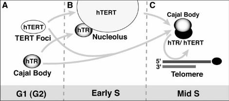
Figure 6. Model for cell cycle regulated trafficking of telomerase subunits to
telomeres during S phase. The predominant phase-specific localization of hTR and
hTERT is shown for G1 (A), early S (B) and mid-S (C). The arrows indicate possible
trafficking pathways accounting for the observed localizations. See text for details.


![[Regulation of telomeres length: making the telomeres accessible?]](https://static.fdokumen.com/doc/165x107/633f1028d121719806096682/regulation-of-telomeres-length-making-the-telomeres-accessible.jpg)