Characterization of telomeres and telomerase expression in Xiphophorus
16
Characterization of telomeres and telomerase expression in Xiphophorus ✰ Kevin P. Downs, Yingjia Shen, Amanda Pasquali, Ion Beldorth, Markita Savage, Katelyn Gallier, Tzintzuni Garcia, Rachell E. Booth * , and Ronald B. Walter Department of Chemistry and Biochemistry, Molecular Biosciences Research Group, 419 Centennial Hall, Texas State University, 601 University Drive, San Marcos, TX 78666, USA Abstract Research investigating telomere lengths and telomerase expression in vertebrates has progressively become important due to the association of these two biological endpoints with cellular aging and cancer in humans. Studies that rely upon the traditional use of laboratory mice have been faced with limitations largely due to inbred mice possessing large telomeres and ubiquitous expression of telomerase. Recently, a number of small fish species have been shown to provide potentially informative models for examining the role of telomeres and telomerase within intact vertebrate animals. Xiphophorus fishes represent a new world live-bearing genus that has not previously been assessed for telomere length or telomerase expression. To add to the knowledge base of telomere and telomerase biology in vertebrates we assessed telomere length and telomerase expression among several species of Xiphophorus. The telomere lengths in several organs (gill, brain, eyes, testis, ovary and liver) in three species (X. hellerii, X. maculatus, X. couchianus) and also in F 1 interspecies hybrids were approximately 2–6 kb. This size was consistent within the same organs of the same species, as well as between species and F 1 hybrids. Despite possessing relatively short telomere lengths compared to humans, the consistency of size among Xiphophorus species and organs may allow experimental detection of telomere shortening. The relative expression of telomerase reverse transcriptase (TERT) was determined by quantitative real-time PCR. Expression levels of TERT was measured in seven organs (ovary, testis, liver, gill, brain, heart, skin) from X. maculatus, X. hellerii and in control and ultraviolet light (UVB) exposed skin samples from X. maculatus, X. hellerii, and F 1 interspecies hybrids. TERT gene expression was significantly higher in ovary and testis, while all other organs showed low relative TERT expression. Detectable increases in TERT expression were found in skin samples upon UVB exposure. Our findings suggest Xiphophorus may serve as a suitable model for future studies investigating the association of telomere length and telomerase expression in regard to aging and disease. ✰ This paper is based on a presentation given at the 5th Aquatic Annual Models of Human Disease conference: hosted by Oregon State University and Texas State University-San Marcos, and convened at Corvallis, OR, USA September 20–22, 2010. © 2011 Elsevier Inc. All rights reserved. * Corresponding author. [email protected], Phone: (512) 245-2327 ; Fax: (512) 245-2374, Department of Chemistry & Biochemistry, 401 CENT, Texas State University, 601 University Drive, San Marcos, TX, 78666, USA. Publisher's Disclaimer: This is a PDF file of an unedited manuscript that has been accepted for publication. As a service to our customers we are providing this early version of the manuscript. The manuscript will undergo copyediting, typesetting, and review of the resulting proof before it is published in its final citable form. Please note that during the production process errors may be discovered which could affect the content, and all legal disclaimers that apply to the journal pertain. ✰ This paper is based on a presentation given at the 5th Aquatic Annual Models of Human Disease conference: hosted by Oregon State University and Texas State University-San Marcos, and convened at Corvallis, OR, USA September 20–22, 2010. NIH Public Access Author Manuscript Comp Biochem Physiol C Toxicol Pharmacol. Author manuscript; available in PMC 2013 January 1. Published in final edited form as: Comp Biochem Physiol C Toxicol Pharmacol. 2012 January ; 155(1): 89–94. doi:10.1016/j.cbpc. 2011.05.005. NIH-PA Author Manuscript NIH-PA Author Manuscript NIH-PA Author Manuscript
-
Upload
independent -
Category
Documents
-
view
1 -
download
0
Transcript of Characterization of telomeres and telomerase expression in Xiphophorus

Characterization of telomeres and telomerase expression inXiphophorus✰
Kevin P. Downs, Yingjia Shen, Amanda Pasquali, Ion Beldorth, Markita Savage, KatelynGallier, Tzintzuni Garcia, Rachell E. Booth*, and Ronald B. WalterDepartment of Chemistry and Biochemistry, Molecular Biosciences Research Group, 419Centennial Hall, Texas State University, 601 University Drive, San Marcos, TX 78666, USA
AbstractResearch investigating telomere lengths and telomerase expression in vertebrates hasprogressively become important due to the association of these two biological endpoints withcellular aging and cancer in humans. Studies that rely upon the traditional use of laboratory micehave been faced with limitations largely due to inbred mice possessing large telomeres andubiquitous expression of telomerase. Recently, a number of small fish species have been shown toprovide potentially informative models for examining the role of telomeres and telomerase withinintact vertebrate animals. Xiphophorus fishes represent a new world live-bearing genus that hasnot previously been assessed for telomere length or telomerase expression. To add to theknowledge base of telomere and telomerase biology in vertebrates we assessed telomere lengthand telomerase expression among several species of Xiphophorus. The telomere lengths in severalorgans (gill, brain, eyes, testis, ovary and liver) in three species (X. hellerii, X. maculatus, X.couchianus) and also in F1 interspecies hybrids were approximately 2–6 kb. This size wasconsistent within the same organs of the same species, as well as between species and F1 hybrids.Despite possessing relatively short telomere lengths compared to humans, the consistency of sizeamong Xiphophorus species and organs may allow experimental detection of telomere shortening.
The relative expression of telomerase reverse transcriptase (TERT) was determined byquantitative real-time PCR. Expression levels of TERT was measured in seven organs (ovary,testis, liver, gill, brain, heart, skin) from X. maculatus, X. hellerii and in control and ultravioletlight (UVB) exposed skin samples from X. maculatus, X. hellerii, and F1 interspecies hybrids.TERT gene expression was significantly higher in ovary and testis, while all other organs showedlow relative TERT expression. Detectable increases in TERT expression were found in skinsamples upon UVB exposure. Our findings suggest Xiphophorus may serve as a suitable model forfuture studies investigating the association of telomere length and telomerase expression in regardto aging and disease.
✰This paper is based on a presentation given at the 5th Aquatic Annual Models of Human Disease conference: hosted by Oregon StateUniversity and Texas State University-San Marcos, and convened at Corvallis, OR, USA September 20–22, 2010.© 2011 Elsevier Inc. All rights reserved.*Corresponding author. [email protected], Phone: (512) 245-2327 ; Fax: (512) 245-2374, Department of Chemistry &Biochemistry, 401 CENT, Texas State University, 601 University Drive, San Marcos, TX, 78666, USA.Publisher's Disclaimer: This is a PDF file of an unedited manuscript that has been accepted for publication. As a service to ourcustomers we are providing this early version of the manuscript. The manuscript will undergo copyediting, typesetting, and review ofthe resulting proof before it is published in its final citable form. Please note that during the production process errors may bediscovered which could affect the content, and all legal disclaimers that apply to the journal pertain.✰This paper is based on a presentation given at the 5th Aquatic Annual Models of Human Disease conference: hosted by Oregon StateUniversity and Texas State University-San Marcos, and convened at Corvallis, OR, USA September 20–22, 2010.
NIH Public AccessAuthor ManuscriptComp Biochem Physiol C Toxicol Pharmacol. Author manuscript; available in PMC 2013January 1.
Published in final edited form as:Comp Biochem Physiol C Toxicol Pharmacol. 2012 January ; 155(1): 89–94. doi:10.1016/j.cbpc.2011.05.005.
NIH
-PA Author Manuscript
NIH
-PA Author Manuscript
NIH
-PA Author Manuscript

KeywordsXiphophorus; telomerase; telomerase reverse transcriptase (TERT); induced gene expression;ultraviolet light (UVB); quantitative real time PCR
1. IntroductionTelomeres are regions of repetitive nucleotide structures located on the ends ofchromosomes that, along with various associated proteins, function to protect and stabilizechromosomes ends (Blackburn 1991, Blackburn 2001). Most vertebrates possess telomeresmade up of the tandem repeat TTAGGG that extends in the 5’ to 3’ direction toward thedistal ends of each chromosome (Moyzis et al. 1988). The average number of telomericrepeat units in organisms can vary greatly. For example, human telomere lengths range from8–12 kb, while some laboratory mice strains are close to 100 kb. Variations in telomerelength have also been reported across tissues within the same organism and even within asingle cell (De Lange et al. 1990). The shortening of telomeres is, in part, largely due to theinability of standard polymerases to fully replicate the ends of chromosomes, leading to agradual loss of telomeric regions as the organism ages. Interestingly, certain cell types (i.e.,germ line cells) are able to prevent telomere loss through the activation of an enzyme/RNAcomplex termed telomerase (Holt et al. 1999).
Telomerase synthesizes telomeric repeats at the ends of chromosomes using a catalyticprotein subunit known as telomerase reverse transcriptase (TERT) and an integral RNAsubunit as the template (Legassie et al. 2005). Although other proteins are involved in thefunction of the telomerase complex, these two essential components are conserved amongall eukaryotes (Weinrich et al. 1997). In humans, it has been shown that telomeres shortenwith age in somatic tissues due to a relative absence of telomerase activity, and thattelomerase activity is limited to highly proliferative cell types, such as stem cells, germ cells,and tumor cells (Greider 1998). Interestingly, abnormally high expression of telomerase ispresent in a majority of cancer cell lines and associated with 80–90% of cancers (Stewart etal. 2006, Kim et al. 1994). To determine the mechanisms by which telomerase activationoccurs during carcinogenesis, various studies investigating telomerase activity in skin cancerhave been reported. In particular, several studies provided evidence that exposure toultraviolet light (UV) induces telomerase expression in human skin, although the molecularmechanism by which this occurs is poorly understood (Taylor et al. 1996, Ueda et al. 1997).The human health conditions correlated with alterations in telomerase activity underscorethe importance of developing vertebrate experimental models that may provide a morecomplete understanding of how this enzyme is regulated under various physiologicalconditions.
Limitations of in vivo studies associated with telomeres and telomerase expression has beenlargely due to lack of appropriate model organisms. To identify potential alternativevertebrate models, recent investigations have begun to examine the role of telomeres andtelomerase in aquatic species. Studies with aquatic vertebrates such as zebrafish, flounder,pufferfish, and medaka have shown that telomerase expression is not limited to reproductiveorgans, but is ubiquitously expressed at high levels. Several aquatic species have also beenfound to possess telomere lengths that are similar to those in humans (McChesney et al.2005). In addition, telomere shortening linked to aging appears to be present in fish such asthe Japanese medaka (Oryzias latipes) and Nothobranchius furzeri despite ubiquitoustelomerase expression (Hatakeyama et al. 2008, Hartman et al. 2009). The expression oftelomerase in somatic tissues of fish is thought to mainly serve a role in tissue regenerationprocesses (Elmore et al. 2008, Lund et al. 2009).
Downs et al. Page 2
Comp Biochem Physiol C Toxicol Pharmacol. Author manuscript; available in PMC 2013 January 1.
NIH
-PA Author Manuscript
NIH
-PA Author Manuscript
NIH
-PA Author Manuscript

An aquatic organism that has yet to be investigated in its potential to serve as a vertebratemodel for studying telomeres and telomerase is Xiphophorus. Xiphophorus fishes comprisea freshwater genus composed of 27 species that are derived from Mexico, Guatamala,Belize, and Honduras. In the late 1920’s it was recognized that various interspecies hybridsof Xiphophorus could be produced that were fertile. Subsequently, crossing of X. maculatuswith the swordtail X. hellerii was shown to segregate backcross hybrid progeny (i.e,backcross to the X. hellerii parent) that developed melanoma according to Mendelianexpectations (Gordon, 1927). In the years since these early experiments, Xiphophorusinterspecies hybrids have developed into valuable experimental models to study the geneticsunderlying spontaneous and induced carcinogenesis (for review see; Walter and Kazianis,2001).
Herein, we report telomere lengths determined by terminal restriction fragment (TRF)southern blot analysis across multiple organs in three different species of Xiphophorus, aswell as F1 interspecies hybrids. Telomere lengths were compared among the organs of eachindividual species and these data compared with telomere lengths between species. Aportion of the TERT transcript and translated protein sequence was identified inXiphophorus and compared to those previously found in other aquatic species includingDanio rerio, N. furzeri, and O. latipes. Relative TERT expression was also determined byquantitative real-time PCR in multiple organs (testis, liver, gill, brain, heart, skin) of X.maculatus and X. hellerii as well as in UVB exposed skin from X. maculatus, X. hellerii andF1 interspecies hybrids.
2. Materials and Methods2.1. Xiphophorus strains and usage
Young adult Xiphophorus including X. maculatus Jp 163 A, X. maculatus Jp 163 B, X.hellerii, X. couchianus, and interspecies hybrids produced from crossing these parental lineswere obtained from the Xiphophorus Genetic Stock Center (XGSC; Texas State University,San Marcos, TX, USA). All fish in the XGSC were maintained as pedigreed lines accordingto standard XGSC protocols (see www.xiphophorus.txstate.edu). The X. maculatus Jp 163 Aand B lines, carrying the spot dorsal (Sd) and spot side (Sp) macromelanophore pigmentpatterns, were originally derived from a single female (X. maculatus Jp 163) collected fromthe wild. Upon establishment in the XGSC the X. maculatus Jp 163 line was spilt into Jp 163A (Sd) and B (Sp) lines based on the pigment patterns and have been maintained as twoindependent pedigrees for over 100 generations (Kallman 2001, Kallman and Kazianis,2006, Walter et al. 2006a,b). First generation hybrids (F1) from three interspecies crosseswere utilized in the studies presented. The interspecies crosses were as follows; (1) X.maculatus Jp 163 A (x) X. couchianus (herein designated, Sd-Xc), (2) X. maculatus Jp 163 A(x) X. hellerii (designated Sd-Xh), and (3) X. maculatus Jp 163 B (x) X. hellerii (designatedSp-Xh).
2.1. UVB ExposureX. maculatus Sd, X. maculatus Sp, X. hellerii, and the F1 interspecies hybrid (Sp-Xh) malefish were exposed to 300 J/m2/day (8:45 min. exposure time) for 5 days resulting in a totaldose of 1500 J/m2. The UVB exposure chamber allowed free-swimming fish to be irradiatedconcurrently on both sides within UV transparent chambers. UV exposure was performedusing Phillips TL20W/01 RS UVB lamps filtered through Kodacel (K-6805). Fish weremaintained in the dark 24 h before the first exposure and after each exposure. After the finalUVB exposure the fish were dissected and select organs immediately flash frozen for RNAisolation.
Downs et al. Page 3
Comp Biochem Physiol C Toxicol Pharmacol. Author manuscript; available in PMC 2013 January 1.
NIH
-PA Author Manuscript
NIH
-PA Author Manuscript
NIH
-PA Author Manuscript

2.3. Telomere length measurementsGenomic DNA was extracted from the liver, gill, brain, eyes, and testis of male and ovariesof female tissue from X. maculatus Sd, X. hellerii, X. couchianus and F1 interspecieshybrids, Sd-Xc and Sd-Xh, using the Genomic-tip system (Qiagen). For each assay, 2 µg ofgenomic DNA was digested with restriction enzymes RsaI and HinfI for ~2 h at 37 °C.Digested DNA was resolved by electrophoresis (30 V) on 0.8% agarose gels using TAEbuffer (40 mM Tris-acetate, pH 7.5, 1 mM EDTA). DNA fragments in the agarose gel weretransferred by capillary transfer and UV cross-linked to a Hybond-N+ membrane.Immobilized DNA fragments were incubated with a digoxigenin (DIG)-labeled probespecific for telomeric repeats (Roche), washed, and then incubated with a DIG-specificantibody covalently coupled to alkaline phosphate (Roche). A highly sensitivechemiluminescent substrate, CDP-Star (Roche), was used to visualize telomeres on theseblots. The average terminal restriction fragment (TRF) length was determined bycomparison of the intensity of telomere sample to a DIG-labeled molecular weight standardrun on the same gel.
2.4. Retrieval of Xiphophorus telomerase (TERT) sequenceRNA-seq data derived from X. maculatus Jp 163 A testis and ovaries mRNA wereassembled de novo (unpublished results) using the Velvet/Oases short read assemblerpackage (Zirbino and Birney 2008). Raw data comprised ~42 million 60 nt read pairssequenced on the Illumina GAII platform. The assembly contained 112,822 contigs with anN50 (length-weighted median) of 1,000 bp with an average length of 613.9 bp. The longestcontig in the assembly was 15,566 bp. Using TERT mRNA sequences obtained from thepublic databases from Oryzias latipes (DQ248968.1), Takifugu rubripes (AY861384), andDanio rerio (BC163300) we performed a similarity search (Blast) of the X. maculatusassembly to identify a set of contigs with high similarity to the queries. This set ofXiphophorus TERT contigs were used to manually splice together a consensus draft ofTERT mRNA.
2.5. RT-PCR and PCR amplification of TERTTotal RNA was extracted from each organ using TRIZOL (Invitrogen) and first strandcDNA was synthesized with Multiscribe Reverse Transcriptase (Applied Biosystems). Genespecific primers homologous to the medaka TERT sequence (NCBI, NM_001104816) weredesigned using the X. maculatus transcriptome based draft TERT sequence. Standard PCRand qRT-PCR were performed. The sequences of these primers are: TERT-F (5’-CTGGGACATGGCTCAGTATGC-3’; sense) and TERT-R (5’-AGCTCCACCGCCTCGTATTGC-3’; antisense).
PCR reactions were carried out at 95 °C for 5 min, followed by 30 cycles of 95 °C for 30 s,56 °C for 45 s, 72 °C for 45 s and a final extension step of 72 °C for 5 min. The PCRproducts were resolved in a 2.5% agarose gel with TAE buffer at 125 V. A 50 bp Ladder(Invitrogen) was used for determining product sizes. Visible amplification products were gelextracted and purified using the Wizard SV Gel and PCR Clean-Up System (Promega).Sequencing of the purified DNA was performed by Davis Sequencing (Davis, CA, USA).
2.6 Quantitative real-time PCRQuantitative real-time PCR (qRT-PCR) was used to assess the relative gene expressionlevels of TERT within various tissues of X. maculatus Sd and X. hellerii. Primer sequencesused in these reactions included gene specific primers TERT-F and TERT-R, 18S rRNAnormalization primers (18S-F 5’-CGGAAAGGATTGACAGATTGA-3’ and 18S-R 5’-CTCAATCTCGTGTGGCTGAA-3’) and MAPK14A normalization primers (MAPK-F 5’-
Downs et al. Page 4
Comp Biochem Physiol C Toxicol Pharmacol. Author manuscript; available in PMC 2013 January 1.
NIH
-PA Author Manuscript
NIH
-PA Author Manuscript
NIH
-PA Author Manuscript

CCTCTTGCTGTAGACTTGCTGG-3’ and MAPK-R 5’-GCTCTTCCACTCCTCGATTTCC-3’). Real-time PCR reactions were performed with a7500 Fast Real-Time PCR System using the Fast SYBR Green Master Mix detectionmethod (Applied Biosystems). For each assay a total of 100 ng cDNA was used in triplicate.To normalize target gene expression for differences in cDNA input, cDNA was diluted anadditional 1/500 for measuring 18S rRNA levels. Diluted cDNAs (8 µL aliquots) wereadded to the wells of a 96-well plate with 10 µL of Fast SYBR Green Master Mix and 0.5µM of each primer pair. For quantification of PCR results cycle threshold (CT) values weredetermined for each reaction. A standard curve was constructed from data derived using adilution series of ovary cDNA, a tissue expected to possess significant TERT expression.The standard curve allowed assessment of the relationship between the quantity of startingmaterial and the CT values as an indicator of the efficiency of TERT amplification. RelativeTERT expression was determined using the comparative ΔCT method by comparing theTERT CT values for each tissue sample with the 18S or MAPK14A rRNA CT values. Foreach assay, the tissue sample with the lowest normalized TERT expression values waschosen for calculating the fold change in expression.
3. Results3.1 Telomere length analysis
The lengths of telomeres in six organs (gill, liver, eye, brain, testis, ovary) of three species ofXiphophorus (X. maculatus Jp Sd, X. hellerii, and X. couchianus), as well as eye and liverorgans derived from two F1 interspecies hybrids (Sp-Xc and Sp-Xh) were determined usingterminal restriction fragment analysis (Fig. 1). Average telomere lengths were determined tobe between 2 to 6 kb in each of the six organs from each Xiphophorus species examined(Table 1). Relatively low heterogeneity in telomere lengths was observed when comparingindividual organs within each species (Table 1). Tissues from X. hellerii (e.g., eyes)possessed the smallest range of telomere lengths (3.5–5 kb) relative to those observed for X.maculatus Sd and X. couchianus (2.7–5 kb and 2.5–5 kb, respectively).
3.2 Comparison of Xiphophorus TERT transcript and proteinThe O. latipes TERT sequence (ABB76648.1) was utilized to search (tBlastn) for theXiphophorus TERT mRNA in the assembled contigs. Comparison of the translatedXiphophorus TERT protein sequence with other fish species; E. coioides (ABC47309.1,78% identity), O. latipes (ABB76648.1, 71% identity), T. rubripes (AAX59693.1, 72%identity), N. furzeri (ACN38321.1, 68% identity), D. rerio (AAI63300.1, 49% identity) andO. melastigma (ABB92622.1, 63% identity) revealed ≥ 49 % conservation at the proteinlevel. Conserved TERT motifs were identified in Xiphophorus by multiple sequencealignments with other fish TERTs and relatively high (>50%) sequence identities wererevealed (Fig. 2).
3.3 Telomerase expression in XiphophorusTelomerase gene expression was assessed by PCR and qRT-PCR of RNA samples isolatedfrom multiple organs (brain, liver, skin, gill, heart, testis, ovary) of adult X. maculatus and X.hellerii as well as skin samples from X. maculatus (Sd and Sp), X. hellerii and F1interspecies hybrids (Sp-Xh). TERT PCR products were only observed in the testis andovaries samples (results not shown). qRT-PCR expression analysis revealed significantlevels (≥ 85-fold normalized to liver) of TERT in both testis and ovaries for X. maculatus Sdand X. hellerii (Fig. 3, Table 2). In contrast, very low levels of detectible gene expression (<3% relative to ovaries) were observed in the RNA derived from liver, brain, gills, and skinorgans for either species. Surprisingly, X. maculatus Sd heart tissue appeared to have highgene expression compared to the other organs but was still very low compared to expression
Downs et al. Page 5
Comp Biochem Physiol C Toxicol Pharmacol. Author manuscript; available in PMC 2013 January 1.
NIH
-PA Author Manuscript
NIH
-PA Author Manuscript
NIH
-PA Author Manuscript

in testis and ovaries (8.7% relative to ovaries). Expression of X. maculatus Sd TERT inmultiple organs (brain, skin, gill, liver, heart) was also compared to the expression ofanother gene, mitogen-activated protein kinase 14a (MAP14KA). Previous work in ourlaboratory had determined the MAP14KA transcript to be ubiquitously expressed atrelatively high and balanced levels in most all Xiphophorus tissues (unpublishedobservations) and so we used expression of MAP14KA as a second gene target to ensure ourreactions were valid. Normalization of the two genes (TERT and MAP14KA) to 18S rRNAlevels revealed significantly lower expression of telomerase compared to MAPK14A inmost tissues. An average difference in expression between TERT and MAPK14A wasdetermined at about 30 fold for expression in liver, brain, skin, and heart of X. maculatus Sd.However, the expression of MAPK14A and TERT in testis and ovaries were determined tobe about equal. Comparison of relative telomerase and MAPK14A expression supports theobservation that TERT is expressed at very low levels in most X. maculatus organs.
Detectable increases in relative TERT expression was observed in RNA isolated from skinof fish exposed to UVB radiation (Fig. 4). As shown in Table 3, the largest induction ofTERT expression in UVB exposed skin was observed in X. maculatus Sp (7.2 ± 1.0 fold)while significant but smaller changes were found for X. maculatus Sd (5.0 ± 0.3 fold), X.hellerii (2.7 ± 0.3 fold), and the Sp-Xh interspecies hybrid between these two parental lines(4.2 ± 0.9 fold).
4. DiscussionThe association of telomere shortening in human aging and disease has led to an interest indeveloping translational models for studying telomere molecular biology. Limitations of invitro culture systems and laboratory mice strains are implied by their extremely largetelomeres lengths compared to human telomeres. This has led investigators to search foralternative models among aquatic species (McChesney et al. 2005). Several aquatic speciesincluding D. rerio, O. latipes, O. melastigma, and N. furzeri (Au et al. 2008, Hartmann et al.2009) have been shown to possess telomere lengths similar to human lengths.
In this report we show results indicating telomere length in various organs and species ofXiphophorus average about 2 to 6 kb. Compared to other fish species (e.g., O. latipes and D.rerio) the average telomere lengths observed in Xiphophorus were more homogenous acrossorgans and between species. For example, similar telomere lengths were observed for liverin X. maculates Sd (2.5 to 5.5 kb), X. hellerii (3.5 to 6.0 kb), X. couchianus (2.5 to 5.5 kb),and for two F1 interspecies hybrids (Fig. 1, Table 1). This is in contrast to previous reportsshowing telomere lengths to be highly heterogenous in the liver of two medaka species O.latipes (6–12 kb) and O. melastigma (0.5–12 kb; Au et al. 2009). Telomere lengths observedin different tissues of zebrafish have also been found to possess highly variable lengths aswell (McChesney et al. 2005). The consistency of telomere length in Xiphophorus may bedue the pedigreed breeding program maintained at the XGSC (Walter at al. 2006a). The X.maculatus and X. couchianus lines are highly inbred, over 100 and 76 generations,respectively, while the X. hellerii line used in these studies (i.e., Sarabia) has beenmaintained in the XGSC since 1963 (Walter et al, 2006b). The relatively constant telomerelengths found in Xiphophorus suggests these fish may be a good model to assess the effectsof age or environmental perturbation (e.g., oxidative stress) on telomere length.
The expression of telomerase in humans is largely restricted to germ line cells and stemcells, and tends toward a gradual telomere shortening in somatic tissues. The activation oftelomerase has been found associated with 85% of cancers and up to 99% of tumor derivedcell lines, although the exact mechanism by which telomerase becomes activated is not wellunderstood. In contrast to absence of telomerase expression in most human tissues,
Downs et al. Page 6
Comp Biochem Physiol C Toxicol Pharmacol. Author manuscript; available in PMC 2013 January 1.
NIH
-PA Author Manuscript
NIH
-PA Author Manuscript
NIH
-PA Author Manuscript

ubiquitous telomerase expression has been reported in many tissues of various fish species(McChesney et al. 2005). Specifically, ubiquitous expression of the catalytically activeportion of telomerase (TERT) has been detected in all tissues of zebrafish, medaka, and N.furzeri. The ability of zebrafish to regenerate damaged tissues at an exceedingly high ratehas been suggested as an explanation for the relatively high expression of telomerase.Relatively high telomerase activity has also been observed in the liver, kidney, intestine,muscle, gonad, heart, brain, spleen, and gill of medaka irrespective of age (Pfennig et al.2008). Furthermore, telomerase activity in the majority of medaka samples examined was 2to 4 times higher than that of a human cervical cancer cell line (Siha cells; Hatakeyama et al.2008). Telomerase is highly expressed in the testes and ovaries of X. maculatus Sd and X.hellerii while other tissues including skin, liver, brain, and gill exhibited very low levels ofexpression. Although studies in other fish species have indicated that telomerase expressionis normally higher in testes and ovaries compared to other tissues, we observed a moredrastic difference of expression ratio between gonads and somatic organs for Xiphophorus.Even when TERT expression is normalized to heart, the somatic tissue that displayed thehighest levels of TERT expression in X. maculatus Sd, the relative change in TERTexpression for testis and ovaries is 84 and 192 fold, respectively. This pattern moreresembles mammalian TERT expression then that of other fishes.
The effects of UVB exposure on TERT expression was investigated in skin samplesobtained from X. maculatus (Sd and Sp), X. hellerii, and an F1 interspecies hybrid (Sp-Xh).We observed UVB induction of TERT expression in skin samples after 5 days of UVBexposure. The UVB exposure protocol utilized mirrored that often employed in UV inducedmelanoma studies (Nairn et al., 2001, Mitchell et al., 2007). The levels of TERT inductionby UVB ranged from 5 to 7 fold in skin samples from X. maculatus Sd and Sp, respectively.A ~3 fold UVB induction was observed for X. hellerii TERT. The F1 interspecies hybrid(Sp-Xh) exhibited an intermediate response (~4 fold) between the two parental phenotypesfor UVB induced TERT expression. In previous DNA repair studies it was that F1interspecies hybrids may exhibit the repair phenotype of one parent, or the other, and insome cases exhibit DNA repair capability much different than either parent. Further, theseDNA repair phenotypes were shown to be different and variable between organs in the samefish (David et al., 2004). UV exposure studies in the hairless mouse model showedsignificant increases in telomerase activity (12 fold) occurred 4 weeks after the UVBexposure (Balasubramanian et al. 1999). In humans, evidence of increased telomeraseactivity has also been found in malignant skin conditions and areas of skin exposed to higherlevels of sunlight (Ueda et al. 1997). The mechanisms of UV induction of telomerase andwhat affects this may have on cellular damage, repair, and downstream induction ofcarcinogenesis should be further explored in future work. Xiphophorus offers several UVBinduced cancer models that may be informative for such studies (Mitchell et al. 2007, Nairnet al. 2001). Thus, Xiphophorus appears to be a unique and applicative model whencompared to other aquatic species for the functional study of telomeres and telomerase.
AcknowledgmentsThe authors would like to thank the other employees of the Xiphophorus Genetic Stock Center, Texas StateUniversity, for maintaining the fish lines. The work was supported from an NIH, National Center for ResearchResources grant award R24-RR024790 and support from Texas State University
ReferencesAu DW, Mok HO, Elmore LW, Holt SE. Japanese medaka: A new vertebrate model for studying
telomere and telomerase biology. Comp. Biochem. Physiol. C. 2008; 149:161–167.
Downs et al. Page 7
Comp Biochem Physiol C Toxicol Pharmacol. Author manuscript; available in PMC 2013 January 1.
NIH
-PA Author Manuscript
NIH
-PA Author Manuscript
NIH
-PA Author Manuscript

Balasubramanian S, Kim KH, Ahmad N, Mukhtar H. Activation of telomerase and its association withG1-phase of the cell cycle during UVB-induced skin tumorigenesis in SKH-1 hairless mouse.Oncogene. 1999; 18:1297–1302. [PubMed: 10022811]
Blackburn EH. Structure and function of telomeres. Nature. 1991; 350:569–573. [PubMed: 1708110]Blackburn EH. Switching and signaling at the telomere. Cell. 2001; 106:661–673. [PubMed:
11572773]David WM, Mitchell DL, Walter RB. DNA repair in hybrid fish of the genus Xiphophorus. Comp.
Biochem. Physiol. C. 2004; 138:301–309.De Lange T, Shiue L, Myers RM. Structure and variability of human chromosome ends. Mol. Cell
Biol. 1990; 10:518–527. [PubMed: 2300052]Elmore LW, Norris MW, Sircar S, Bright AT, McChesney PA, Winn RN, Holt SE. Upregulation of
telomerase function during tissue regeneration. Exp. Biol. Med. 2008; 233:958–967.Greider CW. Telomerase activity, cell proliferation, and cancer. Proc. Natl. Acad. Sci. USA. 1998:90–
92. [PubMed: 9419332]Gordon M. The genetics of a viviparous top-minnow Platypoecilus: The inheritance of two kinds of
melanophores. Genetics. 1927; 12:253–283. [PubMed: 17246524]Hartmann N, Reichwald K, Lechel A, Graf M, Kirschner J, Dorn A, Terzibasi E, Wellner J, Platzer M,
Rudolph KL, Cellerino A, Englert C. Telomeres shorten while Tert expression increases duringageing of the short-lived fish Nothobranchius furzeri. Mech. Ageing Dev. 2009; 130:290–296.[PubMed: 19428446]
Hatakeyama H, Nakamura K, Izumiyama-Shimomura N, Ishii A, Tsuchida S, Takubo K, Ishikawa N.The teleost Oryzias latipes shows telomere shortening with age despite considerable telomeraseactivity throughout life. Mech. Ageing Dev. 2008; 129:550–557. [PubMed: 18597819]
Holt SE, Shay JW. Role of telomerase in cellular proliferation and cancer. J. Cell Physiol. 1999;180:10–18. [PubMed: 10362013]
Kallman KD. How the Xiphophorus problem arrived in San Marcos. Texas. Mar. Biotechnol. (NY).2001; 3:S6–S16.
Kallman KD, Kazianis S. The genus Xiphophorus in Mexico and central America. Zebrafish. 2006;3:271–285. [PubMed: 18377209]
Kim NW, Piatyszek MA, Prowse KR, Harley CB, West MD, Ho PL, Coviello GM, Wright WE,Weinrich SL, Shay JW. Specific association of human telomerase activity with immortal cells andcancer. Science. 1994; 266:2011–2015. [PubMed: 7605428]
Legassie JD, Jarstfer MB. Telomerase as a DNA dependent polymerase. Biochemistry. 2005;44:14191–14201. [PubMed: 16245935]
Lund TC, Glass TJ, Tolar J, Blazar BR. Expression of telomerase and telomere length are unaffectedby either age or limb regeneration in Danio rerio. PLoS One. 2009; 4:e7688. [PubMed: 19893630]
McChesney PA, Elmore LW, Holt SE. Vertebrate marine species as model systems for studyingtelomeres and telomerase. Zebrafish. 2005; 1:349–355. [PubMed: 18248213]
Mitchell D, Paniker L, Sanchez G, Trono D, Nairn R. The etiology of sunlight-induced melanoma inXiphophorus hybrid fish. Mol. Carcinog. 2007; 46:679–684. [PubMed: 17477377]
Moyzis RK, Buckingham JM, Cram LS. A highly conserved repetitive DNA sequence (TTAGGG),present at the telomeres of human chromosomes. Proc. Natl. Acad. Sci. USA. 1988; 85:774–776.
Nairn RS, Kazianis S, Della Coletta L, Trono D, Butler AP, Walter RB, Morizot DC. Genetic analysisof susceptibility to spontaneous and UV-induced carcinogenesis in Xiphophorus hybrid fish. Mar.Biotechnol. 2001; 3:S24–S36. [PubMed: 14961297]
Pfennig F, Kind B, Zieschang F, Busch M, Gutzeit HO. Tert expression and telomerase activity ingonads and somatic cells of the Japanese medaka (Oryzias latipes). Dev. Growth Differ. 2008;50:131–141. [PubMed: 18312429]
Stewart SA, Weinberg RA. Telomeres: Cancer to human aging. Annu. Rev. Cell Dev. Biol. 2006;22:531–557. [PubMed: 16824017]
Taylor RS, Ramirez RD, Ogoshi M, Chaggins M, Piatyszek MA, Shay JW. Detection of telomerase inmalignant and nonmalignant skin conditions. J. Invest. Dematol. 1996; 106:759–765.
Downs et al. Page 8
Comp Biochem Physiol C Toxicol Pharmacol. Author manuscript; available in PMC 2013 January 1.
NIH
-PA Author Manuscript
NIH
-PA Author Manuscript
NIH
-PA Author Manuscript

Ueda M, Ouhtit A, Bito T, Nakazawa K, Lubbe J, Ichihashi M, Yamasaki H, Nakazawa H. Evidenceof UV-associated activation of telomerase in human skin. Cancer Res. 1997; 57:370–374.[PubMed: 9012457]
Walter RB, Kazianis S. Xiphophorus interspecies hybrids as genetic models of induced neoplasia.ILAR J. 2001; 42:299–321. [PubMed: 11581522]
Walter RB, Ju Z, Martinez A, Amemiya C, Samollow PB. Genomic resources for Xiphophorusresearch. Zebrafish. 2006; 3:11–22. [PubMed: 18248243]
Walter, RB.; Hazlewood, L.; Kazianis, S. The Xiphophorus Genetic Stock Center Manual. 1st edition.Kallman, KD.; Schartl, M., editors. San Marcos, TX: Texas State University-San Marcos; 2006.
Weinrich SL, Pruzan R, Ma L, Ouellette M, Tesmer VM, Holt SE, Bodnar AG, Lichtsteiner S, KimNW, Trager JB, Taylor RD, Carlos R, Andrews WH, Wright WE, Shay JW, Harley CB, MorinGB. Reconstitution of human telomerase with the template RNA component hTR and the catalyticprotein subunit hTERT. Nat. Genet. 1997; 17:498–502. [PubMed: 9398860]
Zerbino DR, Birney E. Velvet: Algorithms for de novo short read assembly using de Bruijn graphs.Genome Res. 2008; 18:821–829. [PubMed: 18349386]
Downs et al. Page 9
Comp Biochem Physiol C Toxicol Pharmacol. Author manuscript; available in PMC 2013 January 1.
NIH
-PA Author Manuscript
NIH
-PA Author Manuscript
NIH
-PA Author Manuscript
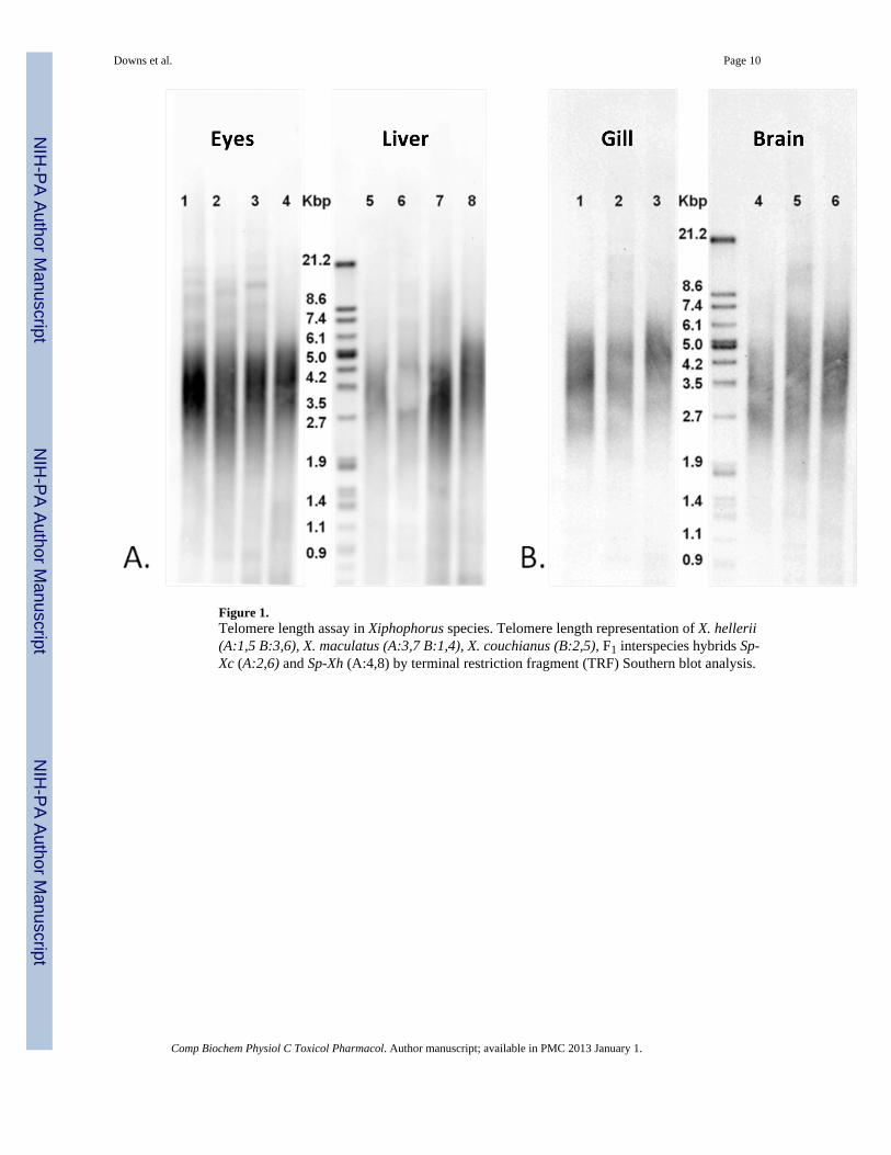
Figure 1.Telomere length assay in Xiphophorus species. Telomere length representation of X. hellerii(A:1,5 B:3,6), X. maculatus (A:3,7 B:1,4), X. couchianus (B:2,5), F1 interspecies hybrids Sp-Xc (A:2,6) and Sp-Xh (A:4,8) by terminal restriction fragment (TRF) Southern blot analysis.
Downs et al. Page 10
Comp Biochem Physiol C Toxicol Pharmacol. Author manuscript; available in PMC 2013 January 1.
NIH
-PA Author Manuscript
NIH
-PA Author Manuscript
NIH
-PA Author Manuscript

Figure 2.Telomerase reverse transcriptase motif alignments across fish species. Alignment includesTERT amino acid motifs from X. maculatus (translated), O. latipes (ABB76648.1), E.coioides (ABC47309.1), D. rerio (AAI63300.1), T. rubripes (AAX59693.1), and N. furzeri(ACN38321.1).
Downs et al. Page 11
Comp Biochem Physiol C Toxicol Pharmacol. Author manuscript; available in PMC 2013 January 1.
NIH
-PA Author Manuscript
NIH
-PA Author Manuscript
NIH
-PA Author Manuscript

Figure 3.Relative gene expression of TERT in multiple tissues of adult X. maculatus Sd (A) and X.hellerii (B) determined by qRT-PCR. Expression values of TERT were normalized against18s rRNA expression and relative fold changes ± SEM (n=3) were calculated with respect toexpression in liver.
Downs et al. Page 12
Comp Biochem Physiol C Toxicol Pharmacol. Author manuscript; available in PMC 2013 January 1.
NIH
-PA Author Manuscript
NIH
-PA Author Manuscript
NIH
-PA Author Manuscript

Figure 4.Relative expression of TERT in UVB exposed skin samples from X. hellerii (XH), X.maculates Sd (XM163A), X. maculatus Sp (XM163B), and a interspecies hybrid made bycrossing parental X. maculatus Sp and X. hellerii (Sp-Xh). Expression values of TERT werenormalized against 18s rRNA expression levels. Relative fold changes ± SEM (n=3) for UVtreated samples were calculated by normalization to un-exposed skin expression for eachspecies.
Downs et al. Page 13
Comp Biochem Physiol C Toxicol Pharmacol. Author manuscript; available in PMC 2013 January 1.
NIH
-PA Author Manuscript
NIH
-PA Author Manuscript
NIH
-PA Author Manuscript

NIH
-PA Author Manuscript
NIH
-PA Author Manuscript
NIH
-PA Author Manuscript
Downs et al. Page 14
Table 1
Telomere lengths in X. hellerii, X. maculatus Sd, and X. couchianus tissues determined by terminal restrictionfragment Southern blot analysis.
Tissue X. maculates(kb)
X. couchianus(kb)
X. hellerii(kb)
Gill 2–5 2.5–6 3–6
Liver 2–5 2–5.5 3.5–6
Brain 2–5 2–5 2.7–5
Eyes 2.7–5 2.5–5 3.5–5
Testis 2–5.5 2–5 2.5–5
Ovary 2–5 2–5 2.7–5
Comp Biochem Physiol C Toxicol Pharmacol. Author manuscript; available in PMC 2013 January 1.

NIH
-PA Author Manuscript
NIH
-PA Author Manuscript
NIH
-PA Author Manuscript
Downs et al. Page 15
Table 2
Relative gene expression of TERT in X. maculatus Sd and X. hellerii tissues. Values were determined by qRT-PCR repeated with three technical repeats and two biological repeats. ND = not determined
Tissue X. helleriifold change
X. maculatusfold change
Ovary 1000.0 ± 13.3 196.0 ± 5.8
Testis 162.0 ± 9.4 85.0 ± 4.3
Liver 1.0 1.0
Gill 20.0 ± 2.0 4.0 ± 0.4
Brain 15.0 ± 1.4 1.0 ± 0.1
Skin 22.0 ± 0.5 6.0 ± 1.1
Heart ND 17.0 ± 0.1
Comp Biochem Physiol C Toxicol Pharmacol. Author manuscript; available in PMC 2013 January 1.

NIH
-PA Author Manuscript
NIH
-PA Author Manuscript
NIH
-PA Author Manuscript
Downs et al. Page 16
Table 3
Relative gene expression of TERT in untreated and UVB treated skin from X. hellerii, X. maculatus Sd, X.maculatus Sp, and an interspecies hybrid made by crossing parental X. maculatus Sp and X. helleri (Sp-Xh).Values were determined by qRT-PCR repeated with three technical repeats and two biological repeats.
Species normal skin UV treated skin
X. hellerii 1.0 2.7 ± 0.3
X. maculatus Sd 1.0 5.0 ± 0.3
X. maculatus Sp 1.0 7.2 ± 1.0
Sp-Xh 1.0 4.2 ± 0.9
Comp Biochem Physiol C Toxicol Pharmacol. Author manuscript; available in PMC 2013 January 1.