Shortened telomeres in individuals with abuse in alcohol consumption
The Rockefeller University Press, 0021-9525/97/04/5/14 $2.00The Journal of Cell Biology, Volume 137, Number 1, April 7, 1997 5–18 5
Telomeres Cluster De Novo before the Initiation of Synapsis:A Three-dimensional Spatial Analysis of Telomere Positionsbefore and during Meiotic Prophase
Hank W. Bass,* Wallace F. Marshall,
‡
John W. Sedat,
‡
David A. Agard,
‡§
and W. Zacheus Cande*
*Department of Molecular and Cell Biology, University of California at Berkeley, Berkeley, California 94720; and
‡
Department of Biochemistry and Biophysics,
§
Howard Hughes Medical Institute, University of California at San Francisco, San Francisco, California 94143
Abstract.
We have analyzed the progressive changes in the spatial distribution of telomeres during meiosis us-ing three-dimensional, high resolution fluorescence mi-
croscopy. Fixed meiotic cells of maize (
Zea mays
L.) were subjected to in situ hybridization under conditions that preserved chromosome structure, allowing identifi-cation of stage-dependent changes in telomere arrange-ments. We found that nuclei at the last somatic prophase before meiosis exhibit a nonrandom, polar-ized chromosome organization resulting in a loose grouping of telomeres. Quantitative measurements on the spatial arrangements of telomeres revealed that, as cells passed through premeiotic interphase and into leptotene, there was an increase in the frequency of large telomere-to-telomere distances and a decrease in the bias toward peripheral localization of telomeres. By leptotene, there was no obvious evidence of telomere
grouping, and the large, singular nucleolus was inter-nally located, nearly concentric with the nucleus. At the end of leptotene, telomeres clustered de novo at the nu-clear periphery, coincident with a displacement of the nucleolus to one side. The telomere cluster persisted throughout zygotene and into early pachytene. The nu-cleolus was adjacent to the cluster at zygotene. At the pachytene stage, telomeres rearranged again by dis-persing throughout the nuclear periphery. The stage-dependent changes in telomere arrangements are sug-gestive of specific, active telomere-associated motility processes with meiotic functions. Thus, the formation of the cluster itself is an early event in the nuclear reor-ganizations associated with meiosis and may reflect a control point in the initiation of synapsis or crossing over.
A
nearly universal element in the choreography of ge-netic material during meiosis is the pairing ofhomologous chromosomes. Exactly how the ho-
mology search and chromosome pairing processes are ac-complished remains a mystery, despite decades of relevantcytological studies (34, 40, 41). The complete pairing ofhomologs for species with large genomes is a formidabletask likely to require the ability to move chromosomes andto reorganize the nucleus. Although some cases of premei-otic pairing have been observed (42, 64), the absence ofpremeiotic pairing is generally well documented (5, 12, 33,54). Thus, some force or active organizing mechanism mustbe operative to ensure timely chromosome alignment andsynapsis. Indications of such forces are found, for example,in the dramatic changes in chromosome morphology asso-ciated with the initiation of synapsis in maize (12). In thatstudy, Dawe et al. described a transition stage called prezy-
gotene, during which sister chromatids were slightly sepa-rated and the normally spherical blocks of heterochromatincalled knobs were axially elongated. Additionally, prezy-gotene marks the first stage at which pairing of homolo-gous knobs could be observed. The telomeric knobs, whichrelocated to the nuclear envelope at prezygotene, were thefirst to pair followed by pairing of interstitial knobs later inzygotene. We wanted to determine whether the behaviorof the telomeric knobs observed by Dawe et al. (12) re-flected the general behavior of all telomeres, and, if so,could we correlate their behavior with the homology searchand chromosome synapsis.
Many studies have drawn attention to a particular orga-nization of meiotic chromosomes in which the ends of thechromosomes are pointed towards the same side of the nu-cleus, usually touching the nuclear envelope. This polar-ized organization of chromosome ends, historically calledthe bouquet stage, occurs during meiotic prophase (16).The bouquet stage is widely conserved in nature, havingbeen observed in yeast, plants, and animals (13). The bou-quet stage always coincides with chromosome pairing, lead-
Please address all correspondence to W.Z. Cande, 341 LSA, Departmentof Molecular and Cell Biology, University of California, Berkeley, CA.Tel.: (510) 642-1669. Fax: (510) 643-6791.
on June 14, 2015jcb.rupress.org
Dow
nloaded from
Published April 7, 1997

The Journal of Cell Biology, Volume 137, 1997 6
ing to the conclusion that the bouquet, as a nuclear struc-ture, is needed for the chromosome pairing process (11,21, 54).
The exact role of the bouquet during meiotic prophaseis difficult to discern because of the inherent complexitiesof chromosome pairing itself. Cytogenetic and moleculargenetic studies have shown that meiotic chromosome pair-ing is composed of two separable events, chromosomealignment and chromosome synapsis. Chromosome align-ment, occasionally referred to as “pairing,” is generallythought to result from homology searching interactions,the timing and nature of which are poorly understood (38,64). Synapsis, on the other hand, can be clearly defined asthe intimate juxtaposition of chromosomes when joined bythe synaptonemal complex, a conserved tripartite struc-ture connecting meiotic chromosomes along their length(62). While alignment of homologues is generally thoughtto precede and contribute to the synapsis, the two pro-cesses can be uncoupled. For instance, homologous chro-mosomes can align, recombine, and segregate in the ab-sence of synapsis (1, 34, 40). In addition, a synaptonemalcomplex can form between nonhomologous chromosomesor chromosome regions, illustrating that synapsis is not de-pendent on homologue alignment (34). The extent to whichsynapsis has occurred delineates the first three stages ofmeiotic prophase: leptotene, zygotene, and pachytene; thesestages are characterized by synapsis of none, part, or all oftheir chromosomes, respectively. The correct identifica-tion of these stages is critical for our goal of defining therelationship between the behavior of meiotic chromosomesand the structure of the meiotic nucleus during the bou-quet stage.
Towards this end, it was necessary to establish the exacttiming and duration of the bouquet stage. To begin, wewanted to know whether the bouquet forms de novo or sim-ply reveals a preexisting nuclear organization that becomesevident upon chromosome condensation. This question hasnot been answered by conventional cytology because theidentification of telomeres requires the resolution of indi-vidual chromosomes. Therefore, the location of telomeresat the onset of chromosome condensation in meiotic pro-phase is ambiguous. To overcome this problem, one canuse fluorescence in situ hybridization (FISH)
1
with probesthat are specific for telomeres. This approach avoids thelimitations described above because it does not rely on theability to resolve the axial paths of individual chromo-somes. In a recent study using telomere FISH probes onsectioned testes of mouse and human, Scherthan et al.found that telomeres undergo dramatic changes in distri-bution during early meiosis (51, 54). Telomeres in mousespermatagonia (premeiotic) nuclei were not clustered,whereas telomeres at zygotene were clustered in the classi-cal bouquet configuration. A progression of telomere ar-rangements was observed within these nuclei; scattered telo-meres at the spermatagonial stage became peripheral atpreleptotene, and eventually peripheral and clustered atzygotene. The inability to identify nuclei at the leptotene
stage resulted in an incomplete description of sequentialdevelopmental stages in which preleptotene was followedby zygotene (54). Therefore, the timing of the bouquetstage relative to the onset of meiotic prophase could notbe directly determined.
To precisely ascertain the organization of the telomeresin nuclei known to be at premeiotic interphase, leptotene,or zygotene, we have applied telomere FISH to intactmaize meiotic cells. The inherent cytological clarity anddevelopmental synchrony of these cells along with the ex-cellent genetics of maize have provided workers with amodel system to investigate structure–function relation-ships operating during meiosis (6, 19, 24, 25, 49, 56). Thechromosomes of maize meiocytes undergo normal chias-mate meiosis (6, 49). In the early literature of McClintockand others describing the meiotic chromosomes of maize,little mention is made of the bouquet stage (49). However,the presence of a bouquet structure in maize is clearly doc-umented in ultrastructural analyses using serial section re-constructions of entire nuclei (22, 23, 46). In these studies,the ends of the chromosomes in zygotene and pachytenestaged nuclei were shown to be polarized, clustering on ornear a small region of the nuclear envelope.
To take full advantage of the maize meiocyte system, wehave adapted a FISH protocol for use with higher plant ma-terial, using methodologies established to study the three-dimensional organization of the nucleus (2, 12, 14, 15, 31,43, 60). Telomere kinetics were monitored in optically sec-tioned nuclei spanning a wide developmental range, in-cluding the last mitosis before meiosis. A quantitative spa-tial analysis of telomere distributions was carried out todocument the extent of the changes that were observed.This work represents the first detailed three-dimensionalanalysis of telomere dynamics at stages before and duringearly meiotic prophase, a period that has been only brieflyexamined in previous studies. As a consequence of ouranalysis, we have found that the bouquet structure formsde novo, at the end of leptotene, just before chromosomesynapsis. Thus, the duration of the bouquet stage was ob-served to be limited to meiotic prophase, suggestive of ameiosis-specific telomere movement system. Finally, wediscuss the significance of this timing in relation to othernuclear changes associated with the initiation of synapsis.
Materials and Methods
Plant Materials and Fixation of Reproductive Organs
Inbred lines of maize (
Zea mays
L., 2
n
5
20) were grown and harvestedthroughout the year at the Oxford greenhouses (Berkeley, CA). Three in-bred lines, KYS
1
, W23
1
, and A344
1
, were used to obtain the majority ofthe nuclei characterized in this study. A total of 57 nuclei was completelymodeled and the models were analyzed mathematically (see below). Inaddition to the standard inbreds, two nuclei (one pachytene and one dia-kinesis) were derived from a BMS line (without B chromosomes) that wasditelocentric and tetrasomic for chromosome 3 (2
n
5
22, gift from R.Staub).
Anthers from preemerged tassels were fixed as previously described(12) with the following modifications. Whole anthers (20–40) were fixed in5 ml 4% freshly prepared formaldehyde in meiocyte buffer A (15 mMPipes-NaOH, pH 6.8, 80 mM KCl, 20 mM NaCl, 0.5 mM EGTA, 2 mMEDTA, 0.15 mM spermine tetra HCL, 0.05 mM spermidine, 1 mM DTT,0.32 M sorbitol) for 2 h at room temperature. After fixation, anthers werewashed (four times, 15 min each) with meiocyte buffer A and used imme-diately or stored in air-tight containers at 4
8
C in sterile-filtered meiocyte
1
. Abbreviations used in this paper
: DAPI 4
9
,6-diamidino-2-phenylindole,dihydrochloride; FISH, fluorescence in situ hybridization; FITC, fluores-cein isothiocyanate.
on June 14, 2015jcb.rupress.org
Dow
nloaded from
Published April 7, 1997
Bass et al.
Meiotic Telomere Behavior
7
buffer A. The majority of data was collected on anthers used immediatelyor within several days of fixation. For some of the zygotene and pachytenestages, fixed anthers were stored for up to 8 wk without any noticeable de-terioration in the cells or nuclei.
Acrylamide Embedding of Meiocytes
We used a novel polyacrylamide embedding technique originally devel-oped for
Drosophila
polytene nuclei (60). A 15-mm-diam ring of nail pol-ish (Sally Hanson’s Hard as Nails) was painted onto ethanol-washed glassslides and allowed to dry for at least 4 h. Fixed anthers, submerged in 60
m
lof meiocyte buffer A, were cut open at their tips and the meiocytes weregently extruded out of the open end. Meiocytes suspended in meiocytebuffer A were transferred (12
m
l) by micropipetting onto the slide fol-lowed by immediate addition of 6
m
l of activated acrylamide stock. Theactivated acrylamide stock was made by addition of 25
m
l of 20% ammo-nium persulfate and 25
m
l of 20% sodium sulfite to 500
m
l of gel stock (15 mMPipes-NaOH, pH 6.8, 80 mM KCl, 20 mM NaCl, 0.5 mM EGTA, 2 mMEDTA, 0.15 mM spermine tetra HCL, 0.05 mM spermidine, 1 mM DTT,0.3 M sorbitol, 15% polyacrylamide [from a 30% 29:1 acrylamide/bis acryl-amide stock]). The slides were rocked and rotated for 15–20 s until mixed,and a lens paper-cleaned coverslip (22
3
22
3
1.5) was placed on top for20 min, and then removed, leaving a thin pad of acrylamide with embed-ded meiocytes attached to the slide. All washing steps below consisted ofplacing a drop (100–200
m
l) on top of the face-up acrylamide pad followedby rotary shaking at the maximum speed possible for 10 min at room tem-perature unless specified otherwise. Solutions were removed by aspira-tion. Although the 5% acrylamide sheet can be rather delicate, typicallythe acrylamide pad made it through the procedure for 70% of the slides.
Oligonucleotide Probe and In Situ Hybridization
We used a modification of the three-dimensional FISH methods estab-lished by Dernburg et al. to be compatible with chromosome preservation(14, 15). This protocol uses a high temperature codenaturation of targetand probe sequences by placement of glass slides on a PCR block. Newlypolymerized acrylamide pads were washed (four times, as described above)with buffer (15 mM Pipes-NaOH, pH 6.8, 80 mM KCl, 20 mM NaCl, 0.5 mMEGTA, 2 mM EDTA) to remove unpolymerized acrylamide, followed byfour equilibration washes with prehybridization buffer (50% deionizedformamide, 2
3
SSC). Hybridization buffer (50% deionized formamide,2
3
SSC, 1–3
m
g/ml telomere oligonucleotide) was added, removed, andadded a second time. The telomere oligonucleotide probe had the se-quence 5
9
-CCCTAAACCCCTAAACCCTAAACCCTAAA-3
9
. The oli-gonucleotide was labeled its 5
9
end by direct incorporation of “Fluores-cein-ON phosphoramidite” (catalogue 5235; Clontech Laboratories, Inc.,Palo Alto, CA) as the last step in the synthesis (HHMI DNA SynthesisFacility, University of California, San Francisco). The initial oligonucleotidepowder was dissolved in 10 mM Tris-HCL (pH 8.0), and its concentrationwas determined spectrophotometrically using the formula 1.0 A
260
5
33
m
g/ml. A final 20–30
m
l drop of hybridization buffer was added, sealed un-der a coverslip using rubber cement, and then placed on a 40
8
C warmingplate for 30 min. The chromosomes were denatured on a PCR block at94
8
C for 6 min, followed by overnight incubation at 37
8
C. The use of vari-ous competitors and blocking reagents including yeast tRNA, calf thymusDNA, unlabeled excess oligonucleotides, and Denhardt’s solution did notshow consistent improvement of signal to noise ratios and was thereforeexcluded from future experiments (data not shown). Because we foundthe optimal denaturation temperature varied between anthers and experi-ments, slides were usually prepared in triplicate and denatured for 6 mineach at 92
8
, 94
8
, or 96
8
C. Too much heat caused noticeable disruption ofchromosomes and cytoplasm, and such samples were not used (see below)despite their typically high signal to noise ratios.
After overnight incubation, the samples were washed sequentially with2
3
PBS containing 0.1% Tween-20 (four times, as described above) and1
3
PBS (three times), and then were stained with 10
m
g/ml 4
9
,6-diami-dino-2-phenylindole (DAPI) in 1
3
PBS for 30 min at room temperature.Excess DAPI was removed by washing with 2
3
PBS (three times), fol-lowed by 1
3
PBS (three times). Slides were equilibrated in an FITC-stabi-lizing medium (95% glycerin, 50 mM Tris base, 2%
N
-propyl gallate)three times for 20 min each and mounted in the same medium. Data setswere collected immediately thereafter and up until 48 h. Within a singleslide, we occasionally found isolated nuclei (no cytoplasm) with relativelybright FISH signals compared with those from intact cells. We reasonedthat the nuclei from intact cells would be less likely to have rearranged asa result of handling or manipulation, despite the potential cost of reduced
signal to noise ratios. For these reasons, data collection was predomi-nantly limited to those nuclei (
.
90% in this paper) within intact cells ortissues (not shown). Since meiocytes have a distinctly dense cytoplasmlacking large vacuoles, assessment of cellular preservation could bereadily made.
Three-dimensional Microscopy and Image Processing
All images were recorded using an IMT-2 wide-field microscope (Olym-pus Corp., Lake Success, NY), making use of one of two oil immersionlenses,
3
60 NA 1.4 PlanApo (Olympus Corp.) or
3
100 NA 1.4 PlanApo(Nikon Inc., Garden City, NY) (30). In both cases, the data were oversam-pled in the X, Y, and Z dimensions with typical XYZ voxel dimensions of0.07
3
0.07
3
0.2
m
m
3
. The computerized light microscope workstationhas been described elsewhere (14, 15). Original data collection was madeon an area extending at least 2
m
m beyond the nuclear border in Z, andextending at least 5
m
m in X and Y from the widest edges of the nuclei.After three-dimensional iterative deconvolution (7) of the original full-sized data sets, individual nuclei were computationally cropped in all di-mensions. The resulting data subsets were used for all subsequent modelbuilding and image display, resulting in the exclusion of the surroundingcytoplasm and neighboring cells from the figures presented. The imagespresented were adjusted for brightness and contrast using linear scaling ofthe minimum and maximum intensities, but no additional image process-ing such as edge or local contrast enhancements were used, except for thevolume-rendered projections in Fig. 3,
C
and
E.
Modeling Building and Spatial Analysis ofTelomere Positions
Individual nuclei were modeled using the Priism software program3DModel (8). Each model file contains a list of points with the real-spacecoordinates of three objects: the edge of the nucleus (Object-1), the edgeof the nucleolus (Object-2), and the telomeres (all the points in Object-3).Negative control experiments were carried out to confirm that none of thebright spots identified as telomeres could be explained as nonspecificbackground artifacts. An FITC-labeled oligonucleotide probe (producedas described above) homologous to the noncoding strand of a single copymaize gene was used under standard conditions (see above). The resultingFITC images lacked bright dots, but exhibited nucleolar staining similar tothe nonspecific background staining seen with the telomere probe (seeFig. 2
B
). In this study, we have decided to define telomeres as the discretespots (three to six pixels across in the X and Y dimensions) in the FISH-treated nuclei. We also required that these spots be brighter (by a factorof
.
1.5-fold) than the average intensity of pixels in the nucleolus, andwithin or touching the DAPI-revealed chromatin of the nucleus. Thesecriteria allowed us to identify telomeres in decondensed chromatin wherethe DAPI images provide little to no assistance in recognizing the ends ofchromosomes. Typically, 30–80% of the expected telomeres were ob-served.
We analyzed the spatial distribution of telomeres in two different ways.The first method compares the distribution of distances (Euclidean point-to-point distances) between pairs of telomeres to the distribution gener-ated from randomized points in nuclei but outside the nucleolus. To ac-complish this task, we used a surface harmonic expansion to fit surfaces tothe boundaries of the nucleus and nucleolus, as defined by the the coordi-nates specified in the 3DModel files (43). To generate a random curve forcomparison, we averaged the pairwise distance values (normalized to themaximum diameter) for 16 nuclei (eight at premeiotic interphase, and eightat leptotene). Monte Carlo simulations were used to generate a list of5,000 randomly placed points within a given nucleus but outside the nucle-olus. The diameter-normalized pairwise distances between all possiblepairs of the 5,000 points (12.5 million distances) were determined. The dis-tributions of random distances were remarkably similar for different nu-clei at premeiotic interphase and leptotene (not shown). The diameter-normalized pairwise distances were determined for the positions of thetelomere signals (yellow spheres in 3DModels), and the values were aver-aged according to stage. A nucleus with 30 telomere signals yields 435nonreciprocal pairwise distances. The largest pairwise distances of Object-1and Object-2 of the 3DModel files are presented in Table I as diameter ofthe nucleus and nucleolus, respectively.
For the determination of telomere positions relative to the inner andouter 50% of the nuclear volume, we used the method described above topartition the space in the nucleus into an inner and outer half, excludingthe space occupied by the nucleolus (43). For the 5,000 randomly placed
on June 14, 2015jcb.rupress.org
Dow
nloaded from
Published April 7, 1997
The Journal of Cell Biology, Volume 137, 1997 8
points in each nucleus, the distance to the nuclear envelope was deter-mined, and the median value of these distances was used as the threshold toclassify telomeres as being in the inner or outer half of the nuclear volume.
Results
Preservation of Nuclear Morphology Is Accomplished with a Three-Dimensional Telomere FISH Protocol
We have used molecular cytology and computerized lightmicroscopy to monitor the dynamic behavior of telomeresduring meiosis. We wished to avoid the limitations andpossible artifacts associated with conventional cytologicalmethods in which samples are routinely squashed, dehy-drated, or physically sectioned. To specifically preservethe morphology of the meiotic nuclei, we have adapted foruse with higher plant cells a novel polyacrylamide-embed-ding technique originally developed for analysis of Drosoph-ila polytene chromosomes (60). The acrylamide-embed-ded, formaldehyde-fixed cells were subjected to FISH with
a fluorescently labeled telomere probe (see below). TheFISH protocol we used was derived from other three-dimensional FISH protocols known to be compatible withthe preservation of chromatin and chromosome structure(14, 15). The final protocol developed for the maize meio-cytes is detailed in the Materials and Methods. Through-out the initial stages of developing these procedures, werequired that the nucleus remain intact and that the chro-mosome morphology should resemble that of cells that hadbeen fixed and stained with the DNA-specific dye, DAPI.
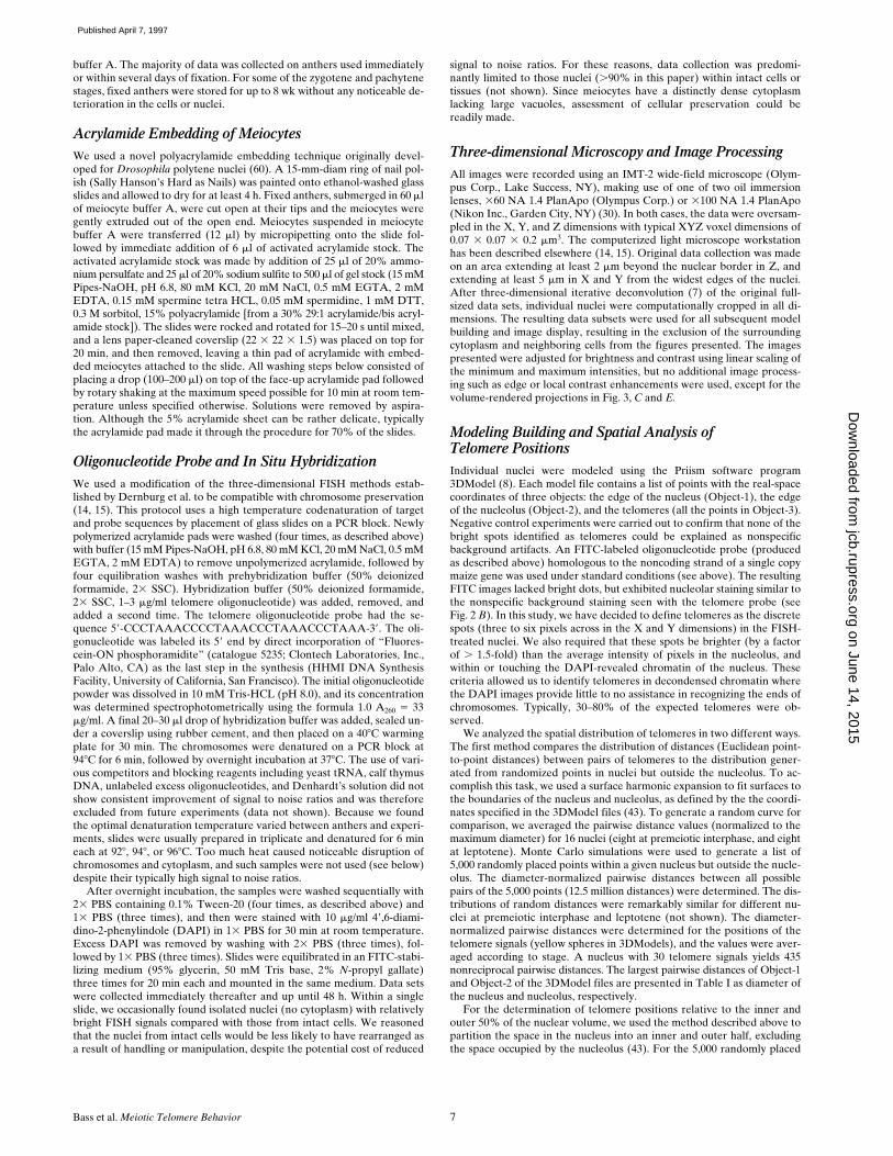
Fig. 1 shows representative DAPI images of staged nu-clei following successful in situ hybridization. These im-ages illustrate that nuclei and the chromosomes withinthem were well preserved. The morphological detail seenin the DAPI images is used in conjunction with severalother criteria to stage the meiocytes. Although meiocytesfrom a single anther are synchronous, we typically pooledmeiocytes from 3–6 similarly sized anthers on each slide.Consequently, stages were identified on a nucleus by nu-cleus basis using the criteria detailed in Fig. 1 (see legend).
Figure 1. Criteria for staging meiocytes after FISH. A summary of the multiple criteria used to determine meiotic stages in maize mei-otic is shown, along with DAPI images of representative nuclei (FITC telomere signals not shown). DAPI images are shown as singleoptical sections (A and B) or projections of optical sections spanning 2–3 microns in the Z dimension (C, D, and E). Each image is froma data stack subvolume originally containing an entire nucleus (see Materials and Methods). Chromatin and chromosome fiber appear-ances, the most definitive characteristics of meiotic stages, are considered together with anther size, nucleolus position, and knob mor-phology in classifying individual nuclei. Asterisks (*) indicate those criteria established in part or in full from this study; additional stag-ing criteria are taken in part or in full from the literature (12, 22, 23, 46). Heterochromatic knobs (k), condensed fibers (f), and theposition of the nucleolus (n) are indicated. The five conventional stages of the prophase of meiosis I (leptotene, zygotene, pachytene,diplotene, and diakinesis) are indicated at the top. Chromosomes of leptotene, zygotene, and pachytene nuclei are not yet synapsed,partially synapsed, or completely synapsed, respectively. Chromosomes of diplotene and diakinesis have desynapsed and are furthercondensed. Additionally, premeiotic interphase and prezygotene (Pzt, and see text) are indicated.
on June 14, 2015jcb.rupress.org
Dow
nloaded from
Published April 7, 1997
Bass et al.
Meiotic Telomere Behavior
9
The multiple criteria for staging are detailed here becauseproper staging is essential for accurately sorting out thetemporal relationships among multiple meiotic processes.
Equally critical to the proper interpretation of molecu-lar cytological data is the assurance that discrete FISH sig-nals are reliably reporting the position of the intended tar-get sequences. For the detection of telomeres, we designedan oligonucleotide probe cosynthetically labeled at the 5
9
end with FITC. The sequence chosen was based on the
Ar-abidopsis
telomere-repeat sequence, (TTTAGGG)
n
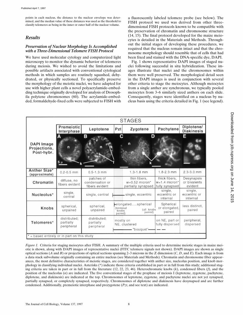
, whichhad been successfully used with other grass species (50).Fig. 2 shows the telomere FISH results from a pachytenenucleus that was purposely ruptured and flattened in orderto reveal isolated chromosome arms. The chromosomes(DAPI image, Fig. 2
A
) and the telomeres (t, FITC image,Fig. 2
B
) from a single optical section capturing most ofthe flattened nucleus are overlaid (Fig. 2
C
) to show thatthe FISH signals were found at the ends of the chromo-somes (see enlarged inset from Fig 2
C
). The restriction oftelomere FISH signals to the ends of chromosomes wasconsistently observed in numerous meiotic nuclei at pachy-tene or later, as well as in somatic nuclei undergoing mito-sis (Bass, H.W., unpublished observations). Occasionally,spots were observed to be outside the nucleus. Subsequentanalyses with multiple wavelength imaging revealed thatthe cytoplasmic spots (from zero to several per cell) typicallyresulted from nonspecific autofluorescence (not shown).The double dots at the ends of the pachytene chromo-somes (e.g., inset Fig. 2
C
) are interpreted to represent theends of the homologous chromosomes. The sister chroma-tids were not resolved in our images (for zygotene orpachytene, and by inference leptotene). From these analy-ses, we found no evidence for telomeric sequences at inter-nal chromosomal loci.
Chromosomes Are Polarized at the Last Prophase before Meiosis
Having established the conditions for direct labeling of telo-meres on pachytene chromosomes, our analysis was ex-tended to include the earlier stages where little is known
about telomere arrangements. It was possible to directlytest whether the bouquet arrangement of the nucleus ex-isted before meiotic prophase. We first asked whether wecould find any evidence for a nonrandom orientation ofchromosome ends in nuclei developmentally committed tothe meiotic pathway. The persistence of the anaphase con-figuration of chromosomes throughout an entire cell cycleresults in a polarized nucleus with telomeres and centro-meres occupying opposite hemispheres of the nucleus. Suchpolarized nuclei are said to be in a Rabl configuration, asfirst described by Rabl in 1885 (47) and subsequently doc-umented for a number of different species (10, 20, 26, 32,43, 47, 55, 61). The Rabl orientation is most easily identi-fied in nuclei at mitotic prophase when condensation re-veals the chromosome arrangements.
We therefore started our analysis at the last mitosis pre-ceding meiosis where we found evidence for a Rabl-likeconfiguration as shown in Fig. 3. The telomeric hemi-sphere was identified by the congregation of telomere FISHsignals (
A
,
green spots
;
B
,
yellow spheres
) near the top ofthis nucleus. To identify the centromeric hemisphere, wetook advantage of previous observations that anaphasemovements of metacentric or submetacentric chromosomescreate “U” or “J” shaped chromosome paths in which theturn around point reveals the centromere position (10, 13,20). In our data, these “U-turns” in the chromosome (
ar-rows
,
A
and
C
, Fig. 3) were interpreted as the approximatecentromere positions, best seen in volume-rendered pro-jections of whole nuclei (Fig. 3,
C
and
E
). We traced thepaths of the chromosomes in the original datasets of thenuclei used to make the projections shown in
C
and
E
(Fig. 3).The nucleus in
A
yielded the traces shown in
D
, and thenucleus used to make the projection in
E
yielded the tracesshown in
F
(see legend). Within each nucleus, the chromo-some paths showed a consistent orientation. Measuringthe size ratios of the chromosome arms on each side of aU-turn, we obtained ratio values (data not shown) thatwere comparable to the actual arm ratio values known formaize (42). Although we were unable to specifically iden-tify each chromosome, we believe that the chromosomepaths and the arm ratios together provide a reasonable es-
Figure 2. Specificity of oligonucleotide telomere FISH. A partially ruptured and flattened pachytene nucleus following FISH with telo-mere oligonucleotide probe (see Materials and Methods). Images show the relationship of chromosomes (A, DAPI image) and telo-meres signals (B, FITC image), by two-colored overlay (C) with inset enlargement. The nucleolus (n) and some telomeres (t) are indi-cated. Bars: (A) 5 mm; (inset) 1 mm.
on June 14, 2015jcb.rupress.org
Dow
nloaded from
Published April 7, 1997
The Journal of Cell Biology, Volume 137, 1997 10
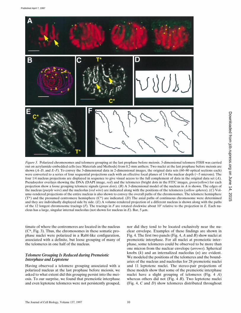
timate of where the centromeres are located in the nucleus(C*, Fig. 3). Thus, the chromosomes in these somatic pro-phase nuclei were polarized in a Rabl-like configuration,associated with a definite, but loose grouping of many ofthe telomeres in one half of the nucleus.
Telomere Grouping Is Reduced during Premeiotic Interphase and Leptotene
Having observed a telomere grouping associated with apolarized nucleus at the last prophase before meiosis, weasked to what extent did this grouping persist into the mei-osis. To our surprise, we found that premeiotic interphaseand even leptotene telomeres were not persistently grouped,
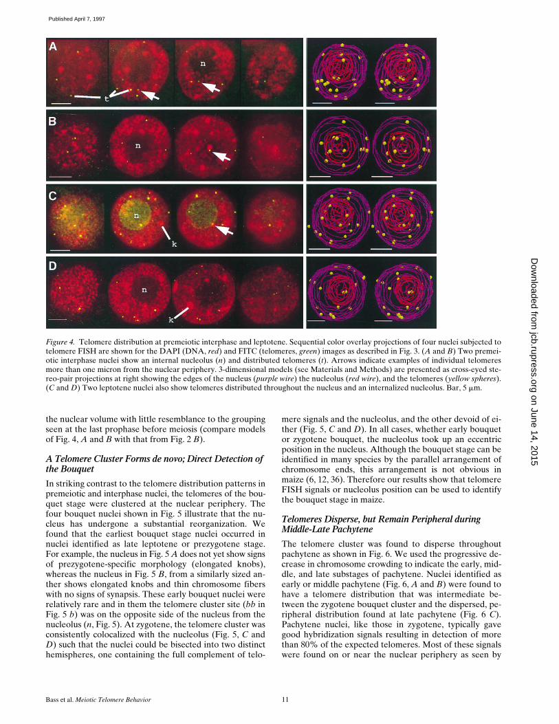
nor did they tend to be located exclusively near the nu-clear envelope. Examples of these findings are shown inFig. 4. The first two panels (Fig. 4,
A
and
B
) show nuclei atpremeiotic interphase. For all nuclei at premeiotic inter-phase, some telomeres could be observed to be more thanone micron from the nuclear envelope (arrows). Sphericalknobs (k) and an internalized nucleolus (n) are evident.We modeled the positions of the telomeres and the bound-aries of the nucleus and nucleolus for 28 premeiotic nucleiand 11 leptotene nuclei. The stereo-pair projections ofthese models show that some of the premeiotic interphasenuclei have a slight grouping of telomeres (Fig. 4 A)whereas others did not (Fig. 4 B). Two leptotene nuclei(Fig. 4, C and D) show telomeres distributed throughout
Figure 3. Polarized chromosomes and telomere grouping at the last prophase before meiosis. 3-dimensional telomere FISH was carriedout on acrylamide-embedded cells (see Materials and Methods) from 0.2-mm anthers. Two nuclei at the last prophase before meiosis areshown (A–D, and E–F). To convey the 3-dimensional data in 2-dimensional images, the original data sets (60–80 optical sections each)were converted to a series of four sequential projections each with an effective focal planes of 1/4 the nuclear depth (z5 microns). Thefour 1/4 nucleus projections are displayed in sequence to give visual access to the full complement of data in the original data set (A).Pseudocolor overlays showing the DNA (DAPI image, red) and the telomeres (bright dots in the FITC images, green/yellow) for eachprojection show a loose grouping telomere signals (green dots). (B) A 3-dimensional model of the nucleus in A is shown. The edges ofthe nucleus (purple wire) and the nucleolus (red wire) are indicated along with the positions of the telomeres (yellow spheres). (C) Vol-ume-rendered projections of the entire nucleus is also shown to convey the overall paths of the chromosomes. The telomere hemisphere(T*) and the presumed centromere hemisphere (C*) are indicated. (D) The axial paths of continuous chromosome were determinedand they are individually displayed side by side. (E) A volume-rendered projection of a different nucleus is shown along with the pathsof the 12 longest chromosome tracings (F). The tracings in F are rotated clockwise about 108 relative to the projection in E. Each nu-cleus has a large, singular internal nucleolus (not shown for nucleus in E). Bar, 5 mm.
on June 14, 2015jcb.rupress.org
Dow
nloaded from
Published April 7, 1997
Bass et al. Meiotic Telomere Behavior 11
the nuclear volume with little resemblance to the groupingseen at the last prophase before meiosis (compare modelsof Fig. 4, A and B with that from Fig. 2 B).
A Telomere Cluster Forms de novo; Direct Detection of the Bouquet
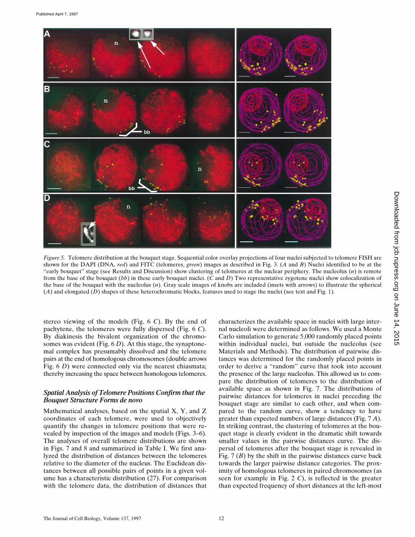
In striking contrast to the telomere distribution patterns inpremeiotic and interphase nuclei, the telomeres of the bou-quet stage were clustered at the nuclear periphery. Thefour bouquet nuclei shown in Fig. 5 illustrate that the nu-cleus has undergone a substantial reorganization. Wefound that the earliest bouquet stage nuclei occurred innuclei identified as late leptotene or prezygotene stage.For example, the nucleus in Fig. 5 A does not yet show signsof prezygotene-specific morphology (elongated knobs),whereas the nucleus in Fig. 5 B, from a similarly sized an-ther shows elongated knobs and thin chromosome fiberswith no signs of synapsis. These early bouquet nuclei wererelatively rare and in them the telomere cluster site (bb inFig. 5 b) was on the opposite side of the nucleus from thenucleolus (n, Fig. 5). At zygotene, the telomere cluster wasconsistently colocalized with the nucleolus (Fig. 5, C andD) such that the nuclei could be bisected into two distincthemispheres, one containing the full complement of telo-
mere signals and the nucleolus, and the other devoid of ei-ther (Fig. 5, C and D). In all cases, whether early bouquetor zygotene bouquet, the nucleolus took up an eccentricposition in the nucleus. Although the bouquet stage can beidentified in many species by the parallel arrangement ofchromosome ends, this arrangement is not obvious inmaize (6, 12, 36). Therefore our results show that telomereFISH signals or nucleolus position can be used to identifythe bouquet stage in maize.
Telomeres Disperse, but Remain Peripheral during Middle-Late Pachytene
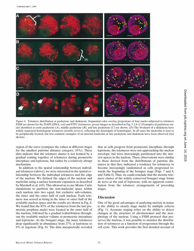
The telomere cluster was found to disperse throughoutpachytene as shown in Fig. 6. We used the progressive de-crease in chromosome crowding to indicate the early, mid-dle, and late substages of pachytene. Nuclei identified asearly or middle pachytene (Fig. 6, A and B) were found tohave a telomere distribution that was intermediate be-tween the zygotene bouquet cluster and the dispersed, pe-ripheral distribution found at late pachytene (Fig. 6 C).Pachytene nuclei, like those in zygotene, typically gavegood hybridization signals resulting in detection of morethan 80% of the expected telomeres. Most of these signalswere found on or near the nuclear periphery as seen by
Figure 4. Telomere distribution at premeiotic interphase and leptotene. Sequential color overlay projections of four nuclei subjected totelomere FISH are shown for the DAPI (DNA, red) and FITC (telomeres, green) images as described in Fig. 3. (A and B) Two premei-otic interphase nuclei show an internal nucleolus (n) and distributed telomeres (t). Arrows indicate examples of individual telomeresmore than one micron from the nuclear periphery. 3-dimensional models (see Materials and Methods) are presented as cross-eyed ste-reo-pair projections at right showing the edges of the nucleus (purple wire) the nucleolus (red wire), and the telomeres (yellow spheres).(C and D) Two leptotene nuclei also show telomeres distributed throughout the nucleus and an internalized nucleolus. Bar, 5 mm. on June 14, 2015
jcb.rupress.orgD
ownloaded from
Published April 7, 1997
The Journal of Cell Biology, Volume 137, 1997 12
stereo viewing of the models (Fig. 6 C). By the end ofpachytene, the telomeres were fully dispersed (Fig. 6 C).By diakinesis the bivalent organization of the chromo-somes was evident (Fig. 6 D). At this stage, the synaptone-mal complex has presumably dissolved and the telomerepairs at the end of homologous chromosomes (double arrowsFig. 6 D) were connected only via the nearest chiasmata;thereby increasing the space between homologous telomeres.
Spatial Analysis of Telomere Positions Confirm that the Bouquet Structure Forms de novo
Mathematical analyses, based on the spatial X, Y, and Zcoordinates of each telomere, were used to objectivelyquantify the changes in telomere positions that were re-vealed by inspection of the images and models (Figs. 3–6).The analyses of overall telomere distributions are shownin Figs. 7 and 8 and summarized in Table I. We first ana-lyzed the distribution of distances between the telomeresrelative to the diameter of the nucleus. The Euclidean dis-tances between all possible pairs of points in a given vol-ume has a characteristic distribution (27). For comparisonwith the telomere data, the distribution of distances that
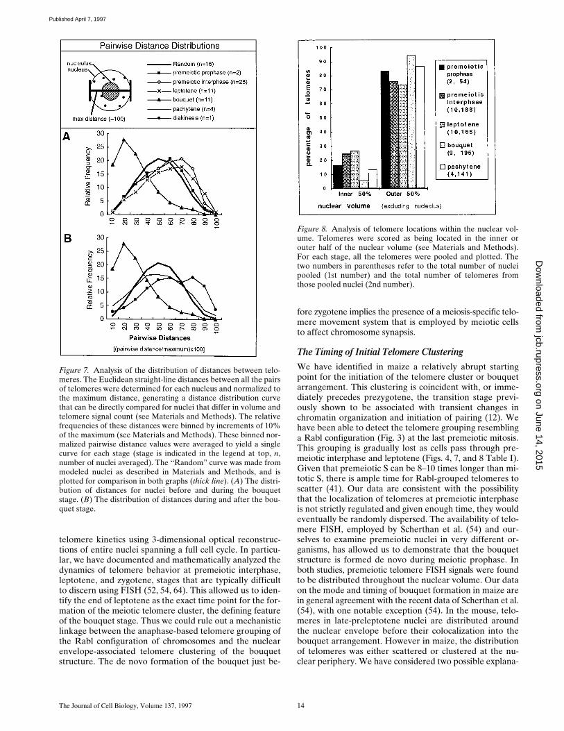
characterizes the available space in nuclei with large inter-nal nucleoli were determined as follows. We used a MonteCarlo simulation to generate 5,000 randomly placed pointswithin individual nuclei, but outside the nucleolus (seeMaterials and Methods). The distribution of pairwise dis-tances was determined for the randomly placed points inorder to derive a “random” curve that took into accountthe presence of the large nucleolus. This allowed us to com-pare the distribution of telomeres to the distribution ofavailable space as shown in Fig. 7. The distributions ofpairwise distances for telomeres in nuclei preceding thebouquet stage are similar to each other, and when com-pared to the random curve, show a tendency to havegreater than expected numbers of large distances (Fig. 7 A).In striking contrast, the clustering of telomeres at the bou-quet stage is clearly evident in the dramatic shift towardssmaller values in the pairwise distances curve. The dis-persal of telomeres after the bouquet stage is revealed inFig. 7 (B) by the shift in the pairwise distances curve backtowards the larger pairwise distance categories. The prox-imity of homologous telomeres in paired chromosomes (asseen for example in Fig. 2 C), is reflected in the greaterthan expected frequency of short distances at the left-most
Figure 5. Telomere distribution at the bouquet stage. Sequential color overlay projections of four nuclei subjected to telomere FISH areshown for the DAPI (DNA, red) and FITC (telomeres, green) images as described in Fig. 3. (A and B) Nuclei identified to be at the“early bouquet” stage (see Results and Discussion) show clustering of telomeres at the nuclear periphery. The nucleolus (n) is remotefrom the base of the bouquet (bb) in these early bouquet nuclei. (C and D) Two representative zygotene nuclei show colocalization ofthe base of the bouquet with the nucleolus (n). Gray scale images of knobs are included (insets with arrows) to illustrate the spherical(A) and elongated (D) shapes of these heterochromatic blocks, features used to stage the nuclei (see text and Fig. 1). on June 14, 2015
jcb.rupress.orgD
ownloaded from
Published April 7, 1997
Bass et al. Meiotic Telomere Behavior 13
region of the curve (compare the values at different stagesfor the smallest pairwise distance category, 10%). Thesedata indicate that the telomere cluster is not formed by agradual coming together of telomeres during premeioticinterphase and leptotene, but rather by a relatively abruptmechanism.
In addition to the spatial relationship between individ-ual telomeres (above), we were interested in the spatial re-lationship between the individual telomeres and the edgeof the nucleus. We defined the edges of the nucleus andnucleolus using a surface harmonic expansion as describedby Marshall et al. (43). This allowed us to use Monte Carlosimulations to partition the non-nucleolar space withineach nucleus into two equal, but exclusive sub-volumes,the inner and the outer half of each nucleus. Each telo-mere was scored as being in the inner or outer half of theavailable nuclear space and the results are shown in Fig. 8.We found that the 83% of the telomeres from the last pre-meiotic prophase nuclei were located in the outer half ofthe nucleus, followed by a gradual redistribution through-out the available nuclear volume at premeiotic interphaseand leptotene. At the bouquet stage, the inner telomeresdrop significantly in abundance from 27% at leptotene to6% at zygotene (Fig. 8). The data unexpectedly revealed
that as cells progress from premeiotic interphase throughleptotene, the telomeres were not approaching the nuclearenvelope, but were increasingly partitioned into the inte-rior spaces in the nucleus. These observations were similarto those derived from the distributions of pairwise dis-tances in that they indicated a tendency for telomeres tobecome increasingly randomized as cells progressed to-wards the beginning of the bouquet stage (Figs. 7 and 8,and Table I). Thus, we could conclude that the meiotic telo-mere cluster of the widely conserved bouquet stage formsde novo at the end of leptotene, with no apparent contri-bution from the telomere arrangements of precedingstages.
DiscussionOne of the great advantages of analyzing meiosis in maizeis the ability to clearly stage nuclei by multiple criteria(Fig. 1). Accurate staging relies on the ability to detectchanges in the structure of chromosomes and the mor-phology of the nucleus. Using a FISH protocol that pre-serves the nucleus, it was possible to monitor the distribu-tion of telomeres as a function of progression through thecell cycle. This work provides the first detailed account of
Figure 6. Telomere distribution at pachytene and diakinesis. Sequential color overlay projections of four nuclei subjected to telomereFISH are shown for the DAPI (DNA, red) and FITC (telomeres, green) images as described in Fig. 3. (A–C) Examples of pachytene nu-clei identified as early pachytene (A), middle pachytene (B), and late pachytene (C) are shown. (D) The bivalents of a diakinesis havewidely separated homologous telomeres (double arrows), reflecting the desynapsis of homologues. In all cases the nucleolus is seen tobe peripherally located, but less common examples of an internal nucleolus at late pachytene and diakinesis have been observed (notshown).
on June 14, 2015jcb.rupress.org
Dow
nloaded from
Published April 7, 1997
The Journal of Cell Biology, Volume 137, 1997 14
telomere kinetics using 3-dimensional optical reconstruc-tions of entire nuclei spanning a full cell cycle. In particu-lar, we have documented and mathematically analyzed thedynamics of telomere behavior at premeiotic interphase,leptotene, and zygotene, stages that are typically difficultto discern using FISH (52, 54, 64). This allowed us to iden-tify the end of leptotene as the exact time point for the for-mation of the meiotic telomere cluster, the defining featureof the bouquet stage. Thus we could rule out a mechanisticlinkage between the anaphase-based telomere grouping ofthe Rabl configuration of chromosomes and the nuclearenvelope-associated telomere clustering of the bouquetstructure. The de novo formation of the bouquet just be-
fore zygotene implies the presence of a meiosis-specific telo-mere movement system that is employed by meiotic cellsto affect chromosome synapsis.
The Timing of Initial Telomere Clustering
We have identified in maize a relatively abrupt startingpoint for the initiation of the telomere cluster or bouquetarrangement. This clustering is coincident with, or imme-diately precedes prezygotene, the transition stage previ-ously shown to be associated with transient changes inchromatin organization and initiation of pairing (12). Wehave been able to detect the telomere grouping resemblinga Rabl configuration (Fig. 3) at the last premeiotic mitosis.This grouping is gradually lost as cells pass through pre-meiotic interphase and leptotene (Figs. 4, 7, and 8 Table I).Given that premeiotic S can be 8–10 times longer than mi-totic S, there is ample time for Rabl-grouped telomeres toscatter (41). Our data are consistent with the possibilitythat the localization of telomeres at premeiotic interphaseis not strictly regulated and given enough time, they wouldeventually be randomly dispersed. The availability of telo-mere FISH, employed by Scherthan et al. (54) and our-selves to examine premeiotic nuclei in very different or-ganisms, has allowed us to demonstrate that the bouquetstructure is formed de novo during meiotic prophase. Inboth studies, premeiotic telomere FISH signals were foundto be distributed throughout the nuclear volume. Our dataon the mode and timing of bouquet formation in maize arein general agreement with the recent data of Scherthan et al.(54), with one notable exception (54). In the mouse, telo-meres in late-preleptotene nuclei are distributed aroundthe nuclear envelope before their colocalization into thebouquet arrangement. However in maize, the distributionof telomeres was either scattered or clustered at the nu-clear periphery. We have considered two possible explana-
Figure 7. Analysis of the distribution of distances between telo-meres. The Euclidean straight-line distances between all the pairsof telomeres were determined for each nucleus and normalized tothe maximum distance, generating a distance distribution curvethat can be directly compared for nuclei that differ in volume andtelomere signal count (see Materials and Methods). The relativefrequencies of these distances were binned by increments of 10%of the maximum (see Materials and Methods). These binned nor-malized pairwise distance values were averaged to yield a singlecurve for each stage (stage is indicated in the legend at top, n,number of nuclei averaged). The “Random” curve was made frommodeled nuclei as described in Materials and Methods, and isplotted for comparison in both graphs (thick line). (A) The distri-bution of distances for nuclei before and during the bouquetstage. (B) The distribution of distances during and after the bou-quet stage.
Figure 8. Analysis of telomere locations within the nuclear vol-ume. Telomeres were scored as being located in the inner orouter half of the nuclear volume (see Materials and Methods).For each stage, all the telomeres were pooled and plotted. Thetwo numbers in parentheses refer to the total number of nucleipooled (1st number) and the total number of telomeres fromthose pooled nuclei (2nd number).
on June 14, 2015jcb.rupress.org
Dow
nloaded from
Published April 7, 1997
Bass et al. Meiotic Telomere Behavior 15
tions for this discrepancy: One is that the two systems arein fact different; in the mouse, telomeres attach to a broadarea of the nuclear envelope before congregating into thebouquet arrangement, but in maize the telomeres mayattach to the nuclear envelope and congregate at the sametime. The other possibility is that we did not detect an at-tachment of maize telomeres to a broad area of the nu-clear envelope because of the brevity of this stage. Werepeatedly made attempts to identify an intermediate be-tween scattered and clustered telomere arrangements. In-variably, meiocytes from anthers expected to yield lateleptotene to early zygotene meiocytes showed either scat-tered or clustered telomere arrangements. Interestingly,these very same samples yielded the relatively rare cells inwhich the nucleolus and the telomere cluster sites werespatially separated. Such nuclei (Fig. 4, A and B) were re-ferred to as “early bouquet” nuclei in which all but thepresumptive NOR-linked telomeres were at the bouquetsite. Our early bouquet stage nuclei could be interpretedas reflecting a transition stage toward bouquet formationin which all telomeres have clustered except for those whichare slower to move because of linkage to the nucleolus.
For every nucleus subjected to data collection, deconvo-lution, and model building, at least five additional nucleiwere imaged or observed while scanning the slides. In allcases where telomere clustering could be detected, the nu-cleolus was at the edge of the nucleus and almost alwaysadjacent to the bouquet. This observation suggests that inmaize, the eccentric position of the nucleolus may be usedas an indicator that the bouquet stage has commenced. Acritical piece of evidence in concluding that telomere clus-tering initiates at the end of leptotene, as opposed toprezygotene or early zygotene, is the observation that therelocation of the nucleolus to the nuclear periphery occursat the end of leptotene (Dawe, R.K., personal communica-tion, see also Fig. 2 C of Dawe et al., 1994). The end of lep-totene is distinct from prezygotene because the knobs arestill spherical (Fig. 1). In fact, the first signs of knob elon-gation are diagnostic for the end of leptotene and the be-ginning of prezygotene (12). Therefore, the nucleus shownin Fig. 5 A provides an example of telomere clustering be-fore prezygotene. Additional evidence in support of thisclaim comes from a partial reconstruction of a leptotenenucleus by serial sectioning electron microscopy (23). Inthis study on maize, the ends of the chromosomes were at-tached to a small region of the nuclear envelope. Similarly,
the bouquet stage has been observed as early as leptotenein human and rye, and at the leptotene-zygotene transitionin locust, respectively (44, 48, 59).
We have illustrated our results in Fig. 9 to summarizethe temporal relationship between the bouquet stage, de-fined by the bouquet structure, and the canonical earlystages of meiotic prophase, leptotene, zygotene, and pachy-tene. The initial telomere clustering is shown to occur atthe end of leptotene just preceding prezygotene. The “earlybouquet” nuclei are the first to exhibit telomere clusteringwhere all but the NOR-linked telomeres have reached thecluster site. The NOR in maize is located near the end ofthe short arm of chromosome 6. The spatial separation ofthe NOR from the telomere cluster site may reflect aslower rate at which the NOR-linked telomeres can moveto the cluster site. Alternatively, the nucleolus may be po-sitioned in these early bouquet nuclei by a telomere-inde-pendent mechanism that overrides the telomere clusteringmechanism (28). Prezygotene is then followed by zygo-tene, during which the most commonly encountered bou-quet arrangement was observed (indicated by “bouquet,”Fig. 9, above zygotene). The dispersal of telomeres duringpachytene is indicated by the predominantly peripheral lo-calization of telomeres near the nuclear envelope (nucleusdiagrammed above “pachytene,” Fig. 9). This summary il-lustrates that the de novo formation of the telomere clus-ter is among the very first steps in nuclear changes associ-ated with synapsis.
We have interpreted our results to indicate that thechanges in telomere arrangements are continuous and es-sentially irreversible. However, the harvesting, fixation, orhybridization of cells could result in alterations of the nu-clear organization that normally occur in vivo. To directlyaddress such a possibility we will need to compare theseresults with those of living cells. Although our study wascarried out on fixed material, we have described thepooled results from three different inbred lines of maize,harvested throughout the year. Thus we have attempted todescribe observations of a general nature, avoiding de-scription of observations that may be peculiar to a particu-lar genetic line or growth condition.
Possible Mechanisms of Meiotic Telomere Movements
The present study describes the sudden formation and thegradual dissolution of the meiotic telomere cluster. In light
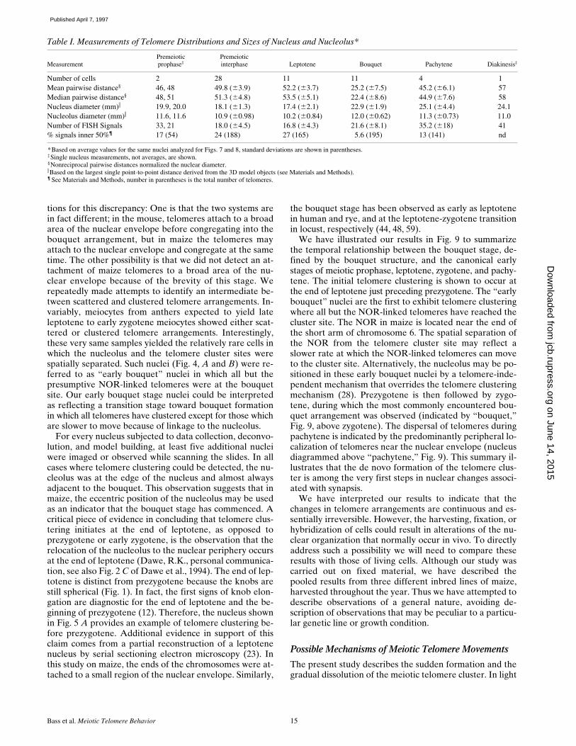
Table I. Measurements of Telomere Distributions and Sizes of Nucleus and Nucleolus*
MeasurementPremeioticprophase‡
Premeioticinterphase Leptotene Bouquet Pachytene Diakinesis‡
Number of cells 2 28 11 11 4 1Mean pairwise distance§ 46, 48 49.8 (63.9) 52.2 (63.7) 25.2 (67.5) 45.2 (66.1) 57Median pairwise distance§ 48, 51 51.3 (64.8) 53.5 (65.1) 22.4 (68.6) 44.9 (67.6) 58Nucleus diameter (mm)i 19.9, 20.0 18.1 (61.3) 17.4 (62.1) 22.9 (61.9) 25.1 (64.4) 24.1Nucleolus diameter (mm)i 11.6, 11.6 10.9 (60.98) 10.2 (60.84) 12.0 (60.62) 11.3 (60.73) 11.0Number of FISH Signals 33, 21 18.0 (64.5) 16.8 (64.3) 21.6 (68.1) 35.2 (618) 41% signals inner 50%¶ 17 (54) 24 (188) 27 (165) 5.6 (195) 13 (141) nd
*Based on average values for the same nuclei analyzed for Figs. 7 and 8, standard deviations are shown in parentheses.‡Single nucleus measurements, not averages, are shown.§Nonreciprocal pairwise distances normalized the nuclear diameter.iBased on the largest single point-to-point distance derived from the 3D model objects (see Materials and Methods).¶ See Materials and Methods, number in parentheses is the total number of telomeres.
on June 14, 2015jcb.rupress.org
Dow
nloaded from
Published April 7, 1997

The Journal of Cell Biology, Volume 137, 1997 16
of these findings, we now consider the possible mecha-nisms underlying these dynamic changes in telomere local-ization. The abruptness of the initial cluster formation al-lows us to rule out one class of mechanisms relying onpassive diffusion. Specifically, our data do not support ascenario in which a region of the nuclear envelope sud-denly acquires an affinity for telomeres, and collects thetelomeres on the basis of chance interaction. What then isthe mechanistic basis for the formation of the telomerecluster? To begin to address this question, two possiblemechanisms of bouquet formation are proposed in Fig. 10.The first mechanism involves a two-step process (move-ments indicated by arrows of nuclei 1A and 1B of Fig. 10)in which the telomeres first attach to any region of the nu-clear envelope (arrows of 1A), followed by the movementof telomeres in the plane of the nuclear envelope (arrowsof 1B) to end up at the final cluster site. The second mech-anism would involve a one-step process (movements indi-cated by arrows of nucleus 2, Fig. 10) in which telomeresmove directly to the region of the nuclear envelope thatwill be the final cluster site, the bouqet base (referred to asbb in Figs. 5 and 6). The two-step clustering mechanism isconsistent with the observations on mouse spermatocytes(54). Our data on meiosis in maize does not allow us todistinguish between the two paths shown in Fig. 10. Al-though this model only addresses the mechanisms respon-sible for telomere movements, we recognize that telomeresare but one of many cellular components that show altereddistributions at the bouquet stage (13).
Evidence that clustering of telomeres is associated withactive forces on chromosomes or nuclei come from a series
of studies on the bouquet stage conducted by Hiraoka(29). In his research on meiotic nuclei in living cells of Eq-uisetum and other plants, he observed that when the bou-quet configuration is formed, nuclei take up an eccentricposition in the cell with the telomeric cluster regions pre-dicting the direction of nuclear migration. Chikashige et al.(9) showed that a similar phenomenon, i.e., meiotic nu-clear migration, explains the dramatic distortions in mei-otic prophase nuclei (the horsetail stage) of the fissionyeast, S. pombe (9). In this case, the entire nucleus, led bythe telomeres and the spindle pole body, is translocatedthrough the cytoplasm. Thus, some of the morphologicalchanges in chromatin and nuclei are consistent with thepossibility that telomeres serve as connection points for acytoplasmic motility system, whose function during meio-sis remains to be discerned. It is noteworthy that theremay be a difference between the forces that form the bou-quet, as drawn in Fig. 10, and the forces that result inmovement of the whole nucleus.
Analysis of nuclear motility along with ultrastructuralstudies on meiotic chromosomes has revealed that themicrotubule cytoskeleton may be involved in these telomere-based actions. These data are based primarily on observa-tions in many species that the base of the bouquet is juxta-posed to the microtubule organizing center, be it a centrioleor spindle pole body (13). It remains to be determinedwhether a microtubule organizing center is near the telo-mere cluster in angiosperms such as maize, which lack cy-tologically distinct microtubule organizing centers. An-other indirect line of evidence linking microtubules tobouquet formation is based on disruption of meiotic chro-mosome pairing by treatment with the microtubule depo-lymerizing drug, colchicine (40). In addition, Driscoll andDarvey provide compelling evidence that chromosomepairing is sensitive to colchicine treatments (18). They ap-plied colchicine to a genetic stock of wheat carrying an iso-chromosome, a compound chromosome in which the rightand left arms are identical, but with mirror image orienta-tion. The isochromosome alone was resistant to the chias-
Figure 9. Diagram of the timing of meiotic telomere clustering. Asummary of the fine scale timing of events associated with the on-set of synapsis is presented based on the observations from thiswork and from Dawe et al. (1994). Leptotene telomeres (legendat right) are shown as either NOR-associated (open circles) ornon-NOR (closed circles, representing the other 38 of 40 maizetelomeres) are scattered throughout the chromatin, and the nu-cleolus is fully internal and approximately concentric with the nu-cleus. The first signs of telomere rearrangements give rise to the“early bouquet” (see text). The early bouquet nucleus (as in Fig.5, A and B) is first seen at the end of leptotene, just precedingprezygotene. Next, prezygotene occurs, comprising the majortransition between leptotene and zygotene as described (12). Byzygotene, all of the telomeres are at the cluster site resulting inthe eccentric nucleolus showing an obligatory colocalization withthe telomere cluster (as in Fig. 5, C and D). At pachytene thetelomere bouquet disperses and the paired homologous telo-meres remain at the nuclear periphery.
Figure 10. Model of possible mechanisms of telomere cluster for-mation. The de novo clustering of randomly distributed telo-meres is proposed to occur by one of two different mechanisms (1and 2). Mechanism 1 proposes a two-step model; 1A, telomeresmove from their positions in the nucleus to the nuclear envelopewhere they become attached, and 1B nuclear envelope-attachedtelomeres move over the nuclear surface to the final cluster site.Mechanism 2 proposes a one-step model in which telomeresmove from their positions in the nucleus directly to the region ofthe nuclear envelope that will be the final cluster site.
on June 14, 2015jcb.rupress.org
Dow
nloaded from
Published April 7, 1997

Bass et al. Meiotic Telomere Behavior 17
mata-reducing effect of colchicine leading the authors toconclude that microtubules participated in the first step ofchromosome pairing, bringing homologous chromosomesinto close spatial proximity (18). This concept is consistentwith the possibility that telomere movements on the nu-clear envelope (as shown for 1B of Fig. 10) may involve in-teractions with the microtubule cytoskeleton.
Even if the cytoskeleton participates in the nuclear mo-tility seen at meiotic prophase or the movement of telo-meres along the nuclear envelope, it remains to be deter-mined how the telomeres arrive at and attach to thenuclear envelope. Although we refer to telomeres in thecytological sense in this paper, we do not know what role,if any, the telomere repeat sequences actually play inmovement or attachment of the ends of chromosomes tothe nuclear envelope. It is also recognized that we did notdirectly localize the nuclear envelope in this study, but in-ferred its location from the edge of the DAPI-stainedchromatin.
Functional Significance of the Bouquet Stage
Having considered the when and how of telomere cluster-ing, we are still left with the basic question of why, i.e., thefunctional significance of the bouquet. The inherentlycomplex nature of the processes taking place during mei-otic prophase preclude any simple assessment of bouquetfunction on the basis of temporal coincidence alone. De-spite its invariant association with zygotene, the bouquetarrangement could have a role in chromosome alignment,homology search, resolution of interlocks, synapsis, synap-tonemal complex formation, or crossover control.
Scherthan et al. (54) have argued that the bouquet mayfunction to facilitate alignment of homologues as extendedchromosome fibers (54). Once aligned, synapsis couldcommence in a relatively efficient manner while minimiz-ing entanglements and search time (54). Dawe et al. (12)showed that heterochromatic, homologous knob loci wereon average more than 7 microns apart at leptotene. Similarobservations were made using a FISH probe for the eu-chromatic 5S rDNA locus (Bass, H.W., unpublished ob-servations). Therefore, homologous loci might have a con-siderable distance to travel to encounter their homologuesin maize, which has a normal diploid genome, similar insize to that of human (3). However, it is not clear that re-duction of the homology search volume is sufficient to ex-plain conservation of the bouquet because even fissionand budding yeast, with small genomes and small nuclei,have a bouquet stage (9, 17, 53). Furthermore, buddingyeast may have already accomplished a substantial degreeof homologous chromosome alignment before the bou-quet stage (38, 64).
A very different type of role for the bouquet might in-volve crossover control. The existence of crossover controlis inferred from the observations that in most species,crossovers (chiasmata) between homologous chromosomesoccur at very low, but non-zero rates, and the distributionof crossovers is not random (i.e., crossover interference).In a recently published model, Kleckner has suggestedthat crossover control occurs “via the imposition and reliefof stress” (37). It is suggested that such stress derives fromdifferential compaction of axis-associated chromatin. In
this scenario, a role for bouquet-related pulling forces onthe ends of the chromosomes could be easily envisioned.The idea that the bouquet, and in particular the forces thatgiving rise to nuclear movement, may be involved in cross-over control is consistent with the observation that the ini-tiation of synapsis, and the formation of chiasmata pre-dominantly occur near the ends of chromosomes in manyspecies (4, 35, 39, 45, 57, 58, 63).
The two possible functions of the bouquet discussedabove might be predicted to manifest quite different phe-notypes in the event of a failure of the bouquet to form orfunction. In the case of a role in chromosome pairing, abouquet failure would be predicted to result in the accu-mulation of unpaired chromosomes at metaphase I, caus-ing a reduction or loss of fertility. Whereas in the case of arole in crossover control, a bouquet failure might result ina change of the position or frequency of chiasmata. Ifthese changes involved a loss of chiasmata, the resultwould be infertility associated with the production of uni-valents. A full understanding of the role of the bouquetstructure will require that we can test its function by ex-perimental or genetic disruption, in conjunction with cyto-logical anlysis. While this work reports on the discoverythat the bouquet forms de novo at the end of leptotene, italso presents a powerful experimental approach that canbe used to elucidate the basic structure-function relation-ships of meiotic nuclei.
We thank H. Scherthan for discussion of unpublished results during theearly stages of this work; A.F. Dernburg and S.J. Parmelee for their excel-lent technical advice on 3-D FISH methodologies; D.D. Hughes and H.Chen for their generous assistance with the distance analyses; and A.E.Franklin, J.L. Paluh, L.C. Harper, and J.C. Fung for critical reading of themanuscript. The first author (H.W. Bass) is especially grateful to W.Z.Cande and J.W. Sedat for their guidance, support, and enthusiasmthroughout this project.
H.W. Bass was supported in the early years of this work by a NationalInstitutes of Health (NIH) grant to J.W. Sedat (R01-GM-25101-16). H.W.Bass is currently supported as a D.O.E. postdoctoral fellow of the LifeSciences Research Foundation. This work was supported by the (NIH)grants to W.Z. Cande (R01-GM-48547) to J.W. Sedat (R01-GM-25101-16), and to D.A. Agard (R01-GM-31627). D.A. Agard is an investigator ofthe Howard Hughes Medical Institute.
Received for publication 7 November 1996 and in revised form 8 January1997.
References
1. Bahler, J., T. Wyler, J. Loidl, and J. Kohli. 1993. Unusual nuclear structuresin meiotic prophase of fission yeast: a cytological analysis. J. Cell Biol.121:241–256.
2. Belmont, A.S., J.W. Sedat, and D.A. Agard. 1987. A three-approach to mi-totic chromosome structure: evidence for a complex hierarchical organi-zation. J. Cell Biol. 105:77–92.
3. Bennett, M.D., J.B. Smith, and J.S. Heslop-Harrison. 1983. Nuclear DNAamounts in angiosperms. Proc. R. Soc. Lond. Ser. B. 216:179–199.
4. Burnham, C.R., J.T. Stout, W.H. Weinheimer, R.V. Knowles, and R.L.Phillips. 1972. Chromosome pairing in maize. Genetics. 71:111–125.
5. Burns, J.A. 1972. Preleptotene chromosome contraction in Nicotiana spe-cies. J. Hered. 63:175–178.
6. Chang, M.T., and M.G. Neuffer. 1989. Maize microsporogenesis. Genome.32:232–244.
7. Chen, H., J.R. Swedlow, M.A. Grote, J.W. Sedat, and D.A. Agard. 1995.The collection, processing, and display of digital three-dimensional im-ages of biological specimens. In Handbook of Biological Confocal Mi-croscopy. J.B. Pawley, editor. Plenum Press, New York. 197–210.
8. Chen, H., D.D. Hughes, T.-A. Chan, J.W. Sedat, and D.A. Agard. 1996.IVE (Image Visualization Environment): a software platform for allthree-dimensional microscopy applications. J. Struct. Biol. 116:56–60.
9. Chikashige, Y., D.-Q. Ding, H. Funabiki, T. Haraguchi, S. Mashiko, M.
on June 14, 2015jcb.rupress.org
Dow
nloaded from
Published April 7, 1997
The Journal of Cell Biology, Volume 137, 1997 18
Yanagida, and Y. Hiraoka. 1994. Telomere-led premeiotic chromosomemovement in fission yeast. Science (Wash. DC). 264:270–273.
10. Cremer, T., A. Kurz, R. Zirbel, S. Dietzel, B. Rinke, E. Schrock, M.R. Spei-cher, U. Mathiew, A. Jauch, P. Emmerich, et al. 1993. Role of chromo-somal territories in the functional compartmentalization of the cell nu-cleus. Cold Spring Harbor Symp. Quant. Biol. 43:777–792.
11. Darlington, C.D., editor. 1937. Recent Advances in Cytology. Second edi-tion. Churchill, Ltd., London. 650 pp.
12. Dawe, R.K., J.W. Sedat, D.A. Agard, and W.Z. Cande. 1994. Meiotic chro-mosome pairing in maize is associated with a novel chromatin organiza-tion. Cell. 76:901–912.
13. Dernburg, A.F., J.W. Sedat, W.Z. Cande, and H.W. Bass. 1995. Cytology oftelomeres. In Telomeres. E.H. Blackburn and C.W. Grieder, editors.Cold Spring Harbor Laboratory Press, Cold Spring Harbor, NY. 295–338.
14. Dernburg, A.F., K.W. Broman, J.C. Fung, W.F. Marshall, J. Philips, D.A.Agard, and J.W. Sedat. 1996. Perturbation of nuclear architecture bylong-distance chromosome interactions. Cell. 85:745–759.
15. Dernburg, A.F., J.W. Sedat, and R.S. Hawley. 1996. Direct evidence of arole for heterochromatin in meiotic chromosome segregation. Cell. 86:135–146.
16. Digby, L. 1919. On the archesporial and meiotic mitoses of Osmunda. Ann.Bot. (Lond.). 33:135–172.
17. Dresser, M.E., and C.N. Giroux. 1988. Meiotic chromosome behavior inspread preparations of yeast. J. Cell. Biol. 106:567–574.
18. Driscoll, C.J., and N.L. Darvey. 1970. Chromosome pairing: effect of colchi-cine on an isochromosome. Science (Wash. DC). 162:290–291.
19. Freeling, M., and V. Walbot, editors. 1994. The Maize Handbook.Springer-Verlag, Inc, New York. 759 pp.
20. Fussell, C.P. 1984. Interphase chromosome order: a proposal. Genetica. 62:193–201.
21. Gelei, J. 1921. Weitere Studien über die Oogenese des Dendrocoelum lac-teum. II. Die Längskonjugation der Chromosomen. Arch. Zellforsch. 16:88–169.
22. Gillies, C.B. 1973. Ultrastructural analysis of maize pachytene karyotypesby three-dimensional reconstruction of the synaptonemal complexes.Chromosoma (Berl.). 43:145–176.
23. Gillies, C.B. 1975. An ultrastructural analysis of chromosomal pairing inmaize. Carlsberg Res. Commun. 40:135–162.
24. Golubovskaya, I.N. 1989. Meiosis in maize: mei genes and conception ofgenetic control of meiosis. Adv. Genet. 26:149–192.
25. Golubovskaya, I., N.A. Avalkina, and W.F. Sheridan. 1992. Effects of sev-eral meiotic mutations on female meiosis in maize. Dev. Genet. 13:411–424.
26. Gruenbaum, Y., M. Hochstrasser, D. Mathog, H. Saumwever, D.A. Agard,and J.W. Sedat. 1984. Spatial organization of the Drosophila nucleus: athree-dimensional cytogenetic study. J. Cell Sci. Suppl. 1:223–234.
27. Hammersley, J. M. 1950. The distribution of distances in a hypersphere.Ann. Math. Stat. 21:447–452.
28. Hiraoka, T. 1952. Observational and experimental studies of meiosis withspecial reference to the bouquet stage. XI. Locomotory movement of thenucleolus in the bouquet stage. Cytologia (Tokyo). 17:201–209.
29. Hiraoka, T. 1952. Observational and experimental studies of meiosis withspecial reference to the bouquet stage. XIV. Some considerations on aprobable mechanism of the bouquet formation. Cytologia (Tokyo). 17:292–299.
30. Hiraoka, Y., J.R. Swedlow, M.R. Paddy, D.A. Agard, and J.W. Sedat. 1991.Three-dimensional multiple wavelength fluorescence microscopy for thestructural analysis of biological phenomena. Semin. Cell Biol. 2:153–165.
31. Hiraoka, Y., A.F. Dernburg, S.J. Parmelee, M.C. Rykowski, D.A. Agard,and J.W. Sedat. 1993. The onset of homologous chromosome pairing dur-ing Drosophila melanogaster embryogenesis. J. Cell Biol. 120:591–600.
32. Hochstrasser, M., D. Mathog, Y. Gruenbaum, H. Saumweber, and J.W. Se-dat. 1986. Spatial organization of chromosomes in the salivary gland nu-clei of Drosophila melanogaster. J. Cell Biol. 102:112–123.
33. John, B. 1976. Myths and mechanisms of meiosis. Chromosoma (Berl.). 54:295–325.
34. John, B., editor. 1990. Meiosis. In Developmental and Cell Biology Series.Cambridge University Press, New York. 396 pp.
35. Kasha, K.J., and C.R. Burnham. 1965. The location of interchange break-points in barley. II. Chromosome pairing and the intercross method. Can.J. Genet. Cytol. 7:620–632.
36. Kezer, J., S.K. Sessions, and P. Leon. 1989. The meiotic structure and be-havior of the strongly heteromorphic X/Y sex chromosomes of neotropi-
cal plethodontid salamanders of the genus Oedipina. Chromosoma(Berl.). 98:433–442.
37. Kleckner, N. 1996. Meiosis, how could it work? Proc. Natl. Acad. Sci. USA.93:8167–8174.
38. Kleckner, N., and B.M. Weiner. 1993. Potential advantages of unstable in-teractions for pairing of chromosomes in meiotic, somatic, and premei-otic cells. Cold Spring Harbor Symp. Quant. Biol. 43:553–565.
39. Laurie, D.A., and M.A. Hulten. 1985. Further studies on chiasma distribu-tion and interference in the human male. Ann. Hum. Genet. 49:203–214.
40. Loidl, J. 1990. The initiation of meiotic pairing: the cytological view. Ge-nome. 33:759–778.
41. Lu, B.C.K. 1996. Chromosomes, mitosis, and meiosis. In Fungal Genetics.C.J. Bos, editor. Marcel Dekker, Inc., New York. 119–176.
42. Maguire, M. 1967. Evidence for homologous pairing of chromosomes priorto meiotic prophase in maize. Chromosoma (Berl.). 21:221–231.
43. Marshall, W.F., A.F. Dernburg, B. Harmon, D.A. Agard, and J.W. Sedat.1996. Specific interactions of chromatin with the nuclear envelope: posi-tional determination within the nucleus in Drosophila melanogaster. Mol.Biol. Cell. 7:825–842.
44. Moens, P.B. 1969. The fine structure of meiotic chromosome polarizationand pairing in Locusta migratoria spermatocytes. Chromosoma (Berl.).28:1–25.
45. Moens, P.B., C. Bernelot-Moens, and B. Spyropoulos. 1989. Chromosomecore attachment to meiotic nuclear envelope regulates synapsis in Chlo-ealtis (Orthoptera). Genome. 32:601–610.
46. Mogensen, H.L. 1977. Ultrastructural analysis of female pachynema andthe relationship between synaptonemal complex length and crossing-over in Zea mays. Carlsberg Res. Commun. 42:475–497.
47. Rabl, C. 1885. Über Zelltheilung. Morpholog. Jahrbuch. 10:214–330.48. Rasmussen, S.W., and P.B. Holm. 1978. Human meiosis II. Chromosome
pairing and recombination nodules in human spermatocytes. CarlsbergRes Commun. 43:275–327.
49. Rhoades, M.M. 1950. Meiosis in maize. J Hered. 41:59–67.50. Richards, E.J., and F.M. Ausubel. 1988. Isolation of a higher eukaryotic
telomere from Arabidopsis thaliana. Cell. 53:127–136.51. Scherthan, H. 1996. Chromosome behavior in earliest meiotic prophase. In
Chromosomes Today. J.S. Parker and M. Puertas, editors. Capman andHall, London.
52. Scherthan, H., and T. Cremer. 1994. Nonisotopic in situ hybridization inparaffin-embedded tissue sections. Methods Mol. Genet. 5:223–238.
53. Scherthan, H., J. Bahler, and J. Kohli. 1994. Dynamics of chromosome or-ganization and pairing during meiotic prophase in fission yeast. J. CellBiol. 127:273–285.
54. Scherthan, H., S. Weich, H. Schwegler, C. Heyting, M. Härle, and T. Cre-mer. 1996. Centromere and telomere movements during early meioticprophase of mouse and man are associated with the onset of chromo-some pairing. J. Cell Biol. 134:1109–1125.
55. Schwarzacher, T., A.R. Leitch, M.D. Bennett, and J.S. Heslop-Harrison.1989. In situ localization of parental genomes in a wide hybrid. Ann. Bot.(Lond.). 64:315–324.
56. Sprague, G.F., and J.W. Dudley. 1988. Corn and corn improvement. InAgronomy. Vol. 18. ASA, CSSA, SSSA, New York.
57. Tabata, M. 1962. Chromosome pairing in intercrosses between stocks of in-terchanges involving the same two chromosomes in maize. Diakinesisconfigurations. Cytologia (Tokyo). 27:410–417.
58. Tabata, M. 1963. Chromosome pairing in intercrosses between stocks of in-terchanges involving the same two chromosomes in maize. II. Pachyteneconfigurations. Cytologia (Tokyo). 28:278–292.
59. Thomas, J.B., and P.J. Kaltsikes. 1976. A bouquet-like attachment plate fortelomeres in leptotene of rye revealed by heterochromatin staining. He-redity. 36:155–162.
60. Urata, Y., S.J. Parmelee, D.A. Agard, and J.W. Sedat. 1995. A three-dimen-sional structural dissection of Drosophila polytene chromosomes. J. CellBiol. 131:279–295.
61. Vanderlyn, L. 1948. Somatic mitosis in the root tip of Allium cepa—a re-view and orientation. Bot. Rev. 14:270–318.
62. von Wettstein, D., S.W. Rasmussen, and P.B. Holm. 1984. The synaptone-mal complex in genetic segregation. Ann. Rev. Genet. 18:331–413.
63. Wallace, B.M.N., and M.A. Hulten. 1985. Meiotic chromosome pairing inthe normal human female. Ann. Hum. Genet. 49:215–226.
64. Weiner, B.A., and N. Kleckner. 1994. Chromosome pairing via multiple in-terstitial interactions before and during meiosis in yeast. Cell. 77:977–991.
on June 14, 2015jcb.rupress.org
Dow
nloaded from
Published April 7, 1997
Copyright © 2022 FDOKUMEN


![[Regulation of telomeres length: making the telomeres accessible?]](https://static.fdokumen.com/doc/165x107/633f1028d121719806096682/regulation-of-telomeres-length-making-the-telomeres-accessible.jpg)