Clinico-immunological changes post-immunotherapy with Periplaneta americana
Upload
independentCategory
view
5download
0
ORIGINAL ARTICLE
Immunological responses in cancer patients after vaccinationwith the therapeutic telomerase-specific vaccine Vx-001
Eleni-Kyriaki Vetsika • Georgios Konsolakis • Despoina Aggouraki • Athanasios Kotsakis •
Elisavet Papadimitraki • Soultana Christou • Jeanne Menez-Jamet • Kostas Kosmatopoulos •
Vassilis Georgoulias • Dimitris Mavroudis
Received: 3 March 2011 / Accepted: 26 July 2011 / Published online: 20 August 2011
� Springer-Verlag 2011
Abstract Vx-001, an HLA-A*0201 restricted telomerase
(TERT)-specific anti-tumor vaccine, is composed of the
9-mer cryptic TERT572 peptide and its optimized variant
TERT572Y. We have previously shown that Vx-001 is non-
toxic, highly immunogenic and in vaccinated NSCLC
patients early specific immune response is associated with
prolonged survival. The aim of the present study was to
investigate the specific T-cell immune response against
Vx-001. Fifty-five patients with chemo-resistant advanced
solid tumors were vaccinated with TERT572Y (2 subcuta-
neous injections) followed by TERT572 peptide (4 subcu-
taneous injections) every 3 weeks. Specific immune
response was evaluated by IFN-c and perforin ELISpot
and intracellular cytokine staining assays. TERT-reactive
T cells were detected in 27 (51%) out of 53 evaluable
patients after the 2nd vaccination and in 22 (69%) out of 32
evaluable patients after the completion of 6 vaccinations.
Immune responses developed irrespective of the stage of
disease and disease status before vaccination. Patients
with disease progression at study entry who developed a
post-vaccination-induced immunological response had a
significant overall survival benefit compared to the post-
vaccination non-responders. The Vx-001 vaccine is a
promising candidate for cancer immunotherapy since it can
induce a TERT-specific T-cell immune response that is
associated with prolonged survival.
Keywords Immunotherapy � Vaccine � CD8? T cells �Telomerase � Cryptic peptide
Introduction
Cancer immunotherapy is based on the protective role of
the immune system against cancer mainly via the capacity
of the CD8? cytotoxic T lymphocytes (CTLs) to recognize
and kill cancer cells [1, 2]. CTLs, when recognizing
tumoral antigenic peptides on the surface of tumor cells in
association with major histocompatibility complex (MHC)
class I molecules, may become activated and can lyse the
cancer cells expressing these antigens [3].
The human telomerase is a complex of the telomerase
RNA subunit (TERC) and the telomerase reverse trans-
criptase (TERT), which is correlated with telomerase
activity. Telomerase adds telomeric repeats to the chro-
mosome ends and therefore prevents the replication-
dependent loss of telomere and cellular senescence in
highly dividing cells such as tumor cells [4]. TERT is
highly expressed in more than 85% of human cancers
whereas it is also detected in some rare normal cells and
Electronic supplementary material The online version of thisarticle (doi:10.1007/s00262-011-1093-4) contains supplementarymaterial, which is available to authorized users.
E.-K. Vetsika (&) � G. Konsolakis � D. Aggouraki �V. Georgoulias � D. Mavroudis
Laboratory of Tumor Biology, Medical School,
University of Crete, Voutes, 71110 Heraklion, Crete, Greece
e-mail: [email protected]; [email protected]
A. Kotsakis � E. Papadimitraki � S. Christou � V. Georgoulias �D. Mavroudis
Department of Medical Oncology,
University General Hospital of Heraklion, Crete, Greece
J. Menez-Jamet � K. Kosmatopoulos
Vaxon Biotech, Paris, France
V. Georgoulias
Department of Medical Oncology, ‘‘IASO’’ General
Hospital of Athens, Athens, Greece
123
Cancer Immunol Immunother (2012) 61:157–168
DOI 10.1007/s00262-011-1093-4

tissues such as hematopoietic precursors, activated B and T
cells and germinal B cells [5]. TERT plays a role in the
development of human cancer [6], and its inhibition in
tumor cells has been shown to lead to growth arrest in vitro
[7]. For these reasons, TERT can be considered as an
appealing universal tumor antigen.
A major problem of cancer immunotherapy is that
almost all human tumor-associated antigens are self-
proteins, and therefore, their specific T cells, mainly those
with the highest affinity, are often tolerized. Consequently,
overcoming tumor-specific self-tolerance is a major goal in
tumor immunotherapy. Self-tolerance is commonly direc-
ted against ‘‘dominant’’ (high affinity for HLA) but not
against ‘‘cryptic’’ (low affinity for HLA) peptides [8, 9].
We have recently demonstrated that a simple way to cir-
cumvent this tolerance is to use cryptic peptides, providing
their immunogenicity has been previously optimized [10].
Based on this concept, we have developed an optimized
‘‘cryptic’’ peptide, the TERT572Y (YLFFYRKSV), which
differs from the native TERT572 at position 1 where a
tyrosine has been substituted for an arginine. This substi-
tution enhances its affinity for HLA-A*0201, the most
frequently expressed HLA allele found in 45% of Cauca-
sians, Hispanics and Asians [10]. The TERT572Y peptide
has been shown to induce tumor immunity but not auto-
immunity in HLA-A*0201 transgenic mice [11, 12]. In
addition, in vitro studies in healthy volunteer donors and
prostate cancer patients have shown that TERT572Y stim-
ulated TERT-specific cytotoxic T lymphocytes (CTLs)
with anti-tumor activity [12, 13].
In order to target TERT-expressing tumors in humans,
we have developed the Vx-001 vaccine, which is composed
of the 9-mer cryptic TERT572 peptide and its optimized
variant TERT572Y, administered separately and in
sequence. Vx-001 has recently been tested in a phase I/II
clinical study in 116 patients with advanced cancer.
Analysis of the first 19 patients, enrolled in the phase I part
of the trial, showed that the vaccine was non-toxic and
highly immunogenic in all tested doses [14]. Moreover,
subgroup analysis of 22 vaccinated patients with advanced
non-small-cell lung cancer (NSCLC) showed that the
vaccine-induced specific early immune response correlated
with prolonged survival [15].
The aim of this study was to investigate the ex vivo
reactivity and function of vaccine-induced CTLs in the
cohort of patients enrolled in the fixed dose phase II part of
the trial. The results reported herein indicate that the vac-
cination with Vx-001 stimulates TERT572-specific reactive
T cells in the peripheral blood in the majority of patients
irrespective of the disease stage or clinical status before
vaccination. More importantly, the vaccine-induced late
immune response correlated with longer overall survival of
vaccinated patients.
Materials and methods
Patients
Fifty-five HLA-A*0201-expressing patients with various
types of chemo-resistant advanced solid tumors (stages III
and IV) were enrolled in this Vx-001 vaccination study.
Additional inclusion criteria were as follows: prior
administration of all available treatments, which could be
considered as ‘‘standard of care’’ including at least one
prior chemotherapy regimen; age [18 years; performance
status (WHO) of 0–2; adequate hematologic parameters
(absolute neutrophil count C 1.500/mm3, absolute lym-
phocyte count C 1.300/mm3, platelets [ 100.000/mm3,
hemoglobin [ 10 g/dl), and renal (creatinine \ 1.5 mg/dl)
and hepatic (bilirubin \ 1.5 times the upper normal value)
function tests. No other concomitant treatment with pos-
sible anti-tumor activity (i.e., chemotherapy, radiotherapy
and glucocorticosteroids) or administration of immuno-
suppressive drugs was allowed 4 weeks before or during
the course of vaccination. The clinical study was approved
by the Ethics and Scientific Committees of the University
Hospital of Heraklion and the National Drug Administra-
tion (EOF) of Greece and according to the Declaration of
Helsinki. All patients signed a written informed consent in
order to participate in the clinical study.
Peptides
The Vx-001 vaccine consisted of the 9-mer cryptic native
TERT572 (RLFFYRKSV) peptide and its optimized variant
TERT572Y (YLFFYRKSV). Both peptides were synthe-
sized at the Faculty of Pharmacy, University of Patras
(Greece) by means of solid-phase Fmoc/Bu chemistry.
Quality assurance studies included confirmation of identity,
sterility and purity ([95% for both peptides) as indicated
by analytical high-performance liquid chromatography and
were validated for identity by mass spectroscopy, as pre-
viously described [15]. No decrease in purity or concen-
tration was observed after more than 2 years of storage at
-80�C. Each peptide was prepared as a lyophilized powder
for reconstitution and dilution in sterile water.
Vaccination protocol
All patients were vaccinated as previously described [15].
In brief, patients received two subcutaneous (s.c) injections
with 2 mg of the optimized TERT572Y peptide followed by
four s.c injections with 2 mg of the native TERT572 pep-
tide, administered every 3 weeks. Both peptides were
emulsified with Montanide ISA51 (Seppic Inc, Paris,
France) immediately prior to injection. Patients received
the 6-vaccination schedule unless disease progression
158 Cancer Immunol Immunother (2012) 61:157–168
123
occurred, and treatment was discontinued. Patients who
completed the 6-vaccination schedule and experienced
disease stabilization or objective clinical response received
boost vaccinations with the native TERT572 peptide every
3 months until disease progression.
Patient samples for immunomonitoring
Patients’ peripheral blood in EDTA (100 ml) was collected
before the first vaccination, after the 2nd and 6th vacci-
nations and before each boost dose for continuing patients.
Peripheral blood mononuclear cells (PBMCs) were isolated
by Ficoll-Hypaque (Sigma, UK) density centrifugation and
cryopreserved in recovery cell culture freezing medium
(Gibco/Invitrogen, Paisley, Scotland, UK) at -80�C until
used for the immune-assessment assays.
Enzyme-linked immunosorbent spot (ELISpot) assays
The IFN-c and Perforin ELISpot assays were used to assess
the reactivity and cytotoxic activity of specific T cells,
respectively, in response to TERT572 and TERT572Y pep-
tides. Both assays were performed according to the manu-
facturer’s protocol (IFN-c ELISPOT, Diaclone, Besancon,
France; Perforin ELISPOT, Mabtech, Sweden). The kits
provided all the reagents used, unless otherwise stated.
Briefly, nitrocellulose bottomed 96-well plates (MultiScreen
MAIP N45; Millipore) were pre-wetted with 50 ll of 70%
EtOH in distilled water for 2 min at room temperature (RT).
The plates were washed five times with 200 ll sterile water
and coated with capture anti-human IFN-c or 30 lg/ml
coating anti-human perforin (Pf-80/164) in PBS overnight at
4�C. The wells were washed with PBS and blocked for 1 h at
room temperature (RT) with 200 ll RPMI-1640 (Gibco/
Invitrogen, Paisley, Scotland, UK) supplemented with 10%
human (AB) serum (PAA, Linz, Austria). Then, 50 ll of
thawed 2x10⁵ PBMCs in RPMI-1640 supplemented with
10% AB serum was added in the presence or absence of
10 lM of peptide (TERT572 or TERT572Y), and the cells
were incubated overnight at 37�C in 5%CO2 in air. After
incubation, the cell suspension was discarded and the plates
were washed with 200 ll PBS. Then, 100 ll of detection
anti-human biotinylated IFN-c 1% (v/v) BSA/PBS or 1 lg/
ml in PBS/1% BSA anti-human biotinylated perforin (Pf-
344-biotin) was added and incubated for 2 h at RT. After 6
washes, 100 ll of alkaline phosphatase-conjugated strepta-
vidin diluted at a ratio of 1:1,000 (v/v) in PBS was added and
incubated for 1 h at RT. After washing, peroxidase substrate
NBT/BCIP was added and incubated for 40 min. Upon
appearance of dark spots in the negative control wells, the
reaction was terminated by washing with running tap water.
The spots were counted using Axio Imager.M1 analyzer and
KS Elispot software (Zeiss, Germany).
In all ELISpot tests, six wells were tested for each group
in three independent experiments. Negative controls were
the cells alone (spontaneous IFN-c and perforin release),
whereas positive controls were cells treated with 5 lg/ml
concanavalin A (ConA; Sigma, UK) or 5 lg/ml staphylo-
coccus enterotoxin B (SEB; Sigma, UK) for IFN-c and
perforin ELISpot assays, respectively. The definition of a
positive response at the IFN-c ELISpot assay included a
difference of more than 10 spot-forming cells and a sta-
tistically significant difference (P B 0.05) between pep-
tide-stimulated and negative control wells using the
Student’s t test. The number of the vaccine-reactive T cells
above background was calculated as the difference
between the numbers of the counted cells in peptide-
stimulated and non-stimulated wells. Responses after the
2nd and 6th vaccination were normalized by subtracting
the pre-vaccination responses. Results are presented as the
number of peptide-reactive cells per 2 9 105 PBMCs. The
definition of a positive response at the perforin ELISpot
assay included a statistically significant difference
(P B 0.05) between peptide-stimulated and negative con-
trol wells using the Student’s t test. Results are presented as
the number of peptide-reactive cells per 5 9 105 PBMCs.
Intracellular cytokine staining
Peptide-specific CD8? T cells were identified by IFN-cintracellular staining using flow cytometry. Thawed 106
PBMCs were incubated in RPMI-1640 supplemented with
10% (v/v) AB serum in the presence or absence of 10 lg/
ml peptides or 5 lg/ml SEB, overnight at 37�C in 5%CO2
in air. Brefeldin A (10 lg/ml; Sigma, UK) was added 1 h
after the initiation of the stimulation in order to inhibit the
secretion of newly synthesized cytokines. The cells were
then washed with FACS buffer (1% (v/v) FCS/0.1% (v/v)
sodium azide in PBS) and stained with fluorochrome-
conjugated monoclonal antibodies against cell surface
molecules (anti-CD8-PerCP and anti-CD3-APC; BD Bio-
sciences, UK) for 30 min at 4�C. After washing, the cells
were fixed and permeabilized by using the Cytofix/Cyto-
perm kit (BD Biosciences, UK) for 20 min at 4�C. After
washing twice with Perm/wash solution, cells were stained
with FITC-conjugated anti-IFN-c (BD Biosciences, UK)
for 40 min at 4�C. Cells were washed with Perm/wash
solution and resuspended in BD Cell Fix buffer (BD Bio-
sciences, UK) and analyzed by flow cytometry using
FACSCalibur (BD Biosciences, UK); the acquired cyto-
fluorographic data were analyzed using Cell Quest Pro
software (BD Biosciences, UK). Results are expressed as
the percentages of CD8? IFN-c? T cells of the gated CD3?
T cells in the dot plots. The number of T cells in the graphs
was calculated as: (2x10⁵/100) 9 [(experimental - spon-
taneous CD8? IFN-c?-releasing cells)].
Cancer Immunol Immunother (2012) 61:157–168 159
123

TERT572Y tetramer staining and clones
TERT572Y-specific CD8? T cells were obtained from the
PBMCs of a responding vaccinated patient by flow cytom-
etry sorting of TERT572Y-tetramer?/CD8? T cells. 1 9 106
thawed unstimulated PBMCs were incubated with phyco-
erythrin-conjugated (PE)-HLA-A*0201/TERT572Y (Proim-
mune Ltd, UK) for 30 min at RT, and then anti-CD8-APC
and anti-CD3-FITC (BD Biosciences, UK) were added and
incubated for an additional 30 min at 4�C. Cells were washed
once and sorted by a flow cytometry cell sorting. Sorted cells
were used to set up limiting dilution cultures and in vitro
expanded in the presence of 1 lg/ml PHA and 150 U/ml rIL-
2 for 7 days and used for the chromium-release assay.
Chromium-release assay
Antigen recognition was assessed using target cell lines
[TERT? (N18/TERT and NA8) and TERT- (N418 and
Me290) kindly provided by Prof. P. Romero, Ludwig Center
for Cancer Research, Lausanne, Switzerland] labeled with51Cr for 1 h at 37�C and washed three times. Labeled target
cells (1,000 cells in 50 ll) were then added to varying
numbers of effector cells (100 ll) in V-bottom microwells.
Chromium release was measured in the supernatant (100 ll)
harvested after 4-h incubation at 37�C. The percentage-
specific lysis was calculated as: 100 9 [(experimental -
spontaneous release)/(total - spontaneous release)].
Statistical analysis
Progression-free (PFS) and overall survival (OS) was
estimated from the date of study entry to the date of the
first evidence of disease progression and the last contact or
death, respectively. PFS and OS of all patients or their
subgroup according to progressive disease (PD) or stable
disease (SD) at study entry was compared according to the
development of early (after the 2nd vaccination) or late (at
the completion of 6-vaccination) immune response by the
log-rank test. The probability of survival was estimated
using the Kaplan–Meier method. The 95% confidence
interval (95% CI) was calculated. The frequencies of
vaccine responding patients were compared using the
paired t test. All tests were considered significant when the
resulting P value was \ 0.05.
Results
Patients’ demographics and vaccine administration
Fifty-five HLA-A*0201-positive patients with chemo-
resistant advanced solid tumors were enrolled in this study.
Patients’ demographics are presented in Table 1. The
enrolled patients had various tumor types, including breast
cancer (20%), melanoma (13%) and prostate cancer (20%)
among others. At study entry, 93% of patients had distant
metastases (Stage IV) and 71% presented documented
disease progression (PD) during the last chemotherapy
regimen. Twenty (36%) patients had received C2 chemo-
therapy regimens before enrollment. All patients received
at least the first two vaccinations and 34 (62%) completed
the 6-vaccination schedule. The vaccination protocol was
prematurely terminated in the remaining patients due to
disease progression. Moreover, nine (16.4%) patients
received at least one boost vaccination.
TERT-specific T-cell responses
Baseline immune reactivity to TERT572 peptide was
assessed in 55 patients, and the vaccine-induced immune
response after the 2nd vaccination was assessed in all but
two patients; in addition, the immune response was asses-
sed in 32 (94%) out of 34 patients after the 6th vaccination
(post-vaccination). Immune responses were evaluated by
detecting peripheral blood TERT-specific T cells using the
IFN-c ELISpot assay. Moreover, the immune response was
Table 1 Patients’ characteristics
Characteristics N0 (%)
Age (median, range) 57 (31–84)
Sex
Male 37 67
Female 18 33
Cancer type
Breast 11 20
colorectal 3 5
Ovarian 1 2
Head and neck 2 4
Pancreas/cholangio 9 16
Melanoma 7 13
Hepatocellular 2 4
Renal 7 13
Prostate 11 20
Other 2 4
Disease stage at study entry
III 4 7
IV 51 93
Disease status at study entry
Progressive disease 39 71
Stable disease 16 29
Lines of treatment prior to study entry
one chemotherapy regimen 35 64
C2 (range, 2–9) chemotherapy regimens 20 36
160 Cancer Immunol Immunother (2012) 61:157–168
123
also evaluated by the IFN-c intracellular staining assay in
35 out of 55 (63.6%) patients in whom sufficient PBMCs
were available.
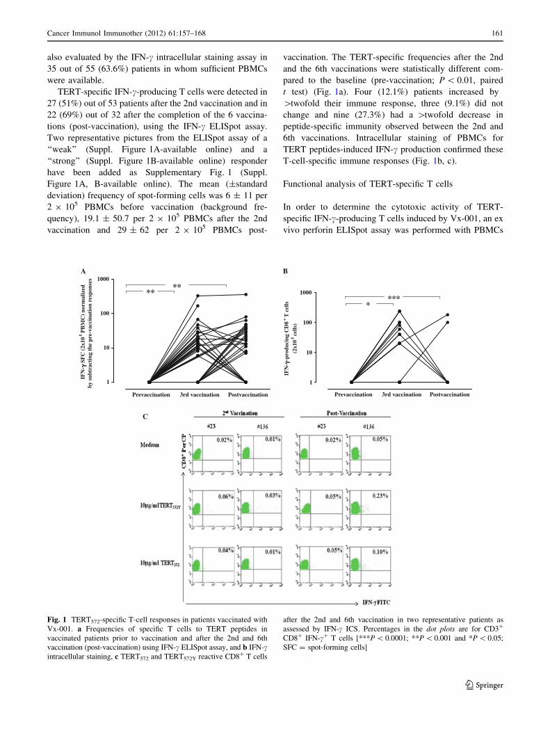
TERT-specific IFN-c-producing T cells were detected in
27 (51%) out of 53 patients after the 2nd vaccination and in
22 (69%) out of 32 after the completion of the 6 vaccina-
tions (post-vaccination), using the IFN-c ELISpot assay.
Two representative pictures from the ELISpot assay of a
‘‘weak’’ (Suppl. Figure 1A-available online) and a
‘‘strong’’ (Suppl. Figure 1B-available online) responder
have been added as Supplementary Fig. 1 (Suppl.
Figure 1A, B-available online). The mean (±standard
deviation) frequency of spot-forming cells was 6 ± 11 per
2 9 105 PBMCs before vaccination (background fre-
quency), 19.1 ± 50.7 per 2 9 105 PBMCs after the 2nd
vaccination and 29 ± 62 per 2 9 105 PBMCs post-
vaccination. The TERT-specific frequencies after the 2nd
and the 6th vaccinations were statistically different com-
pared to the baseline (pre-vaccination; P \ 0.01, paired
t test) (Fig. 1a). Four (12.1%) patients increased by
[twofold their immune response, three (9.1%) did not
change and nine (27.3%) had a [twofold decrease in
peptide-specific immunity observed between the 2nd and
6th vaccinations. Intracellular staining of PBMCs for
TERT peptides-induced IFN-c production confirmed these
T-cell-specific immune responses (Fig. 1b, c).
Functional analysis of TERT-specific T cells
In order to determine the cytotoxic activity of TERT-
specific IFN-c-producing T cells induced by Vx-001, an ex
vivo perforin ELISpot assay was performed with PBMCs
B
1
10
100
1000
****
IFN
- γ-p
rodu
cing
CD
8+ T
cel
ls
(2x1
05
cells
)
C
A
Prevaccination 3rd vaccination PostvaccinationPrevaccination 3rd vaccination Postvaccination
1
10
100
1000
****
IFN
- γ S
FC
(2x
105
PB
MC
) no
rmal
ized
by s
ubtr
acti
ng t
he p
re-v
acci
nati
on r
espo
nses
Fig. 1 TERT572-specific T-cell responses in patients vaccinated with
Vx-001. a Frequencies of specific T cells to TERT peptides in
vaccinated patients prior to vaccination and after the 2nd and 6th
vaccination (post-vaccination) using IFN-c ELISpot assay, and b IFN-cintracellular staining, c TERT572 and TERT572Y reactive CD8? T cells
after the 2nd and 6th vaccination in two representative patients as
assessed by IFN-c ICS. Percentages in the dot plots are for CD3?
CD8? IFN-c? T cells [***P \ 0.0001; **P \ 0.001 and *P \ 0.05;
SFC = spot-forming cells]
Cancer Immunol Immunother (2012) 61:157–168 161
123

from 6 selected patients for whom biological samples were
available from post-vaccination time point. Patients’
demographics are presented in Supplementary Table 1. For
all those 6 patients, an immunological response had pre-
viously been demonstrated by the IFN-c ELISpot assay.
Results showed that five (83%) patients presented CTL
activity with the ability to specifically produce detectable
levels of perforin ex vivo in the presence of TERT572
peptide (Fig. 2a). Three of these six patients were also able
to produce perforin in response to the optimized TERT572Y
peptide. In one patient, T-cell-specific perforin release
could not be detected in response to either peptide.
In addition, the functional specificity of sorted
hTERT572Y-tetramer? CD8? T cells from one vaccinated
patient was assessed in recognizing and killing of TERT-
expressing cells by a chromium-release assay. The TERT?/
HLA-A*0201? (N18/TERT and NA8) but not the TERT-/
HLA-A*0201? (N418 and Me290) cells lines were lysed
by hTERT572Y-tetramer sorted CD8? T cells (Fig. 2b).
Immune responses according to disease stage
and clinical status
In patients with locally advanced disease (stage III), TERT-
specific IFN-c-producing T cells could be detected in three
out of four patients after the 2nd vaccination and in two out
of three patients after the completion of the 6-vaccination
protocol. One patient significantly enhanced its immune
response, whereas one had a decrease in peptide-specific
immunity after more than 3 vaccinations. Moreover, in
patients with stage IV disease, TERT-specific IFN-c-pro-
ducing T cells were induced in 24 (49%) out of 49 patients
after the 2nd vaccination and 20 (69%) out of 29 patients
after the completion of the vaccination protocol (Fig. 3a).
Three (10%) patients increased further their immune
response, three (10%) did not change, and seven (24%) had
a twofold decrease in peptide-specific immunity observed
between the 3rd and after the 6th vaccination. The TERT-
specific frequencies after the 2nd and the 6th vaccination
were statistically different compared to the baseline (pre-
vaccination; P \ 0.001, paired t test).
Vx-001 was similarly immunogenic in patients who
entered the vaccination protocol with either stable (SD) or
progressive disease (PD), as almost 70% of the patients had
developed TERT-specific IFN-c-producing T cells after the
completion of the 6-vaccination protocol (Fig. 3b).
Kinetics of TERT-specific T-cell response
PBMCs from 12 random patients were also collected
before each vaccination dose in order to assess the kinetics
of induction of peptide-specific T-cell responses. As shown
in Fig. 4 A, the induction of peptide-specific immune
response varied from patient to patient; however, the
majority of patients mounted an immune response after the
2nd administration of the TERT572Y peptide. Patients were
also able to generate an immune response at different time
points during the course of the 6-vaccination protocol,
but no patient developed an immune response after
the administration of only the first vaccination dose. The
magnitude of vaccine-induced T-cell response after the
completion of the 6 vaccinations was similar in all patients
independently of the time of the induction of the TERT-
specific immune response.
Moreover, prolonged vaccination maintained the num-
ber of peptide-specific CD8? T cells in nine (82%) out of
11 patients who received boost vaccinations with the native
TERT572 peptide as assessed by IFN-c ELISpot assay
(Fig. 4b) and IFN-c intracellular staining (Fig. 4c).
Immune response and clinical outcome
In order to determine whether there is an association
between the development of TERT-specific IFN-c immune
reactivity and PFS as well as OS, the outcome of patients
enrolled onto the vaccination protocol was analyzed.
Overall, there was no significant difference in either PFS or
OS between patients who developed an early (after the 2nd
vaccination) or late (post-vaccination) immunological
response during vaccination versus the ones who did not
(Fig. 5a, b).
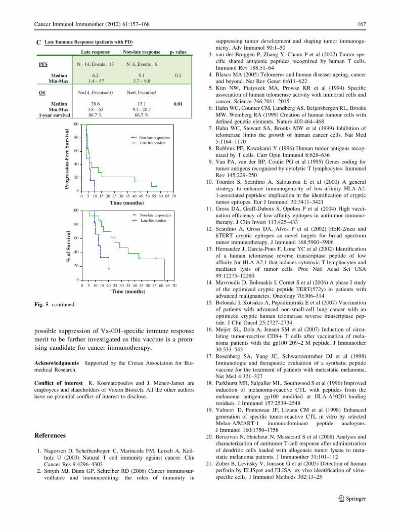
However, in the subgroup analysis of patients who
entered the study with progressive disease, those who
developed a late immune response had a significantly longer
OS compared with that of patients without a post-vaccina-
tion immune response (28.6 months vs. 13.1 months; log-
rank test P = 0.01; Fig. 5c). The enrollment of patients
entering the study with progressive disease was not biased
with regard to the cancer type. The patients had various
cancer types such as renal 13%, breast 13%, cholangio/
pancreas 18%, prostate 26%, ovarian 3%, melanoma 13%,
hepatocellular 5%, colorectal 3%, head and neck 3%, other
3%. Their life expectancy was also variable according to the
disease type and previous treatment but in most cases
estimated to be 6 months or less. In addition, the induction
of either early or late immune response versus no-response
(v2 = 8.3, P = 0.5 and v2 = 8.9, P = 0.3, respectively) in
the progressive disease patients was irrespective of their
cancer type.
Discussion
In a previous study, we showed that CD8? T-cell immune
responses could be detected in 22 HLA-A*0201 patients
with advanced NSCLC vaccinated with Vx-001 [15]. In the
162 Cancer Immunol Immunother (2012) 61:157–168
123

present study, we assessed a larger cohort of HLA-A*0201
patients with different (other than NSCLC) types of cancer,
and we analyzed and further characterized the immune
responses induced by the Vx-001 vaccine. The findings of
the current study confirm our previous observations since
the administration of Vx-001 induced specific CD8? T
cells against TERT peptides, which exhibited in vitro
effector functions including IFN-c and perforin production.
Since many tumor antigens are normal non-mutated
‘‘self-proteins,’’ ‘‘dominant’’ peptides derived from these
0
200
400
600
800
1000
1200
***
‡
# 48
Per
fori
n SF
C/5
x10
5P
BM
C
0
250
500
750
1000
**
# 50
Per
fori
n SF
C/5
x10
5P
BM
C
0
200
400
600
800
1000
1200
*****
# 79
Per
fori
n SF
C/5
x10
5P
BM
C
Medium 10 μg/ml TERT572Y 10 μg/ml TERT572 5 ng/ml SEB Medium 10 μg/ml TERT572Y 10 μg/ml TERT572 5 ng/ml SEB
Medium 10 μg/ml TERT572Y 10 μg/ml TERT572 5 ng/ml SEBMedium 10 μg/ml TERT572Y 10 μg/ml TERT572 5 ng/ml SEB
0
200
400
600
800A
B
*
# 58
Per
ofri
n SF
C/5
x10
5P
BM
C
Fig. 2 a Frequencies of cytotoxic-specific T cells to TERT572 peptide
as assessed by perforin production in the post-vaccination samples of
four representative patients using ELISpot. Dashed line represents the
threshold for ‘‘positive’’ response [***P \ 0.0001; **P \ 0.001 and
*P \ 0.05; SFC = spot-forming cells] b The lytic activity of the
sorted TERT572Y-tetramer?/CD8? T cells from one strongly respond-
ing patient was assessed by a chromium-release assay against TERT?
and TERT- cell lines
Cancer Immunol Immunother (2012) 61:157–168 163
123

proteins are often affected by T-cell tolerance; therefore,
‘‘cryptic’’ peptides derived from these proteins could be
better candidates for inducing an anti-tumor immune
response since, due to their low affinity and recognition
efficiency, they are not affected by tolerance and/or igno-
rance. In previous studies, we have shown that the substi-
tution of an arginine by a tyrosine at the position 1 of the
‘‘native’’ TERT572 peptide significantly increases the
immunogenicity of the ‘‘cryptic’’ peptide [5]; such modi-
fied peptides have also been used by other groups in order
to induce a stronger T-cell activation than that induced by
the native peptides [16–19].
In the present study, patients were vaccinated with two
doses of the ‘‘optimized’’ peptide (TERT572Y) followed by
four doses with the ‘‘native’’ peptide (TERT572). This
vaccination schedule was based on in vivo preclinical
studies which have shown that vaccination of HLA-A*0201
transgenic HHD mice with the optimized TERT572Y fol-
lowed by the native TERT572 peptide induced CTLs with
higher avidity and stronger anti-tumor efficacy than serial
vaccinations with the optimized TERT572Y peptide alone
[11]. This could be explained by the hypothesis that the
optimized TERT572Y peptide first generates peptide-specific
T cells and then the stimulation with the native TERT572
peptide selects among T cells those with the highest spec-
ificity for the native TERT572 peptide that is presented by
tumor cells. Indeed, the presented data demonstrate that
TERT-specific immune responses could be induced in 51
and 70% of the vaccinated patients after the 2nd and
6th vaccinations, respectively, as assessed by both IFN-cELISpot and intracellular cytokine staining assays (Fig. 1);
these findings clearly indicate that our vaccination strategy
efficiently circumvented the presumed immune tolerance
against TERT [8, 10]. Furthermore, the specific T cells
present in blood after the completion of the 6 vaccinations
were of high TCR avidity, relative to the native peptide,
compared to those present after the 2nd vaccination, which
were of low avidity (data under publication).
The induction of TERT572-specific immune response
was independent of the stage of disease or the disease
clinical status at enrollment; moreover, the kinetics of
immune response varied from patient to patient since some
A
B
1
10
100
1000 Locally advanced
1
10
100
1000
******
Metastatic
1
10
100
1000PD Pre-vaccination
******
Prevaccination 3rd vaccination Postvaccination Prevaccination 3rd vaccination Postvaccination
Prevaccination 3rd vaccination PostvaccinationPrevaccination 3rd vaccination Postvaccination
1
10
100
1000 SD Pre-vaccination
*****
IFN
-γ
SFC
(2x
105
PB
MC
)IF
N-
γ SF
C (
2x10
5P
BM
C)
IFN
-γ
SFC
(2x
105
PB
MC
)IF
N-
γ SF
C (
2x10
5P
BM
C)
Fig. 3 TERT572-specific T-cell responses according to disease stage
and disease status before study entry and disease evolution in
vaccinated patients. Magnitude of T-cell response to TERT572 peptide
in vaccinated patients prior to vaccination and after the 2nd and 6th
vaccinations (post-vaccination) according to the a disease stage and
b disease status using ELISpot. [SFC = spot-forming cells;
[***P \ 0.0001; **P \ 0.002, paired t test]
164 Cancer Immunol Immunother (2012) 61:157–168
123

patients required more than two doses of the vaccine in
order to mount a detectable immune response (Fig. 4a), as
already reported with other vaccines [20]. Furthermore,
boost vaccinations with the native peptide resulted in the
maintenance of specific immune response that had been
induced by the 6-vaccination schedule (Fig. 4b, c). These
TERT-specific T cells were functional, since they specifi-
cally released perforin following stimulation with
TERT peptides (Fig. 2a); it is well established that the
perforin ELISpot correlates with cytotoxicity assays [21].
hTERT572Y-tetramer? CD8? T cells from one vaccinated
patient were able to kill TERT-expressing tumor cells
(Fig. 2b).
In the current study, we observed that in some patients
the vaccine-induced TERT-reactive T cells detected in
the blood after the 2nd vaccination disappeared after the
completion of the 6-vaccination protocol (Fig. 1a); this
observation clearly suggests that either the CTLs
migrated to the tumor sites and therefore became unde-
tectable in the blood or were subjected to cell death. It
has been previously reported by two different groups that
tumor-reactive T cells could be easily detected in the
skin and lymph node biopsies but not in the blood of
patients after vaccination [22, 23]. Moreover, Zaks et al.
[24] reported that the re-stimulation of T cells at the
peak of their expansion or activation may cause activa-
tion-induced cell death. Recent studies have proposed
that one mechanism of immune escape used by the
tumors is the production of immunosuppressive type II
cytokines at the tumor sites [25–27]. Alternatively, we
cannot exclude an involvement of other homeostatic
mechanisms such as enhanced expression of surface
CTLA-4 (cytolytic T lymphocyte-associated antigen 4)
molecule, which has higher affinity and effectively
competes with CD28 for B7.1 and B7.2 binding, thus
inducing inhibitory signals to effector T cells [28–33] or
an increased expansion of T regulatory cells (Tregs) that
can suppress effector T cells [34–38]. In addition, the
decreased immune response observed in a proportion of
patients who completed the six vaccinations could be due
to initial stimulation of the immune response by the
modified peptide that was subsequently faded off due to
further stimulation by the native peptide. Hence, con-
sidering all the above possible mechanisms, those
responsible for explaining our findings need to be
investigated further. This could not be achieved in the
present study due to the complete utilization of the
biological material collected from patients.
A
Prevaccination
2nd vaccination
3rd vaccination
4th vaccination
5th vaccination
postvaccination
1
10
100GARBZIAKKANDSERAFSTEFNIKHTGELEMAOLDRONMIRISARIMAT
IFN
- γ S
FC
(2x
105
PB
MC
)
B
Prevac
cinati
on
3rd v
accin
ation
Postva
ccina
tion
1st R
evac
2nd R
evac
3rd R
evac
4th R
evac
5th R
evac
6th R
evac
7th R
evac
8th R
evac
9th R
evac
10th
Revac
11th
Revac
12th
Revac
13th
Revac
14th
Revac
15th
Revac
1
10
100
1000
13/MAIO
34/KOSI
44/NIKHT
45/NIKA
48/PEMA
60/MARE
111/DRON
167/FOTO
168/ALATH
201/KNIK
59/HRPA
IFN
- γ S
FC
(2x
105
PB
MC
)C
Fig. 4 Kinetics of TERT-specific T-cell response development after
immunization. TERT-specific immune response during the course of
six cycles of vaccination a and in boosted patients b as assessed by
IFN-c ELISpot assay c Immune response in two representative
boosted patients after the 8th (#45) and 10th (#13) boosts as assessed
by IFN-c ICS. The data in the graphs are presented as the mean value
of 3 independent experiments. Background frequencies have been
subtracted. [SFC = spot-forming cells]
Cancer Immunol Immunother (2012) 61:157–168 165
123

Finally, we observed a significant correlation between
late (after the 6th vaccination) TERT-specific IFN-cimmune response and overall survival of vaccinated
patients who entered the study with progressive disease.
Indeed, late immune responders had a significantly better
overall survival compared to that of non-responding
patients (Fig. 5c). This observation seems to indicate that
the failure of induction of immune response at the end of
vaccination protocol may define a subgroup of patients
who cannot derive a clinical benefit from the vaccination.
However, this observation should be interpreted with
caution since the present study was not designed to
investigate this question and the patient population was
very heterogeneous in terms of type of cancer and pre-
vious treatments that had been administered. Alterna-
tively, the observed survival difference may merely reflect
the overall better clinical status of patients including their
ability to mount an immune response following Vx-001
vaccination.
In summary, the results of the current study demonstrate
that Vx-001 is able to induce a TERT-specific immune
response in vaccinated patients with different types of solid
tumors and irrespective of the stage of disease and clinical
status. The mechanisms regulating the induction or
A Early Immune Response (all patients)
Early response Non-Early response p- value
PFS
MedianMin-Max
N= 28, Events= 27
4.11.4 – 53
N=25, Events=22
4.81.4 – 57
0.9
OS
MedianMin-Max
1-year survival
N=28, Events=22
203.9 – 62.8
71.4 %
N=25, Events=18
17.11.7 – 57
60%
0.8
0 5 10 15 20 25 30 35 40 45 50 55 60 650
20
40
60
80
100Non-early responders
Early Responders
Time (months)
Pro
gres
sion
-Fre
eS
urv
ival
0 5 10 15 20 25 30 35 40 45 50 55 60 650
20
40
60
80
100Non-early responders
Early Responders
Time (months)
%of
Surv
ival
B Late Immune Response (all patients)
Late response Non-late response p- value
PFS
MedianMin-Max
N= 23, Events= 19
6.53.7 – 57
N=9, Events= 9
6.43.7 – 15.7
0.2
OS
MedianMin-Max
1-year survival
N=23, Events=15
24.96 – 62.891.3 %
N=9, Events=7
16.49.4– 56.4
63 %
0.2
0 5 10 15 20 25 30 35 40 45 50 55 60 65 700
20
40
60
80
100Non-late respondersLate Responders
Time (months)P
rogr
essi
on-F
ree
Su
rviv
al
0 5 10 15 20 25 30 35 40 45 50 55 60 65 70
0
20
40
60
80
100Non-late respondersLate Responders
Time (months)
%of
Surv
ival
Fig. 5 Progression-free and overall survival of all vaccinated
patients. The progression-free and overall survival of all patients
was assessed according to the presence (green line) of absence (blueline) of TERT-specific immune response after the 2nd vaccination (a)
and at the completion of the 6-vaccination protocol (b) Overall
survival of vaccinated patients with PD at study entry according to
post-vaccination immune response (c)
166 Cancer Immunol Immunother (2012) 61:157–168
123
possible suppression of Vx-001-specific immune response
merit to be further investigated as this vaccine is a prom-
ising candidate for cancer immunotherapy.
Acknowledgments Supported by the Cretan Association for Bio-
medical Research.
Conflict of interest K. Kosmatopoulos and J. Menez-Jamet are
employees and shareholders of Vaxon Biotech. All the other authors
have no potential conflict of interest to disclose.
References
1. Nagorsen D, Scheibenbogen C, Marincola FM, Letsch A, Keil-
holz U (2003) Natural T cell immunity against cancer. Clin
Cancer Res 9:4296–4303
2. Smyth MJ, Dunn GP, Schreiber RD (2006) Cancer immunosur-
veillance and immunoediting: the roles of immunity in
suppressing tumor development and shaping tumor immunoge-
nicity. Adv Immunol 90:1–50
3. van der Bruggen P, Zhang Y, Chaux P et al (2002) Tumor-spe-
cific shared antigenic peptides recognized by human T cells.
Immunol Rev 188:51–64
4. Blasco MA (2005) Telomeres and human disease: ageing, cancer
and beyond. Nat Rev Genet 6:611–622
5. Kim NW, Piatyszek MA, Prowse KR et al (1994) Specific
association of human telomerase activity with immortal cells and
cancer. Science 266:2011–2015
6. Hahn WC, Counter CM, Lundberg AS, Beijersbergen RL, Brooks
MW, Weinberg RA (1999) Creation of human tumour cells with
defined genetic elements. Nature 400:464–468
7. Hahn WC, Stewart SA, Brooks MW et al (1999) Inhibition of
telomerase limits the growth of human cancer cells. Nat Med
5:1164–1170
8. Robbins PF, Kawakami Y (1996) Human tumor antigens recog-
nized by T cells. Curr Opin Immunol 8:628–636
9. Van PA, van der BP, Coulie PG et al (1995) Genes coding for
tumor antigens recognized by cytolytic T lymphocytes. Immunol
Rev 145:229–250
10. Tourdot S, Scardino A, Saloustrou E et al (2000) A general
strategy to enhance immunogenicity of low-affinity HLA-A2.
1-associated peptides: implication in the identification of cryptic
tumor epitopes. Eur J Immunol 30:3411–3421
11. Gross DA, Graff-Dubois S, Opolon P et al (2004) High vacci-
nation efficiency of low-affinity epitopes in antitumor immuno-
therapy. J Clin Invest 113:425–433
12. Scardino A, Gross DA, Alves P et al (2002) HER-2/neu and
hTERT cryptic epitopes as novel targets for broad spectrum
tumor immunotherapy. J Immunol 168:5900–5906
13. Hernandez J, Garcia-Pons F, Lone YC et al (2002) Identification
of a human telomerase reverse transcriptase peptide of low
affinity for HLA A2.1 that induces cytotoxic T lymphocytes and
mediates lysis of tumor cells. Proc Natl Acad Sci USA
99:12275–12280
14. Mavroudis D, Bolonakis I, Cornet S et al (2006) A phase I study
of the optimized cryptic peptide TERT(572y) in patients with
advanced malignancies. Oncology 70:306–314
15. Bolonaki I, Kotsakis A, Papadimitraki E et al (2007) Vaccination
of patients with advanced non-small-cell lung cancer with an
optimized cryptic human telomerase reverse transcriptase pep-
tide. J Clin Oncol 25:2727–2734
16. Meijer SL, Dols A, Jensen SM et al (2007) Induction of circu-
lating tumor-reactive CD8? T cells after vaccination of mela-
noma patients with the gp100 209–2 M peptide. J Immunother
30:533–543
17. Rosenberg SA, Yang JC, Schwartzentruber DJ et al (1998)
Immunologic and therapeutic evaluation of a synthetic peptide
vaccine for the treatment of patients with metastatic melanoma.
Nat Med 4:321–327
18. Parkhurst MR, Salgaller ML, Southwood S et al (1996) Improved
induction of melanoma-reactive CTL with peptides from the
melanoma antigen gp100 modified at HLA-A*0201-binding
residues. J Immunol 157:2539–2548
19. Valmori D, Fonteneau JF, Lizana CM et al (1998) Enhanced
generation of specific tumor-reactive CTL in vitro by selected
Melan-A/MART-1 immunodominant peptide analogues.
J Immunol 160:1750–1758
20. Bercovici N, Haicheur N, Massicard S et al (2008) Analysis and
characterization of antitumor T-cell response after administration
of dendritic cells loaded with allogeneic tumor lysate to meta-
static melanoma patients. J Immunother 31:101–112
21. Zuber B, Levitsky V, Jonsson G et al (2005) Detection of human
perforin by ELISpot and ELISA: ex vivo identification of virus-
specific cells. J Immunol Methods 302:13–25
C Late Immune Response (patients with PD)
Late response Non-late response p- value
PFS
MedianMin-Max
N= 14, Events= 13
6.21.4 – 57
N=6, Events= 6
5.13.7 – 9.8
0.1
OS
MedianMin-Max
1-year survival
N=14, Events=10
28.63.8 – 6386.7 %
N=6, Events=5
13.19.4– 20.766.7 %
0.01
0 5 10 15 20 25 30 35 40 45 50 55 60 65 700
20
40
60
80
100
Non-late respondersLate Responders
Time (months)
Pro
gres
sion
-Fre
e Su
rviv
al
0 5 10 15 20 25 30 35 40 45 50 55 60 65 700
20
40
60
80
100Non-late respondersLate Responders
Time (months)
% o
f Su
rviv
al
Fig. 5 continued
Cancer Immunol Immunother (2012) 61:157–168 167
123

22. De Vries IJM, Lesterhuis WJ, Barentsz JO et al (2005) Magnetic
resonance tracking of dendritic cells in melanoma patients for
monitoring of cellular therapy. Nat Biotechnol 23:1407–1413
23. Slingluff CL Jr, Petroni GR, Yamshchikov GV et al (2004)
Immunologic and clinical outcomes of vaccination with a mul-
tiepitope melanoma peptide vaccine plus low-dose interleukin-2
administered either concurrently or on a delayed schedule. J Clin
Oncol 22:4474–4485
24. Zaks TZ, Chappell DB, Rosenberg SA, Restifo NP (1999)
Fas-mediated suicide of tumor-reactive T cells following acti-
vation by specific tumor: selective rescue by caspase inhibition.
J Immunol 162:3273–3279
25. Aruga A, Aruga E, Tanigawa K, Bishop DK, Sondak VK, Chang
AE (1997) Type 1 versus type 2 cytokine release by Vbeta T cell
subpopulations determines in vivo antitumor reactivity: IL-10
mediates a suppressive role. J Immunol 159:664–673
26. Lattime EC, Mastrangelo MJ, Bagasra O, Li W, Berd D (1995)
Expression of cytokine mRNA in human melanoma tissues.
Cancer Immunol Immunother 41:151–156
27. Yang AS, Lattime EC (2003) Tumor-induced interleukin 10
suppresses the ability of splenic dendritic cells to stimulate CD4
and CD8 T-cell responses. Cancer Res 63:2150–2157
28. Gabriel EM, Lattime EC (2007) Anti-CTL-associated antigen 4:
are regulatory T cells a target? Clin Cancer Res 13:785–788
29. Hodi FS, Mihm MC, Soiffer RJ et al (2003) Biologic activity of
cytotoxic T lymphocyte-associated antigen 4 antibody blockade
in previously vaccinated metastatic melanoma and ovarian car-
cinoma patients. Proc Natl Acad Sci USA 100:4712–4717
30. Korman A, Yellin M, Keler T (2005) Tumor immunotherapy:
preclinical and clinical activity of anti-CTLA4 antibodies. Curr
Opin Investig Drugs 6:582–591
31. Krummel MF, Allison JP (1995) CD28 and CTLA-4 have
opposing effects on the response of T cells to stimulation. J Exp
Med 182:459–465
32. Phan GQ, Yang JC, Sherry RM et al (2003) Cancer regression
and autoimmunity induced by cytotoxic T lymphocyte-associated
antigen 4 blockade in patients with metastatic melanoma. Proc
Natl Acad Sci USA 100:8372–8377
33. Sun J, Schiffman J, Raghunath A, Ng TD, Chen H, Sharma P
(2008) Concurrent decrease in IL-10 with development of
immune-related adverse events in a patient treated with anti-
CTLA-4 therapy. Cancer Immun 8:9
34. Ling KL, Pratap SE, Bates GJ et al (2007) Increased frequency of
regulatory T cells in peripheral blood and tumour infiltrating
lymphocytes in colorectal cancer patients. Cancer Immun 7:7
35. O’Mahony D, Morris JC, Quinn C et al (2007) A pilot study of
CTLA-4 blockade after cancer vaccine failure in patients with
advanced malignancy. Clin Cancer Res 13:958–964
36. Sakaguchi S (2000) Regulatory T cells: key controllers of
immunologic self-tolerance. Cell 101:455–458
37. Sakaguchi S (2005) Naturally arising Foxp3-expressing CD25?
CD4? regulatory T cells in immunological tolerance to self and
non-self. Nat Immunol 6:345–352
38. Yamaguchi T, Sakaguchi S (2006) Regulatory T cells in immune
surveillance and treatment of cancer. Semin Cancer Biol
16:115–123
168 Cancer Immunol Immunother (2012) 61:157–168
123
Copyright © 2022 FDOKUMEN


![NO Q RSTUVWXYZW [V \Z]]^XYZW _``Y[VWXV]]V VX ...](https://static.fdokumen.com/doc/165x107/633db03fa7b2edc6000ec873/no-q-rstuvwxyzw-v-zxyzw-yvwxvv-vx-.jpg)