Geometrically Repatterned Immunological Synapses Uncover Formation Mechanisms
8
Geometrically Repatterned Immunological Synapses Uncover Formation Mechanisms Marc Thilo Figge 1* , Michael Meyer-Hermann 2 1 Institute for Theoretical Physics, Johann Wolfgang Goethe University, Frankfurt am Main, Germany 2 Frankfurt Institute for Advanced Studies, Johann Wolfgang Goethe University, Frankfurt am Main, Germany The interaction of T cells and antigen-presenting cells is central to adaptive immunity and involves the formation of immunological synapses in many cases. The surface molecules of the cells form a characteristic spatial pattern whose formation mechanisms and function are largely unknown. We perform computer simulations of recent experiments on geometrically repatterned immunological synapses and explain the emerging structure as well as the formation dynamics. Only the combination of in vitro experiments and computer simulations has the potential to pinpoint the kind of interactions involved. The presented simulations make clear predictions for the structure of the immunological synapse and elucidate the role of a self-organizing attraction between complexes of T cell receptor and peptide–MHC molecule, versus a centrally directed motion of these complexes. Citation: Figge MT, Meyer-Hermann M (2006) Geometrically repatterned immunological synapses uncover formation mechanisms. PLoS Comput Biol 2(11): e171. doi:10.1371/journal.pcbi.0020171 Introduction The recognition of pathogens by the T cells of the immune system relies on antigen-presenting cells (APCs) that process pathogen-derived molecules and present them with major histocompatibility complex (MHC) molecules. The surface of APCs is scanned by T cells that bind to peptide–MHC (pMHC) complexes with their specific T cell receptors (TCRs). This interaction can initiate the dynamic formation of an immunological synapse (IS), which is an adhesive junction with a nanometer scale gap between the two cells [1–3]. Depending on the cellular partners, the IS can adopt different topologies. A fixed plan for a stable common structure does not exist but rather a diversity of structures dictated by the diversity of interacting cells [1]. The prototypical IS matures within minutes into a well- organized structure with a characteristic bull’s-eye pattern that may remain stable for hours [2,3]. This pattern is composed of an outer ring, which is referred to as peripheral supramolecular activation cluster (p-SMAC), consisting of bound complexes of the T cell’s adhesion molecule leukocyte function–associated antigen-1 (LFA-1) and the APC’s inter- cellular adhesion molecule-1 (ICAM-1). The center of the IS, the central supramolecular activation cluster (c-SMAC), consists of bound TCR–pMHC complexes. The hypothesis that this pattern may enhance and sustain TCR signaling and thus the T cell activation has become a matter of controversy during recent years [4–6]. According to these measurements on naive T cells, TCR signaling occurs primarily at the periphery of the synapse and is ceasing before a c-SMAC has formed. Therefore, the bull’s-eye pattern might well be the signature of cell–cell interaction rather than a necessary condition for information processing. Recently, K. D. Mossman et al. [5] performed in vitro experiments in which the IS between a living T cell and a synthetic surface that acts as an artificial APC was geometri- cally repatterned. The repatterning of the IS is enforced by inhibiting the movement of TCR–pMHC and LFA-1–ICAM-1 receptor–ligand complexes in the bilayer across artificially imposed nanometer-scale chromium barriers within the synthetic surface. A schematic cross-section representation of the T cell–synthetic APC interface is shown in Figure 1, which indicates the impact of barriers on the molecular organization of ISs. Various aspects of the IS have been successfully analyzed by in silico experiments [7], which allow with relative ease manipulation of each part of a system individually and monitoring of its impact on the system as a whole. Different approaches and theoretical models for the IS are summarized in a recent review [8]. The model for dynamical IS pattern formation by S. Y. Qi et al. [9] is based on a set of partial differential equations and has taken the role of a standard model, which has been frequently used in modified versions as a starting point of later studies [10–13]. Dynamical aspects of the IS pattern formation were recently also studied with an agent-based approach [6,14,15]. In this approach, receptor– ligand complexes are treated as discrete objects (agents) that move and interact with each other on a lattice representing the spatial surrounding. In particular, Weikl et al. [14,15] introduced a model that describes the c-SMAC formation under the assumption of centrally directed TCR–pMHC motion. It is based on a Hamiltonian containing contribu- tions of the elastic energy of the membrane and interaction energies of the receptors, ligands, and glycoproteins. The T Editor: Sebastian Bonhoeffer, Eidgeno ¨ ssische Technische Hochschule-Zu ¨ rich, Switzerland Received June 6, 2006; Accepted October 4, 2006; Published November 10, 2006 Copyright: Ó 2006 Figge and Meyer-Hermann. This is an open-access article distributed under the terms of the Creative Commons Attribution License, which permits unrestricted use, distribution, and reproduction in any medium, provided the original author and source are credited. Abbreviations: APC, antigen-presenting cell; c-SMAC, central supramolecular activation cluster; ICAM-1, intercellular adhesion molecule-1; IS, immunological synapse; LFA-1, leukocyte function–associated antigen-1; MHC, major histocomp- ability complex; pMHC, peptide–major histocompability complex; p-SMAC, peripheral supramolecular activation cluster; TCR, T cell receptor * To whom correspondence should be addressed. E-mail: [email protected]. de PLoS Computational Biology | www.ploscompbiol.org November 2006 | Volume 2 | Issue 11 | e171 0001
Transcript of Geometrically Repatterned Immunological Synapses Uncover Formation Mechanisms

Geometrically Repatterned ImmunologicalSynapses Uncover Formation MechanismsMarc Thilo Figge
1*, Michael Meyer-Hermann
2
1 Institute for Theoretical Physics, Johann Wolfgang Goethe University, Frankfurt am Main, Germany 2 Frankfurt Institute for Advanced Studies, Johann Wolfgang Goethe
University, Frankfurt am Main, Germany
The interaction of T cells and antigen-presenting cells is central to adaptive immunity and involves the formation ofimmunological synapses in many cases. The surface molecules of the cells form a characteristic spatial pattern whoseformation mechanisms and function are largely unknown. We perform computer simulations of recent experiments ongeometrically repatterned immunological synapses and explain the emerging structure as well as the formationdynamics. Only the combination of in vitro experiments and computer simulations has the potential to pinpoint thekind of interactions involved. The presented simulations make clear predictions for the structure of the immunologicalsynapse and elucidate the role of a self-organizing attraction between complexes of T cell receptor and peptide–MHCmolecule, versus a centrally directed motion of these complexes.
Citation: Figge MT, Meyer-Hermann M (2006) Geometrically repatterned immunological synapses uncover formation mechanisms. PLoS Comput Biol 2(11): e171.doi:10.1371/journal.pcbi.0020171
Introduction
The recognition of pathogens by the T cells of the immunesystem relies on antigen-presenting cells (APCs) that processpathogen-derived molecules and present them with majorhistocompatibility complex (MHC) molecules. The surface ofAPCs is scanned by T cells that bind to peptide–MHC(pMHC) complexes with their specific T cell receptors (TCRs).This interaction can initiate the dynamic formation of animmunological synapse (IS), which is an adhesive junctionwith a nanometer scale gap between the two cells [1–3].Depending on the cellular partners, the IS can adoptdifferent topologies. A fixed plan for a stable commonstructure does not exist but rather a diversity of structuresdictated by the diversity of interacting cells [1].
The prototypical IS matures within minutes into a well-organized structure with a characteristic bull’s-eye patternthat may remain stable for hours [2,3]. This pattern iscomposed of an outer ring, which is referred to as peripheralsupramolecular activation cluster (p-SMAC), consisting ofbound complexes of the T cell’s adhesion molecule leukocytefunction–associated antigen-1 (LFA-1) and the APC’s inter-cellular adhesion molecule-1 (ICAM-1). The center of the IS,the central supramolecular activation cluster (c-SMAC),consists of bound TCR–pMHC complexes. The hypothesisthat this pattern may enhance and sustain TCR signaling andthus the T cell activation has become a matter of controversyduring recent years [4–6]. According to these measurementson naive T cells, TCR signaling occurs primarily at theperiphery of the synapse and is ceasing before a c-SMAC hasformed. Therefore, the bull’s-eye pattern might well be thesignature of cell–cell interaction rather than a necessarycondition for information processing.
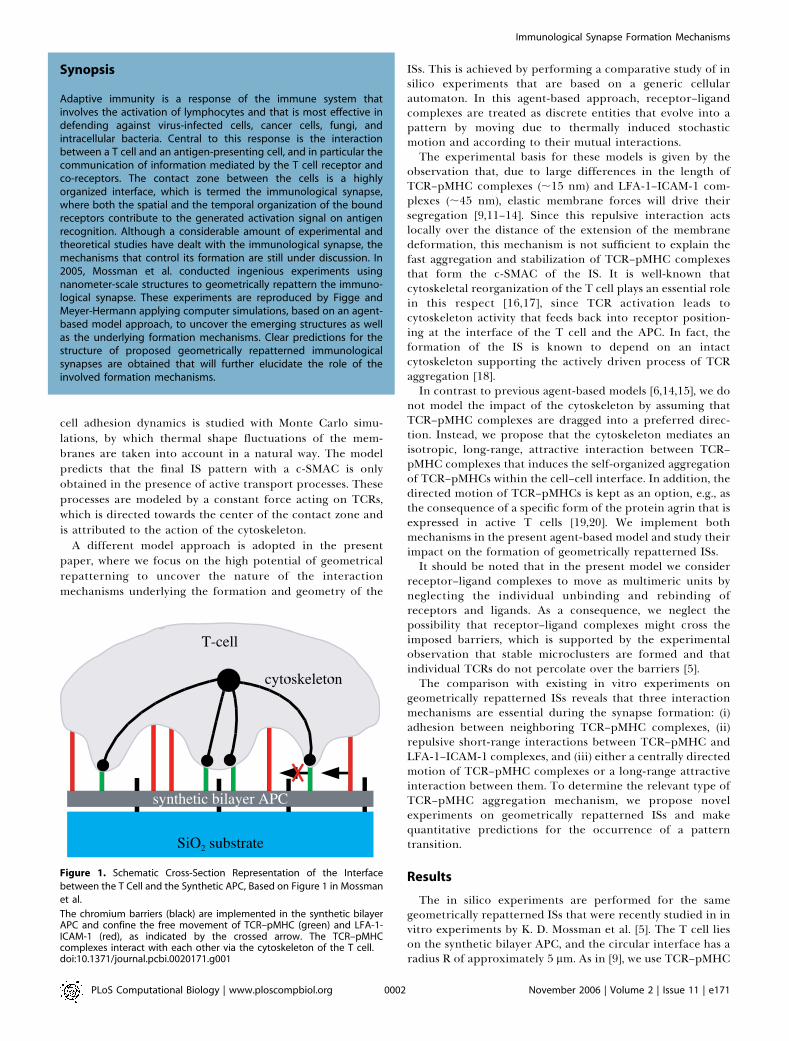
Recently, K. D. Mossman et al. [5] performed in vitroexperiments in which the IS between a living T cell and asynthetic surface that acts as an artificial APC was geometri-cally repatterned. The repatterning of the IS is enforced byinhibiting the movement of TCR–pMHC and LFA-1–ICAM-1receptor–ligand complexes in the bilayer across artificially
imposed nanometer-scale chromium barriers within thesynthetic surface. A schematic cross-section representationof the T cell–synthetic APC interface is shown in Figure 1,which indicates the impact of barriers on the molecularorganization of ISs.Various aspects of the IS have been successfully analyzed by
in silico experiments [7], which allow with relative easemanipulation of each part of a system individually andmonitoring of its impact on the system as a whole. Differentapproaches and theoretical models for the IS are summarizedin a recent review [8]. The model for dynamical IS patternformation by S. Y. Qi et al. [9] is based on a set of partialdifferential equations and has taken the role of a standardmodel, which has been frequently used in modified versionsas a starting point of later studies [10–13]. Dynamical aspectsof the IS pattern formation were recently also studied with anagent-based approach [6,14,15]. In this approach, receptor–ligand complexes are treated as discrete objects (agents) thatmove and interact with each other on a lattice representingthe spatial surrounding. In particular, Weikl et al. [14,15]introduced a model that describes the c-SMAC formationunder the assumption of centrally directed TCR–pMHCmotion. It is based on a Hamiltonian containing contribu-tions of the elastic energy of the membrane and interactionenergies of the receptors, ligands, and glycoproteins. The T
Editor: Sebastian Bonhoeffer, Eidgenossische Technische Hochschule-Zurich,Switzerland
Received June 6, 2006; Accepted October 4, 2006; Published November 10, 2006
Copyright: � 2006 Figge and Meyer-Hermann. This is an open-access articledistributed under the terms of the Creative Commons Attribution License, whichpermits unrestricted use, distribution, and reproduction in any medium, providedthe original author and source are credited.
Abbreviations: APC, antigen-presenting cell; c-SMAC, central supramolecularactivation cluster; ICAM-1, intercellular adhesion molecule-1; IS, immunologicalsynapse; LFA-1, leukocyte function–associated antigen-1; MHC, major histocomp-ability complex; pMHC, peptide–major histocompability complex; p-SMAC,peripheral supramolecular activation cluster; TCR, T cell receptor
* To whom correspondence should be addressed. E-mail: [email protected]
PLoS Computational Biology | www.ploscompbiol.org November 2006 | Volume 2 | Issue 11 | e1710001
cell adhesion dynamics is studied with Monte Carlo simu-lations, by which thermal shape fluctuations of the mem-branes are taken into account in a natural way. The modelpredicts that the final IS pattern with a c-SMAC is onlyobtained in the presence of active transport processes. Theseprocesses are modeled by a constant force acting on TCRs,which is directed towards the center of the contact zone andis attributed to the action of the cytoskeleton.
A different model approach is adopted in the presentpaper, where we focus on the high potential of geometricalrepatterning to uncover the nature of the interactionmechanisms underlying the formation and geometry of the
ISs. This is achieved by performing a comparative study of insilico experiments that are based on a generic cellularautomaton. In this agent-based approach, receptor–ligandcomplexes are treated as discrete entities that evolve into apattern by moving due to thermally induced stochasticmotion and according to their mutual interactions.The experimental basis for these models is given by the
observation that, due to large differences in the length ofTCR–pMHC complexes (;15 nm) and LFA-1–ICAM-1 com-plexes (;45 nm), elastic membrane forces will drive theirsegregation [9,11–14]. Since this repulsive interaction actslocally over the distance of the extension of the membranedeformation, this mechanism is not sufficient to explain thefast aggregation and stabilization of TCR–pMHC complexesthat form the c-SMAC of the IS. It is well-known thatcytoskeletal reorganization of the T cell plays an essential rolein this respect [16,17], since TCR activation leads tocytoskeleton activity that feeds back into receptor position-ing at the interface of the T cell and the APC. In fact, theformation of the IS is known to depend on an intactcytoskeleton supporting the actively driven process of TCRaggregation [18].In contrast to previous agent-based models [6,14,15], we do
not model the impact of the cytoskeleton by assuming thatTCR–pMHC complexes are dragged into a preferred direc-tion. Instead, we propose that the cytoskeleton mediates anisotropic, long-range, attractive interaction between TCR–pMHC complexes that induces the self-organized aggregationof TCR–pMHCs within the cell–cell interface. In addition, thedirected motion of TCR–pMHCs is kept as an option, e.g., asthe consequence of a specific form of the protein agrin that isexpressed in active T cells [19,20]. We implement bothmechanisms in the present agent-based model and study theirimpact on the formation of geometrically repatterned ISs.It should be noted that in the present model we consider
receptor–ligand complexes to move as multimeric units byneglecting the individual unbinding and rebinding ofreceptors and ligands. As a consequence, we neglect thepossibility that receptor–ligand complexes might cross theimposed barriers, which is supported by the experimentalobservation that stable microclusters are formed and thatindividual TCRs do not percolate over the barriers [5].The comparison with existing in vitro experiments on
geometrically repatterned ISs reveals that three interactionmechanisms are essential during the synapse formation: (i)adhesion between neighboring TCR–pMHC complexes, (ii)repulsive short-range interactions between TCR–pMHC andLFA-1–ICAM-1 complexes, and (iii) either a centrally directedmotion of TCR–pMHC complexes or a long-range attractiveinteraction between them. To determine the relevant type ofTCR–pMHC aggregation mechanism, we propose novelexperiments on geometrically repatterned ISs and makequantitative predictions for the occurrence of a patterntransition.
Results
The in silico experiments are performed for the samegeometrically repatterned ISs that were recently studied in invitro experiments by K. D. Mossman et al. [5]. The T cell lieson the synthetic bilayer APC, and the circular interface has aradius R of approximately 5 lm. As in [9], we use TCR–pMHC
Figure 1. Schematic Cross-Section Representation of the Interface
between the T Cell and the Synthetic APC, Based on Figure 1 in Mossman
et al.
The chromium barriers (black) are implemented in the synthetic bilayerAPC and confine the free movement of TCR–pMHC (green) and LFA-1-ICAM-1 (red), as indicated by the crossed arrow. The TCR–pMHCcomplexes interact with each other via the cytoskeleton of the T cell.doi:10.1371/journal.pcbi.0020171.g001
PLoS Computational Biology | www.ploscompbiol.org November 2006 | Volume 2 | Issue 11 | e1710002
Synopsis
Adaptive immunity is a response of the immune system thatinvolves the activation of lymphocytes and that is most effective indefending against virus-infected cells, cancer cells, fungi, andintracellular bacteria. Central to this response is the interactionbetween a T cell and an antigen-presenting cell, and in particular thecommunication of information mediated by the T cell receptor andco-receptors. The contact zone between the cells is a highlyorganized interface, which is termed the immunological synapse,where both the spatial and the temporal organization of the boundreceptors contribute to the generated activation signal on antigenrecognition. Although a considerable amount of experimental andtheoretical studies have dealt with the immunological synapse, themechanisms that control its formation are still under discussion. In2005, Mossman et al. conducted ingenious experiments usingnanometer-scale structures to geometrically repattern the immuno-logical synapse. These experiments are reproduced by Figge andMeyer-Hermann applying computer simulations, based on an agent-based model approach, to uncover the emerging structures as wellas the underlying formation mechanisms. Clear predictions for thestructure of proposed geometrically repatterned immunologicalsynapses are obtained that will further elucidate the role of theinvolved formation mechanisms.
Immunological Synapse Formation Mechanisms
and LFA-1–ICAM-1 densities on the order 40 lm�2 and 100lm�2, respectively. The receptor–ligand complexes performrandom moves within the interface region and interactamong each other. The same diffusion constant, D¼0.06 lm2/s, is chosen for TCR–pMHC and LFA-1–ICAM-1, whichcorresponds to a typical value for these membrane-anchoredmacromolecules [21,22]. We account for the observed TCR–pMHC assembly into microclusters [5] by an adhesive forcebetween direct TCR–pMHC neighbors, and the adhesionstrength is characterized by the parameter a. The repulsive,attractive, and directed interactions are characterized by theinteraction length Li and the relative interaction strength wi
with I ¼ rep, att, and dir, respectively. The details of thecellular automaton are summarized in Materials and Meth-ods.
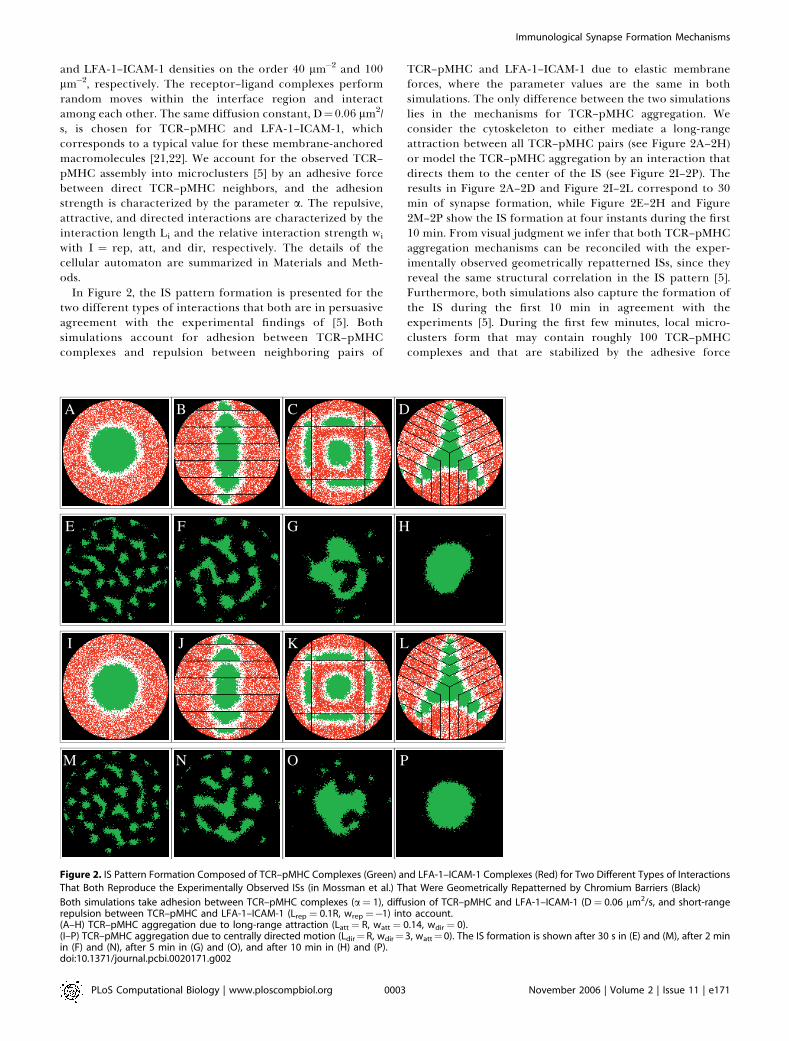
In Figure 2, the IS pattern formation is presented for thetwo different types of interactions that both are in persuasiveagreement with the experimental findings of [5]. Bothsimulations account for adhesion between TCR–pMHCcomplexes and repulsion between neighboring pairs of
TCR–pMHC and LFA-1–ICAM-1 due to elastic membraneforces, where the parameter values are the same in bothsimulations. The only difference between the two simulationslies in the mechanisms for TCR–pMHC aggregation. Weconsider the cytoskeleton to either mediate a long-rangeattraction between all TCR–pMHC pairs (see Figure 2A–2H)or model the TCR–pMHC aggregation by an interaction thatdirects them to the center of the IS (see Figure 2I–2P). Theresults in Figure 2A–2D and Figure 2I–2L correspond to 30min of synapse formation, while Figure 2E–2H and Figure2M–2P show the IS formation at four instants during the first10 min. From visual judgment we infer that both TCR–pMHCaggregation mechanisms can be reconciled with the exper-imentally observed geometrically repatterned ISs, since theyreveal the same structural correlation in the IS pattern [5].Furthermore, both simulations also capture the formation ofthe IS during the first 10 min in agreement with theexperiments [5]. During the first few minutes, local micro-clusters form that may contain roughly 100 TCR–pMHCcomplexes and that are stabilized by the adhesive force
Figure 2. IS Pattern Formation Composed of TCR–pMHC Complexes (Green) and LFA-1–ICAM-1 Complexes (Red) for Two Different Types of Interactions
That Both Reproduce the Experimentally Observed ISs (in Mossman et al.) That Were Geometrically Repatterned by Chromium Barriers (Black)
Both simulations take adhesion between TCR–pMHC complexes (a¼ 1), diffusion of TCR–pMHC and LFA-1–ICAM-1 (D¼ 0.06 lm2/s, and short-rangerepulsion between TCR–pMHC and LFA-1–ICAM-1 (Lrep ¼ 0.1R, wrep¼�1) into account.(A–H) TCR–pMHC aggregation due to long-range attraction (Latt¼ R, watt¼ 0.14, wdir ¼ 0).(I–P) TCR–pMHC aggregation due to centrally directed motion (Ldir¼R, wdir¼ 3, watt¼0). The IS formation is shown after 30 s in (E) and (M), after 2 minin (F) and (N), after 5 min in (G) and (O), and after 10 min in (H) and (P).doi:10.1371/journal.pcbi.0020171.g002
PLoS Computational Biology | www.ploscompbiol.org November 2006 | Volume 2 | Issue 11 | e1710003
Immunological Synapse Formation Mechanisms

between these complexes (see Figure 2E–2F and Figure 2M–2N). Note that microclusters do not form in the absence ofadhesion. In accordance with the experimentally observedtime scale, our model describes the migration of micro-clusters as a whole to the center of the IS, where they coalesceto form a c-SMAC.
It cannot be excluded that other types of interactions arepresent, e.g., adhesive forces between LFA-1–ICAM-1 com-plexes; however, the comparison with the experimentallyobserved geometrically repatterned ISs indicates that theincluded mechanisms are sufficient, are all required, andseem to be the most important ones. In addition, dependingon the precise interaction parameters, a rich variety of ISpatterns is observed. In Figure 3, the results of in silicoexperiments for the same geometrically repatterned ISs as inFigure 2 are presented with various interaction mechanismsbeing changed in a stepwise manner. It is confirmed that eachof the previously considered ingredients delivers an impor-tant contribution to the formation of the geometricallyrepatterned ISs. In particular, the size of the attraction length
Latt has a strong impact on the IS pattern. This can be seen inFigure 3A–3D where the simulation results after 30 min ofsynapse formation are shown for different interaction lengthsLatt, starting from the same simulation parameters as inFigure 2A. It is clearly observed that several TCR–pMHCclusters form in the case of reduced interaction lengths Latt ,
R/2. In the context of immature T cells (thymocytes),multifocal synapse patterns have been attributed to thereduced density of TCRs and thermal fluctuations [10].However, multifocal synapse patterns are also observed formature T cells [23]. We find that a reduced interaction lengthin the attractive long-range interaction between pairs ofTCR–pMHC represents a possible explanation for multifocalsynapse patterns. Note that the IS patterns in Figure 3B–3Dare metastable and may, after an unrealistically long time, stillevolve into a bull’s-eye pattern by diffusion and coalescenceof the clusters.In Figure 3E–3H, the geometrically repatterned ISs are
shown for short-range repulsion between TCR–pMHC andLFA-1–ICAM-1, and for adhesion between direct neighbors
Figure 3. IS Pattern Formation Composed of TCR–pMHC Complexes (Green) and LFA-1–ICAM-1C Complexes (Red) with Various Interaction Mechanisms
Being Changed in a Stepwise Manner
(A–D) Same parameters as in Figure 2A–2D for varying attraction length: (A) Latt ¼ R, (B) Latt¼ 0.43R, (C) Latt¼ 0.29R, and (D) Latt ¼ 0.15R.(E–H) Same parameters as in Figure 2A–2D in the absence of long-range attraction (watt¼ 0) for the geometrically repatterned ISs.(I–L) IS pattern formation in the absence of long-range attraction between TCR–pMHCs (watt¼ 0) and short-range repulsion between TCR–pMHC andLFA-1–ICAM-1 (wrep ¼ 0), and with strong TCR–pMHC adhesion: a¼ 5.(M–P) Same parameters as in Figure 2A–2D in the absence of short-range repulsion (wrep ¼ 0).doi:10.1371/journal.pcbi.0020171.g003
PLoS Computational Biology | www.ploscompbiol.org November 2006 | Volume 2 | Issue 11 | e1710004
Immunological Synapse Formation Mechanisms
of TCR–pMHC complexes, while no long-range attraction asmediated by the cytoskeleton and no directed motion asinduced by proteins are taken into account. The simulationtime corresponds again to 30 min of synapse formation, andit can be seen that the obtained patterns do not reproducethose observed in the experiment. It is intuitively clear that inthe absence of any TCR–pMHC aggregation mechanism a c-SMAC in Figure 3E could only be formed if the TCR–pMHCclusters happen to meet and form larger clusters. Two effectscounteract and retard this process: (i) the repulsion betweenTCR–pMHC and LFA-1–ICAM-1 hinders the coalescence oftwo clusters, and (ii) the larger the clusters become the slowerthey move due to the TCR–pMHC adhesion.
An interesting aspect can be observed for the geometricallyrepatterned ISs in Figure 3F–3H, where TCR–pMHC clustersare found to exist preferentially on opposite sides of barriers.This visualizes the repulsive interaction between TCR–pMHCand LFA-1–ICAM-1 acting across the barrier. Even though aTCR–pMHC cluster that has formed on one side of thebarrier cannot cross this barrier, which is implemented in thesynthetic APC, it will nevertheless counteract membranedeformations in the T cell and by that favor the accumulationof a TCR–pMHC cluster on the other side of this barrier.
The formation of the bull’s-eye pattern is observed if long-and short-range interactions are omitted while adhesionbetween pairs of TCR–pMHC complexes is increased instrength (see Figure 3I–3L). However, it should be noted thateven under these conditions the IS patterns are obtained onlyafter about one day of synapse formation. The unrealisticlong simulation time before the bull’s-eye pattern emerges inFigure 3I is again related to the fact that large clusters ofTCR–pMHC have to be displaced in order to join and form ac-SMAC. In addition, it is found that the bull’s-eye patternonly evolves in a narrow region of TCR–pMHC adhesionaround a ¼ 5. For a , 5, the emergence of the bull’s-eyepattern is prevented by TCR–pMHC diffusion, while for a . 5its formation time increases by orders of magnitude(unpublished data). It should be noted, however, that evenfor a ¼ 5 the geometrically repatterned ISs are not inagreement with the experimental observations in [5]. Theexperimentally observed structural correlations across thebarriers in the geometrically repatterned ISs are absent sincethe various geometric compartments became in fact inde-pendent. This stresses once again the requirement of eitheran attractive long-range interaction between TCR–pMHCsmediated by the cytoskeleton or a centrally directed TCR–pMHC motion induced by proteins to explain the ISformation with its characteristic patterns on a reasonabletime scale.
We finally show in Figure 3M–3P the synapse formationafter 30 min in the presence of adhesion and long-rangeattraction (both as in Figure 2A–2D) but in the absence of theshort-range repulsive interaction that stems from elasticmembrane forces between neighboring TCR–pMHC andLFA-1–ICAM-1 complexes. The in silico experiments repro-duce correctly the experimentally observed patterns. How-ever, a complete segregation between receptors of differentlengths is not found. Once a swelling outer ring of TCR–pMHC is formed, it becomes increasingly unlikely that itbreaks up again to drive more LFA-1–ICAM-1 out of thecenter of the bull’s-eye. In other words, the pattern inversionfrom an outer TCR–pMHC ring into an outer LFA-1–ICAM-1
ring, which has been observed in the early synapse formationof in vitro experiments [4,18,24], is indicative for theimportance of the repulsive interaction between TCR–pMHCand LFA-1–ICAM-1. This statement holds independent of theunderlying aggregation mechanism, i.e., long-range attractionbetween TCR–pMHCs or centrally directed TCR–pMHCmotion.
Discussion
To reproduce the experimentally observed geometricallyrepatterned ISs by in silico experiments, three relevantinteraction mechanisms play an important role: (i) adhesionbetween neighboring TCR–pMHC complexes, (ii) repulsiveshort-range interactions between TCR–pMHC and LFA-1–ICAM-1 complexes, and (iii) either a centrally directedmotion of TCR–pMHC complexes mediated by aggregationproteins, or a long-range attractive interaction betweenTCR–pMHC pairs mediated by the cytoskeleton. To answerthe question by which aggregation mechanism TCR–pMHCsaccumulate at the center of the IS, we propose a conclusiveprocedure that makes once again use of the high potential ofgeometrical repatterning experiments. The difference be-tween an attractive long-range interaction and a directedmotion of TCR–pMHC can be made visible by realizing thatthe former interaction depends in a crucial way on thedistribution of TCR–pMHC complexes, whereas the lattermechanism is governed by the distribution of proteins. It thusfollows that the two mechanisms can be distinguished if thenumber of TCR–pMHC complexes is geometrically confinedin such a way that these mechanisms give rise to clearlydistinguishable IS patterns. We propose experiments wherethe freedom of TCR–pMHC movement is geometricallyconfined by a barrier that subdivides the IS into an innerand an outer region, respectively, with an inner TCR–pMHCnumber, Ni, and an outer TCR–pMHC number, No. Varyingthe size of the inner compartment is accompanied by achange in the ratio No/Ni of the TCR–pMHC numbers in theouter to the inner region. In the presence of directed TCR–pMHC motion, the resulting IS pattern will not changequalitatively as a function of No/Ni. However, we expect thatin the presence of an attractive long-range interactionbetween TCR–pMHCs, the c-SMAC will only form if No ,,
Ni, whereas for No .. Ni the TCR–pMHCs of the innerregion will be attracted towards the geometric boundary. Toprevent the blurring of the desired effect by the repulsionbetween TCR–pMHC and LFA-1–ICAM-1 that is acting acrossthe barrier, as has been discussed in connection with Figure3E–3H, the in silico experiments will be performed for innerregions with linear extensions well above Lrep.In Figure 4 the results are presented of two in silico
experiments for a circular and a quadratic geometry thatsubdivide the cell–cell interface into an outer and innerregion. These results are obtained for the same parametersthat successfully reproduced the experimentally observed ISsin Figure 2, and we checked that all patterns shown after 30min of synapse formation remain qualitatively unchanged forat least another 30 min (see, e.g., Figure 4C, 4G, 4K, and 4O).In the case of TCR–pMHC aggregation due to the long-rangeattraction, the IS pattern is seen in Figure 4A–4D to changequalitatively as a function of the radius r of the geometricalbarrier. In Figure 4A no c-SMAC is formed after 30 min of
PLoS Computational Biology | www.ploscompbiol.org November 2006 | Volume 2 | Issue 11 | e1710005
Immunological Synapse Formation Mechanisms

synapse formation; instead, the TCR–pMHCs are observed toaccumulate at the geometric boundary since they areattracted by the large number of TCR–pMHCs in the outerregion. The radius of the circular geometry is r¼0.36R in thiscase. The pattern is similar for a slightly larger radius r ¼0.43R after 30 min, although it seems that a c-SMAC may stilldevelop (see Figure 4B). In Figure 4C, we show for the sameradius r ¼ 0.43 R that even after 60 min a clear c-SMAC hasnot been formed. However, increasing the radius to r¼0.50R,the pattern changes into a c-SMAC, which is clearly visibleafter 30 min of synapse formation (see Figure 4D). Asexpected, no pattern transition is observed in Figure 4E–4Hfor the same parameters in the case of directed TCR–pMHCmotion. A similar result is found for the quadratic boundaryas a function of the side length s (see Figure 4I–4P).
A quantitative estimate for the occurrence of the patterntransition is obtained as follows. Assuming the initial randomdistribution of TCR–pMHCs to be homogeneous, the ratio nr
¼No/Ni is directly related to the areas of the outer and innerregions, respectively, Ao and Ai. The area of the outer region
may be expressed in terms of the total interface area A¼ pR2.We then estimate:
nr ’A=Ai � 1
where Ai ¼ pr2 for the circular geometry and Ai ¼ s2 for thequadratic geometry. The pattern transition takes place at acritical value of the ratio, nr ¼ nc, where nc may depend ongeometrical constraints, diffusion, and adhesion, as well as oneffects of the repulsive interaction. The pattern transition isfound to occur at the critical extensions rc ’ 0.43R and sc ’
0.78R, respectively, for the circular and quadratic geometry.This implies sc/rc ’ p1/2 and, thus, that the pattern transitionoccurs for these geometries at approximately the samecritical area for the inner region: Ai ’ 0.2A. Inserting thisvalue for Ai into the expression for nr yields a quantitativeestimate for the critical ratio:
nc ’ 4
Since this value is roughly the same for both the circular andquadratic geometry, it may be concluded that its deviation
Figure 4. Pattern Transition for Geometrically Repatterned IS: (A–H) with a Circular Geometry and (I–P) with a Quadratic Geometry
The parameters are the same as in Figure 2, and TCR–pMHC complexes (green), LFA-1–ICAM-1 complexes (red), and chromium barriers (black) areshown.(A–D) and (I–L) TCR–pMHC aggregation due to long-range attractive interaction (Latt ¼ R, watt ¼ 0.14, wdir ¼ 0).(E–H) and (M–P) TCR–pMHC aggregation due to centrally directed motion (Ldir ¼ R, wdir ¼ 3, watt ¼ 0). The radius of the circular geometry and thesimulation time are (A) and (E) r¼ 0.36R after 30 min, (B) and (F) r¼ 0.43R after 30 min, (C) and (G) r¼ 0.43R after 60 min, (D) and (H) r¼ 0.50R after 30min. The side length of the quadratic geometry and the simulation time are (I) and (M) s¼ 0.57R after 30 min, (J) and (N) s¼ 0.71R after 30 min, (K) and(O) s¼ 0.71R after 60 min, (L) and (P) s ¼ 0.85R after 30 min.doi:10.1371/journal.pcbi.0020171.g004
PLoS Computational Biology | www.ploscompbiol.org November 2006 | Volume 2 | Issue 11 | e1710006
Immunological Synapse Formation Mechanisms

from 1 is essentially governed by the residual interactions andnot by the constraints of the considered geometries.
It should be noted that, in principle, the IS may be formedby a combination of the long-range attraction and thedirected motion of TCR–pMHC. In this case, the transitionis expected to be shifted to a larger ratio nc . 4 and thus to asmaller critical value for the area of the inner region, Ai ,
0.2A. The quantitative estimate for the critical ratio, No/Ni ’
Ao/Ai ’ 4, may still serve as a guideline for the experimentalrealization of the pattern transition in the IS formation.
We conclude by once again emphasizing the high potentialof geometrical repatterning of ISs with respect to gaining newinsight into the underlying mechanisms that govern ISformation. The computer simulations are performed in theclassical spirit of an interdisciplinary approach [7], where onthe basis of these in silico experiments we propose new invitro experiments that will advance the understanding of themechanisms contributing to the IS formation in vivo.
Materials and Methods
A cellular automaton is used to perform in silico experiments onthe formation of geometrically repatterned ISs. To keep the numberof involved parameters as small as possible, a minimal phenomeno-logical model is considered where the cell–cell interface is repre-sented by a square lattice of circular geometry with radius R and Nsites. To simulate a T cell with a diameter of approximately 10 lm,the lattice constant is set to a¼70 nm and the radius is set to R¼70a,which gives rise to a lattice of circular geometry with roughly N¼153103 sites. Each site has four nearest-neighbors and four (diagonal)next-nearest-neighbors, and can be either empty or occupied by oneof the NTM and NLI complexes of TCR–pMHC and LFA-1–ICAM-1 inthe system, respectively. The number of receptor–ligand complexesrelative to the number of sites that are not excluded by the presenceof barriers, are in all simulations fixed around 0.2 and 0.47,respectively, for TCR–pMHC and LFA-1–ICAM-1 [9].
Initially all complexes of TCR–pMHC and of LFA-1–ICAM-1 aredistributed randomly on the lattice, i.e., we neglect the recruitment ofTCR and LFA-1 from the backside of the T cell since it is known thatthe cell–cell interface is fully developed during the first 30 s ofsynapse formation [18]. The time evolution of the system is governedby applying a set of rules at each time step. In practice, we chooseNocc ¼ NTM þ NLI sites per time step at random and change thesystem configuration locally due to the random motion of receptor–
ligand complexes and due to their interactions among each other.This procedure is represented by the flowchart in Figure 5 andexplained in detail below.
If a chosen site is occupied, the receptor–ligand complex is allowedto move randomly with a probability ps. In the case of LFA-1–ICAM-1, this move is performed if the neighbor site, which is randomlychosen from the eight nearest-neighbor and next-nearest-neighborsites, is empty. In this procedure, the probability ps for moving to oneof the four next-nearest-neighbors is reduced by a factor 2�1/2. In thecase of TCR–pMHC, whether or not the move is performed dependsin addition on adhesive forces between TCR–pMHC complexes at thefour nearest-neighbor sites. Adhesive forces are taken into account byan adhesive factor that reduces the probability ps for the move. In themodel, the adhesive factor is given by fa(Nnn)¼1/(1þNnn)
a, where Nnnis the actual number of nearest TCR–pMHC-neighbors (0 � Nnn � 4),and the parameter a is a measure for the strength of the adhesiveforce. In all simulations presented in this paper we have chosen ps¼1,from which we estimate the time step for a freely moving membrane-anchored macromolecule with diffusion constant D¼0.06 lm2/s to bes ¼ a2/(4D)¼ 0.02 s.
Furthermore, a randomly chosen receptor–ligand complex mayundergo interactions with other receptor–ligand complexes andmove according to these interactions with probability pi. In the caseof TCR–pMHC, this move is again subjected to adhesive forces due toits nearest-neighbor TCR–pMHC complexes. In all simulationspresented in this paper we have chosen pi ¼ 0.3, which implies ageneral dominance of the number of randomly induced moves overthe number of moves that are induced by interactions. In otherwords, the ratio pi/ps is comparable to the ratio of the potential to thekinetic energy, and pi/ps , 1 has been chosen in the spirit of a fluiditymodel for the plasma membrane.
Different types of interactions between receptor–ligand complexesare considered. The first type of interaction is related to elasticmembrane forces that arise due to the different lengths of TCR–pMHC and LFA-1–ICAM-1 when they are close together. Thisrepulsive interaction of weight wrep is responsible for the segregationof TCR–pMHC and LFA-1–ICAM-1 driving them away from eachother if the distance between them is less than the length Lrep. Thedistance is related to the region of membrane distortion and istypically on the order of several lattice sites, Lrep ¼ 0.1R ,, R. Thesecond type of interaction gives rise to the aggregation of TCR–pMHC at the c-SMAC of the IS. Two possibilities for the origin of thisinteraction are considered, which are referred to as model A andmodel B in Figure 5: (i) the cytoskeleton represents an active sourceof the central organization of TCR–pMHC. In the model, this iscaptured by an attractive force between pairs of TCR–pMHC type,which is considered to be long-range in nature with a characteristiclength Latt (model A); (ii) a centrally directed motion of TCR–pMHCmediated by aggregation proteins that enhance the TCR–pMHCaccumulation at a specific point. The interaction range is defined byLdir (model B). In the simulations presented here we either use theinteraction of type (i) or (ii) with, respectively, Latt¼ R or Ldir¼ R, ifnot stated otherwise.
The precise functional dependence of the involved forces is notknown and depends on numerous complicated factors, e.g., the time-dependent changes of the membrane under the formation of the ISthat have not been monitored in experiments. Thus, we apply thefollowing intuitive rule: if the randomly chosen lattice site is occupiedby a TCR–pMHC complex, we calculate the unit vectors in thedirection of all LFA-1–ICAM-1 complexes that are less than thedistance Lrep apart, sum them up, and give this direction a weight wrep, 0 that is related to the strength of the repulsive force. Next, in thecase of interaction type (i), we calculate the unit vectors in thedirection of all TCR–pMHC complexes that are less than the distanceLatt apart, sum them up, and give this direction a weight watt . 0(model A). In the case of interaction type (ii), we calculate the unitvector in the direction of the center of the lattice and give thisdirection a weight wdir . 0 (model B). In both cases, the twocomputed vectors are added and the resulting vector is normalized.The latter vector points in the direction of one of its eightneighboring lattice sites, in which the TCR–pMHC complex moveswith a probability subjected to the adhesive factor fa(Nnn).
We proceed in a corresponding manner if the randomly chosenlattice site is occupied by an LFA-1–ICAM-1 complex; however, inthis case we only have to account for the repulsive force due tomembrane distortions by surrounding TCR–pMHC complexes.
In the present in silico experiments, strict barriers are imposed,i.e., receptor–ligand complexes are not allowed to cross the barriers.Related to this issue, at this stage we do not account for theunbinding and rebinding of receptor and ligand, which might induce
Figure 5. Flowchart of the Cellular Automaton
See the text for details.doi:10.1371/journal.pcbi.0020171.g005
PLoS Computational Biology | www.ploscompbiol.org November 2006 | Volume 2 | Issue 11 | e1710007
Immunological Synapse Formation Mechanisms
a small portion of barrier crossing. Furthermore, the recruitment ofTCR and LFA-1 from the backside of the T cell during the first fewseconds of the IS formation could be taken into account. This wouldgive rise to a time-dependent increase of the TCR–pMHC and LFA-1–ICAM-1 densities at the cell–cell interface that is accompanied by apattern inversion from an outer TCR–pMHC ring into an outer LFA-1–ICAM-1 ring, as is experimentally observed during early ISformation [4,18,24]. However, in the case of geometrically repat-terned ISs, it can be argued that the impact of this effect may benegligible, since experimental observations suggest that barriercrossings are rare events [5], which implies that a time-dependentincrease of the receptor–ligand densities may be mainly restricted tothe outer region of the geometric pattern of barriers. While theseeffects may be included in a next developmental step, the charm ofthe present minimal model is to be simple and at the same time fully
appropriate in view of describing the experimentally observedgeometrically repatterned ISs.
Acknowledgments
Author contributions. MTF and MMH conceived the idea of thepaper. MTF designed and performed the numerical experiments.MTF and MMH analyzed the data. MTF and MMH wrote the paper.
Funding. MTF acknowledges support by Siemens CorporateTechnology. The Frankfurt Institute for Advanced Studies issupported by Altana AG.
Competing interests. The authors have declared that no competinginterests exist.
References1. Trautmann A, Valitutti S (2003) The diversity of immunological synapses.
Curr Opin Immunol 15: 249–254.2. Shaw AS, Dustin ML (1997) Making the T cell receptor go the distance: A
topological view of T cell activation. Immunity 6: 361–369.3. Dustin ML (2005) A dynamic view of the immunological synapse. Sem
Immunol 17: 400–410.4. Lee KH, Holdorf AD, Dustin ML, Chan AC, Allen PM, et al. (2002) T cell
receptor signaling precedes formation of the immunological synapse.Science 295: 1539–1542.
5. Mossman KD, Campi G, Groves JT, Dustin ML (2005) Altered TCR signalingfrom geometrically repatterned immunological synapses. Science 310:1191–1193.
6. Lee KH, Dinner AR, Tu C, Campi G, Raychaudhuri S, et al. (2003) Theimmunological synapse balances T cell receptor signaling and degradation.Science 302: 1218–1222.
7. Chakraborty AK, Dustin ML, Shaw AS (2003) In silico models for cellularand molecular immunology: Successes, promises and challenges. NatureImmunol 4: 933–936.
8. Coombs D, Goldstein B (2005) T cell activation: Kinetic proofreading, serialengagement and cell adhesion. J Comput Appl Mathem 184: 121–139.
9. Qi SY, Groves JT, Chakraborty AK (2001) Synaptic pattern formationduring cellular recognition. Proc Natl Acad Sci U S A 98: 6548–6553.
10. Lee SJE, Hori Y, Chakraborty AK (2003) Low T cell receptor expression andthermal fluctuations contribute to formation of dynamic multifocalsynapses in thymocytes. Proc Natl Acad Sci U S A 100: 4383–4388.
11. Hori Y, Raychaudhuri S, Chakraborty AK (2002) Analysis of patternformation and phase segregation in the immunological synapse. J ChemPhys 117: 9491–9501.
12. Burroughs NJ, Wulfing C (2002) Differential segregation in a cell–cellcontact interface: The dynamics of the immunological synapse. Biophys J83: 1784–1796.
13. Raychaudhuri S, Chakraborty AK, Kardar M (2003) An effective membranemodel of the immunological synapse. Phys Rev Lett 91: 208101.
14. Weikl TR, Groves JT, Lipowsky R (2002) Pattern formation during adhesionof multicomponent membranes. Europhys Lett 59: 916–922.
15. Weikl TR, Lipowsky R (2004) Pattern formation during T cell adhesion.Biophys J 87: 3665–3678.
16. Blanchard N, Hivroz C (2002) The immunological synapse: The more youlook the less you know. . . . Biol Cell 94: 345–354.
17. Dustin ML, Cooper JA (2000) The immunological synapse and the actincytoskeleton: Molecular hardware for T cell signaling. Nature Immunol 1:23–29.
18. Grakoui A, Bromley SK, Sumen C, Davis MM, Shaw AS, et al. (1999) Theimmunological synapse: A molecular machine controlling T cell activation.Science 285: 221–227.
19. Khan AA, Bose C, Yam LS, Soloski MJ, Rupp F (2001) Physiologicalregulation of the immunological synapse by agrin. Science 292: 1681–1686.
20. Dustin ML (2002) Membrane domains and the immunological synapse:Keeping T cells resting and ready. J Clin Invest 109: 155–160.
21. Sloan-Lancaster J, Presley J, Ellenberg J, Yamazaki T, Lippincott-Schwartz J,et al. (1998) The dynamic nature of ZAP-70 and TCR zeta association:Imaging binding and diffusion in different intracellular locations. J CellBiol 143: 613–624.
22. Favier B, Burroughs NJ, Wedderburn L, Valitutti S (2001) TCR dynamics onthe surface of living T cells. Int Immunol 13: 1525–1532.
23. Brossard C, Feuillet V, Schmitt A, Randriamampita C, Romao M, et al.(2005) Multifocal structure of the T cell–dendritic cell synapse. Eur JImmunol 35: 1741–1753.
24. Johnson KG, Bromley SG, Dustin ML, Thomas ML (2000) A supramolecularfor CD45 tyrosine phosphatase regulation in sustained T cell activation.Proc Natl Acad Sci U S A 97: 10138–10143.
PLoS Computational Biology | www.ploscompbiol.org November 2006 | Volume 2 | Issue 11 | e1710008
Immunological Synapse Formation Mechanisms