The Synergistic Relationship between Painting and Poetry in ...
Upload
independentCategory
view
1download
0
Synergistic tumor suppression by combined inhibitionof telomerase and CDKN1ARomi Guptaa, Yuying Donga, Peter D. Solomona, Hiromi I. Wetterstenb, Christopher J. Chengc,d, JIn-Na Mina,e,Jeremy Hensonf,g, Shaillay Kumar Dograh, Sung H. Hwangi, Bruce D. Hammocki, Lihua J. Zhuj, Roger R. Reddelf,g,W. Mark Saltzmanc, Robert H. Weissb,k, Sandy Changa,e, Michael R. Greenl,1, and Narendra Wajapeyeea,1
Departments of aPathology and eLaboratory Medicine, Yale University School of Medicine, New Haven, CT 06510; iDepartment of Entomology and bDivisionof Nephrology, Department of Internal Medicine, University of California, Davis, California 95616; Departments of cBiomedical Engineering and dMolecularBiophysics and Biochemistry, Yale University, New Haven, CT 06511; fSydney Medical School, University of Sydney, NSW 2006, Australia; gCancer ResearchUnit, Children’s Medical Research Institute, Westmead, NSW 2145, Australia; hSingapore Institute of Clinical Sciences, Agency for Science Technology andResearch (A*STAR), Brenner Center for Molecular Medicine, Singapore 117609; lHoward Hughes Medical Institute and jPrograms in Gene Function andExpression and Molecular Medicine, University of Massachusetts Medical School, Massachusetts 01605; and kDepartment of Medicine, Mather VA MedicalCenter, Sacramento, CA 9565
Contributed by Michael R. Green, June 19, 2014 (sent for review June 8, 2014)
Tumor suppressor p53 plays an important role in mediating growthinhibition upon telomere dysfunction. Here, we show that loss ofthe p53 target gene cyclin-dependent kinase inhibitor 1A (CDKN1A,also known as p21WAF1/CIP1) increases apoptosis induction followingtelomerase inhibition in a variety of cancer cell lines and mousexenografts. This effect is highly specific to p21, as loss of othercheckpoint proteins and CDK inhibitors did not affect apoptosis. Intelomerase, inhibited cell loss of p21 leads to E2F1- and p53-medi-ated transcriptional activation of p53-upregulated modulator ofapoptosis, resulting in increased apoptosis. Combined genetic orpharmacological inhibition of telomerase and p21 synergisticallysuppresses tumor growth. Furthermore, we demonstrate that simul-taneous inhibition of telomerase and p21 also suppresses growth oftumors containing mutant p53 following pharmacological restora-tion of p53 activity. Collectively, our results establish that inacti-vation of p21 leads to increased apoptosis upon telomeraseinhibition and thus identify a genetic vulnerability that can beexploited to treat many human cancers containing either wild-typeor mutant p53.
Aprominent feature that distinguishes cancer cells from theirnormal counterparts is the expression of telomerase. Telo-
merase is a specialized ribonucleoprotein reverse transcriptasethat synthesizes the telomeric DNA ends to maintain telomerelength (1). During early tumorigenesis, telomerase expression isnecessary to bypass replicative senescence, enabling immortali-zation of human cells (2). Notably, telomerase also represents anattractive target for cancer therapy because a large majority ofcancer cells depend on telomerase expression for survival. Ac-cordingly, genetic or pharmacological inhibition of telomerasehas been shown to suppress growth of cancer cells (3). In fact,the telomerase inhibitor imetelstat, an oligonucleotide that in-hibits telomerase activity by binding to the RNA component ofhuman telomerase RNA (hTR), has advanced to the clinic fortreatment of various hematological malignancies and solidtumors. To date, however, telomerase-based monotherapieshave not been successful, underscoring the need to understandin greater detail how cancer cells respond to telomerase inhibition.Previous studies have shown that the tumor suppressor p53
pathway plays a central role in regulating the cellular response totelomerase inhibition and telomere shortening (4). Genetic de-letion of p53 in mice or RNA interference (RNAi)-mediatedinhibition of p53 counteracts the growth suppression that occursfollowing telomerase inhibition. Furthermore, p53 loss cooper-ates with telomerase dysfunction to promote tumorigenesis (4).CDKN1A (also known as p21WAF1/CIP1) is a cyclin-dependent ki-nase inhibitor and a direct transcriptional target of p53 (5, 6). p21mediates several important physiological effects of p53, includ-ing DNA-damage–induced cell cycle checkpoints (7, 8). In ad-
dition to its role in cell cycle regulation, p21 has been shown ina variety of studies to repress apoptosis (9–13).Here, we study the role of p21 in the context of telomerase in-
hibition. We find that abrogation of p21 function induces apoptosisin cancer cells following telomerase inhibition through up-regula-tion of p53-upregulated modulator of apoptosis (PUMA), a proa-poptotic protein. Based upon these results, we go on to show thatsimultaneous genetic or pharmacological inhibition of telomeraseand p21 can synergistically suppress tumor growth, even in p53pathway-defective cancers.
ResultsInduction of Apoptosis Following Telomerase Inhibition in Cancer CellsLacking p21. As described above, p53 is known to play an impor-tant role in the cellular response to telomere dysfunction, andp21 is a major target of p53. However, the specific role of p21 inhuman cancer cells with dysfunctional telomeres has not beenexamined. Therefore, we asked whether cancer cells responddifferently to telomerase inhibition and consequential telomereshortening in the presence or absence of p21. Toward this end, wetreated HCT116 cells and HCT116 p21 knockout cells (HCT116p21KO) with the telomerase inhibitor imetelstat (14). We foundthat imetelstat inhibited proliferation of HCT116 p21KO cells
Significance
Over 90% of cancer cells express telomerase, which is requiredfor their survival. However, telomerase inhibitors alone haveso far failed to provide any significant clinical benefit. There-fore, identifying and targeting genes that can enhance theeffects of telomerase inhibitors will greatly benefit a largepopulation of cancer patients. We find that simultaneous in-hibition of p21 and telomerase synergistically suppresses tu-mor growth. We also show that our approach is useful fortreating p53 mutant cancers, when used with therapies thatrestore the function of mutant p53. We anticipate that simul-taneous targeting of p21 and telomerase will overcome thecurrent limitation of single-agent telomerase therapeutics andprovide an effective method to treat cancers that rely ontelomerase activity for survival.
Author contributions: R.G. and N.W. designed research; R.G., Y.D., P.D.S., C.J.C., J.-N.M.,and N.W. performed research; R.G., H.I.W., C.J.C., J.H., S.K.D., S.H.H., B.D.H., R.R.R., W.M.S.,R.H.W., S.C., M.R.G., and N.W. contributed new reagents/analytic tools; R.G., Y.D., P.D.S.,S.K.D., L.J.Z., S.C., M.R.G., and N.W. analyzed data; and R.G., M.R.G., and N.W. wrotethe paper.
The authors declare no conflict of interest.1To whom correspondence may be addressed. Email: [email protected] [email protected].
This article contains supporting information online at www.pnas.org/lookup/suppl/doi:10.1073/pnas.1411370111/-/DCSupplemental.
E3062–E3071 | PNAS | Published online July 14, 2014 www.pnas.org/cgi/doi/10.1073/pnas.1411370111

much more strongly than that of HCT116 cells (Fig. 1 A and B).Notably, telomerase inhibition in HCT116 and HCT116 p21KOcells was comparable (Fig. 1C). Additional experiments revealedthat growth inhibition of HCT116 p21KO cells was largely due toincreased apoptosis (Fig. 1 D–F). Furthermore, we knocked downtelomerase using two different short hairpin RNAs (shRNAs) inHCT116 and HCT116 p21KO cells. Similar to the results withimetelstat, we found that shRNA-mediated knockdown of telo-merase inhibited proliferation of HCT116 p21KO cells more ef-ficiently than that of HCT116 cells (SI Appendix, Fig. S1).Guided by these cell culture results, we injected HCT116 or
HCT116 p21KO cells s.c. into athymic nude mice and monitoredtumor growth after treatment with imetelstat or a control mis-match oligonucleotide. Similar to the cell culture results, we foundthat imetelstat inhibited growth of HCT116 p21KO tumors moreeffectively than that of HCT116 tumors (4.0-fold inhibition forHCT116 p21KO versus 1.6-fold inhibition for HCT116 cells)(Fig. 1G).To determine the generality of these results, we used RNAi
to knock down p21 in HCT116 cells and the unrelated ACHN(renal) and RKO (colorectal) human cancer cell lines (SI Ap-pendix, Figs. S2 and S3). Cells transduced with p21 shRNAs ora nonspecific control shRNA were treated with imetelstat or amismatch oligonucleotide and monitored for proliferation. Asobserved in HCT116 p21KO cells, shRNA-mediated knockdownof p21 enhanced growth inhibition by imetelstat in HCT116,ACHN, and RKO cells by inducing apoptosis (SI Appendix, Fig.S2 and Fig. 2 A–J). In complete agreement with our cell cultureexperiments, we observed that treatment with imetelstat inhibitedthe growth of p21 shRNA expressing ACHN and RKO tumorsin mice much more strongly than ACHN and RKO tumors ex-pressing a nonspecific control shRNA (Fig. 2 K and L).We also analyzed the imetelstat sensitivity of four additional
human cancer cell lines—LOX IMVI (melanoma), UACC62(melanoma), CAKI (clear cell carcinoma), and NCI H460 (lungadenocarcinoma)—that express either high or low levels of p21.Similar to the results presented above, cell lines expressing a lowlevel of p21 (NCI H460) were sensitive to imetelstat-mediatedgrowth inhibition, whereas cell lines expressing a high level ofp21 (LOX IMVI, UACC62, and CAKI) were not (SI Appendix,Fig. S3). In fact, proliferation of imetelstat-treated LOX IMVI
and UACC62 cells was higher than that of the mismatch oligo-nucleotide-treated cells (SI Appendix, Fig. S3), possibly due tothe activation of Alternative Lengthening of Telomeres (ALT)pathway (SI Appendix, Fig. S3). Taken together, these resultsindicate that loss of p21 sensitizes diverse cancer cell lines totumor inhibition and apoptosis following abrogation of telo-merase activity.
Role of Other Checkpoint Proteins and Other CDK Inhibitors inTelomerase Inhibition-Induced Apoptosis. p53 is necessary forDNA-damage–mediated transcriptional activation of p21 (15),and genetic deletion of p21 abrogates p53-mediated G1 and G2/Mcheckpoints (8, 16). We therefore asked whether knockdown ofother checkpoint proteins also sensitizes cancer cells to telome-rase inhibition-mediated apoptosis. Toward this end, we ana-lyzed two previously described checkpoint proteins, mediator ofDNA damage checkpoint protein 1 (MDC1) and Nijmegenbreakage syndrome 1 (NBS1) (17–19). Notably, MDC1 has beenshown to have a role in detection and repair of human andmouse telomeres that are rendered dysfunctional through in-hibition of TRF2 (20), whereas MRE11–RAD50–NBS1 has beenshown to associate with TRF2 and human telomeres (21).To test the effect of these proteins, MDC1 and NBS1 were
knocked down in HCT116 cells, followed by treatment with ime-telstat. As a control, HCT116 cells expressing a nonspecific shRNAwere analyzed in parallel. In contrast to the results with p21, de-pletion of NBS1 or MDC1 did not increase the sensitivity ofHCT116 cells to imetelstat-mediated growth suppression (SIAppendix, Fig. S4).Additionally, we also tested the role of a second cyclin-dependent
kinase inhibitor CDKN1B (also known as p27). In contrast to p21loss, knockdown of p27 did not sensitize HCT116 cells to imetelstat-induced apoptosis (SI Appendix, Fig. S5). Furthermore, althoughthe cancer cell lines used in our studies lacked CDKN2A (alsoknown as p16) (SI Appendix, Table S1) (22, 23), they varied intheir response to imetelstat. These results indicate that p16 ex-pression also does not determine the response of cancer cells totelomerase inhibition. Collectively, these results show that unlikep21, loss of other checkpoint proteins (e.g., MDC1 and NBS1) orother CDK inhibitors (e.g., p27 and p16) does not cooperate withimetelstat to induce apoptosis.
E
A C
Tum
or v
olum
e (m
m3 )
DB
Days
0
300
600
900
1200
1500
7 14 21 28
Mis
mat
chIm
etel
stat
Wild type p21KOHCT116
Cel
l via
bilit
y (%
)
0
20
40
60
80
100
Wild type p21KO Imetelstat - +- +
Telo
mer
ase
activ
ity (%
)
0
20
40
60
80
100
Imetelstat Wild type p21KO
- +- +
p21KO
p21KO
Wild type
Wild typeImetelstat:
Mismatch:
*** ***
HCT116F G
Wild type p21KOImetelstat - + - +
Cleaved Caspase 3
Actin
0
10
20
30
40
50
% A
nnex
in V
pos
itive
cel
ls ***
Imetelstat Wild type p21KO
- +- +
Imetelstat 0
20
40
60
80
100
120
- + - +
% C
ell p
opul
atio
n
Wild type p21KO
G1SG2/M
Sub G1
***
Fig. 1. Telomerase inhibition induces apoptosis in the absence of p21. Indicated cell lines were treated with mismatch oligonucleotide or imetelstat for 6 wk.(A) Crystal violet staining of HCT116 wild-type and p21KO colonies. Representative wells are shown. (B) Relative cell viability was monitored by trypan blueexclusion assay. (C) Telomerase activities of HCT116 wild-type and HCT116 p21KO cells. (D) Flow cytometry analysis to monitor apoptotic cells. (E) Cleavedcaspase 3 immunoblot to measure apoptosis. Actin was used as a loading control. (F) % Annexin V–FITC–positive cells under indicated treatment conditions.(G) Average tumor volumes for indicated cell lines are presented at indicated conditions. ***P < 0.0001.
Gupta et al. PNAS | Published online July 14, 2014 | E3063
GEN
ETICS
PNASPL
US

We also tested whether a general cellular stress could cooperatewith either imetelstat treatment or p21 loss to induce apoptosis.Our results show that tunicamycin, which induces ER stress, hadno cooperative effect with either imetelstat treatment or p21 losson cell proliferation or apoptosis (SI Appendix, Fig. S6).
Apoptosis Induction After Telomerase Inhibition in Cancer CellsLacking p21 Does Not Involve Telomere Attrition or ALT. We nextsought to understand the mechanism by which apoptosis is in-duced by imetelstat in HCT116 p21KO cells. First, we examinedwhether loss of p21 affects the ability of imetelstat to inducetelomere shortening. SI Appendix, Fig. S7 A–D shows that therewas no significant difference between imetelstat-treated HCT116
and HCT116 p21KO cells in either the extent of telomere short-ening or the number of signal-free chromosomal ends. Althoughin most cancer cells maintenance of telomere length depends ontelomerase activity, in about 10–15% of cancers telomere length ismaintained through an alternative ALT pathway (24). The mech-anism of ALT has not been fully elucidated, however a generalconsensus is that it requires homologous recombination (24).Furthermore, previous studies have shown that, following telo-merase inhibition, cancer cells can survive by activating the ALTpathway (24, 25). We therefore tested whether the ALT pathwaywas more active in HCT116 cells than HCT116 p21KO cells afterimetelstat treatment by monitoring partially single-stranded telo-meric (CCCTAA)n DNA circles (C-circles), a characteristic, quan-
A B C
D
ACHN
0
200
400
600
800
1000
Tum
or v
olum
e (m
m3 )
Imetelstat:
Mismatch:
***
Days7 14 21 28
L
0
20
40
60
80
100
Imetelstat
shRNA: #2p21
NS #1
F
RKO
0
200
400
600
800
1000
1200
Tum
or v
olum
e (m
m3 )
p21#1NS
Imetelstat:
Mismatch:
p21#2
Days7 14 21 28
K
0
20
40
60
80
100
shRNA: #2p21
NS #1
Imetelstat
Mis
mat
chIm
etel
stat
NS #1 #2p21
Mis
mat
chIm
etel
stat
NS #1 #2p21
E
0
20
40
60
80
100
shRNA: #2p21
NS #1
Imetelstat
0
20
40
60
80
100
shRNA: #2p21
NS #1
Imetelstat
MismatchMismatch
Mismatch Mismatch
Telo
mer
ase
activ
ity (%
)Te
lom
eras
e ac
tivity
(%)
Cel
l via
bilit
y (%
) C
ell v
iabi
lity
(%)
*********
** ** ***
G H
I J
NS #1Imetelstat - + - +
Cleaved Caspase 3
Actin
- +#2
p21
RKO
ACHN
NS #1Imetelstat
#2p21
Cleaved Caspase 3
Actin
0
10
20
30
40
50
0
10
20
30
40
50%
Ann
exin
V
posi
tive
cells
shRNA: #2p21
NS #1
shRNA: #2p21
NS #1
% A
nnex
in V
po
sitiv
e ce
lls
****
- + - + - +
*** ***ImetelstatMismatch
ImetelstatMismatch
p21#1NS
p21#2p21#1NS
p21#2
p21#1NS
p21#2
***
Fig. 2. shRNA-mediated p21 knockdown in unrelated human cancer cell lines sensitizes them to telomerase inhibition-mediated apoptosis. Analysis of RKO(A–E) and ACHN (F–J) cells stably transduced with nonspecific control (NS) or two different p21 shRNAs. (A–E and F–J) Cells were treated with mismatcholigonucleotide or with imetelstat for 6 wk. (A and F) Colony formation monitored by crystal violet staining. (B and G) Cell viability was measured by trypanblue exclusion assay. Cell viability relative to mismatch oligonucleotide is plotted. (C and H) Telomerase activity as measured by the TRAP assay and plottedrelative to the mismatch oligonucleotide. (D and I) Cleaved caspase 3 immunoblot to measure apoptosis. Actin was used as a loading control. (E and J) %Annexin V–FITC–positive cells under indicated treatment conditions. (K and L) Average tumor volumes for indicated cell lines for indicated conditions areshown. **P < 0.001; ***P < 0.0001.
E3064 | www.pnas.org/cgi/doi/10.1073/pnas.1411370111 Gupta et al.

tifiable marker of ALT activity (26). As expected, the previouslydescribed ALT-positive osteosarcoma cell line U2OS producedC-circles, whereas ALT-negative HeLa cells did not (SI Appendix,Fig. S7E). Notably, we did not detect C-circles in either HCT116or HCT116 p21KO cells, before or after imetelstat treatment,indicating that ALT activity does not explain the differential re-sponse to imetelstat. As an additional control, we analyzed theeffect of telomerase inhibition in cancer cell line U2OS, in whichthe ALT pathway is active, and thus these cells do not dependupon telomerase expression for survival (27). As expected, treat-ment with imetelstat did not affect the proliferation of U2OS cellsin the absence or presence of p21 shRNAs (SI Appendix, Fig. S8).Collectively, these results further confirm that imetelstat inhibitstelomerase activity to prevent growth of cancer cells that are de-pendent upon telomerase activity for survival.
P53- and E2F1-Mediated PUMA Activation in Cells Lacking p21 AfterTelomerase Inhibition. In addition to promoting cell cycle arrest inresponse to DNA damage, the tumor suppressor p53 activatesproapoptotic genes such as BAX, BAK, and PUMA to induceapoptosis (28–32). We therefore monitored expression of BAX,BAK, and PUMA in HCT116 and HCT116 p21KO cells treatedwith imetelstat. Unexpectedly, imetelstat treatment induced PUMAexpression to substantially higher levels in HCT116 p21KO cellscompared with HCT116 cells (Fig. 3 A and B). Likewise, shRNA-mediated knockdown of p21 in RKO and ACHN cells led to a largeincrease in PUMA expression following imetelstat treatment (SI
Appendix, Fig. S9 A and B as well as G and H). By contrast, fol-lowing imetelstat treatment, BAK expression was actually higher inHCT116 cells than in HCT116 p21KO cells, and BAX expressionwas comparable in the two cell lines (Fig. 3 A and B).Previous studies have shown that the E2F1 transcription factor
is an activator of PUMA transcription and that p21 can negativelyregulate E2F1 activity (33, 34). These two findings suggested thatE2F1 might activate PUMA expression in HCT116 p21KO cellsfollowing imetelstat treatment. Consistent with this idea, fol-lowing knockdown of E2F1 in HCT116 p21KO cells, imetelstattreatment no longer activated PUMA expression (Fig. 3 C and D).Furthermore, following shRNA-mediated knockdown of E2F1 inHCT116 p21KO cells, imetelstat failed to inhibit cellular prolif-eration (Fig. 3E and SI Appendix, Fig. S10) or efficiently induceapoptosis (Fig. 3 F and G). Likewise, PUMA transcription was notactivated by imetelstat in HCT116 p53KO cells following p21knockdown (Fig. 3 H and I). PUMA expression was comparable inimetelstat-treated cells containing or depleted of the checkpointproteins NBS1 and MDC1 (SI Appendix, Fig. S4 F and L) as wellas in cells depleted of the CDK inhibitor p27 (SI Appendix, Fig.S5F), again confirming the specific role of p21 in regulating ap-optosis following telomerase inhibition. Thus, in the absence ofp21, E2F1 and p53 activate PUMA expression in telomerase-inhibited cells.Previous work has shown that p53 deficiency prevents the
growth inhibitory effects of telomere dysfunction (35). Indeed,we found that HCT116 p53KO cells were more resistant to
A
BAXBAKPUMA1248
163264
128256512 Wild type
p21KO
Fold
upr
egul
atio
n(Im
etel
stat
/ C
ontro
l)
B Wild type p21KO
BAX
BAK
PUMA
p53
p21
Actin
G
0
0.2
0.4
0.6
0.8
1.0
0
20
40
60
80
100
Rel
ativ
e ex
pres
sion
(%)
HCT116 p53KO
NS #1 #2shRNA:shRNA: #2p21
NSp21
#1
Fold
upr
egul
atio
n(Im
etel
stat
/ C
ontro
l)
PUMA
Cel
l via
bilit
y (%
)
0
20
40
60
80
100
Imetelstat Wild type p53KO
- +- +
Ip21
Imetelstat - +- +C HCT116 p21KO
shRNA:0
20
40
60
80
100
Rel
ativ
e ex
pres
sion
(%)
NS #1 #2shRNA:E2F1
1248
163264
128256512
#2E2F1
NS #1
PUMA
Fold
upr
egul
atio
n(Im
etel
stat
/ C
ontro
l)
E2F1 D
E F
J
Cel
l via
bilit
y (%
)
0
20
40
60
80
100
shRNA:NS #1 #2
E2F1
****
*** * *
Imetelstat - + - + - +
Cleaved caspase 3
Actin % A
nnex
in V
po
sitiv
e ce
lls
shRNA:NS #1 #2
E2F1
Imetelstat - + - + - +
H
0
10
20
30
40
50 ***
* *
Imetelstat NS #1 #2
E2F1
- + - + - +
Fig. 3. Telomerase inhibition activates E2F1- and p53-dependent PUMA transcription in the absence of p21. Indicated cell lines were treated with eithera mismatch oligonucleotide or imetelstat. (A) Fold change in PUMA, BAK, and BAX transcript levels measured by quantitative RT-PCR (qRT-PCR) after 6 wk oftreatment. (B) Immunoblot analysis was performed for indicated proteins. (C) PUMA expression was measured in imetelstat-treated HCT116 p21KO cells afterE2F1 or control (NS) shRNA knockdown. (D) E2F1 knockdown was confirmed by qRT-PCR. (E) Relative cell viability was measured by trypan blue exclusionassay. (F) Cleaved caspase 3 was measured by immunoblot in mismatch oligonucleotide or imetelstat-treated HCT116 p21KO cells after E2F1 or control (NS)shRNA knockdown. Actin expression was monitored as a control. (G) Annexin V–FITC–positive cells were quantified by FACS analysis in mismatch oligonu-cleotide or imetelstat-treated HCT116 p21KO cells after E2F1 or control (NS) shRNA knockdown. (H) PUMA expression in imetelstat-treated HCT116 p53KOafter p21 or control (NS) shRNA knockdown. (I) p21 knockdown was confirmed by qRT-PCR. (J) Cell viability of HCT116 wild-type and p53KO cells after 6 wk ofimetelstat treatment monitored by trypan blue exclusion. *P < 0.01; ***P < 0.0001.
Gupta et al. PNAS | Published online July 14, 2014 | E3065
GEN
ETICS
PNASPL
US
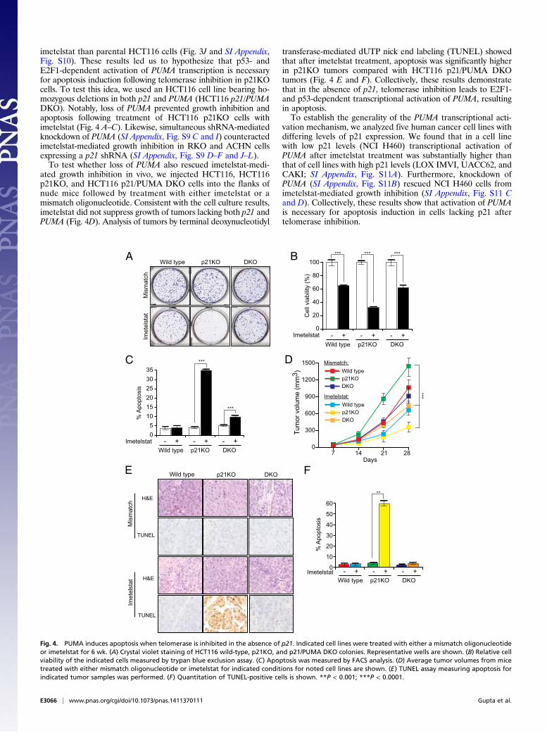
imetelstat than parental HCT116 cells (Fig. 3J and SI Appendix,Fig. S10). These results led us to hypothesize that p53- andE2F1-dependent activation of PUMA transcription is necessaryfor apoptosis induction following telomerase inhibition in p21KOcells. To test this idea, we used an HCT116 cell line bearing ho-mozygous deletions in both p21 and PUMA (HCT116 p21/PUMADKO). Notably, loss of PUMA prevented growth inhibition andapoptosis following treatment of HCT116 p21KO cells withimetelstat (Fig. 4 A–C). Likewise, simultaneous shRNA-mediatedknockdown of PUMA (SI Appendix, Fig. S9 C and I) counteractedimetelstat-mediated growth inhibition in RKO and ACHN cellsexpressing a p21 shRNA (SI Appendix, Fig. S9 D–F and J–L).To test whether loss of PUMA also rescued imetelstat-medi-
ated growth inhibition in vivo, we injected HCT116, HCT116p21KO, and HCT116 p21/PUMA DKO cells into the flanks ofnude mice followed by treatment with either imetelstat or amismatch oligonucleotide. Consistent with the cell culture results,imetelstat did not suppress growth of tumors lacking both p21 andPUMA (Fig. 4D). Analysis of tumors by terminal deoxynucleotidyl
transferase-mediated dUTP nick end labeling (TUNEL) showedthat after imetelstat treatment, apoptosis was significantly higherin p21KO tumors compared with HCT116 p21/PUMA DKOtumors (Fig. 4 E and F). Collectively, these results demonstratethat in the absence of p21, telomerase inhibition leads to E2F1-and p53-dependent transcriptional activation of PUMA, resultingin apoptosis.To establish the generality of the PUMA transcriptional acti-
vation mechanism, we analyzed five human cancer cell lines withdiffering levels of p21 expression. We found that in a cell linewith low p21 levels (NCI H460) transcriptional activation ofPUMA after imetelstat treatment was substantially higher thanthat of cell lines with high p21 levels (LOX IMVI, UACC62, andCAKI; SI Appendix, Fig. S11A). Furthermore, knockdown ofPUMA (SI Appendix, Fig. S11B) rescued NCI H460 cells fromimetelstat-mediated growth inhibition (SI Appendix, Fig. S11 Cand D). Collectively, these results show that activation of PUMAis necessary for apoptosis induction in cells lacking p21 aftertelomerase inhibition.
C
A BWild type p21KO DKO
Imet
elst
atM
ism
atch
0
20
40
60
80
100
Cel
l via
bilit
y (%
)
Imetelstat Wild type p21KO
- +- +DKO- +
% A
popt
osis
Imetelstat Wild type
- +p21KO- +
DKO- +
p21KO
p21KO
Wild type
Wild type
DKO
DKO
Imetelstat:
Mismatch:D
0
300
600
900
1200
1500
Days 7 14 21 28
Tum
or v
olum
e (m
m3 )
05
101520253035
E
***
0
10
20
30
40
50
60
Imetelstat Wild type
- +p21KO- +
DKO- +
% A
popt
osis
*** ***
***
***
***
**
FWild type p21KO DKO
H&E
TUNEL
H&E
TUNEL
Mis
mat
chIm
etel
stat
Fig. 4. PUMA induces apoptosis when telomerase is inhibited in the absence of p21. Indicated cell lines were treated with either a mismatch oligonucleotideor imetelstat for 6 wk. (A) Crystal violet staining of HCT116 wild-type, p21KO, and p21/PUMA DKO colonies. Representative wells are shown. (B) Relative cellviability of the indicated cells measured by trypan blue exclusion assay. (C) Apoptosis was measured by FACS analysis. (D) Average tumor volumes from micetreated with either mismatch oligonucleotide or imetelstat for indicated conditions for noted cell lines are shown. (E) TUNEL assay measuring apoptosis forindicated tumor samples was performed. (F) Quantitation of TUNEL-positive cells is shown. **P < 0.001; ***P < 0.0001.
E3066 | www.pnas.org/cgi/doi/10.1073/pnas.1411370111 Gupta et al.

Synergistic Tumor Suppression by RNAi-Mediated p21 Depletion andImetelstat Treatment. The results described above suggested thatsimultaneous inhibition of p21 and telomerase could synergisti-cally suppress tumor growth. We therefore carried out a series ofexperiments in which p21 function was abrogated using differentapproaches and telomerase was inhibited with imetelstat. In thefirst approach, we used a polymer nanoparticle-based system todeliver a p21 small interfering RNA (siRNA) (36, 37). Thesepoly(lactic-co-glycolic acid) (PLGA) nanoparticles were coatedwith the PEGylated cell-penetrating peptide, N terminus ofthe CPP penetratin (ANTP), and loaded with a p21 siRNA. Asa control, a nonspecific negative control siRNA was similarlyencapsulated into modified PLGA nanoparticles. We then s.c.injected HCT116 and ACHN cells into athymic nude mice andsystemically treated the mice with imetelstat and nanoparticlesencapsulated in siRNA. In good agreement with our cell cul-ture results, the combination of a p21 siRNA and imetelstatresulted in significantly stronger tumor suppression comparedwith imetelstat alone (SI Appendix, Fig. S12 A and B and TableS2). Furthermore, as expected, analyses of tumor lysates revealedreduced p21 expression in p21 siRNA nanoparticle-injectedtumors (SI Appendix, Fig. S12 C and D), increased apoptosisupon simultaneous inhibition of p21 and telomerase (SI Appendix,Fig. S12 C and D), and reduced telomerase activity in imetelstat-treated tumor samples (SI Appendix, Fig. S12 E and F).
Synergistic Tumor Suppression by Pharmacological Inhibition ofTelomerase and p21. The p21 siRNA results described aboveprovide proof-of-principle that systemic in vivo targeting of p21can substantially enhance tumor suppression when combined with
telomerase inhibition. We next performed experiments in whichp21 function was abrogated by pharmacological inhibition in con-junction with imetelstat treatment. We first inhibited p21 expres-sion in HCT116 cells using sorafenib (Fig. 5A), which promotesproteasome-mediated degradation of p21 (38). As predicted, wefound that sorafenib sensitized cells to imetelstat-mediated growthinhibition and apoptosis induction (Fig. 5 B–D). Notably, reintro-duction of p21 in HCT116 cells treated with imetelstat and sor-afenib substantially counteracted growth inhibition and apoptosisinduction (Fig. 5 E–H), confirming that sorafenib functions bydown-regulating p21 activity. Next, we tested the combined effectof imetelstat and sorafenib in suppressing tumor growth inmouse xenografts. We found that simultaneous treatment withimetelstat and sorafenib was substantially more effective at sup-pressing growth of HCT116 xenografts than either drug alone andthus functions in a synergistic manner (Fig. 5I and SI Appendix,Table S2).Toxicity analyses revealed that there were no significant dif-
ferences in the body weight or alanine aminotransferase (ALT),aspartate aminotransferase (AST), alkaline phosphatase (AP),whole blood counts, and renal activity markers in mice that weretreated with imetelstat sorafenib, or both, in comparison with thecontrol group (SI Appendix, Fig. S13). Finally, to determine thegenerality of these results, we analyzed the effect of combinedimetelstat and sorafenib treatment on xenografts formed fromfive additional human cancer cell lines representing four differ-ent tissue origins. Notably, in all cases, treatment with both ime-telstat and sorafenib was substantially more effective at suppressingtumor growth than either drug alone (SI Appendix, Fig. S14 andTable S2).
BA
p21
p53
Actin
DMSOSora
fenib
C D
E
0
100
200
300
400
500
600
700
800
Days 7 14 21 28
VehicleSorafenibImetelstatSorafenib + Imetelstat
I
Tum
or v
olum
e (m
m3 )
% A
popt
osis
05
10152025303540
Cel
l via
bilit
y (%
)
0
20
40
60
80
100
0
20
40
60
80
100
120
Cel
l via
bilit
y (%
)
Vector p210
5
10
15
20
25
% A
popt
osis
Vector p21
Sorafenib
2.5 µM
0 µM1 µM
Sorafenib0 µM1 µM
***
**
** **
**
**
**
***
**
***
Imetelstat + +- - Imetelstat + +- -
F
0
5
10
15
20
25
% A
nnex
in V
pos
itive
cel
ls
Vector p21
Sorafenib
2.5 µM
0 µM1 µM
**
**G
Cleaved Caspase 3
Actin
Vector p21Sorafenib (µM)0 1 2.5 0 1 2.5
p53
p21
Actin
Cleaved Caspase 3
Sorafenib- - + +Imetelstat- + - +
H
Sorafenib0 µM1 µM
Fig. 5. Simultaneous pharmacological inhibition of p21 by sorafenib and telomerase prevents tumor growth in mice. (A) Immunoblot of p21 expression inHCT116 cells treated with DMSO or sorafenib (1 μM) for 24 h. (B) Relative cell viability measured by trypan blue exclusion assay of HCT116 cells treated withimetelstat and sorafenib. (C) Apoptosis of HCT116 cells treated with imetelstat and sorafenib was measured by FACS analysis. (D) Immunoblot for indicatedproteins in HCT116 cells treated with sorafenib, imetelstat, or both. (E) Relative cell viability measured by trypan blue exclusion assay of imetelstat-treatedHCT116 cells transfected with control or p21 expression vectors and then treated with sorafenib. (F) Apoptosis measured by FACS analysis of imetelstat-treated HCT116 cells transfected with control or p21 expression vectors and then treated with sorafenib. (G) Annexin V–FITC–positive cells were quantified byFACS analysis of imetelstat-treated HCT116 cells transfected with control or p21 expression vectors and then treated with sorafenib. (H) Cleaved caspase 3 wasmeasured by immunoblot in imetelstat-treated HCT116 cells transfected with control or p21 expression vectors and then treated with sorafenib. (I) Averagetumor volumes from mice treated with vehicle, sorafenib alone, imetelstat alone, or both drugs. **P < 0.001; ***P < 0.0001.
Gupta et al. PNAS | Published online July 14, 2014 | E3067
GEN
ETICS
PNASPL
US

Sorafenib has been shown to have cellular targets other than p21,including BRAF and VEGF (39). Therefore, we asked whether analternative pharmacological inhibitor that more selectively tar-gets p21 expression can, like sorafenib, cooperate with imetelstat tosuppress tumor growth. Toward this end, we used a recently iden-tified pharmacological p21 inhibitor, UC2288 (Fig. 6A), whichdown-regulates p21 levels through transcriptional and post-transcriptional mechanisms (40). Furthermore, unlike Sorafenib,UC2288 does not inhibit RAF kinase or VEGF activities (40). Inagreement with previous studies, we observed that treatment of
cancer cells with UC2288 led to decreased p21 levels (Fig. 6B).Furthermore, whereas UC2288 alone had only modest growthinhibitory effects, combining UC2288 with imetelstat potentlyinhibited cancer cell growth by inducing apoptosis (Fig. 6 C–E).The ability of UC2288 to inhibit growth was largely dependentupon its ability to down-regulate p21 expression, because ectopicexpression of p21 counteracted growth inhibition and apoptosisinduction in UC2288 and imetelstat-treated cancer cells (Fig. 6 F–H). Finally, we tested whether UC2288 can inhibit tumor growthin vivo. Toward this end, we injected HCT116 and ACHN cells
BA
p21
Actin
Days 7 14 21 28
VehicleUC2288ImetelstatUC2288 + Imetelstat
I
Tum
or v
olum
e (m
m3 )
Cel
l via
bilit
y (%
)
UC2288 + +- -
UC2288 (µM)- 1.0 2.5 5.0
D E
F
VehicleUC2288ImetelstatUC2288 + Imetelstat
J
Tum
or v
olum
e (m
m3 )
HCT116 ACHN
Cl
F3C NH
oo
NH
N
CF3
UC2288
C
G
Cleaved Caspase 3
Actin
UC2288- - + +
0
20
40
60
80
100
0
10
20
30
40
50
60
UC2288 + +- -
% A
nnex
in V
pos
itive
cel
ls
Cel
l via
bilit
y (%
)
Vector p21
UC2288
2.5 µM
0 µM1 µM
0
20
40
60
80
100
0
10
20
30
40
50
60
% A
nnex
in V
pos
itive
cel
ls
H
Cleaved Caspase 3
Actin
Vector p21
0
100
200
300
400
500
600
700
800
Days 7 14 21 28
Vector p21
0
100
200
300
400
500
600
700
800
UC2288 (µM)0 1 2.5 0 1 2.5
Imetelstat - + - +* *** ***
**
******
***
***
***
ImetelstatMismatch ImetelstatMismatch
K L
p21
Cleaved Caspase 3
Actin
UC2288
Vehicl
e
HCT116 ACHN
UC2288
+Imete
lstat
Imete
lstat
p21
Cleaved Caspase 3
Actin
UC2288
Vehicl
e
UC2288
+Imete
lstat
Imete
lstat
p53
p21**
*
Fig. 6. Simultaneous pharmacological inhibition of p21 by UC2288 and telomerase prevents tumor growth in mice. (A) Chemical structure of UC2288. (B)Immunoblot of p21 expression in HCT116 cells treated with DMSO or with indicated concentrations of UC2288 for 24 h. (C) Relative cell viability measured bytrypan blue exclusion assay of HCT116 cells treated with imetelstat, UC2288 (2.5 μM), or both. (D) % Annexin V–FITC–positive HCT116 cells treated withimetelstat and UC2288 was measured by FACS analysis. (E) Immunoblot for indicated proteins in HCT116 cells following treatment with U2288, imetelstat, orboth. (F) Relative cell viability measured by trypan blue exclusion assay of imetelstat-treated HCT116 cells transfected with control or p21 expression vectorsand then treated with UC2288. (G) % Annexin V–FITC–positive cells was measured by FACS analysis of imetelstat-treated HCT116 cells transfected with controlor p21 expression vectors and then treated with UC2288. (H) Cleaved caspase 3 was measured by immunoblot in imetelstat-treated HCT116 cells transfectedwith control or p21 expression vectors and then treated with UC2288. (I) Average tumor volumes of HCT116 xenograft frommice treated with vehicle, UC2288alone, imetelstat alone, or both drugs. **P < 0.001; ***P < 0.0001. (J) Average tumor volumes of ACHN xenograft from mice treated with vehicle, UC2288alone, imetelstat alone, or both drugs. (K) Mouse-derived HCT116 xenograft tumors under indicated treatment conditions were analyzed for indicatedproteins by immunoblot analysis. (L) Mouse-derived ACHN xenograft tumors under indicated treatment conditions were analyzed for indicated proteins byimmunoblot analysis. *P < 0.01; **P < 0.001; ***P < 0.0001.
E3068 | www.pnas.org/cgi/doi/10.1073/pnas.1411370111 Gupta et al.

into the flanks of athymic nude mice and followed it by treat-ment with UC2288, imetelstat, or both drugs. Notably, combinedtreatment with UC2288 and imetelstat synergistically suppressedtumor growth (Fig. 6 I–L and SI Appendix, Table S2).Toxicity analyses revealed that there were no significant dif-
ferences in the body weight or ALT, AST, AP, whole bloodcounts, and renal activity markers in mice that were treated withimetelstat, UC2288, or both drugs, in comparison with the con-trol group (SI Appendix, Fig. S15).
Targeting Tumors Containing Mutant p53 Following PharmacologicalRestoration of p53 Activity. The results described above show thatin the absence of p21, p53 and E2F1 cooperate to activate PUMAexpression, which is necessary for apoptosis induction (Figs. 3 and4). Accordingly, cells that lack functional p53 are less sensitive toimetelstat-mediated growth inhibition compared with cells con-taining wild-type p53 (Fig. 3J).
Approximately 50% of human cancers lack a functional p53pathway, largely due to inactivating mutations in the p53 gene(41). We therefore asked whether simultaneous inhibition oftelomerase and p21 could be adapted to treat cancers harboringp53 mutants. Previous studies have shown that the function ofseveral different cancer-associated p53 mutants can be restoredby treatment with small molecules such as CP-31398 (42, 43).CP-31398 alters the conformation of p53, thereby promoting itsability to bind to DNA (42, 43). As expected, treatment of p53mutant cancer cell lines DLD1 (Ser241Phe), SW480 (Arg273Hisand Pro309Ser), and A375.S2 (Arg249Ser) with CP-31398 re-stored p53 activity, as evidenced by increased expression of itstranscriptional target p21 (Fig. 7A). Next, using shRNAs, weknocked down p21 in DLD1 and A375.S2 cells (Fig. 7B) andtreated these cells with imetelstat and CP-31398. Treatment withboth CP-31398 and imetelstat inhibited the growth of DLD1 andA375 cells expressing a p21 shRNA more strongly compared with
A
p21
Actin
shRNA:NS #1 #2 NS #1 #2
p21 p21
C D E
DLD1 A375.S2B
p21
Actin
DLD1 SW480 A375.S2CP-31398 (µg/ml) 0 2 5 0 2 5 0 2 5
F
ImetelstatCP-31398
- + - + - + - + - + - +- - + + - - + + - - + +
shRNA: NS #1 #2p21
0
20
40
60
80
100
Cel
l Via
bilit
y (%
)
01020304050607080
0
10
20
30
40
50
60
01020304050607080
0
20
40
60
80
100
120
G H
% A
nnex
in V
pos
itive
cel
ls
Rel
ativ
e P
UM
A ex
pres
sion
- + - + - + - + - + - +- - + + - - + + - - + +
NS #1 #2p21
- + - + - + - + - + - +- - + + - - + + - - + +
NS #1 #2p21
ImetelstatCP-31398
- + - + - + - + - + - +- - + + - - + + - - + +
shRNA: NS #1 #2p21
0
20
40
60
80
100
Cel
l Via
bilit
y (%
)
- + - + - + - + - + - +- - + + - - + + - - + +
NS #1 #2p21
- + - + - + - + - + - +- - + + - - + + - - + +
NS #1 #2p21
Rel
ativ
e P
UM
A e
xpre
ssio
n
% A
nnex
in V
pos
itive
cel
ls
DLD1
DLD1 DLD1 DLD1
A375.S2 A375.S2 A375.S2
I J
Tum
or v
olum
e (m
m3 )
Tum
or v
olum
e (m
m3 )
VehicleCP-31398ImetelstatCP-31398+ Imetelstat
0
100
200
300
400
500
600
700
800
0
200
400
600
800
1000
VehicleCP-31398ImetelstatCP-31398+ Imetelstat
NS
siR
NA
p21
siR
NA
*****
*
Days7 14 21 28
Days7 14 21 28
* * *
*
** **
*
** **
* * *
Fig. 7. Pharmacological restoration of p53 activity allows for the targeting of p53 mutant cancers by simultaneous p21 and telomerase inhibition. (A) In-dicated p53 mutant cancer cell lines were treated at indicated concentrations of CP-31398 and analyzed for p21 expression by immunoblot. Actin was used asa loading control. (B) Indicated p53 mutant cancer cell lines expressing either an NS shRNA or shRNA targeting p21 were analyzed for p21 expression byimmunoblot. Actin was used as a loading control. (C and F) DLD1 cells (C) or A375.S2 cells (F) expressing indicated shRNAs were treated with mismatcholigonucleotide or imetelstat for 6 wk and were either untreated or treated with CP-31398 for 2 d. Cell viability was measured by trypan blue assay, andrelative cell viability is plotted. (D and G) DLD1 cells (D) or A375.S2 cells (G) expressing indicated shRNAs were treated with mismatch oligonucleotide orimetelstat for 6 wk and were either untreated or treated with CP-31398 for 2 d. Apoptosis was measured by Annexin V staining, and % apoptosis is plotted.(E and H) DLD1 cells (E) or A375.S2 cells (H) expressing indicated shRNAs were treated with mismatch oligonucleotide or imetelstat for 6 wk and were eitheruntreated or treated with CP-31398 for 2 d. PUMA expression was measured by qRT-PCR. (I and J) Average tumor volumes of DLD1 xenograft (I) or A375.S2 (J)from mice treated with vehicle, CP-31398 alone, imetelstat alone, or both drugs with simultaneous treatment with nanoparticles with nonspecific or p21siRNA are shown. *P < 0.01; **P < 0.001; ***P < 0.0001.
Gupta et al. PNAS | Published online July 14, 2014 | E3069
GEN
ETICS
PNASPL
US

cells expressing a nonspecific shRNA (Fig. 7 C and F and SIAppendix, Fig. S16). Growth inhibition correlated with increasedapoptosis (Fig. 7 D and G) and significantly higher PUMA ac-tivation (Fig. 7 E and H). Finally, we tested this approach forsuppressing tumor growth in vivo. We found that addition of CP-31398 combined with simultaneous inhibition of telomerase andp21 substantially suppressed growth of tumors containing mutantp53 (Fig. 7 I and J). Collectively, these results show that simul-taneous inhibition of telomerase and p21 can also be used totreat cancers containing mutant p53.
DiscussionA large majority of diverse tumor types express telomerase, whichis critical for cancer cell survival. Thus, inhibition of telomeraseactivity can block tumor cell growth both in vitro and in vivo. Dueto such widespread expression and the requirement of telomerasefor cancer cell survival, telomerase inhibitors can, in principle, beused to treat a broad spectrum of cancer types. In fact, telomeraseinhibitors such as imetelstat have advanced into the clinic for thetreatment of human cancers. However, for several reasons, single-agent telomerase therapies have not proven effective. First, becausetelomere shortening and consequential tumor growth inhibitionrequire many cell divisions, single-agent telomerase inhibitorsrequire substantial time to significantly decrease tumor growth.Second, telomerase inhibition typically results in only a cytostaticeffect, which allows tumor cells to acquire secondary genetic andepigenetic alterations resulting in drug resistance. Finally, single-agent telomerase therapeutics often fail due to activation of ALTpathways, which bypasses the requirement for telomerase (25).In this report, we have studied the role of p21 in telomerase
inhibition-mediated growth suppression. We found that p21 lossincreases apoptosis induction following telomerase inhibition ina variety of cancer cell lines and mouse xenografts. We furthershowed that apoptosis induction is specifically due to up-regulationof PUMA expression and that up-regulation of PUMA expressionis dependent upon both p53 and E2F1. Although our results areconsistent with other studies reporting that p21 can regulate ap-optosis (9–13), the specific apoptotic mechanism we identify hasnot been previously described.Previous studies have shown that p21 loss sensitizes cells to
DNA-damage–induced apoptosis (9–13). In our experiments,treatment of cells with imetelstat induces a DNA damage re-sponse, which upon simultaneous loss of p21, results in apo-ptosis. Thus, the loss of p21 effectively converts the response ofimetelstat from simple growth inhibition to apoptosis. Accordingto this mechanism, the specificity of growth inhibition is due toimetelstat, which will selectively induce a DNA damage response
in telomerase-positive cancer cells but not telomerase-negativenormal cells.Our finding that p21 is required to prevent apoptosis following
telomerase inhibition reveals a critical genetic vulnerability oftelomerase-expressing cancer cells. Accordingly, using both RNAi-based and pharmacological approaches, we showed that simulta-neous inhibition of p21 and telomerase induces apoptosis intelomerase-positive human cancer cell lines and synergisticallysuppresses tumor growth. Specifically, we found that two unrelatedpharmacological inhibitors of p21, sorafenib and UC2288, couldfunction synergistically with imetelstat. Notably, in both cases,growth inhibition could be counteracted by ectopic expression ofp21, indicating that sorafenib and UC2288 functioned by inhibitingp21 and not through an off-target effect. The induction of apo-ptosis by simultaneous inhibition of p21 and telomerase maygreatly reduce the likelihood that resistance to telomeraseinhibitors will develop through additional secondary geneticalterations or activation of ALT pathways. Finally, we show thatsimultaneous inhibition of telomerase and p21 also suppressesgrowth of tumors containing mutant p53 following pharmaco-logical restoration of p53 activity.Overall, we anticipate that simultaneous inhibition of telo-
merase and p21 can potentially overcome the current limitationof single-agent telomerase therapeutics and provide an effectivemethod to treat a large number of cancers that rely on telo-merase activity for survival, including the p53 mutant cancers.
Materials and MethodsMaterials and procedures for all experiments are supplied in SI Appendix.Included in SI Appendix are the mammalian cell culture procedures, detailsregarding tumorigenesis assay, protocols for TRAP assay to measure telo-merase activity, Terminal Restriction Fragment analysis to measure telomerelength, and ALT activity assay to monitor C-Circle amplification. Also pro-vided are additional figures and tables providing p16 status of the cell linesused in our study, primer sequence information and antibody details usedfor immunolotting, and the statistical analyses of drug combinations to es-tablish synergistic effects.
ACKNOWLEDGMENTS. We thank Bert Vogelstein, William G. Kaelin, andWilliam Hahn for reagents. We thank Katia Bassett and Kevin Eng (GeronCorporation) for helpful discussions and Darryl Conte for editorial assistance.R.H.W. is supported by Grants 1R01CA135401-01A1 and 1R01DK082690-01A1 and B.D.H. is supported by Grant R01 ES002710. S.C. is supportedby the Grant RO1 CA129037 from the National Cancer Institute. N.W. is atranslational scholar of the Sidney Kimmel Foundation for Cancer Researchand is supported by the Young Investigator grants from the National LungCancer Partnership, Uniting Against Lung Cancer Foundation, InternationalAssociation for the Study of Lung Cancer, and Melanoma Research Allianceand Melanoma Research Foundation. M.R.G. is an investigator of theHoward Hughes Medical Institute.
1. Blackburn EH, Collins K (2011) Telomerase: An RNP enzyme synthesizes DNA. ColdSpring Harb Perspect Biol 3(5).
2. Shay JW, Wright WE (2011) Role of telomeres and telomerase in cancer. Semin CancerBiol 21(6):349–353.
3. Hahn WC, et al. (1999) Inhibition of telomerase limits the growth of human cancercells. Nat Med 5(10):1164–1170.
4. Chin L, et al. (1999) p53 deficiency rescues the adverse effects of telomere lossand cooperates with telomere dysfunction to accelerate carcinogenesis. Cell97(4):527–538.
5. el-Deiry WS, et al. (1993) WAF1, a potential mediator of p53 tumor suppression. Cell75(4):817–825.
6. Harper JW, Adami GR, Wei N, Keyomarsi K, Elledge SJ (1993) The p21 Cdk-interactingprotein Cip1 is a potent inhibitor of G1 cyclin-dependent kinases. Cell 75(4):805–816.
7. Polyak K, Waldman T, He TC, Kinzler KW, Vogelstein B (1996) Genetic determinants ofp53-induced apoptosis and growth arrest. Genes Dev 10(15):1945–1952.
8. Waldman T, Kinzler KW, Vogelstein B (1995) p21 is necessary for the p53-mediated G1arrest in human cancer cells. Cancer Res 55(22):5187–5190.
9. Javelaud D, Besancon F (2002) Inactivation of p21WAF1 sensitizes cells to apoptosisvia an increase of both p14ARF and p53 levels and an alteration of the Bax/Bcl-2 ratio.J Biol Chem 277(40):37949–37954.
10. Broude EV, et al. (2007) p21 (CDKN1A) is a negative regulator of p53 stability. CellCycle 6(12):1468–1471.
11. Xia M, Knezevic D, Vassilev LT (2011) p21 does not protect cancer cells from apoptosisinduced by nongenotoxic p53 activation. Oncogene 30(3):346–355.
12. Dong C, Li Q, Lyu SC, Krensky AM, Clayberger C (2005) A novel apoptosis pathway
activated by the carboxyl terminus of p21. Blood 105(3):1187–1194.13. Chen Y, Zhang L, Jones KA (2011) SKIP counteracts p53-mediated apoptosis via se-
lective regulation of p21Cip1 mRNA splicing. Genes Dev 25(7):701–716.14. Röth A, Harley CB, Baerlocher GM (2010) Imetelstat (GRN163L)—Telomerase-based
cancer therapy. Recent Results Cancer Res 184:221–234.15. Macleod KF, et al. (1995) p53-dependent and independent expression of p21 during
cell growth, differentiation, and DNA damage. Genes Dev 9(8):935–944.16. Bunz F, et al. (1998) Requirement for p53 and p21 to sustain G2 arrest after DNA
damage. Science 282(5393):1497–1501.17. Goldberg M, et al. (2003) MDC1 is required for the intra-S-phase DNA damage
checkpoint. Nature 421(6926):952–956.18. Lou Z, Minter-Dykhouse K, Wu X, Chen J (2003) MDC1 is coupled to activated CHK2 in
mammalian DNA damage response pathways. Nature 421(6926):957–961.19. Stewart GS, Wang B, Bignell CR, Taylor AM, Elledge SJ (2003) MDC1 is a mediator of
the mammalian DNA damage checkpoint. Nature 421(6926):961–966.20. Dimitrova N, de Lange T (2006) MDC1 accelerates nonhomologous end-joining of
dysfunctional telomeres. Genes Dev 20(23):3238–3243.21. Zhu XD, Küster B, Mann M, Petrini JH, de Lange T (2000) Cell-cycle-regulated asso-
ciation of RAD50/MRE11/NBS1 with TRF2 and human telomeres. Nat Genet 25(3):
347–352.22. Kubo A, et al. (1999) The p16 status of tumor cell lines identifies small molecule in-
hibitors specific for cyclin-dependent kinase 4. Clin Cancer Res 5(12):4279–4286.
E3070 | www.pnas.org/cgi/doi/10.1073/pnas.1411370111 Gupta et al.

23. Lee EJ, et al. (2008) Histone deacetylase inhibitor scriptaid induces cell cycle arrest and
epigenetic change in colon cancer cells. Int J Oncol 33(4):767–776.24. Cesare AJ, Reddel RR (2010) Alternative lengthening of telomeres: Models, mecha-
nisms and implications. Nat Rev Genet 11(5):319–330.25. Hu J, et al. (2012) Antitelomerase therapy provokes ALT and mitochondrial adaptive
mechanisms in cancer. Cell 148(4):651–663.26. Henson JD, et al. (2009) DNA C-circles are specific and quantifiable markers of
alternative-lengthening-of-telomeres activity. Nat Biotechnol 27(12):1181–1185.27. Scheel C, et al. (2001) Alternative lengthening of telomeres is associated with chro-
mosomal instability in osteosarcomas. Oncogene 20(29):3835–3844.28. Miyashita T, Reed JC (1995) Tumor suppressor p53 is a direct transcriptional activator
of the human bax gene. Cell 80(2):293–299.29. Polyak K, Xia Y, Zweier JL, Kinzler KW, Vogelstein B (1997) A model for p53-induced
apoptosis. Nature 389(6648):300–305.30. Nakano K, Vousden KH (2001) PUMA, a novel proapoptotic gene, is induced by p53.
Mol Cell 7(3):683–694.31. Degenhardt K, Chen G, Lindsten T, White E (2002) BAX and BAK mediate p53-
independent suppression of tumorigenesis. Cancer Cell 2(3):193–203.32. Villunger A, et al. (2003) p53- and drug-induced apoptotic responses mediated by
BH3-only proteins puma and noxa. Science 302(5647):1036–1038.33. Carnevale J, Palander O, Seifried LA, Dick FA (2012) DNA damage signals through
differentially modified E2F1 molecules to induce apoptosis. Mol Cell Biol 32(5):
900–912.
34. Hershko T, Ginsberg D (2004) Up-regulation of Bcl-2 homology 3 (BH3)-only proteinsby E2F1 mediates apoptosis. J Biol Chem 279(10):8627–8634.
35. Wang Y, Shen MF, Chang S (2011) Essential roles for Pot1b in HSC self-renewal andsurvival. Blood 118(23):6068–6077.
36. Woodrow KA, et al. (2009) Intravaginal gene silencing using biodegradable polymernanoparticles densely loaded with small-interfering RNA. Nat Mater 8(6):526–533.
37. Cheng CJ, Saltzman WM (2011) Enhanced siRNA delivery into cells by exploiting thesynergy between targeting ligands and cell-penetrating peptides. Biomaterials 32(26):6194–6203.
38. Inoue H, Hwang SH, Wecksler AT, Hammock BD, Weiss RH (2011) Sorafenib attenu-ates p21 in kidney cancer cells and augments cell death in combination with DNA-damaging chemotherapy. Cancer Biol Ther 12(9):827–836.
39. Wilhelm SM, et al. (2008) Preclinical overview of sorafenib, a multikinase inhibitorthat targets both Raf and VEGF and PDGF receptor tyrosine kinase signaling. MolCancer Ther 7(10):3129–3140.
40. Wettersten HI, et al. (2013) A novel p21 attenuator which is structurally related tosorafenib. Cancer Biol Ther 14(3):278–285.
41. Brown CJ, Lain S, Verma CS, Fersht AR, Lane DP (2009) Awakening guardian angels:Drugging the p53 pathway. Nat Rev Cancer 9(12):862–873.
42. Foster BA, Coffey HA, Morin MJ, Rastinejad F (1999) Pharmacological rescue of mu-tant p53 conformation and function. Science 286(5449):2507–2510.
43. Takimoto R, et al. (2002) The mutant p53-conformation modifying drug, CP-31398,can induce apoptosis of human cancer cells and can stabilize wild-type p53 protein.Cancer Biol Ther 1(1):47–55.
Gupta et al. PNAS | Published online July 14, 2014 | E3071
GEN
ETICS
PNASPL
US
Copyright © 2022 FDOKUMEN