A Synergistic Approach for Internet of Things (IoT) and Cloud ...
Upload
independentCategory
view
3download
0
SYNERGISTIC ACTIVITY OF LETROZOLE AND SORAFENIB ON BREAST CANCER
CELLS
Mara A. Bonelli1*, Claudia Fumarola1*, Roberta R. Alfieri1, Silvia La Monica1, Andrea Cavazzoni1, Maricla Galetti1, Rita Gatti1, Silvana Belletti1, Adrian L. Harris2, Stephen B. Fox3, Dean B. Evans4, Mitch Dowsett5, Lesley-Ann Martin6, Alberto Bottini7, Daniele
Generali7,8, Pier Giorgio Petronini1 1 Department of Experimental Medicine, University of Parma, Italy 2 Molecular Oncology Laboratories, Weatherall Institute of Molecular Medicine, University
of Oxford, John Radcliffe Hospital, Oxford, UK 3 Peter MacCallum Cancer Centre, St Andrews Place, East Melbourne, Victoria, Australia 4 Novartis Institutes of BioMedical Research, Oncology Research, Basel, Switzerland 5 Academic Department of Biochemistry, Royal Marsden Hospital; 6 Breakthrough Breast
Cancer Centre, Institute of Cancer Research, London, UK 7 Unità di Patologia Mammaria-Breast Cancer Unit; 8 Centro di Medicina Molecolare, Istituti
Ospitalieri di Cremona, Italy
*These authors contributed equally to this work
Running title: Letrozole and sorafenib combination in breast cancer cells.
Keywords: letrozole, sorafenib, breast cancer, mTORC1
Footnote: Dean Evans is employed by Novartis Pharma AG
Grant support: Associazione Davide Rodella, Montichiari (BS); Lega Italiana per la lotta
contro i tumori, Parma; A.VO.PRO.RI.T., Parma; Fondazione Banca Popolare di Cremona;
CONAD, Bologna (Italy)
Requests for reprints:
Claudia Fumarola
Department of Experimental Medicine Via Volturno, 39
43100 Parma (Italy) Phone: +390521903762 Fax: +390521903742
e-mail: [email protected]
2
ABSTRACT
Estrogens induce breast tumour cell proliferation by directly regulating gene expression via
the estrogen receptor (ER) transcriptional activity and by affecting growth factor signalling
pathways such as mitogen-activated protein kinase (MAPK) and AKT/mammalian target of
rapamycin Complex1 (mTORC1) cascades. In this study we demonstrated the preclinical
therapeutic efficacy of combining the aromatase inhibitor letrozole with the multi -kinase
inhibitor sorafenib in aromatase-expressing breast cancer cell lines. Treatment with
letrozole reduced testosterone-driven cell proliferation, by inhibiting the synthesis of
estrogens. Sorafenib inhibited cell proliferation in a concentration-dependent manner; this
effect was not dependent on sorafenib-mediated inhibition of Raf1, but involved the down-
regulation of mTORC1 and its targets p70S6K and 4E-binding protein 1 (4E-BP1). At
concentrations of 5-10μM the growth-inhibitory effect of sorafenib was associated with the
induction of apoptosis, as indicated by release of cytochrome c and Apoptosis-Inducing
Factor into the cytosol, activation of caspase-9 and caspase-7, and PARP-1 cleavage.
Combination of letrozole and sorafenib produced a synergistic inhibition of cell proliferation
associated with an enhanced accumulation of cells in the G0/G1 phase of the cell cycle and
with a down-regulation of the cell cycle regulatory proteins c-myc, cyclin D1 and phospho-
Rb. In addition, longer experiments (12 weeks) demonstrated that sorafenib may be
effective in preventing the acquisition of resistance towards letrozole . Together, these
results indicate that combination of letrozole and sorafenib might constitute a promising
approach to the treatment of hormone-dependent breast cancer.
3
INTRODUCTION
Approximately 60% of primary breast cancers express the ER-alpha and are dependent on
estrogens for their growth. ER is a member of the nuclear hormone receptor superfamily
and 17 -estradiol (E2) is the most potent ligand. Estrogens activate ER through genomic
and non-genomic pathways, stimulating nuclear and cytoplasmatic processes that promote
breast cancer cell proliferation. In the nucleus, ER regulates gene transcription either by
direct binding to estrogen response elements (ERE) [1] or via protein-protein interactions
with other transcription factors such as AP-1, SP-1, and NF-kB [2]. In addition, estrogens
exert rapid non-genomic actions mediated by the interaction between membrane-bound
ER and membrane kinase receptors such as IGF-IR, EGFR, and HER2 [1], which results
in the activation of signalling cascades including the Ras/Raf/MAPK pathway [3] and the
phosphatidylinositol-3-kinase (PI3K)/AKT/mTOR pathway [2]. Conversely, ER can be
directly activated in the absence of estrogens by various kinases, including MAPK and
PI3K/AKT [4, 5].
In postmenopausal women, estrogens are generated from androgens through aromatase-
dependent enzymatic conversion [6]. Letrozole (Femara®) is a third-generation
nonsteroidal aromatase inhibitor (AI) [7] approved for treatment of postmenopausal women
with hormone-dependent breast cancer. By eradicating estrogens, AIs suppress both
genomic and non-genomic action of ER and are proved to be effective as first-line therapy
in advanced/metastatic breast cancer as well as in neoadjuvant, early adjuvant and
extended adjuvant strategies [8]. However, either intrinsic or acquired resistance to AIs
may occur, arising from activation of novel signalling mechanisms that involve crosstalk
between ER and growth factor receptors [9]. This provides a strong rationale for combining
endocrine therapies with signal transduction inhibitors to achieve a more potent anti-
tumour effect or to bypass acquired resistance [10, 11].
4
Generali and co-workers demonstrated the role of mTOR, MAPK and HIF-1α in endocrine
resistance in patients receiving letrozole-based treatment supporting the development of
new treatment strategies based on the combination of AIs with signal transduction
inhibitors targeting mainly MAPK but also mTOR and HIF-1alpha [12].
Sorafenib (Nexavar, BAY43-9006) is a multi-kinase inhibitor recently approved for
treatment of advanced renal cell and hepatocellular carcinoma [13, 14]. It has been shown
to block tumour cell proliferation and angiogenesis by inhibiting serine/threonine kinases
(C-Raf, mutant and wild-type B-Raf) as well as vascular endothelial growth factor
receptors (VEGFR2, VEGFR3) [15], and other tyrosine-kinase receptors such as PDGFR-
, FLT-3, Ret and c-KIT [15, 16]. In animal models, sorafenib effectively reduces
melanoma, breast, colon and lung cancer growth [15, 17]. In addition, it has been reported
that sorafenib induces apoptosis in a variety of tumour cell lines [18-22]. Recently, it was
also shown that sorafenib modulates either mTOR signalling and its targets (p70S6K,
S6R, 4EBP1) expression or HIF-1alpha expression [23, 24].
The purpose of this study was to evaluate the potential for combining letrozole and
sorafenib in breast cancer cell lines constitutively expressing the aromatase gene.
5
MATERIALS AND METHODS
Cell culture
The human breast cancer cell lines MCF-7/AROM-1 and T47D/AROM (clone 1 and 2),
expressing high levels of aromatase, were generated by stable transfection of full-length
human aromatase under control of the cytomegalovirus promoter. BT474/AROM were
stably transduced with a retroviral construct pBabeAROM expressing aromatase. Cells
were cultured as recommended [25, 26].
Before the experiments cells were steroid-deprived for 4 days using phenol red-free
medium supplemented with 5% charcoal-stripped FCS (HyClone, Logan, UT) and then
treated with 1nM E2 or stimulated with 100nM testosterone (T) in the absence or presence
of the drugs. Media and drugs were changed every three days.
Compounds
Letrozole was provided by Novartis Institutes for BioMedical Research (Basel,
Switzerland). Sorafenib was from Bayer HealthCare LLC (Tarrytown, NY). Drugs and
hormones (E2 and T, Sigma-Aldrich, St. Louis, MO) were prepared in DMSO. The
concentration of DMSO never exceeded 0.1% (v/v) and equal amounts of the solvent were
added to control cells.
Antibodies and reagents
Media and FBS were from Euroclone (Devon, UK). Antibodies against cytochrome c,
Apoptosis-Inducing Factor (AIF), p53, p21WAF1 were from Santa Cruz Biotechnology
(Santa Cruz, CA). Antibodies against PARP-1, caspase-7, caspase-8, caspase-9,
phospho-ERK1/2(Thr202/Tyr204), ERK1/2, phospho-AKT(Ser473), AKT, phospho-
mTOR(Ser2448), phospho-4E-BP1(Ser65), phospho-p70S6K(Thr389), p70S6K, c-myc,

6
cyclin D1, p27Kip1, phospho-Rb(Ser780), and Rb were from Cell Signaling Technology
(Beverly, MA). Anti-actin antibody was from Sigma-Aldrich. Horseradish peroxidase-
conjugated secondary antibodies and chemiluminescence system were from Millipore
(Millipore, MA). Reagents for electrophoresis and blotting analysis were from BIO-RAD
Laboratories (Hercules, CA). Calcein-AM and propidium iodide (PI) were from Invitrogen
(Milano, Italy), Draq5 was from Alexis Biochemicals (Lausen, Switzerland). Rapamycin
and all other reagents were from Sigma-Aldrich.
Western blotting
Procedures for protein extraction and analysis by 1-D PAGE are described elsewhere [27].
Cytosolic and mitochondrial fractions were generated as previously described [28].
Protein levels were quantified by densitometric analysis (UN-SCAN-ITTMgel) and
normalized to the actin levels; values are expressed as percent versus T-stimulated cells.
Analysis of cell proliferation, cell death and cell cycle
Cell proliferation was evaluated by cell counting and tetrazolium dye [3-(4,5-
dimethylthiazol-2-yl)-2,5-diphenyltetrazolium-bromide] (MTT) assay, as previously
described [27]. Data are expressed as percent inhibition of cell proliferation versus T-
stimulated cells, calculated by subtracting the amount of proliferation independent of
hormone stimulation. A percent value of inhibition >100% indicates that hormone-
independent cell proliferation was also inhibited.
Cell death was assessed as previously described [28]. In addition, real time analysis was
performed using a Zeiss LSM510 Meta confocal microscope (CLSM) (Carl Zeiss, Jena,
Germany) with a 63X NA 1.4 plan apo objective. Viability and morphology were assessed
after cell loading with calcein-AM, PI and Draq5 excited with 488, 543, 633 laser lines
7
respectively. Acquisition was carried out in the multitrack mode, namely through
consecutive and independent optical pathways.
Distribution of the cells in the cell cycle was determined by PI staining and flow cytometry
as described elsewhere [27, 28].
Statistical Analysis
Statistical significance of differences among data was estimated by two -tailed Student’s t
test or one-way ANOVA (with Tukey test for pairwise comparisons). Differences were
considered significant at p<0.05.
IC50 values, expressed as mean of three independent determinations ( SD), were
calculated by fitting the experimental data with a hyperbolic function and constraining Ymax
to 100 (GraphPad-Prism 4.00).
The nature of the interaction between letrozole and sorafenib was calculated using the
Bliss interaction model [29, 30]. A theoretical dose-response curve was calculated for
combined inhibition using the equation Ebliss=EA+EB-EAxEB, where EA and EB are the
percent of inhibition versus testosterone-stimulated cells, obtained by drug A (sorafenib)
and B (letrozole) alone and Ebliss is the percent of inhibition that would be expected if the
combination was exactly additive. If the experimental percent of inhibition is >Ebliss the
combination is considered synergistic, if it is <Ebliss the combination is antagonistic.
8
RESULTS
Letrozole and sorafenib inhibit estrogen-driven proliferation of MCF-7/AROM-1 cells.
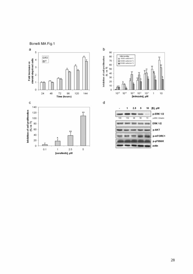
MCF-7/AROM-1 cells were firstly analyzed for their estrogen sensitivity (fig.1a). Cell
proliferation was slowed by steroid-deprivation and stimulated in a time-dependent manner
by either E2 or testosterone, thus indicating that these cells mainly depend on estrogens
for their growth. The effect of letrozole on cell proliferation was evaluated in testosterone-
stimulated cells seeded at different densities. A clear concentration-dependent inhibition of
testosterone-driven proliferation was observed in cells cultured at low density (fig.1b); in
contrast, in cells cultured at higher density letrozole, even at 10μM, was unable to inhibit
cell proliferation by more than 20%, indicating that the growth-inhibitory effect of the drug
is affected by cell density. Indeed, in high density cell cultures a higher concentration of
autocrine growth factors is presumably achieved [31], thus rendering cell proliferation less
dependent on estrogen production. Treatments with letrozole up to 6 days, even at the
highest concentrations, were never associated with induction of cell death (not shown).
Then we evaluated the effects of sorafenib on testosterone-driven proliferation. As
demonstrated by cell counting (not shown) and MTT assay (fig.1c), sorafenib impaired cell
proliferation in a concentration-dependent manner. In addition, sorafenib inhibited ERK1/2
phosphorylation only at concentrations higher than the IC50 value (3µM) after 2h of
treatment, whereas the activation status of the AKT/mTORC1 pathway was unaffected
(fig.1d). It is noteworthy that the range of concentrations of sorafenib here used was below
the plasma concentrations achieved in vivo at the clinically approved doses [32, 33].
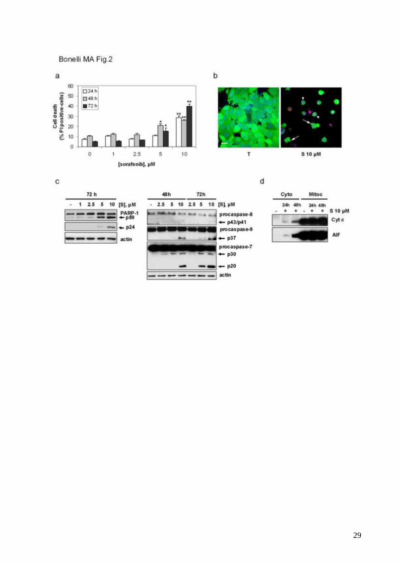
Sorafenib at higher concentrations induces cell death by apoptosis.
Treatment with sorafenib at higher concentrations (5-10µM) was associated with the
induction of cell death (fig.2a). Flow cytometry revealed the presence of Annexin V-FITC-
9
positive cells excluding PI at early time intervals (not shown), suggesting that sorafenib
induced cell death by apoptosis. This result was confirmed by CSLM analysis (fig.2b).
Sorafenib-treated cells exhibited some morphological hallmarks of apoptosis: rounded
shape, nuclear condensation, blebbing and calcein-positivity until late stages [34]; the
morphological changes associated with caspase-3 activity, such as fragmentation in
apoptotic bodies, were not detected because MCF-7/AROM-1 cells are null for caspase-3
[35]. As shown in fig.2c sorafenib induced cleavage of the caspase substrate PARP-1 [36]
and activation of the effector caspase-7. Also procaspase-9 was processed into active
subunits, whereas procaspase-8 remained uncleaved, indicating that sorafenib-mediated
apoptosis was associated with the activation of the mitochondrial intrinsic pathway. In line
with this observation, cytochrome c was detected in the cytosol (fig.2d). Concomitantly,
AIF was released from the mitochondria, suggesting that activation of both caspase-
dependent and -independent pathways could be involved in the pro -apoptotic effects of
sorafenib.
Combination of letrozole and sorafenib results in synergistic anti-proliferative
activity in aromatase-expressing breast cancer cell lines.
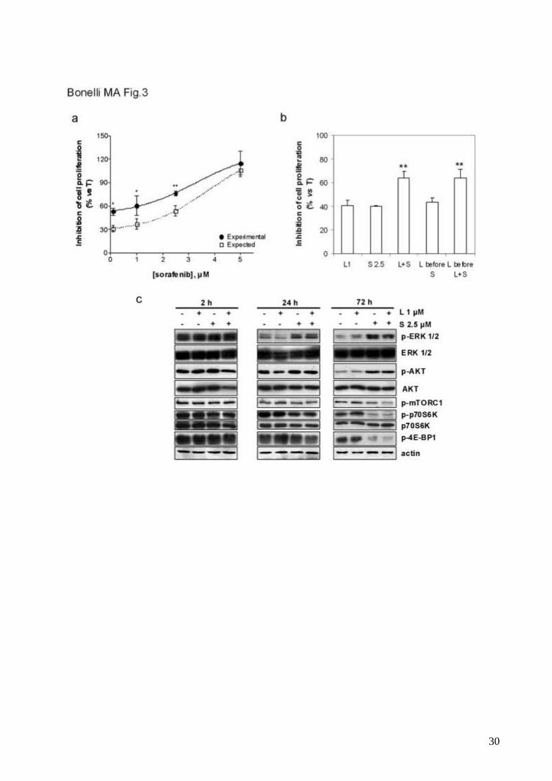
Treatment of MCF-7/AROM-1 cells with 1μM letrozole and increasing concentrations of
sorafenib (0.1-2.5μM) produced a synergystic inhibition of cell proliferation, as calculated
by the Bliss interaction model (fig.3a). Combination with 5μM sorafenib failed to produce
any enhancement of the growth-inhibitory effects, presumably because of the cytotoxic
action of sorafenib at this concentration.
To evaluate whether the synergistic anti-proliferative action of letrozole and sorafenib
required the simultaneous presence of both drugs from the start of treatment, we
compared three different schedules: “simultaneous”, “sequential”, and “up-front” (letrozole
before combination) treatment. As shown in fig.3b, the “sequential” treatment was not
10
effective in enhancing the anti-proliferative activity of the single agents. In contrast, the
“up-front” schedule showed the same enhanced anti-proliferative effect as that produced
by the “simultaneous” schedule.
We then examined the impact of the synergistic drug treatment on growth/survival
signalling pathways. As shown in fig.3c, letrozole alone had no effect on the
phosphorylation/expression of ERK1/2, AKT, mTORC1 and p70S6K. In contrast, sorafenib
significantly decreased the phosphorylation of mTORC1 and its targets p70S6K and 4E-
BP1 after 24-72h; concomitantly the phospho-AKT and phospho-ERK1/2 level increased,
presumably as consequence of release of mTORC1-mediated inhibition on PI3K/AKT
signalling [37]. The combination with letrozole did not produce any additional effect on
these changes.
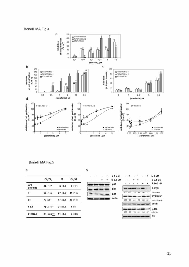
To evaluate whether the synergistic anti-proliferative effect of letrozole/sorafenib
combination was not restricted to MCF7/AROM-1 cells, treatments with letrozole and
sorafenib either alone or in combination were performed in other aromatase-expressing
breast cancer cell lines, i.e. T47D/AROM cells (clone 1 and 2) and BT474/AROM cells. As
demonstrated in MCF-7/AROM-1 cells, letrozole slowed cell proliferation (fig.4a).
Treatment with sorafenib inhibited cell proliferation in a concentration-dependent manner
(fig.4b) and was associated with inhibition of the mTORC1 pathway (not shown); in
addition induction of cell death was detected on treatment with higher concentrations of
the drug (fig.4c). Most importantly, the combined treatment produced a synergistic anti-
proliferative effect, as demonstrated by the Bliss interaction model (fig.4d), thus
strengthening the potential of combining letrozole and sorafenib for treatment of hormone-
dependent breast cancer.
Letrozole and sorafenib cooperate to induce G0/G1 cell cycle arrest without inducing
cell death.
11
Cell cycle analysis of MCF-7/AROM-1 cells (fig.5a) demonstrated that after 3 days of
steroid-deprivation the cells were almost completely blocked in G0/G1 phase. Treatment
with either letrozole or sorafenib also induced accumulation of cells in G0/G1 phase with a
concomitant decrease of cells in S phase. The accumulation of cells in G0/G1 phase was
further increased by the drug combination, thus confirming the synergistic growth-inhibitory
effects. Western blot analysis demonstrated that neither the single drug treatme nt nor the
combination affected the expression of p53 and p27 (fig.5b). Letrozole induced p21
expression; however, no further induction was observed in the presence of both drugs. In
contrast, a combined effect was detectable on the expression of c-myc, cyclin D1 and
phospho-Rb, whose levels were reduced after 3 days of treatment. Comparable effects
were obtained combining letrozole with the mTORC1 inhibitor rapamycin (fig.5b),
suggesting that the down-regulation of these cell cycle regulatory proteins required the
simultaneous inhibition of the estrogen-dependent signalling and the mTORC1 pathway.
Fluorescence microscopy demonstrated that letrozole 1μM combined with sorafenib 1-2.5
μM up to 6 days failed to promote cell death (not shown), indicating that the drug
combination had cytostatic and not cytotoxic effects.
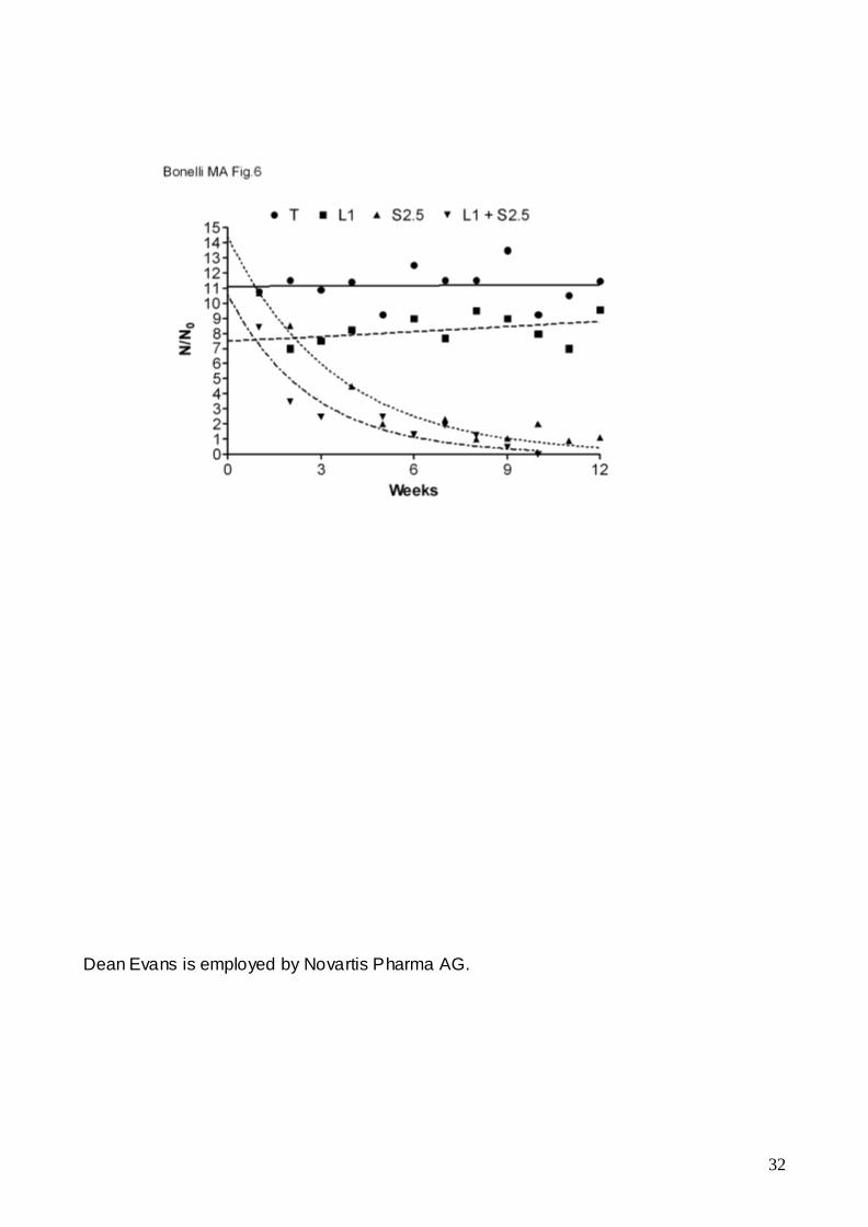
Sorafenib treatment prevents the acquisition of resistance towards letrozole.
Acquisition of resistance to letrozole has been shown to eventually occur after prolonged
treatments with the drug [38]. To evaluate whether sorafenib may prevent or delay the
development of resistance to letrozole, long-lasting treatments with the single agents or
the combination were performed in MCF-7/AROM-1 cells. As shown in fig.6, the
proliferation index of cells continuously cultured in the presence of letrozole was lower
than that observed in testosterone-stimulated cells. However, a more prolonged treatment
with letrozole eventually promotes the acquisition of resistance to the drug. Also sorafenib
treatment produced a decline in the proliferation index, however the cells continued to

12
proliferate, although at a very low rate. In contrast, continuous treatment with letrozole and
sorafenib induced a rapid decline of the proliferation index followed by death of the entire
cell population (N/N0<1 after 9 weeks), indicating that MCF-7/AROM-1 cells were not able
to adapt to the drug combination and therefore to acquire any form of resistance to
letrozole.
13
DISCUSSION
In recent years, crosstalk between ER and growth factor receptors has been shown to
contribute to both the failure of endocrine therapy as well as the development of resistance
in breast cancer. Therefore, a clear rationale has been developed for combining endocrine
therapies with signal transduction inhibitors to enhance endocrine responsiveness and to
potentially circumvent or delay the onset of acquired resistance [10, 11].
The multi-kinase inhibitor sorafenib has been recently approved for treatment of advanced
renal and hepatocellular carcinoma [13, 14] and has shown potent activity against a variety
of tumour cell types [39]. In breast cancer cells sorafenib reduces cell proliferation, and
sensitizes to chemotherapy via down-regulation of the Bcl-2-like anti-apoptotic protein Mcl-
1 [22, 40]. Moreover, sorafenib combined with nanoliposomal ceramide has been shown to
inhibit survival of MDA-MB-231 breast cancer cells and decrease tumour development in
vivo [41].
Here we provide evidence that letrozole combined with sorafenib produces synergistic
anti-proliferative effects in aromatase-expressing breast cancer cell lines (MCF-7/AROM-1,
T47D/AROM and BT474/AROM cells).
Proliferation of these cells is affected by estrogen or testosterone stimulation and letrozole,
by preventing testosterone aromatization, inhibited cell proliferation.
Also sorafenib inhibited cell proliferation in a concentration-dependent manner. It could be
argued that sorafenib’s anti-proliferative effect may result via direct inhibition of aromatase,
however recent studies have shown this is not the case [26]. In addition, when sorafenib
was used at higher concentrations (5-10μM) the inhibition of cell proliferation was
associated with apoptosis induction, via the mitochondrial intrinsic pathway.
In testosterone-stimulated MCF-7/AROM-1 cells, sorafenib at 2.5μM significantly
decreased the phosphorylation of mTORC1 and its targets p70S6K and 4E-BP1. A
14
sorafenib-mediated inhibition of the mTOR pathway has been described in melanoma cells
as a consequence of MAPK signalling inhibition and ascribed to the existence of crosstalk
between MAPK and mTOR signalling [42]. Under our experimental conditions, however,
inhibition of the MAPK pathway was only observed with higher concentrations of sorafenib
(5-10µM), suggesting that other mechanisms are involved in the down-regulation of the
mTORC1 signalling. Sorafenib-mediated inhibition of mTORC1 signalling was downstream
of AKT, being associated with AKT phosphorylation/activation. In this scenario, sorafenib
seems to mimic a phenomenon that has been widely described in various cellular models,
including MCF-7 cells, on treatment with rapamycin, i.e. the release of mTOR-dependent
negative regulation of RTK/IRS-1/PI3K/AKT signalling [37]. In addition, the recent
demonstration that mTORC1 inhibition increases RTK/IRS-1/PI3K activity towards
Ras/MAPK [43] may provide a mechanistic explanation for how treatment with 2.5 μM
sorafenib also leads to ERK1/2 phosphorylation/activation. Together, these results
demonstrate that the anti-proliferative effects of sorafenib were not dependent on its
activity as a Raf1 inhibitor, but involved the inhibition of the mTORC1 pathway.
Combination of letrozole and sorafenib significantly enhanced the anti -proliferative activity
compared with single agents alone in MCF7/AROM-1 cells as well as in T47D/AROM and
BT474/AROM cells, with statistical analysis indicating synergistic interaction. The growth-
inhibitory effects produced by a 6-days combined treatment were maintained by an “up-
front” schedule (a 3-days treatment with letrozole followed by 3 days of exposure to both
drugs). Considering the advantage of shortening drug treatments, this result suggests that
the “up-front” schedule may provide the optimal strategy for combining letrozole with
sorafenib.
In MCF7/AROM-1 cells, the synergistic inhibition of cell proliferation was associated with
accumulation of cells in the G0/G1 phase of the cell cycle and with down-regulation of
cyclin D1, c-myc and phospho-Rb expression. In the experimental conditions adopted in
15
the present study, both testosterone and serum growth factors contributed to sustain the
expression of cyclin D1 and c-myc thus promoting cell proliferation. Estrogens induce the
expression of these genes through a “non-classical” pathway in which ER binds to AP-1
proteins and functions as a coactivator to stimulate gene transcription [44]. Growth factor-
mediated contribution to cyclin D1 and c-myc expression is quite complex and primarily
involves the mTORC1 signalling cascade. In particular, mTORC1 can regulate the
synthesis of both cyclin D1 and c-myc by controlling cap-dependent mRNA translation.
However, when this pathway is inhibited, for example by rapamycin or sorafenib,
translation can be initiated via IRES-mediated mechanisms [45]. Moreover, mTORC1 can
induce cyclin D1 and c-myc gene transcription by activating transcriptional factors such as
STAT3 [46]. Since the regulation of these genes is governed by at least these two different
mechanisms of control (ER-mediated genomic pathway and mTORC1 pathway), both
have to be inhibited to reduce cyclin D1 and c-myc expression and to block cell cycle
progression.
As previously mentioned, development of resistance to letrozole is a clinical issue. It has
been reported that letrozole-resistant cell lines may be generated by prolonged culturing
(more than 50 weeks) in the presence of the drug [38]. Research into the mechanism of
endocrine responsiveness and resistance in breast cancer has revealed that various
growth factor pathways, such as EGFR/HER2 or PI3K/AKT family, and oncogenes
involved in the signal transduction cascades become activated and utilized by breast
cancer cells to bypass the effects of endocrine treatments [47]. Such cascades would
antagonize the anti-proliferative effect of anti-estrogens and thus represent attractive
targets for pharmacologic intervention with signal transduction inhibitors that target
aberrantly or excessively expressed oncogene products . Here we demonstrate that when
letrozole is used in combination with sorafenib under long-term culture conditions (12
16
weeks), the cell proliferation rate rapidly drops as compared with letrozole-treated cells,
and the cells ultimately die.
In conclusion, to our knowledge, this is the first in vitro study demonstrating the efficacy of
combining letrozole with the multi-kinase inhibitor sorafenib for the treatment of breast
cancer either to inhibit cell growth or to prevent the acquisition of resistance to AIs therapy.
To prove the clinical benefits of the association of letrozole with sorafenib a phase II
clinical trial in breast cancer neo-adjuvant setting is on-going between our Institutions.

17
Acknowledgements: We thank Bayer HealthCare Pharmaceuticals for providing
sorafenib
18
REFERENCES
(1). Osborne CK, Shou J, Massarweh S, Schiff R. (2005) Crosstalk between estrogen
receptor and growth factor receptor pathways as a cause for endocrine therapy resistance
in breast cancer. Clin Cancer Res 11:865s-70s
(2). Bjornstrom L, Sjoberg M. (2005) Mechanisms of estrogen receptor signaling:
convergence of genomic and nongenomic actions on target genes. Mol Endocrinol 19:833-
42
(3). Migliaccio A, Di Domenico M, Castoria G, et al. (1996) Tyrosine
kinase/p21ras/MAP-kinase pathway activation by estradiol-receptor complex in MCF-7
cells. EMBO J 15:1292-300
(4). Le Goff P, Montano MM, Schodin DJ, Katzenellenbogen BS. (1994)
Phosphorylation of the human estrogen receptor. Identification of hormone-regulated sites
and examination of their influence on transcriptional activity. J Biol Chem 269:4458-66
(5). Kato S, Endoh H, Masuhiro Y, et al. (1995) Activation of the estrogen receptor
through phosphorylation by mitogen-activated protein kinase. Science 270:1491-4
(6). Smith IE, Dowsett M. (2003) Aromatase inhibitors in breast cancer. N Engl J Med
348:2431-42
(7). Haynes BP, Dowsett M, Miller WR, Dixon JM, Bhatnagar AS. (2003) The
pharmacology of letrozole. J Steroid Biochem Mol Biol 87:35-45
(8). Bhatnagar AS. (2006) Review of the development of letrozole and its use in
advanced breast cancer and in the neoadjuvant setting. Breast 15 Suppl 1:S3-13
(9). Schiff R, Massarweh SA, Shou J, Bharwani L, Mohsin SK, Osborne CK. (2004)
Cross-talk between estrogen receptor and growth factor pathways as a molecular target
for overcoming endocrine resistance. Clin Cancer Res 10:331S-6S
19
(10). Johnston SR, Martin LA, Leary A, Head J, Dowsett M. (2007) Clinical strategies for
rationale combinations of aromatase inhibitors with novel therapies for breast cancer. J
Steroid Biochem Mol Biol 106:180-6
(11). Gligorov J, Azria D, Namer M, Khayat D, Spano JP. (2007) Novel therapeutic
strategies combining antihormonal and biological targeted therapies in breast cancer:
focus on clinical trials and perspectives. Crit Rev Oncol Hematol 64:115-28
(12). Generali D, Buffa FM, Berruti A, et al. (2009) Phosphorylated ERalpha, HIF-1alpha,
and MAPK signaling as predictors of primary endocrine treatment response and resistance
in patients with breast cancer. J Clin Oncol 27:227-34
(13). Kane RC, Farrell AT, Saber H, et al. (2006) Sorafenib for the treatment of advanced
renal cell carcinoma. Clin Cancer Res 12:7271-8
(14). Kane RC, Farrell AT, Madabushi R, et al. (2009) Sorafenib for the treatment of
unresectable hepatocellular carcinoma. Oncologist 14:95-100
(15). Wilhelm SM, Carter C, Tang L, et al. (2004) BAY 43-9006 exhibits broad spectrum
oral antitumor activity and targets the RAF/MEK/ERK pathway and receptor tyrosine
kinases involved in tumor progression and angiogenesis. Cancer Res 64:7099-109
(16). Plaza-Menacho I, Mologni L, Sala E, et al. (2007) Sorafenib functions to potently
suppress RET tyrosine kinase activity by direct enzymatic inhibition and promoting RET
lysosomal degradation independent of proteasomal targeting. J Biol Chem 282:29230-40
(17). Sharma A, Trivedi NR, Zimmerman MA, Tuveson DA, Smith CD, Robertson GP.
(2005) Mutant V599EB-Raf regulates growth and vascular development of malignant
melanoma tumors. Cancer Res 65:2412-21
(18). Liu L, Cao Y, Chen C, et al. (2006) Sorafenib blocks the RAF/MEK/ERK pathway,
inhibits tumor angiogenesis, and induces tumor cell apoptosis in hepatocellular carcinoma
model PLC/PRF/5. Cancer Res 66:11851-8
20
(19). Panka DJ, Wang W, Atkins MB, Mier JW. (2006) The Raf inhibitor BAY 43-9006
(Sorafenib) induces caspase-independent apoptosis in melanoma cells. Cancer Res
66:1611-9
(20). Huether A, Hopfner M, Baradari V, Schuppan D, Scherubl H. (2007) Sorafenib
alone or as combination therapy for growth control of cholangiocarcinoma. Biochem
Pharmacol 73:1308-17
(21). Rahmani M, Davis EM, Bauer C, Dent P, Grant S. (2005) Apoptosis induced by the
kinase inhibitor BAY 43-9006 in human leukemia cells involves down-regulation of Mcl-1
through inhibition of translation. J Biol Chem 280:35217-27
(22). Ding Q, Huo L, Yang JY, et al. (2008) Down-regulation of myeloid cell leukemia-1
through inhibiting Erk/Pin 1 pathway by sorafenib facilitates chemosensitization in breast
cancer. Cancer Res 68:6109-17
(23). Huynh H, Ngo VC, Koong HN, et al. (2009) Sorafenib and Rapamycin Induce
Growth Suppression in Mouse Models of Hepatocellular Carcinoma. J Cell Mol Med DOI:
10.1111/j.1582-4934.2009.00692.x
(24). Kumar SM, Yu H, Edwards R, et al. (2007) Mutant V600E BRAF increases hypoxia
inducible factor-1alpha expression in melanoma. Cancer Res 67:3177-84
(25). Macaulay VM, Nicholls JE, Gledhill J, Rowlands MG, Dowsett M, Ashworth A.
(1994) Biological effects of stable overexpression of aromatase in human hormone-
dependent breast cancer cells. Br J Cancer 69:77-83
(26). Banerjee S, Zvelebil M, Furet P, et al. (2009) The vascular endothelial growth factor
receptor inhibitor PTK787/ZK222584 inhibits aromatase. Cancer Res 69:4716-23
(27). Cavazzoni A, Petronini PG, Galetti M, et al. (2004) Dose-dependent effect of FHIT-
inducible expression in Calu-1 lung cancer cell line. Oncogene 23:8439-46
21
(28). Fumarola C, La Monica S, Alfieri RR, Borra E, Guidotti GG. (2005) Cell size
reduction induced by inhibition of the mTOR/S6K-signaling pathway protects Jurkat cells
from apoptosis. Cell Death Differ 12:1344-57
(29). Goldoni M, Johansson C. (2007) A mathematical approach to study combined
effects of toxicants in vitro: evaluation of the Bliss independence criterion and the Loewe
additivity model. Toxicol In Vitro 21:759-69
(30). La Monica S, Galetti M, Alfieri RR, et al. (2009) Everolimus restores gefitinib
sensitivity in resistant non-small cell lung cancer cell lines. Biochem Pharmacol 78:460-8
(31). Aesoy R, Sanchez BC, Norum JH, Lewensohn R, Viktorsson K, Linderholm B.
(2008) An autocrine VEGF/VEGFR2 and p38 signaling loop confers resistance to 4-
hydroxytamoxifen in MCF-7 breast cancer cells. Mol Cancer Res 6:1630-8
(32). Lathia C, Lettieri J, Cihon F, Gallentine M, Radtke M, Sundaresan P. (2006) Lack of
effect of ketoconazole-mediated CYP3A inhibition on sorafenib clinical pharmacokinetics.
Cancer Chemother Pharmacol 57:685-92
(33). Strumberg D, Clark JW, Awada A, et al. (2007) Safety, pharmacokinetics, and
preliminary antitumor activity of sorafenib: a review of four phase I trials in patients with
advanced refractory solid tumors. Oncologist 12:426-37
(34). Gatti R, Belletti S, Orlandini G, Bussolati O, Dall'Asta V, Gazzola GC. (1998)
Comparison of annexin V and calcein-AM as early vital markers of apoptosis in adherent
cells by confocal laser microscopy. J Histochem Cytochem 46:895-900
(35). Kagawa S, Gu J, Honda T, et al. (2001) Deficiency of caspase-3 in MCF7 cells
blocks Bax-mediated nuclear fragmentation but not cell death. Clin Cancer Res 7:1474-80
(36). Oliver FJ, de la Rubia G, Rolli V, Ruiz-Ruiz MC, de Murcia G, Murcia JM. (1998)
Importance of poly(ADP-ribose) polymerase and its cleavage in apoptosis. Lesson from an
uncleavable mutant. J Biol Chem 273:33533-9
22
(37). O'Reilly KE, Rojo F, She QB, et al. (2006) mTOR inhibition induces upstream
receptor tyrosine kinase signaling and activates Akt. Cancer Res 66:1500-8
(38). Belosay A, Brodie AM, Njar VC. (2006) Effects of novel retinoic acid metabolism
blocking agent (VN/14-1) on letrozole-insensitive breast cancer cells. Cancer Res
66:11485-93
(39). Wilhelm S, Chien DS. (2002) BAY 43-9006: preclinical data. Curr Pharm Des
8:2255-7
(40). Yu C, Bruzek LM, Meng XW, et al. (2005) The role of Mcl-1 downregulation in the
proapoptotic activity of the multikinase inhibitor BAY 43-9006. Oncogene 24:6861-9
(41). Tran MA, Smith CD, Kester M, Robertson GP. (2008) Combining nanoliposomal
ceramide with sorafenib synergistically inhibits melanoma and breast cancer cell survival
to decrease tumor development. Clin Cancer Res 14:3571-81
(42). Molhoek KR, Brautigan DL, Slingluff CL, Jr. (2005) Synergistic inhibition of human
melanoma proliferation by combination treatment with B-Raf inhibitor BAY43-9006 and
mTOR inhibitor Rapamycin. J Transl Med 3:39
(43). Carracedo A, Ma L, Teruya-Feldstein J, et al. (2008) Inhibition of mTORC1 leads to
MAPK pathway activation through a PI3K-dependent feedback loop in human cancer. J
Clin Invest 118:3065-74
(44). DeNardo DG, Cuba VL, Kim H, Wu K, Lee AV, Brown PH. (2007) Estrogen receptor
DNA binding is not required for estrogen-induced breast cell growth. Mol Cell Endocrinol
277:13-25
(45). Shi Y, Sharma A, Wu H, Lichtenstein A, Gera J. (2005) Cyclin D1 and c-myc
internal ribosome entry site (IRES)-dependent translation is regulated by AKT activity and
enhanced by rapamycin through a p38 MAPK- and ERK-dependent pathway. J Biol Chem
280:10964-73

23
(46). Zhou J, Wulfkuhle J, Zhang H, et al. (2007) Activation of the PTEN/mTOR/STAT3
pathway in breast cancer stem-like cells is required for viability and maintenance. Proc
Natl Acad Sci U S A 104:16158-63
(47). Nicholson RI, McClelland RA, Robertson JF, Gee JM. (1999) Involvement of steroid
hormone and growth factor cross-talk in endocrine response in breast cancer. Endocr
Relat Cancer 6: 373-87
24
FIGURE LEGENDS
Fig.1 MCF-7/AROM-1 cell proliferation is stimulated by estrogens and inhibited by
letrozole and sorafenib
a, Steroid-deprived cells (104cells/cm2) were treated with E2 or T up to 144h. Cell
proliferation was assessed by MTT assay. Data are expressed as fold increase versus
steroid-deprived cells.
b, Cells were seeded at the indicated density in 96-multiwell plates and 24h later were
treated with T in the absence or presence of increasing concentrations of letrozole. After 6
days of incubation cell proliferation was assessed by MTT assay.
c, Steroid-deprived cells (104cells/cm2) were treated with T in the absence or presence of
increasing concentrations of sorafenib (0.1-5µM). After 6 days cell proliferation was
assessed by MTT assay. The IC50 value, calculated as reported in Material and Methods,
was of 3µM.
Data in b and c are expressed as percent of inhibition of cell proliferation versus T-
stimulated cells. Columns, means of three separate experiments; bars, SD. *p<0.05;
**p<0.01.
d, Cells were pre-incubated for 30min with sorafenib (S, 1-10μM) and then stimulated with
T for 2h in the absence or presence of the drug at the indicated concentrations. The cells
were lysed and protein expression was assessed by Western blot analysis. Data are from
a representative experiment. Each experiment, repeated three times, yielded similar
results.
Fig. 2 Sorafenib at high concentrations induces cell death by apoptosis
25
Steroid-deprived MCF-7/AROM-1 cells were treated with T in the absence or presence of
various concentrations of sorafenib (S, 1-10 µM) up to 72h.
a, Cell death was quantitated at the indicated time intervals by fluorescence microscopy on
Hoechst 33342/PI-stained cells. Data are expressed as percent values. Columns, means
of three separate experiments; bars, SD. *p<0.05; **p<0.01.
b, Morpho-functional analysis of cell death was evaluated by real time CLSM after 48h of
treatment with sorafenib 10µM using the vital stain calcein-AM (green colour), and the
nuclear dyes Draq5 (blue colour) and PI (red colour) to discriminate between viable/early
apoptotic and dead cells. Arrows, blebs; arrowheads, calcein-AM positivity unti l late stages
of cell death. Bar, 20µm.
c, Cleavage of PARP-1, procaspase-8, -9 and -7 were assessed at the indicated time by
Western blot analysis.
d, Cytochrome c and AIF release from mitochondria was assessed after 24 and 48h of
incubation with sorafenib 10µM by Western blot analysis of cell cytosolic and mitochondrial
fractions.
Images shown in b and results presented in c and d are from a representative experiment.
Each experiment, repeated three times, yielded similar results.
Fig. 3 Combination of letrozole and sorafenib potentiates the inhibition of cell proliferation
a, Steroid-deprived MCF-7/AROM-1 cells (104cells/cm2) were treated with T in the
absence or presence of 1μM letrozole combined with various concentrations of sorafenib
(0.1-5μM). After 6 days cell proliferation was assessed by MTT assay and the effect of the
drug combination was evaluated using the Bliss interaction model. Data are expressed as
percent of inhibition of cell proliferation versus T-stimulated cells and are means of three
separate experiments. Bars, SD. *p<0.05; **p<0.01.
26
b, Cells (104cells/cm2) were treated for 6 days with T in the absence or presence of 1μM
letrozole (L1), 2.5μM sorafenib (S2.5) or three different combinations of the drugs: 6 days
of continuous exposure to letrozole and sorafenib (L+S); a 3 days-treatment with letrozole
followed by a 3 days-treatment with sorafenib (L before S); a 3 days-treatment with
letrozole followed by 3 days of combined treatment (L before L+S). Cell proliferation was
assessed by MTT assay. Data are expressed as percent of inhibition of cell proliferation
versus T-stimulated cells. Columns, means of three separate experiments; bars, SD.
*p<0.05; **p<0.01 (one-way ANOVA).
c, Cells were treated with T in the absence or presence of 1μM letrozole (L), 2.5μM
sorafenib (S) or the combination of both. The cells were lysed at the indicated time and
protein expression was assessed by Western blot analysis. The results are from a
representative experiment. Each experiment, repeated three times, yielded similar results.
Fig. 4 Combination of letrozole and sorafenib produces a synergistic inhibition of cell
proliferation in T47D/AROM and BT474/AROM cells
Steroid-deprived cells (104cells/cm2) were treated with T in the absence or presence of
increasing concentrations of letrozole (a) or sorafenib (b). After 6 days of incubation cell
proliferation was assessed by MTT assay. Data are expressed as percent of inhibition of
cell proliferation versus T-stimulated cells. Columns, means of three separate
experiments; bars, SD. *p<0.05; **p<0.01.
c, Steroid-deprived cells were treated with T in the absence or presence of various
concentrations of sorafenib up to 72h. Cell death was quantitated by fluorescence
microscopy on Hoechst 33342/PI-stained cells. Data are expressed as percent values.
Columns, means of three separate experiments; bars, SD. *p<0.05; **p<0.01.
d, Steroid-deprived cells (104cells/cm2) were treated with T in the absence or presence of
letrozole (1μM for T47D/AROM clone 1 and 2; 10-2μM for BT474/AROM cells) combined
27
with increasing concentrations of sorafenib. After 6 days cell proliferation was assessed by
MTT assay and the effect of the drug combination was evaluated using the Bliss
interaction model. Data are expressed as percent of inhibition of cell proliferation versus T-
stimulated cells and are means of three separate experiments. Bars, SD. *p<0.05;
**p<0.01.
Fig. 5 Letrozole and sorafenib cooperate to inhibit cell cycle progression in G1 phase
Steroid-deprived MCF-7/AROM-1 cells were untreated (without steroids) or treated with T
in the absence or presence of 1μM letrozole (L1), 2.5μM sorafenib (S2.5) or a combination
of both.
a, After 72h the cells were analyzed by flow cytometry for cell cycle-phase distribution.
Mean percentages ±SD of cells residing in each cycle phase are reported in the table
(*p<0.05; **p<0.01, Student's t test versus T-stimulated cells; ### p<0.001, one-way
ANOVA versus single agent treatment).
b, After 72h of treatment the cells were lysed and protein expression was assessed by
Western blot analysis. R: rapamycin. The results are from a representative experiment.
Each experiment, repeated three times, yielded similar results.
Fig. 6 Effect of long-lasting treatments with letrozole and sorafenib on MCF-7/AROM-1 cell
proliferation
MCF-7/AROM-1 T-stimulated cells were continuously cultured in the presence of 1μM
letrozole (L1), 2.5μM sorafenib (S2.5), or the combination of both (L1+S2.5) up to 12
weeks. Once a week the cells were counted and reseeded at the density of 8 x10³
cells/cm2. Data are expressed as proliferation index N/N0, where N is the number of cells
counted after 7 days of culture and N0 is the number of cells initially seeded.
28
29
30
31
32
Dean Evans is employed by Novartis Pharma AG.
Copyright © 2022 FDOKUMEN