The role of polycystic ovary syndrome (PCOS) and overweight ...

MOLECULAR AND CELLULAR BIOLOGY,0270-7306/00/$04.0010
June 2000, p. 3764–3771 Vol. 20, No. 11
Copyright © 2000, American Society for Microbiology. All Rights Reserved.
Induction of hTERT Expression and Telomerase Activity byEstrogens in Human Ovary Epithelium Cells
SILVIA MISITI,1 SIMONA NANNI,1 GIULIA FONTEMAGGI,1 YU-SHENG CONG,2 JIANPING WEN,2
HAL W. HIRTE,3 GIULIA PIAGGIO,1 ADA SACCHI,1 ALFREDO PONTECORVI,1,4
SILVIA BACCHETTI,2* AND ANTONELLA FARSETTI1,5*
Molecular Oncogenesis Laboratory, Regina Elena Cancer Institute,1 and Institute of Experimental Medicine,National Research Council,5 Rome, and Institute of Medical Pathology, Catholic University, Milan,4 Italy,
and Department of Pathology and Molecular Medicine2 and Department of Medicine,3
McMaster University, Hamilton, Ontario, Canada
Received 10 November 1999/Returned for modification 21 December 1999/Accepted 2 March 2000
In mammals, molecular mechanisms and factors involved in the tight regulation of telomerase expressionand activity are still largely undefined. In this study, we provide evidence for a role of estrogens and theirreceptors in the transcriptional regulation of hTERT, the catalytic subunit of human telomerase and, conse-quently, in the activation of the enzyme. Through a computer analysis of the hTERT 5*-flanking sequences, weidentified a putative estrogen response element (ERE) which was capable of binding in vitro human estrogenreceptor a (ERa). In vivo DNA footprinting revealed specific modifications of the ERE region in ERa-positivebut not ERa-negative cells upon treatment with 17b-estradiol (E2), indicative of estrogen-dependent chroma-tin remodelling. In the presence of E2, transient expression of ERa but not ERb remarkably increased hTERTpromoter activity, and mutation of the ERE significantly reduced this effect. No telomerase activity wasdetected in human ovary epithelial cells grown in the absence of E2, but the addition of the hormone inducedthe enzyme within 3 h of treatment. The expression of hTERT mRNA and protein was induced in parallel withenzymatic activity. This prompt estrogen modulation of telomerase activity substantiates estrogen-dependenttranscriptional regulation of the hTERT gene. The identification of hTERT as a target of estrogens representsa novel finding which advances the understanding of telomerase regulation in hormone-dependent cells andhas implications for a potential role of hormones in their senescence and malignant conversion.
Most human somatic cells do not express telomerase, theribonucleoprotein that elongates telomeric DNA, or its cata-lytic protein, hTERT, which is limiting for enzyme activity (33).In humans, telomerase is regulated in a tissue-specific mannerduring development (42); the enzyme is present in early em-bryogenesis but is repressed upon cell differentiation in so-matic tissues (27, 42). Loss of enzymatic activity is accompa-nied by loss of the full-length transcript of hTERT and/or bythe appearance of alternatively spliced transcripts that are un-likely to encode functional proteins (21, 42). In the adult, te-lomerase persists only in germ line cells and in progenitor cellsof somatic tissues with self-renewing potential, in agreementwith the requirement for the enzyme for sustained cell prolif-eration (16). How hTERT silencing is achieved and which fac-tors contribute to this process are presently unknown, althoughthe regulation of hTERT expression appears to be primarily atthe transcriptional level (42). An understanding of the molec-ular mechanisms underlying the regulation of telomerase ac-tivity might allow the modulation of telomerase expression and,consequently, of cell life span (4, 43), with important potentialtherapeutic applications in aging and malignancy.
Several lines of evidence suggest that sex steroid hormonesmay be good candidates as physiological regulators of hTERT
expression. Recent findings are consistent with the hypothesisthat telomerase activity is potentially under hormonal controlin some estrogen-targeted tissues, such as the endometrium(25, 37, 40), and the prostate (30), and in epithelial cells withhigh renewing potential from estrogen-regulated tissues (3).Physiological responses to estrogen are mediated, within spe-cific tissues, by at least two members of the nuclear hormonereceptor superfamily, the estrogen receptors (ERs) ERa andERb (2). These are ligand-dependent transcription factors be-longing to a large family of structurally related proteins thatare able to modulate the expression of a variety of genesinvolved in diverse biological functions, such as cell prolifera-tion, morphogenesis, cellular differentiation, and programmedcell death. ERs act by direct interaction of the hormone-re-ceptor complex with a set of specific DNA sequences, the es-trogen response elements (EREs), localized in the 59-flankingregions of hormone-regulated genes. Distinct ligand-directedconformational changes of the hormone-receptor complex mayresult, in turn, in transcriptional silencing or activation of tar-get genes (2, 28). Alternative mechanisms of ER activation,involving coregulatory proteins (19, 20), transcription factorssuch as AP-1 or Sp1 (35, 44), and/or phosphorylation signalingpathways, have also been reported (2).
The recent cloning and characterization of the hTERT geneand its promoter region (6, 17, 40, 45) has provided essentialreagents for the investigation of the molecular mechanismsinvolved in the regulation of telomerase in different cell back-grounds. In this study, we provide evidence for a potential roleof estrogens and ERs in the transcriptional regulation ofhTERT by demonstrating that a noncanonical ERE within thehTERT promoter is functional in vitro and in vivo and that theaddition of estrogen to human ovary epithelium cell cultures
* Corresponding author. Mailing address for Silvia Bacchetti: De-partment of Pathology and Molecular Medicine, McMaster University,Hamilton, Ontario L8N 3Z5, Canada. Phone: (905) 525-9140, ext.22296. Fax: (905) 546-9940. E-mail: [email protected]. Mail-ing address for Antonella Farsetti: Molecular Oncogenesis Laboratory,Regina Elena Cancer Institute, Via delle Messi d’Oro 156, 00158Rome, Italy. Phone: (39-06) 4985-2531. Fax: (39-06) 4180-526. E-mail:[email protected].
3764
on February 24, 2016 by guest
http://mcb.asm
.org/D
ownloaded from

results in the induction of hTERT expression and of telomer-ase activity.
MATERIALS AND METHODS
Cells. Human ovarian surface epithelium (HOSE) cell strains GRO, LLO, andLEA and immortal HOSE cell line WOO were cultured in E3 medium with 3%fetal calf serum (FCS) (8). The human ovarian cancer cell line OVCA-433 (36)was grown in RPMI 1640 with 10% FCS, while cervical cancer HeLa cells, breastcancer MCF-7 and MDA-MB231 cells, and mouse NIH 3T3 fibroblasts weregrown in Dulbecco modified essential medium with 10% FCS. Forty-eight hoursprior to experimental use, the cells were switched to medium supplemented withhormone-deprived serum (18).
Plasmids and transfections. Plasmids P-1009 and P-330 and the pGL2-En-hancer vector have been described previously (6). P-1009Mut, with a mutatedERE, was generated using a QuickChange Site-Directed Mutagenesis Kit (Strat-agene, La Jolla, Calif.) and the following oligonucleotide sequence: 59-CCTCCCCCTTGTGCGGGCATGATGTGATCAGATGTTGGCC-39.
The hTERT-ERE-TK reporter was generated by inserting double-strandedoligonucleotides encompassing the hTERT promoter (2956 to 2930) into thelinker region of pBLCAT2 (12) upstream of the thymidine kinase (TK) promoterat bp 2105. All constructs were sequenced using the dideoxynucleotide method(38). pSG5-HEO, encoding human ERa (13), and the human ERb expressionvector (24) were gifts from P. Chambon (Strasbourg, France) and J. A. Gustafs-son (Huddinge, Sweden), respectively. The reporter vector for the Xenopus laevisvitellogenin B1 (VIT) promoter has been previously described (12). pCMV-b-galwas used as an internal control to monitor transfection efficiency. Cells wereelectroporated as described previously (12) and assayed for luciferase and b-ga-lactosidase activities using reagents and protocols from Promega (Madison,Wis.).
Electrophoretic mobility shift assay. A 32P-labeled double-stranded oligonu-cleotide containing the hTERT ERE (59-GCATGTGTGTGCGGGCGGGATGTGACCAGATGTGATCC-39; bp 2949 to 2935 upstream of the ATG) wasassayed for binding to extracts from Spodoptera frugiperda Sf9 cells infected witha baculovirus expressing human ERa (5) or ERb (Alexis Biochemicals, Milan,Italy) or with a control baculovirus. As a control for ER binding, a 32P-labeleddouble-stranded oligonucleotide spanning the canonical X. laevis VIT ERE wasused. Competition experiments were performed by adding to the binding mixtureincreasing amounts of unlabeled oligonucleotides containing the hTERT ERE,the human coaggulation factor XII ERE (12), or the mutant hTERT ERE 20min prior to the addition of the 32P-labeled probe. Supershift experiments withERb were carried out with anti-ERb antibody L-20X (Santa Cruz, Santa Cruz,Calif.). Binding reactions and native polyacrylamide gel electrophoresis werecarried out as previously described (12).
Western blot analysis. For detection of ERa expression, cell lysates wereresolved by sodium dodecyl sulfate-polyacrylamide gel electrophoresis and trans-ferred to nitrocellulose membranes (Bio-Rad, Hercules, Calif.). Immunostainingof proteins was done with antibody HC-20 according to supplier instructions(Santa Cruz), and detection was done by enhanced chemiluminescence (Amer-sham Corp., Arlington Heights, Ill.).
Genomic footprinting. Cells were treated with the DNA-alkylating reagentdimethyl sulfate (DMS) (0.1% for 2 min for MCF-7, MDA-MB231, and HeLacells and 0.2% for 4 min for OVCA-433 cells), and their DNA was cleaved withpiperidine. Genomic footprinting was performed by ligation-mediated (LM)PCR (9) with Vent DNA polymerase for first-strand extension and subsequentPCR amplification. To generate footprints for the endogenous hTERT gene, thefollowing primers, specific for the region of interest, were used (coding strand):primer 1, GAATCGGCCTAGGCTGTG; primer 2, ACCGGGCGCCTCACACCAGCC; and primer 3, ACCGGGCGCCTCACACCAGCCACAACGG. La-beled PCR products were resolved on a 6% polyacrylamide–8 M urea sequencinggel. Control samples consisted of chromatin-free DNA from each cell linetreated in vitro with 0.125% DMS for 2 min. Volumetric integration of signalintensities was performed with NIH Image software (version 1.58), and quanti-tation was done as described by Dey et al. (9). Briefly, the average value of eachband from three independent experiments was normalized to the value of thecorresponding band in the control guanine ladder. Methylation percentages wereobtained by normalization of values for ERa-positive (MCF-7 and OVCA-433)cells to those for ERa-negative (MDA-MB231 and HeLa) cells, which showedno protection or hypersensitivity. Values of ,15% were considered not signifi-cant (32).
RT-PCR analysis of hTERT mRNA. Expression of hTERT mRNA was ana-lyzed by semiquantitative reverse transcription (RT)-PCR amplification. TotalRNA was prepared from HOSE cells using RNAzol B (Biotech, Rome, Italy)according to the manufacturer’s protocol. One microgram of total RNA wasreverse transcribed at 37°C for 45 min in the presence of random hexamers andMoloney murine leukemia virus reverse transcriptase (Gibco-BRL). hTERTmRNA analysis was carried out by PCR amplification of a fragment of 145 bpusing primers and conditions described by Ulaner et al. (42). The housekeepingaldolase mRNA, used as an external standard, was amplified from the samecDNA reaction mixture using specific primers (31). The exponential phase ofamplification was previously determined by serial dilution of RT reaction mix-tures for each cDNA template used and PCR performed under these conditions.
Amplified PCR products were electrophoresed on a 3% agarose gel containingethidium bromide (0.5 mg/ml) and visualized under UV light.
Immunofluorescence. LEA and LLO cells, grown in E3 medium with hor-mone-deprived serum, were seeded in 35-mm plates. After 16 h, the medium wasreplaced with fresh medium containing 17b-estradiol (E2) at a final concentra-tion of 1027 M or the equivalent volume of vehicle alone. Cells were immuno-stained with the telomerase-specific antibody K-370 as described by Martin-Rivera et al. (29), and nuclei were stained with Hoechst 33258. Images werecaptured with a Zeiss fluorescence microscope.
Telomerase assay. Extracts from GRO, LEA, LLO, and WOO cells wereprepared by detergent lysis, and enzymatic activity was detected by the PCR-based telomere repeat amplification protocol (TRAP) (22).
RESULTS AND DISCUSSION
Ligand-dependent occupancy of the hTERT promoter invitro and in vivo. A computer-assisted analysis of the hTERT59-flanking sequence (6) revealed a composite regulatory unitcomprising an imperfect palindromic consensus sequence forthe ERE (59-GGCGGGATGTGACCA-39, at positions 2949to 2935 relative to the ATG), partially overlapping an AP1binding site and adjacent to an SP1 motif (Fig. 1a).
We first evaluated the ability of the hTERT promoter tobind in vitro ligand-activated human ERs. Incubation of a32P-labeled oligonucleotide spanning the hTERT ERE withextracts from S. frugiperda (Sf9) cells infected with a baculovi-rus expressing ERa (Fig. 1b, lane 3) resulted in the formationof a specific complex that was progressively inhibited by in-creasing concentrations of unlabeled oligonucleotides corre-sponding to the hTERT ERE (Fig. 1b, lanes 4 to 6) or to thewell-characterized FXII ERE (12) (Fig. 1b, lanes 7 to 9). Com-pared to the hTERT ERE, the FXII ERE was a less efficientcompetitor, likely because of sequence divergence between thetwo oligonucleotides within the 59 half-site. No inhibition wasobserved upon the addition of a 250-fold molar excess of anunrelated oligonucleotide (Fig. 1b, lane 10), and no complexwas formed when the hTERT ERE was incubated with extractsfrom SF9 cells infected with wild-type virus (Fig. 1b, lane 2),underscoring the specificity of the interaction between ERaand the hTERT ERE. An oligonucleotide containing a mu-tated hTERT ERE competed weakly for ER binding (data notshown), in agreement with functional data showing that muta-tions strongly reduced the estrogen response of the element(see Fig. 3a and b). Incubation of 32P-labeled hTERT EREwith Sf9 cells infected with a baculovirus expressing ERb didnot show any interaction, whether in the presence or absenceof E2 or of the antiestrogen 4-hydroxytamoxifen (TAM). As acontrol for the functionality of ERb, the canonical X. laevisVIT ERE was used (Fig. 1c). In the presence of E2, ERbbound the VIT ERE and was supershifted upon the addition ofanti-ERb antibody (Fig. 1c, lanes 2 and 4, respectively). Theaddition of TAM to the binding mixture inhibited the forma-tion of the ERb-VIT ERE complex (Fig. 1c, lane 3).
To assess the functionality of the hTERT ERE in cells fromestrogen-regulated tissues, we compared the in vivo DNA foot-prints over this region in ERa-positive (MCF-7 and OVCA-433) and ERa-negative (MDA-MB231 and HeLa) cells (Fig.2a) cultured in the presence or absence of E2 (1027 M) forvarious times. Cells were treated with DMS, and their DNAwas analyzed by LM PCR with primers specific for the regionof the hTERT promoter from positions 21025 to 2917 rela-tive to the ATG (Fig. 1a). The extent of protection or hyper-sensitivity of specific residues was evaluated by densitometry(Table 1) (Materials and Methods). A comparison of DNAextracted from DMS-treated cells to purified DNA treatedwith DMS in vitro (Fig. 2b to e) revealed a mixed pattern ofprotected and hyperreactive G residues in cells constitutivelyexpressing ERa (e.g., MCF-7 and OVCA-433). In particular,
VOL. 20, 2000 ESTROGEN INDUCTION OF hTERT GENE EXPRESSION 3765
on February 24, 2016 by guest
http://mcb.asm
.org/D
ownloaded from

in MCF-7 cells grown in the absence of E2, we observed pro-tection (;70%) of the G at position 2954; protection was lesspronounced (;40%) in cells grown with the hormone (Fig. 2b,compare lane 2 with lanes 1 and 3). The addition of estrogen
resulted in hypermethylation (30 to 55%) of two clusters of Gresidues, at positions 2949 to 2945, immediately upstream ofand within the hTERT ERE 59 half-site; this result was com-patible with unmasking of binding sites normally inaccessible
FIG. 1. (a) Schematic diagram and nucleotide sequence of the hTERT gene 59-flanking sequences. The region extending to bp 1009 upstream of the hTERT ATG(11) and the locations of the two ERE half-sites and of an additional downstream half-site (black triangles) are indicated. The hTERT promoter sequence betweenbp 21009 and 2755 is shown below the diagram. The boxes define the composite regulatory unit comprising an imperfect palindromic ERE at positions 2949 to 2935,a partially overlapping AP1 binding site, an adjacent SP1 motif, and the single ERE half-site at positions 2794 to 2789. The asterisks indicate G residues altered inthe genomic footprints shown in Fig. 2. (b) ERa binding to the hTERT ERE. A 32P-labeled double-stranded oligonucleotide containing the hTERT ERE sequencewas incubated with extracts of Sf9 cells infected with wild-type (wt) baculovirus (lane 2) or recombinant baculovirus expressing human ERa (lanes 3 to 10). Lane 1,probe alone; lane 3, recombinant ERa alone; lanes 4 to 9, like lane 3 but with 25-, 100-, and 250-fold molar excesses of unlabeled oligonucleotides containing the hTERT(lanes 4 to 6) or FXII (lanes 7 to 9) ERE sequences; lane 10, like lane 3 but with a 250-fold molar excess of an unrelated unlabeled oligonucleotide (NS). (c) ERbbinding to EREs. 32P-labeled double-stranded oligonucleotides containing the VIT ERE (lanes 1 to 4) or the hTERT ERE (lanes 5 to 8) were incubated with extractsof Sf9 cells infected with recombinant baculovirus expressing human ERb (lanes 2 to 4 and 6 to 8) in the presence of (E2) (lanes 2, 4, 6, and 8) or of TAM (lanes 3and 7). Anti-ERb antibodies (lanes 4 and 8) were used for supershifting ERb-ERE complexes. Lane 1, VIT ERE probe alone; lane 5, hTERT ERE probe alone.
3766 MISITI ET AL. MOL. CELL. BIOL.
on February 24, 2016 by guest
http://mcb.asm
.org/D
ownloaded from

to transcription factors, as suggested for the uteroglobingene enhancer in hormone-treated endometrial cells (39).In OVCA-433 cells (Fig. 2d), estrogen treatment resulted ina different pattern, with consistent protection (35 to 45%) offour G residues (at positions 2954, 2950, 2946, and 2944)upstream of and within the 59 half-site of the ERE. In addition,G residues at positions 2931 and 2927 downstream of theERE were protected, in comparison to the results for in vitro-treated DNA or DNA from cells grown without estrogen. Pro-tection of all these regions may be mediated by the conforma-tional and functional changes that the ER undergoes following
transition from an inactive to an active state (2, 28). The pres-ence of a binding site for AP1 and SP1 adjacent to or within theERE may also contribute to this effect, since these nuclearproteins have been shown to interact with ERa in certaincontexts involving estrogen-regulated promoters (35, 44).
In MDA-MB231 and HeLa cells, both of which do not ex-press ERa (Fig. 2a), no genomic footprint was detectable overthe region comprising the ERE, regardless of hormone treat-ment (Fig. 2c and e); this result suggests that no factors arebound to this region, at least in these cell backgrounds. Thelack of a footprint in MDA-MB231 cells, which express low
FIG. 2. (a) Expression of endogenous ERa by Western blot analysis. ERa-negative (HeLa and MDA-MB231) and ERa-positive (OVCA-433 and MCF-7) cellswere lysed directly in protein sample buffer, and equal amounts of protein were separated on a denaturing 12% polyacrylamide gel. Immunostaining was performedwith anti-ERa antibody HC-20. As a loading control, proteins were stained with Ponceau S (data not shown). Treatment with E2 did not affect the levels of ERa inthe ER-positive cells (data not shown). (b to e) DMS genomic footprinting of the hTERT promoter. Cells were treated with the DNA-alkylating reagent DMS, andtheir DNA was cleaved with piperidine and analyzed by LM PCR with primers specific for the region of the hTERT promoter from bp 21025 to 2917 relative to theATG (Fig. 1a). (b) Breast cancer MCF-7 cells. (c) Breast cancer MDA-MB231 cells. (d) Ovarian cancer OVCA-433 cells. (e) Cervical cancer HeLa cells. Length (inhours) of treatment with E2 (lanes 2, 3, 5 to 9, and 11 to 13) is indicated. In vitro-methylated DNA from each cell line is shown in lanes 1, 4, 10, and 14. Protectedguanine residues over the ERE region are indicated by open arrows, while relevant hypermethylated guanine residues are indicated by filled arrows (b and d).Corresponding G residues unmodified in ERa-negative cells are indicated by arrowheads (c and e). The asterisks (in panel d) indicate two protected guanine residues,of unknown significance, downstream of the ERE region.
VOL. 20, 2000 ESTROGEN INDUCTION OF hTERT GENE EXPRESSION 3767
on February 24, 2016 by guest
http://mcb.asm
.org/D
ownloaded from

levels of human ERb (1), further indicates that this receptordoes not interact in vivo with the hTERT ERE. Overall, theseresults demonstrate that distinct and cell-type specific remod-elling of chromatin takes place over the hTERT ERE uponhormonal induction and that the expression of ERa is requiredfor this effect.
Functional characterization of the hTERT ERE. Chimericconstructs containing the luciferase reporter fused to two frag-ments of the hTERT promoter, P-1009 and P-330 (6), only thefirst of which contains the ERE, were cotransfected with anERa expression vector into NIH 3T3 murine fibroblasts andimmortal HOSE WOO cells in the presence or absence of E2(1027 M). In NIH 3T3 cells, the combination of ERa andestrogen resulted in about 40-fold enhancement in the activityof the P-1009 promoter. Mutation of the hTERT ERE inP-1009Mut essentially abrogated the estrogen responsivenessof this construct (Fig. 3a). No estrogen-mediated activationwas observed with the ERE-negative construct P-330. Similarresults were obtained with WOO cells (Fig. 3b), although in
TABLE 1. Densitometry of genomic footprintsa
Cells Guanineposition
% Methylation in the presenceof E2 at the following h:
0 3 12
MCF-7 2954 31 ND 612949 105 ND 1302945 114 ND 155
OVCA-433 2954 98 62 462950 79 72 592946 86 76 632944 73 67 50
a Genomic footprints (Fig. 2) were quantitated by densitometry. In vivo per-cent methylation of each guanine residue was calculated relative to methylationat the correspondnig guanine position in free-DNA (in vitro) reactions, followedby normalization of values obtained in ER-positive (MCF-7 and OVCA-433)cells to those obtained in ER-negative (MDA-MB231 and HeLa) cells. Values of,15% were taken as nonsignificant; ND, not determined.
FIG. 3. Effects of E2 and ERs on hTERT promoter activity. (a and b) NIH 3T3 or WOO cells, grown in the presence or absence of 1027 M E2, were cotransfectedwith 5 mg of the hTERT promoter-luciferase reporter plasmids (P-1009, P-1009Mut, and P-330) or the control vector pGL2-Enhancer (pGL2), 5 mg of the ERaexpression vector, and 250 ng of pCMV-bgal. Cells were assayed for luciferase and b-galactosidase activities after 48 to 72 h. Data are expressed as lightunits/b-galactosidase units in the presence (1) or absence (2) of hormone. Results represent the average (6 standard error [SE]) of a minimum of three independentexperiments, each performed in duplicate. (c) NIH 3T3 cells were transfected with P-1009, alone (2 ER) or in combination with expression vectors for ERa (1 ERa)or ERb (1 ERb), in the presence of E2 or TAM. The VIT promoter (nucleotides 2596 to 18), containing a perfect ERE, was used as a control reporter (VIT). Resultsrepresent the average (6 SE) of three independent experiments, each performed in duplicate, and values are expressed as fold induction (ratio with and without ligand).(d) NIH 3T3 cells were cotransfected with (1 ERa) or without (2 ERa) the expression vector for ERa and the hTERT-ERE-TK and TK reporters as indicated andcultured in the absence or presence of E2. Results of a representative experiment out of two, each performed in triplicate, are expressed as fold induction as describedfor panel c.
3768 MISITI ET AL. MOL. CELL. BIOL.
on February 24, 2016 by guest
http://mcb.asm
.org/D
ownloaded from

this case the hormone-dependent induction of the P-1009 pro-moter was substantially less pronounced (about fourfold). Thisreduced responsiveness could be accounted for by the higherbasal level of the promoter in WOO cells and/or by the effectsof other cell-specific factors that may contribute to hTERTtranscriptional regulation in ovarian cells.
The oncogene c-myc has been shown to activate telomerasethrough a variety of sites (45), and estrogens have been shownto activate c-myc (10). The two c-myc binding sites within thefirst 250 nucleotides upstream of the ATG are present in boththe P-1009 and the P-330 constructs; therefore, the observeddifferences in the activation of these two reporters cannot beexplained by the presence of these sites. Moreover, the lack ofactivation with P-1009Mut demonstrates specific dependenceon the hTERT ERE. Thus, our data rule out an indirect effectof estrogens mediated by the activation of c-myc.
To expand on the results of the electrophorectic mobilityshift assays and DNA footprinting experiments, the potentialrole of ERb in hTERT promoter function was evaluated di-rectly by cotransfection of the ERb expression vector and theP-1009 reporter in NIH 3T3 cells (Fig. 3c). Unlike the resultsobtained with ERa and in agreement with the results of theband shift assays, no induction of promoter activity by ERbover basal levels was observed in the presence of E2. More-over, treatment with TAM, which is reported to activate ERbvia an AP1 binding site (34), did not elicit a promoter re-sponse. As expected, the addition of TAM virtually abrogatedERa transactivation. These results demonstrate that the EREcontained within the proximal 1 kb of the hTERT promoter isfunctional and is necessary for transcriptional regulation byligand-activated ERa. In contrast, ERb does not appear tomediate E2 induction of the hTERT promoter.
The ability of hTERT ERE to respond to E2 on its own wasassessed by cloning a single copy of this element upstream ofthe TK promoter (hTERT-ERE-TK construct). With the con-trol vector (TK), in which the reporter is under the control ofthe TK promoter (positions 2105 to 151), there was nochange in chloramphenicol acetyltransferase activity upon co-transfection with ERa and treatment with E2 (Fig. 3d). Incontrast, the hTERT ERE was able to confer E2 inducibility
(threefold) to the heterologous TK promoter, demonstratingthe regulatory properties of this element.
Estrogen induction of telomerase activity and hTERT ex-pression. To further investigate the potential role of sex steroidhormones in the regulation of hTERT expression and of te-lomerase activity, we made use of normal HOSE cells, derivedfrom estrogen-responsive tissue from which over 90% of ovar-ian tumors arise (23). HOSE cells express substantial levels ofERs (11, 15; data not shown), and specific transcripts for bothERa and ERb as well as androgen and progesterone receptorsare detectable in these cells in postmenopausal women (15).These properties, together with the lack of telomerase activity,make HOSE cells a suitable model to study the role of steroidhormones in telomerase regulation.
To assess whether transcriptional induction of hTERT pro-moter activity by E2 was paralleled by the induction of hTERTmRNA expression in estrogen-sensitive tissues, hTERT mRNAlevels were measured in HOSE cells by semiquantitativeRT-PCR. In the absence of hormone, no expression of hTERTmRNA was detected, whereas treatment with E2 for 6 h re-sulted in the appearance of a product corresponding to theamplified hTERT cDNA fragment (Fig. 4). The prompt estro-gen induction of hTERT mRNA is in agreement with a regu-latory mechanism acting at the transcriptional level. In addi-tion to the mRNA, expression of the hTERT protein in thesame set of primary ovarian cells was monitored by indirect
FIG. 4. Expression of hTERT mRNA in HOSE cells upon estrogen treat-ment. Total RNA was extracted from LEA (lanes 1 and 2) and LLO (lanes 3 and4) cells grown in the presence (1) or absence (2) of 1027 M E2 for 6 h. RT-PCRanalysis was performed using primers specific for the hTERT and housekeepingaldolase genes and the conditions described in Materials and Methods. Lane 5,HeLa cell RNA as a control; lane 6, no cDNA template. Positions of molecularsize markers are indicated.
FIG. 5. Induction of hTERT expression by E2. LEA (a, b, e, and f) and LLO(c, d, g, and h) cells were grown in the absence (a to d) or presence (e to h) ofE2 and stained with anti-hTERT antibody K-370 (a, c, e, and g) or with Hoechst33258 (b, d, f, and h). Uninduced cells (a and c) did not express hTERT, whiletreatment with the hormone for 6 h resulted in abundant nuclear accumulationof the protein (e and g). Magnification, 385.
VOL. 20, 2000 ESTROGEN INDUCTION OF hTERT GENE EXPRESSION 3769
on February 24, 2016 by guest
http://mcb.asm
.org/D
ownloaded from
immunofluorescence. Figure 5 shows the results obtained withLEA (Fig. 5a, b, e, and f) and LLO (Fig. 5c, d, g, and h) cellsstained with antibody K-370 to the telomerase protein (Fig. 5a,c, e, and g). As a control, telomerase-positive, ERa-negativeHeLa cells were included in each assay (data not shown). Inthe absence of hormone, no fluorescent signal was detected(Fig. 5a and c), in agreement with the lack of hTERT tran-scripts. The addition of E2 (Fig. 5e and g) for 6 h resulted inspecific staining in over 70% of the cells. The staining patternwas predominantly nuclear and punctuated, as described forhTERT (14, 29). However, in the majority of LEA cells, stain-ing extended to the cytoplasm (Fig. 5e); the reason for this dualpattern in not clear, but the morphology of cells with nuclearand cytoplasmic staining suggested that they might be ap-proaching senescence.
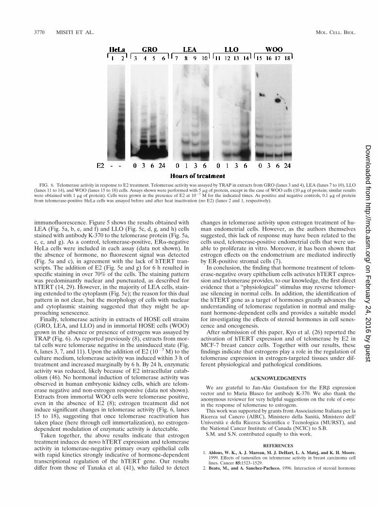
Finally, telomerase activity in extracts of HOSE cell strains(GRO, LEA, and LLO) and in immortal HOSE cells (WOO)grown in the absence or presence of estrogens was assayed byTRAP (Fig. 6). As reported previously (8), extracts from mor-tal cells were telomerase negative in the uninduced state (Fig.6, lanes 3, 7, and 11). Upon the addition of E2 (1027 M) to theculture medium, telomerase activity was induced within 3 h oftreatment and increased marginally by 6 h. By 24 h, enzymaticactivity was reduced, likely because of E2 intracellular catab-olism (46). No hormonal induction of telomerase activity wasobserved in human embryonic kidney cells, which are telom-erase negative and non-estrogen responsive (data not shown).Extracts from immortal WOO cells were telomerase positive,even in the absence of E2 (8); estrogen treatment did notinduce significant changes in telomerase activity (Fig. 6, lanes15 to 18), suggesting that once telomerase reactivation hastaken place (here through cell immortalization), no estrogen-dependent modulation of enzymatic activity is detectable.
Taken together, the above results indicate that estrogentreatment induces de novo hTERT expression and telomeraseactivity in telomerase-negative primary ovary epithelial cellswith rapid kinetics strongly indicative of hormone-dependenttranscriptional regulation of the hTERT gene. Our resultsdiffer from those of Tanaka et al. (41), who failed to detect
changes in telomerase activity upon estrogen treatment of hu-man endometrial cells. However, as the authors themselvessuggested, this lack of response may have been related to thecells used, telomerase-positive endometrial cells that were un-able to proliferate in vitro. Moreover, it has been shown thatestrogen effects on the endometrium are mediated indirectlyby ER-positive stromal cells (7).
In conclusion, the finding that hormone treatment of telom-erase-negative ovary epithelium cells activates hTERT expres-sion and telomerase provides, to our knowledge, the first directevidence that a “physiological” stimulus may reverse telomer-ase silencing in normal cells. In addition, the identification ofthe hTERT gene as a target of hormones greatly advances theunderstanding of telomerase regulation in normal and malig-nant hormone-dependent cells and provides a suitable modelfor investigating the effects of steroid hormones in cell senes-cence and oncogenesis.
After submission of this paper, Kyo et al. (26) reported theactivation of hTERT expression and of telomerase by E2 inMCF-7 breast cancer cells. Together with our results, thesefindings indicate that estrogens play a role in the regulation oftelomerase expression in estrogen-targeted tissues under dif-ferent physiological and pathological conditions.
ACKNOWLEDGMENTS
We are grateful to Jan-Ake Gustafsson for the ERb expressionvector and to Maria Blasco for antibody K-370. We also thank theanonymous reviewer for very helpful suggestions on the role of c-mycin the response of telomerase to estrogens.
This work was supported by grants from Associazione Italiana per laRicerca sul Cancro (AIRC), Ministero della Sanita, Ministero dell’Universita e della Ricerca Scientifica e Tecnologica (MURST), andthe National Cancer Institute of Canada (NCIC) to S.B.
S.M. and S.N. contributed equally to this work.
REFERENCES
1. Aldous, W. K., A. J. Marean, M. J. DeHart, L. A. Matej, and K. H. Moore.1999. Effects of tamoxifen on telomerase activity in breast carcinoma celllines. Cancer 85:1523–1529.
2. Beato, M., and A. Sanchez-Pacheco. 1996. Interaction of steroid hormone
FIG. 6. Telomerase activity in response to E2 treatment. Telomerase activity was assayed by TRAP in extracts from GRO (lanes 3 and 4), LEA (lanes 7 to 10), LLO(lanes 11 to 14), and WOO (lanes 15 to 18) cells. Assays shown were performed with 5 mg of protein, except in the case of WOO cells (10 mg of protein; similar resultswere obtained with 1 mg of protein). Cells were grown in the presence of E2 at 1027 M for the indicated times. As positive and negative controls, 0.1 mg of proteinfrom telomerase-positive HeLa cells was assayed before and after heat inactivation (no E2) (lanes 2 and 1, respectively).
3770 MISITI ET AL. MOL. CELL. BIOL.
on February 24, 2016 by guest
http://mcb.asm
.org/D
ownloaded from
receptors with the transcription initiation complex. Endocrinol. Rev. 17:587–609.
3. Bednarek, A. K., Y. Chu, and C. M. Aldaz. 1998. Constitutive telomeraseactivity in cells with tissue-renewing potential from estrogen-regulated rattissues. Oncogene 16:381–385.
4. Bodnar, A. G., M. Ouellette, M. Frolkis, S. E. Holt, C. P. Chiu, G. B. Morin,C. B. Harley, J. W. Shay, S. Lichtsteiner, and W. E. Wright. 1998. Extensionof life-span by introduction of telomerase into normal human cells. Science279:349–352.
5. Brown, M., and P. A. Sharp. 1990. Human estrogen receptor forms multipleprotein-DNA complexes. J. Biol. Chem. 265:11238–11243.
6. Cong, Y. S., J. Wen, and S. Bacchetti. 1999. The human telomerase catalyticsubunit hTERT: organization of the gene and characterization of the pro-moter. Hum. Mol. Genet. 8:137–142.
7. Cooke, P. S., D. L. Buchanan, P. Young, T. Seawan, J. Brody, K. S. Korach,J. Taylor, D. B. Lubhan, and G. R. Cunha. 1997. Stromal estrogen receptorsmediate mitogenic effects of estradiol on uterine epithelium. Proc. Natl.Acad. Sci. USA 94:6535–6540.
8. Counter, C. M., H. W. Hirte, S. Bacchetti, and C. B. Harley. 1994. Telom-erase activity in human ovarian carcinoma. Proc. Natl. Acad. Sci. USA91:2900–2904.
9. Dey, A., A. M. Thornton, M. Lonergan, S. M. Weissman, J. W. Chamberlain,and K. Ozato. 1992. Occupancy of upstream regulatory sites in vivo coincideswith major histocompatibility complex class I gene expression in mousetissues. Mol. Cell. Biol. 12:3590–3599.
10. Dubik, D., and R. P. C. Shiu. 1992. Mechanism of estrogen activation ofc-myc oncogene expression. Oncogene 7:1587–1594.
11. Enmark, E., M. Pelto-Huikko, K. Grandien, S. Lagercrantz, J. Lagercrantz,C. Nordenskjold, and J. A. Gustafsson. 1997. Human estrogen receptorbeta-gene structure, chromosomal localization and expression pattern.J. Clin. Endocrinol. Metab. 82:4258–4265.
12. Farsetti, A., S. Misiti, F. Citarella, A. Felici, M. Andreoli, A. Fantoni, A.Sacchi, and A. Pontecorvi. 1995. Molecular basis of estrogen regulation ofHageman factor XII gene expression. Endocrinology 136:5076–5083.
13. Green, S., I. Issemann, and E. Sheer. 1998. A versatile in vivo and in vitroeucaryotic expression vector for protein engineering. Nucleic Acids Res.16:369.
14. Harrington, L., W. Zhou, T. McPhail, R. Oulton, D. S. K. Yeung, V. Mar,M. B. Bass, and M. O. Robinson. 1997. Human telomerase contains evolu-tionarily conserved catalytic and structural subunits. Genes Dev. 11:3109–3115.
15. Hillier, S. G., R. A. Anderson, A. R. Williams, and M. Tetsuka. 1998.Expression of oestrogen receptor alpha and beta in cultured human ovariansurface epithelial cells. Mol. Hum. Reprod. 4:811–815.
16. Holt, S. E., and J. W. Shay. 1999. Role of telomerase in cellular proliferationand cancer. J. Cell. Physiol. 180:10–18.
17. Horikawa, I., P. L. Cable, C. Afshari, and J. C. Barrett. 1999. Cloning andcharacterization of the promoter region of human telomerase reverse tran-scriptase gene. Cancer Res. 59:826–830.
18. Horwitz, K. B., and W. L. McGuire. 1987. Estrogen control of progesteronereceptor in human breast cancer. J. Biol. Chem. 266:1008–1013.
19. Horwitz, K. B., T. A. Jackson, D. L. Bain, J. K. Richer, G. S. Takimoto, andL. Tung. 1996. Nuclear receptor coactivators and corepressors. Mol. Endo-crinol. 10:1167–1177.
20. Katzenellenbogen, J. A., B. W. O’Malley, and B. S. Katzenellenbogen. 1996.Tripartite steroid hormone receptor pharmacology: interaction with multipleeffector sites as a basis for the cell- and promoter-specific action of thesehormones. Mol. Endocrinol. 10:119–131.
21. Kilian, A., D. D. L. Bowtell, H. E. Abud, G. R. Hime, D. J. Venter, P. K.Keese, E. L. Duncan, R. R. Reddel, and R. A. Jefferson. 1997. Isolation of acandidate human telomerase catalytic subunit gene, which reveals complexsplicing patterns in different cell types. Hum. Mol. Genet. 6:2011–2019.
22. Kim, N. W., and F. Wu. 1997. Advances in quantification and characteriza-tion of telomerase activity by the telomeric repeat amplification protocol(TRAP). Nucleic Acids Res. 25:2595–2597.
23. Kommos, F. 1996. Gynaecological cancer, p. 541–572. In J. R. Pasqualini andB. S. Katzenellenbogen (ed.), Hormone-dependent cancer. Marcel Dekker,Inc., New York, N.Y.
24. Kuiper, G. G., J. G. Lemmen, B. Carlsson, J. C. Corton, S. H. Safe, P. T. vander Saag, B. van der Burg, and J. A. Gustafsson. 1998. Interaction ofestrogenic chemicals and phytoestrogens with estrogen receptor beta. En-docrinology 139:4252–4263.
25. Kyo, S., M. Takakura, T. Kohama, and M. Inoue. 1997. Telomerase activityin human endometrium. Cancer Res. 57:610–614.
26. Kyo, S., M. Takakura, T. Kanaya, W. Zhuo, K. Fujimoto, Y. Nishio, A.Orimo, and M. Inoue. 1999. Estrogen activates telomerase. Cancer Res.59:5917–5921.
27. Lau, K. M., S. C. Mok, and S. M. Ho. 1999. Expression of mouse telomerasecatalytic subunit in embryos and adult tissues. Proc. Natl. Acad. Sci. USA96:5722–5727.
28. Lazennec, G., T. R. Ediger, L. N. Petz, A. M. Nardulli, and B. S. Katzenel-lenbogen. 1997. Mechanistic aspects of estrogen receptor activation probedwith constitutively active estrogen receptors: correlations with DNA andcoregulator interactions and receptor conformational changes. Mol. Endo-crinol. 11:1375–1386.
29. Martin-Rivera, L., E. Herrera, J. P. Albar, and M. A. Blasco. 1998. Expres-sion of mouse telomerase catalytic subunit in embryos and adult tissues.Proc. Natl. Acad. Sci. USA 95:10471–10476.
30. Meeker, A. K., H. J. Sommerfeld, and D. S. Coffey. 1996. Telomerase isactivated in the prostate and seminal vesicles of castrated rats. Endocrinol-ogy 137:5743–5746.
31. Moretti, F., A. Farsetti, S. Soddu, S. Misiti, M. Crescenzi, S. Filetti, M.Andreoli, A. Sacchi, and A. Pontecorvi. 1997. P53 re-expression inhibitsproliferation and restores differentiation of human thyroid anaplastic carci-noma cells. Oncogene 14:729–740.
32. Mueller, P. R., S. J. Salser, and B. Wold. 1988. Constitutive and metal-inducible protein: DNA interactions at the mouse metallothionein I pro-moter examined by in vivo and in vitro footprinting. Genes Dev. 2:412–427.
33. Nugent, C. I., and V. Lundblad. 1998. The telomerase reverse transcriptase:components and regulation. Genes Dev. 12:1073–1085.
34. Paech, K., P. Webb, G. G. J. M. Kuiper, S. Nilsson, J.-A. Gustafsson, P. J.Kushner, and T. S. Scanlan. 1997. Differential ligand activation of estrogenreceptors ERa and ERb at AP-1 site. Science. 277:1508–1510.
35. Porter, W., B. Saville, D. Hoivik, and S. Safe. 1997. Functional synergybetween the transcription factor Sp1 and the estrogen receptor. Mol. Endo-crinol. 11:1569–1580.
36. Rodriguez, G. C., A. Berchuck, R. S. Whitaker, D. Schlossman, D. L. Clarke-Pearson, and R. C. Bast, Jr. 1991. Epidermal growth factor expression innormal ovarian epithelium and ovarian cancer. Am. J. Obstet. Gynecol.164:745–750.
37. Saito, T., A. Schneider, N. Martel, H. Mizumoto, M. Bulgay-Moerschel, R.Kudo, and H. Nakazawa. 1997. Proliferation-associated regulation of telom-erase activity in human endometrium and its potential implication in earlycancer diagnosis. Biochem. Biophys. Res. Commun. 231:610–614.
38. Sanger, F., S. Nicklen, and A. R. Coulson. 1977. DNA sequencing withchain-terminating inhibitors. Proc. Natl. Acad. Sci. USA 74:5463–5467.
39. Scholz, A., M. Truss, and M. Beato. 1999. Hormone-dependent recruitmentof NF-Y to the uteroglobin gene enhancer associated with chromatin re-modeling in rabbit endometrial epithelium. J. Biol. Chem. 274:4017–4026.
40. Takakura, M., S. Kyo, T. Kanaya, H. Hirano, J. Takeda, M. Yutsudo, and M.Inoue. 1999. Cloning of human telomerase catalytic subunit (hTERT) genepromoter and identification of proximal core promoter sequences essentialfor transcriptional activation in immortalized and cancer cells. Cancer Res.59:551–557.
41. Tanaka, M., M. Takakura, T. Kanaya, T. Sagawa, K. Yamashita, Y. Okada,E. Hiyama, and M. Inoue. 1998. Expression of telomerase activity in humanendometrium is localized to epithelial glandular cells and regulated in amenstrual phase-dependent manner correlated with cell proliferation. Am. J.Pathol. 153:1985–1991.
42. Ulaner, G. A., J. F. Hu, T. H. Vu, L. C. Giudice, and R. Hoffman. 1998.Telomerase activity in human development is regulated by human telomer-ase reverse transcriptase transcription and by alternative splicing of hTERTtranscripts. Cancer Res. 58:4168–4172.
43. Vaziri, H., and S. Benchimol. 1998. Reconstitution of telomerase activity innormal human cells leads to elongation of telomeres and extended replica-tive life span. Curr. Biol. 8:279–282.
44. Webb, P., G. N. Lopez, R. M. Uht, and P. J. Kushner. 1995. Tamoxifenactivation of the estrogen receptor/AP-1 pathway: potential origin for thecell-specific estrogen-like effects of antiestrogens. Mol. Endocrinol. 9:443–456.
45. Wu, K.-J., C. Grandori, M. Amacker, N. Simon-Vermot, A. Polack, J. Ling-ner, and R. Dalla-Favera. 1999. Direct activation of TERT transcription byc-Myc. Nat. Genet. 21:220–224.
46. Zimmermann, H., R. Koytchev, O. Mayer, A. Borner, U. Mellinger, and H.Breitbarth. 1998. Pharmacokinetics of orally administered estradiol-valerate.Results of a single-dose cross-over bioequivalence study in postmenopausalwomen. Arzneimittelforschung 48:941–947.
VOL. 20, 2000 ESTROGEN INDUCTION OF hTERT GENE EXPRESSION 3771
on February 24, 2016 by guest
http://mcb.asm
.org/D
ownloaded from
Copyright © 2022 FDOKUMEN