VDAC and ERα interaction in caveolae from human cortex is altered in Alzheimer's disease
Upload
independentCategory
view
4download
0
ORIGINAL ARTICLE JJBMR
Estrogens Attenuate Oxidative Stress and theDifferentiation and Apoptosis of Osteoblasts byDNA-Binding-Independent Actions of the ERaMaria Almeida, Marta Martin-Millan, Elena Ambrogini , Robert Bradsher III , Li Han, Xiao-Dong Chen,Paula K Roberson, Robert S Weinstein, Charles A O’Brien, Robert L Jilka, and Stavros C Manolagas
Division of Endocrinology and Metabolism, Center for Osteoporosis and Metabolic Bone Diseases, University of Arkansas for MedicalSciences and the Central Arkansas Veterans Health Care System, Little Rock, AR, USA
ABSTRACTEstrogens diminish oxidative stress in bone and bone marrow, attenuate the generation of osteoblasts, and decrease the prevalence of
mature osteoblast apoptosis. We have searched for the molecular mechanism of these effects using as tools a mouse model bearing an
estrogen receptor a (ERa) knock-in mutation that prevents binding to DNA (ERaNERKI/�) and several osteoblast progenitor cell models
expressing the wild-type ERa or the ERaNERKI/�. We report that the ability of estrogens to diminish the generation of reactive oxygen
species, stimulate the activity of glutathione reductase, and decrease the phosphorylation of p66shc, as well as osteoblastogenesis and
osteoblast number and apoptosis, were fully preserved in ERaNERKI/� mice, indicating that the DNA-binding function of the ERa is
dispensable for all these effects. Consistent with the attenuation of osteoblastogenesis in this animal model, 17b-estradiol attenuated
bone morphogenetic protein 2 (BMP-2)–induced gene transcription and osteoblast commitment and differentiation in murine and
human osteoblastic cell lines. Moreover, 17b-estradiol attenuated BMP-2-induced differentiation of primary cultures of calvaria- or bone
marrow–derived osteoblastic cells from ERaNERKI/� mice as effectively as in cells from wild-type littermates. The inhibitory effect of the
hormone on BMP-2 signaling resulted from an ERa-mediated activation of ERKs and the phosphorylation of Smad1 at the linker region of
the protein, which leads to proteasomal degradation. These results illustrate that the effects of estrogens on oxidative stress and the birth
and death of osteoblasts do not require the binding of ERa to DNA response elements, but instead they result from the activation of
cytoplasmic kinases. � 2010 American Society for Bone and Mineral Research.
KEY WORDS: REACTIVE OXYGEN SPECIES; P66SHC; ERKS; BMP-2; ESTROGEN RECEPTOR
Introduction
Work from our group during the last 20 years has elucidated
that estrogens protect the adult skeleton against bone loss
by slowing the rate of bone remodeling and by maintaining a
focal balance between bone formation and resorption.(1–3)
Slowing of bone remodeling results from the attenuating effects
of sex steroids on the birth rate of osteoclast and osteoblast
progenitors.(4,5) Maintenance of a focal balance between
formation and resorption results from opposite effects on the
lifespan of osteoclasts and osteoblasts/osteocytes: a proapopto-
tic effect on osteoclasts and an antiapoptotic effect on
osteoblasts and osteocytes.(6–8) The effects of estrogens on
osteoclast and osteoblast apoptosis are exerted by a mechanism
that is distinct from that requiring direct interaction of their
receptors with DNA (hormone-response element) or protein-
protein interaction between the receptor and other transcription
Received in original form March 27, 2009; revised form July 31, 2009; accepted Oc
Address correspondence to: Stavros C Manolagas, MD, PhD, 4301 West Markham, N
Journal of Bone and Mineral Research, Vol. 25, No. 4, April 2010, pp 769–781
DOI: 10.1359/jbmr.091017
� 2010 American Society for Bone and Mineral Research
factors. Instead, the effect of estrogens on the apoptosis of either
cell type is the result of an extranuclear action of the classical
receptors that cause activation of cytoplasmic kinases, including
extracellular signal-regulated kinases (ERKs) and kinase-depen-
dent changes in the activity of transcription factors.(6,8,9) The
mechanistic basis for the divergence of estrogens’ effect on the
survival of the two cell types downstream from ERKs is evidently
dependent on the kinetics of ERK phosphorylation and the
length of time that phospho-ERKs are retained in the nucleus,
perhaps by determining the activation of a distinct set of
transcription factors.(9)
We have demonstrated previously that the number of
osteoblast progenitors, as measured by colony-forming units–
osteoblast (CFU-OB), increase after loss of estrogens in mice(10)
and that this change is partially preserved in mice treated with
bisphosphonates, which significantly decrease osteoclast num-
ber, strongly suggesting that bone resorption (and the release of
tober 9, 2009. Published online October 12, 2009.
o. 587, Little Rock, AR 72205-7199, USA. E-mail: [email protected]
769
growth factors from the bone matrix) is not required for the
increase in osteoblast precursors. Therefore, estrogens must
suppress osteoblastogenesis by direct actions on early osteo-
blast precursors. Further, we have shown that most CFU-OBs are
early transit-amplifying progenitors (i.e., dividing cells with
limited self-renewal capacity) and that their replication is indeed
attenuated by estrogens.(5)
We and others also have shown previously that estrogens
attenuate the transcription of bone morphogenetic protein 2
(BMP-2) target genes.(11–13) BMPs are members of the
transforming growth factor b (TGF-b) superfamily and play an
essential role in skeletal development and repair.(14,15) Specifi-
cally, BMPs promote embryonic and postnatal osteogenesis by
inducing the commitment of mesenchymal cells to the
osteoblastic lineage and promoting osteoblast differentia-
tion.(16,17) Binding of BMPs to their receptor serine/threonine
kinases results in the phosphorylation of Smads 1, 5, and 8(18) at
the carboxy terminus and translocation into the nucleus after
heterodimerization with Smad4. In the nucleus, the complex
either binds to DNA sequences directly or can interact with
several transcription factors to control the activity of hundreds of
downstream target genes.(19,20) The Smad proteins consist of two
globular domains (MH1 andMH2 domains) connected by a linker
region. In Smad 1, 5, and 8, the latter contains four MAPK
phosphorylation sites and two putative GSK-3b sites.(19,21)
Importantly, MAPK phosphorylation of the linker region inhibits
Smad function and therefore BMP-induced transcription both in
vitro and in vivo.(22–24)
More recently, we and others have obtained evidence that the
protective effects of estrogens on bone result from their ability to
attenuate oxidative stress and that loss of estrogens accelerates
the effects of aging. Specifically, we have shown that C57BL/6
mice lose bone strength and mass progressively between the
ages of 4 and 31 months(25) and that these changes are
temporally associated with decreased osteoblast and osteoclast
numbers and decreased bone-formation rate as well as increased
osteoblast and osteocyte apoptosis. These changes are also
temporally linked with increased reactive oxygen species (ROS)
levels and decreased glutathione reductase (GSR) activity in the
bone marrow, as well as a corresponding increase in the
phosphorylation of p66shc—an adapter protein that serves as a
key component of a signaling cascade that is activated by ROS
and influences apoptosis and lifespan in invertebrates and
mammals.(26) Indeed, proapoptotic signals, including ROS,
release p66shc from an inhibitory complex, and active p66shc
serves as a redox enzyme that catalyzes reduction of O2 to H2O2
through electron transfer from cytochrome c. H2O2, in turn,
causes opening of the mitochondrial permeability transition
pore, swelling, and apoptosis. An increase in ROS production and
p66shc phosphorylation, as well as decreased GSR activity, was
reproduced acutely in our previous work by gonadectomy in
either female or male C57BL/6 mice and prevented by the
antioxidant N-acetyl-cysteine (NAC).(25) In agreement with our in
vivo findings, results of in vitro experiments demonstrated that
the effects of estrogens on osteoclastogenesis and osteoclast
and osteoblast apoptosis result from cell autonomous antiox-
idant actions of the hormone on the respective cell types, are
dependent on the estrogen receptor (ER), and are mediated via
770 Journal of Bone and Mineral Research
ERKs. Moreover, we have shown that estrogens attenuate the
phosphorylation of p66shc in osteoblastic cells and that this effect
is also mediated via ERKs. Based on these findings, we have
proposed that loss of estrogens accelerates the effects of aging
on bone by decreasing the defense against oxidative stress.
In this study we have investigated the molecular actions of
ERa on osteoblasts using as a tool amousemodel bearing an ERa
knock-in mutation that prevents binding to DNA (ERaNERKI/�).(27)
We previously determined that ERaNERKI/� mice have an atrophic
uterus despite normal estrogen levels and that estrogen
replacement does not restore it in ovariectomized (OVX)
ERaNERKI/� mice, but it does induce the activation of ERKs and
the ERK-mediated phosphorylation of the transcription factor
Elk-1 in vertebrae.(13) In addition, in this study we have
investigated in vitro the signaling cascades downstream from
ERa that are responsible for its effects on osteoblast commit-
ment and differentiation. We show that the effects of estrogens
on oxidative stress and the birth and death of osteoblasts are
fully preserved in ERaNERKI/� mice. Consistent with the attenua-
tion of osteoblastogenesis in the ERaNERKI/� mice, 17b-estradiol
(E2) attenuates BMP-2-induced gene transcription and differ-
entiation of preosteoblastic cell lines as well as primary cultures
of calvaria- or bone marrow–derived osteoblastic cells from
ERaNERKI/� mice as effectively as in cells from wild-type
littermates. This effect is due to an ERa-mediated activation of
ERKs and the phosphorylation of Smad1 at the linker region of
the protein, which leads to proteasomal degradation.
Materials and Methods
Chemicals, reagents, and plasmids
Etoposide, H2O2, and E2 were purchased from Sigma-Aldrich (St.
Louis, MO, USA). BMP-2 and fibroblast growth factor 2 (FGF2)
recombinant proteins were purchased from R&D Systems
(Minneapolis, MN, USA). PD98059 was purchased from Cell
Signaling Technology, Inc. (Danvers, MA, USA). The BMP-
responsive luciferase reporter construct (BRE)-Luc was obtained
from Peter ten Dijke (Leiden University Medical Center, Leiden,
The Netherlands).(28)
Animal experimentation
Mice heterozygous for an ERa knock-in mutant in a 129SvJ
background were provided by J Larry Jameson (Northwestern
University, Chicago, IL, USA).(27) Mice harboring an inactivating
mutation in the ERa locus (ERaþ/�) in a C57BL/6 background
were provided by Andree Krust and Pierre Chambon (Institute for
Genetics and Cellular and Molecular Biology, Strasbourg,
France).(29) ERaNERKI/þ mice were crossed with heterozygous
ERaþ/� female mice to produce animals carrying only one NERKI
allele (ERaNERKI/�). Five-month-old female ERaNERKI/� mice of
the F1 generation and their ERaNERKI/þ, ERaþ/�, and ERaþ/þ
littermates were electronically tagged (Biomedic Data System,
Inc., Maywood, NJ, USA), and bone mineral density (BMD)
measurements were performed on each mouse. Animals then
were sham-operated or ovariectomized (OVX). The following
day, OVX animals were subcutaneously injected with vehicle or
with replacement doses of E2 (30 ng/g/day; n¼ 12 per group),
ALMEIDA ET AL.
and sham-operated animals were injected with vehicle. After
6 weeks of treatment, animals were sacrificed and the tissues
dissected for further analyses. BMD, vertebral dimensions,
osteoblast number, and osteoblast apoptosis were obtained
as described previously.(7,30,31) ERa�/� and corresponding wild-
type (WT) littermate control mice were generated by crossing
heterozygous ERaþ/� mice.
Western blot analysis
The phosphorylation status of p66shc was analyzed by
immunoblotting in fifth lumbar vertebra lysates, as described
previously,(8) using a mouse monoclonal antibody recognizing
Ser36 phosphorylated p66shc (Calbiochem, San Diego, CA, USA).
Protein levels of p66shc were analyzed using a rabbit polyclonal
antibody recognizing p66shc (BD Biosciences, Palo Alto, CA, USA).
The antibody recognizing p-Smad1/5/8 was purchased from Cell
Signaling. The antibody recognizing phospho-Smad1 (Ser214)
was kindly provided by EM De Robertis (Howard Hughes Medical
Institute and University of California, Los Angeles, CA, USA).(32)
The antibodies against Smad4, p-ERK1/2, and ERK1/2 were
purchased from Santa Cruz Biotechnology (Santa Cruz, CA, USA).
Quantification of the intensity of the bands in the autoradio-
grams was performed using a VersaDoc imaging system (Bio-Rad
Laboratories, Hercules, CA, USA).
Micro-Computed Tomography (mCT)
mCT analysis of the sixth lumbar vertebrae was done after the
bones were dissected, cleaned, fixed in 10% Millonig’s formalin,
transferred to ethanol, loaded into 12.3mm diameter scanning
tubes, and imaged (mCT40, Scanco Medical, Basserdorf, Switzer-
land). Scans were integrated into 3D voxel images (1024� 1024
pixel matrices for each individual planar stack), and a Gaussian
filter (s¼ 0.8, support¼ 1) was used to reduce signal noise. A
threshold of 200 was applied to all analyzed scans. Scans were
done at medium resolution (E¼ 55 kVp, I¼ 145 mA, integration
time¼ 200ms). The entire vertebral body was scanned with a
transverse orientation excluding the pedicles and articular
processes. Manual analysis excluded the cortical bone and the
primary spongiosa from the analysis. All trabecular measure-
ments were made by manually drawing contours every 10 to 20
slices and using voxel counting for bone volume per tissue
volume and sphere-filling distance-transformation indices with-
out assumptions about the bone shape as a rod or plate for
trabecular microarchitecture. Cortical thickness was measured at
the tibial mid-diaphysis.
Cell culture, transfections, and luciferase activity
Osteoblastic cells derived from mouse calvaria or bone marrow
were obtained and cultured as described previously,(33) and
during exposure to E2, the cultures were maintained in 2%
charcoal-stripped serum. Osteoblast differentiation was analyzed
using freshly isolated cells cultured in 12 well tissue culture plates
at 5� 106 cells per well in amodified essential medium (a-MEM)
containing 10% fetal bovine serum (FBS) for 10 days. Half the
mediumwas replaced every 5 days. FBS then was reduced to 2%,
and 10�8M E2 was added in the presence or absence of 25 ng/
mL BMP-2. Two days later, 10mM b-glycerophosphate was
NONCLASSICAL ERa ACTIONS IN OSTEOBLASTS
added to the medium, and the cultures were maintained for an
additional 2 weeks. The mineralized matrix was stained with
40mM alizarin red, pH 4.2. Alizarin red was quantified after
extraction with 10mM sodium phosphate, 10% cetylpyridinium
chloride, pH 7, and absorbance determination at 562 nm against
a known alizarin red standard. For assay of caspase 3 activity, the
medium was changed to serum-free prior to the addition of the
different compounds. Colony-forming units–fibroblast (CFU-F)
and CFU-OB number were determined as described previously,(5)
using guinea pig feeder cells,(34) 15% FBS, and 1mM ascorbate-2-
phosphate. Half the medium was replaced every 5 days. CFU-Fs
were enumerated at 10 days of culture after staining for alkaline
phosphatase, and CFU-OBs were enumerated at 25 days of
culture after von Kossa staining. Colonies containing more than
50 fibroblastic cells were enumerated and plotted as a function
of the number of cells seeded. C2C12 and 2T3 cells were
maintained in Dulbecco’s modified Eagle’s medium supplemen-
ted with 10% FBS and 1% each of penicillin, streptomycin, and
glutamine and 1% sodium pyruvate. U2OS cells stably expressing
tetracycline-inducible ERa (U2OS-ERa) were kindly provided by
DC Leitman (University of California, San Francisco, CA, USA).(35)
U2OS-ERa cells were maintained in phenol red–free McCoy
medium supplemented with 10% FBS and 1% each of penicillin,
streptomycin, and glutamine. Cells were incubated for 24 hours
with or without doxycycline (1mg/mL) and serum starved for
another 16 hours previous to the addition of BMP-2 or E2. Mouse
embryonic fibroblasts (MEFs) from WT or Smad1L/L mice, kindly
provided by P Soriano (Fred Hutchinson Cancer Research Center,
Seattle, WA, USA),(24) were cultured in DMEM supplemented
with 10% FBS. Plasmid constructs were introduced into cells
by transient transfection using Lipofectamine Plus (Invitrogen,
Carlsbad, CA. USA). Cells were plated in 48 well plates and
transfected 16 hours later with a total of 0.4 mg of
DNA. Luciferase activity assays were performed as described
previously.(36)
Alkaline phosphatase (AP) activity and osteocalcinproduction
C2C12 or 2T3 cells were seeded at a density of 2� 104/cm2 in
medium containing 10% FBS. The following day, before
treatment, the medium was replaced with 5% serum-containing
medium. Cells were lysed in 100mM glycine, 1mM MgCl2, and
1% Triton X-100 at pH 10. AP activity in the cell lysate was
determined using a buffer containing 2-amino-2-methylpropa-
nol and p-nitrophenylphosphate (Sigma-Aldrich. Inc.). The
amount of osteocalcin secreted in the medium was determined
using an ELISA kit (Biomedical Technologies, Inc., Stoughton, MA,
USA). Both activities were normalized for total protein
concentration, determined using a Bio-Rad DC protein assay kit.
Mineralization assay
Freshly isolated murine bone marrow cells pooled from three
mice were seeded on 12 well tissue culture plates at 5� 106 cells
per well in standard culture medium and cultured for 10 days.
Half the medium was replaced every 5 days. Calvaria cells
isolated from adult mice were seeded at 0.02� 106 cells per well
and cultured for 3 days. FBS then was reduced to 2%, and 50 ng/
Journal of Bone and Mineral Research 771
mL BMP-2 was added in the presence or absence of 10�8M E2 in
both types of cells. Two days later, 10mM b-glycerolphosphate
was added to themedium. Themineralizationmatrix was stained
with 40mM alizarin red solution 2 weeks later.
Quantitative RT-PCR
Total RNA was extracted using Ultraspec (Biotecx Laboratories,
Houston, TX, USA) and reverse-transcribed using the High-
Capacity cDNA Archive Kit (Applied Biosystems) according to the
manufacturer’s instructions. Taqman quantitative reverse-tran-
scriptase polymerase chain reaction (RT-PCR) was performed as
described previously.(36) The primers and probes for murine
Smad6 and rRNA18S were manufactured by the TaqMan Gene
Expression Assays service (Applied Biosystems). Gene expression
was quantified by subtracting the rRNA18S threshold cycle (Ct)
value from the Ct value of the gene of interest and expressed as
2�DCt, as described by the protocol of the manufacturer.
Other assays
Intracellular ROS were quantified with dichlorodihydrofluores-
cein diacetate dye,(37) using bone marrow cells flushed from
femurs and washed with PBS. Glutathione reductase activity was
assayed with a kit from Cayman Chemical Company (Ann Arbor,
MI, USA). Apoptosis in cultured cells was determined by
measuring caspase-3 activity by cleavage of the fluorogenic
substrate Ac-DEVD-AFC (Biomol, Plymouth Meeting, PA, USA), as
described previously.(38)
Statistical analysis
ANOVA was used to detect effects of various in vivo and in vitro
treatments after establishing that the data were normally
distributed and equivalency of variances. Bonferroni’s method
was used to perform appropriate pairwise comparisons of
treatment groups. In cases where one or both of the
requirements for performing ANOVA were not met, Kruskal-
Wallis ANOVA on ranks was used, followed by Dunn’s method, to
perform pairwise comparisons of treatment groups. Unless
otherwise stated, results are presented as mean� SD and in vitro
assays performed in triplicate and repeated at least one time.
Results
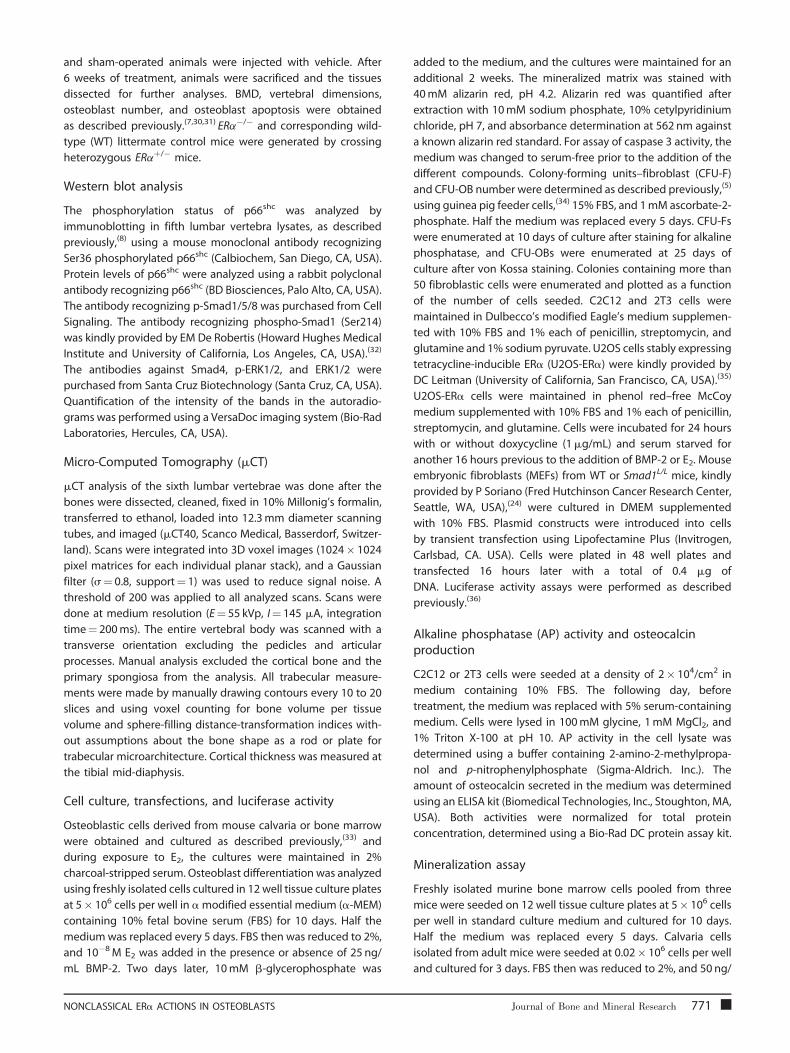
ERa NERKI is capable of mediating the antiapoptoticeffects of E2 on osteoblasts
To investigate whether direct binding of the ERa to DNA is
required for the ability of this receptor to mediate the effects of
estrogens on the lifespan of bone cells in vivo, we first
investigated whether the ERa NERKI was capable of mediating
the known antiapoptotic effects of estrogens on osteoblasts in
vitro and in vivo. In agreement with previous findings from our
group,(6,25) we found that E2 prevented apoptosis of osteoblasts
induced by H2O2 or the topoisomerase inhibitor etoposide as
effectively in osteoblastic cells isolated from calvaria of ERaNERKI/�
mice, as it did in cells from WT mice (Fig. 1A). In line with these
in vitro effects, the prevalence of osteoblast apoptosis was
increased in vivo in both WT and ERaNERKI/� mice 6 weeks
772 Journal of Bone and Mineral Research
following OVX, as determined by in situ end labeling of vertebral
sections (see Fig. 1B). Moreover, E2 replacement prevented the
OVX-induced increase in osteoblast apoptosis in both WT and
ERaNERKI/� mice.
Estrogens suppress oxidative stress in vivo independentlyof the DNA-binding function of the ERa
Wenext compared the effects of OVX and E2 replacement on ROS
levels and GSR activity in the bone marrow and on p66shc
phosphorylation in vertebral lysates in WT and ERaNERKI/� mice
6 weeks after the hormonal manipulation (see Fig. 1C, D). We
also compared these changes in mice haploinsufficient for ERa
(ERaþ/�) andmice inwhichonecopyof theERahasbeen replaced
by the NERKI mutant (ERaNERKI/þ). As we had seen in our earlier
work, ROS levels were increased (see Fig. 1C) and GSR activity was
decreased (see Fig. 1D) following OVX, and these changes were
prevented by E2 replacement in WT animals. Similar results were
obtained in ERaþ/�, ERaNERKI/þ, and ERaNERKI/� mice, indicating
that the antioxidant properties of estrogens are independent of
the DNA-binding function of the ERa.
We also observed an increase in p66shc phosphorylation 6
weeks following OVX and its reversal by E2 replacement in WT,
ERaþ/�, ERaNERKI/þ, and ERaNERKI/� mice, demonstrating that the
negative regulation of p66shc phosphorylation by estrogens in
vivo is mediated via a mechanism that does not require ERa
binding to DNA (see Fig. 1E). The increase in p66shc
phosphorylation in vertebral lysates after OVX was reproduced
in both WT and ERaNERKI/� mice in a second experiment in which
the mice were sacrificed 5 days after OVX (data not shown).
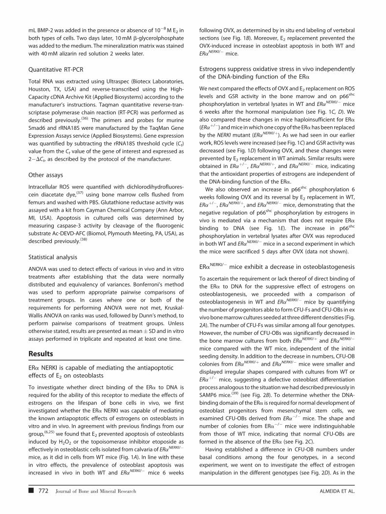
ERaNERKI/� mice exhibit a decrease in osteoblastogenesis
To ascertain the requirement or lack thereof of direct binding of
the ERa to DNA for the suppressive effect of estrogens on
osteoblastogenesis, we proceeded with a comparison of
osteoblastogenesis in WT and ERaNERKI/� mice by quantifying
the number of progenitors able to formCFU-Fs and CFU-OBs in ex
vivobonemarrowcultures seededat threedifferentdensities (Fig.
2A). The number of CFU-Fs was similar among all four genotypes.
However, the number of CFU-OBs was significantly decreased in
the bone marrow cultures from both ERaNERKI/þ and ERaNERKI/�
mice compared with the WT mice, independent of the initial
seeding density. In addition to the decrease in numbers, CFU-OB
colonies from ERaNERKI/þ and ERaNERKI/� mice were smaller and
displayed irregular shapes compared with cultures from WT or
ERaþ/� mice, suggesting a defective osteoblast differentiation
process analogous to the situationwehaddescribedpreviously in
SAMP6 mice.(39) (see Fig. 2B). To determine whether the DNA-
binding domain of the ERa is required for normal development of
osteoblast progenitors from mesenchymal stem cells, we
examined CFU-OBs derived from ERa�/� mice. The shape and
number of colonies from ERa�/� mice were indistinguishable
from those of WT mice, indicating that normal CFU-OBs are
formed in the absence of the ERa (see Fig. 2C).
Having established a difference in CFU-OB numbers under
basal conditions among the four genotypes, in a second
experiment, we went on to investigate the effect of estrogen
manipulation in the different genotypes (see Fig. 2D). As in the
ALMEIDA ET AL.

Fig. 1. E2 effects on osteoblast apoptosis, ROS levels, GSR activity, and p66shc phosphorylation are preserved in ERaNERKI/� mice. (A) Caspase-3 activity in
calvaria cells isolated from wild-type (WT) control or ERaNERKI/� mice pretreated with vehicle (veh) or E2 (10�8M) for 1 hour. Cells then were treated with
veh, etoposide (5� 10�5M), or H2O2 (5� 10�5M) for 6 hours. (B) Osteoblast apoptosis determined by in situ end labeling in sections of undecalcified
vertebral bone (L1–5) of 5-month-old WT or ERaNERKI/� mice sham operated or OVX. OVX animals received veh or E2 replacement for 6 weeks (n¼ 4 to
6 animal/group). (C) ROS levels and (D) GSR activity in bone marrow cells from 5-month-old WT, ERaþ/�, ERaNERKI/þ, and ERaNERKI/� mice sham operated or
OVX and treated as described in B (n¼ 4 animals/group). (E) Phosphorylation of p66shc determined by Western blot analyses in vertebral lysates from the
same mice as in C; each lane represents one animal. yp< .05 versus respective vehicle; �p< .05 versus OVX.
preceding experiment, ERaNERKI/�mice exhibited strikingly fewer
CFU-OBs than WT and ERaþ/� mice. In this experiment, CFU-OBs
from ERaNERKI/� mice were discernibly decreased compared with
ERaNERKI/þ mice. Nonetheless, all four genotypes exhibited an
increase in CFU-OB numbers following OVX, and the OVX-
induced increase was prevented in all four genotypes by E2replacement. In agreement with these results, the number of
osteoblasts in vertebral cancellous bone also was decreased in
sham operated ERaNERKI/� mice compared with WT sham-
operated controls. More important, the number of osteoblasts
from ERaNERKI/� mice was increased following OVX, and this was
prevented by E2, just as was observed in WT mice (see Fig. 2E).
ERaNERKI/� mice have low basal BMD and lose cancellousbone following OVX
ERaNERKI/� mice exhibited low bone mass at baseline in both
femur and spine (Fig. 3A). Thesemice also exhibited a decrease in
vertebral length and volume, as well as femoral bone area
NONCLASSICAL ERa ACTIONS IN OSTEOBLASTS
(Fig. 3B). Interestingly, when comparing mice carrying two
copies of the WT ERa (ERaþ/þ) or one copy (ERaþ/�) or the ERa
NERKI mutant together with the WT ERa (ERaNERKI/þ) or alone
(ERaNERKI/�), there was a decrease at baseline BMD in the femur
and spine of ERaþ/�, ERaNERKI/þ, and ERaNERKI/� mice compared
with ERaþ/þ mice. In agreement with the dual-energy X-ray
absorptiometry (DXA) results, unstained longitudinal sections
of bone viewed in dark field confirmed the decreased
vertebral size and diminished cancellous and cortical bone in
the ERaNERKI/� mice (see Fig. 3C). 3D BMD, cortical thickness, and
trabecular thickness, as determined by mCT, also were decreased
in the ERaNERKI/� mice (but trabecular number and trabecular
separation were indistinguishable) compared with WT controls
under basal conditions (Table 1). Moreover, similar to the WT
controls, the ERaNERKI/� mice lost cancellous (see Fig. 3D) but not
cortical (see Fig. 3E) bone following OVX. Nonetheless, E2replacement at the dose used in our study did not prevent the
loss of cancellous bone. A similar phenomenon was observed in
the studies of Syed and colleagues,(40) but cancellous bone loss
Journal of Bone and Mineral Research 773
Fig. 2. Osteoblastogenesis is decreased in ERaNERKI/� mice. (A) CFU-Fs or
CFU-OBs in the bone marrow from femora of intact mice of the indicated
genotypes. Cells from three mice were pooled and plated in duplicate at
three different densities for each genotype. CFU-Fs were stained for
alkaline phosphatase after 10 days, and CFU-OBs were stained with von
Kossa to detect mineral after 25 days (left panel). The graphs on the right
represent the quantification of CFUs depicted on the left. (B) Photo-
micrographs show representative CFU-OB colonies (50�) obtained from
WT (ERaþ/þ) or ERaNERKI/� mice or (C) from WT or ERa�/� mice; þ/þ and
WT refer to the respective littermate controls, as detailed in ‘‘Materials
and Methods.’’ The graph on the right represents the quantification of
CFU-OBs depicted on the right. (D) CFU-OBs obtained from femora of
mice used in the experiment described in Fig. 1C. Cells from three mice
were pooled and plated in triplicate at 106 cells per well. The graph on the
bottom represents the quantification of CFU-OBs depicted on the top. (E)
Osteoblast numbers on longitudinal undecalcified sections of L1–4
vertebrae from mice used in the experiment described in
Fig. 1C (n¼ 6 animals per group). �p< .05 versus OVX; yp< .05 versus
þ/þ sham.
in the ERaNERKI/� mice was prevented by a higher dose of E2replacement in that earlier study.
E2 attenuates BMP-2-induced osteoblast differentiationand the expression of BMP-2 target genes via ERa
Prompted by the evidence from the ERaNERKI/� mouse
suggesting that estrogens attenuate osteoblastogenesis
independently of the ability of ERa to interact directly with
774 Journal of Bone and Mineral Research
DNA, we went on to investigate whether estrogens attenuate
osteoblastogenesis via a cell autonomous mechanism and
whether such an effect is exerted via an extranuclear action of
the ERa mediated through the activation of cytoplasmic
kinases. To establish the generality of such a mechanism, we
used several cell models: two different established murine cell
lines, a human osteosarcoma cell line with conditional
expression of the ERa (U2OS-ERa), and primary cultures of
osteoblast progenitors obtained from calvaria or the bone
marrow of C57BL/mice or ERaNERKI/� mice. As shown in Fig. 4A,
BMP-2 dose dependently stimulated the differentiation of the
preosteoblast cell line 2T3, as determined by AP activity. This
effect was noticeable as early as day 1, reached a peak at day 3,
and decreased by day 5. Addition of E2 attenuated the effect of
BMP-2. Similarly, BMP-2 stimulated osteoblast differentiation in
the uncommitted mesenchymal progenitor cell line C2C12 with
a maximal effect at 3 days of culture, and E2 attenuated the
effect of BMP-2 (see Fig. 4B). Consistent with its suppressive
effect on BMP-2-induced osteoblast differentiation, E2 also
attenuated the stimulating effect of BMP-2 on the secretion of
osteocalcin, an osteoblast-specific biosynthetic product, at days
3 and 5 of the cultures (see Fig. 4B). Moreover, E2 antagonized
BMP-2-induced gene transcription in C2C12 cells, as deter-
mined by the expression of the BMP-2 target gene Smad6 (see
Fig. 4C).
To establish that the effect of E2 on BMP-2-induced
transcription was indeed mediated via the ERa, we examined
the effects of E2 on BMP-2-induced Smad6 mRNA in the human
osteoblast-like osteosarcoma cell line U2OS stably expressing
doxycycline-inducible ERa. BMP-2 stimulated Smad6 transcrip-
tion in U2OS cells both in the absence or presence of ERa.
However, whereas E2 attenuated the effect of BMP-2 in U2OS
cells expressing the ERa, the effect of E2 was abolished in cells
lacking the ERa (see Fig. 4D).
The findings with the established cell lines were readily
reproduced in primary cultures of bone marrow–derived stromal
cells or primary cultures of calvaria-derived cells, models that
more closely represent normal osteoblastic cells in vivo. As
expected, addition of BMP-2 to these primary cultures strongly
promoted osteoblast differentiation and maturation, as deter-
mined by osteocalcin secretion (Fig. 5A) and mineralization (Fig.
5B). In full agreement with the evidence from the cell lines, E2decreased BMP-2-induced AP and osteocalcin secretion as well
as mineralization in both the bone marrow– and the calvaria-
derived primary osteoblastic cell cultures (see Fig. 5). Moreover,
practically identical results were obtained in cultures of calvaria
cells from WT (ERaþ/þ) and the ERaNERKI/� mice (see Fig. 5C, D),
establishing that the DNA-binding function of ERa is indeed
dispensable for the attenuating effect of estrogens on osteoblast
differentiation.
E2 stimulates Smad1 phosphorylation at the linker regionand attenuates BMP-induced transcription via ERKs
Based on the finding that estrogens inhibit osteoblastogenesis in
both WT and ERaNERKI/� mice and cells, we next tested the
hypothesis that the attenuating effect of E2 on BMP-2-induced
transcription and osteoblast commitment/differentiation was
ALMEIDA ET AL.
Table 1. mCT Measurements of Lumbar Cancellous Bone (L5) and Mid-Diaphyseal Tibial Cortical Bone in ERaþ/þ, ERaþ/�, ERaNERKI/þ, and
ERaNERKI/� Female Mice
mCT Measurements ERaþ/þ ERaþ/� ERaNERKI/þ ERaNERKI/�
3D-BMD (mg HA/cm3) 298.9� 24.7 238.0� 48.4 271.1� 61.3 200.5� 48.7�
BV/TV % 39.5� 3.8 32.3� 6.2 37.7� 9.4 28.0� 6.6�
Trabecular number (per mm) 4.98� 0.32 4.56� 0.57 5.31� 0.96 4.79� 0.56
Trabecular thickness (mm) 70.7� 2.7 65.5� 5.7 64.5� 5.8 56.9� 5.5�
Trabecular separation (mm) 209.8� 16.8 224.3� 32.6 200.5� 34.4 209.6� 27.9
Cortical thickness (mm) 0.22� 0.011 0.21� 0.012 0.21� 0.016 0.19� 0.012�
�p< .05 versus ERaþ/þ controls, n¼ 7–11.
mediated by the activation of cytoplasmic kinases, such as ERKs.
As shown in Fig. 6A, E2 attenuated BMP-2-induced phosphoryla-
tion of Smad1/5/8 in the C2C12 cell model, in agreement with
earlier studies of ours.(13) In addition, E2 attenuated the BMP-2-
induced transcriptional activation of the Smad6-luciferase
construct (see Fig. 6B). Importantly, the specific MEK inhibitor
PD98059 reversed the attenuating effect of E2 on both BMP-2-
induced Smad1/5/8 phosphorylation and activation of transcrip-
tion. However, PD98059 by itself had no effect on the BMP-2-
induced Smad1/5/8 phosphorylation or transcription, indicating
that these effects of BMP-2 do not require ERK activation. Total
Smad1 levels were not affected by any one of these treatments
(data not shown).
Furthermore, E2 as well as FGF2, used here as a positive
control, stimulated the phosphorylation of ERKs as well as the
phosphorylation of Smad1 at p-Serine 214 in its linker region (see
Fig. 6C). Ser214 phosphorylation is the direct result of ERK
activation(23,41) and triggers Smad1 proteasomal degradation
leading to a decrease in Smad1 transcriptional activity.(21,32) In
agreement with this evidence, PD98059 prevented the ability of
E2 to stimulate ERKs as well as Smad1 Ser214 phosphorylation. As
expected, PD98059 also prevented FGF2-induced ERKs and
Smad1 phosphorylation (see Fig. 6C). Finally, to verify that the
MAPK sites in the linker region of Smad1 are important for the
inhibitory actions of E2 on BMP-2 signaling, we used MEFs from
WT or Smad1L/L mice that carry a Smad1 allele lacking all four
MAPK sites in the linker region.(24) Addition of FGF2 to WT MEFs
decreased BMP-2-dependent activation of a BMP transcriptional
reporter construct, whereas addition of FGF2 to Smad1L/L cells
had no effect, as seen before by others.(21) Importantly, E2 also
failed to inhibit BMP-induced transcription in Smad1L/L cells (see
Fig. 6D).
Discussion
The evidence presented in this report illustrates that estrogens
attenuate oxidative stress as well as the differentiation and
apoptosis of osteoblasts by a nonclassical mechanism of ERa
action. Specifically, our data reveal that the ability of estrogens to
suppress oxidative stress and thereby attenuate the apoptosis of
osteoblasts does not require binding of the ERa to DNA. The
demonstration of estrogens’ ability to suppress ROS levels and
increase GSR activity in the bone marrow of ERaNERKI/� mice
NONCLASSICAL ERa ACTIONS IN OSTEOBLASTS
strongly suggests that the dispensability of ERa binding to DNA
for the antioxidant properties of estrogens extends beyond
osteoclasts and osteoblasts and therefore must be a common
mechanism of this property of estrogens in all their other target
tissues.
Using the ERaNERKI/� mouse model, we have obtained
compelling evidence that binding of ERa to DNA is also
dispensable for the attenuating effects of estrogens on
osteoblastogenesis. Moreover, using a variety of osteoblastic
models from mice and humans, including primary cultures of
calvaria- and bone marrow–derived osteoblastic cells, we
demonstrate herein that E2 attenuates BMP-2-induced transcrip-
tion and thereby osteoblastogenesis via ERK activation and
downstream phosphorylation of Smad1 at the linker region of
the protein, which leads to Smad1 proteasomal degradation.
Consistent with this mode of action, E2 attenuated BMP-2-
induced differentiation of primary cultures of calvaria- or bone
marrow–derived osteoblastic cells from ERaNERKI/� mice as
effectively as in cells from WT littermates, establishing that the
DNA-binding function of the ERa is indeed dispensable for this
effect. Thesemechanisms are summarized in themodel depicted
in Fig. 7.
The evidence that estrogens attenuate osteoblast apoptosis
and oxidative stress by an ERa-mediated mechanism that does
not depend on the DNA-binding function of this receptor is
consistent with extensive work of others showing that estrogens
are indeed able to influence cells in their numerous reproductive
and nonreproductive target organs, in part, by extranuclear
actions of the ER involving kinase-mediated signaling.(42–44) In
addition, the findings of this study are in agreement with earlier
studies of ours showing that the suppressive effect of E2 on H2O2-
induced phosphorylation of p66shc—a cumulative index of
oxidative stress— is kinase-mediated and is inhibited by the ERK-
specific inhibitor PD98059.(25)
ERb is intact in ERaNERKI/� mice, and therefore, we cannot
categorically exclude the possibility that some of the effects of E2on this model are mediated by ERb. However, ERb expression in
murine bone is two to three orders of magnitude lower than
ERa,(25) and osteoblast number and bone mass were unaffected
in mice lacking ERb.(45) In addition, studies in mice with a
genuine null mutation of ERb indicate that with the exception of
impaired ovarian function, this isotype of the ER is not required in
the mouse for the development and homeostasis of the major
body systems.(46) In addition to regulating kinases, steroid
Journal of Bone and Mineral Research 775
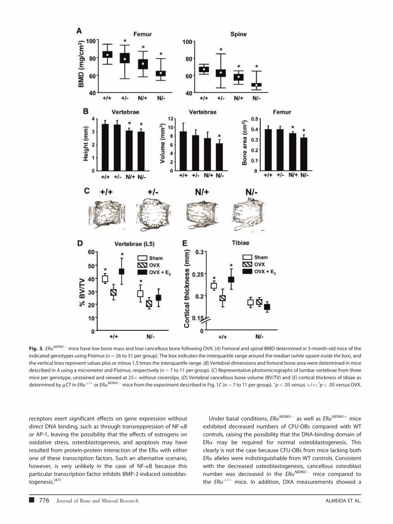
Fig. 3. ERaNERKI/�mice have low bone mass and lose cancellous bone following OVX. (A) Femoral and spinal BMD determined in 5-month-old mice of the
indicated genotypes using Piximus (n¼ 26 to 31 per group). The box indicates the interquartile range around themedian (white square inside the box), and
the vertical lines represent values plus or minus 1.5 times the interquartile range. (B) Vertebral dimensions and femoral bone area were determined inmice
described in A using a micrometer and Piximus, respectively (n¼ 7 to 11 per group). (C) Representative photomicrographs of lumbar vertebrae from three
mice per genotype, unstained and viewed at 25� without coverslips. (D) Vertebral cancellous bone volume (BV/TV) and (E) cortical thickness of tibiae as
determined bymCT in ERaþ/þ or ERaNERKI/�mice from the experiment described in Fig. 1C (n¼ 7 to 11 per group). �p< .05 versusþ/þ; yp< .05 versus OVX.
receptors exert significant effects on gene expression without
direct DNA binding, such as through transreppression of NF-kB
or AP-1, leaving the possibility that the effects of estrogens on
oxidative stress, osteoblastogenesis, and apoptosis may have
resulted from protein-protein interaction of the ERa with either
one of these transcription factors. Such an alternative scenario,
however, is very unlikely in the case of NF-kB because this
particular transcription factor inhibits BMP-2-induced osteoblas-
togenesis.(47)
776 Journal of Bone and Mineral Research
Under basal conditions, ERaNERKI/� as well as ERaNERKI/þ mice
exhibited decreased numbers of CFU-OBs compared with WT
controls, raising the possibility that the DNA-binding domain of
ERa may be required for normal osteoblastogenesis. This
clearly is not the case because CFU-OBs from mice lacking both
ERa alleles were indistinguishable from WT controls. Consistent
with the decreased osteoblastogenesis, cancellous osteoblast
number was decreased in the ERaNERKI/� mice compared to
the ERaþ/þ mice. In addition, DXA measurements showed a
ALMEIDA ET AL.
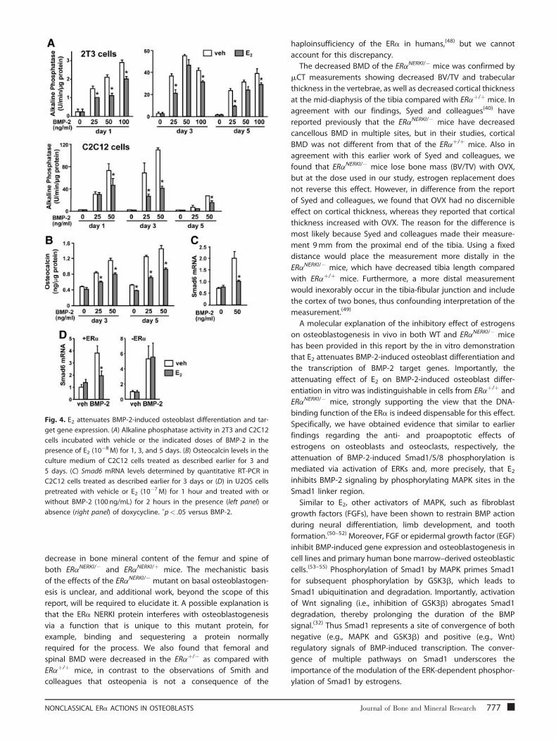
Fig. 4. E2 attenuates BMP-2-induced osteoblast differentiation and tar-
get gene expression. (A) Alkaline phosphatase activity in 2T3 and C2C12
cells incubated with vehicle or the indicated doses of BMP-2 in the
presence of E2 (10�8M) for 1, 3, and 5 days. (B) Osteocalcin levels in the
culture medium of C2C12 cells treated as described earlier for 3 and
5 days. (C) Smad6 mRNA levels determined by quantitative RT-PCR in
C2C12 cells treated as described earlier for 3 days or (D) in U2OS cells
pretreated with vehicle or E2 (10�7M) for 1 hour and treated with or
without BMP-2 (100 ng/mL) for 2 hours in the presence (left panel) or
absence (right panel) of doxycycline. �p< .05 versus BMP-2.
decrease in bone mineral content of the femur and spine of
both ERaNERKI/� and ERaNERKI/þ mice. The mechanistic basis
of the effects of the ERaNERKI/� mutant on basal osteoblastogen-
esis is unclear, and additional work, beyond the scope of this
report, will be required to elucidate it. A possible explanation is
that the ERa NERKI protein interferes with osteoblastogenesis
via a function that is unique to this mutant protein, for
example, binding and sequestering a protein normally
required for the process. We also found that femoral and
spinal BMD were decreased in the ERaþ/� as compared with
ERaþ/þ mice, in contrast to the observations of Smith and
colleagues that osteopenia is not a consequence of the
NONCLASSICAL ERa ACTIONS IN OSTEOBLASTS
haploinsufficiency of the ERa in humans,(48) but we cannot
account for this discrepancy.
The decreased BMD of the ERaNERKI/� mice was confirmed by
mCT measurements showing decreased BV/TV and trabecular
thickness in the vertebrae, as well as decreased cortical thickness
at the mid-diaphysis of the tibia compared with ERaþ/þ mice. In
agreement with our findings, Syed and colleagues(40) have
reported previously that the ERaNERKI/� mice have decreased
cancellous BMD in multiple sites, but in their studies, cortical
BMD was not different from that of the ERaþ/þ mice. Also in
agreement with this earlier work of Syed and colleagues, we
found that ERaNERKI/� mice lose bone mass (BV/TV) with OVX,
but at the dose used in our study, estrogen replacement does
not reverse this effect. However, in difference from the report
of Syed and colleagues, we found that OVX had no discernible
effect on cortical thickness, whereas they reported that cortical
thickness increased with OVX. The reason for the difference is
most likely because Syed and colleagues made their measure-
ment 9mm from the proximal end of the tibia. Using a fixed
distance would place the measurement more distally in the
ERaNERKI/� mice, which have decreased tibia length compared
with ERaþ/þ mice. Furthermore, a more distal measurement
would inexorably occur in the tibia-fibular junction and include
the cortex of two bones, thus confounding interpretation of the
measurement.(49)
A molecular explanation of the inhibitory effect of estrogens
on osteoblastogenesis in vivo in both WT and ERaNERKI/� mice
has been provided in this report by the in vitro demonstration
that E2 attenuates BMP-2-induced osteoblast differentiation and
the transcription of BMP-2 target genes. Importantly, the
attenuating effect of E2 on BMP-2-induced osteoblast differ-
entiation in vitro was indistinguishable in cells from ERaþ/þ and
ERaNERKI/� mice, strongly supporting the view that the DNA-
binding function of the ERa is indeed dispensable for this effect.
Specifically, we have obtained evidence that similar to earlier
findings regarding the anti- and proapoptotic effects of
estrogens on osteoblasts and osteoclasts, respectively, the
attenuation of BMP-2-induced Smad1/5/8 phosphorylation is
mediated via activation of ERKs and, more precisely, that E2inhibits BMP-2 signaling by phosphorylating MAPK sites in the
Smad1 linker region.
Similar to E2, other activators of MAPK, such as fibroblast
growth factors (FGFs), have been shown to restrain BMP action
during neural differentiation, limb development, and tooth
formation.(50–52) Moreover, FGF or epidermal growth factor (EGF)
inhibit BMP-induced gene expression and osteoblastogenesis in
cell lines and primary human bone marrow–derived osteoblastic
cells.(53–55) Phosphorylation of Smad1 by MAPK primes Smad1
for subsequent phosphorylation by GSK3b, which leads to
Smad1 ubiquitination and degradation. Importantly, activation
of Wnt signaling (i.e., inhibition of GSK3b) abrogates Smad1
degradation, thereby prolonging the duration of the BMP
signal.(32) Thus Smad1 represents a site of convergence of both
negative (e.g., MAPK and GSK3b) and positive (e.g., Wnt)
regulatory signals of BMP-induced transcription. The conver-
gence of multiple pathways on Smad1 underscores the
importance of the modulation of the ERK-dependent phosphor-
ylation of Smad1 by estrogens.
Journal of Bone and Mineral Research 777
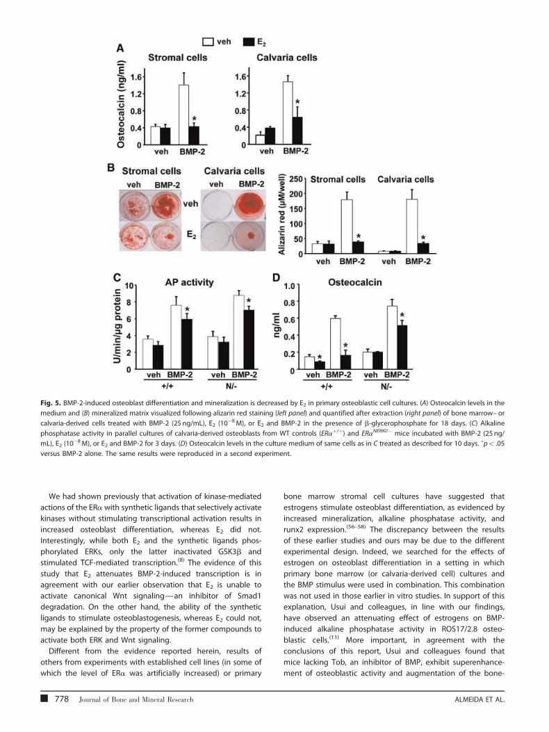
Fig. 5. BMP-2-induced osteoblast differentiation and mineralization is decreased by E2 in primary osteoblastic cell cultures. (A) Osteocalcin levels in the
medium and (B) mineralized matrix visualized following alizarin red staining (left panel) and quantified after extraction (right panel) of bone marrow– or
calvaria-derived cells treated with BMP-2 (25 ng/mL), E2 (10�8M), or E2 and BMP-2 in the presence of b-glycerophosphate for 18 days. (C) Alkaline
phosphatase activity in parallel cultures of calvaria-derived osteoblasts from WT controls (ERaþ/þ) and ERaNERKI/� mice incubated with BMP-2 (25 ng/
mL), E2 (10�8M), or E2 and BMP-2 for 3 days. (D) Osteocalcin levels in the culture medium of same cells as in C treated as described for 10 days. �p< .05
versus BMP-2 alone. The same results were reproduced in a second experiment.
We had shown previously that activation of kinase-mediated
actions of the ERa with synthetic ligands that selectively activate
kinases without stimulating transcriptional activation results in
increased osteoblast differentiation, whereas E2 did not.
Interestingly, while both E2 and the synthetic ligands phos-
phorylated ERKs, only the latter inactivated GSK3b and
stimulated TCF-mediated transcription.(8) The evidence of this
study that E2 attenuates BMP-2-induced transcription is in
agreement with our earlier observation that E2 is unable to
activate canonical Wnt signaling—an inhibitor of Smad1
degradation. On the other hand, the ability of the synthetic
ligands to stimulate osteoblastogenesis, whereas E2 could not,
may be explained by the property of the former compounds to
activate both ERK and Wnt signaling.
Different from the evidence reported herein, results of
others from experiments with established cell lines (in some of
which the level of ERa was artificially increased) or primary
778 Journal of Bone and Mineral Research
bone marrow stromal cell cultures have suggested that
estrogens stimulate osteoblast differentiation, as evidenced by
increased mineralization, alkaline phosphatase activity, and
runx2 expression.(56–58) The discrepancy between the results
of these earlier studies and ours may be due to the different
experimental design. Indeed, we searched for the effects of
estrogen on osteoblast differentiation in a setting in which
primary bone marrow (or calvaria-derived cell) cultures and
the BMP stimulus were used in combination. This combination
was not used in those earlier in vitro studies. In support of this
explanation, Usui and colleagues, in line with our findings,
have observed an attenuating effect of estrogens on BMP-
induced alkaline phosphatase activity in ROS17/2.8 osteo-
blastic cells.(11) More important, in agreement with the
conclusions of this report, Usui and colleagues found that
mice lacking Tob, an inhibitor of BMP, exhibit superenhance-
ment of osteoblastic activity and augmentation of the bone-
ALMEIDA ET AL.
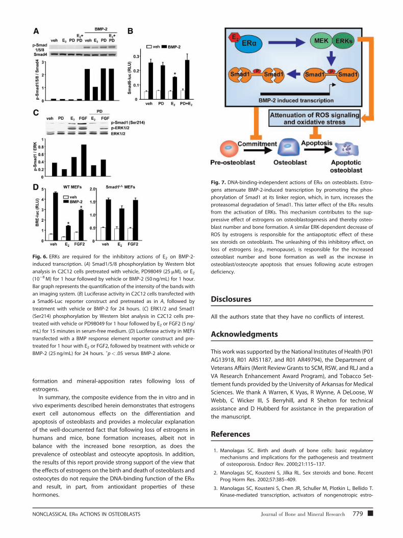
Fig. 6. ERKs are required for the inhibitory actions of E2 on BMP-2-
induced transcription. (A) Smad1/5/8 phosphorylation by Western blot
analysis in C2C12 cells pretreated with vehicle, PD98049 (25mM), or E2(10�8M) for 1 hour followed by vehicle or BMP-2 (50 ng/mL) for 1 hour.
Bar graph represents the quantification of the intensity of the bands with
an imaging system. (B) Luciferase activity in C2C12 cells transfected with
a Smad6-Luc reporter construct and pretreated as in A, followed by
treatment with vehicle or BMP-2 for 24 hours. (C) ERK1/2 and Smad1
(Ser214) phosphorylation by Western blot analysis in C2C12 cells pre-
treated with vehicle or PD98049 for 1 hour followed by E2 or FGF2 (5 ng/
mL) for 15 minutes in serum-free medium. (D) Luciferase activity in MEFs
transfected with a BMP response element reporter construct and pre-
treated for 1 hour with E2 or FGF2, followed by treatment with vehicle or
BMP-2 (25 ng/mL) for 24 hours. �p< .05 versus BMP-2 alone.
Fig. 7. DNA-binding-independent actions of ERa on osteoblasts. Estro-
gens attenuate BMP-2-induced transcription by promoting the phos-
phorylation of Smad1 at its linker region, which, in turn, increases the
proteasomal degradation of Smad1. This latter effect of the ERa results
from the activation of ERKs. This mechanism contributes to the sup-
pressive effect of estrogens on osteoblastogenesis and thereby osteo-
blast number and bone formation. A similar ERK-dependent decrease of
ROS by estrogens is responsible for the antiapoptotic effect of these
sex steroids on osteoblasts. The unleashing of this inhibitory effect, on
loss of estrogens (e.g., menopause), is responsible for the increased
osteoblast number and bone formation as well as the increase in
osteoblast/osteocyte apoptosis that ensues following acute estrogen
deficiency.
formation and mineral-apposition rates following loss of
estrogens.
In summary, the composite evidence from the in vitro and in
vivo experiments described herein demonstrates that estrogens
exert cell autonomous effects on the differentiation and
apoptosis of osteoblasts and provides a molecular explanation
of the well-documented fact that following loss of estrogens in
humans and mice, bone formation increases, albeit not in
balance with the increased bone resorption, as does the
prevalence of osteoblast and osteocyte apoptosis. In addition,
the results of this report provide strong support of the view that
the effects of estrogens on the birth and death of osteoblasts and
osteocytes do not require the DNA-binding function of the ERa
and result, in part, from antioxidant properties of these
hormones.
NONCLASSICAL ERa ACTIONS IN OSTEOBLASTS
Disclosures
All the authors state that they have no conflicts of interest.
Acknowledgments
This work was supported by the National Institutes of Health (P01
AG13918, R01 AR51187, and R01 AR49794), the Department of
Veterans Affairs (Merit Review Grants to SCM, RSW, and RLJ and a
VA Research Enhancement Award Program), and Tobacco Set-
tlement funds provided by the University of Arkansas for Medical
Sciences. We thank A Warren, K Vyas, R Wynne, A DeLoose, W
Webb, C Wicker III, S Berryhill, and R Shelton for technical
assistance and D Hubberd for assistance in the preparation of
the manuscript.
References
1. Manolagas SC. Birth and death of bone cells: basic regulatory
mechanisms and implications for the pathogenesis and treatment
of osteoporosis. Endocr Rev. 2000;21:115–137.
2. Manolagas SC, Kousteni S, Jilka RL. Sex steroids and bone. Recent
Prog Horm Res. 2002;57:385–409.
3. Manolagas SC, Kousteni S, Chen JR, Schuller M, Plotkin L, Bellido T.
Kinase-mediated transcription, activators of nongenotropic estro-
Journal of Bone and Mineral Research 779
gen-like signaling (ANGELS), and osteoporosis: a different perspec-tive on the HRT dilemma. Kidney Int Suppl. 2004;91:S41–49.
4. Jilka RL, Hangoc G, Girasole G, et al. Increased osteoclast development
afterestrogen loss:mediationby interleukin-6.Science.1992;257:88–91.
5. DiGregorio G, Yamamoto M, Ali A, et al. Attenuation of the self-renewal of transit amplifying osteoblast progenitors in the murine
bone marrow by 17b-estradiol. J Clin Invest. 2001;107:803–812.
6. Kousteni S, Bellido T, Plotkin LI, et al. Nongenotropic, sex-nonspecificsignaling through the estrogen or androgen receptors: dissociation
from transcriptional activity. Cell. 2001;104:719–730.
7. Kousteni S, Chen J-R, Bellido T, et al. Reversal of bone loss in mice by
nongenotropic signaling of sex steroids. Science. 2002;298:843–846.
8. Kousteni S, Han L, Chen J-R, et al. Kinase-mediated regulation of
common transcription factors accounts for the bone-protective
effects of sex steroids. J Clin Invest. 2003;111:1651–1664.
9. Chen JR, Plotkin LI, Aguirre JI, et al. Transient Versus SustainedPhosphorylation and Nuclear Accumulation of ERKs Underlie Anti-
Versus Pro-apoptotic Effects of Estrogens. J Biol Chem. 2005;280:
4632–4638.
10. Jilka RL, Takahashi K, Munshi M, Williams DC, Roberson PK, Mano-lagas SC. Loss of estrogen upregulates osteoblastogenesis in the
murine bone marrow: evidence for autonomy from factors released
during bone resorption. J Clin Invest. 1998;101:1942–1950.
11. Usui M, Yoshida Y, Tsuji K, et al. Tob deficiency superenhances
osteoblastic activity after ovariectomy to block estrogen defi-
ciency-induced osteoporosis. Proc Natl Acad Sci U S A. 2004;101:
6653–6658.
12. Yamamoto T, Saatcioglu F, Matsuda T. Cross-talk between bone
morphogenic proteins and estrogen receptor signaling. Endocrinol-
ogy. 2002;143:2635–2642.
13. Kousteni S, Almeida M, Han L, Bellido T, Jilka RL, Manolagas SC.Induction of osteoblast differentiation by selective activation of
kinase-mediated actions of the estrogen receptor. Mol Cell Biol.
2007;27:1516–1530.
14. Rosen V. BMP and BMP inhibitors in bone. Ann N Y Acad Sci.
2006;1068:19–25.
15. Canalis E, Economides AN, Gazzerro E. Bonemorphogenetic proteins,
their antagonists, and the skeleton. Endocr Rev. 2003;24:218–235.
16. Yamaguchi A, Komori T, Suda T. Regulation of osteoblast differentia-
tion mediated by bone morphogenetic proteins, hedgehogs, and
Cbfa1. Endocr Rev. 2000;21:393–411.
17. Abe E, Yamamoto M, Taguchi Y, et al. Essential requirement of BMPs-2/4 for both osteoblast and osteoclast formation in murine bone
marrow cultures from adult mice: antagonism by noggin. J Bone
Miner Res. 2000;15:663–673.
18. Massague J, Seoane J, Wotton D. Smad transcription factors. GenesDev. 2005;19:2783–2810.
19. Shi Y, Massague J. Mechanisms of TGF-b signaling from cell mem-
brane to the nucleus. Cell. 2003;113:685–700.
20. Feng XH, Derynck R. Specificity and versatility in TGF-b signaling
through Smads. Annu Rev Cell Dev Biol. 2005;21:659–693.
21. Sapkota G, Alarcon C, Spagnoli FM, Brivanlou AH, Massague J.
Balancing BMP signaling through integrated inputs into the Smad1linker. Mol Cell. 2007;25:441–454.
22. Kretzschmar M, Doody J, Massague J. Opposing BMP and EGF
signalling pathways converge on the TGF-b family mediator Smad1.
Nature. 1997;389:618–622.
23. Pera EM, Ikeda A, Eivers E, De Robertis EM. Integration of IGF, FGF, and
anti-BMP signals via Smad1 phosphorylation in neural induction.
Genes Dev. 2003;17:3023–3028.
24. Aubin J, Davy A, Soriano P. In vivo convergence of BMP and MAPK
signaling pathways: impact of differential Smad1 phosphorylation on
development and homeostasis. Genes Dev. 2004;18:1482–1494.
780 Journal of Bone and Mineral Research
25. Almeida M, Han L, Martin-Millan M, et al. Skeletal involution by age-associated oxidative stress and its acceleration by loss of sex steroids.
J Biol Chem. 2007;282:27285–27297.
26. Giorgio M, Migliaccio E, Orsini F, et al. Electron transfer between
cytochrome c and p66Shc generates reactive oxygen species thattrigger mitochondrial apoptosis. Cell. 2005;122:221–233.
27. Jakacka M, Ito M, Martinson F, Ishikawa T, Lee EJ, Jameson JL. An
estrogen receptor (ER) a deoxyribonucleic acid-binding domainknock-in mutation provides evidence for nonclassical ER pathway
signaling in vivo. Mol Endocrinol. 2002;16:2188–2201.
28. Korchynskyi O, Ten Dijke P. Identification and functional character-
ization of distinct critically important bone morphogenetic protein-specific response elements in the Id1 promoter. J Biol Chem. 2002;
277:4883–4891.
29. Dupont S, Krust A, Gansmuller A, Dierich A, Chambon P, Mark M.
Effect of single and compound knockouts of estrogen receptors a(ERa) and b (ERb) onmouse reproductive phenotypes. Development.
2000;127:4277–4291.
30. Weinstein RS, Jilka RL, Parfitt AM, Manolagas SC. Inhibition of
osteoblastogenesis and promotion of apoptosis of osteoblasts andosteocytes by glucocorticoids: potential mechanisms of their dele-
terious effects on bone. J Clin Invest. 1998;102:274–282.
31. Weinstein RS, Chen JR, Powers CC, et al. Promotion of osteoclastsurvival and antagonism of bisphosphonate-induced osteoclast
apoptosis by glucocorticoids. J Clin Invest. 2002;109:1041–1048.
32. Fuentealba LC, Eivers E, Ikeda A, et al. Integrating patterning signals:
Wnt/GSK3 regulates the duration of the BMP/Smad1 signal. Cell.2007;131:980–993.
33. Jilka RL, Weinstein RS, Bellido T, Parfitt AM, Manolagas SC. Osteoblast
programmed cell death (apoptosis): modulation by growth factors
and cytokines. J Bone Miner Res. 1998;13:793–802.
34. Kuznetsov S, Robey PG. Species differences in growth requirements
for bone marrow stromal fibroblast colony formation in vitro. Calcif
Tissue Int. 1996;59:265–270.
35. Kian TM, Rogatsky I, Tzagarakis-Foster C, et al. Estradiol and selective
estrogen receptor modulators differentially regulate target genes
with estrogen receptors a and b. Mol Biol Cell. 2004;15:1262–1272.
36. Almeida M, Han L, Bellido T, Manolagas SC, Kousteni S. Wnt proteinsprevent apoptosis of both uncommitted osteoblast progenitors and
differentiated osteoblasts by b-catenin-dependent and -indepen-
dent signaling cascades involving Src/ERK and phosphatidylinositol
3-kinase/AKT. J Biol Chem. 2005;280:41342–41351.
37. Huang X, Frenkel K, Klein CB, Costa M. Nickel induces increased
oxidants in intact cultured mammalian cells as detected by dichlor-
ofluorescein fluorescence. Toxicol Appl Pharmacol. 1993;120:29–36.
38. Plotkin LI, Weinstein RS, Parfitt AM, Roberson PK, Manolagas SC,Bellido T. Prevention of osteocyte and osteoblast apoptosis by
bisphosphonates and calcitonin. J Clin Invest. 1999;104:1363–1374.
39. Jilka RL, Weinstein RS, Takahashi K, Parfitt AM, Manolagas SC. Linkageof decreased bone mass with impaired osteoblastogenesis in a
murine model of accelerated senescence. J Clin Invest. 1996;97:
1732–1740.
40. Syed F, Modder U, Fraser D, et al. Skeletal effects of estrogen aremediated by opposing actions of classical and non-classical estrogen
receptor pathways. J Bone Miner Res. 2005;20:1992–2001.
41. Kuroda H, Fuentealba L, Ikeda A, Reversade B, De Robertis EM. Default
neural induction: neuralization of dissociated Xenopus cells ismediated by Ras/MAPK activation. Genes Dev. 2005;19:1022–1027.
42. Hall JM, Couse JF, Korach KS. The Multifaceted Mechanisms of
Estradiol and Estrogen Receptor Signaling. J Biol Chem. 2001;276:36869–36872.
43. Manolagas SC, Kousteni S. Perspective: Nonreproductive Sites of
Action of Reproductive Hormones. Endocrinology. 2001;142:2200–
2204.
ALMEIDA ET AL.
44. Hammes SR, Levin ER. Extranuclear steroid receptors: nature andactions. Endocr Rev. 2007;28:726–741.
45. Sims NA, Dupont S, Krust A, et al. Deletion of estrogen receptors
reveals a regulatory role for estrogen receptors-b in bone remodeling
in females but not in males. Bone. 2002;30:18–25.
46. Antal MC, Krust A, Chambon P, Mark M. Sterility and absence
of histopathological defects in nonreproductive organs of a
mouse ERb-null mutant. Proc Natl Acad Sci USA. 2008;105:2433–2438.
47. Li Y, Li A, Strait K, Zhang H, Nanes MS, Weitzmann MN. Endo-
genous TNFa lowers maximum peak bone mass and inhibits osteo-
blastic Smad activation through NF-kB. J Bone Miner Res. 2007;22:646–655.
48. Smith EP, Specker B, Bachrach BE, et al. Impact on Bone of an
Estrogen Receptor-a Gene Loss of Function Mutation. J Clin Endo-
crinol Metab. 2008;93:3088–3096.
49. Bab I, Hajbi-Yonissi C, Gabet Y, Muller R. Tibio-fibular complex and
knee joint, Fig. 7. In Micro-Tomographic Atlas of the Mouse Skeleton.
New York, NY: Springer, 2007:177.
50. De Robertis EM, Kuroda H. Dorsal-ventral patterning and neuralinduction in Xenopus embryos. Annu Rev Cell Dev Biol. 2004;20:
285–308.
51. Niswander L, Martin GR. FGF-4 and BMP-2 have opposite effects onlimb growth. Nature. 1993;361:68–71.
NONCLASSICAL ERa ACTIONS IN OSTEOBLASTS
52. Neubuser A, Peters H, Balling R, Martin GR. Antagonistic interactionsbetween FGF and BMP signaling pathways: a mechanism for posi-
tioning the sites of tooth formation. Cell. 1997;90:247–255.
53. Nakayama K, Tamura Y, Suzawa M, et al. Receptor tyrosine kinases
inhibit bone morphogenetic protein-Smad responsive promoteractivity and differentiation of murine MC3T3-E1 osteoblast-like cells.
J Bone Miner Res. 2003;18:827–835.
54. Osyczka AM, Leboy PS. Bone morphogenetic protein regulation ofearly osteoblast genes in humanmarrow stromal cells is mediated by
extracellular signal-regulated kinase and phosphatidylinositol 3-
kinase signaling. Endocrinology. 2005;146:3428–3437.
55. Chaudhary LR, Hofmeister AM, Hruska KA. Differential growth factorcontrol of bone formation through osteoprogenitor differentiation.
Bone. 2004;34:402–411.
56. Dang ZC, Van Bezooijen RL, Karperien M, Papapoulos SE, Lowik CW.
Exposure of KS483 cells to estrogen enhances osteogenesis andinhibits adipogenesis. J Bone Miner Res. 2002;17:394–405.
57. Okazaki R, Inoue D, Shibata M, et al. Estrogen promotes early
osteoblast differentiation and inhibits adipocyte differentiation in
mouse bonemarrow stromal cell lines that express estrogen receptor(ER) a or b. Endocrinology. 2002;143:2349–2356.
58. Qu Q, Perala-Heape M, Kapanen A, et al. Estrogen enhances differ-
entiation of osteoblasts in mouse bone marrow culture. Bone.1998;22:201–209.
Journal of Bone and Mineral Research 781
Copyright © 2022 FDOKUMEN