
Molecular clustering based on ERα and EIG121 predicts survival in high-grade serous carcinoma of...
19
Molecular Clustering Based on ERα and EIG121 Predicts Survival in High-Grade Serous Carcinoma of the Ovary/ Peritoneum Matthew P. Schlumbrecht 1 , Su-Su Xie 2 , Gregory L. Shipley 3 , Diana L. Urbauer 4 , and Russell R. Broaddus 2 1 Department of Gynecologic Oncology, The University of Texas M. D. Anderson Cancer Center, Houston, TX 2 Department of Pathology, The University of Texas M. D. Anderson Cancer Center, Houston, TX 3 Department of Integrative Biology & Pharmacology, The University of Texas Health Science Center at Houston, Houston, TX 4 Division of Quantitative Sciences, The University of Texas M. D. Anderson Cancer Center, Houston, TX Abstract Assessment of estrogen receptor expression by immunohistochemistry has yielded inconsistent results as a prognostic indicator in ovarian carcinoma. In breast and endometrial carcinomas, panels of estrogen-induced genes have shown improved prognostic capability over the use of estrogen receptor immunohistochemistry alone. For both breast and endometrial cancer, over- expression of estrogen-induced genes is associated with better prognosis. We hypothesized that analysis of a panel of estrogen-induced genes can predict outcome in ovarian carcinoma and potentially differentiate between tumors of varying hormonal responsiveness. From a cohort of 219 women undergoing ovarian cancer surgery from 2004 −2007, 83 patients were selected for inclusion. All patients had advanced stage ovarian/primary peritoneal high-grade serous carcinoma and underwent primary surgical debulking followed by adjuvant treatment with platinum and taxane agents. The expression of ERα and six genes known to be induced by estrogen in the female reproductive tract (EIG121, sFRP1, sFRP4, RALDH2, PR, IGF-1) was measured using qRT-PCR. Unsupervised cluster analyses were used to categorize patients as high or low gene expressors. Gene expression results were then compared to those for estrogen receptor immunohistochemistry. Clusters were compared using chi-square analyses, and Cox proportional hazards were used to evaluate survival outcomes. Median follow-up time was 38.7 months (range 1–68). A cluster defined by EIG121 and ERα segregated tumors into distinct groups of high and low gene expressors. Shorter overall survival was associated with high gene expression (HR 2.84 [1.11–7.30], p=0.03), even after adjustment for other covariates. No difference in estrogen receptor immunohistochemistry expression was noted between gene clusters. In contrast to other hormonally-driven cancers, high expression of ERα and the estrogen-induced gene EIG121 predicts shorter overall survival in patients with high-grade serous ovarian carcinoma. Such a Users may view, print, copy, download and text and data- mine the content in such documents, for the purposes of academic research, subject always to the full Conditions of use: http://www.nature.com/authors/editorial_policies/license.html#terms Corresponding Author, Russell R. Broaddus, MD, PhD, Associate Professor, Department of Pathology, The University of Texas M. D. Anderson Cancer Center, 1515 Holcombe Blvd, Unit 0085, Houston, TX 77030, Phone: 713-745-2794, [email protected]. Conflict of Interest. The authors report no conflict of interest. NIH Public Access Author Manuscript Mod Pathol. Author manuscript; available in PMC 2011 September 1. Published in final edited form as: Mod Pathol. 2011 March ; 24(3): 453–462. doi:10.1038/modpathol.2010.211. NIH-PA Author Manuscript NIH-PA Author Manuscript NIH-PA Author Manuscript
-
Upload
mdanderson -
Category
Documents
-
view
3 -
download
0
Transcript of Molecular clustering based on ERα and EIG121 predicts survival in high-grade serous carcinoma of...

Molecular Clustering Based on ERα and EIG121 PredictsSurvival in High-Grade Serous Carcinoma of the Ovary/Peritoneum
Matthew P. Schlumbrecht1, Su-Su Xie2, Gregory L. Shipley3, Diana L. Urbauer4, andRussell R. Broaddus2
1Department of Gynecologic Oncology, The University of Texas M. D. Anderson Cancer Center,Houston, TX2Department of Pathology, The University of Texas M. D. Anderson Cancer Center, Houston, TX3Department of Integrative Biology & Pharmacology, The University of Texas Health ScienceCenter at Houston, Houston, TX4Division of Quantitative Sciences, The University of Texas M. D. Anderson Cancer Center,Houston, TX
AbstractAssessment of estrogen receptor expression by immunohistochemistry has yielded inconsistentresults as a prognostic indicator in ovarian carcinoma. In breast and endometrial carcinomas,panels of estrogen-induced genes have shown improved prognostic capability over the use ofestrogen receptor immunohistochemistry alone. For both breast and endometrial cancer, over-expression of estrogen-induced genes is associated with better prognosis. We hypothesized thatanalysis of a panel of estrogen-induced genes can predict outcome in ovarian carcinoma andpotentially differentiate between tumors of varying hormonal responsiveness. From a cohort of219 women undergoing ovarian cancer surgery from 2004 −2007, 83 patients were selected forinclusion. All patients had advanced stage ovarian/primary peritoneal high-grade serous carcinomaand underwent primary surgical debulking followed by adjuvant treatment with platinum andtaxane agents. The expression of ERα and six genes known to be induced by estrogen in thefemale reproductive tract (EIG121, sFRP1, sFRP4, RALDH2, PR, IGF-1) was measured usingqRT-PCR. Unsupervised cluster analyses were used to categorize patients as high or low geneexpressors. Gene expression results were then compared to those for estrogen receptorimmunohistochemistry. Clusters were compared using chi-square analyses, and Cox proportionalhazards were used to evaluate survival outcomes. Median follow-up time was 38.7 months (range1–68). A cluster defined by EIG121 and ERα segregated tumors into distinct groups of high andlow gene expressors. Shorter overall survival was associated with high gene expression (HR 2.84[1.11–7.30], p=0.03), even after adjustment for other covariates. No difference in estrogenreceptor immunohistochemistry expression was noted between gene clusters. In contrast to otherhormonally-driven cancers, high expression of ERα and the estrogen-induced gene EIG121predicts shorter overall survival in patients with high-grade serous ovarian carcinoma. Such a
Users may view, print, copy, download and text and data- mine the content in such documents, for the purposes of academic research,subject always to the full Conditions of use: http://www.nature.com/authors/editorial_policies/license.html#terms
Corresponding Author, Russell R. Broaddus, MD, PhD, Associate Professor, Department of Pathology, The University of Texas M.D. Anderson Cancer Center, 1515 Holcombe Blvd, Unit 0085, Houston, TX 77030, Phone: 713-745-2794,[email protected].
Conflict of Interest. The authors report no conflict of interest.
NIH Public AccessAuthor ManuscriptMod Pathol. Author manuscript; available in PMC 2011 September 1.
Published in final edited form as:Mod Pathol. 2011 March ; 24(3): 453–462. doi:10.1038/modpathol.2010.211.
NIH
-PA Author Manuscript
NIH
-PA Author Manuscript
NIH
-PA Author Manuscript

biomarker panel may potentially be used to guide management with estrogen antagonists in thispatient population.
Keywordsestrogen-induced genes; ovarian high-grade serous carcinoma; hormone antagonism
IntroductionOvarian cancer is the fifth most common cause of cancer death in American women (1).Carcinoma of the ovary is typically diagnosed in women older than 50 years of age, and themajority of cancers are advanced at time of diagnosis (1). The majority of ovarian cancertypes are epithelial in origin, and of these, high-grade serous carcinoma is the most common.For the majority of patients, the treatment of high-grade serous carcinoma is surgicaldebulking followed by adjuvant chemotherapy with combination platinum and taxaneagents. The five-year overall survival for women with high-grade serous carcinoma is lessthan 30%, due in part to its asymptomatic progression from localized tumor to metastaticdisease, high rate of recurrence, development of chemoresistance, and the paucity of agentseffective at treating recurrent disease (1–3).
Because ovarian function is, at least in part, hormonally regulated, it is generally presumedthat ovarian carcinoma should be somewhat responsive to hormonal manipulation. Given thesuccess of hormone manipulating agents in the treatment of breast, endometrial, and prostatecarcinomas, incorporating agents aimed at antagonizing hormone-dependent proliferativepathways into the ovarian cancer treatment algorithm has been proposed. Agents targetingestrogen pathways are of particular interest, especially since the inhibition of estrogen inestrogen receptor (ER) positive breast cancers translates into reduced risk of recurrence andincreased survival (4). Despite the fact that more than 60% of ovarian cancers demonstrateER positivity (5), inhibition of estrogen-associated pathways has not translated intosignificant improvements in patient outcomes. In a meta-analysis of 18 ovarian cancer trialswhich treated patients with tamoxifen, a selective estrogen receptor modulator, the collectiveresponse rate was only 13% (6). Pre-treatment determination of tumor ER status has alsodemonstrated poor predictive ability of response to treatment. For example, Schwartz et al(7) performed a prospective trial of chemotherapy with or without tamoxifen in patients withadvanced stage ovarian cancer and noted that there was no relation between hormonereceptor status and survival outcomes. More recent studies evaluating the activity ofaromatase inhibitors in ER-positive patients have reported response rates between 3–17%,with stable disease achieved in up to 26% (8,9). Taken together, these investigations suggestthat there is a subset of women with ovarian cancer who will have some degree of responseto hormone antagonism, but ER immunohistochemistry may not be a sufficient means ofidentifying these patients.
An alternative to ER immunohistochemical assessment is to evaluate genes known to beinduced by estrogen, a strategy which has resulted in improved capability to segregatetumors based on hormone sensitivity in other malignancies. In breast cancer, quantitativeexamination of estrogen-regulated genes helps to detect subgroups within ER-positivetumors with differing survival parameters, even when accounting for tumor characteristicssuch as lymph node positivity, tumor size, and the use of chemotherapy (10). Validatedusing specimens provided by a several different investigators, a gene panel proposed by Ohet al (10) accurately predicted patients with invasive breast ductal carcinoma who hadmarkedly different relapse-free survivals. Similar findings have been reported in endometrialcancer. In 2009, Westin et al (11) described a panel of estrogen-induced genes in patients
Schlumbrecht et al. Page 2
Mod Pathol. Author manuscript; available in PMC 2011 September 1.
NIH
-PA Author Manuscript
NIH
-PA Author Manuscript
NIH
-PA Author Manuscript
with endometrial carcinoma which identified two distinct clusters based on degree of geneexpression. Higher estrogen-regulated gene expression was predictive of improvedrecurrence-free survival and was able to distinguish between high/intermediate- and low-risktumors with a false negative rate of only 4.8% (11).
Given the findings in breast and endometrial carcinoma that estrogen-regulated genesdemonstrate prognostic capability, it is possible that analyzing estrogen-regulated geneexpression may have similar utility for ovarian cancer patients. Determining which subset ofwomen with ovarian cancer who may potentially respond to estrogen antagonism wouldafford the oncologist the ability to initiate such treatment earlier in the disease course, eitheralone or in combination with other therapies. Our primary aim was to quantify theexpression of estrogen-induced genes in a cohort of women with the most common ovariancancer, high-grade serous carcinoma, and determine if differential expression was predictiveof clinical outcomes. Secondarily, we compared gene expression to immunohistochemicalassessment of ER, the current standard for judging hormone sensitivity, to determine ifimmunohistochemistry accurately predicts tumor molecular profiles. We hypothesized thatthis examination of estrogen-induced genes would identify subsets of patients with differentclinical characteristics and distinct survival outcomes. Because higher estrogen-inducedgene expression portends improved prognosis in other hormone-sensitive tumors, weexpected that a similar relationship would be observed in this cohort of ovarian cancerpatients.
Materials and MethodsPatient Selection and Clinical Data Acquisition
After IRB approval, a review of the institutional Tumor Bank identified two-hundrednineteen (219) patients from whom ovarian or primary peritoneal carcinoma specimens wereobtained at the time of tumor-reductive surgeries between 2004 and 2007. Pathologicdiagnoses were made by gynecologic pathologists after microscopic review of hematoxylinand eosin-stained slides derived from surgical specimens containing ovarian or primaryperitoneal carcinomas. Patient clinical characteristics were obtained by a review ofelectronic medical records and included date of birth, race, anthropometric variables, date ofsurgical staging, debulking status, primary and secondary chemotherapy regimens, date ofrecurrence, date of last follow-up, and disease status at last follow-up. Both clinical andpathologic features were utilized to determine inclusion criteria. Patients selected forinclusion in the study demonstrated only advanced stage (III or IV), high-grade serousovarian or primary peritoneal carcinoma. Additionally, all patients received treatment withplatinum and taxane agents as first-line adjuvant chemotherapy. Specific exclusion criteriaincluded treatment with neoadjuvant chemotherapy, consolidation/maintenancechemotherapy, and first-line treatment regimens that were experimental protocols or notplatinum-based. Body mass indices were categorized by World Health Organizationdefinitions of normal weight, overweight, and obese. Chemotherapy resistance wasdetermined using Gynecologic Oncology Group criteria, which include 1) diseaseprogression while on a first-line platinum-based regimen; 2) tumor progression within sixmonths of completion of platinum-based therapy; and 3) persistent clinically measurabledisease with best response as stable disease at the completion of first-line therapy (12).
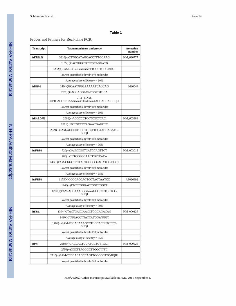
Gene Selection and RNA PreparationSeven genes (ERα, PR, EIG121, IGF-1, RALDH2, sFRP1, sFRP4) were included fortranscript quantification by qRT-PCR. Our research group has previously used a genomicsapproach to identify that EIG121, RALDH2, sFRP1, and sFRP4 are highly induced byestrogen in the human female reproductive tract (11,13,14). PR and IGF-1 are classical
Schlumbrecht et al. Page 3
Mod Pathol. Author manuscript; available in PMC 2011 September 1.
NIH
-PA Author Manuscript
NIH
-PA Author Manuscript
NIH
-PA Author Manuscript
estrogen-induced genes (15,16). Transcript analysis of ERα was included for subsequentcomparisons to ER immunohistochemistry studies. After microscopic confirmation of tumorhistology and presence of greater than 70% viable tumor, RNA was extracted from frozentissues, prepared using the TriReagent (Molecular Research Center, Cincinnati, OH), andprecipitated with isopropyl alcohol. Ultraviolet spectrometry was used to confirm adequateconcentrations of RNA in each prepared specimen. RNA was applied to RNeasy spincolumns (QIAGEN, Valencia, CA), eluted, and treated with 10% RNAse-free DNAsesolution. Specimens were incubated for 30 minutes at 37°C and subsequently treated for 10minutes at 75°C to inactivate DNAse I. Post-incubation RNA was stored at −80°C untilquantitative RT-PCR is performed.
Reverse Transcription and Real-Time Polymerase Chain ReactionProbes and primers (TaqMan® probe) for the gene panel are shown in Table 1. Aliquots(100 ng) of each RNA were reverse transcribed in quadruplicate (including a no reversetranscriptase control) with 300 nM assay specific reverse primer, 4 mM MgCl2, 500 µMdNTPs and 10 Unit of MMLV Superscript II reverse transcriptase (Invitrogen, Carlsbad,CA) at 50°C for 30 min, followed by 72°C for 5 min. Forty µl of PCR mix containing 1XPCR buffer, 300 nM specific forward and reverse primers, 4 mM MgCl2, Taq DNApolymerase and 100 nM fluorogenic probe were then added to each 10-µl RT reaction.Amplification was performed by use of the ABI Prism 7700 Sequence Detection System(Applied Biosystems, Foster City, CA) at 95°C for 1min, followed by 40 cycles of a 12-secstep at 95°C and a 1-min step at 60°C. Synthetic RNA or single strand DNA ampliconstandards were serially diluted in water containing 100 ng/µl yeast tRNA (Invitrogen,Carlsbad, CA).
ImmunohistochemistryImmunohistochemistry for ERα (clone 6F11, 1:35; Novacastra/Leica Microsystems,Chicago, IL) was performed using standard histological techniques. Antigen retrieval wasaccomplished with citrate buffer for 30 min at 100 °C. Stained slides were microscopicallyreviewed in a blinded fashion by the study primary author (MPS) and a gynecologicpathologist (RRB). ERα protein expression was scored semi-quantitatively as per centtumor with 2+ or 3+ nuclear staining. Tumors were considered positive if ≥10% of tumorcell nuclei demonstrated strong (2+ or 3+) expression.
Statistical AnalysesStatistical analyses were performed using STATA 10.0 (StataCorp, College Station, TX)and SPSS 17.0 (Chicago, IL). Summary statistics were generated to describe the studypopulation. The Wilcoxon rank-sum test was used to compare mean gene expression inrelation to clinical variables, and linear regression was used to compare gene expressionwith continuous clinical variables (body mass index, age). Chi-square analyses were used todescribe the relationship between categorical variables, and logistic regression was used todetermine associations between clinical variables and categorical outcomes. Recurrence-freesurvival (RFS) and overall survival (OS) for each gene was analyzed using Cox proportionalhazards models and the Kaplan-Meier method. RFS was defined as the interval betweeninitial surgical treatment and date of recurrence. OS was defined as the interval betweeninitial surgical treatment and date of last follow-up or death. A p-value of <0.05 wasconsidered statistically significant, and all tests were two-sided.
Unsupervised cluster analyses were performed for each gene separately, and in variouspermutations, to include all six estrogen-induced genes and ERα. Specific clusters wereselected based on their potential ability to segregate tumors into distinct groups, andanalyzed to compare differences in survival, outcome, and demographic characteristics
Schlumbrecht et al. Page 4
Mod Pathol. Author manuscript; available in PMC 2011 September 1.
NIH
-PA Author Manuscript
NIH
-PA Author Manuscript
NIH
-PA Author Manuscript
between these groups. Characteristics between cluster groups were compared using chi-square or Fisher’s exact analyses, when appropriate. Univariate Cox proportional hazardsmodels were used to analyze gene associations between survival, gene expression clusters,and other patient clinical features. Variables with p-values <0.15 in the univariate analysiswere included in the multivariate model. The Kaplan-Meier method and log-rank test wereused to compare survival outcomes.
The Wilcoxon rank-sum test was used to compare percentage of ER immunohistochemicalexpression between tumor samples from the different clusters. Fisher’s exact test was usedto compare clusters when protein expression was characterized categorically. All tests weretwo-sided, and a p-value of <0.05 was considered statistically significant.
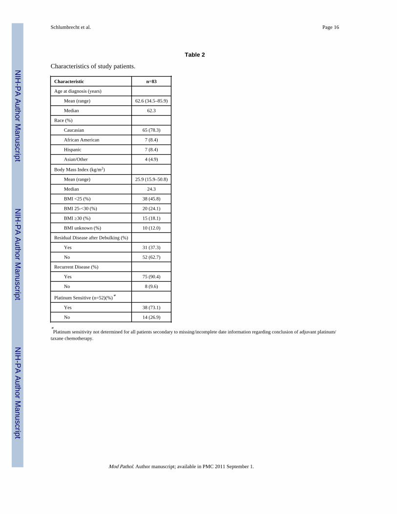
ResultsTwo hundred nineteen (219) women underwent surgery for ovarian or primary peritonealcarcinoma during the study interval. Application of the inclusion criteria resulted in arelatively homogenous cohort for study numbering 113 patients of which frozen tissues for83 were available from the institutional Gynecologic Oncology Tumor Bank. Patients werefollowed for a median of 38.7 months (range 0.5–67.8) from the time of their cancerdiagnoses. The demographic features of the cohort of 83 patients are summarized in Table 2.The mean age at diagnosis was 62.6 years (range 34.5–85.9), and the majority of patientswere Caucasian. A significant proportion of patients were overweight or obese (42.2%), andmore than 18% had a BMI ≥ 30 kg/m2. Fifty-two (62.7%) patients had an optimal surgicaldebulking, defined as less than 1 cm of residual disease at the conclusion of primarycytoreductive therapy, and more than 90% of the patients recurred during the follow-upinterval. No association between either age (p=0.22) or race (p=0.88) and frequency ofoptimal debulking was observed. However, as BMI increased, the frequency of optimaldebulking decreased (p=0.03).
In a univariate analysis, increased expression of EIG121 demonstrated a statisticallysignificant association with worse OS (HR 1.21 [1.09–1.35], p<0.001) (Table 3). IncreasedRALDH2 expression was also associated with reduced OS, though to a lesser degree (HR1.18 [1.03–1.35], p=0.016). There was a trend towards a negative association betweensFRP1 expression and OS, but the association was not statistically significant. In amultivariate model which included gene expression and patient clinical characteristics, bothsFRP1 (HR 1.04 [1.00–1.07], p=0.028) and EIG121 (HR 1.20 [CI 1.08–1.35], p=0.001)remained independently prognostic of worse OS. When RFS was considered, greaterEIG121 expression was associated with shorter time to recurrence (HR 1.13 [CI 1.02–1.26],p=0.021). EIG121 was the only gene in the panel whose expression demonstrated such aneffect. When accounting for patient characteristics in a multivariate model, EIG121remained predictive of RFS (HR 1.14 [CI 1.02–1.27], p=0.02).
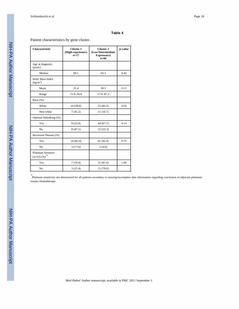
Unsupervised cluster analyses based on gene expression were performed and resulted in atotal of 246 unique clusters. All clusters were reviewed, and those which appeared to bestsegregate patients into two distinct groups were considered for more detailed analysis.Ultimately, the cluster defined by EIG121 and ERα was selected for further examinationbecause that combination appeared to reasonably segregate tumors into distinct groups ofhigh expressors (Cluster 1) and low/intermediate expressors (Cluster 2) (Figure 1). Thecharacteristics of the patients in each cluster are described in Table 4. There was nodifference between clusters with regards to BMI, age at diagnosis, frequency of optimaldebulking, or recurrence. There was a greater proportion of non-white patients in Cluster 1than Cluster 2 (41% vs. 20%, p=0.03).
Schlumbrecht et al. Page 5
Mod Pathol. Author manuscript; available in PMC 2011 September 1.
NIH
-PA Author Manuscript
NIH
-PA Author Manuscript
NIH
-PA Author Manuscript
A univariate analysis of patient characteristics and gene expression clusters in relation to OSis shown in Table 5. Age at diagnosis (p=0.56), race (p=0.57), and BMI (p=0.60) were notsignificantly associated with OS. The presence of residual disease at the time of surgicaldebulking is a classical indicator of poor prognosis in ovarian carcinoma (17). In our cohort,we similarly found that presence of residual disease following surgical debulkingsignificantly reduced OS (HR 2.65 [1.13–6.18], p=0.02), as did categorization into Cluster 1(HR 3.02 [1.19–7.65], p=0.02). In a multivariate analysis, both residual disease (HR 2.53[1.08–5.95], p=0.03) and Cluster 1 gene expression (HR 2.84 [1.11–7.30], p=0.03) remainedsignificantly associated with shorter OS. A post-hoc statistical evaluation determined thatthe power to detect an association of this magnitude between Cluster 1 gene expression andOS was 99%. The Kaplan-Meier curve depicted in Figure 2 demonstrates significantlydifferent OS between patients in Cluster 1 and Cluster 2 (log-rank p=0.014).
Table 5 also shows the results of a univariate analysis evaluating gene expression clustersand patient characteristics in relation to RFS. As was seen in OS, age was not associatedwith RFS (p=0.87). Interestingly, while gene cluster was not predictive of RFS (p=0.15),nonwhite race (HR 2.01 [1.06–3.84], p=0.03) residual disease (HR 1.85 [1.07–3.21],p=0.03), and BMI (HR 1.05 [1.01–1.09], p=0.02) were all associated with reduced RFS. In amultivariate analysis, however, none of these variables remained statistically significant. Asexpected, the vast majority (90%) of patients in our cohort had recurrent disease followingsurgical debulking and first-line chemotherapy. It is possible that the wide variety oftreatment approaches employed for recurrent disease is obscuring any effects of gene clusterand other variables on RFS.
ER immunohistochemistry is currently the accepted standard method of determining atumor’s potential sensitivity to ER antagonists or anti-estrogen agents. Eighty-eight percent(88%) of all tumors in this study demonstrated strong (2+ or 3+) positive ER staining in>10% of tumor cell nuclei. The mean percentage of ER positive cells per tumor was notsignificantly different between Cluster 1 and Cluster 2 (69% vs. 59%, p=0.29). When ERexpression was categorized as either negative (≤10%) or positive (>10%), the proportion ofsamples staining positive was not significantly different between gene clusters (p=0.14).Finally, ER expression by immunohistochemistry did not correlate with gene clusterassignment (R=0.29, p=0.1).
The results of ER immunohistochemistry were assessed to determine if percent ER-positivetumor cells correlated to survival. When percentage of ER-positive tumor cells is analyzedas a continuous variable to predict outcomes, there is no association between percentage ofER-positive cells and either OS (HR 1.00, p=0.89) or RFS (HR 0.99, p=0.60). The meanpercentage of ER-positive tumor cells was quite high overall (62 ± 5.0%; mean ± SE). Using30% ER-positive tumor cells as a cut-off (because this was the lowest value to generateadequate groups for comparison), univariate analysis demonstrated no significant differencein OS (HR 1.20 [0.33–4.30], p=0.78) or RFS (HR 0.70 [0.27–1.80], p=0.46). Using 10% or20% ER-positive cells as cut-offs similarly yielded statistically non-significant findings(data not shown); however, the number of subjects in the ER-low group for each of thesecut-offs was extremely low. ER immunohistochemical expression was not significantlyassociated with patient age, BMI, race, surgical debulking status, recurrence, or platinumsensitivity (data not shown).
DiscussionEstrogen is a potent steroid hormone responsible for normal physiologic functions inwomen, including ovulation and the maintenance of bone mineral density. In the setting ofaberrations to normal physiology, most notably in the setting of a cancer diagnosis, estrogen
Schlumbrecht et al. Page 6
Mod Pathol. Author manuscript; available in PMC 2011 September 1.
NIH
-PA Author Manuscript
NIH
-PA Author Manuscript
NIH
-PA Author Manuscript

takes on a different role. For hormone-sensitive malignancies, estrogen can stimulate cancercell proliferation. However, the presence of estrogen sensitivity may also be a positiveprognostic marker. In both endometrial and breast cancers, higher expression of estrogen-induced genes is associated with tumors that tend to be low-grade and less biologicallyaggressive (10,11). Greater estrogen sensitivity and increased expression of genes inducedby estrogen are also predictive of improved survival (10,11,18). Importantly, theidentification of tumor estrogen sensitivity allows physicians to consider using hormoneantagonistic treatments which are less toxic than traditional chemotherapy and lesssignificantly impact patient quality of life.
The fact that ER immunohistochemistry did not correlate well with the gene-basedclustering approach of high grade serous ovarian carcinoma is important. From previousclinical trials of tamoxifen and aromatase inhibitors in women with ovarian cancer, we canestimate a response rate of 13–26% (6,8,9). However, as shown here, 88% of high gradeserous ovarian carcinomas have sufficient immunohistochemical expression of ER to beconsidered “positive” and thus eligible for tamoxifen, letrozole, or other similar inhibitors ofestrogen action. Thus, ER immunohistochemistry is significantly over-estimating thesubgroup of ovarian cancer patients who may benefit from such hormonal approaches. Incontrast, the approach of using transcript quantification of ERα and EIG121 of this same setof tumors demonstrated that only 20% of these patients had higher expression of ERα andEIG121; this figure more closely approximates the 13–26% response rate of estrogen-directed therapies for ovarian cancer. Therefore, this provides good preliminary evidencethat quantification of transcripts from a gene panel may be a more accurate clinical methodof identifying women who would benefit most from such estrogen-directed therapeuticapproaches. A prospective clinical trial will be necessary to definitively prove this point,however.
Abundant data exist demonstrating the poor prognostic capabilities of hormone receptorimmunohistochemistry for ovarian cancer (19–25). For example, Slotman et al (21)observed that there was no correlation between ER expression and overall patient survival ina group of women with ovarian carcinoma, whereas other authors have argued thatcombined ER/PR expression can distinguish between groups of patients with markedlydifferent survivals (20). Even when used to gauge potential response to hormonalantagonistic therapies, receptor positivity correlates poorly with treatment outcomes. Rose etal (23) examined 123 ovarian carcinomas for PR and ER expression between 1981 and1989. In a population of mixed histologies, ER and PR were not significantly associatedwith survival, chemotherapy response, or second-look findings in a multivariate analysis. Analternative approach, quantification of panels of transcripts, is already widely used in themanagement of breast cancer. Oncotype DX, a multigene classifier that analyzes RNAexpression from paraffin-embedded tumor tissue, is currently recommended by theAmerican Society of Clinical Oncology in early-stage breast cancer as a prognostic tool toboth determine risk for disease recurrence and predict response to treatment (26,27). Asecond gene classifier for breast cancer, MammaPrint, is currently under investigation, but ithas shown similar prognostic capability (27).
In the current study it was initially hypothesized that improved survival would be seen inwomen whose ovarian tumors expressed high levels of ERα and estrogen-induced genes.However, this was not observed. High expression of ERα and estrogen-induced genes wasnot only associated with worse overall survival, but it was a negative prognostic factorindependent of other patient-dependent covariates such as age, race, and BMI. Such afinding suggests that the molecular mechanisms underlying ovarian tumorigenesis may infact be quite different in high-grade serous ovarian cancer compared to hormone sensitivetumors at other disease sites, or perhaps even different ovarian histologies. It is known that
Schlumbrecht et al. Page 7
Mod Pathol. Author manuscript; available in PMC 2011 September 1.
NIH
-PA Author Manuscript
NIH
-PA Author Manuscript
NIH
-PA Author Manuscript
exposure to unopposed estrogen is associated with an increased risk of developing ovariancarcinoma (28). However, more detailed epidemiological studies have shown that a specifictype of ovarian carcinoma, the less common histotype endometrioid adenocarcinoma, ismost closely linked to estrogen exposure, while high-grade serous carcinoma is not linked toestrogen exposure (29). Therefore, the findings reported for our current study may bespecific for ovarian high-grade serous carcinoma and not applicable to endometrioid-typeovarian tumors.
There is some experimental data that estrogen and/or genes induced by estrogen mayactually promote adverse biological properties of ovarian cancer cells. Murdoch and VanKirk (30) showed that ovarian cancer cells treated with estrogen had a significantlydecreased ability to undergo apoptosis. In addition, after causing cellular stress by treatmentwith cisplatin, ovarian cancer cells exposed to estrogen had significantly increased DNArepair activity (30). The authors suggested that estrogen antagonized the apoptotic pathwaytriggered by chemotherapy-induced DNA damage. While the pathways by which estrogenachieved these changes were not specifically elucidated, these findings are significantbecause platinum drugs are one of two primary agents used for the treatment of ovariancarcinoma, including high grade serous carcinoma.
Estrogen’s role in stimulating ovarian cancer progression may also lie in the activation ofnon-genomic signaling mechanisms. Park et al (31) found that when estrogen-sensitiveovarian cancer cell lines are exposed to estrogen, the cells undergo an epithelial-to-mesenchymal transition in which the cells lose expression of E-cadherin, become morespindle-shaped, and develop pseudopodia (31). Estrogen-associated cross-talk with theepidermal growth factor receptor (EGFR) pathway may further promote ovarian tumorprogression. In ovarian cancer, EGFR expression is a negative prognostic indicator ofsurvival, and overexpression is associated with poor responses to chemotherapy in both invitro and clinical studies (32,33). Research on other estrogen-sensitive tissues suggestspotential mechanisms to explain this phenomenon. McBean et al (34) reported that estrogentreatment increased endometrial tissue expression of EGFR mRNA and EGFR protein byIHC. Filardo (35) later described a signaling cascade in breast cancer cells initiated byestrogen’s interaction with a membrane-bound G-protein-coupled receptor that subsequentlyactivated EGFR and downstream Mek (35).
EIG121 was significantly associated with poorer OS in our cohort and has been investigatedas a prognostic marker in other hormone-sensitive tumors. Interestingly, in both breast andendometrial cancers, increased expression of EIG121 predicts improved survival (10,11).Recent mechanistic studies suggest a function of EIG121 which may help to support ourcurrent findings that its increased expression in ovarian cancer is associated with pooroutcome (36). Specifically, EIG121 protein is localized to the late endosome-lysosomecompartments and regulates autophagy, a cellular pro-survival mechanism activated when acell is stressed by lack of nutrients or chemotherapy (36). It was shown that EIG121conferred protection against the induction of apoptosis on cells exposed to serum starvationand treatment with taxane chemotherapy (36).
When considering these possible mechanisms to explain our findings of decreased survivalassociated with increased expression of ERα and EIG121, it is important to also keep inmind that the first-line therapy of ovarian high grade serous carcinoma is very different fromthat of ER-positive breast carcinoma or endometrioid-type endometrial carcinoma. Manylow grade endometrioid-type endometrial carcinomas are treated with surgery alone;chemotherapy is reserved for recurrences or when there are metastases outside the uterus.Many ER-positive breast carcinomas are well-differentiated and respond well to tamoxifenor other pharmacological methods to antagonize estrogen’s action. It is possible that
Schlumbrecht et al. Page 8
Mod Pathol. Author manuscript; available in PMC 2011 September 1.
NIH
-PA Author Manuscript
NIH
-PA Author Manuscript
NIH
-PA Author Manuscript
potential hormonal responsiveness in ovarian high grade serous carcinoma is masked by thefront-line use of cytotoxic chemotherapy.
Identifying biomarkers is important to adequately counsel individual patients regarding theirprognoses, but biomarkers may also provide insight into potential therapeutic interventions.Given the finding that high expression of ERα and estrogen-induced genes in high-gradeserous ovarian tumors is associated with a poorer prognosis, and that the downstreamproducts of estrogen stimulation may enhance cancer cell survival, it is reasonable toconsider the addition of an estrogen antagonist to current treatment regimens. At present, thestandard of care for ovarian carcinoma is therapy with platinum and taxane agents, both ofwhich are cytotoxic and function independently of hormone-mediated pathways (37,38). Inselected patients with high ERα and EIG121 expression, supplementing this regimen withan estrogen-antagonizing drug may prevent ovarian cancer cells from activating self-preservation pathways, thus making them more susceptible to the lethal effects ofchemotherapeutic agents.
AcknowledgmentsNIH T32 Training Grant (MPS), NIH 1P50CA098258-01 SPORE in Uterine Cancer (RRB), and NIH P50CA83639SPORE in Ovarian Cancer (RRB). This original research was presented in part at the 99th United States andCanadian Academy of Pathology Annual Meeting, March 20–26, 2010, Washington, DC, and at the 41st Society ofGynecologic Oncologists Annual Meeting on Women’s Cancer, March 13–17, 2010, San Francisco, CA. Theauthors would like to thank Sally W. Vernon, PhD, and Thomas H. Stock, PhD, MPH, for their continued supportduring the completion of this manuscript.
References1. Jemal A, Siegel R, Ward E, et al. Cancer Statistics, 2009. CA Cancer J Clin. 2009; 59:225–245.
[PubMed: 19474385]
2. Aghajanian C, Blessing J, Darcy K, et al. A phase II evaluation of bortezomib in the treatment ofrecurrent platinum-sensitive ovarian or primary peritoneal cancer: a Gynecologic Oncology Groupstudy. Gynecol Oncol. 2009; 115:215–220. [PubMed: 19712963]
3. Schorge, J.; Schaffer, J.; Halvorson, L., et al., editors. Williams Gynecology. New York: McGrawHill; 2008.
4. Hunt, K.; Robb, G.; Strom, E.; Ueno, N., editors. Breast Cancer. 2nd edition. New York: Springer;2008.
5. Rao BR, Slotman BJ. Endocrine role in ovarian cancer. Endocrine-Related Cancer. 1996; 3:309–326.
6. Perez-Gracia J, Carrasco E. Tamoxifen therapy for ovarian cancer in the adjuvant and advancedsetting: systematic review of the literature and implications for future research. Gynecol Oncol.2002; 84:201–209. [PubMed: 11812075]
7. Schwartz P, Chambers J, Kohort E, et al. Tamoxifen in combination with cytotoxic chemotherapy inadvanced epithelial ovarian cancer. A prospective randomized trial. Cancer. 1989; 63:1074–1078.[PubMed: 2917311]
8. Ramirez P, Schmeler K, Milam M, et al. Efficacy of letrozole in the treatment of recurrent platinum-and taxane-resistant high-grade cancer of the ovary or peritoneum. Gynecol Oncol. 2008; 110:56–59. [PubMed: 18457865]
9. Smyth J, Gourley C, Walker G, et al. Antiestrogen therapy is active in selected ovarian cancer cases:the use of letrozole in estrogen-receptor positive patients. Clin Cancer Res. 2007; 13:3617–3622.[PubMed: 17575226]
10. Oh D, Troester M, Usary J, et al. Estrogen-regulated genes predict survival in hormone receptor-positive breast cancers. J Clin Oncol. 2006; 24:1656–1664. [PubMed: 16505416]
11. Westin S, Broaddus R, Deng L, et al. Molecular clustering of endometrial carcinoma based onestrogen-induced gene expression. Cancer Biol Ther. 2009; 8:2126–2135. [PubMed: 19755863]
Schlumbrecht et al. Page 9
Mod Pathol. Author manuscript; available in PMC 2011 September 1.
NIH
-PA Author Manuscript
NIH
-PA Author Manuscript
NIH
-PA Author Manuscript

12. Johns Hopkins Pathology. Ovarian Cancer. 2001 November 14 [cited 2010 May 18]. Availablefrom: http://ovariancancer.jhmi.edu/treatment.cfm
13. Deng L, Broaddus R, McCampbell A, et al. Identification of a novel estrogen-regulated gene,EIG121, induced by hormone replacement therapy and differentially expressed in type I and typeII endometrial cancer. Clin Cancer Res. 2005; 11:8258–8264. [PubMed: 16322283]
14. Deng L, Shipley G, Loose-Mitchell D, et al. Coordinate regulation of the production and signalingof retinoic acid by estrogen in the human endometrium. J Clin Endocrinol Metab. 2003; 88:2157–2163. [PubMed: 12727970]
15. O'Toole S, Dunn E, Sheppard B, et al. Oestrogen regulated gene expression in normal andmalignant endometrial tissue. Maturitas. 2005; 51:187–198. [PubMed: 15917160]
16. Rutanen E. Insulin-like growth factors in endometrial function. Gynecol Endocrinol. 1998;12:399–406. [PubMed: 10065165]
17. Winter W, Maxell G, Tian C, et al. Tumor residual after surgical cytoreduction in prediction ofclinical outcome in stage IV epithelial ovarian cancer: a Gynecologic Oncology Group Study. JClin Oncol. 2008; 26:83–89. [PubMed: 18025437]
18. Yu J, Yu J, Cordero K, et al. A transcriptional fingerprint of estrogen in human breast cancerpredicts patient survival. Neoplasia. 2008; 10:79–88. [PubMed: 18231641]
19. Geisler J, Wiemann M, Miller G, Geisler H. Estrogen and progesterone receptor status asprognostic indicators in patients with optimally cytoreduced stage IIIc serous cystadenocarcinomaof the ovary. Gynecol Oncol. 1996; 60:424–427. [PubMed: 8774651]
20. Arias-Pulido H, Smith H, Joste N, et al. Estrogen and progesterone receptor status and outcome inepithelial ovarian cancers and low malignant potential tumors. Gynecol Oncol. 2009; 114:480–485. [PubMed: 19560192]
21. Slotman B, Kuhnel R, Rao B, et al. Importance of steroid receptors and aromatase activity in theprognosis of ovarian cancer: high tumor progesterone levels correlate with longer survival.Gynecol Oncol. 1989; 33:76–81. [PubMed: 2703171]
22. Munstedt K, Steen J, Knauf A, et al. Steroid hormone receptors and long term survival in invasiveovarian cancer. Cancer. 2000; 89:1783–1791. [PubMed: 11042574]
23. Rose P, Reale F, Longcope C, Hunter R. Prognostic significance of estrogen and progesteronereceptors in epithelial ovarian cancer. Obstet Gynecol. 1990; 76:258–263. [PubMed: 2371031]
24. Tangjitgamol S, Manusirivithaya S, Khunnarong J, Jesadapatarakul S, Tanwanich S. Expressionsof estrogen and progesterone receptors in epithelial ovarian carcinoma: a clinicopathologic study.Int J Gynecol Cancer. 2009; 19:620–627. [PubMed: 19509560]
25. Hogdall E, Christensen L, Hogdall C, et al. Prognostic value of estrogen receptor and progesteronereceptor tumor expression in Danish ovarian cancer patients: from the 'MALOVA' Ovarian CancerStudy. Oncol Rep. 2007; 18:1051–1059. [PubMed: 17914554]
26. Ross J. Multigene classifiers, prognostic factors, and predictors of breast cancer clinical outcome.Adv Anat Pathol. 2009; 16:204–215. [PubMed: 19546609]
27. Dobbe E, Gurney K, Kiekow S, Lafferty J, Kolesar J. Gene-expression assays: new tools toindividualize treatment of early-stage breast cancer. Am J Health Syst Pharm. 2008; 65:23–28.[PubMed: 18159035]
28. Danforth K, Tworoger S, Hecht J, et al. A prospective study of postmenopausal hormone use andovarian cancer risk. Br J Cancer. 2007; 96:151–156. [PubMed: 17179984]
29. Purdie D, Bain C, Siskind V, et al. Hormone replacement therapy and risk of epithelial ovariancancer. Br J Cancer. 1999; 81:559–563. [PubMed: 10507786]
30. Murdoch W, Van Kirk E. Oestradiol inhibits spontaneous and cisplatin-induced apoptosis inepithelial ovarian cancer cells: relationship to DNA repair capacity. Apoptosis. 1997; 2:478–484.[PubMed: 14646531]
31. Park S, Cheung L, Wong A, Leung P. Estrogen regulates snail and slug in the down-regulation ofE-cadherin and induces metastatic potential of ovarian cancer cells through estrogen receptoralpha. Mol Endocrinol. 2008; 22:2085–2098. [PubMed: 18550773]
32. Fischer-Colbrie J, Witt A, Heinzl H, et al. EGFR and steroid receptors in ovarian carcinoma:comparison with prognostic parameters and outcome of patients. Anticancer Res. 1997; 17:613–619. [PubMed: 9066588]
Schlumbrecht et al. Page 10
Mod Pathol. Author manuscript; available in PMC 2011 September 1.
NIH
-PA Author Manuscript
NIH
-PA Author Manuscript
NIH
-PA Author Manuscript

33. Bull, Phelps S.; Schorge, J.; Peyton, M., et al. Implications of EGFR inhibition in ovarian cancercell proliferation. Gynecol Oncol. 2008; 109:411–417. [PubMed: 18423824]
34. McBean J, Brumsted J, Stirewalt W. In vivo estrogen regulation of epidermal growth factorreceptor in human endometrium. J Clin Endocrinol Metab. 1997; 82:1467–1471. [PubMed:9141535]
35. Filardo E. Epidermal growth factor receptor (EGFR) transactivation by estrogen via the G-protein-coupled receptor, GPR30: a novel signaling pathway with potential significance for breast cancer.J Steroid Biochem Mol Biol. 2002; 80:231–238. [PubMed: 11897506]
36. Deng L, Feng J, Broaddus R. The novel estrogen-induced gene EIG121 regulates autophagy andpromotes cell survival under stress. Cell Death & Disease. 2010; 1:e32. [PubMed: 21072319]
37. McGuire W, Hoskins W, Brady M, et al. Cyclophosphamide and cisplatin compared withpaclitaxel and cisplatin in patients with stage III and stage IV ovarian cancer. N Engl J Med. 1996;334:1–6. [PubMed: 7494563]
38. Piccart M, Bertelsen K, James K, et al. Randomized intergroup trial of cisplatin-paclitaxel versuscisplatin-cyclophosphamide in women with advanced epithelial ovarian cancer: three-year results.J Natl Cancer Inst. 2000; 92:699–708. [PubMed: 10793106]
Schlumbrecht et al. Page 11
Mod Pathol. Author manuscript; available in PMC 2011 September 1.
NIH
-PA Author Manuscript
NIH
-PA Author Manuscript
NIH
-PA Author Manuscript

Figure 1.Unsupervised cluster analysis using ERα and EIG121. Cluster 1 has relatively higherexpression of these genes, while Cluster 2 has lower expression. Normalized median geneexpression (x104) is shown in the table below.
Schlumbrecht et al. Page 12
Mod Pathol. Author manuscript; available in PMC 2011 September 1.
NIH
-PA Author Manuscript
NIH
-PA Author Manuscript
NIH
-PA Author Manuscript

Figure 2.Kaplan-Meier curve of overall survival by gene cluster. Patients in Cluster 1 (high geneexpression) had a significant overall survival disadvantage.
Schlumbrecht et al. Page 13
Mod Pathol. Author manuscript; available in PMC 2011 September 1.
NIH
-PA Author Manuscript
NIH
-PA Author Manuscript
NIH
-PA Author Manuscript
NIH
-PA Author Manuscript
NIH
-PA Author Manuscript
NIH
-PA Author Manuscript
Schlumbrecht et al. Page 14
Table 1
Probes and Primers for Real-Time PCR.
Transcript Taqman primers and probe Accessionnumber
hEIG121 3210(+)CTTGCATAGCACCTTTGCAAG NM_020777
3135(−)CAGTGGGTGTTGCAGGATG
3232(+)FAM-CYGCGGCGATTTGGGTGCC-BHQ1
Lowest quantifiable level=240 molecules
Average assay efficiency = 90%
hIGF-1 146(+)GCAATGGGAAAAATCAGCAG M26544
237(−)GAGGAGGACATGGTGTGCA
217(−)FAM-CTTCACCTTCAAGAAATCACAAAAGCAGCA-BHQ-1
Lowest quantifiable level=160 molecules
Average assay efficiency = 99%
hRALDH2 2002(+)AGGCCCTCCTCGCTCAC NM_003888
2071(−)TCTGCCCCAGAATGAGCTC
2021(+)FAM-ACCCCTCCCTCTCTTCCAAGGAGATC-BHQ1
Lowest quantifiable level=210 molecules
Average assay efficiency = 96%
hsFRP1 720(+)GAGCCGGTCATGCAGTTCT NM_003012
786(−)CCTCCGGGAACTTGTCACA
740(+)FAM-CGGCTTCTACTGGCCCGAGATCG-BHQ1
Lowest quantifiable level=210 molecules
Average assay efficiency = 95%
hsFRP4 1175(+)GCGCACCAGTCGTAGTAATCC AF026692
1246(−)TTCTTGGGACTGGCTGGTT
1202(+)FAM-ACCAAAGGGAAAGCCTCCTGCTCC-BHQ1
Lowest quantifiable level=200 molecules
Average assay efficiency = 99%
hERα 1394(+)TACTGACCAACCTGGCAGACAG NM_000125
1490(−)TGGACCTGATCATGGAGGGT
1466(−)FAM-TCCACAAAGCCTGGCACCCTCTTC-BHQ1
Lowest quantifiable level=150 molecules
Average assay efficiency = 95%
hPR 2689(+)GAGCACTGGATGCTGTTGCT NM_000926
2754(−)GGCTTAGGGCTTGGCTTTC
2710(+)FAM-TCCCACAGCCAGTTGGGCGTTC-BQH1
Lowest quantifiable level=220 molecules
Mod Pathol. Author manuscript; available in PMC 2011 September 1.

NIH
-PA Author Manuscript
NIH
-PA Author Manuscript
NIH
-PA Author Manuscript
Schlumbrecht et al. Page 15
Transcript Taqman primers and probe Accessionnumber
Average assay efficiency = 93%
h18s Primers:
(1335+)CGGCTTAATTTGACTCAACAC M10098
(1401−)ATCAATCTGTCAATCCTGTCC
Probe:
(1359+)AAACCTCACCCGGCCCG
Amplicon-68 bases in length
Mod Pathol. Author manuscript; available in PMC 2011 September 1.
NIH
-PA Author Manuscript
NIH
-PA Author Manuscript
NIH
-PA Author Manuscript
Schlumbrecht et al. Page 16
Table 2
Characteristics of study patients.
Characteristic n=83
Age at diagnosis (years)
Mean (range) 62.6 (34.5–85.9)
Median 62.3
Race (%)
Caucasian 65 (78.3)
African American 7 (8.4)
Hispanic 7 (8.4)
Asian/Other 4 (4.9)
Body Mass Index (kg/m2)
Mean (range) 25.9 (15.9–50.8)
Median 24.3
BMI <25 (%) 38 (45.8)
BMI 25-<30 (%) 20 (24.1)
BMI ≥30 (%) 15 (18.1)
BMI unknown (%) 10 (12.0)
Residual Disease after Debulking (%)
Yes 31 (37.3)
No 52 (62.7)
Recurrent Disease (%)
Yes 75 (90.4)
No 8 (9.6)
Platinum Sensitive (n=52)(%)*
Yes 38 (73.1)
No 14 (26.9)
*Platinum sensitivity not determined for all patients secondary to missing/incomplete date information regarding conclusion of adjuvant platinum/
taxane chemotherapy.
Mod Pathol. Author manuscript; available in PMC 2011 September 1.

NIH
-PA Author Manuscript
NIH
-PA Author Manuscript
NIH
-PA Author Manuscript
Schlumbrecht et al. Page 17
Table 3
Univariate analysis of overall survival and recurrence free survival by normalized gene expression (x104).
OverallSurvival
Recurrence FreeSurvival
Gene HR (95% CI) p-value HR (95% CI) p-value
ER-alpha 0.99 (0.94–1.03) 0.54 1.02 (0.99–1.04) 0.27
EIG121 1.21 (1.09–1.35) <0.001 1.13 (1.02–1.26) 0.02
sFRP1 1.03 (0.99–1.06) 0.09 1.02 (0.98–1.06) 0.32
sFRP4 1.00 (0.99–1.01) 0.89 1.00 (0.99–1.01) 0.26
RALDH2 1.18 (1.03–1.35) 0.02 1.06 (0.95–1.18) 0.32
PR 0.99 (0.95–1.02) 0.39 0.99 (0.99–1.01) 0.75
IGF-1 1.02 (0.97–1.07) 0.53 1.03 (0.99–1.07) 0.14
Mod Pathol. Author manuscript; available in PMC 2011 September 1.
NIH
-PA Author Manuscript
NIH
-PA Author Manuscript
NIH
-PA Author Manuscript
Schlumbrecht et al. Page 18
Table 4
Patient characteristics by gene cluster.
Characteristic Cluster 1(High-expressors)
n=17
Cluster 2(Low/Intermediate
Expressors)n=66
p-value
Age at diagnosis(years)
Median 60.1 63.3 0.42
Body Mass Index(kg/m2)
Mean 25.4 28.3 0.12
Range 15.9–50.8 17.9–47.3
Race (%)
White 10 (58.8) 55 (83.3) 0.03
Non-white 7 (41.2) 11 (16.7)
Optimal Debulking (%)
Yes 9 (52.9) 44 (67.7) 0.14
No 8 (47.1) 22 (33.3)
Recurrent Disease (%)
Yes 14 (82.4) 61 (92.4) 0.75
No 3 (17.6) 5 (4.6)
Platinum Sensitive
(n=52) (%)*
Yes 7 (18.4) 31 (81.6) 1.00
No 3 (21.4) 11 (78.6)
*Platinum sensitivity not determined for all patients secondary to missing/incomplete date information regarding conclusion of adjuvant platinum/
taxane chemotherapy.
Mod Pathol. Author manuscript; available in PMC 2011 September 1.

NIH
-PA Author Manuscript
NIH
-PA Author Manuscript
NIH
-PA Author Manuscript
Schlumbrecht et al. Page 19
Table 5
Univariate analysis of overall survival and recurrence free survival by patient characteristics.
OverallSurvival
RecurrenceFree Survival
Variable HR (with 95%CI)
p-value HR (with 95%CI)
p-value
Age at diagnosis(months)
1.00 (0.99–1.01) 0.56 1.00 (0.99–1.00) 0.87
Race (white vs.nonwhite)
1.33 (0.49–3.62) 0.57 2.01 (1.06–3.81) 0.03
Residual diseasefollowing
debulking surgery
2.65 (1.13–6.18) 0.02 1.85 (1.07–3.21) 0.03
Post-operativeBMI
1.02 (0.95–1.09) 0.60 1.05 (1.01–1.09) 0.02
Cluster (1 vs. 2) 3.02 (1.19–7.65) 0.02 1.65 (0.84–3.22) 0.15
Mod Pathol. Author manuscript; available in PMC 2011 September 1.