The Genetics of Central Serous Chorioretinopathy Rosa ...
296
PDF hosted at the Radboud Repository of the Radboud University Nijmegen The following full text is a publisher's version. For additional information about this publication click this link. http://hdl.handle.net/2066/201198 Please be advised that this information was generated on 2022-02-05 and may be subject to change.
-
Upload
khangminh22 -
Category
Documents
-
view
1 -
download
0
Transcript of The Genetics of Central Serous Chorioretinopathy Rosa ...

PDF hosted at the Radboud Repository of the Radboud University
Nijmegen
The following full text is a publisher's version.
For additional information about this publication click this link.
http://hdl.handle.net/2066/201198
Please be advised that this information was generated on 2022-02-05 and may be subject to
change.

The Genetics of Central Serous Chorioretinopathy
Rosa Louise Schellevis
373T
HE
GE
NE
TIC
S O
F C
NE
TR
AL
SE
RO
US
CH
OR
IOR
ET
INO
PAT
HY
Rosa Louise Schellevis

The Genetics of Central Serous Chorioretinopathy
Rosa Louise Schellevis

This project was funded by the Radboud Institute for Molecular Life Sciences.
The work presented in this thesis was carried out within the Department of Ophthalmology, Donders Institute for Brain, Cognition and Behaviour, Radboud university medical center
The printing of this thesis was financially supported by the Radboud University Nijmegen, Stichting Blindenhulp, de Rotterdamse Stichting Blindenbelangen, Stichting AF Deutman Oogheelkunde Researchfonds andLandelijke Stichting voor Blinden en Slechtzienden
Design, cover, illustrations and layout: Janine SchellevisPrinting: Ipskamp Printing
No part of this book may be reproduced or transmitted, in any form or by any means, without written permission of the publisher holding the copyright of the published articles. Except for the published articles, parts of this book may copied, distributed, displayed and derivative works and remixes based on it may be made only if they give the author the credits and only for non-commercial purposes.
ISBN: 978-94-6284-187-1Donders thesis nummer: 373
Copyright: Rosa Louise Schellevis

The Genetics of Central Serous Chorioretinopathy
Proefschriftter verkrijging van de graad van doctor
aan de Radboud Universiteit Nijmegen
op gezag van de rector magnificus prof. dr. J.H.J.M. van Krieken,
volgens besluit van het college van decanen
in het openbaar te verdedigen op vrijdag 1 maart 2019
om 10.30 uur precies
door
Rosa Louise Schellevis
geboren op 15 oktober 1991
te Doetinchem

Promotor: Prof. dr. A.I. den Hollander
Copromotor: Dr. E.K. de Jong
Manuscriptcommissie:Prof. dr. J.A. Veltman (voorzitter)Prof. dr. M. Vermeulen Dr. J.J.W. Kuiper (University Medical Center Utrecht)
Paranimphs:Maartje GeerlingsLaura Lorés de MottaMaarten Schellevis

“The potential people who could have been here in my place but who will in fact never see the light of day outnumber the sand grains of Arabia. Certainly those unborn ghosts include greater poets than Keats, scientists greater than Newton.
We know this because the set of possible people allowed by our DNA so massively exceeds the set of actual people. In the teeth of these stupefying odds it is you and I, in our ordinariness, that are here. We privileged few, who won the lottery of birth
against all odds.”
Richard Dawkins, Unweaving the Rainbow: Science, Delusion and the Appetite for Wonder
“Life before death,
strength before weakness,
journey before destination.”
First Ideal of the Knights Radiant - The stormlight archive by Brandon Sanderson
Voor mijn opa’s en oma’s


LIST OF ABBREVIATIONS..........................................................................................................................8
1. INTRODUCTION...................................................................................................................................12
2.TARGETED APPROACHES..................................................................................................................302.1 Chronic central serous chorioretinopathy is associated with genetic variants implicated in age-related macular degeneration......................................................................................................................................................32
2.2 Genomic copy number variations of the complement component C4B gene are associated with chronic Central Serous Chorioretinopathy................................................................................................................................52
2.3 Association of a haplotype in the NR3C2 gene, encoding the mineralocorticoid receptor, with chronic Central Serous Chorioretinopathy................................................................................................................................66
2.4 Genetic risk factors in acute Central Serous Chorioretinopathy....................................................................80
3.UNBIASED APPROACHES..................................................................................................................983.1 Role of the complement system in chronic Central Serous Chorioretinopathy: a genome-wide association study...........................................................................................................................................................100
3.2 Exome sequencing identifies PIGZ, DUOX1, LAMB3 and RSAD1 as susceptibility genes for chronic Central Serous Chorioretinopathy in females..........................................................................................................................136
4.FAMILIAL CENTRAL SEROUS CHORIORETINOPATHY............................................................1704.1 Familial Central Serous Chorioretinopathy........................................................................................................172
4.2 Exome sequencing in familial chronic Central Serous Chorioretinopathy..................................................196
5.STEROID MEASUREMENTS IN CENTRAL SEROUS CHORIORETINOPATHY...................2245.0 Elevated steroid hormone levels in active chronic Central Serous Chorioretinopathy...........................226
6. GENERAL DISCUSSION....................................................................................................................244
7. SUMMARY/SAMENVATTING - DANKWOORD - LIST OF PUBLICATIONS - CV............270English Summary............................................................................................................................................................272
Nederlandse samenvatting..........................................................................................................................................276
Dankwoord......................................................................................................................................................................281
List of Publications.........................................................................................................................................................285
Curriculum Vitae.............................................................................................................................................................286
Phd Portfolio....................................................................................................................................................................287
Data Management page................................................................................................................................................289
Table of Contents

9
Genes
ADAMTS9 ADAM metallopeptidase with thrombospondin type 1 motif 9
ALX1 ALX homeobox protein 1
APOE Apolipoprotein E
ARFGEF1 Adenosine diphosphate ribosylation factor guanine nucleotide ex-change factor 1
ARMS2 Age-related macular susceptibility 2
B3GALTL Beta 3-glucosyltransferase
C1QL3 Complement c1q like 3
C3 Complement component 3
C4 (A/B) Complement component 4 (A/B)
C5orf63 Chromosome 5 open reading frame 63
CD34 Cluster of differentiation 34
CD46 Cluster of differentiation 46
CDH5 Cadherin 5
CETP Cholesteryl ester transfer protein
CFH Complement factor H
CFHR1/4 CFH related 1/4
CFI Complement factor I
COL8A1 Collagen type 8 alpha 1 chain
COL10A1 Collagen type 10 alpha 1 chain
DUOX1 Dual oxidase 1
G3BP1 Guanosine triphosphatase activating protein (SH3 Domain) binding protein 1
GAPVD1 Guanosine triphosphatase-activating protein and VPS9 domains 1
GATA5 GATA binding protein 5
GSTM1 Glutathione-S-transferase M1
KCNT2 Potassium sodium-activated channel subfamily T member 2
IER3-DDR1 Immediate early response 3-Discoidin Domain receptor tyrosine ki-nase 1
LAMB3 Laminin subunit beta 3
LIPC Lipase C, hepatic type
LIPH Lipase member H
MEGF6 Multiple epidermal growth factor like domains 6
List of abbreviations
NOB1 Nin one 1 binding protein 1 homolog
NOP14 Nucleolar protein 14
NR3C1 Nuclear receptor subfamily 3 group C 1
NR3C2 Nuclear receptor subfamily 3 group C 2
OR5H14 Olfactory receptor family 5 subfamily H member 14
PAI-1 Plasminogen activator inhibitor 1
PIGZ Phosphatidylinositol glycan anchor biosynthesis class Z
PITPNC1 Phosphatidylinositol transfer protein cytoplasmic 1
PTPRB Protein tyrosine phosphatase, receptor type B
RAD51B RAD51 paralog B
RCCX RP-C4-CYP21-TNX
RNF144A Ring finger protein 144A
RORA Retinoic acid receptor-related orphan receptor A
RSL1D1 Ribosomal L1 domain containing 1
RSAD1 Radical S-adenosyl methionine domain containing 1
RSPO2 R-spondin 2
SGK-1 Serum glucocorticoid kinase 1
SKIV2L Ski2 Like RNA Helicase
SLC16A8 Solute carrier family 16 member 8
SPATA7 Spermatogenesis associated 7
SYN3 Synapsin 3
TDGF1 Teratocarcinoma-derived growth factor 1
TGFBR1 Transforming growth factor beta receptor 1
TNFRSF10A Tumor necrosis factor receptor superfamily member 10a
VEGFA Vascular endothelial growth factor A
VIPR2 Vasoactive intestinal peptide receptor 2
ZNF713 Zinc finger protein 713
ZNF300P1 Zinc finger protein 300 pseudogene 1

11
Other abbreviations
17-OHP 17a-Hydroxyprogesterone
ACTH Adrenocorticotropic hormone
aCSC Acute central serous chorioretinopathy
aHUS Atypical hemolytic syndrome
AM Adrenomedullin
AMD Age-related macular degeneration
BCVA Best corrected visual acuity
BM Bruch’s membrane
Bp Base pair
C3G C3 glomerulopathy
CCP Complement control protein
Chr Chromosome
CI Confidence interval
(c)CSC (chronic) central serous chorioretinopathy
DHEA(S) Dehydroepiandrosterone (sulfate)
DHT Dihydrotestosterone
DOC Deoxycorticosterone
E1 Estrone
E2 Estradiol
EDI Enhanced depth imaging
EPACTS Efficient and parallelizable association container toolbox
eQTL Expression quantitative trait locus
ETDRS Early Treatment Diabetic Retinopathy Study
EUGENDA European Genetic Database
FA Fluorescein angiography
FAF Fundus autofluorescence
FH Factor H
GC Glucocorticoids
GR Glucocorticoid receptor
GTEx Gentype-tissue expression
GWAS Genome-wide association study
HPA Hypothalamic-pituitary-adrenal
ICGA Indocyanine green angiography
INDEL Insertion deletion

IQR Interquartile range
LE Left eye
LD Linkage disequilibrium
MAF Minor allele frequency
MAGMA Multi-marker analysis of genomic annotation
MHC Major histocompatability complex
MR Mineralocorticoid receptor
MSigDB Molecular signatures database
NA Not annotated
NBS Nijmegen biomedical study
No. Number
Nr. Number
OCT Optical coherence tomography
OR Odds ratio
PCA Principal component analysis
PCV Polypoidal choroidal vasculopathy
PDT Photodynamic therapy
QC Quality control
RE Right eye
RPE Retinal pigment epithelium
SCR Short consensus repeat
SD Standard deviation
SE Standard error
SKAT(-O) Sequence Kernel Association Test (-Optimal unified test)
SLE Systemic lupus erythematosus
SNP Single nucleotide polymorphism
SRF Subretinal fluid
UTR Untranslated region
VEGAS2 Versatile gene-based association 2

13
Adapted from:
Central serous chorioretinopathy, Section I: Basics, Genetics
Rosa L. Schellevis, Anneke I. den Hollander, Camiel J.F. Boon, Eiko K. de Jong
Elsevier Publishing in press

1. Introduction

15
1
The human eyeOur vision is mediated by a complex process involving our eyes and the visual cortex of the brain. When light initially enters our eyes through the cornea, pupil and lens it is focused on the retina located in the back of the eye. The human retina is comprised of various specialized neuronal layers containing different types of cells. Light is converted into a biochemical signal by the photoreceptor cells through an elaborate process called the phototransduction cascade. The synapses of multiple photoreceptor cells are connected to one bipolar cell, and also have a connection with the horizontal cells, which provide lateral interaction. The signal of multiple bipolar cells in turn is transferred to the ganglion cells, which are laterally interconnected with amacrine cells. The axons on the ganglion cells make up the optic nerve, which sends the signal to the visual cortex in the brain, where it is interpreted and the image is perceived (Figure 1).1,2
There are two different types of photoreceptors cells in the retina: the rods and the cones. The rods are mainly active during dim light conditions and mediate our black, white, and contrast vision, while the cones are important for color and high resolution vision. Rods are mainly present in the peripheral retina, whereas cones are more abundant in the macula, the central part of the retina which is responsible for central and sharp vision.1,2 To maintain proper functioning of the visual process the retinal pigment epithelial (RPE) cells located beneath the neuronal layers and the choroidal blood flow beneath the RPE are of great importance.4,5
The retinal pigment epithelium (RPE)The RPE cells encapsulate the outer segments of the photoreceptors and are connected to each other through tight-junctions (Figure 4A). Functions of the RPE cells, among others, include providing nutrients to the photoreceptor cells, preventing photo-
Figure 1: Schematic representation of the visual process. Light enters the eye through the lens and passes through the eye to fall on the retina (1). In the retina the light signal is received by two types of photoreceptors, the rods and cones (2) and is converted to a biochemical signal. The signals of multiple photoreceptors is passed on to a single bipolar cell, multiple bipolar cells in turn transfer their signal to a ganglion cell. (3) The axons of the ganglion cells make up the optic nerve (4) and pass on the signal to the visual cortex of the brain (5).3

oxidation, recycling waste products of the visual cycle and performing phagocytosis of the photoreceptor outer segment discs.4 Together with the Bruch’s membrane (BM) the RPE cells form the outer blood-retina barrier. This barrier effectively controls the transport of water, ions and nutrients from the neuroretina to the underlying choroidal structure and vice versa.4,6,7 Waste products from the photoreceptors are transported through the subretinal space, to the RPE cells. Here the products move from the apical- to the basolateral side, pass through the Bruch’s membrane (BM) and are taken up by the choriocapillaris, a fine network of permeable blood vessels that allow rapid product exchange. The choriocapillaris transports the waste products towards more distal parts of the choroid, where blood vessels are larger and no longer permeable, eventually releasing the waste products in the blood stream.5 Nutrients and oxygen on the other hand follow the reversed route, and are transported from the choroid through the BM and RPE to the photoreceptor cells.
Due to the large metabolic turnover of the photoreceptor cells, a large volume of water is produced and released in the subretinal space. Elimination of the water from the retina is mediated by the RPE cells through a tightly controlled ion-balance, and generates an adhesion force between the RPE and retina.4 The transport of chloride (Cl-) ions mainly drives the apical to basolateral water transport in the RPE, which is estimated to be 1.4-11 µL/cm2/h.4 Multiple ion channels, ion exchangers and cotransporters are involved in this process (Figure 2).4,6 Additionally, aquaporin 1 molecules, which are known to transport water, are found on both the apical and basolateral side of the RPE, providing additional water efflux.4
Figure 2: Summary of the mechanisms for transepithelial transport adapted from Reichart and Strauss 2014.6 Transepithelial transport of water and Cl-: A gradient for Na+ into the cell is provided by the activity of the apical Na+/K+-ATPase and inward rectifier K+ channel Kir7.1. The gradient is used to transport Cl- into the cell via the activity of the Na+/K+/2Cl- cotransporter, leading to an intracellular Cl- concentration of 40-60 mM. Cl- leaves the cell across the basolateral membrane via different types of Cl channels, osmotically driving water through aquaporins across the epithelium. AQP1: Aquaporin 1; best-1: Bestrofin 1; ClC-2: Cl channel-2 of the ClC family; CFTR: cystic fibrosis transmembrane conductance regulatorl; Kir7.1: inward rectifier K+ channel.

17
1
Figure 3: Clinical features of acute and chronic CSC. Multimodal imaging of the left eye of a 41-year-old male patient (A-F) with acute central serous chorioretinopathy (aCSC) and the right eye of a 40 year-old male patient (G-L) with chronic CSC (cCSC). B) Fluorescein angiography (FA) revealed a single “hot spot” of leakage and no atrophic retinal pigment epithelium (RPE) changes in the aCSC patient. C) On mid-phase indocyanine green angiography (ICGA) a small hyperfluorescent lesion was observed at the location of the “hot spot” on FA. D) Fundus autofluorescence (FAF) imaging showed granular hyper-autofluorescent changes at the site of the serous neuroretinal detachment. E) Optical coherence tomography (OCT) scan at first presentation revealed a subretinal serous fluid (SRF) accumulation , F) which resolved after four weeks. H) FA imaging in the cCSC patient revealed a large area of atrophic RPE changes and multiple leakage spots. I) ICGA imaging in this patient revealed diffuse choroidal hyperpermeability which was slightly larger than the area of leakage visible on FA. J) FAF imaging showed a mixture of intense areas of hyper-autofluorescence together with gran-ular hypo-autofluorescent changes. K) At diagnosis, foveal SRF and a small RPE detachment were observed on the OCT scan of the cCSC patient, L) which both resolved within three weeks after treatment with half-dose photodynamic therapy. Image from chapter 2.4 of this thesis.

Central serous chorioretinopathyOne of the main defining features of central serous chorioretinopathy (CSC) is the accumulation of serous fluid underneath the retina (Figure 3E/K and Figure 4B). This subretinal fluid disrupts the interaction between the photoreceptor cells and the retinal pigment epithelium (RPE) leading to visual complaints such as blurred vision, metamorphopsia and loss of visual acuity when the subretinal fluid is present in the central part of the retina.8-10 The diagnosis of CSC is determined using combined information from different imaging modalities such as fundus photography, optical coherence tomography (OCT), fluorescein angiography (FA) and indocyanine green angiography (ICGA) (Figure 3).
Two forms of CSC can be distinguished: acute and chronic CSC. One of the discriminating factors between the two subtypes is the duration of complaints; where acute CSC (aCSC) usually spontaneously resolves within 3-6 months with minimal visual acuity loss, chronic CSC (cCSC) is characterized by prolonged fluid accumulation.8,11 The subtypes can be further distinguished by the number of retinal abnormalities; patients with acute CSC usually present with only a single focal leakage spot on FA (Figure 3B), whereas chronic CSC is characterized by more widespread RPE alterations or atrophy, more numerous RPE detachments and leakage points (Figure 3H) and in general a poorer visual prognosis.8,11 Besides RPE alterations, increased permeability of the choroid and an overall thickening of the choroid can also be observed in CSC.8,9,12 It is still unclear if acute and chronic CSC represent two distinct entities or whether they belong to a spectrum within the same disease.
The mechanism behind the occurrence of CSC is currently unknown. It has been hypothesized that increased pressure from the (thickened) choroid in combination with a dysfunctional pump-function of the RPE underlie the subretinal fluid accumulation.8,9 However, there is no consensus how both the thickened choroid and RPE dysfunction itself occur. Several clinical risk factors for CSC have been described, the most predominantly being (perceived) stress and the use of corticosteroids.10
Other risk factors include pregnancy, Helicobacter pylori infection, type-A personality, hypertension and Cushing’s disease.8,13 A large meta-analysis of 17 studies on CSC risk factors was performed by Liu et al, providing an overview of the odds ratios associated with the different risk factors for CSC (Table 1).13
Patients with CSC usually present with their first symptoms between 40-55 years of age, when they are still professionally active and visual complaints cause major consequences for their day-to-day life.8 A skewed disease incidence in men compared to women is observed (9.9 cases per 100,000 men, 1.7 per 100,000 women).30 Therefore, it has been suggested that the stress axis, or other hormonal imbalances may underlie the disease.8,31 So far, one animal study demonstrated that features of CSC, namely a thickened choroid and increased leakage, could be modeled in rats after injection of a high dose of the mineralocorticoid aldosterone.32 This animal work was translated into humans by testing mineralocorticoid receptor (MR) antagonists as a potential treatment for CSC.

19
1
Table 1. Overview of reported risk factors for CSC from Liu et al. 201613
Risk factorMeta-analysis
P-value
Meta-analysis
OR (95% CI)
Studies included in
meta-analysis
Coronary heart disease NS 1.31 (0.94-1.82) 14-16
Hypertension 0.0002 1.70 (1.28-2.25) 14-21
Helicobacter pylori infection <0.0001 3.12 (1.81-5.40) 22-24
Steroid usage 0.0002 4.29 (2.01-9.15) 14-17,19,21,25,26
Sleeping disturbance 0.002 1.90 (1.28-1.83) 18-20
Autoimmune disease 0.0001 3.44 (1.90-6.26) 17,21,25
Psychopharmacologic medica-tion use
0.0001 2.69 (1.63-4.45) 15-17
Type-A behavior 0.03 2.53 (1.08-5.96) 21,27,28
Gastroesophageal reflux disease
<0.05 3.29 (1.04-10.34) 17
Peptic Ulcer <0.05 1.56 (1.30-1.88) 14,22
Organ transplantation NS 6.30 (0.85-46.94) 17,19
Urbanization level <0.05 0.86 (0.76-0.98) 14,19
Antihistamines <0.05 2.93 (1.62-5.31) 17,29
Smoking NS 1.16 (0.81-1.67) 17,29
CI: Confidence Interval; NS: Not significant; OR: Odds Ratio
Although some cases of CSC spontaneously resolve, treatment of the disease is warranted in most cases of cCSC. Currently, treatment options for CSC are limited, but most commonly used treatments are either micropulse laser or photodynamic therapy (PDT). For PDT, verteporfin is administered intravenously and a laser beam is used to activate the substance when it enters the choroid.33 In micropulse laser treatment the RPE cells are targeted, and subjected to ultrashort laser pulses.34 Although it has been postulated that PDT reduces choroidal congestion and micropulse laser stimulates RPE pumping function, the exact biological mechanism of action of both techniques is not known.8,33,35 Recent studies indicate a better disease outcome while using half-dose PDT compared to high-density subthreshold micropulse laser.36,37 Additionally, several reports have discussed the effectiveness of MR antagonists in CSC,38-40 but to date no randomized controlled clinical trial has been performed to properly assess superiority over other treatments in CSC.
Familial CSC - The basis of a genetic component for CSCFor CSC, reports about familial CSC occurrence go back many decades with the first report published in 1960 and the latest, to date, published in 2017.41,42 These studies have led to the first hints on a genetic component for CSC and form the basis for the work presented in this thesis.
In 1960, an interesting case was presented in literature about a monozygotic twin pair that almost simultaneously presented with CSC.41 The two twin brothers, 36 years of age, visited an ophthalmologist within 9 months from each other, presenting with unilateral CSC. They were treated with rest, vasodilator drugs and anti-allergics

and vision improved in both instances to normal in one brother (10/10) and almost normal in the other (8/10). Even though the brothers had experienced hardship during World War II, they were described as ‘happy and cheerful men’. The author was not able to pin-point a specific current stressor in their lives, nor any commonality in their exposure to environmental circumstances that might trigger the disease. The author suggested that this particular case supports a theory in which constitutional hereditary factors may play a role in CSC.41
Eight years after this initial report of two twin brothers, the first parent-child relationship was presented. In 1968, Haik et al described the case of a 30-year old housewife from Costa Rica, who had moved to the United States with her husband, presenting with CSC.43 According to the report she had experienced “unaccustomed stresses from loneliness, household duties, and long hours of study to learn English” before waking up one day with a dark spot in the vision of her left eye. Treatment with a vasodilator, tranquilizer and corticosteroids led to disappearance of most of the fluid after four weeks, while vision returned to normal after six weeks. Interestingly, nine days after initial onset of CSC in the patient, her mother also presented in the clinic with complaints of a dark spot in her left eye. The mother was described as being “unduly concerned about illnesses within her family” and she had also suffered a recent loss of uninsured property in Costa Rica around the same time.43 The course of disease followed a similar pattern as that of her daughter, and after eight weeks both the fundus and visual acuity returned to normal.
Figure 4: Graphical representation of the retinal anatomy in a normal and an eye affected with CSC. A) Normal retina, indicated are the various different cell layers involved in the visual process. B) Retina affected with CSC, fluid accumulation is present between the photoreceptor layer and RPE cells, leading to disruption of their interaction. BM: Bruch’s membrane. RPE: Retinal Pigment Epithelium. Original figure courtesy of Maartje Geerlings, PhD.

21
1
During the following years scattered reports of familial occurrence emerged in literature, but in 1996, the first series of eleven families presenting with CSC were described. In each family, two to four family members were affected with CSC.44 Most of these families contained affected siblings, but one family showed CSC in a mother and her son. Of note, in most of these patients both eyes were affected in contrast to the earlier reports where only one eye was affected. Moreover, the mean age of the patients was 44.9 years, which is in the range of the demography of cCSC patients, whereas the patients in the initial reports were quite young, suggesting that they may represent acute cases.44
After this collection of eleven families was described, a few additional families were reported in literature,45-48 but the next major collection of families was described more than 20 years later in 2017.42 This study expanded on the families described in the 1996 publication of Oosterhuis et al, and now included a total of 23 families with 103 subjects.42 Of particular interest in this work was the long-term follow up of subjects that had been enrolled in the initial study, demonstrating that 24% of subjects that were previously unaffected showed (sub)clinical signs of CSC after 20 years.42 These results suggest that familial occurrence is a strong risk factor of developing the disease, underscoring the possible involvement of genetic factors.
Although these observations of familial CSC occurrence do not completely rule out other attributing factors, such as simultaneous exposure to environment triggers or circumstances that are a consequence of nurture rather than nature, they do suggest that a genetic predisposition or susceptibility to such environmental factors can be passed on through the generations, leading to a higher chance of CSC development in family members.
A brief history of human geneticsIn 1866, Mendel discovered that “traits” are transmitted to offspring in certain reproducible patterns,49 a discovery that can be seen as the start of the modern genetics era. However, only after the observation in 1902 that chromosomes were the carriers of these “traits” or also called genes,50,51 and the discovery in 1911 that certain genes are located on specific chromosomes,52 advancements in the genetics field really took flight. In rapid succession, the DNA double helix was discovered to be the carrier of genetic information, RNA was identified to be transcribed from DNA and proteins from RNA, establishing the central dogma of molecular biology.53
Inheritance patternsGenetic diseases can be divided in several categories based on the contribution of the genetic component to the disease. Mendelian diseases are single gene disorders that can be inherited through different inheritance patterns. In a dominant disease, a single (rare) variant in a gene (Box 1) causes the disease, and subsequently the disease is transmitted from generation to generation.49,54 Recessive disorders are caused by the inheritance of two disease-causing variants in a single gene from each parent, only leading to disease when both gene-copies are affected. In addition more complex inheritance patterns may occur, including sex-linked inheritance patterns, which are caused by disease-causing variants on the X- or Y-chromosomes, incomplete penetrance in which individuals that carry a disease-causing variant remain

unaffected, and digenic disorders, in which disease-causing variants in two genes underlie the disease.54 However, the common consensus is that disorders following these inheritance patterns are caused by (rare) variants that often have a large effect on protein function, and are absent or very rare in non-affected individuals.
On the contrary, in complex diseases, such as age-related macular degeneration (AMD), the combined effect of multiple genetic variants (that are generally common in the population) and environmental risk factors determines the disease susceptibility.55,56 In complex diseases, the effect size of common variants on disease is represented by the odds ratios (ORs). These allelic ORs are calculated by comparing the allele (Box 1) frequency of a variant between patients and an unaffected control population. A higher allele frequency of the variant in the patient population compared to healthy individuals indicates that the variant increases risk for disease (OR>1), whereas the opposite applies when a variant is more common in healthy individuals (OR<1) and is thus protective for the disease.
For complex diseases, a concurrence of circumstances might cause disease such as variations in one or multiple genes or pathways, influenced by one or more environment triggers.55,56 These multiple layers of complexity make it challenging to study the genetic origin of complex diseases. By comparing genetic data of large cohorts of affected and unaffected individuals, genetic association studies aim to find
Box 1: Commonly used genetic terminologyAllele: Variation of a gene which can span one or multiple consecutive nucleotides. The most common occurrence of the variation is referred to as the major allele, whereas the less occurring change is indica-ted as the minor allele.
expression Quantitative Trait Loci (eQTLs): A locus or SNP that directly influences the expression of a close by (cis-eQTL) or distal (trans-eQTL) gene. Due to different activation of transcription factors and regulato-ry elements eQTLs are often tissue-specific.
Gene: Region of the genome that is translated into a functional protein. Genes consist of coding regions (exons), non-coding regions (introns), and untranslated regions (UTRs). After transcription, intronic regions are spliced out and the exonic regions are translated into amino-acids forming the functional protein.
Haplotype (block): A stretch of DNA containing multiple alleles inherited together due to the process of recombination and presence of linkage disequilibrium in the genome.
Imputation: The prediction of alleles on the same haplotype block based on the information of alleles that have been genotyped. This method can be used to greatly increase the amount of variants in a study. Linkage disequilibrium (LD): The non-random distribution of alleles at a locus, indicating that in a popu-lation certain alleles are not independently inherited, but co-occur more often than expected by chance. This gives rise to the occurrence of haplotype blocks. The amount of LD between variants is expressed as a R2 value from 0=no linkage to 1=perfect linkage.
Locus: Location of a gene on the genome.
Recombination: Merging and shuffling of chromosomal parts during cell division when the chromosomes are paired. During meiosis maternal and paternal chromosomes can be mixed leading to the formation of new haplotypes. The amount of recombination varies between regions of the genome.
Single-Nucleotide Polymorphism (SNP): A variation in the DNA that spans only one nucleotide and has a frequency of >1% in the population.

23
1
variants that occur at different frequencies in patients compared to controls, and may contribute to the disease.57
Modern genotyping methodsInitially, the only way to observe genetic abnormalities was by karyotyping, which uses microscopy and chromosomal staining to identify relatively large chromosomal abnormalities.58,59 Later, linkage analysis was used in families to pinpoint the approximate location of disease specific mutations. This technique uses SNP information to identify haplotype blocks segregating with the disease due to linkage disequilibrium (Box 1).60 Later, after the development of Sanger sequencing,61 these locations could be sequenced to identify disease-causing genetic variants at the level of single nucleotides. A rapid growth of research into the genetics of diseases followed, identifying an ever growing list of causal genes in for example Huntington’s disease (1983), color blindness (1986), cystic fibrosis (1989), breast cancer (1994) and many others.62-66
Currently, sequencing and genotyping techniques are evolving to cheaper and high-throughput approaches, allowing us to delve deeper into the genetic origins of disease. Depending on the study design, various methods can be used to determine genetic variation. In a classic candidate variant/gene approach, a specific single nucleotide variant (SNP) or gene is selected and genotyped, and its association to a disease is tested. SNPs can be genotyped with various methods including uniplex genotyping assays targeting one particular variant, or multiplex genotyping chips or arrays targeting hundreds of thousands of genetic variants simultaneously.67,68 The entire coding regions of genes can be sequenced with Sanger sequencing, but the field is shifting to faster and scalable new techniques, for example molecular inversion probes (MIPs) combined with next-generation sequencing.69,70 The candidate gene/variant-approach can be very effective when variants are known or suspected to be involved in disease. However, problems with scalability occur when a large number of variants or genes needs to be tested, or when there is no prior knowledge about the genetic loci involved in the disease.71
Therefore, genome-wide unbiased approaches can be used in order to test many variants and genes at the same time and to find new loci in the genome that are associated to disease.57 Hundreds of thousands of genetic variants spread throughout the genome can be genotyped using commercially available chips or arrays.67 Data of these (mostly) common variants can be imputed with reference panels, in which non-genotyped variants are predicted based on known inheritance patterns present in the genome (linkage disequilibrium, Box 1), increasing the data sets to millions of variants.72 In a genome-wide association study (GWAS) the frequency of these (mostly) common variants are compared between affected and unaffected individuals, and regions of the genome can be identified that increase susceptibility to disease.57 As an example, the GWAS method has been very successful in identifying over 52 independent variants at 34 genomic loci associated with AMD.73
Alternatively, all the coding regions of the genome (the exome) or the entire genome of an individual can be sequenced and the frequencies of both rare and common variants can be compared to healthy individuals. However, as on average every individual carries 4.1-5 million variations compared to the reference genome, of

which ~149-182 sites are protein-truncating and 10,000-12,000 variants alter an amino-acid,74 the large datasets generated by such techniques provide a challenge to determine causality. Another challenge when using exome or genome sequencing is that due to the low frequencies of most rare variants, large cohorts are necessary to obtain sufficient statistical power to prove that a rare variant is associated with a disease. To overcome this hurdle, the cumulative effect of multiple variants in one gene can be determined using gene-based tests, or the combined effect of variants in multiple genes can be analyzed using pathway enrichment analyses.75,76

25
1
Aims and outline of this thesisCurrently, the etiology of CSC is unknown and treatment options are limited. Studying the genetic architecture of a disease has the potential to uncover novel insights on the disease pathogenesis and to work towards therapies targeting the biological mechanisms underlying the disease. This approach has been followed for several other multifactorial eye diseases such as AMD, glaucoma and myopia.77 In particular for AMD the genetic architecture of the disease has been thoroughly investigated over the past 15 years, yielding significant insights into the molecular mechanisms of AMD development.
The aim of this doctoral thesis is to investigate the genetic components contributing to cCSC development in order to increase our knowledge of the disease etiology and on the long-term to improve treatment options for patients affected with cCSC.
In Chapter 1, an introduction is given about the eye and the disease characteristics of aCSC and cCSC. The first familial studies on CSC are described, which provided the initial hypothesis that a genetic component may underlie CSC. A brief history of genetics is supplied along with the most commonly used techniques in genetic studies.
Section 2 of this thesis describes various candidate studies performed for CSC. In Chapter 2.1, previously associated AMD SNPs as well as variants in the complement factor H (CFH) gene are investigated in a cohort of cCSC patients. Frequencies of these genetic variants are compared between cCSC patients, healthy controls and AMD patients, and haplotypes of the CFH gene are constructed.
Chapter 2.2 investigates the involvement of copy number variations of the C4 gene in cCSC. The protein encoded by this gene (C4) plays an important role in the complement system, and has also been shown to increase cortisol response, making it an interesting candidate gene for cCSC.
Functional variants in the glucocorticoid receptor gene (NR3C1) and mineralocorticoid receptor (NR3C2) are investigated in cCSC patients in Chapter 2.3. Additionally, haplotypes of both genes are constructed.
In Chapter 2.4 all variants that were previously associated with cCSC are investigated in patients with aCSC. Additionally, minor allele frequencies of the selected variants were compared between cCSC and aCSC patients.
In order to identify new loci in the genome implicated in cCSC Section 3 focuses on unbiased case-control studies for cCSC.
Chapter 3.1 describes the first GWAS study conducted on a large cohort of cCSC patients and population controls. Genetic variants throughout the genome are compared between these groups and information from eQTL databases is used to predict gene expression levels in cCSC patients and controls to identify genes that might be differentially expressed in cCSC patients.
An exome sequencing study on cCSC patients is conducted in Chapter 3.2. In order to find new genes implicated in the disease, multiple variants in the same gene are grouped to perform gene-burden analyses. In addition, males and females are

analyzed separately to identify genetic associations that are sex-specific.
Section 4 focuses on familial cases of CSC. In Chapter 4.1, the disease characteristics of familial CSC patients and their family members are described. A number of families described in a previous study are investigated again to provide long-term follow-up, and more families are included compared to the previous study.
Multiple families included in Chapter 4.1 are investigated on a genetic level in Chapter 4.2. Rare variant segregation analysis is performed for the families, and gene-based analyses of rare variants are also provided.
Chapter 5 investigates levels of 17 steroid hormones in cCSC patients with an active episode of fluid accumulation compared to health controls.
Chapter 6 summarizes the primary findings of this thesis, and provides an overview of all genetic studies performed on cCSC to date. The current knowledge on the genetics of CSC is placed into current literature and future perspectives and follow-up studies are discussed.

27
1
References1. Mannu GS. Retinal phototransduction. Neurosciences (Riyadh). 2014;19(4):275-280.2. Gehring WJ. The evolution of vision. Wiley Interdiscip Rev Dev Biol. 2014;3(1):1-40.3. Unknown. Diagram of the eye rods and cones. http://destiny104.info/diagram-of-the-eye-rods- and-cones.html.4. Strauss O. The retinal pigment epithelium in visual function. Physiol Rev. 2005;85(3):845-881.5. Nickla DL, Wallman J. The multifunctional choroid. Prog Retin Eye Res. 2010;29(2):144-168.6. Reichhart N, Strauss O. Ion channels and transporters of the retinal pigment epithelium. Exp Eye Res. 2014;126:27-37.7. Booij JC, Baas DC, Beisekeeva J, Gorgels TG, Bergen AA. The dynamic nature of Bruch’s mem brane. Prog Retin Eye Res. 2010;29(1):1-18.8. Daruich A, Matet A, Dirani A, et al. Central serous chorioretinopathy: Recent findings and new physiopathology hypothesis. Prog Retin Eye Res. 2015;48:82-118.9. Nicholson B, Noble J, Forooghian F, Meyerle C. Central serous chorioretinopathy: update on pathophysiology and treatment. Surv Ophthalmol. 2013;58(2):103-126.10. Nicholson BP, Atchison E, Idris AA, Bakri SJ. Central serous chorioretinopathy and glucocorti coids: an update on evidence for association. Surv Ophthalmol. 2018;63(1):1-8.11. Liew G, Quin G, Gillies M, Fraser-Bell S. Central serous chorioretinopathy: a review of epide miology and pathophysiology. Clinical & experimental ophthalmology. 2013;41(2):201-214.12. Gemenetzi M, De Salvo G, Lotery AJ. Central serous chorioretinopathy: an update on pathoge nesis and treatment. Eye (Lond). 2010;24(12):1743-1756.13. Liu B, Deng T, Zhang J. RISK FACTORS FOR CENTRAL SEROUS CHORIORETINOPATHY: A Systematic Review and Meta-Analysis. Retina. 2016;36(1):9-19.14. Chen SN, Lian I, Chen YC, Ho JD. Increased incidence of peptic ulcer disease in central serous chorioretinopathy patients: a population-based retrospective cohort study. Retina. 2015;35(2):231-237.15. Tittl MK, Spaide RF, Wong D, et al. Systemic findings associated with central serous chorioreti nopathy. Am J Ophthalmol. 1999;128(1):63-68.16. Zhou H CN, Liu MZ, Cai B. Systematic factors associated with central serous chorioretinopat hy. J Tradit Chin Ophthalmol 2001;11:155.17. Mansuetta CC, Mason JO, 3rd, Swanner J, et al. An association between central serous chorio retinopathy and gastroesophageal reflux disease. Am J Ophthalmol. 2004;137(6):1096-1100.18. Eom Y, Oh J, Kim SW, Huh K. Systemic factors associated with central serous chorioretinopathy in Koreans. Korean journal of ophthalmology : KJO. 2012;26(4):260-264.19. Tsai DC, Chen SJ, Huang CC, et al. Risk of central serous chorioretinopathy in adults prescribed oral corticosteroids: a population-based study in Taiwan. Retina. 2014;34(9):1867-1874.20. Brodie FL, Charlson ES, Aleman TS, et al. Obstructive sleep apnea and central serous choriore tinopathy. Retina. 2015;35(2):238-243.21. Haimovici R, Rumelt S, Melby J. Endocrine abnormalities in patients with central serous chorio retinopathy. Ophthalmology. 2003;110(4):698-703.22. Cotticelli L, Borrelli M, D’Alessio AC, et al. Central serous chorioretinopathy and Helicobacter pylori. European journal of ophthalmology. 2006;16(2):274-278.23. Misiuk-Hojlo M, Michalowska M, Turno-Krecicka A. Helicobacter pylori--a risk factor for the developement of the central serous chorioretinopathy. Klin Oczna. 2009;111(1-3):30-32.24. Roshani M, Davoodi NA, Seyyedmajidi MR, et al. Association of Helicobacter pylori with central serous chorioretinopathy in Iranian patients. Gastroenterol Hepatol Bed Bench. 2014;7(1):63-67.25. Karadimas P, Bouzas EA. Glucocorticoid use represents a risk factor for central serous choriore tinopathy: a prospective, case-control study. Graefe‘s archive for clinical and experimental opht halmology = Albrecht von Graefes Archiv fur klinische und experimentelle Ophthalmolo gie. 2004;242(9):800-802.26. Carvalho-Recchia CA, Yannuzzi LA, Negrao S, et al. Corticosteroids and central serous choriore tinopathy. Ophthalmology. 2002;109(10):1834-1837.27. Xu SH ZA, Wang YF, Fu B. Association between central serous chorioretinopathy and type of personality. Chin Behav Sci Med 1994;3:29–30.28. Yannuzzi LA. Type A behavior and central serous chorioretinopathy. Transactions of the American

Ophthalmological Society. 1986;84:799-845.29. Haimovici R, Koh S, Gagnon DR, Lehrfeld T, Wellik S, Central Serous Chorioretinopathy Case-Control Study G. Risk factors for central serous chorioretinopathy: a case-control study. Ophthalmology. 2004;111(2):244-249.30. Kitzmann AS, Pulido JS, Diehl NN, Hodge DO, Burke JP. The incidence of central serous chorio retinopathy in Olmsted County, Minnesota, 1980-2002. Ophthalmology. 2008;115(1):169-173.31. Nuzzi R, Scalabrin S, Becco A, Panzica G. Gonadal Hormones and Retinal Disorders: A Review. Front Endocrinol (Lausanne). 2018;9:66.32. Zhao M, Celerier I, Bousquet E, et al. Mineralocorticoid receptor is involved in rat and human ocular chorioretinopathy. The Journal of clinical investigation. 2012;122(7):2672-2679.33. Chan WM, Lam DS, Lai TY, Tam BS, Liu DT, Chan CK. Choroidal vascular remodelling in central serous chorioretinopathy after indocyanine green guided photodynamic therapy with vertepor fin: a novel treatment at the primary disease level. The British journal of ophthal mology. 2003;87(12):1453-1458.34. Chen SN, Hwang JF, Tseng LF, Lin CJ. Subthreshold diode micropulse photocoagulation for the treatment of chronic central serous chorioretinopathy with juxtafoveal leakage. Ophthal mology. 2008;115(12):2229-2234.35. Sivaprasad S, Elagouz M, McHugh D, Shona O, Dorin G. Micropulsed diode laser therapy: evolution and clinical applications. Surv Ophthalmol. 2010;55(6):516-530.36. van Dijk EHC, Fauser S, Breukink MB, et al. Half-Dose Photodynamic Therapy versus High-Density Subthreshold Micropulse Laser Treatment in Patients with Chronic Central Serous Chorioretinopathy: The PLACE Trial. Ophthalmology. 2018.37. Breukink MB, Mohr JK, Ossewaarde-van Norel A, et al. Half-dose photodynamic therapy followed by diode micropulse laser therapy as treatment for chronic central serous chorioretinopathy: evaluation of a prospective treatment protocol. Acta Ophthalmol. 2016;94(2):187-197.38. Yang D, Eliott D. Systemic Mineralocorticoid Antagonists in the Treatment of Central Serous Chorioretinopathy. Seminars in ophthalmology. 2017;32(1):36-42.39. Breukink MB, den Hollander AI, Keunen JE, Boon CJ, Hoyng CB. The use of eplerenone in therapy-resistant chronic central serous chorioretinopathy. Acta Ophthalmol. 2014;92(6):e488- 490.40. Bousquet E, Beydoun T, Zhao M, Hassan L, Offret O, Behar-Cohen F. Mineralocorticoid receptor antagonism in the treatment of chronic central serous chorioretinopathy: a pilot study. Retina. 2013;33(10):2096-2102.41. Jonkers GH. Central Serous Retinopathy in a Monozygotic Twin-pair. Acta geneticae medicae et gemellologiae. 1960;9(4):438-441.42. van Dijk EHC, Schellevis RL, Breukink MB, et al. FAMILIAL CENTRAL SEROUS CHORIORETI NOPATHY. Retina. 2017.43. Haik GM, Perez LF, Murtagh JJ. Central serous retinopathy. Consecutive development in daughter and mother. Am J Ophthalmol. 1968;65(4):612-615.44. Oosterhuis JA. Familial central serous retinopathy. Graefe’s archive for clinical and exper imental ophthalmology = Albrecht von Graefes Archiv fur klinische und experimentelle Ophthalmolo gie. 1996;234(5):337-341.45. Park DW, Schatz H, Gaffney MM, McDonald HR, Johnson RN, Schaeffer D. Central serous chorioretinopathy in two families. European journal of ophthalmology. 1998;8(1):42-47.46. Lin E, Arrigg PG, Kim RY. Familial central serous choroidopathy. Graefe’s archive for clinical and experimental ophthalmology = Albrecht von Graefes Archiv fur klinische und experimentelle Oph thalmologie. 2000;238(11):930-931.47. Yoshida M, Yoshida A, Honda K, Honda T, Sakamoto T, Ishibashi T. [Siblings with age-related macular degeneration in a pedigree]. Nippon Ganka Gakkai Zasshi. 2000;104(9):644-647.48. Weenink AC, Borsje RA, Oosterhuis JA. Familial chronic central serous chorioretinopathy. Ophthalmologica Journal international d’ophtalmologie International journal of ophthal mology Zeitschrift fur Augenheilkunde. 2001;215(3):183-187.49. Mendel JG. Versuche über Pflanzenhybriden. Brünn. 1866;Bd. IV für das Jahr 1865.50. Sutton WS. On the morphology of the chromosome group in brachystola magna. Biological Bulletin. 1902;4:24-39.

29
1
51. Boveri T. Über Mehrpolige Mitosen als Mittel zur Analyse des Zellkerns. Verhandlungen der Physikalische-medizinischen Gesellschaft zu Würzburg. 1902;35:67–90.52. Morgan TH. Random Segregation Versus Coupling in Mendelian Inheritance. Science. 1911;34(873):384.53. Crick FH. On protein synthesis. Symp Soc Exp Biol. 1958;12:138-163.54. Wilkie AO. The molecular basis of genetic dominance. J Med Genet. 1994;31(2):89-98.55. Reich DE, Lander ES. On the allelic spectrum of human disease. Trends Genet. 2001;17(9):502- 510.56. Hunter DJ. Gene-environment interactions in human diseases. Nat Rev Genet. 2005;6(4):287- 298.57. McCarthy MI, Abecasis GR, Cardon LR, et al. Genome-wide association studies for complex traits: consensus, uncertainty and challenges. Nat Rev Genet. 2008;9(5):356-369.58. Tommerup N. Mendelian cytogenetics. Chromosome rearrangements associated with mendeli an disorders. J Med Genet. 1993;30(9):713-727.59. Tjio JH, Levan, A. . The chromosome number of man. Hereditas. 1956;42:1-6.60. Wall JD, Pritchard JK. Haplotype blocks and linkage disequilibrium in the human genome. Nat Rev Genet. 2003;4(8):587-597.61. Sanger F, Nicklen S, Coulson AR. DNA sequencing with chain-terminating inhibitors. Proc Natl Acad Sci U S A. 1977;74(12):5463-5467.62. Gusella JF, Wexler NS, Conneally PM, et al. A polymorphic DNA marker genetically linked to Huntington’s disease. Nature. 1983;306(5940):234-238.63. Nathans J, Piantanida TP, Eddy RL, Shows TB, Hogness DS. Molecular genetics of inherited variation in human color vision. Science. 1986;232(4747):203-210.64. Rommens JM, Iannuzzi MC, Kerem B, et al. Identification of the cystic fibrosis gene: chromoso me walking and jumping. Science. 1989;245(4922):1059-1065.65. Miki Y, Swensen J, Shattuck-Eidens D, et al. A strong candidate for the breast and ovarian can cer susceptibility gene BRCA1. Science. 1994;266(5182):66-71.66. McKusick-Nathans Institute of Genetic Medicine JHUB, MD). Online Mendelian Inheritance in Man, OMIM®. 2018; https://omim.org/, 2018.67. LaFramboise T. Single nucleotide polymorphism arrays: a decade of biological, computational and technological advances. Nucleic Acids Res. 2009;37(13):4181-4193.68. Syvanen AC. Accessing genetic variation: genotyping single nucleotide polymorphisms. Nat Rev Genet. 2001;2(12):930-942.69. Hardenbol P, Baner J, Jain M, et al. Multiplexed genotyping with sequence-tagged molecular inversion probes. Nat Biotechnol. 2003;21(6):673-678.70. Hardenbol P, Yu F, Belmont J, et al. Highly multiplexed molecular inversion probe genotyping: over 10,000 targeted SNPs genotyped in a single tube assay. Genome Res. 2005;15(2):269-275.71. Zhu M, Zhao S. Candidate gene identification approach: progress and challenges. Int J Biol Sci. 2007;3(7):420-427.72. Loh PR, Danecek P, Palamara PF, et al. Reference-based phasing using the Haplotype Reference Consortium panel. Nat Genet. 2016;48(11):1443-1448.73. Fritsche LG, Igl W, Bailey JN, et al. A large genome-wide association study of age-related macular degeneration highlights contributions of rare and common variants. Nat Genet. 2016;48(2):134-143.74. Genomes Project C, Auton A, Brooks LD, et al. A global reference for human genetic variation. Nature. 2015;526(7571):68-74.75. Lee S, Abecasis GR, Boehnke M, Lin X. Rare-variant association analysis: study designs and statistical tests. Am J Hum Genet. 2014;95(1):5-23.76. Wang YY, Wang ZX, Hu YD, et al. Current status of pathway analysis in genome-wide associati on study. Yi Chuan. 2017;39(8):707-716.77. Lauwen S, de Jong EK, Lefeber DJ, den Hollander A. Omics Biomarkers in Ophthalmology. Invest Ophthalmol Vis Sci. 2017;58(6):Bio88-bio98.


31
2.1 Chronic central serous chorioretinopathy is associated with genetic variants implicated in age-related macular degeneration
2.2 Genomic copy wnumber variations of the complement component C4B gene are associated with chronic central serous chorioretinopathy
2.3 Association of a haplotype in the NR3C2 gene, encoding the mineralocorticoid receptor, with chronic central serous chorioretinopathy
2.4 Genetic risk factors in acute central serous chorioretinopath

2. Targeted Approaches

33

2.1 Chronic central serous chorioretinopathy is associated with genetic variants implicated in age-related macular degeneration
Eiko K. de Jong*, Myrte B. Breukink*, Rosa L. Schellevis, Bjorn Bakker, Jacqueline K. Mohr, Sascha Fauser, Jan E.E. Keunen, Carel B. Hoyng, Anneke I. den Hollander, Camiel J.F. Boon
Ophthalmology. 2015 Mar;122(3):562-70. doi: 10.1016/j.ophtha.2014.09.026. * These authors contributed equally to this study

35
2.1
AbstractPurpose: In this study, single nucleotide polymorphisms (SNPs) at 19 loci, previously associated with age- related macular degeneration (AMD), were systematically tested for association in patients with chronic central serous chorioretinopathy (cCSC). In addition, we evaluated the effect of detailed phenotyping on these genetic associations.
Design: Case-control study.
Participants: We included 292 cCSC patients, 1147 AMD patients, and 1311 control individuals.
Methods: We genotyped SNPs at 19 AMD-associated loci and 6 additional SNPs at the complement factor H (CFH) locus. Phenotyping of all patients was based on fundoscopy, spectral-domain optical coherence tomography, fluorescein angiography (FA), and indocyanine green angiography.
Main Outcome Measures: We measured the allele frequencies of 25 AMD-associated SNPs and CFH haplotype frequencies in patients with cCSC and the effect of phenotypic subdivision of cCSC on genetic associations.
Results: One SNP in ARMS2 (rs10490924) was significant after Bonferroni correction (Punadjusted = 0.002; odds ratio [OR] = 0.64). The SNPs at 3 other AMD loci (CFH, TNFRSF10A, ADAMTS9) showed a trend toward association with typical cCSC. Further analysis of the CFH locus identified 2 SNPs that significantly conferred increased risk for cCSC and 1 that was protective. The CFH-H3 haplotype was also found to be protective (P=0.01; OR=0.54). Using multimodal imaging, 197 patients were classified as having typical cCSC, 52 patients had unilateral abnormalities on FA that were otherwise typical of cCSC, and 43 patients had a clinical picture that could be compatible with cCSC, but with features that could also indicate other macular diseases. Significant differences of the minor allele frequencies of the tested SNPs were observed between these 3 phenotypic subgroups.
Conclusions: Chronic CSC is associated with genetic variants in ARMS2 and CFH, indicating a genetic and pathophysiologic overlap between cCSC and AMD. Intriguingly, alleles in ARMS2 and CFH that confer risk of AMD may be protective for cCSC, and alleles in CFH that are protective for AMD confer risk for cCSC. Significant differences in allele frequencies were found among the phenotypic subgroups for several SNPs, illustrating the importance of correct clinical classification.

IntroductionCentral serous chorioretinopathy (CSC) is among the most common forms of macular disease in the Western world.1,2 It is characterized by a serous detachment of the neuroretina from the underlying retinal pigment epithelium (RPE) in the macula due to fluid leakage through a dysfunctional RPE. Clinical evidence from multimodal imaging, such as choroidal congestion and thickening and hyperpermeability of the choroid, implies an important role for choroidal abnormalities as an underlying cause for RPE dysfunction and subretinal fluid leakage in CSC.1-3
Two main subtypes of CSC can be distinguished. Patients with acute CSC present with sudden and marked central vision loss. Acute CSC is characterized by a focal leakage spot on fluorescein angiography (FA), beneath a macular neurosensory retinal detachment.1-9 This so-called hot spot indicates leakage at the level of the RPE.1,4,10 The acute form of CSC generally has a favorable prognosis because the accumulated subretinal fluid often subsides spontaneously within 2 to 3 months, with (near-)normal recovery of vision (Figure 1A-C).1 In contrast, chronic CSC (cCSC) is typically not self-limiting and subretinal fluid remains present for >3 months.2,4,10
Patients have more diffuse multifocal leakage on FA and indocyanine green angiography (ICGA), as well as often irregularly distributed widespread RPE changes associated with various degrees of low-grade leakage (Figure 1D-F).1-10 Because of persistent serous neuroretinal detachments with progressive and irreversible photoreceptor damage, cCSC has a poorer visual prognosis than acute CSC.2,6,11,12
Although the etiology of CSC is largely obscure, clinical observations point toward an association with the use of corticosteroids, hypercortisolism, stress, and type A personality.1,10,13 The incidence of CSC is approximately 6 times higher in men than in women,1 although this male-to-female proportion seems to be less pronounced in cCSC and steroid-related CSC.
Figure 1: Examples of fluorescein angiography (FA), indocyanine green (ICG) angiography, and spectral-domain optical coherence tomography (SD OCT) in acute and chronic central serous chorioretinopathy (CSC). A+C, The left eye of a patient with classical acute CSC, showing a small focal leak in the early phase of FA and a typical smokestack fluorescein leak on late-phase FA (A, B); the corresponding SD OCT image showed a large central subretinal fluid (SRF) accumulation associated with a focal, shallow retinal pigment epithelial (RPE) detachment (C). De F, The right eye of a patient with chronic CSC with more widespread leakage on FA (D) and diffuse hyperfluorescent areas on ICG angiography (E), as well as SRF and multiple RPE detachments on SD OCT (F).
A
C
B D E
F
A
C

37
2.1
Interracial differences in the prevalence of cCSC and the familial occurrence of cCSC suggest strong genetic involvement.2,14-16 However, only a limited number of possible genetic associations have been reported so far.17,18 Certain phenotypic features of cCSC, such as serous RPE detachments, neurosensory retinal detachment, and patchy atrophy of the RPE, are also observed in other macular diseases, such as age-related macular degeneration (AMD) and polypoidal choroidal vasculopathy.19 This phenotypic overlap suggests that a genetic overlap may also exist; AMD is known to be a multifactorial and genetically complex disorder, and 19 genetic loci have been associated with the disease.20-22
To assess the degree of phenotypic and genotypic overlap between cCSC and AMD, this study used a combination of detailed phenotyping based on multimodal imaging and systematic analyses of single nucleotide polymorphisms (SNPs) at the 19 known AMD-associated loci in cCSC patients. Furthermore, a detailed analysis of complement factor H (CFH) haplotypes in cCSC patients was performed. Finally, the effect of phenotypic differentiation on these genetic associations was evaluated.
MethodsIn this study, we included 292 patients with cCSC who visited the outpatient clinic of the Department of Ophthalmology at the Radboud University Medical Center, Nijmegen, the Netherlands (Table 1). The diagnosis of cCSC and phenotyping with multimodal imaging was based on an extensive ophthalmologic examination, including fundoscopy, spectral-domain optical coherence tomography (SD-OCT), FA, and ICG angiography. The definition of cCSC used in this study was based on the currently available literature, taking the following characteristics into account (all had to be present): serous subretinal fluid on OCT, 2:1 areas of multifocal diffuse leakage on FA, and corresponding hyperfluorescence on ICG angiography in 2:1 eye.1-10 Patients suffering from acute CSC as recognized by a focal leakage spot or a smokestack pattern on FA, and patients with a disease period of <3 months, were excluded.1-10 A total of 1311 control subjects were recruited from the blood bank of the Radboud University Medical Center, Nijmegen, the Netherlands (n = 177), and the European Genetic Database (EUGENDA, www.eugenda.org; n = 1134). For control subjects recruited from EUGENDA, ophthalmologic grading was performed excluding signs of AMD. In addition, 1147 AMD patients from the EUGENDA (www.eugenda.org) were used to calculate the minor allele frequencies of SNPs at AMD-associated loci (Table 1). The diagnosis of AMD was defined as described previously.23 Informed consent for the use of DNA for genetic studies was obtained from all subjects. The study adhered to the tenets of the Declaration of Helsinki. Institutional review board/ethics committee approval was obtained.
Single Nucleotide Polymorphism GenotypingGenomic DNA was extracted from peripheral blood samples using standard procedures. Nineteen SNPs at loci previously associated with AMD [(rs10490924 (ARMS2), rs12144939 (CFH), rs429608 (C2-CFB), rs2230199 (C3), rs9621532 (TIMP3), rs4420638 (APOE), rs3764261 (CETP), rs943080 (VEGFA), rs13278062 (TNFRSF10A), rs493258 (LIPC), rs10033900 (CFI), rs3812111 (COL10A1), rs13081855 (COL8A1-FILIP1L), rs3130783 (IER3-DDR1), rs8135665 (SLC16A8), rs334353 (TGFBR1),

Table 1. Demographics of the Study Population
Characteristic Subgroup 1 Subgroup 2 Subgroup 3 Controls AMD
No. of subjects 197 52 43 1311 1147
Sex (male/female) 154/43 38/14 26/17 629/682 448/699
Mean age ± SD (yrs) 53±10 55±12 57±13 66±12 76±9
Age range (yrs) 29-74 32-78 30-78 19-97 55-101
AMD=age-related macular degeneration; SD=standard deviation.
rs8017304 (RAD51B), rs6795735 (ADAMTS9), and rs9542236 (B3GALTL)] were genotyped in 292 cCSC patients and 1311 controls by outsourcing to the genotyping service of LGC Genomics (LGC Limited, Fordham, UK).20-22 Genotyping of 6 CFH SNPs (rs1061170, rs3753394, rs800292,rs2284664, rs1329428, rs1065489) was performed with KASP genotyping assays (LGC Genomics) in 292 cCSC patients and 881 controls. The SNP-specific KASP primer mix and KASP master mix were added to 10 ng of DNA according to the manufacturer’s instructions. Polymerase chain reaction amplification was performed (Veriti 384 thermal cycler; Applied Biosystems, Foster City, CA). Fluorescent FAM and HEX signals were read out with the 7900HT Fast Real-Time Polymerase Chain Reaction System (Applied Biosystems) and converted to genotype information with the SDS program (version 2.3; Applied Biosystems).
Statistical AnalysisDifferences in allele frequencies were calculated using the Fisher exact test (2 sided for the 19 AMD loci and 1-sided for CFH SNPs, based on previous reports), and a Bonferroni correction was performed. The individual tests for the 19 AMD loci were considered significant at P < 0.0026, corrected for 19 tests. The significance threshold for the 6 CFH variants was set at P < 0.0083, correcting for 6 tests. Haplotypes were generated with R (version 3.0.2) and R Studio (version 0.98.501), using the haplo.stats package (version 1.6.8). For each SNP, information for both alleles was inserted into R and the haplo.cc command was used to determine associations between haplotype frequencies and cCSC. The most frequent haplotypes (frequency in controls >5%) were named H1 to H5 according to Hageman et al.24 Haplotypes were considered to be significantly associated to CSC if P < 0.01, correcting for 5 tests. Minor allele frequencies of significantly associated SNPs were compared between subgroups of CSC using a Fisher’s exact test and were considered significant if P < 0.0125, correcting for 4 tests.
ResultsPhenotyping and Classification of Chronic Central Serous Chorioretinopathy PatientsAll cCSC patients were classified into 3 subgroups based on their phenotypic characteristics on multimodal imaging. Patients in phenotypic subgroup 1 (n=197) showed the most typical clinical picture of cCSC. This typical picture was defined as the presence of chronic serous subretinal fluid in ≥1 eye on SD OCT, bilateral irregular RPE window defects on FA with ≥1 “hot spot” of leakage in the affected

39
2.1
Figure 2: Examples of fluorescein angiography (FA; of right eyes [REs] in column I and left eyes [LEs] in column III), indocyanine green (ICG) angiography (REs in column II and LEs in column IV), and spectral-domain optical coherence tomography (SD OCT; of REs in upper images of column V and LEs in lower images of column V) imaging of each phenotypic subgroup. A, B, The REs and LEs of 2 patients representing subgroup 1, demonstrat-ing diffuse areas of leakage on FA and ICG angiography (A/I-II, B/III-IV) and subretinal fluid (SRF) beneath the fovea (A/V [upper image], B/V [lower image]), illustrating typical active chronic central serous chorioretinopathy (cCSC). A/I, Classic example of a gravitational tract on FA in cCSC. The fellow eyes of these patients did not have SRF on SD OCT (A/V [lower image], B/V [upper image]) but had clear hyperfluorescent areas on FA and ICG angiography, indicating cCSC without active leakage (A/III- IV, B/I-II). C, D, Imaging of 2 patients belonging to subgroup 2 with unilateral, more localized leakage on FA and ICG angiography (C/III-IV, D/III-IV) and central SRF on SD OCT (C/V [lower image], D/V [lower image]). E, F, The REs and LEs of 2 patients as an example of subgroup 3. In the first patient, an irregular hyperfluorescent pattern was seen on FA and ICG angiography of the right eye (E/I-II). On SD OCT, there was a large retinal pigment epithelium detachment in association with serous SRF (E/V [upper image]). In the fellow eye, central mild hyperfluorescence was seen on FA, with hypofluorescence on ICG angiography (E/ III-IV). Also, minimal central SRF was present on SD OCT (E/V [lower image]). The second patient had diffuse leakage on FA and ICG angiography (F/I-II) accompanied by SRF on SD OCT (F/V [upper image]) in the RE. Imaging of the LE showed multifocal areas of atrophy on FA and ICG angiography (F/III-IV) and retinal atrophy on SD OCT (F/V). Both eyes had choroidal folds on FA and ICG angiography (F/I-IV).

eye(s), and corresponding hyperfluorescent zones on ICG angiography (Figure 2A, B). No evidence of choroidal neovascularization, polypoidal choroidal vasculopathy, or other atypical findings were seen in this subgroup.
Phenotypic subgroup 2 (n=52) included patients with unilateral (instead of bilateral) abnormalities on FA that were otherwise typical for cCSC. In addition, subgroup 2 included patients with no clear “hot spot” and/or more focal leakage on FA, with an absence of subretinal fluid on SD OCT but otherwise typical FA features of cCSC. Also, patients in whom ICG angiography imaging did not show clear hyperfluorescence corresponding with FA abnormalities were included in subgroup 2 (Figure 2C, D). Furthermore, these patients showed no evidence of choroidal neovascularization, polypoidal choroidal vasculopathy, or other atypical findings.
For patients in phenotypic subgroup 3 (n=43), the clinical picture was primarily compatible with cCSC, but with more atypical features suggestive of other macular diseases; alternatively, it possibly constituted a combination of cCSC and another diagnosis. For instance, atypical clinical features in patients in subgroup 3 included evidence of choroidal neovascularization, the presence of drusen, or (highly) myopic fundus changes (Figure 2E, F).
Association of Single Nucleotide Polymorphisms at Age-related Macular Degeneration Loci with Chronic Central Serous ChorioretinopathyTo investigate the extent to which cCSC and AMD overlap genetically, the association of 19 SNPs at previously described AMD loci was tested in 197 typical cCSC patients (subgroup 1) versus 1311 controls (Table 2). Of these 19 loci, only rs10490924 in ARMS2 remained significant (P=0.002; odds ratio [OR]=0.64) after correcting for multiple testing (Table 2).
Association of CFH Single Nucleotide Polymorphisms and Haplotypes with Chronic Central Serous ChorioretinopathyTo investigate the CFH locus in more detail, we tested 6 additional SNPs for association in 197 typical cCSC patients (subgroup 1) and 881 controls (Table 3). After correction for multiple testing, 2 CFH SNPs conferred an increased risk for cCSC (rs800292 [P=7.5x10⁻⁴; OR=1.50] and rs1329428 [P=4.6x10⁻⁴; OR=1.47]) and 1 SNP was protective (rs1065489; P=0.003; OR=0.63; Table 3).
Of the 5 observed CFH haplotypes, H3 (TGTCCT) was significantly associated (P=0.01) with cCSC. The H3 haplotype was protective against the development of cCSC (OR=0.54), being present in 15.7% of the control population and in 9% of cCSC patients. The H2 haplotype (CATTTG) showed a trend toward association (P=0.072), conferring risk for cCSC (OR=1.33) and being present in 20.8% of control individuals and in 25.8% of cCSC patients (Table 4).
Differences in Minor Allele Frequencies of Tested Single Nucleotide Polymorphisms and Haplotypes among Phenotypic Subgroups of Chronic Central Serous ChorioretinopathyTo investigate whether classifying cCSC into 3 distinct phenotypic subgroups had an effect on the underlying genetic associations, the minor allele frequencies of the significantly associated SNPs [(rs10490924 (ARMS2), rs800292 (CFH), rs1329428

41
2.1
(CFH), and rs1065489 (CFH)] were compared between subgroups 1, 2, and 3 (Table 5).
When comparing cCSC subgroups 1 and 2, none of the SNPs showed a difference in frequency, suggesting that these subgroups are genetically similar. When the atypical patients (subgroup 3) were compared with subgroup 1, rs10490924 in ARMS2 (P=0.002) and rs800292 in CFH (P=0.002) showed a significant difference in allele frequencies between these clinical subgroups, suggesting that subgroups 1 and 3 are genetically different (Table 5).
Table 3. Complement Factor H Locus in Chronic Central Serous Chorioretinopathy
SNP Location
Alleles
(Major
/Minor)
MAF in
Subgroup 1
(n=197)
MAF among Controls (n=881)
Un-
adjusted
Allelic
P Value
Allelic Odds Ratio (95% CI)
rs3753394 Promotor C/T 0.245 0.295 0.027 0.78
(0.60-1.00)
rs800292 (I62V)
Exon 2 G/A 0.315 0.235 7.5x10⁻⁴ 1.50
(1.18-1.90)
rs1061170 (Y402H)
Exon 9 T/C 0.310 0.350 0.065 0.83
(0.66-1.05)
rs2284664 Intron 15 C/T 0.276 0.218 0.009 1.37
(1.07-1.76)
rs1329428 Intron 15 C/T 0.526 0.431 4.6 x 10⁻⁴ 1.47
(1.17-1.83)
rs1065489 (D936E)
Exon 18 G/T 0.118 0.176 0.003 0.63
(0.45-0.87)
CI=confidence interval; MAF=minor allele frequency; SNP=single nucleotide polymorphism. For this analysis, 1-sided P values < 0.0083 were considered significant.
Table 4. Complement Factor H Haplotypes in Chronic Central Serous Chorioretinopathy
SNP
HF in Sub-group 1
(n=197)
HF among Controls
(n=881)
Un-
adjusted
P-value
Odds Ratio (95% CI)Hap-
lo-types
rs37
5339
4
rs80
0292
rs10
6117
0
rs22
8466
4
rs13
2942
8
rs10
6548
9
H1 C G C C C G 0.291 0.328 0.164 0.83 (0.59-1.17)
H2 C A T T T G 0.258 0.208 0.072 1.33 (0.93-1.90)
H3 T G T C C T 0.090 0.157 0.010 0.54 (0.32-0.91)
H4 C G T C T G 0.125 0.132 0.480 0.96 (0.60-1.52)
H5 T G T C T G 0.102 0.076 0.150 1.37 (0.81-2.32)
CI=confidence interval; HF=haplotype frequency; SNP=single nucleotide polymorphism. For this analysis, 1-sided P values < 0.01 were considered significant.

Tabl
e 2.
Ana
lysi
s of
19
Age-
Rela
ted
Mac
ular
Deg
ener
atio
n Lo
ci in
Chr
onic
Cen
tral S
erou
s C
horio
retin
opat
hy
Sing
le N
ucle
otid
e Po
lym
orph
ism
(Loc
us)
Alle
les
(Maj
or/M
inor
)M
AF in
Sub
grou
p 1
(n=
197)
MAF
am
ong
Con
trols
(n=
1311
)U
nadj
uste
d Al
lelic
P va
lue
Alle
lic O
dds
Ratio
(9
5% C
I)
rs10
4909
24 (A
RMS2
)G
/T0.
170.
240.
002
0.64
(0.4
9-0.
85)
rs12
1449
39 (C
FH)
G/T
0.25
0.20
0.03
11.
33 (1
.03-
1.70
)
rs42
9608
(C2-
CFB)
G/A
0.16
0.13
0.13
31.
25 (0
.94-
1.68
)
rs22
3019
9(C3
)G
/C0.
190.
170.
393
1.16
(0.8
3-1.
61)
rs96
2153
2 (T
IMP3
)A/
C0.
050.
050.
807
1.05
(0.6
5-1.
68)
rs44
2063
8 (A
POE)
A/G
0.16
0.17
0.47
00.
89 (0
.66-
1.19
)
rs37
6426
1(CE
TP)
G/T
0.30
0.32
0.48
40.
92 (0
.73-
1.16
)
rs94
3080
(VEG
FA)
T/C
0.44
0.48
0.21
10.
87 (0
.70-
1.08
)
rs13
2780
62 (T
NFR
SF10
A)T/
G0.
400.
480.
004
0.73
(0.5
9-0.
90)
rs49
3258
(LIP
C)C
/T0.
490.
460.
064
1.23
(0.9
9-1.
52)
rs10
0339
00 (C
FI)
C/T
0.50
0.48
0.51
41.
08 (0
.87-
1.34
)
rs38
1211
1 (C
OL1
0A1)
A/T
0.34
0.36
0.49
80.
92 (0
.74-
1.15
)
rs13
0818
55 (C
OL8
A1-F
ILIP
1L)
G/T
0.12
0.09
0.07
41.
38 (0
.98-
1.93
)
rs31
3078
3 (IE
R3-D
DR1
)A/
G0.
200.
190.
836
1.03
(0.7
8-1.
34)
rs81
3566
5 (S
LC16
A8)
C/T
0.21
0.22
0.60
00.
92 (0
.71-
1.20
)
rs33
4353
(TG
FBR1
)T/
G0.
260.
250.
662
1.06
(0.8
3-1.
35)
rs80
1730
4 (R
AD51
B)A/
G0.
360.
380.
467
0.92
(0.7
3-1.
14)
rs67
9573
5 (A
DAM
TS9)
C/T
0.46
0.41
0.04
71.
25 (1
.01-
1.54
)
rs95
4223
6 (B
3GAL
TL)
T/C
0.44
0.44
1.00
01.
01 (0
.81-
1.25
)
CI=
conf
iden
ce in
terv
al; M
AF=
min
or a
llele
freq
uenc
y, F
or th
is a
naly
sis,
2-s
ided
P v
alue
s <
0.0
026
wer
e co
nsid
ered
sig
nific
ant.

43
2.1
DiscussionBoth risk-conferring and protective associations of polymorphisms in the ARMS2 and CFH genes with cCSC were identified in this study. Moreover, by introducing a subclassification of cCSC, our data demonstrate clear differences as well as a certain degree of phenotypic and genetic overlap between cCSC and AMD.
Chronic CSC is a heterogeneous disease that sometimes shows a clinical overlap with other macular diseases, such as polypoidal choroidal vasculopathy and AMD. Withdetailed phenotyping based on SD OCT, FA, and ICG angiography, we defined 3 clinically distinctive subgroups, including typical cCSC (subgroup 1), slightly atypical cCSC (subgroup 2), and a third more atypical phenotypic subgroup of cCSC patients who had evidence of an overlap with other macular diseases (subgroup 3).
From an analysis of 19 SNPs at known AMD loci,20-22 1 SNP in ARMS2 remained significant after correction for multiple testing, and 3 SNPs at other AMD loci (CFH, TNFRSF10A, ADAMTS9) showed a trend toward association with typical cCSC (subgroup 1). The association of these SNPs with cCSC was not exactly in concordance with the strength of the previously reported association with AMD.20 Indeed, the effect sizes for ARMS2 (rs10490924) and CFH (rs1061170, Y402H) are greater in AMD (ORs=2.76 and 2.25, respectively),20,24 whereas an inverse and weaker effect was observed in cCSC (ORs=0.64 and 0.83, respectively). A protective effect was observed for TNFRSF10A (rs13278062) in cCSC (OR=0.73), whereas an opposite and weaker effect was previously detected in AMD (OR= 1.15).20 ADAMTS9 (rs6795735) conferred increased risk for both diseases, but with a less strong effect in AMD (OR=1.10) compared with cCSC (OR=1.37).20 For some of these associations the effect was mainly carried by the males (Table 6).
Table 5. Comparison of Allele Frequencies among the Subgroups
SNP Location
MAF in Sub-group 1
(n=197)
MAF in Sub-roup 2
(n=52)
MAF in Sub-group 3
(n=43)
MAF among Controls
(n=1311
/881)
MAF among
Those With AMD (n=1147)
P-value
Sub-group 1 vs.
Sub-group 2
Sub-group 1 vs.
Sub-group 3
rs10490924 (A69S)
ARMS2 exon 1
0.17 0.26 0.31 0.24 0.41 0.033 0.002
rs800292 (I62V)
CFH exon 2
0.31 0.26 0.15 0.24 0.18 0.337 0.002
rs1329428 CFH intron 15
0.53 0.41 0.40 0.43 N/A 0.047 0.032
rs1065489 (D936E)
CFH exon 18
0.12 0.16 0.12 0.18 N/A 0.246 1
AMD=age-related macular degeneration; MAF=minor allele frequency. SNP=single nucleotide polymorphism For this analysis, 2-sided P values < 0.0125 were considered significant.

However, because cCSC predominantly occurs in male patients, the small number of female patients present in our cohort made the interpretation of sex-specific associations unreliable. Genotyping of more female cCSC patients can shed light on the occurrence of a sex-specific effect and on potential differences in disease etiology.
It is well known that AMD is a multifactorial disease in which a number of subcellular systems and pathways are involved, such as the complement system, angiogenesis, lipid metabolism, and the extracellular matrix.20 Our current findings suggest a partial pathophysiologic overlap of cCSC with AMD that could be connected with 2:1 of these associated pathways.
In both AMD and cCSC, the reported association with ARMS2 (rs10490924) has the strongest effect compared with other disease-associated SNPs (OR=2.76 for AMD and OR=0.64 for cCSC).20 Interestingly, the association of ARMS2 (rs10490924) with cCSC is protective, whereas the previously reported association with AMD confers risk.20,25 The potential protective effect of ARMS2 (rs10490924) in cCSC was not reported before. However, an association of ARMS2 (rs10490924) with a lesser incidence of serous retinal detachments has been reported in polypoidal choroidal vasculopathy, a disease that shows clinical overlap with cCSC.26 Intriguingly, a history of CSC is more prevalent in patients with polypoidal choroidal vasculopathy and choroidal hyperpermeability,27 and CSC in combination with a type 1 neovascularization or polypoidal choroidal vasculopathy has also been described.28,29 The protective effect of ARMS2 (rs10490924) in both conditions may suggest a functional involvement of ARMS2 at the level of the RPE and/or choroid. Although the exact role of the ARMS2 protein product is unknown, a recent study demonstrated that ARMS2 interacts with components of the extracellular matrix.30 Disturbances in the extracellular matrix at the level of the RPE and/or choroid could result in an increased susceptibility to cellular detachments, which may indicate a possible link between the supposed molecular function of ARMS2 and the clinical phenotype of cCSC.
Our data also suggest involvement of other extracellular matrix molecules. Although these results did not remain significant after correction for multiple testing and require replication in other cohorts for confirmation, it is worthwhile to consider their association. ADAMTS9 (rs6795735; P=0.045) encodes a metalloproteinase that cleaves large aggregated proteoglycans, has thrombospondin domains, and is an inhibitor of angiogenesis.31 COL8A1 (rs13081855; P=0.074) encodes collagen type VIII that is expressed in the endothelium of blood vessels and is involved in endothelial cell and vascular smooth muscle proliferation and migration.32,33 TNFRSF10A (rs13278062; P= 0.004) is part of the tumor necrosis factor-a receptor superfamily and acts as a death receptor after binding its cytokine ligand TNFSF10/TRAIL, known to play an important role in the induction of apoptosis,34 vascular smooth muscle proliferation, and regulation of angiogenic responses in the brain after stroke.35,36 These potential associations suggest that the extracellular environment as well as angiogenic processes may be involved in cCSC.
We also found a significant association with several SNPs in the CFH gene (rs800292, rs1329428, and rs1065489), but the effect sizes were slightly smaller than those

45
2.1
recently reported in a Japanese CSC cohort (e.g., OR= 1.47 for rs1329428 reported in the current cohort versus 1.79 reported by Miki et al17). This may partially be explained by genetic differences in the frequency of the minor alleles for the tested SNPs at the CFH locus that exist between the Japanese and Western European populations (hapmap.org; Table 7), as well as differences in phenotyping. Similar to our observation for ARMS2, CFH SNPs that confer risk for cCSC were previously found to be protective for AMD, whereas SNPs that were found to be protective for cCSC were previously found to increase risk for AMD.24 The H2 haplotype, previously found to be protective for AMD (OR=0.54),24 was found to increase risk for cCSC (OR=1.33). Conversely, the H3 haplotype that was previously not found to be associated with AMD was found to be protective for cCSC (OR=0.54).24 The observation that CFH SNPs have opposite effects in CSC versus AMD was also recently reported by Miki et al,17 but the underlying mechanisms that could explain these observations are unclear.
The functional implications of the associated CFH SNPs in the present cCSC cohort and their possible significance for the pathogenesis of cCSC are unknown, but there seems to be an important role for the CFH protein in the RPE and choroid. CFH is an inhibitor of the alternative complement pathway (AP), and choroidal cells play a central role in complement AP activity in the eye. C3, an acute phase protein and the central player in complement activation, is most highly expressed in the choroid.37,38 The CFH protein is the predominant cell surface-associated complement inhibitor in the RPE-choroid complex, where it down-regulates the complement AP activation via inhibition of C3 activity.39
The choroid seems to play a pivotal role in the pathogenesis of CSC because choroidal thickening, congestion, and hyperpermeability seem to be the most pronounced and extensive abnormalities in cCSC patients.40 In addition, patients with CSC show increased choroidal blood flow during exercise compared with controls.41 Homozygosity for the CFH rs1061170 (Y402H) risk allele was associated with increased choroidal blood flow and ocular perfusion pressure during exercise using Doppler flowmetry,42 a process that may be linked to the CFH-binding partner adrenomedullin via (steroid-) hormone-dependent nitric oxide bioactivity in vascular endothelial cells.39,43,44 Altered complement AP activity, extravasation of complement related proteins, and deposition of terminal complement membrane attack complexes may lead to RPE damage and dysfunction. Damage to the RPE cell layer, further destabilized by changes in the extracellular matrix and under stress from increasing pressure from the thickened choroid, could eventually result in subretinal fluid leakage.43,44
Three distinct phenotypic subgroups within our analyzed cCSC patient population were identified based on multimodal imaging, ranging from typical to less typical phenotypes. The minor allele frequencies of the SNPs (rs10490924, rs800292, rs1329428, and rs1065489) that were significantly associated with typical cCSC (subgroup 1) differed from the minor allele frequencies observed in the other subgroups. Subgroup 2 differs only slightly from the most typical subgroup (subgroup 1), suggesting that considerable genetic overlap exists. In fact, the major distinction in the reported phenotypic criteria are bilateral versus unilateral occurrence. The atypical subgroup (subgroup 3) differs significantly from subgroup 1 for ARMS2

(rs10490924; P=0.002) and CFH (rs800292; P=0.002) minor allele frequencies, which suggests that subgroup 3 is genetically different from the typical cCSC group. Based on the phenotypic criteria, subgroup 3 shows overlap with other maculopathies such as AMD, highly myopic macular changes, and polypoidal choroidal vasculopathy. The minor allele frequencies of these 2 SNPs in subgroup 3 were even similar to those normally observed in AMD, with the same direction of effect seen in AMD opposed to typical cCSC. Our results demonstrate that detailed phenotyping in cCSC patients is important to obtain reliable genetic results.
In conclusion, we show that genetic variants in ARMS2 and CFH are associated with cCSC. Detailed phenotyping and classification of cCSC are important to establish such genetic associations. Our findings indicate that complement dysregulation in the RPE/choroid, and potentially dysregulation of genes involved in extracellular matrix and angiogenesis factors, are involved in the pathogenesis of cCSC. Further genetic and phenotypic analysis of cCSC, as well as genotype-phenotype correlation analyses, can provide important clues about the genetic background and pathogenesis of cCSC and may lead to the identification of possible new preventive and therapeutic targets.

47
2.1
Acknowledgments. The authors thank Joannes M.M. Groenewoud, PhD, for his statistical support.
Footnotes and Financial DisclosuresSupported by the Macula Vision Research Foundation, MD Fonds, Landelijke Stichting voor Blinden en Slechtzienden, Gelderse Blindenstichting, Stichting Nederlands Oogheelkundig Onderzoek, Stichting Blindenhulp, Stichting A.F. Deutman Oogheelkunde Researchfonds, Nijmeegse Oogonderzoek Stichting, Janivo Stichting and Oogfonds. The sponsor or funding organization had no role in the design or conduct of this research.
The authors have no proprietary or commercial interest in any materials discussed in this article.
Acronyms and Abbreviations:AMD=age-related macular degeneration; AP= alternative complement pathway; CFH=complement factor H; cCSC=chronic central serous chorioretinopathy; CSC=central serous chorioretinopathy; EUGENDA=European Genetic Database; FA=fluorescein angiography; ICG=indocyanine green; OR=odds ratio; RPE=retinal pigment epithelium; SD OCT=spectral-domain optical coherence tomography; SNP=single nucleotide polymorphism.

Supplementary FilesTable 7. Minor Allele Frequencies of CFH SNPs in European (CEU) and Japanese (JPT) Population.
SNPMA
(HapMap-CEU)
MA frequency
HapMap-CEUMA frequency HapMap-JPT
CFH rs3753394 T 0.279 0.509
CFH rs800292 A 0.217 0.416
CFH rs2284664 T 0.195 0.406
CFH rs1329428 T 0.424 0.473
CFH rs1065489 T 0.161 0.469
CFH = complement factor H; SNP = single nucleotide polymorphism; MA = minor allele

49
2.1
Tabl
e 6.
Ana
lysi
s of
19
Age-
rela
ted
Mac
ular
Deg
ener
atio
n Lo
ci in
Chr
onic
Cen
tral S
erou
s C
horio
retin
opat
hy s
plit
base
d on
sex
SNP
(locu
s)
Alle
les
(Ma-
jor/
Gen
der
MAF
MAF
Una
djus
ted
Alle
licAl
lelic
Odd
s Ra
tio
Min
or)
subg
roup
1
(M=
154/
cont
rols
(M
=62
9/P-
val
ue(9
5% C
I)
F=
43)
F=68
2)
rs10
4909
24
G/T
M0.
150.
220.
006
0.63
(0.4
5-0.
88)
(ARM
S2)
F0.
210.
250.
601
0.83
(0.4
9-1.
42)
rs12
1449
39
G/T
M0.
270.
200.
006
1.51
(1.1
3-2.
01)
(CFH
)F
0.18
0.21
0.67
50.
86 (0
.49-
1.54
)
rs42
9608
G
/AM
0.17
0.14
0.27
71.
22 (0
.87-
1.71
)
(C2-
CFB)
F0.
140.
130.
615
1.15
(0.6
1-2.
17)
rs22
3019
9 G
/CM
0.18
0.16
0.35
51.
21 (0
.81-
1.82
)
(C3)
F0.
210.
190.
630
1.16
(0.6
3-2.
17)
rs96
2153
2 A/
CM
0.05
0.05
0.88
30.
93 (0
.51-
1.69
)
(TIM
P3)
F0.
080.
050.
224
1.60
(0.7
1-3.
60)
rs44
2063
8 A/
GM
0.15
0.19
0.18
10.
78 (0
.55-
1.10
)
(APO
E)F
0.18
0.16
0.64
91.
13 (0
.63-
2.01
)
rs37
6426
1 G
/TM
0.31
0.32
0.68
10.
94 (0
.72-
1.23
)
(CET
P)F
0.27
0.32
0.46
80.
81 (0
.50-
1.33
)
rs94
3080
T/
CM
0.44
0.48
0.33
70.
88 (0
.68-
1.13
)
(VEG
FA)
F0.
440.
480.
501
0.85
(0.5
5-1.
33)

rs13
2780
62
T/G
M0.
410.
500.
005
0.69
(0.5
4-0.
89)
(TN
FRSF
10A)
F0.
380.
460.
139
0.71
(0.4
5-1.
12)
rs49
3258
C
/TM
0.49
0.45
0.14
01.
22 (0
.95-
1.56
)
(LIP
C)F
0.48
0.47
1.00
01.
02 (0
.65-
1.58
)
rs10
0339
00
C/T
M0.
500.
480.
443
1.11
(0.8
6-1.
42)
(CFI
)F
0.49
0.48
0.91
11.
03 (0
.66-
1.60
)
rs38
1211
1 A/
TM
0.35
0.35
1.00
01.
00 (0
.77-
1.30
)
(CO
L10A
1)F
0.31
0.37
0.29
50.
76 (0
.48-
1.23
)
rs13
0818
55
G/T
M0.
120.
090.
049
1.49
(1.0
1-2.
21)
(CO
L8A1
-FIL
IP1L
)F
0.10
0.09
0.84
41.
07 (0
.50-
2.26
)
rs31
3078
3 A/
GM
0.22
0.20
0.57
91.
10 (0
.81-
1.49
)
(IER3
-DD
R1)
F0.
120.
180.
184
0.62
(0.3
1-1.
21)
rs81
3566
5 C
/TM
0.21
0.24
0.40
80.
84 (0
.65-
1.18
)
(SLC
16A8
)F
0.18
0.20
0.67
50.
85 (0
.48-
1.51
)
rs33
4353
T/
GM
0.27
0.25
0.51
31.
10 (0
.83-
1.46
)
(TG
FBR1
)F
0.21
0.25
0.60
10.
84 (0
.49-
1.43
)
rs80
1730
4 A/
GM
0.36
0.37
0.74
20.
95 (0
.73-
1.23
)
(RAD
51B)
F0.
350.
380.
563
0.85
(0.5
4-1.
35)
rs67
9573
5 C
/TM
0.47
0.41
0.05
21.
29 (1
.00-
1.66
)
(AD
AMTS
9)F
0.44
0.41
0.57
01.
14 (0
.73-
1.77
)
rs95
4223
6 T/
CM
0.43
0.44
0.74
80.
96 (0
.74-
1.23
)
(B3G
ALTL
)F
0.48
0.44
0.49
81.
18 (0
.76-
1.83
)
SNP=
sing
le n
ucle
otid
e po
lym
orph
ism
; M=
mal
e; F
=fe
mal
e; M
AF=
min
or a
llele
freq
uenc
y; C
I=co
nfid
ence
inte
rval
. For
the
anal
ysis
di
spla
yed
in th
is ta
ble
two
side
d P-
valu
es <
0.0
026
wer
e co
nsid
ered
to b
e si
gnifi
cant
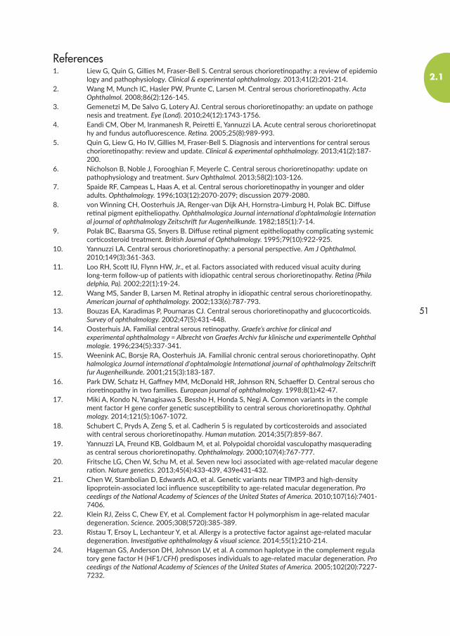
51
2.1
References1. Liew G, Quin G, Gillies M, Fraser-Bell S. Central serous chorioretinopathy: a review of epidemio logy and pathophysiology. Clinical & experimental ophthalmology. 2013;41(2):201-214.2. Wang M, Munch IC, Hasler PW, Prunte C, Larsen M. Central serous chorioretinopathy. Acta Ophthalmol. 2008;86(2):126-145.3. Gemenetzi M, De Salvo G, Lotery AJ. Central serous chorioretinopathy: an update on pathoge nesis and treatment. Eye (Lond). 2010;24(12):1743-1756.4. Eandi CM, Ober M, Iranmanesh R, Peiretti E, Yannuzzi LA. Acute central serous chorioretinopat hy and fundus autofluorescence. Retina. 2005;25(8):989-993.5. Quin G, Liew G, Ho IV, Gillies M, Fraser-Bell S. Diagnosis and interventions for central serous chorioretinopathy: review and update. Clinical & experimental ophthalmology. 2013;41(2):187- 200.6. Nicholson B, Noble J, Forooghian F, Meyerle C. Central serous chorioretinopathy: update on pathophysiology and treatment. Surv Ophthalmol. 2013;58(2):103-126.7. Spaide RF, Campeas L, Haas A, et al. Central serous chorioretinopathy in younger and older adults. Ophthalmology. 1996;103(12):2070-2079; discussion 2079-2080.8. von Winning CH, Oosterhuis JA, Renger-van Dijk AH, Hornstra-Limburg H, Polak BC. Diffuse retinal pigment epitheliopathy. Ophthalmologica Journal international d’ophtalmologie Internation al journal of ophthalmology Zeitschrift fur Augenheilkunde. 1982;185(1):7-14.9. Polak BC, Baarsma GS, Snyers B. Diffuse retinal pigment epitheliopathy complicating systemic corticosteroid treatment. British Journal of Ophthalmology. 1995;79(10):922-925.10. Yannuzzi LA. Central serous chorioretinopathy: a personal perspective. Am J Ophthalmol. 2010;149(3):361-363.11. Loo RH, Scott IU, Flynn HW, Jr., et al. Factors associated with reduced visual acuity during long-term follow-up of patients with idiopathic central serous chorioretinopathy. Retina (Phila delphia, Pa). 2002;22(1):19-24.12. Wang MS, Sander B, Larsen M. Retinal atrophy in idiopathic central serous chorioretinopathy. American journal of ophthalmology. 2002;133(6):787-793.13. Bouzas EA, Karadimas P, Pournaras CJ. Central serous chorioretinopathy and glucocorticoids. Survey of ophthalmology. 2002;47(5):431-448.14. Oosterhuis JA. Familial central serous retinopathy. Graefe’s archive for clinical and experimental ophthalmology = Albrecht von Graefes Archiv fur klinische und experimentelle Ophthal mologie. 1996;234(5):337-341.15. Weenink AC, Borsje RA, Oosterhuis JA. Familial chronic central serous chorioretinopathy. Opht halmologica Journal international d‘ophtalmologie International journal of ophthalmology Zeitschrift fur Augenheilkunde. 2001;215(3):183-187.16. Park DW, Schatz H, Gaffney MM, McDonald HR, Johnson RN, Schaeffer D. Central serous cho rioretinopathy in two families. European journal of ophthalmology. 1998;8(1):42-47.17. Miki A, Kondo N, Yanagisawa S, Bessho H, Honda S, Negi A. Common variants in the comple ment factor H gene confer genetic susceptibility to central serous chorioretinopathy. Ophthal mology. 2014;121(5):1067-1072.18. Schubert C, Pryds A, Zeng S, et al. Cadherin 5 is regulated by corticosteroids and associated with central serous chorioretinopathy. Human mutation. 2014;35(7):859-867.19. Yannuzzi LA, Freund KB, Goldbaum M, et al. Polypoidal choroidal vasculopathy masquerading as central serous chorioretinopathy. Ophthalmology. 2000;107(4):767-777.20. Fritsche LG, Chen W, Schu M, et al. Seven new loci associated with age-related macular degene ration. Nature genetics. 2013;45(4):433-439, 439e431-432.21. Chen W, Stambolian D, Edwards AO, et al. Genetic variants near TIMP3 and high-density lipoprotein-associated loci influence susceptibility to age-related macular degeneration. Pro ceedings of the National Academy of Sciences of the United States of America. 2010;107(16):7401- 7406.22. Klein RJ, Zeiss C, Chew EY, et al. Complement factor H polymorphism in age-related macular degeneration. Science. 2005;308(5720):385-389.23. Ristau T, Ersoy L, Lechanteur Y, et al. Allergy is a protective factor against age-related macular degeneration. Investigative ophthalmology & visual science. 2014;55(1):210-214.24. Hageman GS, Anderson DH, Johnson LV, et al. A common haplotype in the complement regula tory gene factor H (HF1/CFH) predisposes individuals to age-related macular degeneration. Pro ceedings of the National Academy of Sciences of the United States of America. 2005;102(20):7227- 7232.

25. Rivera A, Fisher SA, Fritsche LG, et al. Hypothetical LOC387715 is a second major susceptibility gene for age-related macular degeneration, contributing independently of complement factor H to disease risk. Human molecular genetics. 2005;14(21):3227-3236.26. Sakurada Y, Kubota T, Imasawa M, et al. Role of complement factor H I62V and age-related maculopathy susceptibility 2 A69S variants in the clinical expression of polypoidal choroidal vas culopathy. Ophthalmology. 2011;118(7):1402-1407.27. Koizumi H, Yamagishi T, Yamazaki T, Kinoshita S. Relationship between clinical characteristics of polypoidal choroidal vasculopathy and choroidal vascular hyperpermeability. American journal of ophthalmology. 2013;155(2):305-313 e301.28. Fung AT, Yannuzzi LA, Freund KB. Type 1 (sub-retinal pigment epithelial) neovascularization in central serous chorioretinopathy masquerading as neovascular age-related macular degenerati on. Retina (Philadelphia, Pa). 2012;32(9):1829-1837.29. Park HS, Kim IT. Clinical characteristics of polypoidal choroidal vasculopathy associated with chronic central serous chorioretionopathy. Korean Journal of Ophthalmology 2012;26(1):15-20.30. Kortvely E, Hauck SM, Duetsch G, et al. ARMS2 is a constituent of the extracellular matrix provi ding a link between familial and sporadic age-related macular degenerations. Investigative ophthalmology & visual science. 2010;51(1):79-88.31. Koo BH, Coe DM, Dixon LJ, et al. ADAMTS9 is a cell-autonomously acting, anti-angioge nic metalloprotease expressed by microvascular endothelial cells. The American journal of pathology. 2010;176(3):1494-1504.32. Plenz GA, Deng MC, Robenek H, Volker W. Vascular collagens: spotlight on the role of type VIII collagen in atherogenesis. Atherosclerosis. 2003;166(1):1-11.33. MacBeath JR, Kielty CM, Shuttleworth CA. Type VIII collagen is a product of vascular smooth- muscle cells in development and disease. The Biochemical journal. 1996;319 ( Pt 3):993-998.34. Crowder RN, El-Deiry WS. Caspase-8 regulation of TRAIL-mediated cell death. Experimental on cology. 2012;34(3):160-164.35. Kavurma MM, Schoppet M, Bobryshev YV, Khachigian LM, Bennett MR. TRAIL stimulates proli feration of vascular smooth muscle cells via activation of NF-kappaB and induction of insulin -likgrowth factor-1 receptor. The Journal of biological chemistry. 2008;283(12):7754-7762.36. Buga AM, Margaritescu C, Scholz CJ, Radu E, Zelenak C, Popa-Wagner A. Transcriptomics of post-stroke angiogenesis in the aged brain. Frontiers in aging neuroscience. 2014;6:44.37. Anderson DH, Radeke MJ, Gallo NB, et al. The pivotal role of the complement system in aging and age-related macular degeneration: hypothesis re-visited. Progress in retinal and eye research. 2010;29(2):95- 112.38. Loyet KM, Deforge LE, Katschke KJ, Jr., et al. Activation of the alternative complement pathway in vitreous is controlled by genetics in age-related macular degeneration. Investigative ophthalmology & visual science. 2012;53(10):6628-6637.39. Boon CJ, van de Kar NC, Klevering BJ, et al. The spectrum of phenotypes caused by variants in the CFH gene. Molecular immunology. 2009;46(8-9):1573-1594.40. Imamura Y, Fujiwara T, Margolis R, Spaide RF. Enhanced depth imaging optical coherence to mography of the choroid in central serous chorioretinopathy. Retina (Philadelphia, Pa). 2009;29(10):1469-1473.41. Tittl M, Maar N, Polska E, Weigert G, Stur M, Schmetterer L. Choroidal hemodynamic changes during isometric exercise in patients with inactive central serous chorioretinopathy. Investigative ophthalmology & visual science. 2005;46(12):4717-4721.42. Told R, Palkovits S, Haslacher H, et al. Alterations of choroidal blood flow regulation in young healthy subjects with complement factor H polymorphism. PloS one. 2013;8(4):e60424.43. Udono-Fujimori R, Udono T, Totsune K, Tamai M, Shibahara S, Takahashi K. Adrenomedullin in the eye. Regul Pept. 2003;112(1-3):95-101.44. Dorner GT, Garhofer G, Huemer KH, et al. Effects of adrenomedullin on ocular hemodynamic parameters in the choroid and the ophthalmic artery. Invest Ophthalmol Vis Sci. 2003;44(9):3947-3951.

53

2.2 Genomic copy number variations of the complement component C4B gene are associated with chronic central serous chorioretinopathyMyrte B. Breukink*, Rosa L. Schellevis*, Camiel J. F. Boon, Sascha Fauser, Carel B. Hoyng, Anneke I. den Hollander, and Eiko K. de JongInvest Ophthalmol Vis Sci. 2015 Aug;56(9):5608-13. doi: 10.1167/iovs.15-17343. * These authors contributed equally to this study

55
2.2
AbstractPurpose: Chronic central serous chorioretinopathy (cCSC) has recently been associated to variants in the complement factor H gene. To further investigate the role of the complement system in cCSC, the genomic copy number variations in the complement component 4 (C4) gene were studied.
Methods: C4A and C4B copy numbers were analyzed in 197 cCSC patients and 303 healthy controls using a Taqman copy number determination assay. Copy numbers of C4A, C4B and the total C4 load were compared between cases and controls, using a Fisher Exact test. For this analysis Bonferroni correction was performed for three tests, and P values <0.017 were considered to be significant. A logistic regression model was constructed to calculate the odds ratios (ORs) of each of the C4B copy numbers, using two copies as a reference. For this model P values <0.05 were considered to be significant.
Results: C4B genomic copy numbers differed significantly between cCSC patients and healthy controls (P=0.0018). Absence of C4B significantly conferred risk of cCSC (P=0.039, OR=2.61 [95% confidence interval (CI)=1.05–6.52]), whereas three copies of C4B significantly decreased the risk of cCSC (P=0.014, OR=0.45 [95%CI=0.23–0.85]). The C4A genomic copy numbers and total C4 load did not significantly differ between cases and controls.
Conclusions: This study showed that copy numbers of C4B are significantly associated with cCSC. Carrying no copies of C4B significantly increases the risk of cCSC, whereas carrying three C4B copies is protective. These findings reinforce the hypothesis of a possible involvement of the complement system in the pathogenesis of cCSC.

Introduction Chronic central serous chorioretinopathy (cCSC) is characterized by fluid accumulation under the neuroretina. It has been postulated that this serous fluid derives from the choroid, and that it leaks through a dysfunctional retinal pigment epithelium (RPE) causing a detachment of the neuroretina.1-4 Classically, cCSC patients are relatively young (middle-aged) men who are still professionally active.6 Besides male sex, also the use of corticosteroids, type A personality and stress have been associated with cCSC.4,6,7 Although the exact pathophysiological mechanism of the disease remains unknown, we and others have previously suggested involvement of the complement system, and in particular the complement factor H (CFH) gene in cCSC.8,9 Interestingly, CFH variants that confer increased risk in cCSC have previously been described to be protective in age-related macular degeneration, and vice versa.9-11
The complement system consists of three major pathways: the classical, the lectin, and the alternative pathway.12 CFH is involved in the alternative pathway and can influence C3b production by blocking one of the two C3-convertases (C3bBb). The classical and lectin pathways also play an important role in C3b production mediated by the other C3-convertase (C4b2a), of which the complement component 4 (C4) protein is a key factor.13
Copy number variations of the C4 gene have been associated with several eye diseases and autoimmune disorders (e.g. Vogt- Koyanagi-Harada and Beçhet’s disease).14-16 In addition, Banlaki et al. 17 have found that the genomic copy number of C4B is associated with cortisol release after adrenocorticotropic hormone (ACTH) stimulation. This is of particular interest considering that stress, which appears to be associated with cCSC,4 has a strong influence on the hypothalamic-pituitary-adrenal (HPA) axis and increases ACTH release.18
Because of its role in the complement system, and its association with the HPA axis, we hypothesized that copy number variation in the C4 gene may be associated with cCSC. In this study we assessed the copy number variations of the C4 gene in a cCSC cohort.
Materials and MethodsSubjectsIn this study, 197 patients diagnosed with cCSC who visited the outpatient clinic of the department of Ophthalmology at the Radboud University Medical Center, Nijmegen, the Netherlands were included (Table 1). The diagnosis cCSC was based on an extensive ophthalmological examination including fundoscopy, spectral-domain optical coherence tomography, fluorescein angiography and indocyanine green angiography. The definition of typical cCSC used in this study was based on the previously published subgroups by de Jong et al.,9 and patients in this study were phenotypes by an experienced retina specialist (CJFB) (Figure 1A-F). Additionally, a total of 303 control subjects were recruited from the blood bank of the Radboud University Medical Center, Nijmegen, the Netherlands (n = 154), and the European Genetic Database (EUGENDA, www.eugenda.org; provided in the public domain by the University Hospital of Cologne, Cologne, Germany and Radboud university medical center) (n = 149) (Table 1). For this last group fundus photographs were graded

57
2.2
to rule out any ophthalmological abnormalities at the moment of inclusion. Informed consent for the use of DNA for genetic studies was obtained from all subjects. This study followed the guidelines of the Declaration of Helsinki and was approved by the local ethics committee.
Copy number determinationDNA was isolated from peripheral blood using standard procedures. C4A and C4B copy numbers were determined by real-time PCR using Taqman® genotyping assays (Applied Biosystems, Thermo Fisher Scientific, Waltham, USA). The FAM-labeled C4A (Hs07226349_cn) or C4B (Hs07226350_cn) Taqman copy number assay was combined together with the VIC-labeled Ribonuclease P (RNaseP) reference assay (Catalog nr. 4403326), and Taqman genotyping mastermix (Catalog nr. 4381656). All samples were tested in duplicate for C4A and C4B on 384-wells plates using 10 ng of DNA in a total reaction volume of 10 μl.
Samples with known copy numbers for either C4A (0-4) or C4B (0-3) were kindly provided by C. Yung Yu.19 These samples were included as a reference on each plate to facilitate accurate copy number determination, using the method described previously.19 In each run the amplification efficiencies of the C4A/C4B and RNaseP probes were calculated using a serial dilution (50-1.56 ng) of a sample with two C4A and C4B copies. The primer efficiencies of the probes were compared, and deemed
Figure 1: Example of FA of an RE (A), and an LE (C), ICG angiography of an RE (B), and an LE (D), and spectral-domain optical coherence tomography of an RE (E), and an LE (F) imaging of the phenotypic characteristics of the cCSC cohort used for this study. (A-F) The RE and LE of a patient demonstrate diffuse hyperfluorescent areas of leakage on FA and ICG angiography (A-D) and subretinal fluid beneath the fovea (E), illustrative for typical cCSC. FA, fluorescein angiography; ICG, indocyanine green; LE, left eye; RE, right eye.
Table 1. Demographics of the study population
cCSC patients Controls P value
No. of subjects 197 303 NA
Sex (male/female) 154/43 226/77 0.392
Mean age ±SD (years) 53 (±10) 53 (±11) 0.755
Age range (years) 29-74 29-77 NA
cCSC; chronic central serous chorioretinopathy, NA; not annotated SD; standard deviation

similar if they differed <2%, this was the case in all runs. Therefore, the efficiencies were not incorporated into the calculations of the copy numbers. Polymerase chain reaction was performed with a 7900HT thermocycler (Applied Biosystems, Thermo Fisher Scientific) using the following program: 2 minutes at 50°C, 10 minutes at 95°C, and 40 cycles of 15 seconds at 95°C and 60 secsonds at 60°C. Data were analyzed with the Copycaller software (V2.0; Applied Biosystems, Thermo Fisher Scientific). Copy numbers determined by the Copycaller software were corrected by using the trend line based on the reference samples, as described before.19 If the results were inconsistent between the Copycaller output and the values corrected with the trend line, samples were retested on a new plate in triplicate.
StatisticsThe comparison of C4A, C4B and total C4 copy number distribution between cCSC patients and controls was performed by means of a Fisher Exact test using SPSS Statistics (V20; IBM Corp., Armonk, USA). Bonferroni correction for multiple testing was performed for three tests and P values < 0.017 were considered to be statistically significant. A logistic regression model was constructed to determine the odds ratios (ORs) for the various copy numbers of C4B. According to previously published studies, two genomic copy numbers of C4B are considered to be most common in the healthy population.16,20 We were able to confirm this in our cohort, and therefore this copy number was set as reference. In this model, P values <0.05 were considered to be significant. Graphs were generated using Graphpad Prism (V5; Graphpad Software, San Diego, CA, USA).
ResultsThe copy numbers of C4A and C4B were successfully determined in 197 cCSC cases and 303 controls. No significant difference was observed between cases and controls for the C4A genomic copy number (range: 0-6, P=0.649; Figure 2A). The C4B distribution was significantly different between cCSC patients and controls (range: 0-4, P=0.0018; Figure 2B). Overall, cases carried lower copy numbers of C4B than the control population. The total C4 genomic copy number was not different in cases compared to controls (P=0.148; Figure 2C). Age and sex were not associated with either C4A, C4B, or total C4 genomic copy number (Table 2; data for C4A and total C4 not shown).
To assess the effect size of the different copy numbers of C4B on development of cCSC, a logistic regression was performed (Table 2). The logistic regression model based on the distribution of C4B between cases and controls was significant (P=0.0035, Table 2). Carrying no copies of C4B conferred increased risk of cCSC (P=0.039, OR=2.61, 95% confidence interval [CI]=1.05-6.52). A similar trend was observed for carriers of one copy of C4B, but the results were not significant (P=0.080, OR=1.47, 95% CI=0.96-2.26). Carrying three C4B copies was associated with a significantly decreased risk of cCSC (P=0.014, OR=0.45, 95% CI=0.24-0.85), whereas no significant association with cCSC was observed in individuals carrying four copies of C4B (P=0.81).
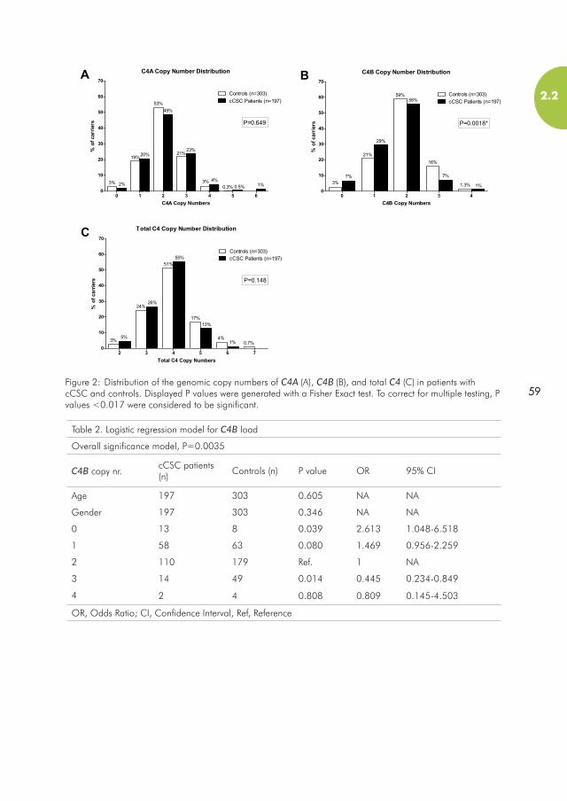
59
2.2
Figure 2: Distribution of the genomic copy numbers of C4A (A), C4B (B), and total C4 (C) in patients with cCSC and controls. Displayed P values were generated with a Fisher Exact test. To correct for multiple testing, P values <0.017 were considered to be significant.
Table 2. Logistic regression model for C4B load
Overall significance model, P=0.0035
C4B copy nr.cCSC patients (n)
Controls (n) P value OR 95% CI
Age 197 303 0.605 NA NA
Gender 197 303 0.346 NA NA
0 13 8 0.039 2.613 1.048-6.518
1 58 63 0.080 1.469 0.956-2.259
2 110 179 Ref. 1 NA
3 14 49 0.014 0.445 0.234-0.849
4 2 4 0.808 0.809 0.145-4.503
OR, Odds Ratio; CI, Confidence Interval, Ref, Reference

DiscussionOur study results demonstrate that cCSC patients have a significantly different C4B load as compared to healthy controls (P=0.0018). Carrying no copies of C4B was associated with an increased risk of cCSC (OR=2.61, 95% CI=1.05-6.52), whereas carrying three C4B copies was associated with a decreased risk of cCSC (OR=0.45, 95% CI=0.23-0.85). No association with cCSC was observed in individuals carrying four C4B copies, which is likely due to the limited sample size of this group (cases, n=4; controls, n=2). No significant differences were observed between cases and controls for C4A and total C4 load.
The C4 gene lies within the RP-C4-CYP21-TNX (RCCX) locus located in the major histocompatability complex (MHC) region III on chromosome 6 of the human genome.20 The MHC region contains an elevated level of genomic copy number variations that are presumably present to increase immunological diversity.21 Duplications and deletions in the region have led to the formation of haplotypes containing variable copies of the RCCX locus in the human population (Figure 3B).5,21,22 Haplotypes containing two or more duplications of the RCCX locus show extensive variability in their gene content, generally with complete duplications of the C4 gene (Figure 3B).22,23 The C4 gene encodes for the C4 protein, of which two variants have been described (C4A/C4B), differing in only four amino acids encoded by exon 26 (Figure 3A).24
Copy number variations of either C4A or C4B have been associated with several systemic diseases with ocular involvement, such as Vogt-Koyanagi-Harada disease,
Figure 3: A schematic illustration of the RCCX locus. (A) A display of the two variants of the C4 gene, differing in four amino acids in exon 26 (p. 1101-1106). (B) An example of the structure of the RCCX locus with a single copy (upper panel) and two copies (lower panel) of the C4 gene. In case of duplication, a complete C4A or C4B and the CYP21 gene are duplicated, while the RP and TNX gene are only partially duplicated. The dotted lines indicate the approximate site were during recombination the duplication has occurred in the past. (Figure based on Banlaki et al. 2013).5
RP
RP RP
C4A/B
C4A/B C4A/B
TNX
TNX
CYP21
CYP21 CYP21
B.
A.
TNX
C4A
C4BY
YPCPVLD
LSPVIH

61
2.2
Behçet’s disease, and systemic lupus erythematosus (SLE).14-16 Hou et al. 14 have shown that a lower copy number of C4A and C4B increases the risk of Vogt-Koyanagi-Harada, an autoimmune disorder characterized by bilateral granulomatous panuveitis. The same group15 has also demonstrated that higher copy numbers of C4A confer risk of Behçet’s disease, an autoinflammatory disease, which presents with acute anterior uveitis. Several studies16,25,26 have shown an association between low copy numbers of C4 and an increased risk for SLE, an autoimmune disease that is typically mediated by immune complexes. In the past, CSC has been described in SLE patients,27,28 but it remains unclear whether this is a primary manifestation of SLE, or whether it is a consequence of corticosteroid treatment for SLE.29,30 Several studies have reported a positive linear correlation between serum C4 and C4 genomic copy number,31-33 suggesting that the lower number of C4B copies in cCSC patients leads to lower systemic C4B levels. This may indicate that an overall lower activity of the complement system might be present in cCSC patients.
Recently, low copy numbers of C4B have been associated with hyperreactivity of the HPA axis.17 Banlaki et al. 17 have shown that in patients with adrenal incidentaloma and low (<2 copies) C4B genomic copy number, baseline ACTH was significantly reduced compared to high (≥2 copies) genomic copy number of C4B. Moreover, a significantly higher cortisol response is observed after ACTH stimulation in the patients with low C4B genomic copy number.17
These results are of interest in the context of cCSC because of the described clinical associations with stress and the use of corticosteroids that both exert physiological effects at the level of the HPA axis.18 Various relatively small studies have studied cortisol levels in cCSC patients. Although 24h urine samples showed elevated cortisol levels in cCSC patients in certain studies,34,35 these results are not observed in single serum measurements during set times in other studies.36,37 These discrepancies could be explained by variable cortisol fluctuations between individuals during the day, and therefore changes in endogenous cortisol levels cannot be ruled out as a hallmark of cCSC. It is possible that patients with cCSC generally have normal cortisol levels, but respond differently to stimulation of the HPA axis. Stress, which also appears to be associated with cCSC,2,38 stimulates the HPA axis and could lead to temporarily elevated cortisol levels in patients as compared to healthy individuals. How high levels of cortisol can lead to subretinal fluid accumulation is currently unknown. A study in rats suggested that the disease mechanism could be mediated by binding of corticosteroids to the mineralocorticoid receptor.39 In this study, activation of the mineralocorticoid receptor causes vascular effects similar to those observed in cCSC,39 but the underlying pathways still remain to be elucidated.
The mechanism through which low copy numbers of C4B may lead to hyperresponsiveness of the HPA axis is unclear. It has been hypothesized that it is not the C4B gene, but rather the neighboring CYP21A2 gene, that mediates this effect.17 The CYP21A2 gene encodes the enzyme 21-hydroxylase, which plays an important role in the steroid metabolism pathway by converting progesterone and 17-α-hydroxyprogesterone to 11-deoxycorticosterone and 11-deoxycortisol, respectively. Because of the genomic structure of the RCCX locus, variation in the CYP21A2 gene is in high linkage disequilibrium with variation in the neighboring C4 gene (C4A or C4B).17 Therefore, further exploration of the precise structure

and specific variations present in the RCCX locus may reveal new insights into the pathogenesis of cCSC.
The current study and previous studies identified an association between complement genes and cCSC,8,9 suggesting that the complement system may be dysregulated in cCSC. Taken together, these findings may indicate that the immune system, influenced by environmental factors, such as stress, could play a pivotal role in the pathophysiology of cCSC. Further studies are necessary to determine the physiological effects of genetic variation at the C4 gene and the RCCX locus in cCSC.

63
2.2
AcknowledgementsWe thank C. Yung Yu for providing the positive control samples (Center for Molecular and Human Genetics, The Research Institute, Ohio, USA). We thank Joannes M.M. Groenewoud (MSc) for his statistical support (Department of Health Evidence, Radboudumc, Nijmegen, The Netherlands).
Footnotes and Financial DisclosuresThis study was supported by the Macula Vision Research Foundation, MD Fonds, Landelijke Stichting voor Blinden en Slechtzienden, Gelderse Blindenstichting, Stichting Nederlands Oogheelkundig Onderzoek, Stichting Blindenhulp, Stichting A.F. Deutman Oogheelkunde Researchfonds, Nijmeegse Oogonderzoek Stichting, Janivo Stichting, Retina Netherlands, Stichting Blinden Penning, Oogfonds, Gisela Thier Fellowship of Leiden University, and the Radboud Institute for Molecular Life Sciences.
The authors alone are responsible for the content and writing of the paper.
No conflicting relationships exist for any author.
Running head: C4B in chronic central serous chorioretinopathy
Keywords: chronic central serous chorioretinopathy, cCSC, complement component 4, C4, C4A, C4B

References1. Wang M, Munch IC, Hasler PW, Prunte C, Larsen M. Central serous chorioretinopathy. Acta
Ophthalmol. 2008;86(2):126-145.2. Gemenetzi M, De Salvo G, Lotery AJ. Central serous chorioretinopathy: an update on pathoge-
nesis and treatment. Eye (Lond). 2010;24(12):1743-1756.3. Nicholson B, Noble J, Forooghian F, Meyerle C. Central serous chorioretinopathy: update on
pathophysiology and treatment. Surv Ophthalmol. 2013;58(2):103-126.4. Liew G, Quin G, Gillies M, Fraser-Bell S. Central serous chorioretinopathy: a review of epide-
miology and pathophysiology. Clin Experiment Ophthalmol. 2013;41(2):201-214.5. Banlaki Z, Szabo JA, Szilagyi A, et al. Intraspecific evolution of human RCCX copy number vari-
ation traced by haplotypes of the CYP21A2 gene. Genome biology and evolution. 2013;5(1):98-112.
6. Yannuzzi LA. Central serous chorioretinopathy: a personal perspective. Am J Ophthalmol. 2010;149(3):361-363.
7. Bouzas EA, Karadimas P, Pournaras CJ. Central serous chorioretinopathy and glucocorticoids. Surv Ophthalmol. 2002;47(5):431-448.
8. Miki A, Kondo N, Yanagisawa S, Bessho H, Honda S, Negi A. Common variants in the comple-ment factor H gene confer genetic susceptibility to central serous chorioretinopathy. Ophthal-mology. 2014;121(5):1067-1072.
9. de Jong EK, Breukink MB, Schellevis RL, et al. Chronic central serous chorioretinopathy is as-sociated with genetic variants implicated in age-related macular degeneration. Ophthalmology. 2015;122(3):562-570.
10. Hageman GS, Anderson DH, Johnson LV, et al. A common haplotype in the complement regu-latory gene factor H (HF1/CFH) predisposes individuals to age-related macular degeneration. Proc Natl Acad Sci U S A. 2005;102(20):7227-7232.
11. Fritsche LG, Chen W, Schu M, et al. Seven new loci associated with age-related macular dege-neration. Nat Genet. 2013;45(4):433-439, 439e431-432.
12. Anderson DH, Radeke MJ, Gallo NB, et al. The pivotal role of the complement system in aging and age-related macular degeneration: hypothesis re-visited. Prog Retin Eye Res. 2010;29(2):95-112.
13. Ricklin D, Hajishengallis G, Yang K, Lambris JD. Complement: a key system for immune surveil-lance and homeostasis. Nature immunology. 2010;11(9):785-797.
14. Hou S, Qi J, Liao D, et al. High C4 gene copy numbers protects against Vogt-Koyanagi-Harada syndrome in Chinese Han. The British journal of ophthalmology. 2014;98(12):1733-1737.
15. Hou S, Qi J, Liao D, et al. Copy number variations of complement component C4 are associa-ted with Behcet’s disease but not with ankylosing spondylitis associated with acute anterior uveitis. Arthritis and rheumatism. 2013;65(11):2963-2970.
16. Yang Y, Chung EK, Wu YL, et al. Gene copy-number variation and associated polymorphisms of complement component C4 in human systemic lupus erythematosus (SLE): low copy number is a risk factor for and high copy number is a protective factor against SLE susceptibility in European Americans. Am J Hum Genet. 2007;80(6):1037-1054.
17. Banlaki Z, Raizer G, Acs B, et al. ACTH-induced cortisol release is related to the copy number of the C4B gene encoding the fourth component of complement in patients with non-functional adrenal incidentaloma. Clin Endocrinol (Oxf). 2012;76(4):478-484.
18. Aguilera G. Regulation of pituitary ACTH secretion during chronic stress. Frontiers in neuroendo-crinology. 1994;15(4):321-350.
19. Wu YL, Savelli SL, Yang Y, et al. Sensitive and specific real-time polymerase chain reaction as-says to accurately determine copy number variations (CNVs) of human complement C4A, C4B, C4-long, C4-short, and RCCX modules: elucidation of C4 CNVs in 50 consanguineous subjects with defined HLA genotypes. Journal of immunology. 2007;179(5):3012-3025.
20. Szilagyi A, Fust G. Diseases associated with the low copy number of the C4B gene encoding C4, the fourth component of complement. Cytogenetic and genome research. 2008;123(1-4):118-130.
21. Olsson LM, Holmdahl R. Copy number variation in autoimmunity--importance hidden in com-plexity? European journal of immunology. 2012;42(8):1969-1976.
22. Chung EK, Yang Y, Rennebohm RM, et al. Genetic sophistication of human complement compo-

65
2.2
nents C4A and C4B and RP-C4-CYP21-TNX (RCCX) modules in the major histocompatibility complex. Am J Hum Genet. 2002;71(4):823-837.
23. Blanchong CA, Zhou B, Rupert KL, et al. Deficiencies of human complement component C4A and C4B and heterozygosity in length variants of RP-C4-CYP21-TNX (RCCX) modules in caucasians. The load of RCCX genetic diversity on major histocompatibility complex-associated disease. The Journal of experimental medicine. 2000;191(12):2183-2196.
24. Yu CY, Belt KT, Giles CM, Campbell RD, Porter RR. Structural basis of the polymorphism of hu-man complement components C4A and C4B: gene size, reactivity and antigenicity. The EMBO journal. 1986;5(11):2873-2881.
25. Lv Y, He S, Zhang Z, et al. Confirmation of C4 gene copy number variation and the association with systemic lupus erythematosus in Chinese Han population. Rheumatology international. 2012;32(10):3047-3053.
26. Ptacek T, Li X, Kelley JM, Edberg JC. Copy number variants in genetic susceptibility and severity of systemic lupus erythematosus. Cytogenetic and genome research. 2008;123(1-4):142-147.
27. Eckstein MB, Spalton DJ, Holder G. Visual loss from central serous retinopathy in systemic lupus erythematosus. The British journal of ophthalmology. 1993;77(9):607-609.
28. Cunningham ET, Jr., Alfred PR, Irvine AR. Central serous chorioretinopathy in patients with systemic lupus erythematosus. Ophthalmology. 1996;103(12):2081-2090.
29. Bouzas E, Mastorakos G. Central serous retinopathy in systemic lupus erythematosus: a manifestation of the disease or of its treatment? The British journal of ophthalmology. 1994;78(5):420-421.
30. Bouyon A, Costedoat-Chalumeau N, Limal N, et al. [Central serous chorioretinopathy and sys-temic diseases report of 2 cases associated with corticotherapy]. La Revue de medecine interne / fondee par la Societe nationale francaise de medecine interne. 2006;27(6):487-491.
31. Margery-Muir AA, Wetherall JD, Castley AS, et al. Establishment of gene copy number-specific normal ranges for serum C4 and its utility for interpretation in patients with chronically low serum C4 concentrations. Arthritis & rheumatology. 2014;66(9):2512-2520.
32. Uko G, Christiansen FT, Dawkins RL, McCann VJ. Reference ranges for serum C4 concentrati-ons in subjects with and without C4 null alleles. Journal of clinical pathology. 1986;39(5):573-576.
33. Yang Y, Chung EK, Zhou B, et al. Diversity in intrinsic strengths of the human complement system: serum C4 protein concentrations correlate with C4 gene size and polygenic variations, hemolytic activities, and body mass index. Journal of immunology. 2003;171(5):2734-2745.
34. Haimovici R, Rumelt S, Melby J. Endocrine abnormalities in patients with central serous chorio-retinopathy. Ophthalmology. 2003;110(4):698-703.
35. Shang Q, Liu C, Wei S, Shi F, Li Y, Qiao L. [Determination of cortisol in plasma and 24-hour urine of patients with central serous chorioretinopathy]. [Zhonghua yan ke za zhi] Chinese journal of ophthalmology. 1999;35(4):297-299.
36. Tufan HA, Gencer B, Comez AT. Serum cortisol and testosterone levels in chronic central serous chorioretinopathy. Graefe‘s archive for clinical and experimental ophthalmology = Albrecht von Graefes Archiv fur klinische und experimentelle Ophthalmologie. 2013;251(3):677-680.


67

2.3 Association of a haplotype in the NR3C2 gene, encoding the mineralocorticoid receptor, with chronic central serous chorioretinopathyElon H. C. van Dijk*, Rosa L. Schellevis*, Maaike G. J.M. van Bergen, Myrte B. Breukink, Lebriz Altay, Paula Scholz, Sascha Fauser, Onno C. Meijer, Carel B. Hoyng, Anneke I. den Hollander, Camiel J. F. Boon, MD, Eiko K. de Jong JAMA Ophthalmol. 2017 May 1;135(5):446-451. doi: 10.1001/jamaophthalmol.2017.0245. * These authors contributed equally to this study

69
2.3
AbstractImportance: Chronic central serous chorioretinopathy (cCSC) is a chorioretinal disease with unknown disease etiology. The glucocorticoid receptor and the mineralocorticoid receptor, 2 glucocorticoid-binding receptors, might be involved in the pathogenesis of cCSC.
Objective: To assess the association of functional variants and haplotypes in the glucocorticoid receptor (NR3C1) and mineralocorticoid receptor genes (NR3C2) with cCSC.
Design, setting, and participants: In this case-control genetic association study, 336 patients with cCSC and 1314 unaffected controls, collected at 3 university medical centers from September 1, 2009, to May 2016, underwent KASP genotyping for selected variants in NR3C1 (rs56149945, rs41423247 and rs6198) and NR3C2 (rs2070951 and rs5522).
Main outcome measure: Genetic associations of 3 NR3C1 variants and 2 NR3C2 variants with cCSC.
Results: Among the 336 patients (274 men and 62 women; mean [SD] age, 52[10] years), after correction for multiple testing, rs2070951 in the NR3C2 gene was significantly associated with cCSC (odds ratio, 1.29; 95% CI, 1.08-1.53; P=.004). Moreover, the GA haplotype of single nucleotide polymorphisms rs2070951 and rs5522 in NR3C2 conferred risk for cCSC (odds ratio, 1.39; 95% CI, 1.15-1.68; P=.004), whereas the CA haplotype decreased risk for cCSC (odds ratio, 0.72; 95% CI, 0.60-0.87; P<.001). Three known variants in NR3C1 that alter the activity of the glucocorticoid receptor (rs56149945, rs41423247 and rs6198) were not associated with cCSC.
Conclusions and Relevance: In this study, the variant rs2070951 and the GA haplotype in NR3C2 were associated with an increased risk for cCSC. Results of this genetic study support a possible role for the mineralocorticoid receptor in the pathogenesis of cCSC. Since these haplotypes have previously been associated with perceived stress, this study provides a clue to bridging clinical risk factors for cCSC to underlying genetic associations.

IntroductionIn central serous chorioretinopathy (CSC), it has been suggested that dysfunction of the retinal pigment epithelium (RPE) due to congestion, thickening, and hyperpermeability of the underlying choroid leads to subretinal fluid accumulation with an associated detachment of the neuroretina.1-5 The exact etiology of the disease is currently unknown, but clinical associations point towards an involvement of steroid signaling. Endogenous hypercortisolism (Cushing’s syndrome), exogenous glucocorticoid exposure, and possibly stress and type A personality are associated with CSC.6-11 It has been hypothesized that the glucocorticoid receptor (GR) and the mineralocorticoid receptor (MR), 2 glucocorticoid-binding receptors, may also be involved in the pathogenesis of CSC.2
The involvement of the MR in the pathogenesis of CSC has been suggested based on the results of studies in rats, in which choroidal findings similar to those seen in CSC occurred after intravitreal injection of either corticosterone or aldosterone.12 Involvement of the MR was further supported by ophthalmological findings in patients with primary hyperaldosteronism (Conn syndrome).13 Moreover, studies evaluating the administration of MR antagonists for patients with cCSC have shown possible beneficial effects.12,14-17 However, clinical results were variable and not permanent, and no prospective randomized placebo-controlled clinical trials have been published to this date to study the role of MR antagonists in the treatment of CSC.2,16
The GR is the most widely expressed cortisol receptor in the body; it regulates metabolism and the cardiovascular system, and it plays a role in immune suppression and stress response.18 As stress and both exogenous and endogenous hypercortisolism may be involved in the etiology of CSC,4,7,10,11 the GR may also be an interesting player in the pathogenesis of CSC.
There are several genetic variants in the genes encoding the MR and GR that are known to alter the MR and GR protein activity.19-26 The MR is encoded by the NR3C2 gene (OMIM 600983), consisting of 10 exons, with 2 alternative 5’-UTR exons 1α and 1β allowing tissue-specific promoter activation.27 The GR is encoded by the NR3C1 gene (OMIM 138040), consisting of 10 exons of which 1 to 9α are translated into the functional GRα receptor.22 In this study, we assessed whether genetic variants in NR3C2 (rs2070951 and rs5522) and NR3C1 (rs56149945, rs41423247, and rs6198) are associated with cCSC.
Key PointsQuestion: Are functional variants and haplotypes in the glucocorticoid receptor (NR3C1) and mineralocorticoid receptor (NR3C2) genes associated with chronic central serous chorioretinopathy (cCSC)?
Findings: In this case-control, genetic association study, rs2070951 in the NR3C2 gene was significantly associated with cCSC. The GA haplotype of single-nucleotide polymorphisms rs2070951 and rs5522 in NR3C2 conferred risk for cCSC, whereas the CA haplotype decreased the risk for cCSC.
Meaning: Results of this genetic study support a possible role for the mineralocorticoid receptor in the pathogenesis of cCSC.

71
2.3
Materials and methodsWe included 336 patients with cCSC in this study. Phenotyping was performed from September 1, 2009, to May 1, 2016, by an experienced retina specialist (C.J.F.B.) and was based on results of a complete ophthalmological examination, including fundoscopy, optical coherence tomography, fluorescein angiography, and indocyanine green angiography. The patients showed the most typical clinical cCSC characteristics (serous subretinal fluid affecting the fovea on optical coherence tomography, a disease period of >3 months, ≥1 area of “hot spot” leakage [the point where fluid is presumed to leak into the serous fluid pocket] or diffuse leakage in combination with irregular retinal pigment epithelial window defects on fluorescein angiography, and a corresponding hyperfluorescence detected on indocyanine green angiography), described as phenotypic subgroup 1 in a previous article on genetic associations in cCSC.28 Patients with high myopia, evidence of choroidal neovascularization, polypoidal choroidal vasculopathy, and other atypical findings were excluded. For this study, neither previous nor current steroid use was considered an exclusion criterion. The patient cohort consisted of 234 patients from the Radboud university medical center (Nijmegen, the Netherlands), 72 patients from the Leiden University Medical Center (Leiden, the Netherlands), and 30 patients from the University Hospital of Cologne (Cologne, Germany). A total of 1314 unaffected individuals (ie, controls) enrolled in the European Genetic Database (www.eugenda.org) were included in the study; they has no signs of either cCSC or age-related macular degeneration when evaluated by multimodal imaging. The study adhered to the tenets of the Declaration of Helsinki,29 and was approved by the institutional review boards and the ethics committees of the Radboud university medical center, the Leiden University Medical Center, and the University Hospital of Cologne. Written informed consent was received from all participants.
Genotyping of selected variants was performed using KASP assays (LGC Genomics) according to manufacturer’s instructions. Specific primers with 5-carboxyfluorescein and 5’-hexachloro-fluorescein labels were designed per variant (NR3C1: rs6198, rs5614994, and rs41423247; NR3C2: rs5522 and rs2070951) (eTable 1 in the Supplement) and the polymerase chain reaction (PCR) conditions per primer pair were provided by LGC Genomics. Data were read with the 7900HT Fast Real-Time PCR system (Applied Biosystems by Life Technologies) and analyzed with SDS version 2.4 (Applied Biosystems).
Table 1. Demographic characteristics of the cCSC patients and controls
n Mean age ± stdev (years) Males (%)
cCSC cases
Nijmegen 234 52 ± 9 188 (80%)
Cologne 30 50 ± 9 24 (80%)
Leiden 72 52 ± 10 62 (86%)
Total 336 52 ± 10 274 (81,5%)
Controls 1314 70 ± 7 549 (42%)
cCSC, chronic central serous chorioretinopathy; stdev, standard deviation

Statistical AnalysisIn IBM SPSS Statistics version 22 (SPSS Inc.), the 2-sided Pearson’s χ2 test was used to compare both the genotype and allele frequencies between cases and controls. Bonferroni correction for multiple testing was performed for 5 variants and P<.01 was considered statistically significant. Logistic regression was performed for the associated rs2070951 variant with Firth bias-corrected likelihood ratio test implemented in EPACTS version 3.2.6 (Efficient and Parallelizable Association Container Toolbox, http://genome.sph.umich.edu/wiki/EPACTS), correcting for sex.30
Using a haplotype analysis, we assessed the combined effect of the 2 variants in NR3C2. Haplotype analysis was performed using R version 3.0.2 (R Core Team; https://www.R-project.org), using the haplo.stats package (version 1.6.8). The 2 most frequent haplotypes were separately used as a reference in the haplo.cc command, to determine odds ratios (ORs) for both haplotypes. A logistic regression analysis (haplo.glm) including sex and haplotypes was performed using the most common haplotype as a reference. Only haplotypes with a frequency higher than 5% are shown. P<.05 was considered statistically significant.
Power analysis was performed with CaTS version 0.0.2 (University of Michigan), using
a multiplicative model in a joint analysis.31 The power per variant was calculated based on the minor allele frequency in controls, a disease prevalence of 0.0001, and a variable genotype relative risk (1-2.6), and the graph was created with Graphpad Prism version 5.03 (Graphpad Software).
ResultsAmong the 336 patients, 274 were men and 62 were women; the mean (SD) age was 52 (10) years. The demographic characteristics of the patients and controls enrolled in this study are summarized in Table 1. All described variants were in Hardy-Weinberg equilibrium, both for controls and patients with cCSC. No statistically significant associations between cCSC and variants in the NR3C1 gene (rs56149945,
rs41423247 and rs6198) were found (Table 2). After correction for multiple testing, a significant association between cCSC and the rs2070951 variant in the NR3C2 gene was observed (OR, 1.29; 95% CI, 1.08-1.53; P=.004,). No association between the variant rs5522 in NR3C2 and cCSC was found (Table 2).
Haplotype analysis of the NR3C2 single-nucleotide polymorphisms rs2070951 and rs5522 showed a significant decreased risk for cCSC for the CA haplotype (OR, 0.72; 95% CI, 0.60-0.87, P<.001) and an increased risk for the GA haplotype (OR, 1.39; 95% CI 1.15-1.68, P=.004) (Table 3). To account for potential confounding effects of sex between cases and controls, we corrected for this factor in a logistic regression model. When including this variable in the model, the association of rs2070951 was independent of sex (OR, 1.28; 95% CI, 1.16-1.41; P=.009). Similarly, when correcting for sex in the haplotype analysis, setting the most common haplotype (GA) as reference, we found that the association of the
A multiplicative model was used to calculate the power of the study, because this model produces a genetic relative risk score that is an estimation of the OR of an allelic model.32 For each variant, the power of detecting an association was calculated

73
2.3
Table 2. Association analysis of variants in NR3C1 and NR3C2 in cCSC patients
Minor Allele Fre-quency
P value
Variant Gene LocationMajor/
Minor allele
Controls PatientsGeno-
typeAllelic OR (95% CI)
rs56149945 NR3C1 Exon 2 A/G 0.0415 0.0357 .37 .500.86
(0.55-1.34)
rs41423247 NR3C1 Intron 2 C/G 0.360 0.390 .16 .151.14
(0.95-1.35)
rs6198 NR3C1Exon 9
UTRA/G 0.169 0.158 .334 .48
0.92
(0.73-1.16)
rs2070951 NR3C2 c.-2 C/G 0.464 0.527 .008 .00401.29
(1.08-1.53)
rs5522 NR3C2 Exon 2 A/G 0.129 0.137 .84 .571.08
(0.83-1.38)
cCSC, chronic central serous chorioretinopathy; CI, confidence interval; MAF, Minor allele frequency; OR, odds ratio; UTR, untranslated region. Bonferroni correction for multiple testing was performed for 5 variants and p-values < 0.01 were deemed significant
Figure 1: Power calculation for each variant in a cohort of 336 patients with chronic central serous chorioretinopathy patients and 1314 unaffected controls. For each variant, the power to detect a certain genotype relative risk was assessed using a multiplicative model, with minor allele frequency in controls and disease frequency of 0.0001 as input. The 80% power detection limits per variant were 1.35 (rs2070951), 1.36 (rs41423247), 1.46 (rs6198), 1.52 (rs5522), and 1.95 (rs56149945).

for the current cohort size. For all variants, genotype relative risks less than 2 could be detected with 80% power using this cohort. The detection limits of the genetic relative risk at 80% power were 1.35, 1.36, 1.46, 1.52, and 1.95 for rs2070951, rs41423247, rs6198, rs5522, and rs56149945, respectively (Figure 1).
DiscussionIn this study, we analyzed a possible association with cCSC for 3 known functiona variants in NR3C1 and 2 known functional variants in NR3C2. The rs2070951 variant in NR3C2 was significantly associated with cCSC, whereas the rs5522 variant was not associated with cCSC. The NR3C2 CA and GA haplotypes were both significantly associated with cCSC, with a protective and a risk-conferring effect, respectively. Odds ratios of the associated NR3C2 variant and the haplotypes were similar to previously described associations for the CFH (OMIM 134370) and ARMS2 (OMIM 611313) genes, and lower compared to the previously described associations in the C4A (OMIM 120810), C4B (OMIM 120820), and CDH5 (OMIM 601120) genes.28,33-35 The 3 variants in the NR3C1 gene (encoding the GR) were not associated with cCSC, which may suggest that MR functionality is more relevant than GR functionality in the etiology of cCSC. However, a larger cohort size is required to exclude the involvement of the 4 variants that were not associated with cCSC in this study.
An abnormal response to the administration of corticosteroids in a subset of patients with cCSC is the strongest risk factor for the disease with described ORs of up to 37.9 However, the precise mechanism of action of steroids in the pathogenesis of cCSC is unknown. One study showed that both mineralocorticoids and glucocorticoids can activate the MR on choroidal endothelial cells in a rat model.12 In this animal model, MR activation resulted in vessel dilation via up-regulation of the endothelial vasodilatory calcium-dependent potassium channel KCa2.3,2,12 producing choroidal thickening that is also commonly observed in patients with cCSC.2 The MR is also present on retinal pigment epithelial cells, and clearance of subretinal fluid through the retinal pigment epithelium towards the choriocapillaris may be influenced by
Table 3. Haplotype analysis of NR3C2 in cCSC patients
Frequency
Haplo-
typers2070951 rs5522 P value Controls Patients
Direction of effect
OR (95% CI)
Direction of effect
OR (95% CI)
H1 C A <.001 0.408 0.336 Base NA Protective0.72
(0.60-0.87)
H2 C G .54 0.127 0.137 NS1.32
(1.00-1.75)
NS0.95
(0.73-1.24)
H3 G A .004 0.463 0.527 Risk1.39
(1.15-1.68)
Base NA
cCSC, chronic central serous chorioretinopathy; CI, confidence interval; Freq, frequency; NA, not annotated; NS, not significant; OR, odds ratio

75
2.3
differences in MR haplotypes.36 In addtion, on Müller glial cells, the MR regulates water homeostasis in the eye; dysregulation of this mechanism may contribute to the intraretinal edema observed in a subset of cCSC patients.2,37 However, direct GR overactivation without MR involvement seems to be sufficient to induce cCSC in some patients because synthetic glucocorticoids with strong selectivity for GR over MR have also described as a risk factor for the disease.7,9
Both variants in NR3C2 that were tested in this study influence the transactivational capacity of the MR after exposure to both cortisol and dexamethasone,21 and they have been shown to affect salivary cortisol levels, especially during the morning cortisol awakening peak.9,21,38 The rs2070951 G variant, which is associated with cCSC in this study, leads to lower expression of MR and reduced transactivation. One study showed that male carriers of the rs2070951 GG genotype in NR3C2 had a higher systolic blood pressure.20 This finding is particularly relevant in the context of cCSC because hypertension is a known risk factor for the disease.39,40 The effect of this genetic variant on systolic blood pressure was observed only in male patients, which is interesting because cCSC is more common in men than in women.41 In our dataset, the association for rs2070951 was also observed only in male cCSC patients when the data was stratified for sex (males: OR, 1.21; 95% CI, 1.04-1.58; P=.02 vs females: OR, 1.21; 95% CI, 0.84-1.75; P=.31, eTable 2 in the Supplement). However, this is likely owing to the limited number of female patients with cCSC included in the analysis, and based on the current data we therefore cannot definitively conclude that sex differences exist in this genetic association.
Haplotypes in NR3C2 have previously been associated with differences in perceived chronic stress,26 another postulated risk factor for cCSC.11,42-44 We found that the haplotype of the single-nucleotide polymorphism rs2070951 and rs5522, GA, which has been previously associated with increased susceptibility to stress,26 conferred risk for developing cCSC in our cohort. The haplotype that was associated with an optimistic attitude (and a tendency to recover from or adjust easily to misfortune or change), CA,45 was protective against the development of cCSC. This finding could indicate that, for patients with cCSC who have the GA haplotype, both the MR-mediated pathways and chronic stress are of significant importance, whereas other not-yet-identified factors could play a bigger role in patients with cCSC who have the CA haplotype. In addition, there is a likelihood that patients with the different haplotypes carry additional unknown genetic variants that might also contribute to an increased or decreased cCSC risk.
Clinical studies that tested the potential of MR antagonists in the treatment of cCSC have yielded mixed results.14-17 Our findings may partly explain this variable response to MR antagonists because carriers of different MR haplotypes may respond differently to MR antagonists. The results of our study may lead to the stratification of patients with cCSC into subgroups, based on MR haplotype. Treatment of these stratified patient subgroups with MR antagonists could result in group-specific effects. For patients with the CA haplotype, other (thus far unknown) factors could contribute to the development of CSC to a greater extent. The results of this study may therefore indicate that a more personalized treatment approach in cCSC may be useful. Further studies on the response to treatment of patients with who have different MR genotypes are needed to test this hypothesis.

LimitationsOur study contains some limitations. The sample size of our cohort is insufficient to detect genetic variants with low ORs. A larger cohort is needed to completely rule out the association of the 4 remaining variants with cCSC.
ConclusionsIn this study rs2070951 in the NR3C2 gene, encoding the MR receptor, is significantly associated with cCSC. Additionally, haplotypes of NR3C2 that have previously been associated with perceived stress also associate with cCSC in this study, which may be a first clue bridging clinical risk factors for cCSC to underlying genetic associations. Functional effects of this variant and the associated haplotype in the MR gene may contribute to the disease mechanisms of cCSC.

77
2.3
Footnotes and Financial DisclosuresThis research was supported by the following foundations: MaculaFonds, Retina Netherlands, BlindenPenning, and Landelijke Stichting voor Blinden en Slechtzienden, that contributed through UitZicht, as well as Rotterdamse Stichting Blindenbelangen, Haagse Stichting Blindenhulp, and Gisela Thier Fellowship of Leiden University (CJFB), and Macula Vision Research Foundation, Stichting Nederlands Oogheelkundig Onderzoek, Stichting Blindenhulp, Stichting A.E. Deutman Oogheelkunde Researchfonds, Radboud Institute of Molecular Life Sciences, Gelderse Blindenstichting, and Nijmeegse Oogonderzoek Stichting (AIdH&EKdJ).
The funding organizations had no role in: the design or conduct of the study; collection, management, analysis, and interpretation of the data; preparation, review, or approval of the manuscript; and decision to submit the manuscript for publication. They provided unrestricted grants.
Author Contributions: Dr van Dijk and Ms Schellevis had full access to all the data in the study and take responsibility for the integrity of the data and the accuracy of the data analysis. Dr van Dijk and Ms Schellevis contributed equally to the article.
Study concept and design: van Dijk, Schellevis, Hoyng, den Hollander, Boon, de Jong.
Acquisition, analysis, or interpretation of data: van Dijk, Schellevis, van Bergen, Breukink, Altay, Scholz, Fauser, Meijer, Hoyng, Boon.
Drafting of the manuscript: van Dijk, Schellevis, Boon, de Jong.
Critical revision of the manuscript for important intellectual content: All authors.
Statistical analysis: Schellevis.
Obtained funding: Hoyng, den Hollander, Boon, de Jong.
Administrative, technical, or material support: van Dijk, Schellevis, Breukink, Altay, Scholz, Fauser, Boon.
Study supervision: Hoyng, den Hollander, Boon, de Jong.
Conflict of Interest Disclosures All authors have completed and submitted the ICMJE Form for Disclosure of Potential Conflicts of Interest and none were reported.

References1. Liew G, Quin G, Gillies M, Fraser-Bell S. Central serous chorioretinopathy: a review of
epidemiology and pathophysiology. Clinical & experimental ophthalmology. 2013;41(2):201-214.
2. Daruich A, Matet A, Dirani A, et al. Central serous chorioretinopathy: Recent findings and new physiopathology hypothesis. Prog Retin Eye Res. 2015;48:82-118.
3. Gemenetzi M, De Salvo G, Lotery AJ. Central serous chorioretinopathy: an update on pathogenesis and treatment. Eye (Lond). 2010;24(12):1743-1756.
4. Yannuzzi LA. Central serous chorioretinopathy: a personal perspective. Am J Ophthalmol. 2010;149(3):361-363.
5. Warrow DJ, Hoang QV, Freund KB. Pachychoroid pigment epitheliopathy. Retina. 2013;33(8):1659-1672.
6. van Dijk EH, Dijkman G, Biermasz NR, van Haalen FM, Pereira AM, Boon CJ. Chronic central serous chorioretinopathy as a presenting symptom of Cushing syndrome. European journal of ophthalmology. 2016;26(5):442-448.
7. Carvalho-Recchia CA, Yannuzzi LA, Negrao S, et al. Corticosteroids and central serous chorioretinopathy. Ophthalmology. 2002;109(10):1834-1837.
8. Jonas JB, Kamppeter BA. Intravitreal triamcinolone acetonide and central serous chorioretinopathy. The British journal of ophthalmology. 2005;89(3):386-387.
9. Haimovici R, Koh S, Gagnon DR, Lehrfeld T, Wellik S. Risk factors for central serous chorioretinopathy: a case-control study. Ophthalmology. 2004;111(2):244-249.
10. Bouzas EA, Scott MH, Mastorakos G, Chrousos GP, Kaiser-Kupfer MI. Central serous chorioretinopathy in endogenous hypercortisolism. Archives of ophthalmology (Chicago, Ill : 1960). 1993;111(9):1229-1233.
11. Yannuzzi LA. Type-A behavior and central serous chorioretinopathy. Retina. 1987;7(2):111-131.12. Zhao M, Celerier I, Bousquet E, et al. Mineralocorticoid receptor is involved in rat and human
ocular chorioretinopathy. The Journal of clinical investigation. 2012;122(7):2672-2679.13. van Dijk EH, Nijhoff MF, de Jong EK, Meijer OC, de Vries AP, Boon CJ. Central serous
chorioretinopathy in primary hyperaldosteronism. Graefe’s archive for clinical and experimental ophthalmology = Albrecht von Graefes Archiv fur klinische und experimentelle Ophthalmologie. 2016;254(10):2033-2042.
14. Bousquet E, Beydoun T, Zhao M, Hassan L, Offret O, Behar-Cohen F. Mineralocorticoid receptor antagonism in the treatment of chronic central serous chorioretinopathy: a pilot study. Retina. 2013;33(10):2096-2102.
15. Bousquet E, Beydoun T, Rothschild PR, et al. SPIRONOLACTONE FOR NONRESOLVING CENTRAL SEROUS CHORIORETINOPATHY: A RANDOMIZED CONTROLLED CROSSOVER STUDY. Retina (Philadelphia, Pa). 2015;35(12):2505-2515.
16. Breukink MB, den Hollander AI, Keunen JE, Boon CJ, Hoyng CB. The use of eplerenone in therapy-resistant chronic central serous chorioretinopathy. Acta Ophthalmol. 2014;92(6):e488-490.
17. Cakir B, Fischer F, Ehlken C, et al. Clinical experience with eplerenone to treat chronic central serous chorioretinopathy. Graefe‘s archive for clinical and experimental ophthalmology = Albrecht von Graefes Archiv fur klinische und experimentelle Ophthalmologie. 2016;254(11):2151-2157.
18. Keenan CR, Lew MJ, Stewart AG. Biased signalling from the glucocorticoid receptor: Renewed opportunity for tailoring glucocorticoid activity. Biochemical pharmacology. 2016;112:6-12.
19. Muhtz C, Zyriax BC, Bondy B, Windler E, Otte C. Association of a common mineralocorticoid receptor gene polymorphism with salivary cortisol in healthy adults. Psychoneuroendocrinology. 2011;36(2):298-301.
20. van Leeuwen N, Caprio M, Blaya C, et al. The functional c.-2G>C variant of the mineralocorticoid receptor modulates blood pressure, renin, and aldosterone levels. Hypertension. 2010;56(5):995-1002.
21. van Leeuwen N, Kumsta R, Entringer S, et al. Functional mineralocorticoid receptor (MR) gene variation influences the cortisol awakening response after dexamethasone. Psychoneuroendocrinology. 2010;35(3):339-349.
22. Derijk RH, Schaaf MJ, Turner G, et al. A human glucocorticoid receptor gene variant that increases the stability of the glucocorticoid receptor beta-isoform mRNA is associated with rheumatoid

79
2.3
arthritis. J Rheumatol. 2001;28(11):2383-2388.23. van Rossum EF, Roks PH, de Jong FH, et al. Characterization of a promoter polymorphism in the
glucocorticoid receptor gene and its relationship to three other polymorphisms. Clin Endocrinol (Oxf). 2004;61(5):573-581.
24. Huizenga NA, Koper JW, De Lange P, et al. A polymorphism in the glucocorticoid receptor gene may be associated with and increased sensitivity to glucocorticoids in vivo. J Clin Endocrinol Metab. 1998;83(1):144-151.
25. van Rossum EF, Koper JW, van den Beld AW, et al. Identification of the BclI polymorphism in the glucocorticoid receptor gene: association with sensitivity to glucocorticoids in vivo and body mass index. Clin Endocrinol (Oxf). 2003;59(5):585-592.
26. van Leeuwen N, Bellingrath S, de Kloet ER, et al. Human mineralocorticoid receptor (MR) gene haplotypes modulate MR expression and transactivation: implication for the stress response. Psychoneuroendocrinology. 2011;36(5):699-709.
27. Zennaro MC, Keightley MC, Kotelevtsev Y, Conway GS, Soubrier F, Fuller PJ. Human mineralocorticoid receptor genomic structure and identification of expressed isoforms. J Biol Chem. 1995;270(36):21016-21020.
28. de Jong EK, Breukink MB, Schellevis RL, et al. Chronic central serous chorioretinopathy is associated with genetic variants implicated in age-related macular degeneration. Ophthalmology. 2015;122(3):562-570.
29. World Medical A. World Medical Association Declaration of Helsinki: ethical principles for medical research involving human subjects. JAMA. 2013;310(20):2191-2194.
30. FIRTH D. Bias reduction of maximum likelihood estimates. Biometrika. 1993;80(1):27-38.31. Skol AD, Scott LJ, Abecasis GR, Boehnke M. Joint analysis is more efficient than replication-
based analysis for two-stage genome-wide association studies. Nat Genet. 2006;38(2):209-213.32. Horita N, Kaneko T. Genetic model selection for a case-control study and a meta-analysis. Meta
gene. 2015;5:1-8.33. Miki A, Kondo N, Yanagisawa S, Bessho H, Honda S, Negi A. Common variants in the complement
factor H gene confer genetic susceptibility to central serous chorioretinopathy. Ophthalmology. 2014;121(5):1067-1072.
34. Schubert C, Pryds A, Zeng S, et al. Cadherin 5 is regulated by corticosteroids and associated with central serous chorioretinopathy. Human mutation. 2014;35(7):859-867.
35. Breukink MB, Schellevis RL, Boon CJ, et al. Genomic Copy Number Variations of the Complement Component C4B Gene Are Associated With Chronic Central Serous Chorioretinopathy. Invest Ophthalmol Vis Sci. 2015;56(9):5608-5613.
36. Golestaneh N, Picaud S, Mirshahi M. The mineralocorticoid receptor in rodent retina: ontogeny and molecular identity. Molecular vision. 2002;8:221-225.
37. Zhao M, Valamanesh F, Celerier I, et al. The neuroretina is a novel mineralocorticoid target: aldosterone up-regulates ion and water channels in Muller glial cells. FASEB journal : official publication of the Federation of American Societies for Experimental Biology. 2010;24(9):3405-3415.
38. DeRijk RH, Wust S, Meijer OC, et al. A common polymorphism in the mineralocorticoid receptor modulates stress responsiveness. The Journal of clinical endocrinology and metabolism. 2006;91(12):5083-5089.
39. Tittl MK, Spaide RF, Wong D, et al. Systemic findings associated with central serous chorioretinopathy. Am J Ophthalmol. 1999;128(1):63-68.
40. Eom Y, Oh J, Kim SW, Huh K. Systemic factors associated with central serous chorioretinopathy in Koreans. Korean journal of ophthalmology : KJO. 2012;26(4):260-264.
41. Kitzmann AS, Pulido JS, Diehl NN, Hodge DO, Burke JP. The incidence of central serous chorioretinopathy in Olmsted County, Minnesota, 1980-2002. Ophthalmology. 2008;115(1):169-173.
42. Wang M, Munch IC, Hasler PW, Prunte C, Larsen M. Central serous chorioretinopathy. Acta Ophthalmol. 2008;86(2):126-145.
43. Spahn C, Wiek J, Burger T. [Operationalized psychodynamic diagnostics (OPD) in patients with central serous chorioretinopathy]. Psychotherapie, Psychosomatik, medizinische Psychologie. 2004;54(2):52-57.
44. Conrad R, Bodeewes I, Schilling G, Geiser F, Imbierowicz K, Liedtke R. [Central serous

chorioretinopathy and psychological stress]. Der Ophthalmologe : Zeitschrift der Deutschen Ophthalmologischen Gesellschaft. 2000;97(8):527-531.
45. de Kloet ER, Otte C, Kumsta R, et al. Stress and Depression: a Crucial Role of the Mineralocorticoid Receptor. Journal of neuroendocrinology. 2016;28(8).

81

2.4 Genetic risk factors in acute central serous chorioretinopathyDanial Mohabati*, Rosa L. Schellevis*, Elon H.C. van Dijk, Lebriz Altay, Sascha Fauser, Carel B. Hoyng, Eiko K. De Jong, Camiel J. F. Boon#, Suzanne YzerRetina. 2018 Oct 8. doi: 10.1097/IAE.0000000000002333. *,# These authors contributed equally to this study

83
2.4
AbstractPurpose: To investigate genetic associations in Caucasian patients with acute central serous chorioretinopathy (aCSC), and to assess genetic differences between aCSC and chronic CSC (cCSC).
Methods: A total of 135 aCSC patients, 272 cCSC patients, and 1385 control individuals were included. Eight single nucleotide polymorphisms (SNPs) were genotyped for ARMS2 (rs10490924), CFH (rs800292, rs1061170, rs1065489, rs1329428, rs2284664, rs3753394), and NR3C2 (rs2070951). Also, C4B gene copy numbers were analyzed.
Results: Three SNPs in the CFH gene were significantly associated with aCSC: rs800292 (P = 0.003, OR = 1.53 [95% CI = 1.15-2.03]), rs1061170 (P = 0.002, OR = 0.64 [95% CI = 0.48-0.86]), and rs1329428 (P = 5.87 × 10⁻⁶, OR = 1.83 [95% CI = 1.40-2.38]). A significant difference was found in the distribution of C4B gene copy numbers in aCSC patients compared to controls (P = 0.0042). No differences could be found among the selected variants between aCSC and cCSC patients.
Conclusions: Three variants in the CFH gene and copy number variations in C4B were found to be significantly associated with the risk of aCSC development. Despite the differences in clinical presentation, acute and chronic CSC may share a similar genetic predisposition based on our present analysis. Other genetic and/or non-genetic risk factors may be more influential in the differentiation toward an acute or a chronic phenotype of CSC.

IntroductionAcute central serous chorioretinopathy (aCSC) is a sudden-onset and relatively common macular disease.1 It is characterized by a neuroretinal detachment with serous subretinal fluid (SRF) accumulation as seen on optical coherence tomography (OCT).2 Patients with aCSC characteristically show a single focal “hot spot” of leakage on fluorescein angiography (FA).3 This leakage occurs because of a small defect in a focally detached retinal pigment epithelium (RPE), which normally constitutes the outer blood-retina barrier.3,4 Acute CSC has been described to be a self-limiting condition and visual acuity recovers completely in most cases.5
In contrast to aCSC, the phenotype of chronic CSC (cCSC) is characterized by prolonged and usually persistent SRF accumulation, larger and/or multiple RPE detachments, often more diffuse RPE leakage, and more extensive multifocal atrophic RPE changes.1 A timely diagnosis and treatment is required in order to accelerate SRF resolution, and to prevent irreversible photoreceptor damage, vision loss and decreased vision-related quality of life.6 It is hypothesized that a congested and hyperpermeable choroid lies at the pathophysiological basis of CSC, as part of the pachychoroid spectrum.1,7 Dysfunction of the RPE, secondary to these choroidal abnormalities, would then result in SRF leakage and neuroretinal detachment, but the exact etiology of the disease is still unknown.7,8 There is ongoing debate about whether aCSC and cCSC form two distinct entities, or whether they belong to a continuum of the same disease.9,10
Recently, single nucleotide polymorphisms (SNPs) in the ARMS2 gene and the CFH gene (involved in the complement system) were found to be significantly associated with cCSC.11-13 An association of these SNPs was previously identified in age-related macular degeneration (AMD), pointing to a genetic and pathophysiologic overlap between CSC and AMD. An important role for the choroid has been postulated in both diseases, which both manifest at the choriocapillaris-Bruch’s membrane-RPE-neuroretina interface. Interestingly, some risk-conferring alleles in ARMS2 and CFH in AMD were found to be protective in cCSC and vice versa.11 We also identified an association of a SNP in the NR3C2 gene that encodes the mineralocorticoid receptor in cCSC.14 Furthermore, genomic copy number variations in the complement component 4 (C4B) gene were shown to be associated with cCSC.15 To the best of our knowledge, no genetic studies have been performed to date in patients with an acute phenotype of CSC characterized by only a single focal leak on FA and without any other signs of chronicity. Additionally, clinically distinct aCSC and cCSC phenotypes have not been compared genetically thus far.
In the present study, we therefore assessed whether SNPs in ARMS2 (rs10490924), CFH (rs800292, rs1061170, rs1065489, rs1329428, rs2284664, rs3753394), and NR3C2 (rs2070951), and the copy numbers of C4B gene are associated with aCSC in a Caucasian patient cohort. Furthermore, these genetic variants were compared between Caucasian aCSC and cCSC patients, to assess whether there are significant differences in these genetic risk factors that could indicate that these disease subtypes are (patho)genetically distinct.

85
2.4
Figure 1: Clinical features visible on multimodal imaging of the left eye of a 41-year-old male patient (A-F) with acute central serous chorioretinopathy (aCSC) and the right eye of a 40 year-old male patient (G-L) with chronic CSC (cCSC). (B) Fluorescein angiography (FA) revealed a single “hot spot” of leakage and no atrophic retinal pigment epithelium (RPE) changes in the aCSC patient. (C) On mid-phase indocyanine green angiography (ICGA) a small hyperfluorescent lesion was observed at the location of the “hot spot” on FA. (D) Fundus autofluorescence (FAF) imaging showed granular hyper-autofluorescent changes at the site of the serous neuroretinal detachment. (E and F) Optical coherence tomography (OCT) scan at first presentation revealed a subretinal serous fluid (SRF) accumulation (E), which resolved after four weeks (F). (H) FA imaging in the cCSC patient revealed a large area of atrophic RPE changes and multiple leakage spots. (I) ICGA imaging in this patient revealed diffuse choroidal hyperpermeability which was slightly larger than the area of leakage visible on FA, and FAF imaging showed a mixture of intense areas of hyper-autofluorescence together with granular hypo-autofluorescent changes. At diagnosis, foveal SRF and a small RPE detachment were observed on the OCT scan of the cCSC patient (K), which both resolved within three weeks after treatment with half-dose photodynamic therapy (L).

MethodsA total of 135 Caucasian subjects with aCSC was included in the study. Subjects were selected from a large cohort of CSC patients from three referral centers: 47 patients from the Department of Ophthalmology at Leiden University Medical Center (Leiden, the Netherlands), 72 patients from the Rotterdam Eye Hospital (Rotterdam, the Netherlands), and 16 patients from University Hospital of Cologne (Cologne, Germany).
Phenotyping of aCSC patients was performed by two experienced retina specialists (SY, CJFB), who had to agree on the aCSC phenotype being typical, which was based on findings on fundoscopy, OCT, FA, and indocyanine green angiography (ICGA) when available. For purposes of comparison to chronic CSC strict criteria for the diagnosis of aCSC were used. Also, only patients who met the definition of aCSC were included when there was at least one follow-up visit and complete resolution of SRF during the first CSC episode (Figure 1). For this study, aCSC was identified on multimodal imaging as a combination of: 1. Documented serous SRF accumulation on OCT; 2. A single focal leakage point (“hot spot”) on FA; 3. Atrophic RPE alterations (including RPE detachments) limited to less than one optic disc diameter in size in the affected eye. Also, the contralateral eyes were not allowed to show any signs of chronicity, such as presence of atrophic RPE changes or chronic SRF leakage. Patients with other possible causes of SRF accumulation such as choroidal neovascularization, or polypoidal choroidal vasculopathy were excluded. In this study, previous steroid use was not an exclusion criterion.
The control group consisted of Caucasian individuals enrolled in the European Genetic Database (EUGENDA; www.eugenda.org), in whom no signs of maculopathy were found when evaluated by multimodal imaging, and 176 subjects from the blood bank of the Radboud University Medical Center (Nijmegen, the Netherlands). The control group for the analysis of ARMS2 and CFH included 826 controls, whereas the analysis of NR3C2 and C4B included 1385 and 250 controls, respectively. Additionally, to assess the genetic difference between aCSC and cCSC we included a cohort of 272 Caucasian patients with typical cCSC (Figure 1), as described in a previous genetic analysis on cCSC by our group.11 Both controls and a subgroup of the cCSC patients were genotyped in previous studies.11,14,15 Approval for this study was obtained at the local institutional review boards in all participating centers and the study was in accordance with the tenets of the Declaration of Helsinki. Written informed consent was obtained from all subjects prior to blood collection for genetic analysis.
SNP and copy number genotypingDNA was isolated from peripheral blood by using standard procedures. The choice of the most relevant genetic variants to be analyzed was based on findings in earlier studies.12-15 Genotyping of the selected SNPs was performed using KASP assays (LGC Genomics; Berlin, Germany) as described previously according to the manufacturer’s instructions. Data were read out with the 7900HT Fast Real-Time PCR system (Applied Biosystems by Life Technologies, Austin, TX, USA) and were analyzed with SDS (version 2.4, Applied Biosystems). C4B copy numbers were measured as previously described using a TaqMan genotyping assay (Hs07226350_cn, Applied Biosystems, Thermo Fisher Scientific, Waltham, MA, USA) with RNaseP as a reference assay.15

87
2.4
Statistical analysisThe allele frequency of the SNPs was compared between aCSC and unaffected controls or cCSC patients using a 2-sided Pearson’s chi-square test (IBM SPSS Statistics, version 22, SPSS Inc., Chicago, IL, USA). The C4B copy numbers distribution was compared with a 2-sided Fisher’s exact test and a logistic model correcting for gender was performed setting two copies of C4B as a reference, as previously described.15 Bonferroni correction for multiple testing was performed for nine variants and P-values <0.0056 were considered statistically significant. The combined effect of the selected six variants in CFH was assessed using a haplotype analysis correcting for gender. Haplotype analysis was performed using R (v3.0.2) using the haplo.stats package (v1.6.8). The two most frequent haplotypes were separately used as a reference in the haplo.glm command to determine odds ratios (ORs) for the haplotypes with a frequency >5% and the aggregate of the haplotypes with a frequency <5%.
ResultsOf the 135 aCSC patients included, 92 patients (68%) were men, with a mean age of 47 ± 10 years (Table 1). Fifty-six aCSC patients (41%) underwent an ICGA imaging and none had a sign of choroidal neovascularisation. Recent steroid use (< 3 months prior to diagnosis) was reported in 29 aCSC patient (21%). The demographic characteristics of aCSC patients, cCSC patients and controls are summarized in Table 1.
Association of SNPs in ARMS2, NR3C2, and CFH genes with aCSCNo association could be found with the rs10490924 variant in ARMS2 in aCSCs (Table 2). An initial significant association in the rs2070951 variant in NR3C2 was lost after correction for multiple testing (Table 2). Among the six tested variants in CFH gene, five variants showed an association with aCSC. Among these, two variants, rs1065489 (P = 0.019, odds ratio (OR) = 0.63 [95% confidence interval (CI) = 0.43-0.93]) and rs2284664 (P = 0.013, OR = 1.44 [95% CI = 1.08-1.93]) showed an association, which was lost after correction for multiple testing (Table 2). Three variants were significantly associated with aCSC after correction for multiple testing: rs800292 (P = 0.003, OR = 1.53 [95% CI = 1.15-2.03]), rs1061170 (P = 0.002, OR = 0.64 [95% CI = 0.48-0.86]), and rs1329428 (P = 5.87 × 10⁻⁶, OR = 1.83 [95% CI = 1.40-2.38]).
Association of CFH haplotypes with aCSCHaplotype analysis corrected for gender identified five haplotypes in the CFH gene
Table 1. Demographic characteristics of the study population
aCSC patients
cCSC patients
Controls ARMS2 & CFH Controls C4B Controls
NR3C2
Number of subjects 135 272 826 250 1385
Number of males 92 (68%) 216 (79%) 424 (51%) 198 (79%) 635 (46%)
Mean age ± SD (years)
47 ± 10 51 ± 10 64 ± 12 51 ± 10 51 ± 10
aCSC = acute central serous chorioretinopathy; cCSC = chronic central serous chorioretinopathy; SD = standard deviation

with a frequency above 5% and an aggregate of the haplotypes with a frequency lower than 5%. When using the most common haplotype (H1) as a reference, an association with aCSC was found for the risk-conferring H2 (P = 0.003, OR = 1.75 [95% CI = 1.21-2.53]), H4 (P = 0.0180, OR = 1.69 [95% CI = 1.09-2.6]) and H5 (P = 0.001, OR = 2.3 [95% CI = 1.39-3.83]), of which H2 and H5 were significant after correction for multiple testing (Table 3). Using the H2 haplotype as a reference, a protective effect for the H1 (P = 0.003 OR = 0.57 [95% CI = 0.39-0.83]) and H3 (P = 0.010, OR = 0.54 [95% CI 0.33-0.86]) haplotypes was identified, but only the association with H1 remained after correction for multiple testing.
C4B copy number determination in aCSCCarriers of two copies of the C4B gene were more frequent in the aCSC group (68%) compared to the control group (57%), whereas carrying three C4B gene copies was observed less frequently in the aCSC group (5.3% versus 18% in controls) (see Figure 2, Supplemental Digital Content 1, which demonstrates the C4B gene copy distribution). The distribution of C4B in aCSC patients compared to controls was significantly different after correction for multiple testing (P = 0.0042). The effect size of different C4B copy numbers on aCSC was assessed by a logistic regression model corrected for gender. The overall model was not significant (P = 0.051) (Table 4), but carriers of three C4B copies appeared to have a reduced risk of aCSC (P = 0.002, OR = 0.27 [95% CI = 0.12-0.63]) (Table 4).
Table 2. Analysis of 8 single nucleotide polymorphisms (SNPs) in acute central serous chorioretinopathy
SNP (gene)Alleles (Major
/Minor)
aCSC
(n)MAF aCSC
Controls
(n)MAF controls
Un-adjusted allelic P-value
Allelic odds ratio (95% CI)
rs10490924 (ARMS2) G/T 132 0.174 812 0.217 0.111
0.76
(0.54-1.07)
rs2070951 (NR3C2) C/G 132 0.538 1385 0.468 0.0287
1.33
(1.03-1.71)
rs800292 (CFH) G/A 133 0.320 798 0.235 3.06×10-31.53
(1.15-2.03)
rs1061170 (CFH) T/C 133 0.259 803 0.353 2.82×10⁻³
0.64
(0.48-0.86)
rs1065489 (CFH) G/T 134 0.119 794 0.177 0.0199
0.63
(0.43-0.93)
rs1329428 (CFH) C/T 133 0.579 787 0.429 5.87×10⁻⁶
1.83
(1.40-2.38)
rs2284664 (CFH) C/T 134 0.287 805 0.219 0.0132
1.44
(1.08-1.93)
rs3753394 (CFH) C/T 131 0.263 800 0.293 0.324
0.86
(0.64-1.16)
aCSC = acute central serous chorioretinopathy; CI = Confidence interval; MAF = minor allele frequency; SNP = single nucleotide polymorphism; P-values < 0.0055 were considered significant.

89
2.4
Tabl
e 3.
Com
plem
ent f
acto
r H
(CFH
) gen
e ha
plot
ypes
in a
CSC
Varia
nts
Hap
lo-
type
srs3753394
rs800292
rs1061170
rs2284664
rs1329428
rs1065489
HF
aCSC
HF
cont
rols
Un-
adju
sted
alle
lic
P-va
lue
Alle
lic o
dds
ratio
(95%
C
I)
Un-
adju
sted
alle
lic
P-va
lue
Alle
lic o
dds
ratio
(95%
CI)
H1
CG
CC
CG
0.24
90.
329
Base
Base
0.00
30.
57
(0.3
9-0.
83)
H2
CA
TT
TG
0.27
20.
209
0.00
31.
75
(1.2
1-2.
53)
Base
Base
H3
TG
TC
CT
0.10
20.
158
0.79
90.
94
(0.5
8-1.
52)
0.01
00.
54
(0.3
3-0.
86)
H4
CG
TC
TG
0.16
40.
133
0.01
81.
69
(1.0
9-2.
6)0.
859
0.96
(0.6
4-1.
46)
H5
TG
TC
TG
0.11
40.
072
0.00
12.
3
(1.3
9-3.
83)
0.28
01.
32
(0.8
-2.1
7)
Rare
**
**
**
0.10
00.
098
0.10
71.
52
(0.9
1-2.
52)
0.57
80.
87
(0.5
3-1.
43)
aCSC
= a
cute
cen
tral s
erou
s ch
orio
retin
opat
hy; C
I = C
onfid
ence
inte
rval
; HF
= h
aplo
type
freq
uenc
y; M
AF =
min
or a
llele
freq
uenc
y; P
-val
ues
< 0
.008
3 w
ere
cons
i-de
red
sign
ifica
nt.

Differences between aCSC and cCSCThe minor allele frequencies of the tested ARMS2, NR3C2, and CFH variants were not significantly different between aCSC and cCSC patients (See Table 5, Supplemental Digital Content 2, which demonstrates minor allele frequencies in aCSC versus cCSC). Haplotype H4 in CFH showed a higher frequency in aCSC compared to cCSC (0.164 in aCSCs versus 0.111 in cCSCs, P = 0.0250, OR = 1.78 [95% CI = 1.08-2.95]), but this was not significant after correction for multiple testing (See Table 6, Supplemental Digital Content 3, which demonstrates CFH haplotypes in aCSC versus cCSC). The distribution of C4B copy numbers was not significantly different between aCSC and cCSC patients (P = 0.345, see Figure 2, Supplemental Digital Content 1), and the logistic regression model was also not significant (P = 0.472) (See Table 7, Supplemental Digital Content 4, which demonstrates logistic regression model for C4B load in aCSC versus cCSC).
DiscussionTo the best of our knowledge, this is the first study to analyze potential genetic associations specifically in aCSC patients, and to compare them with known genetic associations that were previously identified in cCSC. We have found a significant association between three variants in the CFH gene and copy numbers of the C4B gene in patients with aCSC compared to healthy individuals. Among these CFH variants, two SNPs were risk-conferring and one was protective. Additionally, the H1 haplotype in the CFH gene was protective, whereas H2 and H5 were risk-conferring for aCSC. Three copy number of C4B conferred a protective effect for aCSC. No association was found between polymorphisms in ARMS2 or NR3C2 and the risk of aCSC. Finally, no significant differences were identified in these variants between aCSC and cCSC patients.
Genetic variation in different components of the complement system, which is an essential part of innate immunity,16 such as factor H (FH) and complement component 4B (C4B) proteins, have previously been associated with cCSC. Of the six tested variants in the CFH gene, five variants were associated with aCSC of which three were significant after correction for multiple testing. When comparing our findings
Table 4. Logistic regression model for C4B load
Overall significance model P-value = 0.051
Controls (n = 250) aCSC patients (n = 133) P-value Odds ratio (95% CI)
Male sex 198 (79%) 91 (68%) 0.066 1.58 (0.97-2.57)
C4B copy number
0 6 (2.4%) 4 (3.0%) 0.788 1.20 (0.33-4.39)
1 55 (22%) 32 (24%) 0.704 0.91 (0.54-1.51)
2 142 (57%) 90 (68%) Base Base
3 44 (18%) 7 (5.3%) 0.002 0.27 (0.12-0.63)
4 3 (1.2%) 0 0.999 NA
aCSC = acute central serous chorioretinopathy; CI = Confidence interval; NA = not annotated; P-values
.0055 were considered significant.

91
2.4
in aCSC patients with available literature in cCSC patients, our data confirmed the protective effect of rs1061170, and the risk-conferring effects of rs1329428 and rs800292. However, for these variants the observed effect size in aCSC was larger than previously described in Caucasian cCSC patients, respectively, rs1061170 (OR = 0.64 versus 0.83), rs1329428 (OR = 1.83 versus 1.47) and rs800292 (OR = 1.53 versus 1.50).11
Additionally, as observed in cCSC patients, the H1 haplotype in CFH was found to be protective for aCSC. Similar to the single variants, the protective effect of the H1 haplotype was stronger for aCSC compared to cCSC (OR = 0.57 versus 0.83).11 The H2 and H5 haplotypes which were previously reported to increase the risk of cCSC, showed the same association in aCSC patients and their effect size was again larger in aCSC patients compared to cCSC (OR = 1.75 versus 1.33 and OR = 2.30 versus 1.37, respectively).11 It has been suggested that factor H, which is encoded by the CFH gene, can influence the choroidal hemodynamic properties.17 Also, it has been suggested that an altered activity of factor H protein could cause RPE damage and dysfunction,11,17 but the exact mechanism of factor H in the etiology of CSC is still unknown. Both an absence and low copy numbers of C4B are known to increase the risk of cCSC, whereas carrying three copies is protective against cCSC.15 In our study, this protective effect was confirmed in aCSC patients, with an even larger effect size (OR = 0.27) compared to previous reports of cCSC.15
Exogenous administration of glucocorticoids, or an endogenous excess (Cushing syndrome) was previously found as an important risk factor in development of CSC.18 The glucocorticoid receptor and mineralocorticoid receptor are the most important targets for glucocorticoids, and therefore their involvement in the pathogenesis of CSC is conceivable.1 We have previously found a significant association of a genetic variant in the NR3C2 gene, encoding the mineralocorticoid receptor, with an increased risk of cCSC.14 In the present study, we did not find a significant association between the rs2070951 SNP in NR3C2 with aCSC patients after correction for multiple testing, which could have occurred due to a lack of statistical power. Future larger studies can shed a light on whether this finding is indeed due to a lack of power or if it reflects a true difference in NR3C2 rs2070951 risk SNP load between aCSC and healthy individuals.
In a previous study, we have found an association between genetic variations in the ARMS2 gene and cCSC.11 A possible mechanistic explanation for this association was speculated to be the potential interaction of the ARMS2 protein with the extracellular matrix at the level of the choroid and RPE, which are also primarily affected in CSC.11 Although the mechanism of action is not fully understood, presence of the rs10490924 variant in the ARMS2 gene was shown to be protective against cCSC development.11 This association with the rs10490924 SNP in ARMS2 was not found in the current aCSC cohort. Again, this may be due to a lack of power, but could also indicate a difference in genetic predisposition between aCSC and healthy controls.
Acute CSC and cCSC generally show contrasting clinical presentations in terms of extent of retinal abnormalities and final visual outcome.1,19 There is currently no consensus on the classification of CSC, the definition of chronicity, and the exact period of time after which CSC should be considered chronic differs between studies,

ranging from two to six months.20-23 Besides a time based definition, cCSC is usually distinguished from aCSC by its more extensive retinal abnormalities on multimodal imaging, which includes multiple focal or diffuse leakage spots and widespread bilateral RPE alterations.2,9 A typical aCSC, on the other hand, presents with a single leakage spot, with only very few RPE changes. Although some patients with cCSC have a history of aCSC, many patients present with a chronic phenotype at the first presentation.9,24 Therefore, it is still unclear whether these two are part of a continuum with the same pathophysiological background, or if they are essentially different entities. A combination of genetic and non-genetic risk factors such as steroid use, hypertension, and pregnancy,25-27 may play a role in the aspect and severity of CSC, and the risk of progression of aCSC towards a chronic disease course.
Our data suggest a genetic overlap between aCSC and cCSC. No genetic difference could be found when comparing the selected variants in the cCSC and aCSC cohort. However, the effect size of the genetic variants associated with both aCSC and cCSC appears to be systematically larger in aCSC compared to cCSC. The lack of a significant difference between aCSC and cCSC with regard to the associated genetic variants may be partially caused by the small sample size of the current aCSC cohort, and thus a limited power. The larger effect size observed for aCSC suggests that genetic risk factors may play a larger role in the development of aCSC. It has been previously suggested that in multifactorial retinal diseases with genetic involvement such as age-related macular degeneration, patients who develop the disease at a younger age have a stronger genetic predisposition.28,29 A similar mechanism could explain the larger genetic predisposition among aCSC patients, who are generally younger than cCSC patients.9 Other limitations in the present study concern the control groups with different sample sizes, which might have influenced the observed associations. Also, there might be ethnical differences between German and Dutch patients, whom we considered as one Caucasian population.
In conclusion, variants rs800292 and rs1329428 in CFH gene were found to be significantly associated with a higher risk of aCSC, whereas variant rs1061170 in this gene was protective against aCSC. Three copy number of the C4B gene was protective against aCSC, and copy number of the gene differed between aCSC patients and controls. These specific CFH SNPs and the C4B copy numbers showed an even stronger association with aCSC than previously reported for cCSC. Our findings indicate that despite the differences in clinical presentation, acute and chronic CSC might share genetic risk and protective factors, at least among the currently known variants. Presumably, other non-genetic risk factors, or other currently unknown genetic variants are more influential in the differentiation toward an acute or a chronic disease course in CSC. Future genotype-phenotype correlation analyses in larger cohorts may provide important clues about interaction between these different risk factors.

93
2.4
AcknowledgmentsMagda Meester, PhD, from the Department of Ophthalmology of the Erasmus medical center, and coordinator of the CORRBI biobank in Rotterdam Eye Hospital, is thanked for her assistance in the storage and preparation of DNA materials. Also prof. dr. Hein Verspaget, PhD, from the Department of Gastroenterology and Hepatology, and chairman of the biobank facilities of the Leiden University Medical Center, is thanked for his assistance and support.
Footnotes and Financial DisclosuresThis research was supported by the following funding sources: Stichting Leids Oogheelkundig Ondersteuningsfonds, Rotterdamse Stichting Blindenbelangen, Stichting Wetenschappelijk Onderzoek Het Oogziekenhuis, Macula Fonds, Landelijke Stichting voor Blinden en Slechtzienden, Retina Netherlands, and BlindenPenning. C.J.F.B was supported by a Gisela Thier Fellowship from Leiden University and a ZonMw Veni grant from the Netherlands Organization for Scientific Research (NWO). These sponsors and funding organizations played no role in the design or conduct of this research.
Key words: Acute central serous chorioretinopathy, ARMS2 gene, chronic central serous chorioretinopathy, complement factor H, complement component 4, CFH gene, C4B copy numbers, genetic association, NR3C2 gene
Summary Statement: Three variants in the CFH gene and copy numbers of C4B were found to be significantly associated with the risk of acute CSC in Caucasians. Acute and chronic CSC may share a similar genetic predisposition based on the tested genetic variants in ARMS2, CFH, and NR3C2 genes, and copy numbers of the C4B gene.
Running head: Genetics in acute CSC
The authors report no conflicts of interest.

Figure 2: (supplementary) Distribution of C4B copy numbers. (A) Comparison between the C4B distribution among acute central serous chorioretinopathy (CSC) and controls, (B) and acute CSC and chronic CSC.
Supplementary Files
Table 7 (supplementary). Logistic regression model for C4B load aCSC versus cCSC
cCSC patients
(n = 220)aCSC patients (n =133)
P-valueOdds ratio
(95% CI)
Male sex 216 (79%) 91 (68%) 0.026 1.75 (1.07-2.88)
C4B copy number
0 12 (5.4%) 4 (3.0%) 0.247 0.50 (0.16-1.61)
1 66 (30%) 32 (24%) 0.142 0.69 (0.41-1.14)
2 126 (57%) 90 (68%) Base Base
3 15 (6.8%) 7 (5.2%) 0.361 0.64 (0.25-1.66)
4 1 (0.4%) 0 (0%) 1.000 NA
aCSC = acute central serous chorioretinopathy; cCSC = chronic central serous chorioretinopathy; CI = confidence interval; NA = not annotated; P-values <0.0055 were considered significant.

95
2.4
Tabl
e 5
(sup
plem
enta
ry).
Com
paris
on o
f alle
le fr
eque
ncie
s in
aC
SC v
ersu
s cC
SC
SNP
(gen
e)aC
SC (n
)M
AF a
CSC
cCSC
(n)
MAF
cCSC
Una
djus
ted
alle
lic
P-va
lue
Alle
lic o
dds
ratio
(95%
CI)
rs10
4909
24
(ARM
S2)
132
0.17
424
30.
193
0.52
00.
88 (0
.60-
1.30
)
rs20
7095
1
(NR3
C2)
132
0.53
826
90.
520
0.64
21.
07 (0
.80-
1.44
)
rs80
0292
(CFH
)13
30.
320
245
0.29
60.
500
1.12
(0.8
1-1.
54)
rs10
6117
0
(CFH
)13
30.
259
245
0.32
00.
0801
0.74
(0.5
3-1.
04)
rs10
6548
9
(CFH
)13
40.
119
244
w0.
133
0.58
70.
88 (0
.56-
1.39
)
rs13
2942
8
(CFH
)13
30.
579
244
0.51
00.
0707
1.32
(0.9
8-1.
78)
rs22
8466
4
(CFH
)13
40.
287
244
0.27
50.
709
1.07
(0.7
6-1.
48)
rs37
5339
4
(CFH
)13
10.
263
242
0.27
30.
783
0.95
(0.6
8-1.
34)
aCSC
= a
cute
cen
tral s
erou
s ch
orio
retin
opat
hy; c
CSC
= c
hron
ic c
entra
l ser
ous
chor
iore
tinop
athy
; CI =
con
fiden
ce in
terv
al; M
AF =
min
or a
llele
freq
uenc
y; S
NP
=
sing
le n
ucle
otid
e po
lym
orph
ism
; P-v
alue
s <
0.0
055
wer
e co
nsid
ered
sig
nific
ant.
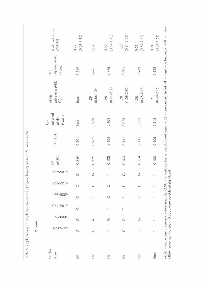
Tabl
e 6
(sup
plem
enta
ry).
Com
plem
ent f
acto
r H
(CFH
) gen
e ha
plot
ypes
in a
CSC
ver
sus
cCSC
Varia
nts
Hap
lo-
type
srs3753394
rs800292
rs1061170
rs2284664
rs1329428
rs1065489
HF
aCSC
HF
cCSC
Un-
adju
sted
alle
lic
P-va
lue
Alle
lic
odds
rat
io (9
5%
CI)
Un-
adju
sted
alle
lic
P-va
lue
Alle
lic o
dds
ratio
(9
5% C
I)
H1
CG
CC
CG
0.24
90.
301
Base
Base
0.21
90.
77
(0.5
1-1.
16)
H2
CA
TT
TG
0.27
20.
262
0.21
91.
29
(0.8
6-1.
94)
Base
Base
H3
TG
TC
CT
0.10
20.
104
0.40
81.
28
(0.7
1-2.
30)
0.97
60.
99
(0.5
7-1.
73)
H4
CG
TC
TG
0.16
40.
111
0.02
51.
78
(1.0
8-2.
95)
0.20
11.
38
(0.8
4-2.
26)
H5
TG
TC
TG
0.11
40.
115
0.37
31.
28
(0.7
5-2.
18)
0.96
30.
99
(0.5
9-1.
66)
Rare
**
**
**
0.10
00.
108
0.51
51.
21
(0.6
8-2.
16)
0.82
20.
94
(0.5
4-1.
64)
aCSC
= a
cute
cen
tral s
erou
s ch
orio
retin
opat
hy; c
CSC
= c
hron
ic c
entra
l ser
ous
chor
iore
tinop
athy
; CI =
con
fiden
ce in
terv
al; H
F =
hap
loty
pe fr
eque
ncy;
MAF
= m
inor
al
lele
freq
uenc
y. P
-val
ues
< 0
.008
3 w
ere
cons
ider
ed s
igni
fican
t.

97
2.4
References1. Daruich A, Matet A, Dirani A, et al. Central serous chorioretinopathy: Recent findings and new
physiopathology hypothesis. Prog Retin Eye Res. 2015;48:82-118.2. Piccolino FC, de la Longrais RR, Ravera G, et al. The foveal photoreceptor layer and visual acuity
loss in central serous chorioretinopathy. Am J Ophthalmol. 2005;139(1):87-99.3. Eandi CM, Ober M, Iranmanesh R, Peiretti E, Yannuzzi LA. Acute central serous chorioretinopathy
and fundus autofluorescence. Retina. 2005;25(8):989-993.4. Fujimoto H, Gomi F, Wakabayashi T, Sawa M, Tsujikawa M, Tano Y. Morphologic changes in acute
central serous chorioretinopathy evaluated by fourier-domain optical coherence tomography. Ophthalmology. 2008;115(9):1494-1500, 1500.e1491-1492.
5. Daruich A, Matet A, Marchionno L, et al. ACUTE CENTRAL SEROUS CHORIORETINOPATHY: Factors Influencing Episode Duration. Retina. 2017;37(10):1905-1915.
6. Breukink MB, Dingemans AJ, den Hollander AI, et al. Chronic central serous chorioretinopathy: long-term follow-up and vision-related quality of life. Clinical ophthalmology (Auckland, NZ). 2017;11:39-46.
7. Guyer DR, Yannuzzi LA, Slakter JS, Sorenson JA, Ho A, Orlock D. Digital indocyanine green videoangiography of central serous chorioretinopathy. Archives of ophthalmology (Chicago, Ill : 1960). 1994;112(8):1057-1062.
8. Ersoz MG, Karacorlu M, Arf S, Hocaoglu M, Sayman Muslubas I. Pachychoroid pigment epitheliopathy in fellow eyes of patients with unilateral central serous chorioretinopathy. The British journal of ophthalmology. 2017.
9. Spaide RF, Campeas L, Haas A, et al. Central serous chorioretinopathy in younger and older adults. Ophthalmology. 1996;103(12):2070-2079; discussion 2079-2080.
10. Wang M, Munch IC, Hasler PW, Prunte C, Larsen M. Central serous chorioretinopathy. Acta Ophthalmol. 2008;86(2):126-145.
11. de Jong EK, Breukink MB, Schellevis RL, et al. Chronic central serous chorioretinopathy is associated with genetic variants implicated in age-related macular degeneration. Ophthalmology. 2015;122(3):562-570.
12. Miki A, Kondo N, Yanagisawa S, Bessho H, Honda S, Negi A. Common variants in the complement factor H gene confer genetic susceptibility to central serous chorioretinopathy. Ophthalmology. 2014;121(5):1067-1072.
13. Moschos MM, Gazouli M, Gatzioufas Z, et al. Prevalence of the Complement Factor H and Gstm1 Genes Polymorphisms in Patients with Central Serous Chorioretinopathy. Retina. 2016;36(2):402-407.
14. van Dijk EHC, Schellevis RL, van Bergen M, et al. Association of a Haplotype in the NR3C2 Gene, Encoding the Mineralocorticoid Receptor, With Chronic Central Serous Chorioretinopathy. JAMA Ophthalmol. 2017;135(5):446-451.
15. Breukink MB, Schellevis RL, Boon CJ, et al. Genomic Copy Number Variations of the Complement Component C4B Gene Are Associated With Chronic Central Serous Chorioretinopathy. Invest Ophthalmol Vis Sci. 2015;56(9):5608-5613.
16. Boon CJ, van de Kar NC, Klevering BJ, et al. The spectrum of phenotypes caused by variants in the CFH gene. Molecular immunology. 2009;46(8-9):1573-1594.
17. Dorner GT, Garhofer G, Huemer KH, et al. Effects of adrenomedullin on ocular hemodynamic parameters in the choroid and the ophthalmic artery. Invest Ophthalmol Vis Sci. 2003;44(9):3947-3951.
18. Carvalho-Recchia CA, Yannuzzi LA, Negrao S, et al. Corticosteroids and central serous chorioretinopathy. Ophthalmology. 2002;109(10):1834-1837.
19. Yannuzzi LA. Central serous chorioretinopathy: a personal perspective. Am J Ophthalmol. 2010;149(3):361-363.
20. Cardillo Piccolino F, Eandi CM, Ventre L, Rigault de la Longrais RC, Grignolo FM. Photodynamic therapy for chronic central serous chorioretinopathy. Retina. 2003;23(6):752-763.
21. Liew G, Quin G, Gillies M, Fraser-Bell S. Central serous chorioretinopathy: a review of epidemiology and pathophysiology. Clinical & experimental ophthalmology. 2013;41(2):201-214.
22. Mehta PH, Meyerle C, Sivaprasad S, Boon C, Chhablani J. Preferred practice pattern in central serous chorioretinopathy. The British journal of ophthalmology. 2017;101(5):587-590.
23. Yannuzzi LA, Slakter JS, Kaufman SR, Gupta K. Laser treatment of diffuse retinal pigment

epitheliopathy. European journal of ophthalmology. 1992;2(3):103-114.24. Mohabati D, van Rijssen TJ, van Dijk EH, et al. Clinical characteristics and long-term visual
outcome of severe phenotypes of chronic central serous chorioretinopathy. Clinical ophthalmology (Auckland, NZ). 2018;12:1061-1070.
25. Tittl MK, Spaide RF, Wong D, et al. Systemic findings associated with central serous chorioretinopathy. Am J Ophthalmol. 1999;128(1):63-68.
26. Haimovici R, Koh S, Gagnon DR, Lehrfeld T, Wellik S. Risk factors for central serous chorioretinopathy: a case-control study. Ophthalmology. 2004;111(2):244-249.
27. Liu B, Deng T, Zhang J. RISK FACTORS FOR CENTRAL SEROUS CHORIORETINOPATHY: A Systematic Review and Meta-Analysis. Retina. 2016;36(1):9-19.
28. Saksens NT, Geerlings MJ, Bakker B, et al. Rare Genetic Variants Associated With Development of Age-Related Macular Degeneration. JAMA Ophthalmol. 2016;134(3):287-293.
29. Winkler TW, Brandl C, Grassmann F, Gorski M, Stark K. Investigating the modulation of genetic effects on late AMD by age and sex: Lessons learned and two additional loci. 2018;13(3):e0194321.

99
3.1 Role of the complement system in chronic central serous chorioretinopathy: a genome-wide association study
3.2 Exome sequencing identifies PIGZ, DUOX1, LAMB3 and RSAD1 as susceptibility genes for chronic central serous chorioretinopathy in females

3. Unbiased Approaches

101

3.1 Role of the complement system in chronic central serous chorioretinopathy: A Genome-wide association study Rosa L. Schellevis, Elon H.C. van Dijk*, Myrte B. Breukink*, Lebriz Altay*, Bjorn Bakker, Bobby P.C. Koeleman, Lambertus A. Kiemeney, Dorine W. Swinkels, Jan E.E. Keunen, Sascha Fauser, Carel B. Hoyng, Anneke I. den Hollander, Camiel J.F. Boon# & Eiko K. de Jong# JAMA Ophthalmol. 2018 Oct 1;136(10):1128-1136. doi: 10.1001/jamaophthalmol.2018.3190 *, # These authors contributed equally to this study

103
3.1
AbstractImportance: To date, several targeted genetic studies on chronic central serous chorioretinopathy (cCSC) have been performed; however, unbiased genome-wide studies into the genetics of cCSC have not been reported. To discover new genetic loci associated with cCSC and to better understand the causative mechanism of this disease, we performed a genome-wide association study (GWAS) on patients with cCSC.
Objective: To discover new genetic loci and pathways associated with cCSC and to predict the association of genetic variants with gene expression in patients with cCSC.
Design, Setting and Participants: This case-control GWAS was completed in the general community, 3 referral university medical centers, and outpatient care on European individuals with cCSC and population-based control participants. Genotype data was collected from May 2013 to August 2017, and data analysis occurred from August 2017 to November 2017.
Main outcomes and measurements: Associations of single-nucleotide polymorphisms, haplotypes, genetic pathways, and predicted gene expression with cCSC.
Results: A total of 521 patients with cCSC (median age 51 years; interquartile range [IQR], 44-59 years; 420 [80.6%] male) and 3577 European population-based control participants (median age, 52 years; IQR, 37-71 years; 1630 [45.6%] male) were included. One locus on chromosome 1 at the complement factor H (CFH) gene reached genome-wide significance and was associated with an increased risk of cCSC (rs1329428; odds ratio [OR], 1.57 [95% CI, 1.38-1.80]; P = 3.12x10⁻¹¹). The CFH haplotypes H1 and H3 were protective for cCSC (H1: OR, 0.64 [95% CI, 0.53-0.77]; P = 2.18 × 10⁻⁶; H3: OR, 0.54 [95% CI, 0.42-0.70]; P = 2.49 × 10⁻⁶), whereas haplotypes H2, H4, H5, and the aggregate of rare CFH haplotypes conferred increased risk (H2: OR, 1.57 [95% CI, 1.30-1.89]; P = 2.18 × 10⁻⁶; H4: OR, 1.43 [95% CI, 1.13-1.80]; P = 2.49 × 10⁻³; H5: OR, 1.80 [95%CI, 1.36-2.39]; P = 4.61 × 10⁻⁵; rare haplotypes: OR, 1.99 [95%CI, 1.43-2.77]; P = 4.59 × 10⁻⁵). Pathway analyses showed involvement of the complement cascade and alternative open reading frame (ARF) pathway in cCSC. Using PrediXcan, we identified changes in predicted expression of complement genes CFH, complement factor H related 1 (CFHR1), complement factor related 4 (CFHR4), and membrane cofactor protein (MCP/CD46). Additionally, the genes potassium sodium-activated channel subfamily T member 2 (KCNT2) and tumor necrosis factor receptor superfamily member 10a (TNFRSF10A) were differentially expressed in cCSC patients.
Conclusions and relevance: In this GWAS on cCSC, we identified a locus on chromosome 1 at the CFH gene that was significantly associated with cCSC, and we report protective and risk-conferring haplotypes in this gene. Pathway analyses were enriched for complement genes, and gene expression analysis suggests a role for CFH, CFHR1, CFHR4, CD46, KCNT2, and TNFRSF10A in the disease. Taken together, these results underscore the potential importance of the complement pathway in the causative mechanism of cCSC.

IntroductionCentral serous chorioretinopathy (CSC) is characterized by subretinal fluid accumulation between the neuroretina and the retinal pigment epithelium (RPE).1-
3 Patients usually present with clinical symptoms of metamorphopsia and central vision impairment.4,5 Although acute CSC can show spontaneous resolution within 3 months, patients with chronic CSC (cCSC) have prolonged presence of fluid with progressive loss of vision, permanent RPE alterations, and a reduced quality of life.4 Generally, CSC occurs more frequently in males (9.9 per 100 000) than females (1.7 per 100 000) and often presents at an age at which patients are still professionally active.6
The exact causative mechanism of cCSC is unknown, but clinical and experimental studies have implicated dysfunction of the RPE and hyperpermeability of the choroid in CSC.4 The disease has been associated with the use of corticosteroids, stress, and type A personality. Reports of familial occurrences of cCSC support a genetic component for the disease.1,3,4,7-11 Genetic studies on cCSC have been limited to candidate gene approaches; several associations have been reported with single-nucleotide polymorphisms (SNPs) in the genes age-related macular degeneration susceptibility 2 (ARMS2), cadherin 5 (CDH5), complement factor H (CFH) and nuclear receptor subfamily 3 group C member 2 (NR3C2), as well as copy number variations in the complement factor 4B (C4B) gene.12-16
Unbiased genome-wide association studies (GWAS) have identified hundreds of genomic loci implicated in complex diseases, such as age-related macular degeneration (AMD), myopia, and glaucoma.17 Such approaches have shed light on the genetic architecture of these diseases; however, establishing functional links between identified loci and disease remains challenging. Besides having direct effects on protein function or structure, SNPs can also influence the expression of nearby (cis) or distal (trans) genes. Such regulatory genetic variants are called expression quantitative trait loci (eQTL).18 Several eQTL databases, linking genotype information to tissue expression of genes, have been established. One of the largest of these projects is the GTEx project, in which expression profiles of 44 tissues of 449 donors have been collected in version 6.18 These eQTL databases offer functional information on SNPs identified in GWAS, contribute to a better understanding of the studied disease trait, and can be used to predict expression in genotyped samples.19
In this study, we performed a GWAS involving European patients with cCSC and population control participants to identify new cCSC disease loci and to increase our knowledge on the causative mechanism of this disease. Additionally, we performed pathway analyses to discover new pathways implicated in cCSC. Using publicly available eQTL data, we aimed to identify new candidate genes predicted to be differentially expressed in patients with cCSC compared with control participants.
Key pointsQuestion: Which genetic loci associate with chronic central serous chorioretinopathy (cCSC) when using an unbiased genome-wide approach?
Findings: In this case-control genome-wide association study, a locus containing multiple variants in the CFH gene was associated with cCSC. Additionally, pathway

105
3.1
analysis implicated the complement system and based on genetic data, components of the complement system were predicted to have an association with altered expression in patients with cCSC.
Meaning: This study of participants with cCSC demonstrates the potential role of the CFH gene and the complement system in the causative mechanism of cCSC.
MethodsStudy participants Genomic DNA was extracted from blood using standard procedures. In total, 546 European patients with cCSC recruited from the outpatient clinics of the Radboud university medical center (Radboudumc), Nijmegen, the Netherlands, University Hospital of Cologne (UHC), Cologne, Germany, and Leiden University Medical Center (LUMC), Leiden, the Netherlands, were included. Grading of all patients was performed by an experienced retinal specialist (C.J.F.B.), and was based on extensive ophthalmological examination including fundoscopy, spectral-domain optical coherence tomography, fluorescein angiography and indocyanine green angiography. Diagnosis of cCSC was based on subgroups 1 and 2, which have been previously described.13 Briefly, patients that were included in this study showed the presence of serous fluid on optical coherence tomography in 1 or both eyes, either bilateral (subgroup 1) or unilateral (subgroup 2) RPE irregularities with 1 or more hot spots of leakage on fluorescein angiography, and corresponding hyperfluorescence on indocyanine green angiography.13 Patients diagnosed with acute CSC as recognized by a focal leakage spot (ink blot) or a smokestack pattern on fluorescein angiography, with less than 1 disc diameter of adjacent atrophic RPE alterations, and/or a duration of disease of less than 3 months were excluded from the study. Patients in whom evidence of another explanatory diagnosis or complication was present (subgroup 3), such as polypoidal choroidal vasculopathy, choroidal neovascularization, drusen or other signs of AMD, were excluded from this study.
This study was carried out in accordance with the tenets of the Declaration of Helsinki and was approved by the local ethics committees of the Radboudumc, LUMC and UHC. Written informed consent was obtained for all subjects involved in the study.
Genotyping data of controls was obtained from the Nijmegen Biomedical Study (NBS), a population-based survey conducted by the Department for Health Evidence and the Department of Laboratory Medicine of the Radboudumc. (eMethods in supplement, http://www.nijmegenbiomedischestudie.nl/).20 In this population-based study, no ophthalmologic grading was performed. Only controls for which genotyping was available on the OmniExpress platform (n=3654) were included in this study. Genotype data was collected from May 2013 to August 2017, and data analysis occurred from August 2017 to November 2017.
The mean age of patients with cCSC and control participants was compared with a Mann-Whitney U test. The sex distribution was compared with a X2 test using SPSS version 22 (IBM). All values of P<.05 were deemed significant.

Genome-wide association and haplotype analyses Genotyping was performed with the OmniExpress-12 or OmniExpress-24 chips, and data were imputed with the Haplotype Reference Consortium release 1.1.2016. After stringent quality control (eMethods in the Supplement), 521 patients with cCSC and 3577 NBS control participants were included in the analysis, carrying 11 261 291 autosomal SNPs and 265 428 X-chromosomal SNPs. Single-variant association analysis was performed using the Firth bias-corrected likelihood ratio test implemented in EPACTS (version 3.2.6, http://genome.sph.umich.edu/wiki/EPACTS; Univeristy of Michigan),21 correcting for sex and the first 2 components of the ancestry analysis, to correct for potential population stratification (eMethods in the Supplement). To assess the potential role of confounding by AMD, the analysis was repeated in patients and controls below younger than 51 years.
Haplotype analysis, combining multiple variants in the CFH gene, was performed with the haplo.stats22 (version 1.7.7) package in R (R Foundation for Statistical Computing).23 The SNPs described by Hageman et al.24 and the CFH SNPs that were previously associated with cCSC were used as input.12,13 Haplotypes with a frequency greater than 1% were analyzed individually, while rare haplotypes (with a frequency less than 1%) were aggregated. Haplotype association with cCSC was performed using the haplo.glm in R, correcting for sex and the first 2 principal components of the ancestry analysis. The analysis was performed using the most common protective haplotype (H1) and cCSC risk-carrying haplotype (H2) as references. The frequency of the haplotypes in patients with cCSC and control participants was obtained using the haplo.cc command in R.
Pathway and PrediXcan analysisWe used GWAS summary statistics to perform competitive gene-set analysis to identify pathways associated with cCSC using 2 different programs: MAGMA (version 1.06, https://ctg.cncr.nl/software/magma) and VEGASv2 (version 2, https://vegas2.qimrberghofer.edu.au/vegas2v2).25,26 Additionally, PrediXcan (Hakyimlab; https://github.com/hakyim/PrediXcan) was used to predict gene expression levels based on publicly available eQTL data of GTEx.27 PrediXcan was performed on all 44 provided GTEx tissues using genetic variants in 17 742 genes. We selected the genes that were associated in at least 1 tissue after Bonferroni correction for multiple testing (P < .05/17742 genes = 2.21x10⁻⁶) and were nominally associated (P < .05) in at least 50% of the tissues they were expressed in. (The eMethods in the Supplement provide further clarification). This study had sufficient power to detect common variants with OR greater than 1.5 and had more than 80% power to detect the lead variant (eMethods and eFigure 1 in the Supplement).
ResultsGenome-wide association analysisA total of 546 patients with cCSC were recruited from Radboudumc (n = 319), UHC (n = 74), and LUMC (n = 153). In addition, 3654 European population-based control participants were included. After quality control (eFigures 2 and 3 in the Supplement), a total of 521 European patients with cCSC (median age, 51 years; interquartile range [IQR], 44-59 years; 420 [80.6%] male) and 3577 controls (median age, 52 years; IQR,

107
3.1
37-71 years; 1630 [45.6%] male; eTable 1 in the Supplement) were analyzed for 11 261 291 autosomal and 265 428 X-chromosomal SNPs. Because of the significant difference in sex between patients with cCSC and control participants (eTable 1 in the Supplement, P = 1.78 x 10-50), sex was included as a covariate in the analysis along with 2 principal components of the ancestry analysis.
The GWAS identified 20 SNPs that reached genome-wide significance (using a cutoff value for significance set at P < 5.0x10⁻⁸) that all resided at 1 locus on chromosome 1 in the CFH gene (lead variant: rs1329428; odds ratio [OR], 1.57 [95% CI, 1.38-1.80]; P = 3.12 x 10⁻¹¹; Figure 1; Table 1; eFigure 5, and eTable 2 in the Supplement). Additionally, six suggestive signals (P < 1x10⁻⁶) were foundon chromosome1 (OR, 0.64 [95%CI,0.54-0.77]; P = 6.69 × 10⁻⁷), chromosome 8 (OR, 42.25 [95% CI, 9.55-186.89]; P = 1.33 × 10⁻⁷), chromosome 9 (OR, 8.36 [95%CI, 3.67-19.06]; P = 6.28 × 10⁻⁷), chromosome 15 (OR, 206.11 [95%CI, 4.50-9436.68]; P = 7.84 × 10⁻⁷), chromosome 17 (OR, 5.25 [95%CI, 2.94-9.48]; P = 1.51 × 10⁻⁷), and chromosome20 (OR,0.67 [95%CI,0.57-0.79]; P = 7.14 × 10⁻⁷; Figure 1; Table 1). Conditioned analysis of the lead SNP on chromosome 1 did not reveal any other independent signal in the CFH gene (eFigure 4 in the Supplement), but did retain the suggestive signal at the CD46 gene on chromosome 1 indicating that the CD46 signal is independent of the CFH association (unconditioned and conditioned data are shown in eFigure 4 the Supplement). Stratified analysis on patients and controls younger than 51 years identified associations with the same 20 SNPs in the CFH gene, but ORs were higher for the lead variant than in the complete cohort (OR, 1.89 [95% CI, 1.56-2.32]; P = 1.23 x 10⁻¹⁰; eTable2 in the Supplement).
Haplotype analysis To further characterize the association at the CFH gene, haplotype analysis was performed. A logistic model corrected for sex and 2 principal components was performed using either the most common cCSC protective (H1) haplotype or the most common cCSC risk-conferring (H2) haplotype as reference (Table 2). The H2, H4, and H5 haplotypes, all containing the minor allele of the lead variant, were associated with an increased risk of cCSC (H2: OR, 1.57 [95% CI, 1.30-1.89]; P = 2.18 × 10⁻⁶; H4: OR, 1.43 [95% CI, 1.13-1.80]; P = 2.49 × 10⁻³; H5: OR, 1.80 [95% CI, 1.36-2.39]; P = 4.61 × 10⁻⁵). The H1 and H3 haplotypes, containing the major allele of the lead variant, were protective (H1: OR, 0.64 [95% CI, 0.53-0.77]; P = 2.18 × 10⁻⁶; H3: OR, 0.54 [95% CI, 0.42-0.70]; P = 2.49 × 10⁻⁶). Additionally, the aggregate of rare haplotypes also increased the risk of cCSC (OR, 1.99 [95% CI, 1.43-2.77]; P = 4.59 × 10⁻⁵). All associations were significant after Bonferroni correction for multiple testing for 10 tests (using a cutoff value of P < .005).
Gene-set/Pathway analysisUsing the GWAS summary statistics, we performed competitive gene-set analysis using 2 programs. MAGMA identified 3 pathways that were associated with cCSC after correction for multiple testing (Table 3, genes in eTable 3 in the Supplement). Two of the pathways are implicated in the complement cascade, and 1 is the ARF pathway. We identified the same reactome pathways of the complement cascade using VEGAS2. However, these findings were not significant after correction for multiple testing (using a cutoff value of P < 1 x 10⁻⁵; Table 3, genes in eTable 4 in the
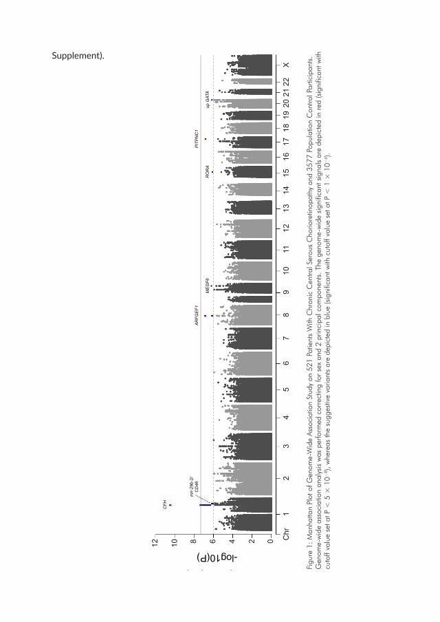
Supplement).
CFH
mir-
29b-
2/C
D46
AR
FGE
F1
ME
GF6
RO
RA
PIT
PN
C1
up G
ATA
-log10(P)
Chr
12
34
56
78
910
1112
1314
1516
1718
1920
2122
X
246 81012 0
Figu
re 1
: Man
hatta
n Pl
ot o
f Gen
ome-
Wid
e As
soci
atio
n St
udy
on 5
21 P
atie
nts
With
Chr
onic
Cen
tral S
erou
s C
horio
retin
opat
hy a
nd 3
577
Popu
latio
n C
ontro
l Par
ticip
ants
. G
enom
e-w
ide
asso
ciat
ion
anal
ysis
was
per
form
ed c
orre
ctin
g fo
r se
x an
d 2
prin
cipa
l com
pone
nts.
The
gen
ome-
wid
e si
gnifi
cant
sig
nals
are
dep
icte
d in
red
(sig
nific
ant w
ith
cuto
ff va
lue
set a
t P <
5 ×
10−
8 ), w
here
as th
e su
gges
tive
varia
nts
are
depi
cted
in b
lue
(sig
nific
ant w
ith c
utof
f val
ue s
et a
t P <
1 ×
10−
6 ).

109
3.1
Tabl
e 1.
Top
hits
in c
hron
ic c
entra
l ser
ous
chor
iore
tinop
athy
GW
AS s
tudy
Top
Sing
le-
Nuc
leot
ide
Poly
-
mor
phis
m
Chr
Base
Pai
r
(hg1
9)
Maj
or
Alel
le/
Min
or a
llele
Gen
e
Min
or A
llele
Fr
eque
ncy
in
Patie
nts
with
cC
SC (n
=52
1)
Min
or A
llele
Fr
eque
ncy
in
cont
rol p
artic
ipan
ts
(n=
3577
)
OR
(95%
CI)
P-va
lue
rs13
2942
8a1
1967
0281
0C
/TCF
H0.
543
0.43
41.
57 (1
.38-
1.80
)3.
12x1
0-11
rs56
1753
392
868
1319
81C
/TAR
FGEF
10.
0070
0.00
0542
.25
(9.5
5-18
6.89
)1.
33x1
0-7
rs76
7664
9817
6541
8162
A/G
PITP
NC1
0.02
10.
005
5.25
(2.9
4-9.
48)
1.51
x10-7
rs11
8083
394
912
3367
552
C/T
MEG
F60.
014
0.00
28.
36 (3
.67-
19.0
6)6.
28x1
0-7
rs48
4439
21
2079
9120
9C
/Gm
ir-29
b-2/
CD46
0.15
90.
230
0.64
(0.5
4-0.
77)
6.69
x10-7
rs23
7912
020
6103
0580
T/A
upst
ream
G
ATA5
0.21
80.
300
0.67
(0.5
7-0.
79)
7.14
x10-7
rs54
1395
042
1561
1753
24C
/TRO
RA0.
006
0.00
206.
11 (4
.50-
9436
.68)
7.84
x10-7
ARFG
EF1:
ade
nosi
ne d
ipho
spha
te ri
bosy
latio
n fa
ctor
gua
nine
nuc
leot
ide
exch
ange
fact
or 1
; bp:
bas
e pa
ir; c
CSC
: chr
onic
cen
tral s
erou
s ch
orio
retin
opat
hy; C
D46
: clu
ster
of
diff
eren
tiatio
n 46
; CFH
: com
plem
ent f
acto
r H; c
hr: c
hrom
osom
e; C
I: co
nfid
ence
inte
rval
; GAT
A5: G
ATA-
5 bi
ndin
g pr
otei
n 5;
GW
AS: g
enom
e-w
ide
asso
ciat
ion
stud
y;
hg:
hum
an g
enom
e; M
AF:
min
or a
llele
freq
uenc
y; M
EGF6
: m
ultip
le e
pide
rmal
gro
wth
fact
or li
ke d
omai
ns 6
; mir-
29b-
2: m
icro
rib
onuc
leic
aci
d 29
b-2;
OR:
odd
s ra
tio;
PITP
NC1
: pho
spha
tidyl
inos
itol t
rans
fer
prot
ein
cyto
plas
mic
1; R
ORA
: ret
inoi
c ac
id r
ecep
tor-
rela
ted
orph
an r
ecep
tor
alph
a; a
This
is a
gen
ome-
wid
e si
gnifi
cant
hit

Tabl
e 2.
Hap
loty
pe a
naly
sis
of th
e C
ompl
emen
t fac
tor
H g
ene
Per
CFH
-H1b
Per
CFH
-H2c
Hap
lo
type
rs3753394
rs529825
rs800292
rs3766404
rs1061170
rs203674
rs3753396
rs2284664
rs1329428a
rs1065489
Frequency in Controls
Frequency in patients with cCSC
Odds Ratio
(95% CI)
P-value Correctedd,e
Odds Ratio (95% CI)
P-value Correctedd,e
H1
CG
GT
CG
AC
CG
0.33
90.
273
1 [R
EF]
1 [R
EF]
0.64
(0.5
3-0.
77)
2.18
x10-6
f
H2
CA
AT
TT
AT
TG
0.22
10.
274
1.57
(1.3
0-1.
89)
2.18
x10-6
f1
[REF
]1
[REF
]
H3
TG
GT
TT
GC
CT
0.13
50.
094
0.85
(0.6
6-1.
09)
0.20
40.
54
(0.4
2-0.
70)
2.49
x10-6
f
H4
CG
GC
TT
AC
TG
0.11
50.
128
1.43
(1.1
3-1.
80)
2.49
x10-3
f0.
91
(0.7
2-1.
15)
0.44
2
H5
TG
GT
TT
AC
TG
0.05
20.
078
1.80
(1.3
6-2.
39)
4.61
x10-5
f1.
15
(0.8
6-1.
53)
0.34
3
H6
TG
GT
TG
AC
CG
0.03
00.
025
1.17
(0.7
5-1.
81)
0.49
20.
74
(0.4
8-1.
16)
0.19
1
H7
TG
GT
CG
AC
CG
0.02
70.
018
0.82
(0.4
8-1.
42)
0.48
50.
53
(0.3
0-0.
91)
0.02
1
H8
TG
GC
TT
AC
TG
0.02
20.
027
1.58
(1.0
0-2.
48)
0.04
81.
01
(0.6
4-1.
59)
0.97
2
H9
CA
AT
TT
GC
CT
0.01
70.
017
1.25
(0.7
3-2.
17)
0.41
80.
80
(0.4
6-1.
38)
0.42
1
RARE
NAg
NAg
NAg
NAg
NAg
NAg
NAg
NAg
NAg
NAg
0.04
20.
066
1.99
(1.4
3-2.
77)
4.59
x10-5
f1.
27
(0.9
1-1.
77)
0.16
3
cCSC
: chr
onic
cen
tral s
erou
s ch
orio
retin
opat
hy; C
I: co
nfid
ence
inte
rval
; NA
: not
app
licab
le; R
EF: r
efer
ence
; a Re
sults
sho
wn
in th
e rs
1329
428
colu
mn
are
top
hits
for
the
geno
me-
wid
e as
soci
-at
ion
stud
y fin
ding
s. b
Anal
ysis
per
form
ed w
ith th
e m
ost c
omm
on p
rote
ctiv
e ha
plot
ype
CFH
-H1
set a
s a
refe
renc
e. c A
naly
sis
perfo
rmed
with
the
mos
t com
mon
ris
k-co
nfer
ring
hapl
otyp
e CF
H-H
2 se
t as
a re
fere
nce.
d Bo
nfer
roni
cor
rect
ion
for
mul
tiple
test
ing
for
10 h
aplo
type
s w
ith s
igni
fican
ce s
et a
t P <
.05/
10 =
.005
. e Ana
lysi
s w
as p
erfo
rmed
cor
rect
ing
for
sex
and
the
first
2 p
rinci
pal
com
pone
nts
of th
e an
cest
ry a
naly
sis.
f Val
ues
are
sign
ifica
nt b
ased
on
a cu
toff
leve
l of P
< .0
05. g
The
data
on
rare
hap
loty
pes
incl
udes
an
accu
mul
atio
n of
alle
les
that
cou
ld b
e ei
ther
min
or o
r m
ajor
.

111
3.1Ta
ble
3. C
ompe
titiv
e G
ene-
Set A
naly
sis
of M
ulti-
Mar
ker
Anal
ysis
of G
enom
ic A
nnot
atio
n (M
AGM
A)
and
Vers
atile
Gen
e-Ba
sed
Asso
ciat
ion
Stud
y 2
(VEG
AS2)
P va
lue
Path
way
nam
eG
enes
, No
Beta
(se)
Nom
inal
Em
peric
al
corr
ecte
d
MAG
MA
Reac
tom
e Re
gula
tion
of C
ompl
emen
t Cas
cade
131.
29 (0
.265
)5.
49x1
0-78.
90x1
0-4a
Reac
tom
e C
ompl
emen
t Cas
cade
290.
786
(0.1
79)
5.56
x10-6
7.37
x10-3
a
Bioc
arta
ARF
pat
hway
170.
848
(0.1
94)
6.49
x10-6
8.65
x10-3
a
VEG
AS2
Reac
tom
e: R
-HSA
-977
606
Regu
latio
n of
Com
plem
ent C
asca
de13
NA
1.03
x10-7
1.60
x10-5
Reac
tom
e: R
-HSA
-166
658
Com
plem
ent C
asca
de22
6.09
x10-7
4.20
x10-5
arf:
alte
rnat
ive
open
rea
ding
fram
e; N
A: n
ot a
nnot
ated
; se:
sta
ndar
d er
ror;
a si
gnifi
cant
afte
r co
rrec
tion
for
mul
tiple
test
ing;
the
MAG
MA
nom
inal
sig
nific
ance
thre
shol
d is
P <
4.1
2 x
10-5 a
nd th
e VE
GAS
2 em
piric
al s
igni
fican
ce th
resh
old
is P
< 1
x 1
0-5

Tabl
e 4.
Pre
diXc
an r
esul
ts, g
enes
diff
eren
tially
exp
ress
ed b
etw
een
cCSC
pat
ient
s an
d co
ntro
ls
Gen
eTi
ssue
s w
ith
expr
essi
on, N
o
Tiss
ues
with
di
ffere
ntia
l ex
pres
sion
,
No.
(%)a
Tiss
uesb
Odd
s Ra
tio
(95%
CI)
P va
lue
CFH
R41
1 (1
00)
Live
r0.
91 (0
.85-
0.98
)1.
29x1
0-6
CD46
2521
(84)
Esop
hagu
s M
uscu
laris
1.05
(1.0
3-1.
07)
1.79
x10-6
Skin
Not
Sun
Exp
osed
Sup
rapu
bic
1.32
(1.1
8-1.
48)
2.26
x10-6
Hea
rt At
rial A
ppen
dage
1.16
(1.0
9-1.
24)
2.39
x10-6
Col
on S
igm
oid
1.07
(1.0
4-1.
09)
2.57
x10-6
TNFR
SF10
A18
15 (8
3)C
ells
Tra
nsfo
rmed
fibr
obla
sts
0.96
(0.9
4-0.
98)
2.41
x10-6
Adre
nal G
land
0.95
(0.9
3-0.
97)
2.60
x10-6
CFH
R112
9 (7
5)
Hyp
otha
lam
us0.
78 (0
.72-
0.85
)1.
73x1
0-8
Adip
ose
Subc
utan
eous
1.15
(1.1
-1.2
1)1.
85x1
0-8
Live
r0.
95 (0
.93-
0.97
)1.
87x1
0-7
Cel
ls T
rans
form
ed fi
brob
last
s0.
79 (0
.71-
0.87
)1.
41x1
0-6
KCN
T25
3 (6
0)Es
opha
gus
Gas
troes
opha
geal
Jun
ctio
n3.
70 (2
.27-
6.04
)1.
62x1
0-7
Ner
ve T
ibia
l1.
13 (1
.08-
1.18
)4.
99x1
0-7
CFH
53
(60)
Adip
ose
Subc
utan
eous
1.09
(1.0
5-1.
13)
1.17
x10-6
Ner
ve T
ibia
l1.
10 (1
.06-
1.14
)1.
63x1
0-6
Abbr
evia
tions
: CD
46: c
lust
er o
f diff
eren
tiatio
n 46
; CFH
: com
plem
ent f
acto
r H
; CFH
R1/4
: com
plem
ent f
acto
r H
rel
ated
1/4
; GTE
x: g
enot
ype-
tissu
e ex
pres
sion
; KCN
T2:
Pota
ssiu
m S
odiu
m-A
ctiv
ated
Cha
nnel
Sub
fam
ily T
Mem
ber
2; O
R: o
dds
ratio
; TN
FRSF
10A:
tum
or n
ecro
sis
fact
or r
ecep
tor
supe
rfam
ily m
embe
r 10
a; a
The
cuto
ff va
lue
for
sign
ifica
nce
in d
iffer
entia
l exp
ress
ion
was
P <
.05;
onl
y th
ose
gene
s th
at s
how
ed e
xpre
ssio
n di
ffere
nces
in m
ore
than
50%
of t
heir
expr
esse
d tis
sues
are
dep
icte
d; b
This
sec
tion
used
a c
utof
f val
ue fo
r si
gnifi
canc
e at
P <
2.8
1 x
10-6.

113
3.1
PrediXcan analysis For each individual, we predicted the association of genotypes with expression levels of 17 742 genes in 44 different GTEx (version 6) tissues using PrediXcan. The predicted gene expression of genes at the CFH locus (CFH, CFHR1, CFHR4 and KCNT2) was different between cCSC patients and controls after correction for multiple testing, using a cutoff value for significance of P < 2.82 x 10⁻⁶ (for example, CFH in subcutaneous adipose tissue: OR, 1.09 [95% CI, 1.05-1.13]; P = 1.17 × 10⁻⁶; CFHR1 in subcutaneous adipose tissue: OR, 1.15 [95% CI, 1.10-1.21]; P = 1.85 × 10⁻⁸; CFHR4 in liver: OR, 0.91 [95% CI, 0.85-0.98]; P = 1.29 × 10⁻⁶; KCNT2 in gastroesophageal junction: OR, 3.70 [95% CI, 2.27-6.04]; P = 1.62 × 10⁻⁷; full results in Table 4). Additionally, we observed altered predicted expression of the CD46 and TNFRSF10A genes (Table 4, results of all tissues in eTable 5 in the Supplement).
DiscussionIn this unbiased genome-wide association study of cCSC, we identified a locus for cCSC on chromosome 1, at the CFH gene, that had previously been described in 2 targeted candidate gene studies.12,13 We discovered protective and risk haplotypes in CFH and found evidence for involvement of rare CFH haplotypes in the disease. Moreover, using gene-set analysis and publicly available expression databases, we uncovered additional evidence for altered regulation of the complement system in cCSC and identified novel candidate genes and pathways implicated in this disease.
So far, to our knowledge, the association of cCSC with the CFH gene is the only genetic association for cCSC that has been replicated in multiple independent studies.12,13,28 The effect size of the lead-variant (rs1329428) in CFH in this unbiased study was higher compared to the previous targeted study,13 but lower compared to the study by Miki et al. 12 (with ORs in the 3 studies 1.57, 1.47, and OR=1.79, respectively). Stratified analysis of patients younger than 51 years to excluded potential confounding with AMD showed the same genome-wide significant 20 SNPs with similar direction of effect, confirming the cCSC specific association. Additionally, haplotype analysis of the CFH locus confirmed a previously reported protective effect of CFH-H3,13 and identified associations with the protective CFH-H1 and risk carrying CFH-H2, CFH-H4, and CFH-H5 haplotypes. The risk associated with the CFH-H5 haplotype was higher than the risk caused by the single lead variant alone and higher to our knowledge than any previously reported SNP in CFH, with an OR of 1.80 for CFH-H5 compared to OR of 1.57 for rs1329428. Interestingly, an aggregate of all haplotypes with a frequency of less than 1% also showed a higher risk for cCSC (OR, 1.99). Higher ORs of CFH-H5 and the rare haplotype aggregate suggest that other (rare) variants in these haplotypes might play a role in cCSC.
The factor H protein, encoded by the CFH gene, is able to block the formation of C3-convertases and therefore is an important regulator of the complement system.29 The CFH gene has been widely studied for its role in AMD, and, interestingly, variants that confer risk for AMD are protective for cCSC and vice versa.13 These genetic associations imply that these 2 diseases might have an opposite disease mechanism, with an overactivation of the complement system in AMD29 and a reduced activity of the complement system in cCSC.

In the current study, we further substantiated the involvement of the complement system in cCSC. Pathway analysis with MAGMA and VEGAS2 showed associations of 2 gene sets of the complement system. We used PrediXcan to predict the expression of genes based on the genotype information of patients with cCSC and control participants. Unfortunately, no eye-specific or retina-specific eQTL database is publicly available, and these tissues are not yet implemented in GTEx nor in any other large eQTL data set. Therefore, as an indicator for general differences, we used all GTEx tissues and observed that the expression of 4 complement genes (CFH, CFHR1, CFHR4 and CD46) was predicted to be different in patients with cCSC compared to control participants. All GTEx tissues that express CFH showed an upregulation of CFH, suggesting that complement system activity may be reduced in cCSC (Table 4). Depending on the tissue, CFHR1 showed upregulation or down-regulation, indicating that regulation of this gene might be tissue specific; meanwhile CFHR4 was consistently down-regulated (Table 4). The factor H-associated (FHR) proteins, encoded by the CFHR genes, show sequence similarities to factor H and can compete with factor H for C3b binding and in this manner can influence complement activation.30 Deletions of CFHR1 have been found to be protective for AMD and CFHR4 deletions have been implicated in atypical hemolytic uremic syndrome,30 but the genes have not previously been associated with cCSC. to our knowledge.
Interestingly, we also observed an association with another complement gene independent of the CFH locus: CD46 was identified as one of the sub-threshold hits in the GWAS (Table 1), and PrediXcan predicted up-regulation of CD46 expression (Table 4). The CD46 gene encodes the membrane cofactor protein (MCP/CD46), a complement inhibitor that blocks all pathways of the complement cascade through inactivation of C3b and C4b.31,32 In light of the clinical characteristics of cCSC, in which patients present with hyperpermeability of the choroid and with fluid leakage through the retinal pigment epithelium, the involvement of CD46 is particularly interesting. It is an important regulator in the maintenance of epithelial cell barrier integrity through interaction with, for example, cadherins and integrins.32,33 Previous studies have shown that the activation of CD46 leads to a decrease in transepithelial resistance and loss of tight junctions in intestinal cells and a decreased membrane adhesion in RPE cells.32,33 The predicted increased CD46 expression in patients with cCSC therefore suggests a downregulation of complement activity, similarly as observed for factor H, and in addition destabilization of the integrity of the RPE, one of the main hallmarks of CSC. Together, both the complement and epithelial regulatory function of CD46 make it an interesting protein for further study in cCSC.
Taken together, the associations and predicted altered expression of CD46, CFH, CFHR1, and CFHR4 with cCSC imply a reduced activity of the complement system in cCSC. To date and to our knowledge, limited information is available on the actual activity of the complement system in patients with cCSC. One study did not find obvious systemic alterations in blood complement components of patients with cCSC, but this study had limited power, and CD46, FHR1, and FHR4 levels were not measured.34 Combined with previously described associations of CFH and C4B12-14 and the associations described here, a central role for the complement system in the disease mechanism of cCSC emerges. Larger studies measuring systemic complement regulators and activation products in patients with cCSC (specifically FH, FHR1,

115
3.1
FHR4, C4B, and CD46) are warranted.
Besides consolidating the involvement of the complement system, we also identified a new cCSC candidate pathway and 2 new cCSC candidate genes. MAGMA identified an association between cCSC and the ARF pathway. This pathway is involved in ribosomal biogenesis, and activation of the pathway leads to termination of ribosomal RNA production and cell cycle arrest.35 In addition, PrediXcan showed decreased expression of TNFRSF10A and up-regulation of KCNT2. Involvement of TNFRSF10A in cCSC was suggested previously in our candidate gene study of the main AMD loci. We showed a protective effect for a TNFRSF10A variant, which was also observed in this study (OR, 0.74 [95% CI, 0.64-0.85]; P = 1.47 x 10⁻⁵; data not shown) and the largest AMD GWAS (OR, 0.90; P = 4.5 x 10⁻¹¹).13,36 The KCNT2 gene encodes a potassium sodium-activated outward rectifier channel (KCNT2) with an unknown function. The ARF pathway and TNFRSF10A and KCNT2 genes are interesting candidates for cCSC, but their exact role in the eye remains to be elucidated.
LimitationsTo our knowledge, this study includes the largest reported cohort of European patients with cCSC; however, even larger sample sizes are necessary to find associations with rare variants or variants with a low effect size. Additionally, in this study, only European individuals were included, and therefore genetic associations in other races/ethnicities still remain to be discovered. Replication of this study by centers in- or outside of Europe will be necessary to increase sample size, determine population specific associations, and replicate the suggestive signals observed in this study.
The use of PrediXcan on the GTEx data allows for prediction of expression levels in 44 tissues, excluding the eye. Although we only regarded those genes that showed differential expression in at least 50% of tissues they were expressed in, this does not necessarily mean that this also applies to expression in the retina. Likewise, retina-specific genes might have been missed due to tissue-specific expression. A retina-specific eQTL database would be necessary to determine these associations, but to date, such a database has not been available.
ConclusionsIn this study, we describe a GWAS for cCSC and confirmed the association of genetic variants in CFH with the disease. Additionally, we identified CFHR1, CFHR4, CD46, KCNT2, and TNFRSF10A as cCSC candidate genes because of their genetic associations and predicted altered expression. With CFH, CFHR1, CFHR4, and CD46 being important regulators of the complement cascade, this study strengthens the involvement of the complement system in cCSC. Further work on the expression of the proteins encoded by these genes is warranted. Additionally, the use of next-generation sequencing techniques, such as exome sequencing, will enable the identification of (rare) coding variants influencing protein function of these genes and might provide more insight in the causative mechanisms of cCSC.

Footnotes and Financial DisclosuresFunding/Support: This study was supported by the Macula Vision Research Foundation, MD Fonds, Landelijke Stichting voor Blinden en Slechtzienden, Gelderse Blindenstichting, Stichting Nederlands Oogheelkundig Onderzoek, Stichting Blindenhulp, Stichting A. F. Deutman Oogheelkunde Researchfonds, Nijmeegse Oogonderzoek Stichting, Gisela Thier Fellowship of Leiden University (Dr Boon), Nederlandse Organisatie voorWetenschappelijk Onderzoek and ZonMW (VENI, Dr Boon), and the Radboud Institute for Molecular Life Sciences. The research leading to these results has received funding from the European Research Council under the European Union’s Seventh Framework Programme (FP/2007-2013; ERC grant agreement 310644 [MACULA]).
Role of the Funder/Sponsor: The funders had no role in the design or conduct of the study; collection, management, analysis, and interpretation of the data; preparation, review, or approval of the manuscript; and decision to submit the manuscript for publication. They provided unrestricted grants.
Author Contributions:Ms Schellevis had full access to all the data in the study and takes responsibility for the integrity of the data and the accuracy of the data analysis. Drs van Dijk, Breukink, Altay, Boon, and de Jong contributed equally to the article. Drs van Dijk, Breukink, and Altay shared the second authorship, while Boon and de Jong share the last authorship position.
Concept and design: Schellevis, Breukink, Keunen, den Hollander, Boon, de Jong.
Acquisition, analysis, or interpretation of data: Schellevis, van Dijk, Breukink, Altay, Bakker, Koeleman, Kiemeney, Swinkels, Keunen, Fauser, Hoyng, Boon, de Jong.
Drafting of the manuscript: Schellevis, de Jong, van Dijk, Keunen, den Hollander, Boon.
Critical revision of the manuscript for important intellectual content: All authors.
Statistical analysis: Schellevis.
Obtained funding: Altay, den Hollander, Boon.
Administrative, technical, or material support: van Dijk, Breukink, Bakker, Koeleman, Kiemeney, Keunen, Fauser, Boon.
Supervision: Kiemeney, Keunen, Hoyng, den Hollander, Boon, de Jong.
Conflict of Interest Disclosures All authors have completed and submitted the ICMJE Form for Disclosure of Potential Conflicts of Interest. Dr den Hollander reports grants from Macula Vision Research Foundation during the conduct of the study, and personal fees from Ionis Pharmaceuticals outside the submitted work. Dr Fauser reports receiving support from Roche Pharmaceuticals outside the submitted work. No other disclosures were reported.

117
3.1AcknowledgmentsWe thank L.G. Fritsche, PhD, Center for Statistical Genetics, Department of Biostatistics, University of Michigan, Ann Arbor, Michigan, USA for providing us with his R-script to perform the power calculations. He was not compensated for his contribution.
Additional InformationThe Nijmegen Biomedical Study is a population-based survey conducted at the Department for Health Evidence, and the Department of Laboratory Medicine of the Radboud university medical center. Principal investigators of the Nijmegen Biomedical Study are L.A.L.M. Kiemeney, A.L.M. Verbeek, D.W. Swinkels and B. Franke.

Supplementary FileseMethodsDNA extraction and study participantsGenomic DNA isolation was performed from blood using a Hamilton Microlab STAR autoload system with an integrated Chemagen MSM I separation module (Hamilton Robotics GmbH, Martinsried, Germany). DNA isolation was performed using the Chemagic DNA blood kit special (PerkinElmer) according to the manufacturer’s instructions. An integrated barcode system was used throughout the process to track all samples and isolated DNA fractions. Following DNA isolation, concentrations of DNA fractions were determined (Hamilton Microlab Starlet robot with integrated Tecan Infinite 200 Pro reader).
In the Nijmegen Biomedical Study 21,756 age- and gender-stratified randomly selected inhabitants of the municipality of Nijmegen received an invitation to complete a postal questionnaire on, e.g., lifestyle and medical history, and to donate an 8.5 ml blood sample in a serum separator tube and a 10 ml EDTA blood sample. The response to the questionnaire was 43% (n=9,350), and 69% (n=6,468) of the responders donated blood samples
Quality Control and FilteringThe 3,654 NBS controls were genotyped with the HumanOmniExpress-12 (n=1034) or Infinium OmniExpress-24 (n=2620) bead chips. cCSC patients (n=547) were genotyped in three separate batches. Using the HumanOmniExpress-12 genotyping of 114 cCSC samples was performed by the Illumina facility of the Department of Genetics, University Medical Center Utrecht, The Netherlands, under supervision of B.P.C. Koeleman in May 2013 (cCSC1). To expand the study, two additional batches of cCSC patients were genotyped at the Genome Analysis Center, Helmholtz Zentrum München, Germany with the Infinium OmniExpress-24 bead chip, 96 samples in December 2014 (cCSC2) and 336 samples in August 2017 (cCSC3).
Quality control steps were applied to the separate batches using PLINK (v1.90b4.1, https://www.cog-genomics.org/plink/1.9/).1,2 Samples with a call rate of <97% or discordant reported gender were removed (71 controls and 7 cCSC patients). In each batch, SNPs that had genotype call rates <98% and/or showed deviations from Hardy-Weinberg equilibrium (P < 10-6) were excluded and only variants with a minor allele frequency (MAF) >1% were retained. The cCSC3 data was lifted from GRCh38 to genome build GRCh37, to match the other batches (http://genome.ucsc.edu/cgi-bin/hgLiftOver).3 Only variants common to all datasets were kept and the data of the various batches was merged into one dataset. Strand inconsistencies were identified between batches with a flipscan implemented in PLINK and variants were flipped until concordance was reached in all datasets. Only variants with a call rate >98% in the full dataset were preserved, leaving 589,487 autosomal and 13,282 X-chromosomal variants that could be used for down-stream analysis.
Population stratification and cryptic relatednessIn order to assess population stratification, the dataset was merged with the Hapmap3 data containing individuals of known genetic ancestry.4 Data was pruned with a window size of 50kb, step size of 5 and variance inflation factor of 2 and

119
3.1
a principal component analysis (PCA) was performed with PLINK. Only individuals of European ancestry were retained for further analysis (Excluded 2 controls and 13 cCSC patients). Cryptic relatedness within the dataset was analyzed with KING (v2.0).5 A kinship coefficient threshold of <0.0884 was used to remove duplicates and individuals with a first or second degree relationship from the dataset (4 controls and 5 cCSC patients). Plots of the PCA and KING analyses were made with R (v3.2.0, https://cran.r-project.org/). In total, 521 cCSC patients and 3,577 controls remained for downstream analysis.
Imputation After quality control a total of 589,487 autosomal and 13,282 X-chromosomal variants were used to impute the dataset. Autosomal genotype data was phased using Eagle (v2.3),6 while the X chromosome was phased with ShapeIT (v2. r790).7 After phasing, the data was imputed with the Haplotype Reference Consortium (HRC) release 1.1.2016 using the Michigan Imputation server (https://imputationserver.sph.umich.edu).8,9 SNPs were filtered on an imputation quality score of R2 > 0.3 for common variants (MAF >5%) and a R2 > 0.8 for low frequency variants (MAF <5%).
Pathway and PrediXcan analysisIn both MAGMA and VEGAS, first gene-based P-values were calculated which were used as input for the competitive gene-set analysis on predefined gene-sets. MAGMA gene-sets were downloaded from the molecular signatures database v6.1 (MSigDB, http://software.broadinstitute.org/gsea/msigdb, C2: curated gene-sets, canonical pathways) and 100,000 permutations were performed to apply empirical multiple testing correction.10 In VEGASv2 permutations ranging from 1,000 to 1,000,000 were performed depending on the p-value, and the provided biosystems gene-pathway annotation file was used, which includes genes from gene ontology, curated gene-sets from MSigDB, PANTHER, and pathway commons.11
Transcriptome prediction models of the GTEx (V6) data containing 44 tissues, based on the 1000 genomes files were downloaded from PredictDB (http://predictdb.hakyimlab.org/).12 As PrediXcan does not allow for covariate implementation, the covariates gender, PC1 and PC2 were regressed out on the phenotype using the lm and resid function in R (fit=lm(Disease~Sex+PC1+PC2), resid(fit)), and the resulting residuals were used as input for the analysis as recommended by PrediXcan in its google forum discussions.13 Because the resulting residuals were continuous, the linear regression option was used to perform the association analysis.
Power calculation Power analysis was performed implementing the effective sample size calculation for alleles [neff=4/(1/ncases+1/ncontrols)]14 in an altered version of the pwr (v.1.2.1) R package provided by Dr. L.G. Fritsche (https://github.com/ilarsf/gwasTools). Power calculation input parameters for the current study were 521 cases and 3,577 controls, α=5x10⁻⁸
and α=1x10⁻⁶. Top variants were plotted according to their odds ratio (OR) and their MAFs in controls.

A. B.
eFigure 2. Principal component analysis (PCA) of the cCSC patients and controls.Graphical representation of the first 2 principal components of the PCA analysis, before (A) and after (B) filtering based on European ancestry. Various colours indicate the Hapmap3 samples according to the legend, orange depicts the controls and light green depicts the cCSC patients. Dotted line in A depicts cut-off used to exclude samples.
Risk Allele Frequency (% in Controls)
Odd
s R
atio
0.01 0.1 1 10 50 90 99 99.9
11.21.5
23
5
10
20
50
100150200250
CFH
up GATA5mir-29b-2/CD46
MEGF6
PITPNC1
ARFGEF1
RORA
eFigure 1. Power of the cCSC GWAS. The black line indicates the power of the current study at genome-wide significance (P=5x10⁻⁸), whereas the green line indicates power to detect suggestive signals (P=1x10⁻⁶). The lead variant in CFH is depicted in red, and suggestive signals are depicted in blue.

121
3.1
A. B.
eFigure 3. Kinship analysis of cCSC patients and controls. Kinship analysis was performed with KING, and estimated kinship coefficients of individuals in the study were calculated (A). Individuals with a kinship > 0.0884 were removed from the analysis (B).
eFigure 5. Quantile-quantile plot of the cCSC GWAS. Each round represents a genetic variant with its expected and observed chi-square statistics. The black line indicates the predicted ratio between observed an expected chi-square statistics. The red line indicates deviations due to inflation Lambda=1.03.

Chr1 cCSC GWAS (Sex, PC1, PC2)
0
2
4
6
8
10
12
−lo
g 10(p−value)
0
20
40
60
80
100
Recom
bination rate (cM/M
b)
●
●●
●
●
●
●
●●
●
●
●
●
●
●●
●
●
●
●
●
●
●
●
●
●●
●
●●●●
●
●
●
●
●
●●
●
●
●
●
●●●
●
●
●●
●
●
●
●
●
●
●
●
●
●
●
●●●
●
●●●●
●
●●● ●●
●
●
●
●
●
●●
●
●●
●
●
●
●
●
●●●
●
●
●
●●
●●
●●
●
●●
●
●●●
●
●●
●●
●
●
●●
●●
●
●●●
●
●
●
●●●●●●
●
●
●●●●●
●
●●
●
●
●
●●
●
●
●
●
●
●●
●
●●
●
●●
●
●
●
●
●
●
●●
●
●●●●●●
●●●●
●
●
●
●
●
●
●●●
●●
●●●
●
●
●●
●
●
●
●
●
●
●●●
●●
●
●●
●●
●
●
●
●
●
●●
●●●
●
●
●●
●
●●
●
●
●●●●
●●●●
●
●
●●●
●
●
●●
●●
●
●●●
●●
●●
●
●
●
●●
●●
●
●
●●
●
●●
●
●●
●●
●●●●●
●●
●
●
●
●
●
●
●
●
●
●●
●
●
●
●
●●
●●●
●
●●●
●
●
●●
●
●●
●
●
●
●
●
●●●
●●
●●
●
●
●
●
●
●●
●
●
●
●
●
●
●●
●
●
●
●
●
●
●●●
●
●●●
●
●●●
●
●
●●●
●
●
●
●●●
●
●●●●
●●
●●
●
●
●
●
●
●
●
●●●●●
●●●
●●●●
●
●
●●
●
●●
●●
●●●
●
●
●
●
●
●
●●●
●
●
●
●●
●
●
●
●●●
●●
●
●●
●●
●
●●
●
●
●
●
●●
●●●
●
●
●●●●
●●
●●
●●●●
●
●
●●●●●●●
●
●●
●
●●●●
●
●
●
●
●●●
●
●
●●●●
●
●
●●
●
●
●
●●
●
●●
●●●
●●●●
●●
●
●
●
●
●
●●
●●●●
●
●●
●●
●
●
●●●●●●
●●
●●
●
●
●●
●●●●●●
●●
●
●
●●
●
●●
●
●
●●
●
●
●
●
●
●
●●
●
●●
●●
●●●
●
●●●
●
●●
●●●
●●
●
●
●
●●
●
●
●
●
●
●●
●
●
●
●
●●●●
●
●●
●
●
●
●●●●
●
●●
●●
●●
●●
●●●
●
●
●
●
●
●●●
●●●●
●
●
●
●
●
●
●●●
●●●
●
●●●●●
●
●
●
●
●●
●●
●
●●●
●
●●
●●●
●●●●
●
●
●●●
●
●
●
●●
●
●●
●●●●●
●
●●
●●●
●
●●●●●●
●●
●●
●
●●
●
●
●
●
●
●
●
●
●
●●
●
●
●
●●
●
●
●●
●●
●
●●
●
●
●
●
●●●●
●●
●
●●
●
●●●
●
●
●●●●●●
●●
●
●
●●
●●
●
●
●
●
●
●●
●
●
●●
●
●●
●●
●●
●
●
●●●
●
●●
●
●
●●●●●
●
●●●●
●
●●●●●●●●●●●●●
●
●
●
●●●●●●●●
●
●
●●●●
●●●●●●
●●
●
●●
●●
●
●
●●●●●●●
●
●
●
●
●
●●●●●●●●●●●●●
●●●●●
●●
●●●
●
●●
●●
●
●●
●
●
●●
●●●
●●
●
●
●
●
●●●●●●
●
●●●
●
●
●●●
●
●●●●●●●●●●●●●●●●●●
●
●●●●●●●●●●●●
●
●●●●●●
●
●
●●●
●●●●●●●●●●●●●●●●●●
●●●
●●●●
●●
●●●
●
●
●●
●●●●●
●
●●
●
●●
●
●●
●
●●●●
●●●●
●
●●●●●●
●●●
●
●●●
●●
●
●
●
●
●
●
●
●
●
●
●
●
●
●
●
●
●●
●
●
●●
●
●
●
●●
●●●●●
●
●●
●
●●●●●
●
●
●
●
●
●
●
●
●
●
●●●●●●
●
●●●
●
●
●●
●
●
●
●
●
●●●
●
●●●●●
●
●
●
●●●●●●●
●
●●●
●
●●
●
●●
●●
●
●
●
●●●
●
●●●●●●●
●
●
●●
●
●
●
●
●
●●●
●
●
●
●●
●●●●
●
●
●
●
●●●
●
●●●●
●
●
●
●●●●
●
●●●●●●●
●
●●
●
●●
●●●
●
●●
●
●●
●
●
●
●
●●●
●
●
●
●●●●●●●
●
●●
●●●
●
●●
●
●●
●
●●
●●
●
●
●●
●
●●
●●
●●
●●●●●●
●
●●
●●●
●
●
●●
●●●●●●
●
●
●
●
●●●
●
●
●
●●●
●
●
●●
●
●●
●
●
●●
●●
●●
●
●
●●
●
●●
●
●●●
●
●●●●
●●
●●●●
●
●●
●
●●
●
●
●●●●●●●●●●●
●
●
●
●●
●
●
●
●
●
●●●●●●
●
●
●
●
●●●●
●
●●
●
●
●●●●
●
●
●●●
●●
●
●
●●
●
●
●
●
●
●
●
●
●
●
●
●
●
●
●●
●●
●
●
●
●●
●●
●●
●
●
●
●
●●
●●
●
●
●
●●●●
●
●●
●
●●●●
●●
●●
●
●
●
●
●●
●
●●
●
●
●
●●●
●
●●
●
●●
●
●
●●●●●●
●
●
●
●●●
●●
●
●
●
●
●
●●
●●
●
●
●
●
●
●
●
●●●
●
●
●●
●
●●
●
●
●●●
●
●●●
●
●
●●
●●
●●
●
●
●●
●
●
●
●●
●
●
●
●
●●●
●
●
●
●●
●
●
●●
●
●
●
●●●●
●
●●
●
●
●
●
●
●
●
●
●
●●●
●
●
●
●●●
●
●
●●●●●
●
●●
●
●●
●
●●
●●●●●●
●
●●●●●●
●
●
●
●
●
●●
●
●●
●
●
●
●
●
●
●
●
●
●
●●
●
●
●●●●
●
●
●
●●
●
●●
●●●●●●
●
●●●●●●
●
●
●●●
●
●
●
●
●
●
●
●●●●●
●●
●●
●
●●●●
●
●
●●
●
●
●●●●●●●●●
●
●
●
●
●●
●
●●
●●
●
●
●
●●
●
●
●
●
●
●
●
●●
●●●
●●
●●●●●
●●
●
●●●●●●●
●
●
●●●●●●●●●
●
●
●●●
●
●
●●●●
●●
●
●
●●●●●
●●●●
●
●●
●
●
●
●
●●●
●●
●●●●
●
●●
●●
●●●●●●
●
●●
●●
●
●
●
●
●●
●
●
●
●
●●
●●
●●
●●●●
●
●●●
●●
●●
●●
●
●
●
●●●
●
●●●●●
●●●
●
●
●●
●
●
●●
●
●●●●●
●
●
●●●
●
●●
●
●
●
●●●
●
●
●
●●●●●●●
●
●
●●
●
●●●●
●
●●
●●
●
●●
●●●
●●●●
●●●●●
●
●
●●●
●
●
●
●●
●●
●
●
●●●●
●
●●
●●
●●●●●●●
●●●●●●●●●●●●●●●●●●●●●●●●●
●●
●●●●●●●
●
●●
●●●●●
●●●
●
●●●●●●●
●●●●●●●●●
●●●●●
●●
●●
●●●●
●●●●●●
●
●
●
●●●●●●●●●●●
●●
● ●●
●
●●●
●●●●
●●●
●
●
●●
●●
●
●
●
●●●●
●
●●
●●●●●●●●●
●●●●
●
●●●●●●●●●●●●●●●●●●●●●●●●●●●●●●●●●
●●●●●●●●
●●●●●●●●●●●●●●●●●●●●●●●●●●●
●●
● ●
●
●●●
●●●●●
●
●●●●
●● ●●●
●●●● ●
●●●●●●●●●●●●●●●●●
chr1:196701284
0.20.40.60.8
r2
KCNT2
MIR4735
CFH
CFHR3
CFHR1
CFHR4
CFHR2
CFHR5
F13B
ASPM
196.4 196.6 196.8 197Position on chr1 (Mb)
Chr1 cCSC GWAS conditioned (Sex, PC1, PC2)
0
2
4
6
8
10
−lo
g 10(p−value)
0
20
40
60
80
100
Recom
bination rate (cM/M
b)
●●●
●●
●
●
●●
●●
●
●
●
●●
●
●●
●
●
●
●●●
●●
●
●●●●●
●
●●
●●●
●
●●
●
●●●
●
●
●●
●● ●
●
● ●●●
●●
●
●●
●
●
●●●●
●●●● ●●
●
●●
●
●
●●●
●
●●
●
●●
●
●●
●●
●
●
●●●●
●
●●
●
●
●
●●●●
●
●
●●
●●
●
●●●
●
●
●
●
●
●
●●
●
●
●●●●●
●●
●
●
●
●●
●●●●●●
●
●
●
●●●●●●●
●
●●●●●●
●
●●
●
●
●
●●●
●
●●●
●●●
●
●●●●
●
●●
●
●
●
●
●
●
●
●
●
●●
●
●●●●
●
●●●●●●●●●●●
●●
●
●
●●
●
●
●
●
●
●●●●
●
●
●
●
●
●
●●●
●
●
●
●●●●●
●●●
●●
●
●
●
●●●
●
●●●●
●
●●
●
●
●●●●●
●
●●●
●●●●●
●
●
●●
●
●
●●●
●
●
●
●
●
●
●●
●
●
●
●
●
●
●
●
●
●
●
●
●
●●
●●
●
●
●●●●
●●
●●●●
●
●●
●
●●
●
●
●●
●
●
●
●●
●
●
●
●●●●●●●
●
●●
●
●
●
●
●
●
●
●
●●
●
●
●●●●
●
●
●●
●●●●●●
●
●●
●
●●
●●
●●●●●
●●
●
●●●
●
●●●●
●●
●●●●
●
●
●●●●●
●●
●
●
●●
●
●●
●●
●
●●●●●
●
●
●●
●●
●●
●●●
●
●
●
●●
●●
●
●●
●●
●●
●●
●
●
●●
●
●●
●
●●
●●●
●●●●●●●●●
●
●
●
●
●
●
●●●
●
●
●
●
●●●
●
●
●●
●●
●
●●●●●
●
●●
●
●●●●
●●
●●
●
●
●
●
●
●
●●●●
●
●●●
●
●●●●●●●●
●
●●●●●●
●
●
●
●●●●●●●●●●●●
●
●
●
●●●●●
●
●●●●●
●
●●
●
●●●
●
●●●●
●
●
●
●
●
●●
●
●
●●
●
●
●
●
●
●
●●●
●●●●
●
●●●●●
●●
●
●
●
●●●
●●
●●●●
●●
●●●
●
●
●●
●●●
●
●
●●
●
●
●●●
●●
●
●●
●●
●●●
●
●
●
●●●
●
●●●
●
●●
●
●
●
●
●●
●
●●
●
●
●
●●●●●●
●
●
●
●●
●●
●
●●
●●●●●
●
●
●●
●
●●●●
●
●●
●
●●●
●●
●●
●
●
●
●
●
●
●●
●●
●●
●
●
●
●
●●
●
●●
●
●
●●
●
●●●
●●
●
●
●
●
●●
●
●●●
●
●●●●
●
●
●
●●
●●
●●
●
●
●
●
●●●●●●
●
●
●
●
●
●●
●
●●●●
●●
●
●
●●●●
●●
●
●
●
●●●
●
●
●●
●
●
●
●●
●●●●●
●●
●●
●
●
●●●
●
●
●
●●●●
●
●●
●●●
●
●
●●●
●
●
●
●
●
●
●
●●●●●
●
●●●●
●
●
●
●
●●●
●
●●●
●●
●●●●
●●
●
●●●
●
●
●
●
●●
●
●●
●●●●●
●●
●
●●
●
●
●●●
●
●●
●
●
●
●
●
●●
●
●●●
●
●
●●●
●
●●
●
●●●●
●
●●
●
●●
●
●
●
●
●
●●
●
●
●
●●
●
●●●●
●●
●●
●
●
●
●
●
●●
●●●●●
●
●
●
●
●
●
●●
●●
●●●
●
●●●
●
●
●
●
●●●
●●
●●●
●●
●
●
●●●
●
●
●
●
●
●●●●●
●●
●
●
●
●●
●
●
●
●●
●
●
●
●●
●
●
●●●
●
●●●●
●
●●●
●●●●
●
●●
●●●●●
●
●
●
●●●●
●
●
●●
●●●
●
●
●●●
●
●
●●●
●●
●●
●
●
●
●
●
●
●●
●
●●
●
●
●
●●
●●
●
●●●●●
●
●
●
●
●●●
●
●
●●
●
●●●●
●
●●
●
●
●
●●
●●●●●
●
●●●●●
●
●●
●
●●
●●●
●
●●
●
●●
●●●●●●
●
●●●●
●
●
●
●●
●
●●
●
●
●●●
●●●
●
●
●●●●●●
●●●
●
●●●●●●●
●●
●●●●●●●
●●
●
●
●
●●●●
●
●●
●●
●
●●
●
●
●
●●●●●●●
●
●
●●●●●●
●
●
●●
●●●
●
●
●
●
●
●
●●●●
●
●●●
●
●●●●
●
●●
●
●
●
●●●
●
●
●●●●●●●
●
●
●●●
●
●
●
●●●
●
●●●●●●●●●●●●●●●●
●●
●●●
●
●●
●●●
●
●●
●
●●●
●
●
●
●
●●●●●●●●●
●
●
●
●
●
●
●
●
●
●
●
●
●
●●●
●●
●
●●●●
●●●
●●
●●●
●
●
●●
●
●
●●●
●
●
●
●●●●
●
●
●
●
●
●
●
●●●●
●
●
●●
●
●
●
●
●
●●
●●●
●●
●
●
●
●●●●●
●
●●●●
●
●●
●●●●
●
●●●●●●●●●●●
●
●●
●
●●●●●●
●
●●
●●●●●●●
●
●●●●●●●●●●●●●
●
●
●
●●●●●
●
●●●●●●
●
●●●
●
●
●
●
●●
●
●
●
●●●●●
●
●●●
●
●
●●●●●●●●●●●●●●●●●●●●●●●●●
●
●●
●
●●
●
●
●●●
●
●●
●●
●
●
●●●●●●
●●
●●●●●
●
●
●
●●●
●●●●
●●●
●●●●
●
●●
●
●
●●
●●
●●
●
●●●●●
●
●●
●
●●●
●
●
●
●
●
●
●
●
●
●●●
●
●●
●
●●●●●
●
●●●
●
●●●
●
●
●●●●●●●●●
●
●
●●●
●●
●●●●
●●●●●●●●●●●
●
●●
●
●
●
●
●●●●●●●●
●
●
●●●●●●
●●
●
●
●
●●●●
●●
●●
●
●●
●
●●
●
●●●
●
●●
●
●
●
●
●
●
●
●
●
●
●●
●
●●●
●
●
●●●
●●●●●
●
●
●
●●●
●
●●●●●
●
●●●●●●●●●●●●●●●●
●
●
●●●●●●●
●
●
●●
●●●●●
●
●●
●
●●●●
●
●
●
●
●●●●
●●
●●
●
●●●●●●●
●
●
●
●●●
●●
●
●●
●
●
●
●
●●●●●●
●●●
●
●
●
●
●
●●
●
●●●
●
●
●
●
●●
●
●●●●●●
●●
●
●
●
●
●
●●●
●
●
●●●●●●●
●
●●
●
●
●
●
●
●●●●●
●●●
●●●●
●●●
●
●
●
●
●●
●●●
●●●
●●●●●
●
●
●●●
●●●
●●
●●
●●●
●
●
●
●●
●
●●●●●●●●●●●
●
●●●
●
●
●●●●
●
●
●
●
●
●●●●●●●
●
●●
●
●
●●●●
●
●●●●
●●●
●
●
●●●●
●
●●
●
●●●●
●
●
●
●●
●●●●●●●
●
●●●●●
●
●●●●●
●
●●●●●●
●●●●●●●●
●
●
●
●
●●
●
●
●
●●
●
●
●●●●
●
●
●●
●
●●
●●●●
●●●
●●
●
●
●
●
●
●
●●
●●●
●●
●
●●
●●
●
●●●
●●●●●
●
●
●●●●
●
●●●
●●●●
●●●●●●●
●●●
●
●●
●
●●●●●●●
●●
●●●
●
●
●●●●
●●
●●●●●
●
●●●●●●
●
●●●
●●
●
●
●
●●●
●
●●
●●●
●
●
●
●
●●
●●
●●●
●
●●●
●
●
●
●
●
●
●
●●
●●●●●●
●
●●●●
●●●
●
●
●
●●●●
●●
●
●●
●
●
●●●
●
●
●●●
●●
●
●
●●●
●●
●●●●●●●●
●●
●
●
●●●●
●
●●●
●●●
●●●●
●
●●●●●
●
●
●●
●●●●●●
●
●●●●●●
●
●
●
●●
●
●
●●
●
●●
●●
●●
●
●●●●●●●●
●
●
●●●
●
●●●●
●
●●●●
●●●●
●●●●●●●●●
●●●
●●
●●
●
●
●●
●●●●
●●
●●●●
●
●
●●
●
●●●●●●
●
●●●●●
●
●
●
●
●●●
●●
●
●
●●●
●
●●
●
●
●
●●
●●
●
●●●
●
●●
●
●
●●●●●●●●
●
●
●●●●●
●●
●
●●●
●
●●
●
●●●
●●●●
●
●●●●●
●
●●●
●
●
●●
●●
●●
●●
●
●
●
●
●
●●
●
●●●
●
●●
●
●
●
●
●●
●●●●●●●
●
●
●●
●
●
●
●
●●
●
●●●●
●
●●●
●●●
●●●
●
●●●
●
●
●
●
●●
●
●●
●
●●
●
●
●●
●●●
●
●
●
●
●●
●●
●●●●●
●●
●●
●●
●
●●
●●●
●●●
●
●
●●●●
●
●
●
●
●
●●●●●●●●
●
●
●
●●●●
●
●●
●
●●●●
●●
●●
●●●
●
●
●●
●
●●●
●●
●
●
●
●●●●●
●
●●
●
●●●●●●
●
●
●
●
●
●●●●●●●●
●●
●
●●●
●
●●
●●
●●●●●●●●
●●
●●
●
●
●
●
●●●●
●
●●
●●●
●
●●●●●●●●●●
●
●●●
●
●●●
●●
●●●●
●
●●
●
●
●
●●●
●
●●●●●●●
●
●
●●
●
●
●●●
●
●●●●
●
●●●●●
●
●●●
●
●
●
●●●
●
●
●
●
●●●●●
●
●●●●●
●
●●
●
●
●
●●
●
●
●●
●
●●●●
●
●●●
●
●●
●
●●
●
●●●
●
●●
●
●●●
●
●
●●
●●
●
●
●
●
●
●●●
●●
●●
●
●●●
●●
●●●
●●
●
●
●●●●
●●
●
●
●
●●●●
●
●●●●
●●
●
●
●
●●
●
●●
●
●
●
●●●●●
●
●
●
●
●
●●
●●●●
●●
●●●
●
●
●
●
●
●●
●●
●
●
●●
●●●
●
●●
●
●●
●
●
●●●
●●
●●●
●●
●
●
●●●●●●●
●●●
●
●
●
●
●●●●
●●●●●●
●
●●●
●
●●
●
●●●●●
●
●●
●●
●
●
●●
●●●
●●●●
●
●
●
●●
●●
●●
●
●●●
●
●●●
●
●●●●
●
●●
●
●●●●
●
●●●●●●●●
●●●●
●
●
●●
●
●
●
●●
●●
●●●
●●
●●●●
●
●
●
●●●
●
●●●●●
●
●
●
●
●●
●●
●
●
●
●
●
●
●
●
●●
●
●
●
●
●
●
●
●●●●●
●●●
●●●
●
●●●
●●●
●
●●
●
●
●●●●●●●●●●●●●
●●●●●●●●●
●●●● ●
●●●●●●●
●
●●
●●●●●
●●●
●
●●●●●●●●●●●●●●●●●●●●●●●●●●●●●●●●
●
●
●●●●●
●●●
●
●
●●●●●
●●
● ●●●●●●●●●●
●●●
●●
●●
●●
●●●●●●●●●●●●●●●●●●● ●●●●
●●●●●●●●●●●●●●●●●●●●●●●●●●●●●●●●●●
●●●●●●●●
●●●●●●●●●●●●●●●●●●●●●●●●●●●●
● ●●● ●●●
●●●●●●
●●●●
●● ●●●●●●● ●
●●●●●●●●●●●●●●●●●
chr1:196701284
0.20.40.60.8
r2
KCNT2
MIR4735
CFH
CFHR3
CFHR1
CFHR4
CFHR2
CFHR5
F13B
ASPM
196.4 196.6 196.8 197Position on chr1 (Mb)
A. B.
Chr1 2nd hit cCSC GWAS (Sex, PC1, PC2)
0
2
4
6
8
10
−lo
g 10(p−value)
0
20
40
60
80
100
Recom
bination rate (cM/M
b)
●●●● ●●●●●
●
●
●●●
●●
●●
●
●●●●
●
●
●
●
●
●
●
●●●●
●●●
●
●●●●
●●
●
●
●
●
●
●
● ●
●●
●
● ● ●
●
●●
●
●●
●●●
●
●
●
●
●
●
●
●
●●
●
●
●
●●
●●
●●
●●
●●●●●●●●●●●●
●●●●●●●●●●
●
●●●
●●●●●●●●●●●●
●●●
●●●
●●●●●●●
●
●●●
●
●●●
●●●●
●
●
●●
●
●
●●●
●
●
●
●●●
●
●●●
●●●●●●
●●
●
●●
●●●
●●●
●
●
●●
●●
●●●●●●
●
●●●
●
●
●
●●●●●
●
●●●●●●
●●●●●●●
●
●●
●
●●●●●●
●
●●●●●
●
●●●
●●
●●●●●
●
●●
●
●●
●
●●●
●
●●●
●●
●●●●●●●●●
●
●●
●
●●●●●●
●●●●●●●●●●
●●●●
●●
●●●
●●●●●●●
●
●●●●●●●
●●●●●●●●●
●
●
●●●●
●
●
●●
●
●
●●●●●●●●●●●
●
●●●●●●
●
●●●●●
●●●
●●●●
●●●●●●●
●●●●
●
●
●●●
●
●●
●
●●
●
●
●
●●●●
●
●●●●●●
●●
●
●●
●
●●●●
●●●
●●●●●
●
●●●
●
●
●
●
●●●
●
●●●●●
●●●●●●●
●●
●
●
●●
●
●●●●
●●
●●●
●●
●
●
●●
●
●
●●●
●
●
●●●●
●●
●
●
●●
●●●●
●
●
●
●●●
●
●●
●●●●
●
●
●
●
●●●●
●
●●
●
●
●●
●
●
●
●●●●
●●
●●●
●
●●●
●
●●
●
●●
●
●●●
●
●●●
●
●
●
●
●●
●●●●
●
●
●●●●
●
●●●●●●●●
●
●
●
●
●
●
●●●●
●●●●
●●
●●
●
●●●●●●●
●●●
●
●
●●
●●
●
●●
●●●●
●
●
●
●
●
●
●●
●●●
●●●
●●
●
●●●●
●
●
●●●●
●
●●●
●
●●
●
●●●●●
●●
●
●
●
●
●
●●
●●●
●
●
●
●
●
●●●●●●●●
●●●●
●●●●●
●
●
●
●●
●
●●
●
●
●
●●●
●
●●
●
●●
●●●
●
●●
●●●
●
●
●
●●●
●
●
●
●
●
●
●
●●●●
●
●●●●
●
●
●
●
●
●●●●●●●
●●
●●
●
●●
●●
●
●
●
●●
●
●
●●
●
●
●
●●●
●
●●●●●●●●●
●
●●●●●●●
●
●
●
●●●●
●
●●
●
●●●●●●●●●●
●●●●
●
●●●●●●●●
●
●●●●●●●●●●●●●●●●●●●●●●●●●●●
●●
●●●●●●●●
●●●●●●●●
●●
●●
●●●●●●●●
●
●
●●●
●●●
●
●●
●●●●●●●●●●
●●●●●●●
●
●●●
●●
●
●
●●●●●●●●●●●●
●●
●●●
●●●
●
●
●
●●●●
●●●●●●●●●●●●●●●●●●●●●●●●●●●●●●●●●●●●●●●●●●●●●●●●●●
●
●
●
●●
●●●●●●●●●●
●
●●
●●
●●
●●
●
●
●
●
●●●●●
●●●●●●
●●●●●
●
●●●●●
●
●
●●●
●●●
●●●●●
●●●
●
●●●●●●
●●●●
●●●●●●●●●●●●●●●●●●●●●●●●●●●●●●●●●●●●●●●●●●●●●
●●●●●●●●●●●●●
●●●●●
●●
●
●●●●●
●
●
●●●●●●●●●●●●●●
●
●●●●
●●●●●●●
●
●
●●
●
●●●●●●●●
●
●●●
●
●
●
●●
●
●
●●●
●●●●●●
●●●●
●●●●●●●●●●●
●
●●
●
●●
●
●
●●●
●●
●●●
●
●
●
●
●●●
●
●
●
●
●
●●●●
●
●
●
●●
●
●
●
●
●
●
●●●●●●●●
●
●
●
●
●●
●
●●●●
●
●
●
●
●●●
●
●
●●
●●
●
●
●
●
●
●●
●
●
●
●
●
●
●
●●●●●
●
●
●
●●●
●●
●
●
●●●●
●
●●
●
●●●
●
●
●
●
●
●
●
●
●●
●
●●
●
●●
●
●
●●●
●
●●
●
●
●
●
●●●
●●
●
●
●●
●●
●
●●
●
●
●
●
●
●
●
●
●●●●●●●
●●
●●
●
●
●
●●●
●
●●
●
●
●
●
●
●
●
●●●
●●
●●
●
●●●●●
●
●●
●
●
●
●
●●●●
●●
●
●●
●
●●
●
●
●
●●●
●
●●●
●
●●●●
●●
●
●
●●●●
●
●●
●
●
●
●●●●
●●
●●
●●
●
●●
●
●●
●
●●●
●
●●
●●
●
●●
●●
●●
●
●
●●
●
●●
●
●
●
●
●
●
●
●●●●●
●
●
●●●●●●
●
●●●
●
●●
●
●
●
●
●●
●
●●●
●
●
●
●
●
●
●
●
●
●
●
●●●●●●
●●
●●●●
●
●
●
●
●
●
●
●●
●
●
●
●
●
●●
●
●
●●●
●
●
●
●
●
●●●
●
●
●
●
●
●
●
●
●
●
●
●
●
●
●
●
●
●
●●●●
●●
●
●
●
●
●
●
●●●●
●
●
●
●●
●
●
●
●
●
●
●●●
●
●●
●
●
●
●
●
●●
●
●●
●●
●
●
●●●
●
●●●
●●●●●●
●●
●●
●
●●
●
●
●
●
●●●●●
●
●●
●
●
●
●
●
●●●●
●
●●
●
●
●
●
●
●
●
●●
●
●
●
●
●●
●●
●
●
●
●
●●●
●
●
●
●●●●
●
●
●●
●
●
●
●
●
●●
●
●
●
●
●●
●●
●
●●
●●●●●
●●
●●
●
●
●
●
●
●
●●
●●●
●
●●●
●
●
●
●
●
●
●●
●
●●●
●●
●●
●
●
●●●●●●
●
●●
●●●●●
●●●●●
●●
●●
●
●
●
●●
●●●
●●
●●●●
●●●
●●●●●
●●
●
●●
●
●●●●
●●
●●
●
●●●●
●
●●
●●
●
●●
●●●●
●
●●●●●●●●●
●
●
●
●●
●
●●●
●
●●●●●
●●
●
●
●
●
●
●
●●●
●
●●●●
●●
●
●●
●●
●
●
●
●
●●
●●●●●
●
●●
●●
●●
●●
●●●●●●●●●●
●
●●●●●●●
●
●
●
●●
●
●●
●●●●●
●
●●
●
●●●●●●
●
●●●●
●
●
●●●●●●
●●●●●●●
●
●●
●●
●●●
●
●●●
●
●
●
●
●●
●●●●
●●●
●
●●
●●●●●
●●
●●
●
●
●
●
●
●
●●●
●●●●●
●●●●●●●●
●
●
●●
●
●
●●●●●●●●
●
●●
●●●
●●●●●●●●●●●●●●
●
●
●●●●●●●●●
●●●●
●
●
●
●
●●●●
●●●●●●
●
●
●
●
●●●●●●●●●●●●●●●●●●●
●
●●
●●
●
●●●●
●
●●●
●●●●
●
●
●
●●●●●●
●
●●●●●●●●●
●
●●●●●●●●
●●●●
●●●●●
●
●●●●
●●●●
●
●
●●
●
●●●●
●●●●●●●●●
●●
●●
●
●●●
●●●●●
●●●
●●
●●
●
●●●●●
●●●●●●●●●●●●●●
●●●
●
●●●
●
●●
●●●●●●●●●●●●●●●●●
●●●
●
●●●●●●●●●
●●
●
●
●●
●●
●
●●
●●●
●●●●●●●●●
●
●
●●●●●
●
●●
●●●●●●●●
●●●●●●
●
●
●
●●●●●
●●●●
●●●●
●
●
●
●
●
●
●●
●
●
●
●●●●●●●●●●●●●●●
●
●●
●●
●
●●
●●●●●
●
●●●●●
●
●●●●●●
●
●●
●
●
●●
●●●
●●
●
●●●●●
●
●●
●
●
●●●●●●●●●●
●●
●
●●●
●
●●●
●
●
●
●●●●●●●
●
●●
●
●●●●●●
●●●●●●●●●●
●
●●●
●●●
●
●●●●●●●●●
●●●●●●●●●
●
●
●●●●
●●
●●
●●
●
●
●
●
●
●●●●●
●
●●●
●
●
●●
●●●●●●●●
●
●●●●
●
●
●●●
●●●●●●●
●
●●●●●
●●●●●●●●●●●●●●●●●●●●●●
●
●●●
●
●
●
●●●●●●
●●●●●●●●
●●●●
●
●●
●●●●●
●●●
●
●
●
●●●
●
●●●●
●
●●●●●
●
●●●●●●●
●
●
●
●
●
●
●●●●●●●●●●●●●●●●●●●●
●●●●●●
●
●●
●
●●
●●●●●●●●●●●
●
●
●
●●
●●
●
●
●●
●●●●●●●
●●●
●●●●
●●●●●●●●●●
●●●●●●●●
●
●●
●
●●●●●●●
●
●
●
●
●●
●●●
●●●
●
●
●
●
●
●
●
●
●●
●●
●
●
●●
●
●
●
●
●
●
●
●
●
●
●
●●●
●
●●
●●●●●
●●●
●
●
●●
●●
●●●
●●
●●●
●
●●●●●●●
●●●
●
●●●
●
●●
●●
●●●●●
●●●●
●
●
●
●●●●●●●●
●
●●●●
●
●
●
●●●
●
●
●
●●
●●●●
●●●
●●●●●●●●
●
●
●●
●●●●●
●●●
●●
●
●●●●●●●●●●
●●
●
●
●
●
●●
●●
●●
●
●●
●
●●
●●
●●●●●●●
●●●
●●●●
●●●●●●
●●●
●
●
●
●●●●●●●●
●●●●●
●
●
●
●
●●●●
●
●●●
●●●
●
●●●
●
●●●●●
●
●●●●●●
●●
●
●●●●●●●●●●●
●●
●●●●●●●●
●
●●●
●●
●●●●●●●●●
●
●●●
●●●
●
●●●●●●●●●●●●●●●●●●●●●●●●●●●●●●●●●●●●
●
●●●●●●●●●●●● ●
●
●●●●●●●●
●
●●
●●●●●
●●●●●
●
● ●●●●●●
●●
●
●●●●●●●
●●●
●
●
●
●●●
●●●●●●●●●
●
●
●
●●●●●
●●●●●●●●●●●●
chr1:207991209
0.20.40.60.8
r2
CR2
CR1
CR1L
CD46
MIR29C
MIR29B2
LOC148696
CD34
PLXNA2
207.6 207.8 208 208.2Position on chr1 (Mb)
Chr1 2nd hit cCSC GWAS conditioned (Sex, PC1, PC2)
0
2
4
6
8
10
−lo
g 10(p−value)
0
20
40
60
80
100
Recom
bination rate (cM/M
b)
●●●●
●●●●●
●
●
●
●●
●●
●●
●
●●
●
●
●
● ●
●
●●
●
●
●●●
●●●
●
●●●●
●●
●
●
●
●
●
●
● ●
●●
●
● ● ●
●
●●
●
●●
●●●
●
●
●
●
●
●
●
●
●
●●
●
●
●
●
●●
●●
●●
●●
●●●●●●●●●●
●●●●●●●●●●
●
●●●●●●●●●●●●●●●
●●●
●●●
●●●●●●
●
●
●●●
●
●●●
●●●●
●
●●●
●
●
●●●
●
●
●
●●●
●
●●●
●●●●●●●●
●●●●●●●●●●
●
●
●
●●●
●
●
●●●●●●
●
●●●
●
●●
●
●●●●●●
●
●
●
●●●●●●
●●●●●●
●●●●
●
●●●
●●●●●●●●
●
●●●●●●●●
●●
●●●
●●
●●●●●●●●●
●
●●●
●
●
●
●
●
●●●●
●
●●●
●
●●●●●
●●●
●
●●●●●
●
●●●
●●●●
●
●●●
●●
●●●●●●●
●●●●●●●●
●
●●●●●●●●●●●●
●
●
●●
●●●●●●●●●●●
●●●
●
●●●●●●●●●●
●
●●●●●●●
●
●
●●●●●
●●●
●
●
●●
●●
●
●●
●●●
●●●
●
●
●
●●●●●●●●●●●●●
●●
●
●●●
●
●●
●
●●●●●●
●
●●●●
●●
●
●
●●●●●●
●
●●
●
●
●●●●
●
●
●
●
●●●●
●
●●●●●
●
●
●
●●●
●
●●
●
●●
●
●●
●
●●
●
●●●●●●
●
●
●
●●●
●
●
●
●●●
●●●●●
●●●●●
●
●●●●
●
●●●●●
●
●
●
●
●●●
●
●●●●●
●●●●●●●●
●●●●
●●
●
●●
●
●●●●
●
●●
●●●
●●
●
●●
●●
●
●
●
●●
●
●●●
●
●
●
●●●
●●
●
●
●
●
●●●●●
●
●●
●●●
●
●●●
●
●●
●●
●
●●
●
●
●
●
●
●
●●●●
●
●
●●
●●
●
●
●●●●●●
●●
●
●●●●
●
●
●
●
●●●
●
●●●●
●
●
●●
●●●
●
●●
●
●●●●●
●
●●
●
●●
●
●
●●
●●
●●●●
●
●
●●●●●●
●
●●●●●●
●●●●
●
●●●
●
●●
●
●
●
●
●●●●●
●
●●●
●●●
●●
●●
●
●
●●●
●●●●
●
●●
●
●
●●
●
●
●
●
●
●
●
●●
●●
●
●
●
●
●
●●
●
●
●
●
●●●
●●●
●●
●
●●●●●
●
●●
●
●
●●●●●
●
●●
●
●
●
●●
●
●●●●●●
●●
●●
●
●
●
●●
●●●
●
●
●
●
●●
●
●●●●
●
●●
●
●●●●●●●●●●
●●
●
●●
●●●
●
●
●
●
●
●
●●
●
●●
●
●
●
●●●●
●●
●
●
●
●
●
●
●
●
●
●
●●
●●
●●
●●
●
●●
●
●●●
●●
●
●●●
●
●
●●
●
●
●
●●
●●
●
●
●
●●●●
●
●
●●
●
●●
●
●●●●
●
●●●
●
●
●
●
●
●
●
●
●
●
●
●●
●●
●●
●●
●●
●●
●●
●●●●
●
●●●
●
●●
●●
●
●
●●●
●●
●
●
●●
●
●●
●
●
●●●
●
●●●●●●●●●●●●
●
●●●●●●●
●
●
●
●●●●●●●●●●
●
●●
●
●●●●●
●
●●●●●
●●
●●●●
●
●●●●●●●●●●●●
●
●●●
●●●●
●
●
●
●●●●●●●●●
●●
●●
●●
●●●●●●●●
●●
●
●●
●●●●
●●●●●●
●●●
●
●●●●●●
●
●
●
●●●
●●●●●●●●●
●
●●
●●●
●●●
●
●●
●●●●●●●●●●●
●●●●●●●●
●
●●●
●●
●
●
●
●
●●●
●●
●●●●●●●●●
●
●
●
●●
●●
●
●
●
●●
●●
●
●●●●
●●●
●●
●
●●●●
●
●●
●
●●●●●●●●●●●●●●●●●●●●●●●●●●●●●●
●
●●●●●●●●●
●●●●●●●●
●
●
●●
●
●
●
●
●
●
●●●●●●●
●●●●●
●
●
●
●●●
●●●
●●
●
●●●
●●●
●
●
●
●
●●
●●
●●●●●
●●●●●●
●●●●
●
●
●
●●●●●●
●
●
●●●
●●
●
●●
●●●
●
●●●●
●
●●●●●●●●
●●●●
●●●●●●●●●●●●
●●
●
●
●●●●●
●●●
●●●●●●●●●●●●●●●●●●●●●●●●●●●
●●●
●
●●●●●●●●●●●
●●●●●●●
●
●●●
●
●
●●●●●●●
●
●●●●●●●●●●●●●●●●●●●
●
●●
●●
●●●●●●●●●●●
●
●●●●●●
●
●●●●●●●●
●●
●
●●●●
●●●
●●●
●●
●●●●●
●
●
●
●●●●●●●●
●
●●●●●
●●●●●●●●
●
●●●●●
●
●●
●●
●
●
●
●●
●●
●●
●
●●
●●
●
●●●●●
●●●●●●
●
●●●
●
●●●●●●●
●
●
●
●●
●●
●●
●
●
●
●
●
●
●●
●●●●●
●
●●
●
●●●
●
●
●
●
●
●●●●●●●●●●●
●
●
●
●
●
●●●●
●
●
●●
●●
●●
●
●
●●
●
●
●
●●●
●
●●
●
●
●
●
●●
●
●
●
●
●
●
●
●●●
●
●●●
●
●●
●●●
●●●●●
●●
●
●
●
●
●
●
●●●●●●●
●
●●●
●
●●
●
●●
●
●●●●●
●
●●●
●
●●
●●●
●
●●
●
●
●
●●●●
●
●●●
●
●●●●●●●
●
●
●
●
●
●
●
●
●●
●
●
●●
●
●
●
●●
●●
●
●
●●
●
●
●
●●
●●
●
●
●
●
●
●●●●
●
●●
●
●
●
●
●
●
●
●
●●●●
●
●●
●
●●●
●
●●●●●●
●
●●
●
●●
●
●
●
●
●
●
●●
●●
●
●
●
●
●●●●●●●
●
●●
●
●
●●
●
●
●●●
●
●
●●●
●
●●●
●
●
●
●
●
●
●●●●●
●
●●
●
●●●●●
●●●●
●●●
●
●
●
●
●●
●●●●
●●
●
●●
●●
●
●
●
●●
●●
●●
●●●
●
●●●●●
●
●●●
●
●
●
●●●●●
●
●●●●
●
●●
●
●●●
●
●●●
●
●
●●●●●●●●●●
●
●●
●
●
●
●
●
●
●●●●●
●●
●●
●●
●
●●
●
●●●●●
●
●●●●●●
●●
●●
●●●
●
●
●
●●
●●●
●
●●
●
●
●
●
●
●
●●
●
●●●
●
●
●
●●
●●●
●●
●
●
●●
●●●●●●
●
●
●●●
●
●●
●
●●
●
●
●
●
●●●
●●
●
●
●●●
●
●●
●
●
●
●
●
●
●
●
●
●
●
●
●●●●●●●●
●●●●
●●
●●●●
●
●
●
●
●
●
●
●
●
●●●●
●
●
●
●
●
●●
●
●
●●●
●●
●
●
●
●
●●●●●
●
●
●
●
●
●
●
●
●
●
●
●
●
●
●●
●
●
●
●
●
●●
●
●
●
●
●●●●
●
●
●
●
●
●
●
●●●●●
●
●
●
●●
●
●
●
●●
●●
●●
●
●●●●
●
●●
●
●
●
●
●
●
●
●
●
●
●
●
●
●
●●●
●
●●
●
●
●●
●
●●
●●●
●
●
●●●●●
●●
●●
●
●●
●
●
●
●
●
●●●●
●
●
●●
●
●
●
●
●
●
●●●●●
●
●●
●
●●
●
●
●
●●
●●●●●●
●
●●●●
●
●●
●
●
●●
●
●
●●●●●
●
●
●●
●
●●●
●
●
●●●●●
●
●
●
●
●
●
●●
●●●
●
●
●
●
●
●
●
●
●
●
●
●●
●●
●
●●
●
●●
●
●●●
●
●
●
●●
●
●●●
●
●●
●●
●●●●●
●
●●●●
●
●
●●●
●
●
●
●
●●
●
●
●●
●●
●
●
●●
●●
●
●
●
●●●●
●
●●
●
●
●
●
●●
●
●●●●
●
●
●●●●
●●●
●
●●
●
●●
●
●
●
●●
●●
●
●
●
●●●●
●●●●●
●●●
●●●
●
●
●
●●●●
●
●●●
●●●●
●●
●●
●
●●●●
●
●
●●
●●
●
●
●
●
●●●●
●
●●●●●●●
●
●●
●
●
●
●●●
●●
●●
●●●
●
●●●●●
●●
●
●
●
●●
●●
●●
●●
●●
●
●
●●
●
●
●●
●
●●
●
●
●
●
●
●
●
●●
●●●●●
●
●●●●
●●
●●●●
●●●●●●●●●
●
●
●●●●
●
●●
●●●
●●●
●
●
●●
●
●●
●●●●
●
●
●
●●
●
●●●●●●
●●
●●●●
●
●
●
●●
●●●●
●●
●
●●
●
●●●●●●
●●●
●●●
●
●
●●
●●
●●●
●
●
●
●●
●●●
●●●●
●●●●
●●●●
●
●●●
●●
●
●●
●
●
●●●●
●
●
●
●●
●
●
●
●
●●●●●●●●
●●
●●●
●
●●●●●
●
●●●●●
●
●●
●
●
●
●●●●
●
●
●
●●●●●●●●●
●
●
●
●
●●●●●
●
●●●●●
●●●●
●
●
●●●●●●●
●
●●●●●●●
●●
●
●●●●●
●
●●●●●●●
●●●●●●●●
●
●
●●●●●
●●●●●●
●
●●●●●●
●
●
●
●
●●
●
●●●●●●●●
●
●
●●●
●
●●●●●●●●●●●●
●
●●●●
●
●
●
●●
●●
●
●●●●●●
●
●●●●●●●●
●●●●
●
●
●
●●●●●●●●●●
●
●●
●
●●●
●
●●●●●●●●●●
●
●●●●●●●●●
●
●●
●●●●●●●●●●●●
●
●
●●
●●
●●●
●
●●●●
●●●●●●●
●
●
●●
●●
●●●●●●●●●
●●●●●●
●●●
●
●●●●●●●
●
●
●
●●
●
●●●●●●
●●
●●●●
●●
●
●
●●
●
●●●●●
●
●
●
●●●●●●●●
●●
●
●●●●●
●
●●●●●●●●
●
●●●
●
●●●●
●●●●
●
●●●●●●
●
●
●●●
●●
●●●●
●●●●●●
●●
●●
●●
●●●
●●●●●●●●●●●
●●
●●
●●●
●●●●●●
●
●●
●●●
●
●●●●●●●●●●●
●
●
●
●
●●●●●●
●●
●●
●●●●●●
●●●●
●●
●●●●●●
●
●
●●●●●●
●●●●●●●●●●●●●
●
●●
●●
●
●
●
●●
●
●●●●
●
●
●
●●●●●●●●●●●●●●
●●●
●
●
●
●●
●●●
●
●●
●●●●
●
●
●
●
●
●●●●●●●
●
●●●●●●●●
●●
●●
●
●●●
●●●
●●
●
●●●●●●
●●
●●●●
●●
●
●●●●●●●●●●
●
●●●
●
●●
●●●●
●●
●●●●●●
●
●●●●●●●●●●●●
●●
●
●●●●●●●●●●●●●●●●●●●
●
●●
●●
●●●
●
●●●●
●
●●●●
●●●●●●●●
●
●●
●
●●●●
●●
●
●
●
●●
●
●
●●●●●●
●●
●●
●●
●
●
●●●●
●●
●●●●●●●
●
●●●●
●
●
●
●
●●●●●●●●●●●
●
●●●●●●
●●
●●
●
●
●●●●●●●●●●●●
●●
●
●●●●●
●●●●●●
●
●●●●●
●●●
●●●●●●●●
●●●●●●●
●
●●●●
●
●
●
●
●
●●●●
●●●●●●●●●●●●●
●●●●
●
●
●
●●●●●●●●●●●
●
●
●●
●●●
●
●●●●
●
●●●
●●
●
●●●●●●●●●
●
●
●
●
●
●
●
●●
●●●●●●●●●●
●●●
●●●
●●●
●●
●●●●●●●●●
●
●●●
●
●●●●
●●●●●●●●●
●
●●●●
●
●●
●
●●
●●
●
●
●●●●●●
●●●●●●●●
●●●●
●●●●●●●●●●●●●●
●●●●●●●●●●
●●
●●●●
●
●●●●
●
●
●●●●●
●
●
●
●
●
●●
●●●
●●●
●
●
●
●
●
●●●●●
●●
●●●●
●
●
●
●
●
●
●
●●●
●
●
●●●●●
●
●
●
●●●
●●●●●●
●●●●●
●●
●●●
●
●
●●
●●
●●●
●
●●●●●●●●●●●●●●
●
●●●●●●●
●●
●●●●●
●●●●
●●
●
●
●●●●●●●●●
●●
●●
●●●●
●
●
●
●●●
●●●
●
●
●
●
●
●
●
●●●
●
●
●●●●●●
●
●●●●
●
●●●
●
●●
●
●
●●
●●●●●●●●
●
●●●●●●●
●
●●●●●●●
●
●●●●●
●●
●●●●
●
●
●●
●●
●●
●
●●
●
●●
●●
●●●●●●●●●●●
●●●●●
●●●
●●●●●●●●●●●
●
●●●
●
●●
●
●●●●●●●●●●●
●●●●●●●●●●●
●●●●●●●●●●
●●●●●●●
●
●●●●
●
●●●●●●●
●●●
●
●●●
●●●●
●
●●●●●●●●●●●
●●
●●●●●●●●
●
●●●
●●
●●●●●●●●●
●
●●●●
●●●●
●●●●●●●●●●●●●●●●●●●●●●●●●●●●●●●●●●●●
●
●●●●●●●●●●●● ●
●
●●●●●●●●
●
●●
●●●
●
●●●●●●
●
● ●●●●●●
●●
●
●●●●●●●
●●●
●
●
●
●●●
●●●●●●●●●
●
●
●
●●●●●
●●●●●●●●●●●●
chr1:207991209
0.20.40.60.8
r2
CR2
CR1
CR1L
CD46
MIR29C
MIR29B2
LOC148696
CD34
PLXNA2
207.6 207.8 208 208.2Position on chr1 (Mb)
C. D.
eFigure 4. Conditioned analysis. Conditioned analysis of the lead variant on chromosome 1was performed to identify independent signals on chromosome 1. No independent signal was found in the CFH gene (B), the additional signal in the CD46 gene was independent of the lead variant (D).

123
3.1
eMethods References1. Chang CC, Chow CC, Tellier LC, Vattikuti S, Purcell SM, Lee JJ. Second-generation PLINK: rising
to the challenge of larger and richer datasets. Gigascience. 2015;4:7.2. Purcell S, Neale B, Todd-Brown K, et al. PLINK: a tool set for whole-genome association and
population-based linkage analyses. American journal of human genetics. 2007;81(3):559-575.3. Tyner C, Barber GP, Casper J, et al. The UCSC Genome Browser database: 2017 update. Nucleic
acids research. 2017;45(D1):D626-D634.4. International HapMap C. The International HapMap Project. Nature. 2003;426(6968):789-796.5. Manichaikul A, Mychaleckyj JC, Rich SS, Daly K, Sale M, Chen WM. Robust relationship inference
in genome-wide association studies. Bioinformatics (Oxford, England). 2010;26(22):2867-2873.6. Loh PR, Danecek P, Palamara PF, et al. Reference-based phasing using the Haplotype Reference
Consortium panel. Nature genetics. 2016;48(11):1443-1448.7. O’Connell J, Gurdasani D, Delaneau O, et al. A general approach for haplotype phasing across the
full spectrum of relatedness. PLoS genetics. 2014;10(4):e1004234.8. McCarthy S, Das S, Kretzschmar W, et al. A reference panel of 64,976 haplotypes for genotype
imputation. Nature genetics. 2016;48(10):1279-1283.9. Das S, Forer L, Schonherr S, et al. Next-generation genotype imputation service and methods.
Nature genetics. 2016;48(10):1284-1287.10. de Leeuw CA, Mooij JM, Heskes T, Posthuma D. MAGMA: generalized gene-set analysis of
GWAS data. PLoS Comput Biol. 2015;11(4):e1004219.11. Mishra A, MacGregor S. A Novel Approach for Pathway Analysis of GWAS Data Highlights Role
of BMP Signaling and Muscle Cell Differentiation in Colorectal Cancer Susceptibility. Twin Res Hum Genet. 2017;20(1):1-9.
12. Carithers LJ, Ardlie K, Barcus M, et al. A Novel Approach to High-Quality Postmortem Tissue Procurement: The GTEx Project. Biopreservation and biobanking. 2015;13(5):311-319.
13. Gamazon ER, Wheeler HE, Shah KP, et al. A gene-based association method for mapping traits using reference transcriptome data. Nature genetics. 2015;47(9):1091-1098.
14. Willer CJ, Li Y, Abecasis GR. METAL: fast and efficient meta-analysis of genomewide association scans. Bioinformatics (Oxford, England). 2010;26(17):2190-2191.

eTable 1. Demographic characteristics of the GWAS cohort
Discovery cohort
n Median age in years (IQR) Males (%)
cCSC patients
Nijmegen 307 51 (44-59) 242 (79%)
Cologne 71 51 (43-59) 55 (78%)
Leiden 143 51 (44-59) 123 (86%)
Total 521 51 (44-59) 420 (81%)
Controls
NBS 3577 52 (37-71) 1630 (46%)
P-value cCSC patients vs Controls 0.192 1.78E-50
cCSC, chronic central serous chorioretinopathy; GWAS, genome-wide association study; NBS, Nijmegen Biomedical Study; Age difference between cCSC patients and controls was compared with a Mann-Whitney U test, and gender differences were compared with a chi-square test.

125
3.1
eTab
le 2
. Gen
ome-
wid
e si
gnifi
cant
hits
in th
e CF
H g
ene
Com
plet
e C
ohor
tC
ohor
t 50
year
s an
d yo
unge
r
SNP
Chr
bp (h
g19)
Maj
or/M
i-no
r al
lele
MAF
cC
SC
(n=
521)
MAF
con
trols
(n
=35
77)
P-va
lue
OR
(95%
CI)
MAF
cC
SC
(n=
254)
MAF
con
trols
(n
=17
21)
P-va
lue
OR
(95%
CI)
rs13
2942
81
1967
0281
0C
/T0.
543
0.43
43.
12E-
111.
57 (1
.38-
1.80
)0.
583
0.43
31.
23E-
101.
89 (1
.55-
2.31
)
rs75
4003
21
1967
0128
4C
/T0.
543
0.43
43.
12E-
111.
57 (1
.38-
1.80
)0.
583
0.43
31.
23E-
101.
89 (1
.55-
2.31
)
rs10
9221
081
1967
0147
3A/
T0.
543
0.43
43.
13E-
111.
57 (1
.38-
1.80
)0.
583
0.43
31.
23E-
101.
89 (1
.55-
2.31
)
rs75
1426
11
1967
0091
4G
/A0.
543
0.43
43.
16E-
111.
57 (1
.38-
1.80
)0.
583
0.43
41.
24E-
101.
89 (1
.55-
2.31
)
rs10
8015
591
1967
0420
4G
/A0.
543
0.43
43.
17E-
111.
57 (1
.38-
1.80
)0.
583
0.43
31.
24E-
101.
89 (1
.55-
2.31
)
rs13
2942
71
1967
0455
9C
/T0.
543
0.43
43.
22E-
111.
57 (1
.38-
1.80
)0.
583
0.43
31.
26E-
101.
89 (1
.55-
2.31
)
rs10
9221
091
1967
0463
2C
/A0.
543
0.43
43.
23E-
111.
57 (1
.38-
1.80
)0.
583
0.43
31.
27E-
101.
89 (1
.55-
2.31
)
rs10
8015
581
1966
9904
4T/
G0.
543
0.43
43.
31E-
111.
57 (1
.37-
1.80
)0.
583
0.43
41.
32E-
101.
89 (1
.55-
2.31
)
rs14
1099
61
1966
9693
3G
/A0.
543
0.43
43.
39E-
111.
57 (1
.37-
1.80
)0.
583
0.43
41.
37E-
101.
89 (1
.55-
2.31
)
rs37
6640
51
1966
9516
1C
/T0.
543
0.43
43.
47E-
111.
57 (1
.37-
1.80
)0.
583
0.43
41.
37E-
101.
89 (1
.55-
2.31
)
rs10
9221
061
1966
9146
4A/
G0.
543
0.43
43.
51E-
111.
57 (1
.37-
1.80
)0.
583
0.43
31.
11E-
101.
9 (1
.56-
2.32
)
rs10
9221
051
1966
9025
0A/
C0.
543
0.43
43.
53E-
111.
57 (1
.37-
1.80
)0.
583
0.43
31.
12E-
101.
9 (1
.56-
2.31
)
rs10
9221
041
1966
8773
0A/
G0.
543
0.43
43.
58E-
111.
57 (1
.37-
1.80
)0.
583
0.43
41.
13E-
101.
9 (1
.56-
2.31
)
rs10
4655
861
1966
8732
9A/
T0.
543
0.43
43.
59E-
111.
57 (1
.37-
1.80
)0.
583
0.43
41.
13E-
101.
9 (1
.56-
2.31
)
rs66
8827
21
1966
8439
2G
/T0.
543
0.43
43.
61E-
111.
57 (1
.37-
1.80
)0.
583
0.43
41.
14E-
101.
9 (1
.56-
2.31
)
rs37
5339
51
1966
8665
2A/
T0.
543
0.43
43.
61E-
111.
57 (1
.37-
1.80
)0.
583
0.43
41.
14E-
101.
9 (1
.56-
2.31
)
rs66
7708
91
1966
8431
3A/
C0.
543
0.43
43.
63E-
111.
57 (1
.37-
1.80
)0.
583
0.43
41.
15E-
101.
9 (1
.56-
2.31
)
rs75
3526
31
1966
8234
6G
/A0.
543
0.43
43.
73E-
111.
57 (1
.37-
1.80
)0.
583
0.43
41.
29E-
101.
89 (1
.55-
2.31
)
rs22
7470
01
1966
8294
7G
/A0.
543
0.43
43.
91E-
111.
57 (1
.37-
1.80
)0.
583
0.43
41.
28E-
101.
9 (1
.55-
2.31
)
rs10
7376
801
1966
7945
5A/
C0.
543
0.43
43.
99E-
111.
57 (1
.37-
1.80
)0.
582
0.43
41.
31E-
101.
89 (1
.55-
2.31
)

eTab
le 3
. Gen
e co
nten
ts o
f the
sig
nific
ant M
AGM
A ge
ne-s
ets.
ALPH
A =
0.0
5
TOT_
SETS
= 1
329
SET1
_ SE
T =
BIO
CAR
TA_A
RF_P
ATH
WAY
SET1
_NG
ENES
= 1
7
SET1
_P =
6.4
9E-0
6
SET1
_P_C
ORR
= 0
.008
65
SET1
GEN
EC
HR
STAR
TST
OP
NSN
PSN
PARA
MN
ZSTA
TP
SET1
POLR
1A2
8625
3451
8633
3278
312
4040
981.
3454
0.08
9243
SET1
POLR
1B2
1132
9867
611
3334
727
168
2840
981.
582
0.05
682
SET1
PIK3
CA3
1788
6631
117
8952
500
302
4540
982.
6021
0.00
4632
7
SET1
PIK3
R15
6751
1584
6759
7649
387
9140
982.
5764
0.00
4991
5
SET1
POLR
1C6
4348
4777
4349
7114
3613
4098
1.56
840.
0583
95
SET1
RAC1
764
1412
664
4359
817
232
4098
0.45
552
0.32
437
SET1
TWIS
T17
1903
9315
1915
7295
421
5840
983.
0537
0.00
1130
1
SET1
PIK3
CG7
1065
0572
310
6547
592
116
3540
981.
7093
0.04
37
SET1
MYC
812
8748
315
1287
5368
022
1040
980.
7278
30.
2333
6
SET1
CDKN
2A9
2196
7751
2199
4490
6119
4098
0.43
356
0.33
23
SET1
ABL1
913
3588
266
1337
6306
267
387
4098
1.74
70.
0403
23
SET1
MD
M2
1269
2019
5269
2393
2412
328
4098
-0.9
4153
0.82
678
SET1
POLR
1D13
2819
4880
2824
1559
190
4240
980.
7358
90.
2309
SET1
RB1
1348
8778
8349
0560
2653
445
4098
0.85
083
0.19
743
SET1
TP53
1775
7172
075
9086
881
2540
98-0
.549
420.
7086
4
SET1
TBX2
1759
4772
5759
4868
2731
740
980.
2947
30.
3841
SET1
E2F1
2032
2632
9232
2742
1025
1540
981.
4725
0.07
0444

127
3.1
SET2
_SET
= R
EAC
TOM
E_RE
GU
LATI
ON
_OF_
CO
MPL
EMEN
T_C
ASC
ADE
SET2
_NG
ENES
= 1
3
SET2
_P =
5.4
9E-0
7
SET2
_P_C
ORR
= 0
.000
89
SET2
GEN
EC
HR
STAR
TST
OP
NSN
PSN
PARA
MN
ZSTA
TP
SET2
CFH
119
6621
008
1967
1663
438
342
4098
6.02
648.
38E-
10
SET2
CFH
R31
1967
4392
519
6763
203
287
4098
2.27
130.
0115
65
SET2
C4BP
B1
2072
6221
020
7273
338
3815
4098
1.82
510.
0339
9
SET2
C4BP
A1
2072
7758
320
7318
317
154
3340
980.
9731
50.
1652
4
SET2
CD55
120
7494
817
2075
3431
113
122
4098
1.67
130.
0473
26
SET2
CR1
120
7669
473
2078
1511
041
659
4098
1.00
650.
1570
9
SET2
CD46
120
7925
383
2079
6886
113
823
4098
4.46
863.
94E-
06
SET2
PRO
S13
9359
1881
9369
2934
168
5540
98-0
.855
060.
8037
4
SET2
CFI
411
0661
848
1107
2338
124
533
4098
1.51
190.
0652
81
SET2
C26
3186
5562
3191
3449
211
6040
981.
5594
0.05
9454
SET2
CFB
631
9137
2131
9198
6134
1840
982.
6371
0.00
4180
7
SET2
C4A
631
9498
3431
9704
572
140
98-2
.297
40.
9892
SET2
C319
6677
846
6720
662
202
3240
981.
7533
0.03
9777
SET3
_SET
= R
EAC
TOM
E_C
OM
PLEM
ENT_
CAS
CAD
E
SET3
_NG
ENES
= 2
9
SET3
_P =
5.5
6E-0
6
SET3
_P_C
ORR
= 0
.007
37
SET3
GEN
EC
HR
STAR
TST
OP
NSN
PSN
PARA
MN
ZSTA
TP
SET3
MAS
P21
1108
6580
1110
7296
6610
4098
1.05
150.
1465
1
SET3
C1Q
A1
2296
3118
2296
6175
84
4098
-1.1
141
0.86
738

SET3
C1Q
C1
2296
9969
2297
4603
256
4098
-0.2
6136
0.60
309
SET3
C1Q
B1
2297
9682
2298
8130
4214
4098
-0.9
0879
0.81
827
SET3
C8A
157
3204
4357
3838
9431
549
4098
0.31
115
0.37
784
SET3
C8B
157
3948
8357
4332
1318
245
4098
0.59
985
0.27
43
SET3
CRP
115
9682
079
1596
8439
69
540
98-0
.847
20.
8015
6
SET3
CFH
119
6621
008
1967
1663
438
342
4098
6.02
648.
38E-
10
SET3
CFH
R31
1967
4392
519
6763
203
287
4098
2.27
130.
0115
65
SET3
C4BP
B1
2072
6221
020
7273
338
3815
4098
1.82
510.
0339
9
SET3
C4BP
A1
2072
7758
320
7318
317
154
3340
980.
9731
50.
1652
4
SET3
CD55
120
7494
817
2075
3431
113
122
4098
1.67
130.
0473
26
SET3
CR1
120
7669
473
2078
1511
041
659
4098
1.00
650.
1570
9
SET3
CD46
120
7925
383
2079
6886
113
823
4098
4.46
863.
94E-
06
SET3
PRO
S13
9359
1881
9369
2934
168
5540
98-0
.855
060.
8037
4
SET3
MAS
P13
1869
3387
318
7009
810
363
8740
981.
4588
0.07
231
SET3
CFI
411
0661
848
1107
2338
124
533
4098
1.51
190.
0652
81
SET3
C95
3928
4377
3936
4655
461
5040
981.
5884
0.05
6098
SET3
C75
4090
9599
4098
3041
427
5940
980.
1854
60.
4264
4
SET3
C65
4114
2248
4126
1588
573
8240
980.
1828
70.
4274
5
SET3
C26
3186
5562
3191
3449
211
6040
981.
5594
0.05
9454
SET3
CFB
631
9137
2131
9198
6134
1840
982.
6371
0.00
4180
7
SET3
C4A
631
9498
3431
9704
572
140
98-2
.297
40.
9892
SET3
C59
1237
1461
312
3812
554
314
4340
981.
5377
0.06
2059
SET3
C8G
913
9839
333
1398
4142
615
440
980.
1885
10.
4252
4
SET3
MBL
210
5452
5140
5453
2578
5216
4098
0.42
175
0.33
66
SET3
C1S
1271
6781
971
7833
629
940
98-1
.118
40.
8683
1
SET3
CFD
1985
9665
8636
249
440
981.
4475
0.07
3874
SET3
C319
6677
846
6720
662
202
3240
981.
7533
0.03
9777

129
3.1
eTab
le 4
. Gen
e co
nten
ts o
f the
VEG
AS2
mos
t sig
nific
ant g
ene-
sets
Path
way
nGen
es
Map
ped
nGen
es
Use
dnS
ampl
esO
bser
vedP
empi
rical
PG
enes
1269
250
R-H
SA-9
7760
6 Re
gu-
latio
n of
Com
plem
ent c
asca
de22
1310
0000
01.
03E-
071.
60E-
05C8
B, C
FHR3
, CD
46, P
ROS1
, CFI
, C9,
C6,
C4B
, 2,
C5, C
8G, C
D59
, VTN
, C3
1269
241
R-H
SA-1
6665
8 C
om-
plem
ent c
asca
de36
2250
0000
6.09
E-07
4.20
E-05
MAS
P2, C
1QB,
FCN
3, C
8B, C
RP, C
FHR3
, CD
46,
PRO
S1, M
ASP1
, CFI
, C9,
C6,
C4B
, 2, C
5, F
CN1,
C8
G, M
BL2,
CD
59, C
1R, V
TN, C
FD, C
3
eTab
le 5
. Pre
diXc
an r
esul
ts o
f gen
es s
igni
fican
t afte
r Bo
nfer
roni
cor
rect
ion
for
mul
tiple
test
ing.
Tiss
ueG
ene
Gen
eT
P-va
lue
OR
(95%
CI)
Arte
ry_A
orta
EN
SG00
0001
4403
1AN
KRD
53-4
.725
2.38
E-06
0.84
(0.7
9-0.
91)
Stom
ach
ENSG
0000
0144
031
ANKR
D53
-2.6
987.
00E-
030.
91 (0
.85-
0.98
)
Thyr
oid
ENSG
0000
0144
031
ANKR
D53
-2.5
939.
56E-
030.
89 (0
.81-
0.97
)
Arte
ry_T
ibia
l EN
SG00
0001
4403
1AN
KRD
531.
909
5.64
E-02
1.07
(1-1
.14)
Brai
n_N
ucle
us_a
ccum
bens
_bas
al_g
angl
ia
ENSG
0000
0144
031
ANKR
D53
1.19
32.
33E-
011.
01 (0
.99-
1.03
)
Brai
n_C
orte
x EN
SG00
0001
4403
1AN
KRD
53-0
.471
6.38
E-01
0.99
(0.9
6-1.
03)
Skin
_Not
_Sun
_Exp
osed
_Sup
rapu
bic
ENSG
0000
0144
031
ANKR
D53
-0.4
496.
53E-
010.
99 (0
.94-
1.04
)
Ante
rior_
cing
ulat
e_co
rtex
ENSG
0000
0144
031
ANKR
D53
-0.4
376.
62E-
010.
98 (0
.91-
1.07
)
Col
on_T
rans
vers
e EN
SG00
0001
4403
1AN
KRD
53-0
.042
9.66
E-01
0.98
(0.3
2-2.
96)
Esop
hagu
s_M
uscu
laris
EN
SG00
0001
1733
5CD
464.
783
1.79
E-06
1.05
(1.0
3-1.
07)
Skin
_Not
_Sun
_Exp
osed
_Sup
rapu
bic
ENSG
0000
0117
335
CD46
4.73
62.
26E-
061.
32 (1
.18-
1.48
)
Hea
rt_At
rial_
Appe
ndag
e EN
SG00
0001
1733
5CD
464.
724
2.39
E-06
1.16
(1.0
9-1.
24)
Col
on_S
igm
oid
ENSG
0000
0117
335
CD46
4.70
92.
57E-
061.
07 (1
.04-
1.09
)
Arte
ry_T
ibia
l EN
SG00
0001
1733
5CD
464.
573
4.96
E-06
1.05
(1.0
3-1.
07)

Pitu
itary
EN
SG00
0001
1733
5CD
464.
567
5.10
E-06
1.1
(1.0
5-1.
14)
Brai
n_C
orte
x EN
SG00
0001
1733
5CD
464.
565
5.15
E-06
1.05
(1.0
3-1.
07)
Arte
ry_C
oron
ary
ENSG
0000
0117
335
CD46
4.53
06.
05E-
061.
11 (1
.06-
1.16
)
Brai
n_C
auda
te_b
asal
_gan
glia
EN
SG00
0001
1733
5CD
464.
478
7.73
E-06
1.07
(1.0
4-1.
1)
Arte
ry_A
orta
EN
SG00
0001
1733
5CD
464.
438
9.34
E-06
1.06
(1.0
3-1.
09)
Ner
ve_T
ibia
l EN
SG00
0001
1733
5CD
464.
212
2.58
E-05
1.05
(1.0
3-1.
07)
Hea
rt_Le
ft_Ve
ntric
le
ENSG
0000
0117
335
CD46
4.20
12.
71E-
051.
12 (1
.06-
1.18
)
Brai
n_C
ereb
ella
r_H
emis
pher
e EN
SG00
0001
1733
5CD
463.
999
6.47
E-05
1.05
(1.0
2-1.
07)
Cel
ls_E
BV-tr
ansf
orm
ed_l
ymph
ocyt
es
ENSG
0000
0117
335
CD46
3.92
09.
00E-
051.
05 (1
.03-
1.08
)
Adip
ose_
Subc
utan
eous
EN
SG00
0001
1733
5CD
463.
902
9.68
E-05
1.06
(1.0
3-1.
1)
Esop
hagu
s_G
astro
esop
hage
al_J
unct
ion
ENSG
0000
0117
335
CD46
3.57
73.
52E-
041.
04 (1
.02-
1.06
)
Brai
n_Pu
tam
en_b
asal
_gan
glia
EN
SG00
0001
1733
5CD
463.
327
8.87
E-04
1.05
(1.0
2-1.
08)
Brai
n_C
ereb
ellu
m
ENSG
0000
0117
335
CD46
3.29
01.
01E-
031.
03 (1
.01-
1.05
)
Ova
ry
ENSG
0000
0117
335
CD46
3.24
61.
18E-
031.
08 (1
.03-
1.13
)
Brai
n_H
ippo
cam
pus
ENSG
0000
0117
335
CD46
3.20
61.
36E-
031.
05 (1
.02-
1.08
)
Smal
l_In
test
ine_
Term
inal
_Ile
um
ENSG
0000
0117
335
CD46
2.75
15.
97E-
031.
05 (1
.01-
1.09
)
Adre
nal_
Gla
nd
ENSG
0000
0117
335
CD46
1.58
21.
14E-
011.
03 (0
.99-
1.06
)
Ante
rior_
cing
ulat
e_co
rtex
ENSG
0000
0117
335
CD46
0.71
34.
76E-
011.
01 (0
.99-
1.02
)
Test
is
ENSG
0000
0117
335
CD46
0.42
36.
73E-
011.
01 (0
.98-
1.03
)
Cel
ls_T
rans
form
ed_f
ibro
blas
ts
ENSG
0000
0117
335
CD46
0.13
38.
94E-
011
(0.9
4-1.
07)
Adip
ose_
Subc
utan
eous
EN
SG00
0000
0097
1CF
H4.
869
1.17
E-06
1.09
(1.0
5-1.
13)
Ner
ve_T
ibia
l EN
SG00
0000
0097
1CF
H4.
801
1.63
E-06
1.1
(1.0
6-1.
14)
Ova
ry
ENSG
0000
0000
971
CFH
3.03
22.
44E-
031.
05 (1
.02-
1.09
)
Arte
ry_T
ibia
l EN
SG00
0000
0097
1CF
H1.
045
2.96
E-01
1.03
(0.9
7-1.
09)
Pitu
itary
EN
SG00
0000
0097
1CF
H-0
.376
7.07
E-01
0.99
(0.9
6-1.
03)
Hyp
otha
lam
us
ENSG
0000
0244
414
CFH
R1-5
.648
1.73
E-08
0.78
(0.7
2-0.
85)

131
3.1Ad
ipos
e_Su
bcut
aneo
us
ENSG
0000
0244
414
CFH
R15.
637
1.85
E-08
1.15
(1.1
-1.2
1)
Live
r EN
SG00
0002
4441
4CF
HR1
-5.2
211.
87E-
070.
95 (0
.93-
0.97
)
Cel
ls_T
rans
form
ed_f
ibro
blas
ts
ENSG
0000
0244
414
CFH
R1-4
.831
1.41
E-06
0.79
(0.7
1-0.
87)
Ova
ry
ENSG
0000
0244
414
CFH
R14.
564
5.15
E-06
1.11
(1.0
6-1.
16)
Arte
ry_T
ibia
l EN
SG00
0002
4441
4CF
HR1
4.35
51.
37E-
051.
24 (1
.13-
1.37
)
Esop
hagu
s_G
astro
esop
hage
al_J
unct
ion
ENSG
0000
0244
414
CFH
R14.
346
1.42
E-05
1.1
(1.0
5-1.
15)
Adip
ose_
Visc
eral
_Om
entu
m
ENSG
0000
0244
414
CFH
R1-3
.302
9.68
E-04
0.86
(0.7
9-0.
94)
Sple
en
ENSG
0000
0244
414
CFH
R1-2
.457
1.40
E-02
0.96
(0.9
3-0.
99)
Panc
reas
EN
SG00
0002
4441
4CF
HR1
-1.0
143.
11E-
010.
6 (0
.23-
1.6)
Ner
ve_T
ibia
l EN
SG00
0002
4441
4CF
HR1
0.53
35.
94E-
011.
01 (0
.98-
1.04
)
Lung
EN
SG00
0002
4441
4CF
HR1
0.05
89.
54E-
011
(0.9
2-1.
09)
Live
r EN
SG00
0001
3436
5CF
HR4
-4.8
491.
29E-
060.
88 (0
.83-
0.93
)
Esop
hagu
s_G
astro
esop
hage
al_J
unct
ion
ENSG
0000
0162
687
KCN
T25.
247
1.62
E-07
3.7
(2.2
7-6.
04)
Ner
ve_T
ibia
l EN
SG00
0001
6268
7KC
NT2
5.03
54.
99E-
071.
13 (1
.08-
1.18
)
Arte
ry_T
ibia
l EN
SG00
0001
6268
7KC
NT2
3.54
04.
05E-
041.
11 (1
.05-
1.17
)
Esop
hagu
s_M
uscu
laris
EN
SG00
0001
6268
7KC
NT2
1.57
81.
15E-
011.
02 (1
-1.0
5)
Adip
ose_
Subc
utan
eous
EN
SG00
0001
6268
7KC
NT2
0.89
23.
72E-
011.
06 (0
.93-
1.2)
Ova
ry
ENSG
0000
0120
539
MAS
TL-5
.027
5.19
E-07
0.93
(0.9
-0.9
6)
Adip
ose_
Visc
eral
_Om
entu
m
ENSG
0000
0120
539
MAS
TL-1
.927
5.40
E-02
0.94
(0.8
9-1)
Vagi
na
ENSG
0000
0120
539
MAS
TL-1
.525
1.27
E-01
0.98
(0.9
5-1.
01)
Adip
ose_
Subc
utan
eous
EN
SG00
0001
2053
9M
ASTL
-1.2
282.
20E-
010.
99 (0
.97-
1.01
)
Hea
rt_At
rial_
Appe
ndag
e EN
SG00
0001
2053
9M
ASTL
-1.1
412.
54E-
010.
99 (0
.96-
1.01
)
Brai
n_C
ereb
ella
r_H
emis
pher
e EN
SG00
0001
2053
9M
ASTL
-1.1
062.
69E-
010.
99 (0
.97-
1.01
)
Brai
n_Pu
tam
en_b
asal
_gan
glia
EN
SG00
0001
2053
9M
ASTL
-1.0
722.
84E-
010.
97 (0
.92-
1.03
)
Live
r EN
SG00
0001
2053
9M
ASTL
-1.0
612.
89E-
010.
98 (0
.94-
1.02
)
Tiss
ueG
ene
Gen
eT
P-va
lue
OR
(95%
CI)

Cel
ls_T
rans
form
ed_f
ibro
blas
ts
ENSG
0000
0120
539
MAS
TL-1
.030
3.03
E-01
0.99
(0.9
7-1.
01)
Stom
ach
ENSG
0000
0120
539
MAS
TL-1
.001
3.17
E-01
0.99
(0.9
6-1.
01)
Adre
nal_
Gla
nd
ENSG
0000
0120
539
MAS
TL-0
.961
3.37
E-01
0.99
(0.9
6-1.
01)
Arte
ry_C
oron
ary
ENSG
0000
0120
539
MAS
TL-0
.961
3.37
E-01
0.98
(0.9
5-1.
02)
Esop
hagu
s_G
astro
esop
hage
al_J
unct
ion
ENSG
0000
0120
539
MAS
TL-0
.950
3.42
E-01
0.99
(0.9
7-1.
01)
Hea
rt_Le
ft_Ve
ntric
le
ENSG
0000
0120
539
MAS
TL-0
.948
3.43
E-01
0.98
(0.9
5-1.
02)
Skin
_Sun
_Exp
osed
_Low
er_l
eg
ENSG
0000
0120
539
MAS
TL-0
.946
3.44
E-01
0.99
(0.9
7-1.
01)
Mus
cle_
Skel
etal
EN
SG00
0001
2053
9M
ASTL
-0.9
403.
47E-
010.
99 (0
.97-
1.01
)
Col
on_T
rans
vers
e EN
SG00
0001
2053
9M
ASTL
-0.9
113.
63E-
010.
99 (0
.96-
1.01
)
Arte
ry_T
ibia
l EN
SG00
0001
2053
9M
ASTL
-0.9
093.
63E-
010.
99 (0
.97-
1.01
)
Lung
EN
SG00
0001
2053
9M
ASTL
-0.8
813.
78E-
010.
99 (0
.97-
1.01
)
Esop
hagu
s_M
ucos
a EN
SG00
0001
2053
9M
ASTL
-0.8
773.
80E-
010.
99 (0
.97-
1.01
)
Arte
ry_A
orta
EN
SG00
0001
2053
9M
ASTL
-0.8
753.
82E-
010.
99 (0
.96-
1.01
)
Esop
hagu
s_M
uscu
laris
EN
SG00
0001
2053
9M
ASTL
-0.8
214.
12E-
010.
99 (0
.97-
1.01
)
Brea
st_M
amm
ary_
Tiss
ue
ENSG
0000
0120
539
MAS
TL-0
.782
4.34
E-01
0.99
(0.9
7-1.
01)
Col
on_S
igm
oid
ENSG
0000
0120
539
MAS
TL-0
.759
4.48
E-01
0.99
(0.9
6-1.
02)
Thyr
oid
ENSG
0000
0120
539
MAS
TL-0
.737
4.61
E-01
0.99
(0.9
7-1.
01)
Sple
en
ENSG
0000
0120
539
MAS
TL-0
.722
4.70
E-01
0.99
(0.9
7-1.
02)
Skin
_Not
_Sun
_Exp
osed
_Sup
rapu
bic
ENSG
0000
0120
539
MAS
TL-0
.672
5.02
E-01
0.99
(0.9
7-1.
01)
Brai
n_C
auda
te_b
asal
_gan
glia
EN
SG00
0001
2053
9M
ASTL
-0.6
165.
38E-
010.
99 (0
.96-
1.02
)
Ante
rior_
cing
ulat
e_co
rtex
ENSG
0000
0120
539
MAS
TL-0
.613
5.40
E-01
1 (0
.98-
1.01
)
Brai
n_C
ereb
ellu
m
ENSG
0000
0120
539
MAS
TL-0
.574
5.66
E-01
0.99
(0.9
7-1.
02)
Ner
ve_T
ibia
l EN
SG00
0001
2053
9M
ASTL
-0.5
575.
77E-
010.
99 (0
.98-
1.01
)
Ute
rus
ENSG
0000
0120
539
MAS
TL-0
.536
5.92
E-01
0.99
(0.9
7-1.
02)
Who
le_b
lood
_ass
oc
ENSG
0000
0120
539
MAS
TL-0
.512
6.09
E-01
0.99
(0.9
6-1.
02)
Pitu
itary
EN
SG00
0001
2053
9M
ASTL
-0.4
986.
18E-
010.
99 (0
.97-
1.02
)

133
3.1Pa
ncre
as
ENSG
0000
0120
539
MAS
TL-0
.490
6.24
E-01
0.99
(0.9
7-1.
02)
Smal
l_In
test
ine_
Term
inal
_Ile
um
ENSG
0000
0120
539
MAS
TL-0
.474
6.36
E-01
1 (0
.98-
1.01
)
Cel
ls_E
BV-tr
ansf
orm
ed_l
ymph
ocyt
es
ENSG
0000
0120
539
MAS
TL-0
.459
6.46
E-01
0.99
(0.9
6-1.
03)
Pros
tate
EN
SG00
0001
2053
9M
ASTL
-0.2
817.
79E-
011
(0.9
8-1.
02)
Hyp
otha
lam
us
ENSG
0000
0120
539
MAS
TL0.
105
9.17
E-01
1 (0
.98-
1.02
)
Cel
ls_T
rans
form
ed_f
ibro
blas
ts
ENSG
0000
0104
689
TNFR
SF10
A-4
.722
2.41
E-06
0.96
(0.9
4-0.
98)
Adre
nal_
Gla
nd
ENSG
0000
0104
689
TNFR
SF10
A-4
.707
2.60
E-06
0.95
(0.9
3-0.
97)
Arte
ry_T
ibia
l EN
SG00
0001
0468
9TN
FRSF
10A
-4.2
821.
90E-
050.
88 (0
.83-
0.93
)
Esop
hagu
s_G
astro
esop
hage
al_J
unct
ion
ENSG
0000
0104
689
TNFR
SF10
A-4
.245
2.23
E-05
0.62
(0.5
-0.7
8)
Hea
rt_At
rial_
Appe
ndag
e EN
SG00
0001
0468
9TN
FRSF
10A
-4.2
202.
49E-
050.
87 (0
.81-
0.93
)
Ner
ve_T
ibia
l EN
SG00
0001
0468
9TN
FRSF
10A
-4.1
643.
19E-
050.
74 (0
.64-
0.85
)
Adip
ose_
Subc
utan
eous
EN
SG00
0001
0468
9TN
FRSF
10A
-4.0
515.
19E-
050.
94 (0
.92-
0.97
)
Arte
ry_A
orta
EN
SG00
0001
0468
9TN
FRSF
10A
-3.7
132.
07E-
040.
97 (0
.95-
0.98
)
Adip
ose_
Visc
eral
_Om
entu
m
ENSG
0000
0104
689
TNFR
SF10
A-3
.466
5.33
E-04
0.91
(0.8
7-0.
96)
Esop
hagu
s_M
uscu
laris
EN
SG00
0001
0468
9TN
FRSF
10A
-3.4
465.
76E-
040.
95 (0
.92-
0.98
)
Lung
EN
SG00
0001
0468
9TN
FRSF
10A
-3.2
141.
32E-
030.
93 (0
.9-0
.97)
Col
on_S
igm
oid
ENSG
0000
0104
689
TNFR
SF10
A-3
.080
2.09
E-03
0.96
(0.9
3-0.
98)
Who
le_b
lood
_ass
oc
ENSG
0000
0104
689
TNFR
SF10
A-2
.770
5.63
E-03
0.95
(0.9
1-0.
98)
Test
is
ENSG
0000
0104
689
TNFR
SF10
A-2
.045
4.09
E-02
0.97
(0.9
4-1)
Live
r EN
SG00
0001
0468
9TN
FRSF
10A
-2.0
374.
18E-
020.
97 (0
.94-
1)
Brai
n_C
ereb
ella
r_H
emis
pher
e EN
SG00
0001
0468
9TN
FRSF
10A
-1.3
581.
75E-
010.
99 (0
.97-
1.01
)
Ante
rior_
cing
ulat
e_co
rtex
ENSG
0000
0104
689
TNFR
SF10
A1.
128
2.59
E-01
1.02
(0.9
8-1.
07)
Pros
tate
EN
SG00
0001
0468
9TN
FRSF
10A
-0.9
513.
41E-
010.
98 (0
.95-
1.02
)
Red
colo
r in
dica
tes
p-va
lues
that
rea
ched
gen
ome-
wid
e si
gnifi
canc
e P<
0.05
/177
42=
2.82
x10-6
; Blu
e co
lor
indi
cate
s p-
valu
es <
0.05
Tiss
ueG
ene
Gen
eT
P-va
lue
OR
(95%
CI)

References1. Gemenetzi M, De Salvo G, Lotery AJ. Central serous chorioretinopathy: an update on patho-
genesis and treatment. Eye (Lond). 2010;24(12):1743-1756.2. Wang M, Munch IC, Hasler PW, Prunte C, Larsen M. Central serous chorioretinopathy. Acta
Ophthalmol. 2008;86(2):126-145.3. Yannuzzi LA. Central serous chorioretinopathy: a personal perspective. Am J Ophthalmol.
2010;149(3):361-363.4. Daruich A, Matet A, Dirani A, et al. Central serous chorioretinopathy: Recent findings and new
physiopathology hypothesis. Prog Retin Eye Res. 2015;48:82-118.5. Breukink MB, Dingemans AJ, den Hollander AI, et al. Chronic central serous chorioretinopathy:
long-term follow-up and vision-related quality of life. Clinical ophthalmology (Auckland, NZ). 2017;11:39-46.
6. Kitzmann AS, Pulido JS, Diehl NN, Hodge DO, Burke JP. The incidence of central serous chorio-retinopathy in Olmsted County, Minnesota, 1980-2002. Ophthalmology. 2008;115(1):169-173.
7. Zmuda JM, Cauley JA, Kriska A, Glynn NW, Gutai JP, Kuller LH. Longitudinal relation between endogenous testosterone and cardiovascular disease risk factors in middle-aged men. A 13-year follow-up of former Multiple Risk Factor Intervention Trial participants. American journal of epidemiology. 1997;146(8):609-617.
8. Haimovici R, Koh S, Gagnon DR, Lehrfeld T, Wellik S, Central Serous Chorioretinopathy Case-Control Study G. Risk factors for central serous chorioretinopathy: a case-control study. Ophthalmology. 2004;111(2):244-249.
9. Weenink AC, Borsje RA, Oosterhuis JA. Familial chronic central serous chorioretinopathy. Opht-halmologica Journal international d‘ophtalmologie International journal of ophthalmology Zeitschrift fur Augenheilkunde. 2001;215(3):183-187.
10. Lin E, Arrigg PG, Kim RY. Familial central serous choroidopathy. Graefe‘s archive for clinical and experimental ophthalmology = Albrecht von Graefes Archiv fur klinische und experimentelle Oph-thalmologie. 2000;238(11):930-931.
11. van Dijk EHC, Schellevis RL, Breukink MB, et al. FAMILIAL CENTRAL SEROUS CHORIORETI-NOPATHY. Retina. 2017.
12. Miki A, Kondo N, Yanagisawa S, Bessho H, Honda S, Negi A. Common variants in the comple-ment factor H gene confer genetic susceptibility to central serous chorioretinopathy. Ophthal-mology. 2014;121(5):1067-1072.
13. de Jong EK, Breukink MB, Schellevis RL, et al. Chronic central serous chorioretinopathy is as-sociated with genetic variants implicated in age-related macular degeneration. Ophthalmology. 2015;122(3):562-570.
14. Breukink MB, Schellevis RL, Boon CJ, et al. Genomic Copy Number Variations of the Comple-ment Component C4B Gene Are Associated With Chronic Central Serous Chorioretinopathy. Invest Ophthalmol Vis Sci. 2015;56(9):5608-5613.
15. Schubert C, Pryds A, Zeng S, et al. Cadherin 5 is regulated by corticosteroids and associated with central serous chorioretinopathy. Human mutation. 2014;35(7):859-867.
16. van Dijk EHC, Schellevis RL, van Bergen M, et al. Association of a Haplotype in the NR3C2 Gene, Encoding the Mineralocorticoid Receptor, With Chronic Central Serous Chorioretinopa-thy. JAMA Ophthalmol. 2017;135(5):446-451.
17. Lauwen S, de Jong EK, Lefeber DJ, den Hollander A. Omics Biomarkers in Ophthalmology. Invest Ophthalmol Vis Sci. 2017;58(6):Bio88-bio98.
18. Carithers LJ, Ardlie K, Barcus M, et al. A Novel Approach to High-Quality Postmortem Tissue Procurement: The GTEx Project. Biopreservation and biobanking. 2015;13(5):311-319.
19. Zhu Z, Zhang F, Hu H, et al. Integration of summary data from GWAS and eQTL studies pre-dicts complex trait gene targets. Nat Genet. 2016;48(5):481-487.
20. Galesloot TE, Vermeulen SH, Swinkels DW, et al. Cohort Profile: The Nijmegen Biomedical Study (NBS). Int J Epidemiol. 2017;46(4):1099-1100j.
21. FIRTH D. Bias reduction of maximum likelihood estimates. Biometrika. 1993;80(1):27-38.22. haplo.stats: statistical analysis of haplotypes with traits and covariates when linkage phase
is ambiguous; R package version 1.7.7. https://CRAN.R-project.org/package=haplo.stats. Ac-cessed June 22, 2018.
23. R Foundation for Statistical Computing. R: A language and environment for statistical comput-

135
3.1
ing. https://www.R-project.org/. Accessed June 22, 2018.24. Hageman GS, Anderson DH, Johnson LV, et al. A common haplotype in the complement
regulatory gene factor H (HF1/CFH) predisposes individuals to age-related macular de-generation. Proceedings of the National Academy of Sciences of the United States of America. 2005;102(20):7227-7232.
25. de Leeuw CA, Mooij JM, Heskes T, Posthuma D. MAGMA: generalized gene-set analysis of GWAS data. PLoS Comput Biol. 2015;11(4):e1004219.
26. Mishra A, MacGregor S. A Novel Approach for Pathway Analysis of GWAS Data Highlights Role of BMP Signaling and Muscle Cell Differentiation in Colorectal Cancer Susceptibility. Twin Res Hum Genet. 2017;20(1):1-9.
27. Gamazon ER, Wheeler HE, Shah KP, et al. A gene-based association method for mapping traits using reference transcriptome data. Nat Genet. 2015;47(9):1091-1098.
28. Moschos MM, Gazouli M, Gatzioufas Z, et al. Prevalence of the Complement Factor H and Gstm1 Genes Polymorphisms in Patients with Central Serous Chorioretinopathy. Retina. 2016;36(2):402-407.
29. Kersten E, Paun CC, Schellevis RL, et al. Systemic and ocular fluid compounds as potential biomarkers in age-related macular degeneration. Surv Ophthalmol. 2017.
30. Skerka C, Chen Q, Fremeaux-Bacchi V, Roumenina LT. Complement factor H related proteins (CFHRs). Mol Immunol. 2013;56(3):170-180.
31. Liszewski MK, Atkinson JP. Complement regulator CD46: genetic variants and disease associa-tions. Hum Genomics. 2015;9:7.
32. Yamamoto H, Fara AF, Dasgupta P, Kemper C. CD46: the ‘multitasker’ of complement proteins. Int J Biochem Cell Biol. 2013;45(12):2808-2820.
33. McLaughlin BJ, Fan W, Zheng JJ, et al. Novel role for a complement regulatory protein (CD46) in retinal pigment epithelial adhesion. Invest Ophthalmol Vis Sci. 2003;44(8):3669-3674.
34. van Dijk EHC, Tsonaka R, Klar-Mohamad N, et al. Systemic complement activation in central serous chorioretinopathy. PloS one. 2017;12(7):e0180312.
35. Sugimoto M, Kuo ML, Roussel MF, Sherr CJ. Nucleolar Arf tumor suppressor inhibits ribosomal RNA processing. Mol Cell. 2003;11(2):415-424.
36. Fritsche LG, Igl W, Bailey JN, et al. A large genome-wide association study of age-related macu lar degeneration highlights


137

3.2 Exome sequencing identifies PIGZ, DUOX1, LAMB3 and RSAD1 as susceptibility genes for chronic central serous chorioretinopathy in females Rosa L. Schellevis, Myrte B. Breukink, Christian Gilissen, Camiel J.F. Boon, Carel B. Hoyng, Anneke I. den Hollander* & Eiko K. de Jong*Submitted to Scientific Reports * These authors contributed equally to this study

139
3.2
AbstractBackground: Chronic central serous chorioretinopathy (cCSC) is a multifactorial eye disease characterized by subretinal fluid accumulation that leads to vision loss. Clinically, cCSC is associated with stress, hypercortisolism and corticosteroid use, and is more frequent in males (80%) than in females (20%). Current genetic studies on cCSC have thus far focused on common variants, but familial occurrence of cCSC also suggests a role for rare variants in the disease susceptibility. Therefore, in this study, we performed exome sequencing of cCSC patients to elucidate the role of rare (protein-altering) variants in the disease.
Methods: Exome sequencing was performed on 269 cCSC patients and 1,586 controls. Data were processed according to the Genome-Analysis-Toolkit (GATK) best practices. Principal component analysis was performed to check for genetic ancestry and only unrelated subjects of European descent were retained. Burden, SKAT and SKAT-O tests were performed using 2 different grouping criteria. One group included protein-altering variants only, while the other contained synonymous and splice site variants as well. The gene-based analyses were performed using the SKAT R-package correcting for two principal components using two approaches; (1) on the entire cohort correcting for sex and (2) on males and females separately. Additionally, the gene-based associations of genes at previously reported cCSC loci were investigated.
Results: After filtering, the dataset contained 263 cCSC patients (208 males [79%]) and 1352 controls (671 males [50%]) carrying 197,915 protein-altering variants in 16,370 genes and 330,689 exonic variants in 18,173 genes. Analysis stratified by sex identified significant associations with the PIGZ (PSKAT=9.19x10⁻⁷ & PSKAT-O=2.48x10⁻⁶), DUOX1 (PSKAT=1.03x10⁻⁶), RSAD1 (PSKAT=1.92x10⁻⁷ & PSKAT-O=8.57x10⁻⁸) and LAMB3 (PBurden=1.40x10⁻⁶ & PSKAT-O=1.14x10⁻⁶) genes in female cCSC patients, after correction for multiple testing. The number of rare variant carriers in these genes was significantly higher in the female cCSC cohort compared to female controls (45,5% vs. 18.5%, P=1.92x10⁻⁶, OR=3.67 [95%CI=2.09-6.46]). No significant associations were identified in the entire cohort nor in the male patients.
Conclusions: In this exome study on cCSC patients, we have identified PIGZ, DUOX1, RSAD1 and LAMB3 as new susceptibility genes for cCSC in females. The sex-specific associations suggest a possible interaction between genetic factors and sex for cCSC, and indicate that rare (protein-altering) variants play a role in the etiology of cCSC in females.
Keywords (3-10): exome sequencing, central serous chorioretinopathy, CSC, Burden, SKAT, SKAT-O

BackgroundCentral serous chorioretinopathy (CSC) is a vision-threatening eye disease characterized by serous fluid accumulation between the neuroretina and retinal pigment epithelium (RPE), supposedly secondary to a dysfunctional choroid and outer blood-retina barrier of the RPE.1 The incidence of cCSC is estimated to be 1.7 in 100,000 in women and 9.9 in 100,000 in men with most cohorts confirming this ~80% male predominance.1,2 Two distinct forms of CSC have been described, distinguished by the duration of symptoms and extend of the RPE alterations. Acute CSC usually resolves spontaneously within ~3 months, whereas in chronic CSC (cCSC) fluid accumulation persists and more widespread RPE atrophy occurs.1 The etiology of CSC is unknown, but associations with stress, hypertension and the use of corticosteroids have been described.1 A genetic component for the disease was suggested because of familial occurrence of CSC,3,4 and so far a limited number of genetic studies have been performed for cCSC.
Candidate gene studies have described associations between cCSC and common single nucleotide polymorphisms (SNPs) in the CFH, ARMS2, CDH5 and NR3C2 genes,5-8 as well as copy number variations of the C4B gene.9 Additionally, the first genome-wide association study on cCSC confirmed the role of CFH in the disease.10 Other genes suggested to be involved in the genetics of cCSC include CD46, CFHR1, CFHR4, KCNT2, TNFRS10A and VIPR2.10,11 So far, only common variants have been associated with cCSC, however, familial clustering of cCSC has also hinted towards a possible role for rare variants in disease susceptibility.3,4
Therefore, to elucidate the role of rare (protein-altering) variants in cCSC we performed exome sequencing on 269 cCSC patients. We performed single-variant association and gene-based tests, on the entire cohort and stratifying the analyses for sex. Additionally, we investigated the gene-based associations of genes at loci that have previously been associated with cCSC. Here, we report new genetic susceptibility loci and suggest a sex-specific genetic component for cCSC and further broaden our knowledge on the disease etiology.
Materials & MethodsLibrary preparation and sequencingcCSC patients In this study, 269 cCSC patients who visited the outpatient clinic of the Department of Ophthalmology of the Radboud university medical center, Nijmegen, the Netherlands were included. Patients were diagnosed with cCSC based on multimodal retinal imaging as described previously.6,10 DNA of cCSC patients, isolated from blood, was purified with the QIAamp DNA purification kit (Qiagen), according to manufacturer’s instructions. 3µg of purified DNA was used as input for the SureSelectXT target enrichment system for Illumina paired-end multiplexed sequencing (Version B4, August 2015, Agilent Technologies).
Library preparation was performed according to the manufacturer’s instructions. Samples were hybridized with the SureSelect All Exon V4 (Agilent Technologies) capture library and post-hybridization addition of the index tags was performed on the

141
3.2
Dynabeads MyOne Streptavidin T1 beads (ThermoFisher Scientific). Midway and final quality checks were performed with Tapestation high sensitivity D1000 screentape (Agilent Technologies) and a Qubit dsDNA high sensitivity assay (ThermoFisher Scientific). A total of 8 samples was pooled for sequencing in one lane. Sequencing was performed at the Department of Genetics of Maastricht University Medical Center+, Maastricht, The Netherlands, using an Illumina HiSeq2000 with 2*100bp chemistry.
ControlsAs population controls, 1586 parents that were part of a large cohort of intellectual disability trios were used;12 no ophthalmologic imaging was available for these individuals. Control samples were sequenced with the Agilent SureSelect V4 enrichment kit in a diagnostic setting at the Beijng Genomics Institute (BGI) in Copenhagen between August 2013 and March 2015. Sequencing was performed on an Illumina HiSeq instrument with 101-bp paired-end reads.12 The median coverage of the exomes of both cCSC patients and controls was 75X.
This study was performed according to the guidelines of the Declaration of Helsinki and was approved by the local ethics committee. Written informed consent was obtained for all subjects involved in the study.
Variant callingBam to FastQC extraction was performed for both controls and cCSC patient data with Picard-tools (v 1.90). Files of patients and controls were processed together to minimize batch effects.13 Duplicate reads were marked with Picard-tools and reads were aligned to the reference genome (GRCh37.p5 with alternate haplotypes excluded) using BWA-MEM (version v.0.7.12). Sequencing data was processed according to the Genome-Analysis-Toolkit (GATK) best practices (v3.8). Briefly, base recalibration was performed using the following known sites: dbsnp version 144, Mills and 1000G gold standard and 1000G phase1 high confidence insertion/deletions (INDELs). Variant calling was performed with haplotypecaller in g.vcf mode specified on the SureSelect V4 regions flanked with 200bp. Individual g.vcfs were merged into batches of 200 samples and joint genotyping on the whole cohort was performed per chromosome using genotypeGVCFs with the newQual option. Sites with a minimal average coverage of 15X in both the cCSC patients and control datasets were retained for downstream analyses.
Variant quality score recalibration (VQSR)Variant recalibration was performed with GATK (v3.8) using the recommended settings.13 For SNPs, Hapmap3.3 and OmniExpress 2.5 chip sites were used as truth and training sets, while the 1000G phase1 data was used for training only. For INDELs, the Mills and 1000G gold dataset was used as truth and training sets. For both SNPs and INDELs “dbsnp v138 excluding sites after dbsnp v129 sites” were used to indicate known variants. The following arguments were used during the generation of the variant recalibration model for SNPs: Quality by Depth (QD), Mapping Quality (MQ), MQRankSum, ReadPosRankSum, Fisher strand (FS), Strand odds ratio (SOR), InbreedingCoeff, and MQCap = 60. For INDELs the MQ option was removed, as recommended. The variant recalibration model was applied to the dataset for SNPs

and INDELs separately and the data was filtered based on the 99.5 and 99 sensitivity tranche, respectively.
Population stratification, Cryptic relatedness & Sex-checkAll variants overlapping with the Hapmap3 data were extracted and combined with the original Hapmap3 data containing individuals of various genetic ancestries. The merged dataset was pruned using PLINK (v1.90b3y), with a window size of 50kb, stepsize of 5kb and variant inflation factor of 2. Principal component analysis (PCA) was performed on the data to account for population admixture. The first 2 PCA components were plotted with R (v 3.2.0), and ancestry labels were added to the Hapmap data. After filtering only individuals from European descent were retained (Figure S1 in the Supplement).
To assess cryptic relatedness between individuals in the dataset, kinship coefficients were calculated with KING (v2.0).14 Proportion of zero identity by state (IBS) was plotted against the estimated kinship coefficient with R (v 3.2.0) (Figure S2 in the Supplement). Samples were removed that had a kinship coefficient indicative of being either first degree (0.177-0.354), second degree (0.177-0.0884) or third degree (0.0884-0.0442) relatives. Finally, reported sex was compared with the data available for the X chromosome using the sex-check option in PLINK, with a threshold of F<0.35 for women and F>0.7 for men.
Variant filteringVariants located in low complexity regions of the genome were removed.15 Multiallelic variants were extracted with VCFtools (v0.1.13) and split using the splitMultiallelic and LeftAlingandTrimVariants option in GATK (v3.8). After splitting, biallelic and multiallelic SNPs and INDELs were combined and tested for Hardy-Weinberg equilibrium P>1x10⁻⁸ and a minor allele count of 1. Variants from the adult-onset cancer genes captured in the American College of Medical Genetics and Genomics recommendations for incidental findings (BRCA1, BRCA2, MLH1, MSH2, MSH6, PMS2, MUTYH) were removed to reduce the risk of secondary findings.16 Remaining variants were annotated with TabAnno (https://github.com/zhanxw/anno). In order to perform the downstream gene analysis, two different groupfiles were made using a Python script (available on request). The protein-altering variant groupfile contained nonsynonymous, stop gain/loss, start gain/loss, codon gain/loss, frameshift and essential splice site variants, while the exonic groupfile included both protein-altering variants and synonymous, exon and variants in the complete splice site.
Statistical analysisData was converted to binary PLINK files and imported into the SKAT R-package (v. 1.2.1).17,18 As the SKAT package does not offer a function to analyze a large set of single variants, one large “dummy” geneset was generated containing all the variants in the input file. The association of each variant was calculated with a loop through the “dummy” geneset (Script File 1 in the Supplement). The SKAT R-package uses the efficient resampling method to also include extremely rare variants in the analysis.17 The SKAT null model adjusted for small sample size was performed for a binary phenotype correcting for two principal components and sex (for the entire cohort) with the resampling method “bootstrap.fast”. The generated file was also used to

143
3.2
calculate the variant weights based on minor allele frequency using the “Get_Logistic_Weights” function of the SKAT package (v. 1.2.1).17,18
Next, a minor allele frequency (MAF) weighted Burden, SKAT, and SKAT-O analysis was performed with the “SKATBinary.SSD.All” option. These analyses were performed on either the entire cohort or the cohort stratified by sex using the protein-altering and exonic groupfiles. The default settings were used for kernel (linear.weighted), imputation methods (bestguess), number of resampling (2x10⁶) and p-value calculation (hybrid). Only genes with at least two variants were retained and multiple testing correction was performed using Bonferroni correction. Plots were generated with the R-package CMplot. An example of the gene-based test pipeline is available in Script File 1 in the Supplement. Sanger sequencing was performed to confirm the presence of the variants in the top- and suggestive genes of the different gene-based tests in the cCSC patients (P<5x10⁻⁵). Additionally, Burden, SKAT and SKAT-O p-values of previously associated cCSC genes and genes in their 100kb up- and downstream region were extracted from the dataset.
The number of rare variant carriers (MAF < 0.05) in the four identified cCSC susceptibility genes was counted per gene in female and male cCSC patients and controls. The carrier and non-carrier groups were compared between controls and cCSC patients using a Chi-square test or Fisher’s exact test when at least 25% of the cells had an expected count <5. Odds ratios for single variants in the susceptibility genes were calculated using the Firth corrected likelihood method in SAS (v9.4),19 when variants were significantly associated with cCSC, because the SKAT single variant analysis only provides P-values and no risk estimates. Haplotypes in the LAMB3
BurdenSKATSKAT-O
Chromosome1 2 3 4 5 6 7 8 9 10 11 12 13 14 15 16 17 18 19 20 21 22 X
-log1
0(P
)
DUOX1PIGZ
LAMB3TDGF1 RSL1D1
BurdenSKATSKAT-O
Chromosome1 2 3 4 5 6 7 8 9 10 11 12 13 14 15 16 17 18 19 20 21 22 X
-log1
0(P
)
RSAD1
LAMB3ZNF713 SPATA7C1QL3
GAPVD1ZNF300P1
LIPHOR5H14
A.
B.
Figure 1: Manhattan plot of gene-based analysis in female cCSC patients. Burden (blue), SKAT (green) and SKAT-O (purple) association results are depicted for the female cohort including 55 cCSC patients vs. 681 controls, using the protein-altering (A) and exonic (B) group files. The dark horizontal line indicates the significance threshold after correction for multiple testing: 0.05/17,370 genes=2.88x10⁻⁶ (A), 0.05/14,935 genes=3.35x10⁻⁶ (B), while the dotted line indicates the suggestive threshold of P=5x10⁻⁵.

gene were constructed with the haplo.stats R-package as described before.10,20
ResultsQuality controlIn this study, 269 cCSC patients and 1586 controls were analyzed by exome sequencing. Among these samples 232 controls and six cCSC patients were not of European descent, and were therefore excluded from downstream analysis. Additionally, three control samples were removed due to cryptic relatedness (n=2) or uncertain sex based on the X-chromosome data (n=1). After individual quality control the dataset contained 263 cCSC patients (208 males [79%]) and 1352 controls (671 males [50%]). For these individuals, joint genotyping with GATK resulted in a dataset containing 746,264 SNPs and 45,048 INDELs. Of these 3,793 SNPs and 1,661 INDELs were multi-allelic, and were split. Additional quality control included HWE filtering (P = 1x10⁻⁸) and removal of the low complexity regions of the genome.15 Finally, a total of 579,602 variants remained for downstream analysis, of which 547,595 were SNPs and 32,007 were INDELS.
Unbiased single variant association and gene-based analysesSingle variant association analysis corrected for sex and two principal components did not identify any genome-wide significant hits in the entire datasets, nor in the datasets stratified by sex (Figure S3 in the Supplement). To increase power to detect associations, gene-based analyses using the Burden, SKAT and SKAT-O tests were performed. These analyses were carried out on the entire cohort and on male and female cohorts stratified by sex, testing either all protein-altering variants or using all exonic variants. The number of genes and variants included in the gene-based analyses is described in Table 1.
In the protein-altering variant analysis, genome-wide significant associations with the PIGZ (PSKAT=9.19x10⁻⁷ & PSKAT-O=2.48x10⁻⁶) and DUOX1 (PSKAT=1.03x10⁻⁶) genes were identified in females (Table 2, Figure 1A). Additionally, suggestive associations in the protein-altering analysis (P<5x10⁻⁵) were identified in the entire cohort, as well as the male and female cohort for NOP14, RSPO2, LAMB3, TDGF1 and RSL1D1 (Figure1A, Table S2 and Figure S4 the Supplement). Gene-based analyses of the exonic variants identified genome-wide significant associations in the RSAD1 (PSKAT=1.92x10⁻⁷ & PSKAT-
O=8.57x10⁻⁸) and LAMB3 (PBurden=1.40x10⁻⁶ & PSKAT-O=1.14x10⁻⁶) genes in the female cohort (Table 2, Figure 1B). Gene-based analysis of the exonic variant on the entire
Table 1. Number of variants and genes included in the gene-based analyses
Protein-altering variants# Exonic variants#
Variants (n) Genes (n)
Bonferroni correction
p-value
threshold
Variants (n)
Genes (n)
Bonferroni correction
p-value
threshold
All individuals 197,915 16,370 3.05x10⁻⁶ 330,689 18,173 2.75x10⁻⁶
Males 135,468 15,083 3.31x10⁻⁶ 232,667 17,348 2.88x10⁻⁶
Females 123,898 14,935 3.35x10⁻⁶ 215,244 17,370 2.88x10⁻⁶
#Only genes with more than 2 variants were retained and used for Bonferroni correction

145
3.2
cohort and the male cohort did not identify any genome-wide associations. Suggestive hits for the entire cohort and the stratified male and female cohorts were identified in ALX1, NOP14, G3BP1, RNF144A, C5orf63, NOB1, LOC283332, ZNF713, SPATA7, C1QL3, GAPVD1, ZNF300P1, LIPH and OR5H14 (Figure 1B, Table S2 and Figure S4 in the Supplement).
Rare variant carrier frequency in PIGZ, DUOX1, LAMB3 and RSAD1 in cCSC patients and controlsGene-based analyses identified four cCSC-associated susceptibility genes in the female cohort. An overview of all variants found in PIGZ, DUOX1, LAMB3 and RSAD1 in females is presented in Table S2 in the Supplement. The PIGZ gene-based association was explained by 14 rare (MAF <0.05) and 4 common (MAF >0.05) nonsynonymous variants. The number of rare variant carriers was higher in the female cCSC patient group compared to the female controls (Table 3, P=0.014, Odds ratio (OR)=3.67 [95% confidence interval (CI)=1.42-9.47]). The p.A124V variant in PIGZ was individually associated with cCSC in females (P=0.0003, OR=16.40 [95% CI= 3.64-73.94]) (Table S2 in the Supplement).
The DUOX1 gene-based association was composed of 12 rare and 2 common nonsynonymous variants and 1 rare stop-gain variant. Of the rare variants, 3 were found in the cCSC group, whereas 11 were found in the control group. The number of carriers was significantly higher in the cCSC group compared to controls (Table 3, P= 0.039, OR=3.74 [95%CI=1.19-11.77]) The p.R925W variant was individually associated with cCSC (P=0.03, OR=62.69 [95%CI=1.52->999.99]), and was only found in the female cCSC group (Table S2 in the Supplement).
In the LAMB3 gene, a total of 49 variants was observed in females. These variants included 31 nonsynonymous, 13 synonymous, 3 intronic variants near splice sites and 2 stop-gain variants. Three of the synonymous variants were common, while the remaining 46 variants were rare. In the control group 94 individuals (13.8%) carried at least one LAMB3 rare variant, while 17 (30.9%) female cCSC patients carried one or more rare variants in LAMB3 (Table 3, P=0.00056, OR=2.83 [95%CI=1.53-5.22]). Haplotype analysis identified a significant association with cCSC for a haplotype carrying 7 variants in LAMB3 (p.S97S, p.N181D, p.V527M, p.Y588Y, p.S708S, p.N690S, p.A926D) which had a frequency of 7.3% in the cCSC patients and 1.6% in the control cohort (Table S3 in the Supplement, P=7.22x10⁻⁵, OR=6.60 [95%CI=2.61-16.67]).
Table 2. Significant gene-based association results for the female cCSC cohort using the Burden, SKAT and SKAT-O tests
Gene VariantsBurden
p-value
SKAT
p-value
SKAT-O
p-value
PIGZ Protein-altering 6.99x10⁻³ 9.19x10⁻⁷# 2.48x10⁻⁶#
DUOX1 Protein-altering 7.18x10⁻³ 1.03X10⁻⁶# 4.23x10⁻⁶
RSAD1 Exonic 7.14x10⁻⁵ 1.92x10-7* 8.57x10-8*
LAMB3 Exonic 1.40x10-6* 4.51x10-6 1.14x10⁻⁶*# Significant after Bonferroni correction for 14,935 genes; P≤3.35x10⁻⁶
* Significant after Bonferroni correction for 17,370 genes; P≤2.88x10⁻⁶

A total of 11 variants was present in RSAD1 in females, of these 9 were nonsynonymous (7 rare, 2 common) variants, 1 was a common synonymous variant, and 1 was a rare intronic variant. The number of rare variant carriers was higher among female cCSC patients, compared to the control group (Table 3, P= 0.017, OR=7.80 [95%CI=1.81-33.55]). Three variants were only observed in the patient group (p.P250L, p.V299I, p.R317Q).
In total, 18.5% of the female control individuals carried at least one rare variant in PIGZ, DUOX1, LAMB3 or RSAD1, whereas this number was 45.5% in the female cCSC patients. The number of rare variant carriers in these genes was significantly higher in the female cCSC patients group compared the control group (Table 3, P=1.92x10⁻⁶, OR=3.67 [95%CI=2.09-6.46]). In males, no differences in the number of carriers of PIGZ, DUOX1, LAMB3 or RSAD1 rare variants were observed between controls and cCSC patients (Table S4 in the Supplement). Also, the total number of carriers of at least one variant in these 4 genes was not different in males (Table S4 in the Supplement).
Gene-based association of genes located in known cCSC lociGenes at previously associated cCSC loci and genes within ±100kb of the top-associated variant were extracted from the dataset including the ARMS2-HTRA1, C4-RCCX, CD46, CDH5, KCNT2-CFH-CFHR1/4, NR3C2, TNFRS10A and VIPR2 loci. In total, 56 genes were located in these regions, of which 39 genes contained at least 2 protein-altering variants and 44 genes had at least 2 variants in the exonic regions. The gene-based association results of these genes were extracted from the genome-wide analysis. However, no associations were identified between these genes and cCSC after correction for multiple testing (Table S5 in the Supplement, Threshold P<0.00114 [corrected for 44 genes in the exonic variant analysis] and P<0.00128 [corrected for 39 genes in the protein-altering variant analysis]).
Table 3. Female carriers of rare variants (MAF <0.05) in PIGZ, DUOX1, LAMB3 and RSAD1
Gene Nr. rare
variants
in gene
Nr. carriers
in controls
(ntotal=681)
Nr. carriers
in cCSC patients
(ntotal=55)
P-value OR (95% CI)
PIGZ 14 22 (3.2%) 6 (10.9%) 0.014 3.67 (1.42-9.47)
DUOX1 13 14 (2.1%) 4 (7.3%) 0.039 3.74 (1.19-11.77)
LAMB3 46 93 (13.8%) 17 (30.9%) 5.57x10⁻⁴ 2.83 (1.53-5.22)
RSAD1 8 5 (0.7%) 3 (5.5%) 0.017 7.80 (1.81-33.55)
Variant in
1 or more genes126 (18.5%) 25 (45.5%) 1.92x10⁻⁶ 3.67 (2.09-6.46)
Nr. number; P-values are calculated with Chi-square test or Fisher’s exact when at least 25% of the cells had an expected count <5.

147
3.2
DiscussionIn this study, in order to increase our knowledge and understanding of the genetics of cCSC, we performed exome sequencing on a cohort of cCSC patients. Using a gene-based approach, we identified 4 new candidate genes for the disease in female cCSC patients specifically.
Due to a relative small sample size this study had limited power to identify single-variant associations, especially for rare variants, therefore, three different gene-based analyses with varying assumptions were performed. Previous studies have shown that the CFH gene carried both protective and risk-conferring variants,5,6 therefore, we used the SKAT test, which assumes variants within a gene can have both directions of effect. The Burden test on the other hand relies on the assumption that all variants in a gene have the same direction of effect, and the SKAT-O test combines both assumptions into one test.18 The gene-based analyses were performed using two different inclusion criteria. One analysis contained protein-altering variants only, since they are most likely to affect protein function. The other analysis contained all exonic variants, including synonymous (silent) variants and splice site variants, of which the effect on protein function is more difficult to predict. Nevertheless, such variants may alter gene expression levels and were therefore included in the analysis.21 In the female cohort we identified a significant burden of protein-altering variants in two genes (PIGZ and DUOX1) and a significant burden of exonic variants in two additional genes (LAMB3 and RSAD1).
Nearly half of the females in the cCSC cohort carried at least one variant in the 4 identified cCSC susceptibility genes (45.5%), while this number was significantly lower in the female control cohort (18.5%, P=1.92x10⁻⁶). Carrying a rare variant in one of these genes increases the risk for developing cCSC 3.7-fold in females, while carrying a variant in RSAD1 even increases risk 7.8-fold. Notably, a number of high-risk variants were identified in the gene-based tests, including the p.A124V variant in PIGZ (P=0.0003, OR=16.40 [95% CI= 3.64-73.94]) and the p.R925W variant in the DUOX1 gene (P=0.03, OR=62.69 [95%CI=1.52->999.99]), and three variants (p.P250L, p.V299I, p.R317Q) in RSAD1 were observed only in the patient group but not in the controls. The number of carriers of rare variants in the PIGZ, DUOX1, LAMB3 and RSAD1 genes was not different between male cCSC patients and controls (Table S4 in the Supplement, 15.9% vs. 20.4%, respectively, P=0.146), indicating that the observed associations with cCSC are sex-specific. Replication in an independent female cCSC cohort is recommended to substantiate these findings.
Interestingly, genome-wide significant associations were only observed in female cCSC patients but not in the males. The enrichment of rare variants in these genes in females is likely not due to population stratification, sequencing artefacts or the presence of related individuals, because stringent quality control was performed to avoid these confounders. It remains unclear why no associations in the male cCSC cohort were observed, despite accounting for the largest proportion of the cohort. One could hypothesize that cCSC is more genetically heterogeneous in males, and that a larger sample size is therefore necessary to identify associations in this group. The biological mechanism behind these sex-specific associations remains to be elucidated, but sex-biased gene expression or regulation could be one of the

contributing factors.22,23 Interactions between sex and genetics have been described for other complex traits such as hypertension, schizophrenia and juvenile idiopathic arthritis,23-25 and for cCSC the previously described CDH5 association was observed in males only.7 In light of future clinical trials and genetic studies on cCSC, sex-stratification should be taken into account to further elucidate a possible interaction between sex, genetics and possibly treatment outcome.
For cCSC specifically, an interaction between genetic variants and sex hormone regulation and expression seems likely, since the association of cCSC with the stress axis, hypercortisolism and the use of corticosteroids has been described extensively, and pregnancy is known to increase the risk of CSC in females.1,26 An interaction with the steroid hormone system has already been described for LAMB3 and DUOX1,27,28 and all four identified susceptibility genes are expressed in different tissues of the eye including the retina and RPE (Figure S5 in the Supplement).29
The LAMB3 gene encodes the beta chain of laminin-5, which increases the adhesion properties of RPE cells and promotes the epithelial appearance of these cells in vitro.30 Laminin-5 shows upregulation in endometrial biopsies after treatment with progesterone and 17β-estradiol,27 suggesting a hormonal regulation of the gene. The dual oxidase 1 protein encoded by DUOX1, is a member of the NADPH oxidase family and generates hydrogen peroxide, an important reactive oxygen species (ROS). DUOX1 is mainly expressed in thyroid and lung epithelial cells, however after serum starvation both rods and cones expressed DUOX1 indicating that it plays a role in ROS response in these cells as well.31 Furthermore, the expression of DUOX1 is stimulated by testosterone in skin keratinocytes, indicating a steroid hormone-dependent activation of this gene at least in these cells.28 No steroid response has been described yet for the PIGZ and RSAD1 genes and their general function is still obscure.32,33 In light of the cCSC phenotype, for all four genes the effect of the identified variants on the disease mechanisms needs to be determined. Additionally, future experiments may focus on determining if a steroid hormone response is also present in the retina, RPE and/or choroid for these genes, and if the identified rare variants might alter this response.
ConclusionsIn this exome sequencing study on a cohort of cCSC patients and population controls, we identified PIGZ, DUOX1, LAMB3 and RSAD1 as new susceptibility genes for cCSC in females specifically. Our results suggest that the contribution of rare genetic risk variants is different between the sexes and that the cCSC disease mechanism might differ between males and females. Further genetic studies and clinical trials on cCSC may take a sex-interaction component for cCSC into account.

149
3.2
Footnotes and Financial DisclosuresDeclarations:This study was performed according to the guidelines of the Declaration of Helsinki and was approved by the local ethics committee. Written informed consent was obtained for all subjects involved in the study.
The datasets used and/or analyzed during the current study are available from the corresponding author on reasonable request.
This study was supported by the Macula Vision Research Foundation and the Radboud Institute for Molecular Life Sciences.
The authors declare that they have no competing interests
Authors’ ContributionsRLS performed library preparation of the cCSC samples, performed all bioinformatics analyses including variant calling, filtering, and statistical analysis and drafted the manuscript. MBB was instrumental in collecting DNA samples from cCSC patients. CBH collected DNA samples and supervised MBB. CG provided the control samples used in this study and offered bioinformatics support. CJFB collected DNA samples, supervised MBB, and performed clinical grading of the cCSC patients. EKdJ and AIdH obtained funding for the research and were responsible for design of the study, critical revisions of the manuscript and supervision of RLS. All authors provided critical feedback on the manuscript and approved the final manuscript.
AcknowledgementsWe would like to thank Simone van den Heuvel for her help with the library preparation of the cCSC samples. Also, we would like to thank Marc Pauper and Stefan Lelieveld for the usage of their scripts and Dimitra Zafeiropoulou for her bio-informatics support.

Supplementary Files
A. B.
Figure S2. Kinship analysis of cCSC patients and controls. Kinship analysis was performed with KING, and estimated kinship coefficients of individuals in the study were calculated (A). Individuals with a kinship > 0.0442 were removed from the analysis (B).
−0.05 −0.04 −0.03 −0.02 −0.01 0.00 0.01
−0.01
0.00
0.01
0.02
0.03
0.04
0.05
0.06
PCA analysis Exomes
PC1
PC2
ASWCEUCHBCHDGIHJPTLWKMEXMKKTSIYRIControlsCases
−0.05 −0.04 −0.03 −0.02 −0.01 0.00 0.01
−0.01
0.00
0.01
0.02
0.03
0.04
0.05
0.06
PCA analysis Exomes
PC1
PC2
ASWCEUCHBCHDGIHJPTLWKMEXMKKTSIYRIControlsCases
A. B.
Figure S1. Principal component analysis (PCA) of the cCSC patients and controls.Graphical representation of the first 2 principal components of the PCA analysis, before (A) and after (B) filtering based on European ancestry. Various colours indicate the Hapmap3 samples according to the legend, orange depicts the controls and light green depicts the cCSC patients.

151
3.2
Figure S5. Gene expression of DUOX1, LAMB3, PIGZ and RSAD1 in different tissues. Pan-Human Gene Expression levels of the four susceptibility genes in females according to the eyeintegration database v0.63 (27). Whole blood (red), Cornea (green), retina (blue) and retinal pigment epithelium (RPE, purple) expression levels are depicted.
Figure S3. Single variant analysis of the exome sequencing results in cCSC patients and controls. Single variant analysis was performed with correction for 2 principal components and gender in the complete cohort (263 cCSC patients vs. 1352 controls [blue]), while for the females (55 cCSC patients vs. 681 controls [green]) and males (208 cCSC patients vs. 671 controls [purple]) only correction with the 2 principal components was applied. The dark horizontal line indicates the significance threshold after correction for multiple testing of 5x10-
8, while the dotted line indicates the suggestive threshold of P=1x10⁻⁶.

Tabl
e S1
. Sug
gest
ive
gene
-bas
ed a
ssoc
iatio
ns u
sing
the
Burd
en, S
KAT
and
SKAT
-O te
sts
Gen
eVa
riant
sBu
rden
p-va
lue
SKAT
p-va
lue
SKAT
-O
p-va
lue
All i
ndiv
idua
ls
ALX1
Exon
ic1.
57x1
0-53.
71x1
0-52.
17x1
0-5
NO
P14
Prot
ein-
alte
ring/
Exon
ic
3.02
x10-2
/
2.49
x10-2
3.76
x10-5
/
1.52
x10-5
8.88
x10-5
/
3.88
x10-5
G3B
P1Ex
onic
4.72
x10-5
1.71
x10-3
8.45
x10
-5
Mal
es
RSPO
2Pr
otei
n-al
terin
g3.
81x1
0-51.
95x1
0-24.
37x1
0-5
ALX1
Exon
ic1.
43x1
0-52.
80x1
0-51.
74x1
0-5
RNF1
44A
Exon
ic2.
25x1
0-52.
25x1
0-52.
12x1
0-5
C5or
f63
Exon
ic2.
48x1
0-54.
92x1
0-53.
49x1
0-5
NO
B1Ex
onic
4.24
x10-5
4.24
x10-5
4.24
x10-5
LOC2
8333
2Ex
onic
2.77
x10-4
4.81
x10-5
4.64
x10-5
G3B
P1Ex
onic
2.63
x10-5
1.36
x10-3
4.81
x10-5
Fem
ales
ZNF7
13Ex
onic
1.68
x10-4
3.40
x10-6
4.86
x10-6
SPAT
A7Ex
onic
7.11
x10-6
2.30
x10-5
5.35
x10-6
C1Q
L3Ex
onic
1.68
x10-4
1.03
x10-5
1.10
x10-5
GAP
VD1
Exon
ic1.
45x1
0-27.
43x1
0-61.
99x1
0-5
ZNF3
00P1
Exon
ic3.
33x1
0-53.
33x1
0-53.
16x1
0-5
LIPH
Exon
ic4.
73x1
0-54.
73x1
0-54.
64x1
0-5
TDG
F1Pr
otei
n-al
terin
g4.
89x1
0-54.
89x1
0-54.
64x1
0-5
OR5
H14
Exon
ic4.
67 x
10-5
9.33
x10-4
5.42
x10-5
RSL1
D1
Prot
ein-
alte
ring
3.52
x10-5
4.28
x10-4
9.66
x10-5

153
3.2

Table S2. Variants observed in the DUOX1, PIGZ, LAMB3 and RSAD1 genes in the female cohort
Gene
Variant
Exon
Effe
ct
Nuc
leo
ti-
de c
hang
e
Pro
tein
ch
ange
SKAT
P-
valu
e
SAS
P-va
-lu
e
OR
(95%
C
I)
Wei
ght
Counts female cCSC (n=55)
C o u n t s f e m a l e controls (n=681) M
AF
cCSC
MAF
con
-tro
ls
Exac
_NFE
Phyl
oP
Gra
ntha
m
Mu
tati
on
Ta
st
er
(sco
re)
CAD
D
Sift
(sco
re)
Poly
ph
en
(sco
re)
Trap(score)
DU
OX1
(NM
_175
940)
rs764911806 5 nonsyn c.C411G p.D137E 0,50 0,54 4.09 (0.04-381.40) 1,00E+00 0 1 0 7,34E-04 1,00E-04 0,25 45 N(1) 15,8 T (0.361) B (0.036) 0,147
rs200421952 14 nonsyn c.G1627A p.A543T 0,55 0,64 2.46 (0.06-102.04) 1,00E+00 0 2 0 1,47E-03 7,00E-04 1,61 58 N(0.953) 13,6 T (0.159) B (0.124) 0,001
rs201198610 15 nonsyn c.T1778C p.F593S 0,54 0,54 4.09 (0.04-381.40) 1,00E+00 0 1 0 7,34E-04 6,00E-04 1,29 155 D(0.558) 20,9 T (0.45) B (0.138) 0,012
rs370633829 15 nonsyn c.G1786A p.G596R 0,044 0,12 37.16 (0.40->999.99)
1,00E+00 1 0 9,09E-03
0 7,50E-05 1,53 125 N(0.643) 24,4 T (0.068) D (0.999) 0,234
rs143304688 17 stopgain c.C1948T p.Q650* 0,55 0,54 4.09 (0.04-381.40) 1,00E+00 0 1 0 7,34E-04 1,70E-03 . . A(1) 44,0 . . 0,773
rs368978619 18 nonsyn c.T2152A p.L718M 0,57 0,64 2.46 (0.06-102.04) 1,00E+00 0 2 0 1,47E-03 2,00E-04 0,06 15 N(1) 8,1 T (0.266) B (0.003) 0,004
rs373898472 21 nonsyn c.G2750T p.W917L 0,54 0,54 4.09 (0.04-381.40) 1,00E+00 0 1 0 7,34E-04 3,00E-05 3,56 61 D(1) 17,0 T (0.831) D (0.957) 0,021
rs145668427 21 nonsyn c.C2773T p.R925W 0,0024 0,03 62.69 (1.52->999.99)
1,00E+00 2 0 0,018 0 2,40E-03 0,99 101 D(1) 34,0 D (0.004) D (1) 0,048
rs148902727 21 nonsyn c.C2794T p.R932C 0,57 0,64 2.46 (0.06-102.04) 1,00E+00 0 2 0 1,47E-03 5,00E-04 3,23 180 D(1) 26,6 D (0.014) D (1) 0,038
rs16939752 24 nonsyn c.T3076C p.C1026R 0,33 0,30 1.35 (0.77-2.37) 1,09E-03 15 146 0,136 0,107 0,127 0,98 180 P(0.899) 15,4 T (0.364) B (0) 0,026
rs777590867 24 nonsyn c.G3170C p.G1057A 0,051 0,08 12.41 (0.77-199.80)
1,00E+00 1 1 0,01 7,34E-04 1,52E-05 1,46 60 D(1) 10,9 T (0.38) Prob. D (0.661) 0,014
rs2458236 27 nonsyn c.C3532T p.L1178F 0,99 0,91 0.98 (0.65-1.47) 2,24E-17 37 467 0,336 0,343 0,674 0,85 22 P(0.999) 20,5 T (0.439) B (0.002) 0,09
rs142004175 28 nonsyn c.G3644A p.R1215H 0,56 0,54 4.09 (0.04-381.40) 1,00E+00 0 1 0 7,34E-04 1,60E-03 3,38 29 D(1) 33,0 D (0.002) D (1) 0,059
rs138894830 28 nonsyn c.G3656A p.R1219Q 0,51 0,54 4.09 (0.04-381.40) 1,00E+00 0 1 0 7,34E-04 1,30E-03 3,38 43 D(1) 23,8 T (0.277) Prob. D (0.706) 0,332
rs773444369 33 nonsyn c.C4462T p.R1488C 0,55 0,54 4.09 (0.04-381.40) 1,00E+00 0 1 0 7,34E-04 4,50E-05 2,20 180 D(1) 33,0 D (0) D (1) 0,116
PIG
Z (N
M_0
2516
3)
rs74554271 2 nonsyn c.C65T p.P22L 0,54 0,54 4.09 (0.04-381.40) 1,00E+00 0 1 0 7,34E-04 2,00E-04 0,26 98 N(1) 16,23 D (0.015) B (0.002) 0,021
rs780890452 3 nonsyn c.C308T p.T103I 0,53 0,54 4.09 (0.04-381.40) 1,00E+00 0 1 0 7,34E-04 9,98E-05 2,40 89 D(1) 15,80 D (0.018) B (0.036) 0,214
rs200901195 3 nonsyn c.G323A p.R108K 0,53 0,54 4.09 (0.04-381.40) 1,00E+00 0 1 0 7,34E-04 2,00E-04 0,29 26 D(1) 9,08 T (1) B (0) 0,475
rs199852771 3 nonsyn c.C344T p.P115L 0,60 0,75 1.76 (0.06-54.12) 1,00E+00 0 3 0 2,20E-03 4,00E-04 -0,93 98 N(1) 0,00 T (0.407) B (0) 0,123
rs144088027 3 nonsyn c.C371T p.A124V 0,00046 0,00030 16.40 (3.64-73.94) 1,00E+00 4 3 0,036 2,20E-03 5,70E-03 -1,40 64 N(1) 0,00 T (1) B (0.002) 0,144
rs150001262 3 nonsyn c.G464A p.R155H 0,53 0,54 4.09 (0.04-381.40) 1,00E+00 0 1 0 7,34E-04 1,30E-03 0,66 29 D(1) 23,20 T (0.129) D (1) 0,128
rs148186466 3 nonsyn c.G725T p.G242V 0,63 0,85 1.364 (0.052-36.04)
1,00E+00 0 4 0 2,94E-03 2,00E-04 0,78 109 D(0.821) 16,08 T (0.164) Prob. D (0.934) 0,327
rs574365 3 nonsyn c.G796A p.A266T 0,58 0,56 0.87 (0.55-1.39) 2,11E-12 24 334 0,218 0,245 0,744 0,20 58 P(1) 0,27 T (0.323) B (0.001) 0,063
rs140946995 3 nonsyn c.C821T p.A274V 0,57 0,64 2.46 (0.06-102.04) 1,00E+00 0 2 0 1,47E-03 1,50E-03 1,61 64 N(1) 0,00 T (1) B (0.136) 0,064
rs148546943 3 nonsyn c.C864A p.N288K 0,54 0,54 4.09 (0.04-381.40) 1,00E+00 0 1 0 7,34E-04 1,57E-04 0,52 94 .(.) 0,03 T (0.49) B (0.002) 0,011
rs149631795 3 nonsyn c.C1010T p.A337V 0,55 0,54 4.09 (0.04-381.40) 1,00E+00 0 1 0 7,34E-04 4,00E-04 0,76 64 N(1) 7,99 T (0.164) Prob. D (0.919) 0,078
rs4916589 3 nonsyn c.G1019A p.R340Q 0,75 0,72 0.93 (0.63-1.38) 9,96E-23 43 557 0,391 0,409 0,596 0,46 43 P(1) 8,52 T (0.505) B (0) 0,166
rs150517093 3 nonsyn c.C1078T p.R360W 0,54 0,54 4.09 (0.04-381.40) 1,00E+00 0 1 0 7,34E-04 4,00E-04 -2,76 101 N(1) 0,00 T (0.188) B (0.001) 0,009
rs1147238 3 nonsyn c.T1250C p.V417A 0,47 0,42 1.24 (0.73-2.09) 3,51E-05 18 189 0,164 0,139 0,141 0,98 64 P(0.947) 7,25 T (0.252) B (0.007) 0,081
rs150414171 3 nonsyn c.G1401C p.M467I 0,11 0,09 7.45 (0.71-77.77) 1,00E+00 1 2 9,09E-03
1,47E-03 1,90E-03 3,82 10 D(0.937) 25,20 D (0.036) D (1) 0,043
rs17855662 3 nonsyn c.G1461A p.M487I 0,14 0,19 0.55 (0.23-1.33) 8,97E-02 5 117 0,045 0,086 0,084 2,20 10 N(0.712) 22,10 T (0.143) Prob. D (0.524) 0,431
rs202156268 3 nonsyn c.G1466A p.G489E 0,50 0,54 4.09 (0.04-381.40) 1,00E+00 0 1 0 7,34E-04 3,00E-05 3,82 98 D(0.931) 24,90 D (0.005) D (0.998) 0,297
rs146219795 3 nonsyn c.G1591A p.V531M 0,042 0,12 37.16 (0.40->999.99)
1,00E+00 1 0 9,09E-03
0 5,00E-04 -0,60 21 N(1) 2,73 T (0.18) Prob. D (0.539) 0,01

155
3.2
LAM
B3 (N
M_0
0101
7402
)G
ene
Variant
Exon
Effe
ct
Nuc
leo
ti-
de c
hang
e
Pro
tein
ch
ange
SKAT
P-
valu
e
SAS
P-va
-lu
e
OR
(95%
C
I)
Wei
ght
Coun t s f e m a l e c C S C (n=55)
C o u n t s f e m a l e cont ro ls (n=681) M
AF
cCSC
MAF
con
-tro
ls
Exac
_NFE
Phyl
oP
Gra
ntha
m
Mu
tati
on
Ta
st
er
(sco
re)
CAD
D
Sift
(sco
re)
Poly
ph
en
(sco
re)
Trap(score)

rs140470350 1 syn c.C15T p.F5F 0,53 0,54 4.09 (0.04-381.40) 1,00E+00 0 1 0 7,34E-04 2,60E-03 . . . . . . 0,096
rs2228339 2 syn c.C138T p.T46T 0,77 0,76 1.07 (0.68-1.70) 2,84E-11 26 308 0,236 0,226 0,245 . . . . . . 0,044
rs2076356 3 syn c.A291C p.S97S 0,06 0,07 1.44 (0.97-2.14) 2,06E-18 46 454 0,418 0,333 0,659 . . . . . . 0,023
rs2235542 5 nsyn c.A541G p.N181D 0,00015 <0.0001 5.11 (2.33-11.22) 9,99E-01 9 24 8,18E-02
1,76E-02 2,26E-02 0,953 23 P(1) 5,937 T(0.461) B(0.036) 0
rs150895872 5 nsyn c.G548A p.R183H 0,57 0,54 4.09 (0.04-381.40) 1,00E+00 0 1 0 7,34E-04 1,40E-03 0,015 29 N(1) 12,49 T(0.282) B(0.004) 0,054
rs121912482 6 nsyn c.G628A p.E210K 0,55 0,54 4.09 (0.04-381.40) 1,00E+00 0 1 0 7,34E-04 . 1,461 56 A(0.922) 22,5 T(0.398) B(0.238) 0,988
rs141059189 8 syn c.C927T p.D309D 0,51 0,54 4.09 (0.04-381.40) 1,00E+00 0 1 0 7,34E-04 3,00E-04 . . . . . . 0,028
rs886045867 8 syn c.A943C p.R315R 0,53 0,54 4.09 (0.04-381.40) 1,00E+00 0 1 0 7,34E-04 . . . . . . . 0,875
rs52814161 9 nsyn c.T1015C p.Y339H 0,15 0,15 2.37 (0.72-7.75) 1,00E+00 3 18 2,73E-02
1,32E-02 1,72E-02 1,190 83 N(1) 7,697 T(0.287) B(0) 0,008
rs114875539 9 nsyn c.G1051A p.E351K 0,57 0,64 2.46 (0.06-102.04) 1,00E+00 0 2 0 1,47E-03 1,60E-03 1,145 56 N(1) 17,25 T(0.333) D(0.998) 0
rs548096567 9 nsyn c.C1069T p.R357W 0,037 0,12 37.16 (0.40->999.99)
1,00E+00 1 0 9,09E-03
0 0 0,351 101 N(1) 33 D(0.004) D(1) 0,157
rs2076351 10 syn c.G1149A p.P383P 0,69 0,90 0.82 (0.04-17.55) 1,00E+00 0 7 0 5,14E-03 5,90E-03 . . . . . . 0,003
rs779219301 10 nsyn c.G1189A p.G397R 0,52 0,54 4.09 (0.04-381.40) 1,00E+00 0 1 0 7,34E-04 5,38E-05 5,261 125 D(1) 29,4 D(0.001) D(1) 0,38
1:209801457 10 nsyn c.A1211G p.H404R 0,53 0,54 4.09 (0.04-381.40) 1,00E+00 0 1 0 7,34E-04 . 4,536 29 N(0.859) 13,61 D(0.017) B(0.101) 0,307
rs2229468 11 nsyn c.T1312A p.S438T 0,54 0,54 4.09 (0.04-381.40) 1,00E+00 0 1 0 7,34E-04 3,00E-04 -0,062 58 P(1) 4,981 T(1) B(0.002) 0,056
rs762400142 11 syn c.G1347A p.G449G 0,54 0,54 4.09 (0.04-381.40) 1,00E+00 0 1 0 7,34E-04 0 . . . . . . 0,018
1:209800257 12 stgain c.C1552T p.Q518X 0,54 0,54 4.09 (0.04-381.40) 1,00E+00 0 1 0 7,34E-04 . . . A(1) 38 . . 0,015
rs771727419 12 nsyn c.G1574C p.G525A 0,54 0,54 4.09 (0.04-381.40) 1,00E+00 0 1 0 7,34E-04 1,55E-05 5,575 60 D(1) 24,9 D(0.008) D(1) 0,102
1:209800233 12 nsyn c.G1576A p.D526N 0,56 0,54 4.09 (0.04-381.40) 1,00E+00 0 1 0 7,34E-04 . 1,497 23 N(0.98) 17,93 D(0.016) D(0.974) 0,005
rs2076349 12 nsyn c.G1579A p.V527M 0,00091 0,0005 3.83 (1.79-8.19) 9,99E-01 9 32 8,18E-02
2,35E-02 2,76E-02 -0,266 21 P(1) 10,99 T(0.068) P(0.596) 0,007
rs767004520 13 nsyn c.C1654T p.R552C 0,031 0,12 37.16 (0.40->999.99)
1,00E+00 1 0 9,09E-03
0 1,00E-04 2,557 180 D(0.999) 34 D(0.006) D(1) 0,02
rs2179402 13 syn c.T1716C p.C572C 0,19 0,19 1.32 (0.87-1.99) 2,94E-14 36 369 0,327 0,271 0,747 . . . . . . 0,014
rs370280436 13 nsyn c.G1721A p.R574H 0,57 0,64 2.46 (0.06-102.04) 1,00E+00 0 2 0 1,47E-03 7,64E-05 0,516 29 N(0.978) 21,9 T(0.069) B(0.039) 0,002
rs771720883 13 syn c.G1728A p.P576P 0,52 0,54 4.09 (0.04-381.40) 1,00E+00 0 1 0 7,34E-04 1,00E-04 . . . . . . 0,024
rs2229465 13 syn c.T1764C p.Y588Y 0,00019 <.0001 4.91 (2.25-10.74) 9,99E-01 9 25 8,18E-02
1,84E-02 2,45E-02 . . . . . . 0,002
rs760667887 13 nsyn c.G1853A p.R618H 0,54 0,54 4.09 (0.04-381.40) 1,00E+00 0 1 0 7,34E-04 3,01E-05 -4,245 29 N(1) 0,066 T(0.574) B(0) 0
rs80356682 13 stgain c.C1903T p.R635X 0,14 0,09 7.45 (0.71-77.77) 1,00E+00 1 2 9,09E-03
1,47E-03 1,30E-03 . . A(1) 36 . . 0,007
1:209799027 13 nsyn c.G1942A p.V648M 0,55 0,54 4.09 (0.04-381.40) 1,00E+00 0 1 0 7,34E-04 . 0,301 21 N(0.976) 21,2 T(0.052) B(0.048) 0,017
1:209799011 13 nsyn c.G1958T p.S653I 0,50 0,54 4.09 (0.04-381.40) 1,00E+00 0 1 0 7,34E-04 . 0,199 142 N(1) 24,6 D(0.005) P(0.641) 0,019
rs2229466 14 nsyn c.A2069G p.N690S 0,00014 <0.0001 5.11 (2.33-11.22) 9,99E-01 9 24 8,18E-02
1,76E-02 2,21E-02 0,225 46 P(1) 0,003 T(0.243) B(0.003) 0,271
rs2072937 14 syn c.T2124C p.S708S 0,00014 <0.0001 5.11 (2.33-11.22) 9,99E-01 9 24 8,18E-02
1,76E-02 2,21E-02 . . . . . . 0,041
rs778247685 15 nsyn c.C2209T p.R737C 0,039 0,12 37.16 (0.40->999.99)
1,00E+00 1 0 9,09E-03
0 1,51E-05 -0,134 180 N(1) 16,04 T(0.061) P(0.876) 0,052
rs113745536 15 syn c.G2298A p.K766K 0,57 0,64 2.46 (0.06-102.04) 1,00E+00 0 2 0 1,47E-03 8,00E-04 . . . . . . 0,005

157
3.2
RSAD
1 (N
M_0
1834
6)LA
MB3
(NM
_001
0174
02)
Gen
eVariant
Exon
Effe
ct
Nuc
leo
ti-
de c
hang
e
Pro
tein
ch
ange
SKAT
P-
valu
e
SAS
P-va
-lu
e
OR
(95%
C
I)
Wei
ght
Counts f ema l e c C S C (n=55)
C o u n t s f e m a l e controls (n=681)
M A F c C S C (n=55)
MAF
co
ntro
ls
Exac
_NFE
Phyl
oP
Gra
ntha
m
Mu
tati
on
Ta
st
er
(sco
re)
CAD
D
Sift
(sco
re)
Poly
ph
en
(sco
re)
Trap(score)

1:209796424 16 nsyn c.G2459A p.G820D 0,52 0,54 4.09 (0.04-381.40) 1,00E+00 0 1 0 7,34E-04 . 4,023 94 D(1) 29,5 D(0.004) D(1) 0,088
rs151006337 17 nsyn c.C2564G p.A855G 0,55 0,64 2.46 (0.06-102.04) 1,00E+00 0 2 0 1,47E-03 1,00E-04 0,396 60 N(1) 4,838 T(0.074) B(0.039) 0,01
rs539752226 17 nsyn c.T2588G p.I863S 0,57 0,64 2.46 (0.06-102.04) 1,00E+00 0 2 0 1,47E-03 . 2,704 142 N(0.842) 24,7 D(0.001) P(0.808) 0,452
rs199946321 17 nsyn c.C2632T p.R878C 0,53 0,54 4.09 (0.04-381.40) 1,00E+00 0 1 0 7,34E-04 7,49E-05 1,177 180 D(0.999) 34 D(0) D(1) 0,049
rs2076222 18 nsyn c.C2777A p.A926D 0,00014 <0.0001 5.11 (2.33-11.22) 9,99E-01 9 24 8,18E-02
1,76E-02 2,21E-02 3,355 126 P(0.06) 24,5 D(0.002) D(0.999) 0,001
rs775023132 18 nsyn c.T2813C p.I938T 0,56 0,54 4.09 (0.04-381.40) 1,00E+00 0 1 0 7,34E-04 1,50E-05 2,485 89 D(0.605) 26,5 D(0.021) D(0.979) 0,035
rs777389254 18 nsyn c.G2833A p.V945M 0,54 0,54 4.09 (0.04-381.40) 1,00E+00 0 1 0 7,34E-04 1,50E-05 1,167 21 N(0.986) 27 D(0.049) D(0.999) 0,032
rs143164025 18 syn c.G2871A p.A957A 0,63 0,75 1.76 (0.06-54.12) 1,00E+00 0 3 0 2,20E-03 2,00E-04 . . . . . . 0,031
rs140769823 19 nsyn c.A2945G p.D982G 0,59 0,75 1.76 (0.06-54.12) 1,00E+00 0 3 0 2,20E-03 1,60E-03 2,079 94 N(0.857) 17,12 T(0.574) B(0) 0,578
1:209791343 19 nsyn c.T2960G p.L987R 0,53 0,54 4.09 (0.04-381.40) 1,00E+00 0 1 0 7,34E-04 . 3,490 102 D(0.942) 29 D(0.006) P(0.821) 0,017
rs147527725 20 nsyn c.G3068A p.R1023Q 0,07 0,08 12.41 (0.77-199.80)
1,00E+00 1 1 9,09E-03
7,34E-04 3,00E-04 0,063 43 N(1) 18,77 T(0.55) B(0.244) 0,171
rs147931502 22 nsyn c.A3479G p.H1160R 0,52 0,54 4.09 (0.04-381.40) 1,00E+00 0 1 0 7,34E-04 7,00E-04 1,698 29 N(0.998) 12,25 T(0.119) B(0.201) 0,097
rs745444340 22 nsyn c.G3487A p.G1163R 0,53 0,54 4.09 (0.04-381.40) 1,00E+00 0 1 0 7,34E-04 3,00E-05 -0,135 125 N(1) 0,188 T(0.677) B(0.003) 0,032
rs901375494 Intr15 . G>A 0 0,50 0,54 4.09 (0.04-381.40) 1,00E+00 0 1 0 7,34E-04 . . . . . . . 0,053
rs757259426 Intr2 . G>A 0 0,048 0,12 37.16 (0.40->999.99)
1,00E+00 1 0 9,09E-03
0 0,00E+00 . . . . . . 0,065
rs375846575 Intr6 . G>C 0 0,56 0,54 4.09 (0.04-381.40) 1,00E+00 0 1 0 7,34E-04 6,02E-05 . . . . . . 0,38
rs2290862 3 nsyn c.G355A p.A119T 0,55 0,70 0.93 (0.62-1.38) 1,66E-22 41 534 0,373 0,392 0,613 -0,041 58 P(1) 8,627 T(0.446) B(0) 0,09
rs2290861 3 nsyn c.T377C p.L126S 0,55 0,70 0.93 (0.62-1.38) 1,66E-22 41 534 0,373 0,392 0,614 4,026 145 P(1) 18,49 T(0.455) B(0) 0,304
17:48557374 3 nsyn c.A403C p.T135P 0,54 0,54 4.09 (0.04-381.40) 1,00E+00 0 1 0 7,34E-04 . 0,959 38 D(0.975) 23 D(0.002) D(0.999) 0,066
rs201488851 4 nsyn c.C512T p.T171M 0,50 0,54 4.09 (0.04-381.40) 1,00E+00 0 1 0 7,34E-04 2,00E-04 1,188 81 N(0.983) 24 T(0.105) D(0.996) 0,002
rs7210579 4 syn c.A582G p.V194V 0,55 0,62 1.12 (0.72-1.75) 4,12E-11 28 321 0,255 0,236 0,243 . . . . . . 0,083
rs137932823 4 nsyn c.G745C p.D249H 0,53 0,54 4.09 (0.04-381.40) 1,00E+00 0 1 0 7,34E-04 1,00E-03 3,211 81 D(0.939) 24,3 D(0.003) P(0.74) 0,02
rs541139014 4 nsyn c.C749T p.P250L 0,031 0,1237.16 (0.40->999.99) 1,00E+00 1 0
9,09E-03 0 4,60E-05 1,761 98 D(0.999) 19,12 T(0.059) P(0.895) 0,21
rs143685560 5 nsyn c.G895A p.V299I 0,021 0,1237.16 (0.40->999.99) 1,00E+00 1 0
9,09E-03 0 2,00E-04 0,130 29 N(1) 9,465 T(1) B(0.028) 0,099
rs778299565 6 nsyn c.G950A p.R317Q 0,039 0,1237.16 (0.40->999.99) 1,00E+00 1 0
9,09E-03 0 4,62E-05 3,475 43 D(0.997) 26,6 D(0.038) D(0.992) 0,281
rs374609288 7 nsyn c.C1072G p.L358V 0,52 0,54 4.09 (0.04-381.40) 1,00E+00 0 1 0 7,34E-04 2,00E-04 1,226 32 N(0.997) 22,9 D(0.007) B(0.201) 0,079
17:48561127 0 . G>A 0 0,53 0,54 4.09 (0.04-381.40) 1,00E+00 0 1 0 7,34E-04 . . . . . . . 0,236

159
3.2
Tabl
e S3
. Hap
loty
pes
of L
AMB3
in th
e fe
mal
e co
hort
Cor
rect
ed w
ith P
C1
and
PC2
Haplotype
rs745444340
rs147931502
rs147527725
1:209791343rs140769823
rs143164025
rs777389254
rs775023132
rs2076222
rs199946321
rs539752226
rs151006337
1:209796424
rs901375494
rs113745536
rs778247685
rs2072937
rs2229466
1:209799011
1:209799027
rs80356682
rs760667887
rs2229465
rs771720883
rs370280436
rs2179402
rs767004520
rs2076349
1:209800233
rs771727419
1:209800257
rs762400142
rs2229468
1:209801457
rs779219301
rs2076351
rs548096567
rs114875539
rs52814161
rs886045867
rs141059189
rs375846575
rs121912482
rs150895872
rs2235542
rs2076356
rs757259426
rs2228339rs140470350
Pooled HF
Control HFcCSC patients HF
P-value
OR (95% CI)
10
00
00
00
00
00
00
00
00
00
00
00
00
00
00
00
00
00
00
00
00
00
00
00
00
0,508
0,5180,412NA
Base
50
00
00
00
00
00
00
00
00
00
00
00
00
00
00
00
00
00
00
00
00
00
00
10
00
0,012
0,013NA0
0 (0-0)
60
00
00
00
00
00
00
00
00
00
00
00
00
00
00
00
00
00
00
00
00
00
00
10
10
0,143
0,1450,1290,929
1.03 (0.53-2.00)
230
00
00
00
00
00
00
00
00
00
00
00
00
10
00
00
00
00
00
00
00
00
00
10
10
0,062
0,0600,0530,447
1.40 (0.59-3.30)
200
00
00
00
00
00
00
00
00
00
00
00
00
10
00
00
00
00
00
00
00
00
00
00
00
0,116
0,1140,1160,521
1.24 (0.64-2.41)
110
00
00
00
00
00
00
00
00
00
00
00
00
00
00
00
00
00
00
01
00
00
00
00
00
0,014
0,012NA0,108
2.72 (0.80-9.18)
220
00
00
00
00
00
00
00
00
00
00
00
00
10
00
00
00
00
00
00
00
00
00
10
00
0,077
0,0740,1120,045
2.03 (1.02-4.04)
550
00
00
00
01
00
00
00
01
10
00
01
00
00
10
00
00
00
00
00
00
00
01
10
00
0,020
0,0160,0737,22E-05
6.60 (2.61-16.67)
Base
: Hap
loty
pe u
sed
as r
efer
ence
in th
e as
soci
atio
n m
odel
; HF:
Hap
loty
pe fr
eque
ncy;
NA
: Not
ann
otat
ed; P
C: P
rinci
pal c
ompo
nent
of t
he a
nces
try a
naly
sis

Table S4. Male rare variant (MAF <0.05) carriers in PIGZ, DUOX1, LAMB3 and RSAD1
Gene
No. rare
variants
in gene
Controls (n=671)
cCSC patients (n=208)
P-value OR (95% CI)
LAMB3 32 81 (12.1%) 20 (9.6%) 0.38 0.78 (0.46-1.30)
PIGZ 19 35 (5.2%) 7 (3.4%) 0.35 0.63 (0.28-1.45)
DUOX1 23 24 (3.6%) 8 (3.8%) 1.00 1.08 (0.48-2.44)
RSAD1 4 7 (1.0%) 1 (0.5%) 0.69 0.46 (0.06-3.73)
Variant in 1 or more genes 137 (20.4%) 33 (15.9%) 0.15 0.74 (0.49-1.12)

161
3.2
Table S5. Gene-burden results of genes in the known cCSC loci
All individuals Males Females
exonic protein altering exonic protein altering exonic protein altering
Gene Locus Va-ri-ants
Burden SKAT SKAT-O Va-ri-ants
Burden SKAT SKAT-O Vari-ants
Burden SKAT SKAT-O Vari-ants
Bur-den
SKAT SKAT-O Va-ri-ants
Burden SKAT SKAT-O Vari-ants
Burden SKAT SKAT-O
ARMS2 ARMS2-HT-RA1
6 0,358 0,681 0,498 5 0,593 0,865 0,725 5 0,377 0,602 0,500 4 0,635 0,633 0,636 4 0,784 0,781 1,000 4 0,784 0,781 1,000
HTRA1 ARMS2-HT-RA1
10 0,763 0,910 0,918 6 0,844 0,632 0,866 9 0,716 0,859 0,887 5 0,885 0,470 0,637 4 0,807 0,571 0,913 2 0,634 0,634 0,634
PLEKHA1 ARMS2-HT-RA1
18 0,572 0,267 0,432 12 0,118 0,248 0,201 15 0,209 0,149 0,254 10 0,020 0,096 0,036 11 0,157 1,000 0,274 9 0,204 0,854 0,349
CR1L CD46 8 0,542 0,844 0,645 7 0,546 0,877 0,663 7 0,673 1,000 0,779 6 0,671 1,000 0,775 7 0,689 0,801 0,797 6 0,673 0,879 0,773
CD46 CD46 10 0,405 0,689 0,504 7 0,347 0,666 0,431 7 0,163 0,151 0,195 5 0,131 0,147 0,165 7 0,373 0,050 0,072 6 0,389 0,049 0,071
CD34 CD46 16 0,219 0,033 0,047 9 0,113 0,033 0,047 12 1,000 0,575 0,712 7 0,795 0,539 0,667 10 0,0091 0,0016 0,0026 6 0,0067 0,0017 0,0027
BEAN1 CDH5 24 0,758 1,000 0,892 16 0,583 0,668 0,694 16 0,589 0,633 0,756 9 0,525 0,594 0,635 17 1,000 0,390 0,554 10 1,000 1,000 1,000
CDH5 CDH5 39 0,787 0,892 1,000 23 0,867 0,577 0,798 25 0,549 0,596 0,721 15 0,321 0,264 0,434 29 0,122 1,000 0,247 17 0,126 0,982 0,228
CFHR2 CFH 6 0,090 0,389 0,136 4 0,193 0,494 0,270 6 0,249 0,685 0,342 4 0,326 0,729 0,432 4 0,128 0,507 0,193 3 0,424 0,678 0,590
CFHR4 CFH 7 0,294 0,146 0,242 4 0,262 0,709 0,400 6 1,000 0,410 0,607 4 0,255 0,709 0,391 2 0,069 0,069 0,071 NA NA NA NA
KCNT2 CFH 26 0,733 0,188 0,271 11 0,140 0,057 0,119 14 0,552 0,278 0,384 4 0,033 0,130 0,100 17 0,086 0,204 0,135 8 0,957 0,539 0,957
CFHR5 CFH 24 0,374 0,358 0,524 19 0,350 0,338 0,497 17 0,943 0,641 0,813 15 0,952 0,613 0,787 17 0,085 0,050 0,091 13 0,064 0,047 0,080
CFH CFH 33 0,717 0,333 0,515 19 0,276 0,464 0,423 24 0,184 0,073 0,126 14 0,059 0,128 0,099 23 0,182 0,924 0,315 13 0,254 0,562 0,373
NR3C2 NR3C2 15 0,227 0,737 0,348 8 0,109 0,782 0,185 12 0,332 0,881 0,485 5 0,159 0,783 0,233 14 0,465 0,593 0,641 7 0,379 0,677 0,581
ARHGAP10 NR3C2 31 0,570 0,630 0,742 16 0,202 0,932 0,276 24 0,866 0,363 0,523 12 0,282 1,000 0,378 18 0,419 1,000 0,590 7 0,422 1,000 0,548
PPT2 RCCX 4 0,170 0,109 0,151 3 0,789 0,793 1,000 4 0,128 0,143 0,190 3 0,454 0,455 0,455 2 0,210 0,210 0,210 2 0,210 0,210 0,210
STK19 RCCX 10 0,590 0,600 0,754 4 0,187 0,519 0,380 8 0,846 0,965 0,966 3 0,638 0,763 0,741 5 0,058 0,210 0,149 3 0,105 0,294 0,294
ZBTB12 RCCX 8 0,482 0,617 0,566 5 0,372 0,776 0,481 5 0,516 0,599 0,608 2 0,343 0,721 0,721 5 1,000 1,000 1,000 3 0,603 0,603 0,603
FKBPL RCCX 7 0,699 0,729 0,852 6 0,889 0,746 0,885 7 0,740 0,264 0,394 6 1,000 0,283 0,416 6 1,000 0,289 0,433 5 1,000 0,270 0,406
RDBP RCCX 10 0,040 0,018 0,040 8 0,046 0,021 0,028 7 0,035 0,011 0,020 5 0,012 0,010 0,017 7 0,541 0,490 0,710 5 0,718 0,906 0,906
DOM3Z RCCX 19 1,000 0,777 1,000 9 1,000 0,783 1,000 15 1,000 0,502 0,673 8 0,600 0,478 0,586 13 1,000 0,443 0,607 5 0,526 0,299 0,371
EGFL8 RCCX 14 0,655 0,223 0,332 10 0,064 0,162 0,107 10 1,000 0,374 0,495 7 0,119 0,201 0,194 10 0,320 0,323 0,426 7 0,184 1,000 0,298
CFB RCCX 17 0,350 0,703 0,505 11 0,204 0,646 0,310 15 0,836 1,000 1,000 9 0,891 0,895 1,000 13 0,117 0,101 0,149 9 0,0060 0,056 0,012
ATF6B RCCX 21 0,173 0,682 0,268 11 0,248 1,000 0,373 16 0,522 1,000 0,668 8 0,402 0,808 0,578 13 0,070 0,124 0,116 9 0,569 0,804 0,835
SLC44A4 RCCX 25 0,483 0,641 0,656 15 0,404 0,706 0,597 14 0,257 1,000 0,431 10 0,348 0,837 0,527 21 1,000 0,611 0,774 12 0,870 0,863 1,000
EHMT2 RCCX 36 0,208 0,183 0,284 16 0,153 0,073 0,113 22 0,656 0,538 0,745 11 0,548 0,317 0,471 25 0,129 0,108 0,159 12 0,086 0,070 0,101
C2 RCCX 24 0,811 0,645 0,786 20 0,660 0,591 0,728 18 0,682 0,676 0,811 14 0,679 0,654 0,787 14 1,000 0,316 0,430 13 1,000 0,393 0,512
AGER RCCX 31 0,209 0,745 0,324 20 0,275 0,368 0,385 25 0,320 0,818 0,475 15 0,397 0,521 0,535 19 0,384 0,582 0,545 11 0,396 0,713 0,509
SKIV2L RCCX 45 0,922 0,283 0,429 24 0,798 0,215 0,327 33 0,596 0,050 0,080 19 0,343 0,077 0,125 30 0,447 0,420 0,601 14 0,167 0,273 0,262
NOTCH4 RCCX 72 0,685 1,000 0,856 47 0,544 0,939 0,740 55 0,591 1,000 0,769 32 0,756 0,695 0,863 49 0,093 0,220 0,150 29 0,074 0,448 0,128
TNXB RCCX 116 0,519 0,404 0,576 84 0,221 0,346 0,351 87 0,228 0,048 0,085 63 0,048 0,050 0,066 71 0,394 0,159 0,251 52 0,284 0,141 0,238
GPSM3 RCCX 2 0,221 0,221 0,221 NA NA NA NA 2 0,094 0,094 0,094 NA NA NA NA 2 1,000 1,000 1,000 NA NA NA NA
MIR1236 RCCX 3 0,104 0,379 0,196 NA NA NA NA 2 0,076 0,279 0,123 NA NA NA NA 1 NA NA NA NA NA NA NA


163
3.2
All individuals Males Females
exonic protein altering exonic protein altering exonic protein altering
Gene Locus Va-ri-ants
Burden SKAT SKAT-O Va-ri-ants
Burden SKAT SKAT-O Vari-ants
Burden SKAT SKAT-O Vari-ants
Bur-den
SKAT SKAT-O Va-ri-ants
Burden SKAT SKAT-O Vari-ants
Burden SKAT SKAT-O

RNF5 RCCX 3 0,530 0,482 0,575 NA NA NA NA 3 0,591 1,000 0,716 NA NA NA NA 2 0,037 0,037 0,037 NA NA NA NA
RNF5P1 RCCX 3 0,544 0,544 0,544 NA NA NA NA 3 0,372 0,372 0,372 NA NA NA NA 3 0,729 0,729 0,729 NA NA NA NA
AGPAT1 RCCX 4 0,793 0,357 0,723 NA NA NA NA 3 0,347 0,747 0,541 NA NA NA NA 3 0,115 0,115 0,115 NA NA NA NA
TNFRSF10C TNFRSF10A 7 0,755 0,554 0,699 3 0,970 0,267 0,269 3 0,786 0,809 0,926 NA NA NA NA 7 0,176 0,335 0,338 3 0,079 0,079 0,079
CHMP7 TNFRSF10A 6 0,544 0,791 0,815 6 0,544 0,791 0,815 4 0,527 0,753 0,787 4 0,527 0,753 0,787 2 0,585 0,585 0,585 2 0,585 0,585 0,585
R3HCC1 TNFRSF10A 18 0,936 0,829 1,000 8 0,674 0,980 0,779 15 0,836 0,687 0,813 6 0,632 1,000 0,736 13 0,785 1,000 1,000 6 0,934 1,000 1,000
TNFRSF10D TNFRSF10A 20 0,483 0,030 0,051 10 0,330 0,505 0,454 12 0,786 0,114 0,181 8 0,317 0,535 0,439 18 0,337 0,179 0,275 8 0,823 0,782 1,000
TNFRSF10A TNFRSF10A 18 0,893 0,049 0,089 15 0,271 0,017 0,030 14 0,575 0,119 0,203 12 0,890 0,057 0,100 12 0,250 0,193 0,317 10 0,097 0,118 0,143
LOXL2 TNFRSF10A 36 0,881 0,518 0,707 15 0,470 0,061 0,112 29 0,360 0,182 0,290 11 1,000 0,603 0,806 23 0,131 0,569 0,218 10 0,079 0,102 0,148
VIPR2 VIPR2 19 0,608 0,659 0,809 7 0,818 0,649 0,815 11 0,603 0,754 0,798 4 0,764 0,683 0,781 15 0,867 0,590 0,800 5 0,954 0,307 0,307
WDR60 VIPR2 41 0,499 0,282 0,415 27 0,547 0,843 0,733 29 0,417 0,355 0,512 20 0,396 0,817 0,569 22 0,979 0,596 0,754 13 1,000 0,495 0,674
Yellow color indicated P-values < 0.05. Correction for multiple testing was applied for 42 for the exonic groupfile (P<0.0119) and for 37 genes
for the protein altering groupfile (P<0.001351). No genes were significantly associated after correction for multiple testing.

165
3.2
A.
B.
C.
D.
Figure S4. Gene-burden analysis all cCSC individuals and the male cCSC cohort. Burden (blue), SKAT (green) and SKAT-O (purple) association results are depicted for the complete cohort of 263 cCSC patients and 1352 controls corrected for gender and 2 principal components, using the Exonic (A) and Protein Altering (B) group files. The results the gene-burden test in the 208 male cCSC patients and 671 male controls, for both group files are also depicted (C+D). The dark horizontal line indicates the significance threshold after correction for multiple testing: 0.05/18,173 genes=2.75x10⁻⁶ (A), 0.05/16,370 genes=3.05x10⁻⁶ (B), 0.05/17,348 genes=2.88x10⁻⁶ (C) and 0.05/15,083 genes=3.31x10⁻⁶ (D), while the dotted line indicates the suggestive threshold of P=5x10⁻⁵.

Script File S1#Convert data to plink files and make a dummy gene list based on the variants in the bim files#In the following format:#dummygene SNP1#dummygene SNP2awk ‘{print(“dummygene””\t”$2)}’ input.bim | sort -k2 -u > SNPsets_dummy.txt
#Generate weight file based on minor allele frequencyRlibrary(SKAT)#Generate the File.SSD and File.InfoGenerate_SSD_SetID(“input.bed”, “input.bim”, “input.fam”, “SNPsets_dummy.txt”, “File.SSDdummy”, “File.Infodummy”)#read the covariate file and fam file if applicableFAM_Cov<-Read_Plink_FAM_Cov(“input.fam”, “input_cov.txt”, Is.binary=TRUE, flag1=0, cov_header=TRUE)SSD.INFOdummy<-Open_SSD(“File.SSDdummy”, “File.Infodummy”)#Get genotype matrix of the dummy genesetdummy_geneset<-Get_Genotypes_SSD(SSD.INFOdummy, 1, is_ID = TRUE)weight<-Get_Logistic_Weights(dummy_geneset, par=0.07, par2=150)weights<-data.frame(weight)weights[,2] <- colnames(dummy_geneset)weights = weights[, c(2,1)]write.table(weights, file=”snp_weights.txt”, col.names=FALSE, row.names=FALSE, sep=”\t”, quote = FALSE)
#perform single variant association with dummy geneset dataset#Define the covariates if applicablePhenotype = FAM_Cov$PhenotypePC1 = FAM_Cov$PC1PC2 = FAM_Cov$PC2Sex = FAM_Cov$Sex#Run the null modelobj<-SKAT_Null_Model_MomentAdjust(Phenotype ~ PC1 + PC2 + Sex, type.Resampling=”bootstrap.fast”)#Run the loop through the genotype matrix and output the p-valuessingle_pvalue <- NULLfor(snp in colnames(dummy_geneset)){out <- SKATBinary_Single(dummy_geneset[,snp], obj); single_pvalue <- rbind(single_pvalue, data.frame(snp, out$p.value, out$method.bin, out$m))}#print the output filewrite.table(single_pvalue, file=”Single_pvalue_all.txt”, col.names=TRUE, row.names=FALSE, sep=”\t”, quote = FALSE)
#Run the burden, SKAT and SKAT-O test using the snp_weights_file.txtGenerate_SSD_SetID(“input.bed”, “input.bim”, “input.fam”, “groupfile.txt”, “File.SSD_groupfile”, “File.Info_groupfile”)#read the covariate file and fam file if applicableFAM_Cov<-Read_Plink_FAM_Cov(“input.fam”, “input_cov.txt”, Is.binary=TRUE, flag1=0, cov_header=TRUE)SSD.INFO_groupfile<-Open_SSD(“File.SSD_groupfile”, “File.Info_groupfile”)Phenotype = FAM_Cov$PhenotypePC1 = FAM_Cov$PC1PC2 = FAM_Cov$PC2Sex = FAM_Cov$Sex.xobj<-SKAT_Null_Model_MomentAdjust(Phenotype ~ Sex + PC1 + PC2, type.Resampling=”bootstrap.fast”)

167
3.2
custom_weights<-Read_SNP_WeightFile(“snp_weights.txt”)#Burden testout.burden<-SKATBinary.SSD.All(loaded_SSD, obj, method=”Burden”, obj.SNPWeight=custom_weights, kernel=”linear.weighted”)#SKAT testout.skat<-SKATBinary.SSD.All(loaded_SSD, obj, method=”SKAT”, obj.SNPWeight=custom_weights, kernel=”linear.weighted”)#SKAT-O testout.skato<-SKATBinary.SSD.All(loaded_SSD, obj, method=”SKATO”, obj.SNPWeight=custom_weights, kernel=”linear.weighted”)
#keep only genes with more than 2 variants and perform correction for multiple testingout.burden_select <- out.burden[out.burden$N.Marker.Test>1,] out.burden_select$FDR <- p.adjust(out.burden_select$P.value, method=”fdr”)out.burden_select$Bonf <- p.adjust(out.burden_select$P.value, method=”bonferroni”)write.table(out.burden_select, file=”burden_output_groupfile.txt”, col.names=TRUE, row.names=FALSE, sep=”\t”, quote = FALSE)
out.skat_select <- out.skat[out.skat$N.Marker.Test>1,] out.skat_select$FDR <- p.adjust(out.skat_select$P.value, method=”fdr”)out.skat_select$Bonf <- p.adjust(out.skat_select$P.value, method=”bonferroni”)write.table(out.skat_select, file=”skat_output_groupfile.txt”, col.names=TRUE, row.names=FALSE, sep=”\t”, quote = FALSE)
out.skato_select <- out.skato[out.skato$N.Marker.Test>1,] out.skato_select$FDR <- p.adjust(out.skato_select$P.value, method=”fdr”)out.skato_select$Bonf <- p.adjust(out.skato_select$P.value, method=”bonferroni”)write.table(out.skato_select, file=”skato_output_groupfile.txt”, col.names=TRUE, row.names=FALSE, sep=”\t”, quote = FALSE)
#plot the data in CMplotlibrary(CMplot)ref_genes=read.table(“genes_location.txt”, header=T)data_combined1 <- merge(out.burden_select[,c(1,2)], out.skat_select[,c(1,2)], by=”SetID”, all=TRUE)data_combined2 <- merge(data_combined1, out.skato_select[,c(1,2)], by=”SetID”, all=TRUE)data_combined3 <- merge(ref_genes, data_combined2 , by=1, no.dups=TRUE)colnames(data_combined3) = c(“GENE”,”CHR”,”BP”,”burden”,”skat”,”skato”)nrow(data_combined3)#genome-wide significant line --> 0.05/nrow=pvalue_cutoffdata_combined3[is.na(data_combined3)] <- 1
CMplot(data_combined3, plot.type=”m”, multracks=TRUE, threshold=c(pvalue_cutoff,suggestive_line),threshold.lty=c(1,2), threshold.lwd=c(1,1), threshold.col=c(“black”,”grey”), amplify=TRUE,bin.size=1e6, chr.den.col=NULL, signal.col=c(“red”,”green”),signal.cex=c(1,1),
file=”pdf”,memo=”results”)

References1. Daruich A, Matet A, Dirani A, et al. Central serous chorioretinopathy: Recent findings and new
physiopathology hypothesis. Prog Retin Eye Res. 2015;48:82-118.2. Kitzmann AS, Pulido JS, Diehl NN, Hodge DO, Burke JP. The incidence of central serous
chorioretinopathy in Olmsted County, Minnesota, 1980-2002. Ophthalmology. 2008;115(1):169-173.
3. van Dijk EHC, Schellevis RL, Breukink MB, et al. FAMILIAL CENTRAL SEROUS CHORIORETINOPATHY. Retina. 2017.
4. Weenink AC, Borsje RA, Oosterhuis JA. Familial chronic central serous chorioretinopathy. Ophthalmologica Journal international d’ophtalmologie International journal of ophthalmology Zeitschrift fur Augenheilkunde. 2001;215(3):183-187.
5. Miki A, Kondo N, Yanagisawa S, Bessho H, Honda S, Negi A. Common variants in the complement factor H gene confer genetic susceptibility to central serous chorioretinopathy. Ophthalmology. 2014;121(5):1067-1072.
6. de Jong EK, Breukink MB, Schellevis RL, et al. Chronic central serous chorioretinopathy is associated with genetic variants implicated in age-related macular degeneration. Ophthalmology. 2015;122(3):562-570.
7. Schubert C, Pryds A, Zeng S, et al. Cadherin 5 is regulated by corticosteroids and associated with central serous chorioretinopathy. Human mutation. 2014;35(7):859-867.
8. van Dijk EH, Schellevis RL, van Bergen MG, et al. Association of a Haplotype in the NR3C2 Gene, Encoding the Mineralocorticoid Receptor, With Chronic Central Serous Chorioretinopathy. JAMA ophthalmology. 2017.
9. Breukink MB, Schellevis RL, Boon CJ, et al. Genomic Copy Number Variations of the Complement Component C4B Gene Are Associated With Chronic Central Serous Chorioretinopathy. Invest Ophthalmol Vis Sci. 2015;56(9):5608-5613.
10. Schellevis RL, van Dijk EHC, Breukink MB, et al. Role of the Complement System in Chronic Central Serous Chorioretinopathy: A Genome-Wide Association Study. JAMA ophthalmology. 2018.
11. Hosoda Y, Yoshikawa M, Miyake M, et al. CFH and VIPR2 as susceptibility loci in choroidal thickness and pachychoroid disease central serous chorioretinopathy. Proc Natl Acad Sci U S A. 2018;115(24):6261-6266.
12. Lelieveld SH, Reijnders MR, Pfundt R, et al. Meta-analysis of 2,104 trios provides support for 10 new genes for intellectual disability. Nat Neurosci. 2016;19(9):1194-1196.
13. McKenna A, Hanna M, Banks E, et al. The Genome Analysis Toolkit: a MapReduce framework for analyzing next-generation DNA sequencing data. Genome research. 2010;20(9):1297-1303.
14. Manichaikul A, Mychaleckyj JC, Rich SS, Daly K, Sale M, Chen WM. Robust relationship inference in genome-wide association studies. Bioinformatics. 2010;26(22):2867-2873.
15. Li H. Toward better understanding of artifacts in variant calling from high-coverage samples. Bioinformatics. 2014;30(20):2843-2851.
16. Green RC, Berg JS, Grody WW, et al. ACMG recommendations for reporting of incidental findings in clinical exome and genome sequencing. Genetics in medicine : official journal of the American College of Medical Genetics. 2013;15(7):565-574.
17. Lee S, Fuchsberger C, Kim S, Scott L. An efficient resampling method for calibrating single and gene-based rare variant association analysis in case-control studies. Biostatistics. 2016;17(1):1-15.
18. Lee S, Emond MJ, Bamshad MJ, et al. Optimal unified approach for rare-variant association testing with application to small-sample case-control whole-exome sequencing studies. American journal of human genetics. 2012;91(2):224-237.
19. FIRTH D. Bias reduction of maximum likelihood estimates. Biometrika. 1993;80(1):27-38.20. haplo.stats: statistical analysis of haplotypes with traits and covariates when linkage phase is
ambiguous; R package version 1.7.7. https://CRAN.R-project.org/package=haplo.stats. Accessed June 22, 2018.
21. Sauna ZE, Kimchi-Sarfaty C. Understanding the contribution of synonymous mutations to human disease. Nat Rev Genet. 2011;12(10):683-691.
22. Ellegren H, Parsch J. The evolution of sex-biased genes and sex-biased gene expression. Nat Rev Genet. 2007;8(9):689-698.

169
3.2
23. Ober C, Loisel DA, Gilad Y. Sex-specific genetic architecture of human disease. Nat Rev Genet. 2008;9(12):911-922.
24. Haasnoot AJW, Schilham MW, Kamphuis S, et al. Identification of an Amino Acid Motif in HLA-DRbeta1 That Distinguishes Uveitis in Patients With Juvenile Idiopathic Arthritis. Arthritis Rheumatol. 2018;70(7):1155-1165.
25. Shifman S, Johannesson M, Bronstein M, et al. Genome-wide association identifies a common variant in the reelin gene that increases the risk of schizophrenia only in women. PLoS Genet. 2008;4(2):e28.
26. Liu B, Deng T, Zhang J. RISK FACTORS FOR CENTRAL SEROUS CHORIORETINOPATHY: A Systematic Review and Meta-Analysis. Retina. 2016;36(1):9-19.
27. Zhao Y, Garcia J, Kolp L, Cheadle C, Rodriguez A, Vlahos NF. The impact of luteal phase support on gene expression of extracellular matrix protein and adhesion molecules in the human endometrium during the window of implantation following controlled ovarian stimulation with a GnRH antagonist protocol. Fertil Steril. 2010;94(6):2264-2271.
28. Ko E, Choi H, Kim B, et al. Testosterone stimulates Duox1 activity through GPRC6A in skin keratinocytes. J Biol Chem. 2014;289(42):28835-28845.
29. Bryan JM, Fufa TD, Bharti K, Brooks BP, Hufnagel RB, McGaughey DM. Identifying core biological processes distinguishing human eye tissues with precise systems-level gene expression analyses and weighted correlation networks. Human Molecular Genetics. 2018:ddy239.
30. Aisenbrey S, Zhang M, Bacher D, Yee J, Brunken WJ, Hunter DD. Retinal pigment epithelial cells synthesize laminins, including laminin 5, and adhere to them through alpha3- and alpha6-containing integrins. Investigative ophthalmology & visual science. 2006;47(12):5537-5544.
31. Bhatt L, Groeger G, McDermott K, Cotter TG. Rod and cone photoreceptor cells produce ROS in response to stress in a live retinal explant system. Mol Vis. 2010;16:283-293.
32. Eisenhaber B, Sinha S, Wong WC, Eisenhaber F. Function of a membrane-embedded domain evolutionarily multiplied in the GPI lipid anchor pathway proteins PIG-B, PIG-M, PIG-U, PIG-W, PIG-V, and PIG-Z. Cell Cycle. 2018;17(7):874-880.
33. Haskamp V, Karrie S, Mingers T, et al. The radical SAM protein HemW is a heme chaperone. J Biol Chem. 2018;293(7):2558-2572.


171
4.1 Familial central serous chorioretinopathy
4.2 Exome sequencing in familial chronic central serous chorioretinopathy
4. Familial Central SerousChorioretinopathy
173

4.1 Familial central serous chorioretinopathy Elon H.C. van Dijk*, Rosa L. Schellevis*, Myrte B. Breukink, Danial Mohabati, Greet Dijkman, Jan E. E. Keunen, Suzanne Yzer, Anneke I. den Hollander, Carel B. Hoyng, Eiko K. de Jong, Camiel J.F. BoonRetina. 2017 Nov 28. doi: 10.1097/IAE.0000000000001966. * These authors contributed equally to this study

175
4.1
AbstractPurpose: To assess ophthalmological characteristics in patients and unaffected individuals in families with multiple members affected by central serous chorioretinopathy (CSC), both at presentation and long-term follow-up.
Methods: In 103 subjects from 23 families, with at least 2 affected patients with CSC per family, prospective extensive ophthalmological examination was performed, including best-corrected visual acuity, indirect ophthalmoscopy, digital color fundus photography, optical coherence tomography, fundus autofluorescence, and fluorescein angiography imaging. From these, 24 individuals from 6 families had undergone extensive ophthalmological examination in either 1994 or 1995 and were followed up in this study.
Results: Subretinal fluid accumulation on optical coherence tomography and/or “hot spots” of leakage on fluorescein angiography indicative of CSC were detected in 45 of 103 phenotyped subjects (44%). Findings suggestive of CSC, but without the presence of subretinal fluid on optical coherence tomography and/or “hot spots” of leakage on fluorescein angiography, were observed in an additional 27 family members (26%). In 4 out of 17 previously non-affected subjects (24%) from the 24 individuals that were followed up after more than 20 years, we found more severe abnormalities.
Conclusion: Extensive ophthalmological phenotyping resulted in the detection of (suggestive) CSC in 52% of family members of patients with CSC. Genetic factors may play an important role in these specific CSC cases. Moreover, during follow-up progressive disease can occur in a noteworthy number of patients.

IntroductionCentral serous chorioretinopathy (CSC) is a chorioretinal disease characterized by a detachment of the neurosensory retina because of serous subretinal fluid (SRF). The disease is hypothesized to occur as a result of hyperpermeability and thickening of the choroid (pachychoroid), and subsequent damage to the retinal pigment epithelium (RPE).1-5 Exposure to exogenous corticoids is the most important risk factor for CSC, with odds ratios of up to 37.6-8 Genetic risk and protective factors have also been identified in chronic CSC, although the exact pathogenetic mechanism of the disease is still unclear.9-13 Single nucleotide polymorphisms (SNPs) in the age-related maculopathy susceptibility 2 (ARMS2), complement factor H (CFH), cadherin 5 (CDH5), and nuclear receptor subfamily 3 group C member 2 (NR3C2, mineralocorticoid receptor) genes, and the absence of the complement component 4B (C4B) gene have been associated with an increased risk for CSC in whites.10-13 The risk-conferring CFH association was also observed in an Asian cohort.9 In addition, the presence of three copies of the C4B gene and several other single nucleotide polymorphisms in the CFH gene were associated with a protective effect for CSC.11,13
Familial occurrence of CSC has been described previously, but its occurrence appears to be rare.14-17 A recent study described a thickened choroid (>395 µm) in 50% of eyes from family members of patients with CSC, suggestive of an autosomal-dominant inheritance pattern for pachychoroid.18 A previous study in which ophthalmological imaging was performed in at least 2 members per family described multiple affected family members in 52% of 27 families and fundus lesions suggestive of CSC in 44% out of 80 screened unaffected relatives.15 That work, performed over 20 years ago, did not address whether any progression was observed in the unaffected family members that had suspected lesions. In our current study, through extensive ophthalmological imaging in newly recruited families with multiple members affected by CSC, we aimed to corroborate earlier observations on the occurrence of suspected lesions in unaffected family members. Moreover, we followed up a number of individuals with reported suspected lesions over 20 years ago to ascertain whether these individuals demonstrated any sign of progression of the disease.
MethodsSubject selectionIn this multicenter prospective cohort study, 103 subjects from 23 families including probands were phenotyped either at the Department of Ophthalmology of the Radboud university medical center ([Radboudumc] Nijmegen, the Netherlands), the Leiden University Medical Center ([LUMC] Leiden, the Netherlands), or the Rotterdam Eye Hospital (Rotterdam, the Netherlands). All patients were recruited at the outpatient clinic of the participating hospitals, after the proband had reported a history of having additional family members affected by CSC. Thirty subjects (from six families) out of 103, were previously phenotyped at the Department of Ophthalmology of the LUMC, before the era of optical coherence tomography (OCT) and multimodal imaging, using only indirect ophthalmoscopy and fluorescein angiography (FA), and were invited for a long-term follow-up visit. At the previous visit, findings characteristic for CSC were seen in 9 subjects (30%) and a suspicion of CSC in 16 subjects (53%).15 To update and extend the available ophthalmological information for these patients, we were able

177
4.1
to include 24 of these 30 subjects for new imaging. Five additional new members of these families were also recruited and studied. Another 72 members from 17 other families, in which the proband reported either a possible or a confirmed CSC diagnosis in one or more additional family members were invited for phenotyping (Figure 1).
In total, before ophthalmologic phenotyping for this study, in 39 subjects (30 male subjects and 9 female subjects) of the 23 families, the diagnosis of CSC had already been confirmed. Of these 39 patients, 37 patients could be phenotyped again for this study. Two patients with confirmed diagnosis of familial CSC could not be invited for new phenotyping, but were included in the analysis. Subjects were included from April 2011 to August 2016. Written informed consent for the enrollment was obtained from all subjects. The study adhered to the tenets of the Declaration of Helsinki. Approval of the institutional review board and the ethics committee was obtained for all centers involved.
Ophthalmological imagingSubjects included in this study received an extensive ophthalmological examination. First, an Early Treatment of Diabetic Retinopathy Study (ETDRS) best-corrected visual acuity (BCVA) measurement was performed. A previously published method was used to convert Snellen BCVA to ETDRS BCVA, when this was not available.19 Next, pupils were dilated with 1% tropicamide and 5% phenylephrine. Subsequently, indirect ophthalmoscopy and digital color fundus photography (Topcon Corp., Tokyo, Japan)
Families = 6Subjects with old imaging = 30
Known families
Families = 6Subjects with old and new imaging = 24
Subjects with only old imaging* = 6Subjects without imaging* = 4Newly included subjects = 5
Families = 17Subjects with new imaging = 72
Families = 23Subjects =103
New families
Previously diagnosed CSC: new imaging = 37
Familymembers = 64
Newly diagnosedCSC = 6
SuggestiveCSC = 27
Unaffected = 31
Previously diagnosed CSC: only old imaging = 2
Figure 1: Outline of selection of patients with CSC who underwent ophthalmologic phenotyping. *Family members who were excluded for ophthalmologic analysis, except for the two family members in whom CSC had been previously diagnosed.

were performed. Using the spectral-domain OCT device (SPECTRALIS HRA+OCT; Heidelberg Engineering, Dublin, CA), regular OCT, enhanced depth imaging (EDI) OCT, fundus autofluorescence (FAF), and FA images were obtained. Fluorescein angiography imaging was performed at 10, 15, 20, 25, and 30 minutes after oral administration of 10 ml of 20% fluorescein, after a subject had fasted for at least 3 hours. When treatment or follow-up was necessary on the basis of the obtained images, subjects were invited for a regular visit to the outpatient clinic. An experienced retina specialist (C.J.F.B.) assessed the obtained images in a masked manner.
Subjects were categorized as having CSC when serous SRF could be detected on OCT and when ≥1 “hot spot” of leakage or diffuse leakage in combination with irregular RPE window defects was present on FA. No signs of either polypoidal choroidal vasculopathy or age-related macular degeneration, or other atypical findings could be present. In subjects who could not be reinvited for phenotyping and for whom OCT images were not available, a description of a serous neurosensory detachment on ophthalmoscopy in combination with FA abnormalities was considered confirmative for the diagnosis of CSC. Based on multimodal imaging, a diagnosis of a clinical picture suggestive of a CSC background (suggestive CSC) could be established in subjects who did show RPE abnormalities either typical for CSC without SRF on OCT or typical for pachychoroid pigment epitheliopathy.5
Choroidal thickness (CT) was defined as the distance from the outer part of the hyperreflective RPE layer to the hyperreflective line of the inner surface of the sclera, and was assessed manually using the caliper tool provided by the SPECTRALIS HRA+OCT machine in subjects for whom EDI-OCT was available. Subjects with a CT of >395 µm were considered to have pachychoroid.18
Statistical analysisStatistical analyses were performed in IBM SPSS Statistics, version 23.0 (IBM Corp., Armonk, NY) and GraphPad Prism, version 5.03 (GraphPad Software Inc., La Jolla, CA). The distribution of the ETDRS BCVA and CT values of affected and unaffected eyes, and eyes in which suggestive CSC was found at moment of diagnosis were analyzed with the D’Agostino-Pearson omnibus normality test. Non-parametric tests were used for further analysis, when at least 1 of the compared groups was
Table 1. Clinical characteristics of different subgroups
Known CSC New CSC
Suspected CSC
Un-affected Total
No. of patients 39 6 27 31 103
% Males 77 50 44 42 56
Mean age ± SD (years) 49 ± 12 48 ± 15 53 ± 9 52 ± 11 51 ± 11
History of cardiovascular diseases, % 41 50 52 36 43
History of depression, % 8 0 0 0 3
Hypertension, % 36 33 44 23 34
Steroid use <1 year diagnosis (%) 4/39 (10) 0 5/27 (18) 7/31 (23) 16/103 (16)

179
4.1
not normally distributed. The CT values of affected and unaffected eyes, and eyes with suggestive CSC at moment of diagnosis were compared using a Kruskal-Wallis test with Dunn’s post-hoc comparison adjustment. ETDRS BCVA of affected eyes at moment of diagnosis and at follow-up was compared using Wilcoxon’s matched-pairs signed-rank test. P values <0.05 were considered to be statistically significant.
ResultsIn total, 23 families with multiple individuals diagnosed with CSC were included in this study, resulting in the inclusion of 103 subjects (Figure 1). This included 39 previously diagnosed CSC patients and 64 undiagnosed family members, who were extensively phenotyped. Families were of variable size, with a range of 2 to 10 family members participating in the study (Figure 2 and Supplementary Figure 1). In 6 families (26%), CSC could be diagnosed in multiple generations, with a father-to-son transmission in three families. Reduced penetrance seemed to be present in 2 families (Supplementary Figure 1 P and W), and possible maternal transmission was found in one family (Supplementary Figure 1O).
Characteristics of previously diagnosed CSC patientsThirty-nine patients (30 men and 9 women), who were previously diagnosed with CSC, were phenotyped in this study. The age at diagnosis of these patients was 49 ± 12 years (Table 1). The median ETDRS BCVA of the 63 affected eyes at diagnosis was 80 letters (Snellen equivalent: 20/25; Q1:70-Q3:85) and 85 (Snellen equivalent: 20/20; Q1:81-Q3:89) in the 15 unaffected eyes. The median ETDRS BCVA of the 56 affected eyes, for which new ophthalmological phenotyping could be performed after
A B CNNI
D E F G H INNI
J
A
I
II
III
A.? ?
A B CNNI
D E G H I J
I
II
III
B.? ?
?
A B
?
F
a�ected male/female
suggestive male/female
una�ected male/female
deceased male/female
? unknown phenotype?
45 age at diagnosisNNI no new imaging
?
?
59 58 56 74 69 42 44 43 42 40
45
71 37 48 46 45 43 60 36 29
38
Figure 2: Pedigrees of two CSC families (A and B) with multiple affected family members. All family members were phenotyped again for this study with the exception of the individuals indicated with NNI (no new images).

a median follow-up of 38 months (Q1:11-Q3:160), was 80 letters (Snellen equivalent: 20/25; Q1:68-Q3:85) and 84 (Snellen equivalent: 20/21; Q1:78-Q3:89) in the 12 unaffected eyes, which did not differ from the ETDRS BCVA at the moment of CSC diagnosis.
In 14 of the 39 patients (36%), a history of hypertension was present and in a total of 16 patients (41%), cardiovascular diseases had been previously diagnosed. Three patients were clinically diagnosed with depression. One patient was diagnosed with Cushing disease after she had presented with CSC and three patients were previously known with amblyopia. A total of four patients (10%) reported steroid use within a year before the first diagnosis of CSC, out of whom three patients had received a renal transplant, for which low-dose steroids were prescribed at the moment of ophthalmological phenotyping.
Of the 39 patients, 23 had previously received treatment in a total of 27 eyes: 18 eyes received photodynamic therapy, 11 eyes underwent high-density subthreshold micropulse laser, and 6 eyes were treated with focal thermal laser. In eight other eyes of seven patients, intravitreal injections with either bevacizumab (7 eyes) or ranibizumab (1 eye) were performed because of either the suspicion or the presence of a choroidal neovascularization. Of these eight eyes, two eyes of two different patients exclusively received intravitreal anti-vascular endothelial growth factor injections, after which SRF disappeared in one eye.
The remaining six eyes also received other types of treatment, after which SRF leakage disappeared in five eyes (83%). Thirteen eyes had received treatment with a single treatment modality, which lead to complete resolution of SRF in 8 eyes (62%), whereas 14 eyes were treated with multiple treatment modalities, leading to an
Table 2. Performed treatments for central serous chorioretinopathy and outcome of these treatments, in 23 treated probands
Type of treatment(s) No. of patients No. of eyesNo SRF (leakage) at final follow-up
Treatment with one treatment modality 13 13 8/13 (62%)
Focal thermal laser 2 2 2/2 (100%)
HSML 4 4 2/4 (50%)
Intravitreal bevacizumab injections 2 2 1/2 (50%)
PDT 5 5 3/5 (60%)
Treatment with multiple treatment modalities 14 14 10/14 (71%)
Focal thermal laser + intravitreal bevacizumab injections + PDT
1 1 1/1 (100%)
Focal thermal laser + intravitreal bevacizumab injections
1 1 0/1 (0%)
Focal thermal laser + PDT 2 2 2/2 (100%)
HSML + PDT 6 6 4/6 (67%)
HSML + intravitreal ranibizumab injections + PDT 1 1 0/1 (0%)
Intravitreal bevazumab injections + PDT 2 3 3/3 (100%)
HSML, high-density subthreshold micropulse laser; PDT: photodynamic therapy

181
4.1A B C
DE F G
HI J K
L
Figure 3: Spectrum of observed phenotypes in previously diagnosed patients with central serous chorioretinop-athy. A-D. Multimodal imaging of the right eye of a 40-year-old female patient with unilateral chronic CSC. On intravenous FA (A), some hyperfluorescent changes, but no clear “hot spot” of leakage could be detected. Indocyanine green angiography (B) showed areas of hyperfluorescent changes, which were larger compared to changes on FA. Fundus autofluorescence (C) imaging revealed hyperautofluorescent changes in the fovea. The EDI-OCT (D) scan showed a foveal accumulation of SRF, with the presence of debris within the SRF. Moreover, dilated Haller vessels (pachyvessels) were present, and the subfoveal CT was 513 µm. E-H. Multimodal imaging of the left eye of a 55-year old male patient with unilateral chronic CSC. Intravenous FA (E) revealed foveal hyperfluorescent changes with a “hot spot” of leakage. Indocyanine green angiography imaging (F) showed diffuse zones of hyperfluorescent changes, without signs of either polypoidal choroidal vasculopathy or a cho-roidal neovascularisation. On FAF (G), mostly hypoautofluorescent changes were present foveally. On EDI-OCT (H), a foveal SRF accumulation and pachyvessels could be detected. Subfoveal CT was 420 µm.I-L. Multimodal imaging of the right eye of a 76-year-old male patient with bilateral chronic CSC. Oral FA (I) revealed multiple hyperfluorescent areas, with a possible “hot spot” of leakage nasally of the fovea (arrow). Fundus photography (J) showed multiple areas of alterations of the RPE. On FAF (K), both hyperautofluorescent and hypoautofluores-cent changes were present. On EDI-OCT (L), SRF could be observed nasally of the fovea. A large pachyvessel could be found below the area of leakage on FA. Subfoveal CT was 245 µm, at that moment.

absence of SRF in 10 eyes (71%). The performed treatments and outcome of these treatments have been summarized in Table 2.
A wide range of phenotypical characteristics typical for CSC could be detected on multimodal imaging of the 39 previously diagnosed CSC patients (Figure 3). Either bilateral SRF on OCT or bilateral “hot spots” of leakage on FA was detected in 24 patients (62%). Until the moment of phenotyping for this study, a recurrence of SRF leakage had appeared in 15 patients (38%). On multimodal imaging, a unilateral epiretinal membrane could be observed in two patients, choroidal folds were found in both eyes of one patient, and cuticular drusen were observed bilaterally in another patient. Median CT in all 48 eyes of 25 patients, for whom EDI-OCT was available, was 323 µm (Q1: 275.5-Q3:365.5). Median CT in the 38 affected eyes was 325 µm (Q1:279-Q3:361), whereas median CT in the 10 unaffected eyes was 287 µm (Q1:247-Q3:372). Pachychoroid could be detected in 6 eyes of 5 patients.
Phenotyping of family membersSixty-four family members (28 men and 36 women), in whom no previous CSC was diagnosed, were phenotyped for this study. Medical history of these subjects revealed hypertension in 22 subjects (34%) and other cardiovascular diseases in 9 other subjects (14%; total 48%). One subject had received two panretinal laser treatments because of a central retinal artery occlusion with secondary ischemia. Twelve of these screened family members (19%) reported steroid use within a year before screening. No link of the outcome of phenotyping with either steroid use or medical history could be detected.
Based on serous SRF on OCT and corresponding abnormalities on multimodal imaging, six screened family members (9%) were diagnosed with CSC. Mean age at diagnosis of these 3 men and 3 women was 48 ± 15 years (Table 1) and median ETDRS BCVA was 84 letters (Snellen equivalent: 20/21; Q1:80-Q3:85) in the 7 affected eyes and 85 (Snellen equivalent: 20/20; Q1:71-Q3:89) in the 5 unaffected eyes (Table 3). Moreover, multimodal imaging showed RPE changes suggestive of CSC in 27 screened subjects (42%). The mean age at diagnosis was 53 ± 9 years for these 27 suggestive CSC subjects, and 44% of these subjects were male (Table 1).
The extent of RPE changes on fundus photography and corresponding either hyperfluorescent or hypofluorescent changes on FAF imaging in suggestive CSC patients were variable. Abnormalities on FA ranged from subtle hyperfluorescent RPE changes to diffuse areas of hyperfluorescence, but without the presence of SRF leakage (Figure 4). On OCT, either subtle or obvious RPE alterations were present, but without SRF (Figure 4). Choroidal thickness was variable among the suggestive patients with CSC, but in only one of these patients bilateral pachychoroid was present.
Within the group of screened family members without a previous history of CSC, multimodal imaging revealed bilateral cuticular drusen in two subjects, bilateral choroidal folds in two subjects, a unilateral macular epiretinal membrane in three subjects, a macular cystoid lesion in one eye, a lamellar macular hole in one eye, and findings characteristic of a previous vascular occlusion in one eye.

183
4.1
Choroidal thickness in affected and suspected patients and unaffected individualsChoroidal thickness data of CSC affected, CSC suggestive, and unaffected eyes in the three subgroups were combined (Table 3). Choroidal thickness measurements on EDI-OCT images of 44 affected eyes of patients in whom CSC had been diagnosed showed a median subfoveal CT of 325 µm (Q1:282-Q3:360), wheareas 41 eyes of suggestive CSC patients with both detected abnormalities and available EDI-OCTs, had a median CT of 272 µm (Q1:237-Q3:337), and the 84 unaffected eyes showed a CT of 280 µm (Q1:240-Q3:335). There was a significant difference in CT between affected and
A
D
G
C
F
I
B
E
H
Figure 4: Spectrum of observed phenotypes in family members of patients with CSC. Various phenotypes resem-bling CSC were observed in family members of patients with CSC. A-C. Multimodal imaging of the right eye of a 61-year-old female patient. On oral FA (A) and FAF (B) imaging, subtle hyper (auto) fluorescent changes suggestive of CSC could be detected inferior of the fovea. Moreover, OCT (C) showed subtle RPE abnormalities, and a subfoveal CT of 247 µm. Similar hyperfluorescent lesions on FA and FAF could be detected in the left eye of this patient. D-F. Multimodal imaging of the left eye of a 60-year-old female patient. On oral FA (D) diffuse areas of hyperfluorescence were found without an obvious “hot spot” of leakage, which was considered to be suggestive of CSC. Both hyperautofluorescent and hypoautofluorescent changes were present on FAF (E), with an area of likely prior SRF superiorly. On OCT (F), a detachment of the RPE could be detected temporally of the fovea, without the presence of serous SRF. Subfoveal CT was 400 µm in this eye, and dilated Haller layer (pachyvessels) and bilateral choroidal folds were also present in this patient. G-I. Multimodal imaging of the right eye of a 60-year-old female patient. On oral FA (G), a characteristic “hot spot” of leakage superonasally of the fovea could be detected, with a surrounding area of hyper (auto) fluorescence on FA and FAF (H). On OCT (I), unilateral SRF could be detected superonasally of the fovea. A pachyvessel was present below the SRF, and the subfoveal CT in this eye was 528 µm.

both the unaffected and suggestive CSC eyes (P < 0.05). No significant difference was found between unaffected and suggestive CSC eyes (Table 3). Pachychoroid could be detected in 18 eyes in 12 family members in both the affected (6 patients, eight eyes), suggestive CSC (2 patients, four eyes), and unaffected (4 family members, six eyes) subgroups.
Follow-up of individuals who received ophthalmological phenotyping in 1994-1995Out of the 30 patients who were previously phenotyped at the Department of Ophthalmology of the LUMC,15 24 patients could be included for new imaging, which was performed at a median follow-up of 20.8 years (Q1: 20.7-Q3:21.2) after the original phenotyping. In 7 out of these 24 patients, the CSC diagnosis had already been established at baseline. In 4 of the remaining 17 patients (24%), the outcome of new phenotyping led to a diagnosis of more severe abnormalities. Namely, 2 patients out of 13 patients (15%) in whom RPE alterations were previously observed proved to have active CSC at the moment of new imaging. Moreover, in one out of four previously non-affected subjects (25%), findings suggestive of CSC were found during our phenotyping, and in another nonaffected subject (25%), active CSC was found.
DiscussionBased on the results of this extensive phenotyping, occurrence of CSC and RPE abnormalities suggestive of CSC appears to be relatively common in families with CSC. Family members of patients with CSC could therefore be at risk to develop CSC. Within this study, SRF on OCT and/or “hot spots” of leakage on FA could be detected in 45 of the 103 included subjects (44%) and a CSC suspicion in 27 others (26%). In 31 subjects (30%), no signs of CSC were present. The 45 individuals affected by CSC included 6 previously undiagnosed family members of diagnosed CSC patients. Moreover, in 24% of patients who were invited for a long-term follow-up visit after phenotyping more than 2 decades ago, either progression or new occurrence of CSC could be detected.
Active CSC was found only in a minority of patients in whom findings characteristic for CSC could be detected during ophthalmological screening. Based on the outcome of current ophthalmological screening, none of the screened patients in whom CSC was newly diagnosed required treatment for active CSC. In this study, the range of ophthalmological abnormalities in subjects who were diagnosed as having suggestive CSC was wide (Figure 4) and the clinical relevance of RPE abnormalities suggestive of CSC remains to be elucidated because similar findings have been described not to be present in healthy controls.20 Therefore, the exact relevance of the suggestive CSC diagnosis is unclear, as is the exact overlap between suggestive CSC and CSC.5 The diagnosis of suggestive CSC has features similar to the previously describedpachychoroid pigment epitheliopathy, for which either unilateral or bilateral reduced fundus tessellation with overlying RPE changes on fundoscopy and abnormalities on FAF have been described.5 However, in contrast with pachychoroid pigment epitheliopathy, an increased subfoveal CT was not seen in all patients with suggestive CSC.5 In this study, either unilateral or bilateral pachychoroid could be detected in only 6 of the 61 screened family members (10%), for whom EDI-OCT imaging was available. Pachychoroid, using a CT cut-off point of 395 µm, occurred

185
4.1
Table 3. Ophthalmologic characteristics of different subgroups
Known CSC New CSCSuggestive CSC
Unaffected Total
Affected subjects 39 6 27 31 103
Amblyopia 3 - 1 1 5
Bilateral choroidal folds 1 1 1 - 3
Bilateral cuticular drusen 1 - 2 - 3
Central retinal artery occlusion - - 1 - 1
Glaucoma 1 - - - 1
Ocular hypertension 1 1 - - 2
Unilateral epiretinal membrane 2 1 2 - 5
Unaffected eyes
No. of eyes with available BCVA/CT
15/10 5/4 10/10x 62/60 92/84
Median BCVA (ETDRS) (Q1-Q3)
85 (81-89) 85 (71-89) 87 (73-90) 89 (82-91) 88 (81-90)
Median BCVA (Snellen equi-valent)
20/20 20/20 20/18 20/17 20/17
Median CT in µm (Q1-Q3)287
(247-372)
339
(268-386)
270
(252-332)
277
(227-334)
280
(240-335)
Suspected eyes
No. of eyes with available BCVA/CT
- - 43/41 - 43/41
Median BCVA (ETDRS) (Q1-Q3)
- - 89 (85-91) -89
(85-91)ns
Median BCVA (Snellen equi-valent)
- - 20/17 - 20/17
Median CT in µm (Q1-Q3) - -272
(237-337)-
272
(237-337)ns
Affected eyes
No. of eyes with available BCVA/CT
63/38 7/6 - - 70/44
Median BCVA (ETDRS) (Q1-Q3)
80 (70-85) 84 (80-85) - -80
(70-85)**
Median BCVA (Snellen equi-valent)
20/25 20/21 - - 20/25
Median CT in µm (Q1-Q3)325
(279-361)
320
(296-376)- -
325
(282-360)*
ns: non-significant compared to unaffected eyes
x: one eye was excluded due to poor visual acuity after a central retinal artery occlusion
**P value compared with unaffected eyes: < 0.001
* P value compared with unaffected eyes: < 0.05

both in CSC affected, CSC suggestive, and unaffected screened family members.18 Nevertheless, the CT of affected eyes of patients with CSC was significantly higher compared to all eyes of unaffected individuals (Supplementary Figure 2). More recently, both the maximal CT and pathologically dilated outer choroidal vessels (in the Haller layer) have been described to be even more typical than CT for various diseases within the pachychoroid spectrum including pachychoroid pigment epitheliopathy, CSC, and pachychoroid neovasculopathy.21,22 These abnormalities in the choroid, together with a loss in choriocapillary volume, could also be found in our patients (Figures 3 and 4), addressing the importance of the previously reported pachychoroid spectrum.5,21,22
The underlying pathogenesis of familial clustering of CSC and CSC-like RPE abnormalities remains obscure. Age at CSC diagnosis, sex, steroid use, response to different CSC treatments, medical history, and presence of choroidal neovascularisation do not differ when comparing the current familial CSC cohort with the occurrence of these factors in previously described nonfamilial CSC patient cohorts.1-3,23-26 This leads to a challenge in recognizing family members who may be at risk for developing active CSC because we did not find phenotypic characteristics in the probands that could be indicative of a familial occurrence of CSC. However, both the percentage of recurrent and/or bilateral CSC among the probands in the CSC families in our study was relatively high, compared with sporadic CSC patients in available literature.27 The presence of these signs of a relatively severe CSC could lead to a higher risk for family members to develop CSC.
Central serous chorioretinopathy is believed to be a complex, multifactorial disease, in which both environmental and genetic factors play a role, and its inheritance pattern is currently unknown. In the current study, most of the affected family members involved sibships, but in some families, CSC could be detected in several generations. In most families, the mode of inheritance appears to be autosomal dominant (Supplementary Figure 1). The possible autosomal-dominant inheritance pattern is in line with a recent study on the familial occurrence of pachychoroid, in which this pattern has been suggested.18 However, the lack of large families with data from multiple generations could lead to an unreliable determination of the inheritance pattern. Ophthalmologic phenotyping in multiple generations was only performed when members of the younger generation reported a possible history of CSC or were within the expected age range to develop CSC. Our findings could, therefore, be an underestimation of the actual occurrence of clinical findings characteristic for CSC in multiple generations.
Based on the familial occurrence of CSC, it has already been suggested that genetic factors might play an important role in CSC. This hypothesis is supported by studies on common genetic variants (single nucleotide polymorphisms) that have previously been associated with sporadic CSC.9-11 Single nucleotide polymorphisms in the ARMS2, CDH5, and CFH genes and copy number variations in the C4B gene involved in extracellular matrix, cell adhesion, and complement system, respectively, have already been associated with CSC.9-11 The detection of (suggestive) CSC in 52% of family members of CSC patients reported in this study may indicate that there is also a role for more severe rare genetic variants in CSC. This has already been described in families with age-related macular degeneration, a disease that shows overlapping features with CSC.28

187
4.1
In conclusion, CSC and lesions that could be predisposing to CSC can cluster in families, which may indicate that family members of CSC patients are at risk to develop the disease. However, many of these individuals may remain asymptomatic. During a follow-up at which multimodal imaging is performed, progression of disease can occur in a noteworthy number of patients. Comparing next generation genetic sequencing data of both affected and unaffected family members, ideally from multiple generations, may shed a light on which genetic factors are involved in familial CSC. In future studies, a combination of extensive phenotyping and in-depth genotyping can provide new clues on the etiology of CSC.

Footnotes and Financial DisclosuresThis research was supported by the following foundations: MaculaFonds, Retina Netherlands, BlindenPenning, and Landelijke Stichting voor Blinden en Slechtzienden, that contributed through UitZicht, as well as Rotterdamse Stichting Blindenbelangen, Haagse Stichting Blindenhulp, ZonMw VENI Grant, and Gisela Thier Fellowship of Leiden University (CJFB), and Macula Vision Research Foundation, Stichting Nederlands Oogheelkundig Onderzoek, Stichting Blindenhulp, Stichting A.F. Deutman Oogheelkunde Researchfonds, Radboud Institute of Molecular Life Sciences, Gelderse Blindenstichting, and Nijmeegse Oogonderzoek Stichting (AIdH&EKdJ). The funding organizations provided unrestricted grants.
No conflicting relationship exists for any author.
Key words: central serous chorioretinopathy; disease progression; familial; genetics
Brief summary statement: In a noteworthy percentage of family members of patients with central serous chorioretinopathy findings characteristics for this disease can be detected. During follow-up central serous chorioretinopathy developed in some individuals that were previously unaffected, suggesting a higher risk for this disease for family members of an affected proband.

189
4.1
Supplementary Files
Supplementary Figure 1. Pedigrees of the families who have been invited for ophthalmological phenotyping
A. I
II
III
54 56 54 53 50
F G
A B
? ?
?
?
B.
51
D E
48
C
I
II ? ? ?
A B
C.I
IIF
61 59 55 57 55
F G H I J
A
I
II
III
D.
?
A B C D E
48 48
A B C D E
A B C D E
54 53 53 51 48
31
?
? ?
?
?
49 46 4452 48 39
?
suggestive male/female
deceased male/female
? unknown phenotype?
45 age at diagnosisNNI no new imaging

I.I
II
A B
? ?
G.I
IIDCBA
?
H.I
II
A B
A
?
E. I
II
III
A
A B
? ?
B C D E F
C D E F G H I
? ? ? ? ? ? ?
? ? ? ?
?
?
GFE
F.I
II
III ? ? ?
??
? ? ? ?
A B C D E F G H I J
A B C D
? ? ?
?
? ?
60 57 50 46 44
63 62 61 60 56 55 54 52
53 50 39
63
28 29 62 57
? ?

191
4.1
??J.
I
IIDA B C
DCB
? ? ?
A F GE
? ??
?
III
?K.
I
IIA B
D EC
? ??III
C
? ?
?
A B
A
?
?L.
I
IIA B C
D EC
? ??III ? ?
A B
?
? ?
58 ? ?56 49
26
58
39 37 31
59 46 47
?

??N. I
IIB DCA
M. I
II
III
A B C D E
A B
? ?
?
DCB
? ? ?
A F GE
? ???
? ?? ?60 58
?
44
37 62 56 57
O.I
IIA
A
?
54
78
P.I
IIA
A
?
60
82
?
30
AIII
R.I
II
A BNNI
? ?
40 5744
Q.I
II
A B
? ?
55 57? ?
34
A
III
?

193
4.1??
S. I
IIB
NNIDCA
NNI
6343 3547 60
E
T. I
II 41 68 44
F G
?
ANNI
B C D E
50 69 ? ?
??U. I
IIB DCA
50 6146
E
?
? ?

Supplementary Figure 2: Measurement of choroidal thickness in unaffected, central serous chorioretinopathy suggestive, and affected eyes. Medians with interquartile range are depicted in red.

195
4.1
References1. Liew G, Quin G, Gillies M, Fraser-Bell S. Central serous chorioretinopathy: a review of
epidemiology and pathophysiology. Clinical & experimental ophthalmology. 2013;41(2):201-214.2. Daruich A, Matet A, Dirani A, et al. Central serous chorioretinopathy: Recent findings and new
physiopathology hypothesis. Prog Retin Eye Res. 2015;48:82-118.3. Gemenetzi M, De Salvo G, Lotery AJ. Central serous chorioretinopathy: an update on
pathogenesis and treatment. Eye (Lond). 2010;24(12):1743-1756.4. Yannuzzi LA. Central serous chorioretinopathy: a personal perspective. Am J Ophthalmol.
2010;149(3):361-363.5. Warrow DJ, Hoang QV, Freund KB. Pachychoroid pigment epitheliopathy. Retina.
2013;33(8):1659-1672.6. Carvalho-Recchia CA, Yannuzzi LA, Negrao S, et al. Corticosteroids and central serous
chorioretinopathy. Ophthalmology. 2002;109(10):1834-1837.7. Jonas JB, Kamppeter BA. Intravitreal triamcinolone acetonide and central serous
chorioretinopathy. The British journal of ophthalmology. 2005;89(3):386-387.8. Haimovici R, Koh S, Gagnon DR, Lehrfeld T, Wellik S. Risk factors for central serous
chorioretinopathy: a case-control study. Ophthalmology. 2004;111(2):244-249.9. Miki A, Kondo N, Yanagisawa S, Bessho H, Honda S, Negi A. Common variants in the complement
factor H gene confer genetic susceptibility to central serous chorioretinopathy. Ophthalmology. 2014;121(5):1067-1072.
10. Schubert C, Pryds A, Zeng S, et al. Cadherin 5 is regulated by corticosteroids and associated with central serous chorioretinopathy. Human mutation. 2014;35(7):859-867.
11. de Jong EK, Breukink MB, Schellevis RL, et al. Chronic central serous chorioretinopathy is associated with genetic variants implicated in age-related macular degeneration. Ophthalmology. 2015;122(3):562-570.
12. van Dijk EHC, Schellevis RL, van Bergen M, et al. Association of a Haplotype in the NR3C2 Gene, Encoding the Mineralocorticoid Receptor, With Chronic Central Serous Chorioretinopathy. JAMA Ophthalmol. 2017;135(5):446-451.
13. Breukink MB, Schellevis RL, Boon CJ, et al. Genomic Copy Number Variations of the Complement Component C4B Gene Are Associated With Chronic Central Serous Chorioretinopathy. Invest Ophthalmol Vis Sci. 2015;56(9):5608-5613.
14. Park DW, Schatz H, Gaffney MM, McDonald HR, Johnson RN, Schaeffer D. Central serous chorioretinopathy in two families. European journal of ophthalmology. 1998;8(1):42-47.
15. Weenink AC, Borsje RA, Oosterhuis JA. Familial chronic central serous chorioretinopathy. Ophthalmologica Journal international d‘ophtalmologie International journal of ophthalmology Zeitschrift fur Augenheilkunde. 2001;215(3):183-187.
16. Oosterhuis JA. Familial central serous retinopathy. Graefe‘s archive for clinical and experimental ophthalmology = Albrecht von Graefes Archiv fur klinische und experimentelle Ophthalmologie. 1996;234(5):337-341.
17. Lin E, Arrigg PG, Kim RY. Familial central serous choroidopathy. Graefe‘s archive for clinical and experimental ophthalmology = Albrecht von Graefes Archiv fur klinische und experimentelle Ophthalmologie. 2000;238(11):930-931.
18. Lehmann M, Bousquet E, Beydoun T, Behar-Cohen F. PACHYCHOROID: an inherited condition? Retina. 2015;35(1):10-16.
19. Gregori NZ, Feuer W, Rosenfeld PJ. Novel method for analyzing snellen visual acuity measurements. Retina. 2010;30(7):1046-1050.
20. Gupta P, Gupta V, Dogra MR, Singh R, Gupta A. Morphological changes in the retinal pigment epithelium on spectral-domain OCT in the unaffected eyes with idiopathic central serous chorioretinopathy. International ophthalmology. 2010;30(2):175-181.
21. Dansingani KK, Balaratnasingam C, Naysan J, Freund KB. EN FACE IMAGING OF PACHYCHOROID SPECTRUM DISORDERS WITH SWEPT-SOURCE OPTICAL COHERENCE TOMOGRAPHY. Retina. 2016;36(3):499-516.
22. Dansingani KK, Balaratnasingam C, Klufas MA, Sarraf D, Freund KB. Optical Coherence Tomography Angiography of Shallow Irregular Pigment Epithelial Detachments In Pachychoroid Spectrum Disease. Am J Ophthalmol. 2015;160(6):1243-1254.e1242.

23. Loo RH, Scott IU, Flynn HW, Jr., et al. Factors associated with reduced visual acuity during long-term follow-up of patients with idiopathic central serous chorioretinopathy. Retina (Philadelphia, Pa). 2002;22(1):19-24.
24. Spaide RF, Campeas L, Haas A, et al. Central serous chorioretinopathy in younger and older adults. Ophthalmology. 1996;103(12):2070-2079; discussion 2079-2080.
25. Fok AC, Chan PP, Lam DS, Lai TY. Risk factors for recurrence of serous macular detachment in untreated patients with central serous chorioretinopathy. Ophthalmic research. 2011;46(3):160-163.
26. Ozkaya A, Alkin Z, Ozveren M, Yazici AT, Taskapili M. The time of resolution and the rate of recurrence in acute central serous chorioretinopathy following spontaneous resolution and low-fluence photodynamic therapy: a case-control study. Eye (Lond). 2016;30(7):1005-1010.
27. Quillen DA, Gass DM, Brod RD, Gardner TW, Blankenship GW, Gottlieb JL. Central serous chorioretinopathy in women. Ophthalmology. 1996;103(1):72-79.
28. Saksens NT, Geerlings MJ, Bakker B, et al. Rare Genetic Variants Associated With Development of Age-Related Macular Degeneration. JAMA Ophthalmol. 2016;134(3):287-293.

197

4.2 Exome sequencing in familial chronic central serous chorioretinopathyRosa L. Schellevis*, Elon H.C. van Dijk*, Myrte B. Breukink, Jan E. Keunen, Gijs W.E. Santen, Carel B. Hoyng, Eiko K. de Jong, Camiel J.F. Boon, Anneke I. den HollanderAccepted for publication in Molecular Genetics & Genomic Medicine * These authors contributed equally to this study

199
4.2
AbstractCentral serous chorioretinopathy (CSC) is a chorioretinal disease characterized by fluid accumulation between the neuroretina and retinal pigment epithelium with unknown etiology. Family studies have suggested that CSC might be an inherited disease with an autosomal dominant inheritance pattern. We therefore performed exome sequencing on 72 individuals of 18 families with CSC, and determined whether rare genetic variants (minor allele frequency <1%) segregated in two or more families with CSC. In addition, familial gene-burden analysis was performed and compared to gene-based results of an exome sequencing study on a large sporadic CSC case-control cohort. In total, 11 variants were found to segregate in two out of 18 families. One of these variants, c.4145C>T; p.T1382I (rs61758735) in the PTPRB gene, was also associated with CSC in the case-control cohort (P=0.008965). Additionally, in 28 genes two or more different heterozygous variants were found to segregate in two or more families, but none of these genes showed consistent associations in both the family gene-burden results and gene-burden analysis in the case-control cohort. In this study, we identified possible candidate genes for familial CSC. However, Mendelian inheritance of variants in one or a limited number of genes can be excluded based on this study. Instead, familial CSC may be a heterogeneous Mendelian disease caused by variants in many different genes, or alternatively CSC may represent a complex disease to which both environmental factors and genetics contribute.

IntroductionIn central serous chorioretinopathy (CSC), choroidal congestion, thickening, and hyperpermeability have been suggested to cause leakage through the retinal pigment epithelium (RPE). Subsequently, a neuroretinal detachment occurs due to the accumulation of serous subretinal fluid.1-5 The exact etiology of CSC is still unclear, but male gender, administration of exogenous corticoids have been described to be the most pronounced risk factors for CSC.6-8 Other risk factors include endogenous hypercortisolism, stress and pregnancy.2,9,10 Moreover, genetic variants that confer risk or are protective for CSC have been identified by genetic association studies in case-control cohorts.11-16
Although familial occurrence of CSC appears to be rare, several reports on familial CSC and the occurrence of CSC in multiple generations within a single family have been published, pointing to a potential role for genetic factors in familial CSC.17-20 A Mendelian inheritance of CSC has been proposed previously based on observations that at least two family members proved to have findings characteristic for CSC in 52% of 27 families.18 In some families affected individuals were present in multiple generations, suggesting an autosomal dominant mode of inheritance of familial CSC.20 Additionally, in 50% of eyes from screened family members of CSC patients a thickened choroid (pachychoroid) of more than 395µm was detected, which has been described to be the underlying choroidal abnormality in various diseases that are part of the pachychoroid spectrum.21 However, thus far no genetic studies on familial CSC have been conducted.
Whole exome sequencing has proven to be a powerful tool to identify novel disease-associated genes and gene variants in many disorders.22,23 Exons are presumed to harbor about 85% of disease-causing mutations, making them a primary target to search for disease-associated variants in CSC families.24 Therefore in this study, we performed exome sequencing on 72 individuals of 18 families in which multiple members were found to have CSC, in order to determine whether Mendelian inheritance of rare genetic variants causes familial CSC.
Materials and methodsEditorial Policies and Ethical ConsiderationsWritten informed consent for the enrollment was obtained from all subjects. The study adhered to the tenets of the Declaration of Helsinki. Approval of the institutional review board and the ethics committee was obtained for all centers involved.
Subject selectionIn this multicenter study, 72 subjects from 18 families, including patients with CSC and unaffected family members, visited either the Department of Ophthalmology of the Radboud university medical center ([Radboudumc] Nijmegen, the Netherlands) or the Leiden University Medical Center ([LUMC] Leiden, the Netherlands). The participants were recruited at the outpatient clinic of the participating hospitals, after the proband had reported a family history of CSC. The majority of the individuals were included in our previously published study on the phenotypic characteristics of familial CSC and were divided in the following groups: “Affected with CSC”, “Suggestive of CSC” or “Unaffected”, using the criteria described before.20

201
4.2
Briefly, subjects were categorized as having CSC when serous fluid was detected on OCT and when one or more ‘hot spots’ of leakage or diffuse leakage was present in combination with irregular RPE window defects on fluorescein angiography. Patients were excluded if signs of either polypoidal choroidal vasculopathy or age-related macular degeneration (AMD), or other atypical findings were present. Suggestive CSC was characterized by RPE alterations typical for CSC, but without the presence of either subretinal fluid or ’hot spots’ of leakage on fluorescein angiography.20 Unaffected individuals showed no abnormalities on any of the modalities using multimodal imaging.
Exome sequencingLibrary preparation of the 52 family members of 12 families collected at the Radboudumc, Nijmegen, was performed with the SureSelectXT target enrichment system for Illumina paired-end multiplex sequencing according to manufacturer’s instructions (Version B4, August 2015, Agilent Technologies). Finished libraries were sent to the Department of Genetics of Maastricht University Medical Center+, Maastricht, the Netherlands, where sequencing was performed with 8 samples per lane using an Illumina HiSeq2000 with 2*100bp chemistry, together with a large cohort of 269 sporadic CSC patients (Schellevis et al. 2018 submitted).
The 20 samples of the 6 families collected in Leiden University Medical Center, Leiden, the Netherlands were sent to GenomeScan BV, Leiden, for sequencing. For these samples the Agilent SureSelect V5 enrichment kit was used and sequencing was performed with 2*125bp chemistry on the HiSeq2500.
Variant calling and recalibrationData of all individuals were processed according to the Genome-Analysis-Toolkit (GATK) best practices (v3.8) together with the case-control cohort consisting of 269 sporadic cCSC patients and 1586 population controls (Schellevis et al. 2018 submitted) to improve variant calling. Briefly, BAM to FastQC extraction was performed with Picard-tools (v 1.90), duplicate reads were marked with Picard-tools, and reads were aligned to the reference genome (GRCh37.p5 with alternate haplotypes excluded) using BWA-MEM (version v.0.7.12), as described before (Schellevis et al. 2018 submitted). Base recalibration was performed and subsequent variant calling was performed with the HaplotypeCaller algorithm. All GVCFs were merged together, and joint genotyping was performed on the dataset together.
Variant recalibration was performed on the entire dataset with GATK using the recommended settings,25 as described before. Genetic variants located in low complexity regions of the genome were removed.26 Multiallelic variants were extracted with VCFtools (v0.1.13) and split using the splitMultiallelic and LeftAlignandTrimVariants option in GATK (v3.8). Remaining variants were tested for Hardy-Weinberg equilibrium (P=1x10⁻⁸). Variants from the adult-onset genes captured in the American College of Medical Genetics and Genomics recommendations for incidental findings (BRCA1, BRCA2, MLH1, MSH2, MSH6, PMS2, MUTYH) were removed to reduce the risk of secondary findings.27,28 Finally, family members were extracted from the dataset and only variants with a minor allele count ≥ 1 in the combined family file were retained. Data was annotated with Tabanno (https://github.com/zhanxw/anno) and Annovar.29

Variant filteringVariants were filtered based on a minor allele frequency (MAF) of ≤1% or ≥99% in the following populations: 1000Genomes_all, 1000Genomes_European, Exac_All, Exac_NFE, Exac_FIN, and esp6500siv2_all. Additionally, variants with MAF ≥1% in the 1586 controls of the sporadic CSC case-control cohort were removed. Variants that were annotated by Annovar to be present in the exonic or splice site regions were retained and synonymous variants were excluded. Remaining variants included: frameshift insertions and deletions (INDELs), nonframeshift INDELs, nonsynonymous variants, stop gain or loss variants, and variants with unknown effects. All variants present in one or more unaffected individuals in any of the families were removed, with the exception of the unaffected individual of Family 14, because based on the pedigree structure (Supplementary Figure 1) reduced penetrance appeared to be present in this family. Only variants with a CADD score above 20, corresponding to the 1% most deleterious variants of the human genome, or with an unknown CADD score in case of INDELs, were retained.
Next, for each family segregation analysis of variants was performed, where variants were retained if they were present in all affected individuals of the family and not present in unaffected individuals. No filtering was performed for individuals with suggestive CSC. Variants that segregated in two or more families, or genes that contained multiple variants that segregated in two or more families were retained for further evaluation. Familial gene-burden associations were calculated with RareIBD
Figure 1: Segregation analysis of rs61758735G>A in PTPRB for Family 1 and Family 2. Genotypes for rs61758735 are depicted below each individual in the pedigree. Individuals whom were sent in for exome sequencing are indicated with a blue arrow, while other individuals for which DNA was available were sent in for Sanger sequencing. Individual BIIB did not want to participate in the study, and therefore could not be sequenced.

203
4.2
for those genes that carried multiple segregating variants in two or more families.30 The region encompassing the c.4145C>T; p.T1382I (rs61758735) variant in the PTPRB gene was amplified in additional family members of Family 1 using AmpliTaq DNA polymerase with the following PCR program: 5 min. at 95°C, 10 cycles of touchdown starting at 62°C for 45 sec. and lowering the annealing temperature 0.5°C each cycle, followed by 25 cycles of an annealing temperature of 57°C, all these cycles started with 30 sec. at 95°C and ended 45 sec. at 72°C. The PCR was concluded with 5 min. at 72°C and the following primers were used forward primer: AGCCTTTGAGCAGCTTTTTC and reverse primer: TGATGCTAGTGCCCCATAAG. The PCR product was analyzed by Sanger sequencing by the core sequencing facility at the Department of Human Genetics of the Radboudumc.
Results In this exome sequencing study on familial CSC we included 72 individuals from 18 different families. Out of these 72 individuals, 33 subjects were affected with CSC, 18 had characteristics suggestive of CSC, and 21 were unaffected (Table 1, Figure 1 and Supplementary Figure 1 for pedigrees). After variant filtering based on MAF (≤1%), CADD score (≥20), absence in unaffected individuals, and protein-altering function (frameshift INDELs, nonframeshift INDELs, nonsynonymous, stop gain/loss variants and variants with unknown effects), the dataset contained 2806 variants present in 2368 genes.
Segregation analysis was performed for all 18 families, retaining only variants that were present in the affected individuals and absent in unaffected individuals. The average number of segregating variants in each family was 44 and ranged from three to 124 (Table 1). In four families two segregating heterozygous rare variants in the same gene were observed. Family 12 carried two variants in KCNMA1, Family 7 in RBPJL, Family 8 in SLC26A10, and Family 1 in SP9 (Supplementary Table 1). The entire list of segregating variants for each family is available in Supplementary Table 2.
Variants that segregated in two or more families were retained for further evaluation. In total, 11 rare variants were found to segregate in two families, of which one variant in the PTPRB gene was found homozygous in one individual, while the remaining variants in the SETD2, PWP1, ABCA9, AT2B2, ZFAND4, MROH5, ZAN, SHISA6, DCP1A, and PPM1E genes were heterozygous (Table 2). Expression of these genes in the retina and RPE was evaluated using the Eye Integration Database (Supplementary Figure 1).31 The single variant association results of these variants were extracted from the sporadic CSC case-control dataset (Schellevis et al. 2018 submitted), and the variant in the PTPRB gene (c.4145C>T; p.T1382I, rs61758735) was significantly associated with CSC in the case-control cohort (P=0.008965, Odds Ratio=2.83, 95% Confidence Interval=1.34-5.97). Extended segregating analysis of the PTPRB variant in additional available family members of Family 1 revealed that two individuals carried the variant homozygously (one individual is an affected subject and one is a subject with findings suggestive of CSC), six individuals carried the variant heterozygously (two affected individuals, two individuals with findings suggestive of CSC, and two unaffected individuals) and one individual did not carry the variant (this individual had characteristics suggestive of CSC) (Figure 1A). Since two unaffected individuals (of which one individual was 74 years at examination) carried the variant heterozygously,

complete segregation of this variant with the disease in this family was not observed. For Family 2 no additional individuals were available for extended segregation analysis (Figure 1B).
As a next step, genes that contained multiple variants segregating in two or more families were evaluated. In 28 genes, we observed two or more different heterozygous variants that segregated in two or more families, including in two genes known to cause a retinal phenotype (ABCA4 and VCAN, full list of variants in Table 3). Expression of these genes in the retina and RPE was evaluated using the Eye Integration Database, and all genes with the exception of CDMD2 and TCF25 showed moderate to high expression in the RPE or retina (Supplementary Figure 2).31 For all 28 genes the results of the gene-based analysis (Burden, SKAT, and SKAT-O) were extracted from the case-control analysis (Schellevis et al. 2018 submitted). Also, a family gene-burden analysis was performed with RareIBD including all rare variants found in all families in the 28 genes. Multiple genes were nominally associated with CSC, but no significant associations were observed in either of the tests after correction for multiple testing for 28 genes (Table 4). Additionally, no genes showed consistent associations in both the case-control cohort and the family cohort.
Table 1. Results of per family segregation
Family No. Affected individuals
No. Suggestive individuals
No. Unaffected individuals
No. Variants left after segregation
Family 1 2 3 - 75
Family 2 2 - - 69
Family 3 1 3 2 36
Family 4 1 1 4 14
Family 5 1 2 4 3
Family 6 2 2 1 37
Family 7 1 2 3 17
Family 8 3 - 1 17
Family 9 2 - 2 12
Family 10 2 1 - 58
Family 11 2 1 - 79
Family 12 3 - - 29
Family 13 2 - - 71
Family 14* 2 - 1 37
Family 15 2 - - 60
Family 16 2 1 1 21
Family 17 1 1 - 124
Family 18 2 1 2 29
* Possibly reduced penetrance in this family

205
4.2Ta
ble
2. R
are
segr
egat
ing
varia
nts
obse
rved
in tw
o fa
mili
es
Fam
ilyC
hrPo
sitio
nVa
riant
Ref
Alt
Gen
eAc
cess
ion
num
ber
Cod
on c
hang
ePr
otei
n C
hang
e
P-va
lue
(Sch
elle
vis
et a
l. 20
18
subm
itted
)
CAD
D
scor
e
Fam
ily 7
+ 1
43
4716
2897
rs11
4719
990
TC
SETD
2N
M_0
1415
9c.
A322
9Gp.
T107
7A0.
856
23.6
Fam
ily 1
3 +
17
1210
8105
893
rs11
5479
09G
APW
P1N
M_0
0706
2c.
G14
02A
p.E4
68K
0.80
124
Fam
ily 2
+ 1
617
6701
3913
rs14
3651
746
AG
ABCA
9N
M_0
8028
3c.
T278
5Cp.
F929
L0.
167
22.4
Fam
ily 1
+ 1
73
1041
3597
rs14
4118
750
CT
ATP2
B2N
M_0
0168
3c.
G14
20A
p.V4
74I
0.09
221
.8
Fam
ily 9
+ 1
010
4612
2195
rs14
4142
701
AT
ZFAN
D4
NM
_174
890
c.T1
076A
p.L3
59H
0.59
226
.1
Fam
ily 2
+ 1
58
1424
8789
5rs
1476
9139
1T
AM
ROH
5N
M_2
0741
4c.
A134
6Tp.
E449
V0.
273
24
Fam
ily 6
+ 1
77
1003
6554
2rs
1830
1421
9C
AZA
NN
M_1
7305
9c.
C49
49A
p.T1
650K
0.40
724
.7
Fam
ily 3
+ 1
317
1145
9147
rs18
5956
842
CT
SHIS
A6N
M_0
0117
3461
c.C
890T
p.P2
97L
0.24
133
Fam
ily 7
+ 1
43
5332
6592
rs35
9881
97G
CD
CP1A
NM
_018
403
c.C
890G
p.A2
97G
0.27
824
Fam
ily 1
+ 2
1270
9490
14rs
6175
8735
GA
PTPR
BN
M_0
0120
6971
c.C
4145
Tp.
T138
2I0.
0089
627
.8
Fam
ily 1
+ 1
017
5683
3502
rs77
0124
556
GG
CC
CG
AAPP
M1E
NM
_014
906
c.14
4_14
5ins
CC
CG
AAp.
E48d
elin
sEPE
0.37
5-0
.01
Alt,
alte
rnat
ive
alle
le; c
hr, c
hrom
osom
e; R
ef, R
efer
ence
alle
le

Tabl
e 3.
Gen
es w
ith m
ultip
le d
iffer
ent s
egre
gatin
g va
riant
s in
two
or m
ore
fam
ilies
Fam
ilyC
hrPo
sitio
nVa
riant
Ref
Alt
Gen
eAc
cess
ion
num
ber
Cod
on c
hang
ePr
otei
n C
hang
e
P-va
lue
(Sch
elle
vis
et a
l. 20
18
subm
itted
)
CAD
D
scor
e
Fam
ily 1
81
9450
2780
1:94
5027
80C
TAB
CA4
NM
_000
350
c.G
3734
Ap.
S124
5NN
A21
.2
Fam
ily 1
41
9450
8969
rs61
7513
74G
AAB
CA4
NM
_000
350
c.C
3113
Tp.
A103
8V0.
800
20.5
Fam
ily 7
318
3905
991
rs56
6108
440
GA
ABCF
3N
M_0
0135
1298
c.G
614A
p.R2
05Q
0.79
726
.1
Fam
ily 2
318
3908
940
rs77
9795
407
CT
ABCF
3N
M_0
0135
1298
c.C
1448
Tp.
P483
LN
A35
Fam
ily 1
72
2418
1541
1rs
1409
9217
7T
CAG
XTN
M_0
0003
0c.
T836
Cp.
I279
T0.
201
23.3
Fam
ily 1
224
1810
796
rs12
1908
524
TA
AGXT
NM
_000
030
c.T4
54A
p.F1
52I
0.58
828
.3
Fam
ily 7
1613
9482
2rs
1489
6632
3C
TBA
IAP3
NM
_001
1990
96c.
C16
73T
p.T5
58I
0.49
625
.2
Fam
ily 2
1613
9449
1rs
1142
8097
7G
ABA
IAP3
NM
_001
1990
96c.
G15
16A
p.D
506N
0.05
222
Fam
ily 2
813
9833
569
rs14
5361
557
CT
COL2
2A1
NM
_152
888
c.G
1055
Ap.
R352
Q0.
823
23.1
Fam
ily 1
28
1398
3897
1rs
7273
1614
CT
COL2
2A1
NM
_152
888
c.G
899A
p.R3
00Q
NA
23.8
Fam
ily 1
81
3401
5872
rs14
9704
396
CT
CSM
D2
NM
_052
896
c.G
8390
Ap.
R279
7Q0.
079
22.8
Fam
ily 1
41
3406
6488
rs75
5952
714
CT
CSM
D2
NM
_001
2819
56c.
G68
33A
p.G
2278
EN
A24
.9
Fam
ily 1
1719
4487
1rs
2006
2506
4C
TD
PH1
NM
_001
3465
76c.
C77
8Tp.
Q26
0XN
A35
Fam
ily 2
1719
4309
9rs
8015
0196
CG
DPH
1N
M_0
0134
6576
c.C
326G
p.P1
09R
0.42
928
.4
Fam
ily 4
1615
7330
81rs
1501
9675
5C
TM
ARF1
/KI
AA04
30N
M_0
0118
4998
c.G
10A
p.G
4R0.
718
28.6
Fam
ily 6
1615
7296
50rs
1924
3805
3C
TM
ARF1
NM
_001
1849
98c.
G69
4Ap.
G23
2R0.
495
25.8
Fam
ily 1
613
8655
606
rs77
7828
045
AAG
GA
ARFG
EF3
NM
_020
340
c.56
24_5
626d
elp.
1875
_18
76de
l0.
795
-0.0
1
Fam
ily 2
613
8615
130
rs75
5891
726
GT
ARFG
EF3
NM
_020
340
c.G
3369
Tp.
R112
3S0.
799
25.3
Fam
ily 1
81
1097
4017
5rs
9408
3703
5TC
AT
KIAA
1324
NM
_001
2843
53c.
1188
_118
9del
p.I3
96fs
0.58
2-0
.01
Fam
ily 4
110
9734
349
rs41
2796
90G
AKI
AA13
24N
M_0
0128
4353
c.G
533A
p.G
178D
0.37
423
.3
Fam
ily 1
58
2898
9925
rs14
5324
154
CT
KIF1
3BN
M_0
1525
4c.
G28
42A
p.A9
48T
0.57
332

207
4.2
Fam
ily 1
18
2910
2864
8:29
1028
64G
CKI
F13B
NM
_015
254
c.C
148G
p.R5
0GN
A27
.8
Fam
ily 1
37
9187
1373
rs34
3586
65C
TKR
IT1
NM
_001
3506
72c.
G77
Ap.
R26Q
0.54
223
.6
Fam
ily 1
27
9185
1344
7:91
8513
44T
CKR
IT1
NM
_001
0134
06c.
A129
1Gp.
K431
EN
A24
.2
Fam
ily 1
718
4408
9726
rs57
1539
488
GA
LOXH
D1
NM
_001
1454
73c.
C16
9Tp.
P57S
NA
25.6
Fam
ily 7
1844
1402
15rs
7592
3743
7C
CG
AGG
-AC
T-C
CTC
T-G
ATG
AGLO
XHD
1N
M_1
4461
2c.
2891
_289
2ins
CTC
AT-
CAG
AGG
AGTC
CTC
p.S9
64de
-lin
sSSS
EESS
0.38
6-0
.01
Fam
ily 1
1474
9715
38rs
7600
3628
8C
TLT
BP2
NM
_000
428
c.G
4396
Ap.
G14
66R
NA
32
Fam
ily 1
514
7496
7643
14:7
4967
643
AC
LTBP
2N
M_0
0042
8c.
T541
0Gp.
C18
04G
NA
27.2
Fam
ily 6
1710
4266
47rs
7697
7826
9C
TM
YH2
NM
_001
1001
12c.
G55
55A
p.R1
852Q
NA
33
Fam
ily 1
117
1042
4643
rs34
1617
89C
TM
YH2
NM
_001
1001
12c.
G57
80A
p.R1
927Q
0.85
435
Fam
ily 1
710
9513
7125
rs36
7618
675
GA
MYO
FN
M_1
3333
7c.
C20
24T
p.A6
75V
NA
22
Fam
ily 1
110
9507
9636
rs14
6626
145
GA
MYO
FN
M_1
3333
7c.
C55
52T
p.A1
851V
0.24
823
.8
Fam
ily 3
632
1806
84rs
1500
7929
4A
CN
OTC
H4
NM
_004
557
c.T2
443G
p.C
815G
0.55
524
.5
Fam
ily 1
26
3216
3648
rs76
4118
051
GT
NO
TCH
4N
M_0
0455
7c.
C55
78A
p.R1
860S
NA
34
Fam
ily 9
2047
3643
8420
:473
6438
4C
TPR
EX1
NM
_020
820
c.G
253A
p.D
85N
NA
23.2
Fam
ily 1
120
4728
2854
rs14
9524
742
CT
PREX
1N
M_0
2082
0c.
G17
05A
p.V5
69M
0.04
434
Fam
ily 6
2043
9455
73rs
1999
0433
4A
CRB
PJL
NM
_001
2814
49c.
A152
5Cp.
N50
9HN
A25
.1
Fam
ily 7
2043
9421
6420
:439
4216
4G
CRB
PJL
NM
_001
2814
48c.
G67
6Cp.
V226
LN
A27
.1
Fam
ily 7
2043
9421
7020
:439
4217
0A
CRB
PJL
NM
_001
2814
48c.
A682
Cp.
T228
PN
A26
Fam
ily 3
814
5736
819
rs41
5554
16G
ARE
CQL4
NM
_004
260
c.C
3622
Tp.
R120
8C0.
500
23.5
Fam
ily 1
38
1457
3714
2rs
6175
5067
CG
RECQ
L4N
M_0
0426
0c.
G34
24C
p.D
1142
H0.
585
32
Fam
ily 1
83
5312
6560
rs20
1230
044
CT
RFT1
NM
_052
859
c.G
1283
Ap.
S428
NN
A22
Fam
ily 1
23
5312
6512
rs14
7740
901
GA
RFT1
NM
_052
859
c.C
1331
Tp.
T444
M0.
588
22.7
Fam
ilyC
hrPo
sitio
nVa
riant
Ref
Alt
Gen
eAc
cess
ion
num
ber
Cod
on c
hang
ePr
otei
n C
hang
e
P-va
lue
(Sch
elle
vis
et a
l. 20
18
subm
itted
)
CAD
D
scor
e

Fam
ily 1
79
1351
7356
99:
1351
7356
9C
TSE
TXN
M_0
0135
1527
c.G
5679
Ap.
M18
93I
NA
25.1
Fam
ily 1
29
1352
1003
9rs
5273
9444
6T
CSE
TXN
M_0
0135
1527
c.A7
94G
p.D
265G
0.65
928
.5
Fam
ily 1
716
8996
5023
rs14
4328
773
CA
TCF2
5N
M_0
1497
2c.
C10
81A
p.R3
61S
0.67
334
Fam
ily 1
816
8997
2604
rs13
7901
241
GA
TCF2
5N
M_0
1497
2c.
G16
31A
p.R5
44Q
0.81
128
.4
Fam
ily 1
72
1795
5462
4rs
2022
3417
2C
TTT
NN
M_1
3337
8c.
2803
1-1G
>A
-0.
360
26.1
Fam
ily 1
52
1796
2896
9rs
1395
0452
2G
ATT
NN
M_0
0331
9c.
C99
11T
p.P3
304L
NA
23.6
Fam
ily 1
32
1793
9656
8rs
2012
1882
8T
GTT
NN
M_0
0331
9c.
A775
79C
p.E2
5860
A0.
241
22.1
Fam
ily 1
12
1794
1113
72:
1794
1113
7C
TTT
NN
M_0
0331
9c.
G67
726A
p.G
2257
6SN
A23
.3
Fam
ily 1
49
1326
3695
2rs
1427
1475
6G
AU
SP20
NM
_001
0085
63c.
G18
38A
p.R6
13H
NA
32
Fam
ily 1
29
1326
3042
3rs
1484
2501
0G
AU
SP20
NM
_001
0085
63c.
G83
0Ap.
S277
N0.
379
23.2
Fam
ily 1
05
8283
5589
rs14
6630
369
TC
VCAN
NM
_001
1640
97c.
T380
6Cp.
L126
9P0.
796
24.2
Fam
ily 1
15
8285
0808
rs76
8896
921
AG
VCAN
NM
_001
1263
36c.
A146
3Gp.
N48
8SN
A28
.9
Fam
ily 1
314
7524
5347
14:7
5245
347
TGAG
TYL
PM1
NM
_019
589
c.10
72_1
074d
elp.
358_
358d
elN
A-0
.01
Fam
ily 1
114
7526
5490
14:7
5265
490
CG
YLPM
1N
M_0
1958
9c.
C34
90G
p.R1
164G
NA
25.5
Fam
ily 1
73
1021
8307
6rs
3750
3204
7C
TZP
LD1
NM
_175
056
c.C
790T
p.R2
64X
0.60
348
Fam
ily 1
53
1021
7503
63:
1021
7503
6G
AZP
LD1
NM
_175
056
c.37
6-1G
>A
-N
A25
.7
Alt,
alte
rnat
ive
alle
le; c
hr, c
hrom
osom
e; R
ef, R
efer
ence
alle
le
Burd
en, S
KAT
and
SKAT
-O r
esul
ts w
ere
obta
ined
from
our
stu
dy a
nd w
as p
erfo
rmed
with
263
spo
radi
c C
SC p
atie
nts
and
1352
pop
ulat
ion
cont
rols
(Sch
elle
vis
et a
l. 20
18 s
ubm
itted
).

209
4.2
Table 4. Gene-based analysis results of the genes with multiple segregating variants in two or more families.
Case-control analysis Family analysis
Gene Burden SKAT SKAT-O RareIBD
ABCA4 0.074 0.295 0.129 0.051
ABCF3 0.386 1.000 0.523 0.047
AGXT 0.374 0.367 0.517 0.301
ARFGEF3 0.126 0.166 0.198 0.085
BAIAP3 0.076 0.663 0.132 0.143
COL22A1 0.564 0.506 0.684 0.020
CSMD2 0.460 0.266 0.471 0.328
DPH1 0.039 0.220 0.061 0.086
KIAA1324 0.074 0.035 0.051 0.097
KIF13B 0.053 0.026 0.044 0.288
KRIT1 0.345 0.821 0.446 0.021
LOXHD1 0.567 0.960 0.761 0.049
LTBP2 0.378 0.827 0.553 0.177
MARF1 0.710 1.000 0.878 0.094
MYH2 0.858 0.452 0.655 0.037
MYOF 0.016 0.128 0.025 0.144
NOTCH4 0.544 0.939 0.740 0.018
PREX1 0.044 0.165 0.073 0.061
RBPJL 0.131 0.262 0.195 0.014
RECQL4 0.234 0.538 0.391 0.144
RFT1 0.073 0.341 0.130 0.019
SETX 0.587 0.494 0.667 0.169
TCF25 0.782 0.594 0.829 0.191
TTN 0.577 0.435 0.596 0.072
USP20 0.226 0.948 0.354 0.215
VCAN 0.271 1.000 0.414 0.144
YLPM1 0.524 0.871 0.715 0.047
Burden, SKAT and SKAT-O results were obtained from our study and was performed with 263 sporadic CSC patients and 1352 population controls (Schellevis et al. 2018 submitted).

Discussion In this exome sequencing study on familial CSC we included 72 individuals from 18 different families and focused on rare genetic variants that segregated with the disease in these families. We observed 11 variants that segregated in two families, of which one was also associated with CSC in a recent case-control study (Schellevis et al. 2018 submitted). In addition in 28 genes two different variants were found to segregate in two families, and 26 of these genes showed expression in the retina or RPE according to the Eye Integration Database.31
For AMD, a well-studied multifactorial eye disease with phenotypic overlap with CSC, exome sequencing studies in families have been successful in identifying rare variants that fully or partially segregate with the disease.32-37 Most variants in these AMD families were identified in genes of the complement system, in which common and rare variants were previously identified to be associated with AMD in case-control cohorts.38 So far the only well-established genetic association for CSC has been found in common variants in the complement factor H (CFH) gene.11,13-15 However, in this study we did not observe any segregating rare variants in CFH in our families.
In this first unbiased exome sequencing study in familial CSC, we did not identify either a single variant nor multiple variants in a single gene that segregated with the CSC phenotype. This excludes that familial CSC is a Mendelian disease caused by mutations in a single gene. Analysis of exome sequencing data identified numerous variants that segregate with the disease in each individual family. However with the current study setting it is impossible to identify which of these variants might have an effect on the phenotype. Therefore, we focused on variants that segregated with CSC in at least two families, and on genes that carried multiple variants that segregated with CSC in at least two families.
In total, 11 segregating variants were observed in two families, of which the c.4145C>T; p.T1382I (rs61758735) variant in the PTPRB gene showed an association in the sporadic CSC case-control cohort (Schellevis et al. 2018 submitted). However, extended segregation analysis in additional family members excluded complete segregation of the PTPRB variant with the disease in one of two families. This is in line with results obtained in AMD families in which rare variants did not always fully segregate with the disease.32,34,39 Nevertheless, these variants are likely to contribute to the disease in these families, since several rare variants significantly associated with AMD in case-control studies often also show partial segregation in families.32,37
The PTPRB gene encodes the vascular endothelial protein tyrosine phosphatase (VEPTP) protein. VEPTP is an important modulator of vascular endothelium morphogenesis and is involved promoting angiogenesis and regulates endothelial barrier functions by interacting with cadherin.40,41 Additionally, intra-ocular injectionsof anti-VEPTP suppress neovascularization in mice.42 As it is hypothesized that choroidal hyperpermeability and dysfunction is one of the underlying problems in CSC,2 the PTPRB gene is an interesting candidate gene for CSC and should be investigated in future studies. The remaining 11 variants were extremely rare in the case-control cohort, and we can neither rule out nor confirm their possible role in the CSC disease mechanism at this time.

211
4.2
In 28 genes, multiple different segregating variants were observed in at least two families. For these genes gene-based associations in the case-control cohort (using SKAT, SKAT-O, and Burden test) and gene-based associations in the CSC families (using RareIBD) were evaluated. Five genes were nominally associated in the gene-burden analyses of the case-control cohort and 9 genes were nominally associated in the family dataset. However, none of the genes showed consistent associations in both the gene-burden analysis in the case-control cohort and in the family gene-burden analysis. Of the 5 genes associated in the case-control cohort, the DPH1, KIAA1324, and PREX1 genes were close to significance in our families and might be interesting genes for replication in a larger family dataset.
In summary, we aimed to identify rare variants associated with familial CSC. In each family many variants segregated with the disease, but only few were found to segregate in at least two families. One variant was also associated with CSC in a recent case-control cohort (Schellevis et al. 2018 submitted), and this gene, PTPRB, has a function that could be of importance in the etiology of CSC. Therefore, PTPRB might be an interesting candidate gene for future studies on CSC. Future analyses should include more families with more individuals to increase the chance of finding segregating variants in multiple families, and to increase the power of the RareIBD analysis. Additionally, future studies may focus on the possible role for genetic variants in non-coding genetic regions, such as introns and promoter regions or large structural alterations that cannot be detected with exome sequencing, such as copy number variations.
In general, in familial CSC a Mendelian inheritance pattern of variants in one or a limited number of genes can be excluded based on this study. Instead, familial CSC may be a heterogeneous Mendelian disease caused by variants in many different genes, or alternatively CSC may represent a complex disease to which both genetic and environmental factors contribute.

Footnotes and Financial Disclosures Funding/support: This research was supported by the following foundations: MaculaFonds, Retina Netherlands, BlindenPenning, and Landelijke Stichting voor Blinden en Slechtzienden, that contributed through UitZicht, as well as Rotterdamse Stichting Blindenbelangen, Haagse Stichting Blindenhulp, ZonMw VENI Grant, and Gisela Thier Fellowship of Leiden University (CJFB), and Macula Vision Research Foundation, Stichting Nederlands Oogheelkundig Onderzoek, Stichting Blindenhulp, Stichting A.F. Deutman Oogheelkunde Researchfonds, Radboud Institute of Molecular Life Sciences, Gelderse Blindenstichting, and Nijmeegse Oogonderzoek Stichting (AIdH&EKdJ).
Role of funding organizations: The funding organizations had no role in: the design or conduct of the study; collection, management, analysis, and interpretation of the data; preparation, review, or approval of the manuscript; and decision to submit the manuscript for publication. They provided unrestricted grants.
Conflict of interest: no conflicting relationship exists for any author.

213
4.2Supplementary Files
A. I
II
III
54 56 54 53 50
F G
A B
? ?
?
?
B.
51
D E
48
C
I
II ? ? ?
A B
C.I
IIF
61 59 55 57 55
F G H I J
A
I
II
III
D.
?
A B C D E
48 48
A B C D E
A B C D E
54 53 53 51 48
31
?
? ?
?
?
49 46 4452 48 39
?
suggestive male/female
deceased male/female
? unknown phenotype?
45 age at diagnosisNNI no new imaging
Supplementary Figure 1: Pedigrees of families included in the current study. Individuals that were sent in for exome sequencing are depicted with a blue arrow.

I.I
II
A B
? ?
G.I
IIDCBA
?
H.I
II
A B
A
?
E. I
II
III
A
A B
? ?
B C D E F
C D E F G H I
? ? ? ? ? ? ?
? ? ? ?
?
?
GFE
F.I
II
III ? ? ?
??
? ? ? ?
A B C D E F G H I J
A B C D
? ? ?
?
? ?
60 57 50 46 44
63 62 61 60 56 55 54 52
53 50 39
63
28 29 62 57
? ?

215
4.2??J.
I
IIDA B C
DCB
? ? ?
A F GE
? ??
?
III
?K.
I
IIA B
D EC
? ??III
C
? ?
?
A B
A
?
?L.
I
IIA B C
D EC
? ??III ? ?
A B
?
? ?
58 ? ?56 49
26
58
39 37 31
59 46 47
?

??S. I
IIB
NNIDCA
NNI
6343 3547 60
E
T. I
II 41 68 44
F G
?
ANNI
B C D E
50 69 ? ?
??U. I
IIB DCA
50 6146
E
?
? ?

217
4.2
Supp
lem
enta
ry T
able
1. G
enes
with
mul
tiple
var
iant
s w
ithin
g on
e fa
mily
Fam
ilyC
hrPo
sitio
nVa
riant
REF
ALT
Gen
eAc
cess
ion
num
ber
Cod
on
chan
gePr
otei
n ch
ange
P-va
lue
(Sch
el-
levi
s et
al
. 201
8 su
bmit-
ted)
CAD
D
scor
e
Fam
ily 1
210
7939
7312
rs20
0474
297
TC
KCN
MA1
NM
_001
0147
97c.
A89G
p.H
30R
0,37
24,4
Fam
ily 1
210
7939
7236
rs77
8736
171
AGAG
*KC
NM
A1N
M_0
0101
4797
--
NA
NA
Fam
ily 7
2043
9421
6420
:439
4216
4G
CRB
PJL
NM
_001
2814
48c.
G67
6Cp.
V226
LN
A27
,1
Fam
ily 7
2043
9421
7020
:439
4217
0A
CRB
PJL
NM
_001
2814
48c.
A682
Cp.
T228
PN
A26
Fam
ily 8
1258
0178
60rs
1132
0785
6G
ASL
C26
A10
NM
_133
489
c.G
1206
Ap.
W40
2X0,
1439
Fam
ily 8
1258
0186
68rs
1119
2410
4T
GSL
C26
A10
NM
_133
489
c.T1
247G
p.L4
16R
0,19
27,5
Fam
ily 1
217
5202
184
2:17
5202
184
AGC
GG
CG
-G
CG
GC
GG
-C
AGC
GG
CG
-G
CG
GC
G
*SP
9N
M_0
0114
5250
--
NA
NA
Fam
ily 1
217
5202
187
rs88
9377
194
GG
CG
GC
GG
-C
GG
CA
*SP
9N
M_0
0114
5250
--
NA
NA
Alt,
alte
rnat
ive
alle
le; c
hr, c
hrom
osom
e; R
ef, R
efer
ence
alle
le

Supplementary Table 2

219
4.2
Supplementary Figure 2. Expression according to the Eye integration database31 for the 11 genes with the same variant in two families. Adult retina is depicted in red, whereas RPE is blue.

Supplementary Figure 3. Expression according to the Eye integration database31 for the 28 genes with multiple different variant in two families. Adult retina is depicted in red, whereas RPE is blue.

221
4.2


223
4.2
References1 Liew, G., Quin, G., Gillies, M. & Fraser-Bell, S. Central serous chorioretinopathy: a review of
epidemiology and pathophysiology. Clinical & experimental ophthalmology 41, 201-214, doi:10.1111/j.1442-9071.2012.02848.x (2013).
2 Daruich, A. et al. Central serous chorioretinopathy: Recent findings and new physiopathology hypothesis. Prog Retin Eye Res 48, 82-118, doi:10.1016/j.preteyeres.2015.05.003 (2015).
3 Gemenetzi, M., De Salvo, G. & Lotery, A. J. Central serous chorioretinopathy: an update on pathogenesis and treatment. Eye (Lond) 24, 1743-1756, doi:10.1038/eye.2010.130 (2010).
4 Yannuzzi, L. A. Central serous chorioretinopathy: a personal perspective. Am J Ophthalmol 149, 361-363, doi:10.1016/j.ajo.2009.11.017 (2010).
5 Warrow, D. J., Hoang, Q. V. & Freund, K. B. Pachychoroid pigment epitheliopathy. Retina 33, 1659-1672, doi:10.1097/IAE.0b013e3182953df4 (2013).
6 Carvalho-Recchia, C. A. et al. Corticosteroids and central serous chorioretinopathy. Ophthalmology 109, 1834-1837 (2002).
7 Jonas, J. B. & Kamppeter, B. A. Intravitreal triamcinolone acetonide and central serous chorioretinopathy. The British journal of ophthalmology 89, 386-387, doi:10.1136/bjo.2004.054247 (2005).
8 Haimovici, R., Koh, S., Gagnon, D. R., Lehrfeld, T. & Wellik, S. Risk factors for central serous chorioretinopathy: a case-control study. Ophthalmology 111, 244-249, doi:10.1016/j.ophtha.2003.09.024 (2004).
9 Liew, G., Quin, G., Gillies, M. & Fraser-Bell, S. Central serous chorioretinopathy: a review of epidemiology and pathophysiology. Clinical and Experimental Ophthalmology 41, 201-214, doi:10.1111/j.1442-9071.2012.02848.x (2013).
10 van Dijk, E. H. et al. Chronic central serous chorioretinopathy as a presenting symptom of Cushing syndrome. European journal of ophthalmology 26, 442-448, doi:10.5301/ejo.5000790 (2016).
11 Miki, A. et al. Common variants in the complement factor H gene confer genetic susceptibility to central serous chorioretinopathy. Ophthalmology 121, 1067-1072, doi:10.1016/j.ophtha.2013.11.020 (2014).
12 Schubert, C. et al. Cadherin 5 is regulated by corticosteroids and associated with central serous chorioretinopathy. Human mutation 35, 859-867, doi:10.1002/humu.22551 (2014).
13 de Jong, E. K. et al. Chronic central serous chorioretinopathy is associated with genetic variants implicated in age-related macular degeneration. Ophthalmology 122, 562-570, doi:10.1016/j.ophtha.2014.09.026 (2015).
14 Moschos, M. M. et al. Prevalence of the Complement Factor H and Gstm1 Genes Polymorphisms in Patients with Central Serous Chorioretinopathy. Retina 36, 402-407, doi:10.1097/IAE.0000000000000693 (2016).
15 Schellevis, R. L. et al. Role of the Complement System in Chronic Central Serous Chorioretinopathy: A Genome-Wide Association Study. JAMA Ophthalmol, doi:10.1001/jamaophthalmol.2018.3190 (2018).
16 van Dijk, E. H. C. et al. Association of a Haplotype in the NR3C2 Gene, Encoding the Mineralocorticoid Receptor, With Chronic Central Serous Chorioretinopathy. JAMA Ophthalmol 135, 446-451, doi:10.1001/jamaophthalmol.2017.0245 (2017).
17 Lin, E., Arrigg, P. G. & Kim, R. Y. Familial central serous choroidopathy. Graefe’s archive for clinical and experimental ophthalmology = Albrecht von Graefes Archiv fur klinische und experimentelle Ophthalmologie 238, 930-931 (2000).
18 Weenink, A. C., Borsje, R. A. & Oosterhuis, J. A. Familial chronic central serous chorioretinopathy. Ophthalmologica. Journal international d’ophtalmologie. International journal of ophthalmology. Zeitschrift fur Augenheilkunde 215, 183-187, doi:10.1159/000050855 (2001).
19 Oosterhuis, J. A. Familial central serous retinopathy. Graefe’s archive for clinical and experimental ophthalmology = Albrecht von Graefes Archiv fur klinische und experimentelle Ophthalmologie 234, 337-341 (1996).
20 van Dijk, E. H. C. et al. Familial Central Serous Chorioretinopathy. Retina, doi:10.1097/IAE.0000000000001966 (2017).
21 Lehmann, M., Bousquet, E., Beydoun, T. & Behar-Cohen, F. PACHYCHOROID: an inherited condition? Retina 35, 10-16, doi:10.1097/IAE.0000000000000287 (2015).

22 Gilissen, C. et al. Exome sequencing identifies WDR35 variants involved in Sensenbrenner syndrome. Am J Hum Genet 87, 418-423, doi:10.1016/j.ajhg.2010.08.004 (2010).
23 Gilissen, C., Hoischen, A., Brunner, H. G. & Veltman, J. A. Disease gene identification strategies for exome sequencing. Eur J Hum Genet 20, 490-497, doi:10.1038/ejhg.2011.258 (2012).
24 Choi, M. et al. Genetic diagnosis by whole exome capture and massively parallel DNA sequencing. Proc Natl Acad Sci U S A 106, 19096-19101, doi:10.1073/pnas.0910672106 (2009).
25 McKenna, A. et al. The Genome Analysis Toolkit: a MapReduce framework for analyzing next-generation DNA sequencing data. Genome research 20, 1297-1303, doi:10.1101/gr.107524.110 (2010).
26 Li, H. Toward better understanding of artifacts in variant calling from high-coverage samples. Bioinformatics 30, 2843-2851, doi:10.1093/bioinformatics/btu356 (2014).
27 Green, R. C. et al. ACMG recommendations for reporting of incidental findings in clinical exome and genome sequencing. Genetics in medicine : official journal of the American College of Medical Genetics 15, 565-574, doi:10.1038/gim.2013.73 (2013).
28 Kalia, S. S. et al. Recommendations for reporting of secondary findings in clinical exome and genome sequencing, 2016 update (ACMG SF v2.0): a policy statement of the American College of Medical Genetics and Genomics. Genet Med 19, 249-255, doi:10.1038/gim.2016.190 (2017).
29 Yang, H. & Wang, K. Genomic variant annotation and prioritization with ANNOVAR and wANNOVAR. Nat Protoc 10, 1556-1566, doi:10.1038/nprot.2015.105 (2015).
30 Sul, J. H. et al. Increasing Generality and Power of Rare-Variant Tests by Utilizing Extended Pedigrees. Am J Hum Genet 99, 846-859, doi:10.1016/j.ajhg.2016.08.015 (2016).
31 Bryan, J. M. et al. Identifying core biological processes distinguishing human eye tissues with precise systems-level gene expression analyses and weighted correlation networks. Hum Mol Genet 27, 3325-3339, doi:10.1093/hmg/ddy239 (2018).
32 Saksens, N. T. et al. Rare Genetic Variants Associated With Development of Age-Related Macular Degeneration. JAMA Ophthalmol 134, 287-293, doi:10.1001/jamaophthalmol.2015.5592 (2016).
33 Yu, Y. et al. Whole-exome sequencing identifies rare, functional CFH variants in families with macular degeneration. Hum Mol Genet 23, 5283-5293, doi:10.1093/hmg/ddu226 (2014).
34 Hoffman, J. D. et al. Rare complement factor H variant associated with age-related macular degeneration in the Amish. Invest Ophthalmol Vis Sci 55, 4455-4460, doi:10.1167/iovs.13-13684 (2014).
35 Pras, E. et al. Rare genetic variants in Tunisian Jewish patients suffering from age-related macular degeneration. J Med Genet 52, 484-492, doi:10.1136/jmedgenet-2015-103130 (2015).
36 Wagner, E. K. et al. Mapping rare, deleterious mutations in Factor H: Association with early onset, drusen burden, and lower antigenic levels in familial AMD. Sci Rep 6, 31531, doi:10.1038/srep31531 (2016).
37 Geerlings, M. J. et al. The Functional Effect of Rare Variants in Complement Genes on C3b Degradation in Patients With Age-Related Macular Degeneration. JAMA Ophthalmol 135, 39-46, doi:10.1001/jamaophthalmol.2016.4604 (2017).
38 Fritsche, L. G. et al. A large genome-wide association study of age-related macular degeneration highlights contributions of rare and common variants. Nat Genet 48, 134-143, doi:10.1038/ng.3448 (2016).
39 Duvvari, M. R. et al. Whole Exome Sequencing in Patients with the Cuticular Drusen Subtype of Age-Related Macular Degeneration. PloS one 11, e0152047, doi:10.1371/journal.pone.0152047 (2016).
40 Baumer, S. et al. Vascular endothelial cell-specific phosphotyrosine phosphatase (VE-PTP) activity is required for blood vessel development. Blood 107, 4754-4762, doi:10.1182/blood-2006-01-0141 (2006).
41 Nottebaum, A. F. et al. VE-PTP maintains the endothelial barrier via plakoglobin and becomes dissociated from VE-cadherin by leukocytes and by VEGF. The Journal of experimental medicine 205, 2929-2945, doi:10.1084/jem.20080406 (2008).
42 Shen, J. et al. Targeting VE-PTP activates TIE2 and stabilizes the ocular vasculature. The Journal of clinical investigation 124, 4564-4576, doi:10.1172/JCI74527 (2014).

225
5. Elevated steroid hormone levels in active chronic central serous chorioretinopathy

5. Steroid Measurements In Central Serous Chorioretinopathy

227

5 Elevated steroid hormone levels in active chronic central serous chorioretinopathyRosa L. Schellevis, Lebriz Altay, Asha Kalisingh, Vasilena Sitnilska, Carel B. Hoyng, Camiel J.F. Boon, Joannes M.M. Groenewoud, Anneke I. den Hollander, Eiko K. de JongManuscript in preparation

229
5
AbstractImportance: Chronic central serous chorioretinopathy (cCSC) is a vision-threatening eye disease characterized by fluid accumulation between the photoreceptors and the retinal pigment epithelium. The etiology of cCSC is currently unknown, but reported associations with the use of steroids, stress, pregnancy and the male gender suggest an important role for the steroid hormone system in the disease.
Objective: To perform a comprehensive analysis of the steroid hormone system in cCSC by measuring levels of 17 different steroid hormones simultaneously.
Design: Case-control study
Setting: Two referral university medical centers for outpatient care
Participants: 46 male patients with active cCSC and 46 male age-matched controls.
Main outcome measurements: Serum hormone levels of 17 steroid hormones and Spearman’s correlations between steroid hormones. The AbsoluteIDQ stero17 kit was used, measuring cortisol, cortisone, 11-deoxycortisol, aldosterone, corticosterone, deoxycorticosterone [DOC], 17α-hydroxyprogesterone [17-OHP], progesterone, estrone [E1], estradiol [E2], androstenedione, androsterone, dehydroepiandrosterone [DHEA], dehydroepiandrosterone sulfate [DHEAS], dihydrotestosterone [DHT], etiocholanolone and testosterone.
Results: Elevated levels of androsterone, estrone, etiocholanolone and androstenedione were observed in 46 male cCSC patients (mean age: 50 [range: 28-72] years) compared to 46 male controls (mean age: 50 [range: 22-72]). Median hormone levels in cCSC patients vs. controls, respectively, were: androsterone: 0.84 ng/ml (Inter quartile range [IQR]=0.61-1.06) vs. 0.69 ng/ml (IQR=0.48-0.96, P=0.031), estrone: 0.12 ng/ml (IQR=0.10-0.15) vs. 0.10 ng/ml (IQR=0.08-0.11, P=0.0048) and etiocholanolone: 0.19 ng/ml (IQR=0.15-0.29) vs. 0.13 ng/ml (IQR=0.099-0.20, P=0.0061). Mean levels of androstenedione, which were normally distributed, were 3.10 ng/ml (SD=1.03) vs. 2.55 ng/ml (SD=0.95), in cCSC patients vs. controls, respectively. Additionally, Spearman’s correlations between aldosterone and 11-deoxycortisol, androsterone, DHEA, DHEAS and E1 differed between cCSC patients and controls, as well as between andosterone and E1, and between DHT and 17OHP.
Conclusions and relevance: In this study, by simultaneously measuring levels of 17 steroid hormones, a comprehensice overview of the status of the steroid hormone system in active cCSC was obtained. Levels of four hormones were significantly elevated in cCSC patients compared to controls. Also, the relationships between steroid hormones differed between cases and controls, suggesting that the balance in the steroid hormone system is altered in cCSC patients. This study suggests that steroid hormones might be useful for predicting active phases of cCSC, and that restoring steroid hormone homeostasis might be a target for treatment of cCSC.

IntroductionCentral serous chorioretinopathy (CSC) is a multifactorial retinal disease characterized by fluid accumulation between the photoreceptors and the retinal pigment epithelial (RPE) leading to vision impairment.1-3 The etiology of CSC is currently unknown and treatment options are limited. Two forms of CSC are described: acute CSC and chronic CSC (cCSC). Acute CSC usually spontaneously resolves, while patients with chronic CSC (cCSC) have persisting complaints or multiple separate episodes of fluid accumulation accompanied with permanent loss of visual acuity and RPE alterations.1,4 CSC is more frequent in males (~80% of patients) and has been associated with stress, the use of steroids, type A personality and pregnancy,1-3 suggesting an important role for the steroid hormone system in the disease.
Steroid hormones regulate a large number of physiological processes, such as sex differentiation, metabolism and immune responses.5 To date, only a limited number of studies have investigated hormone levels in CSC patients. The most widely studied hormone is cortisol with over 11 studies,6-16 aldosterone was measured in 2 studies,8,17 testosterone levels were measured in 4 studies,7,8,10,12 dehydroepiandrosterone (DHEA) and dehydroepiandrosterone sulfate (DHEAS) were both described in only one study.8,18 However, results between studies were not consistent, showing associations with CSC and cortisol or testosterone levels in some studies, but not in others.6-16 Notably, these studies focused only on a limited number of hormones, while it is known that many complex interactions exist between steroid hormones and that they are highly correlated.5 A comprehensive analysis of the steroid hormone system would not only provide information on differences in levels on individual steroid hormones, but would also allow for the assessments of interactions between hormones.
Therefore, in order to better understand the role of the hormone system and to observe possible disbalances in this system in cCSC, we simultaneously measured 17 different steroid hormones in serum samples of 46 patients with active cCSC and 46 age- and sex-matched controls. With this study we aimed to increase our knowledge on the status of the steroid hormone system in cCSC patients, in order to gain more insight into the mechanisms underlying the disease.
Key PointsQuestion: Which steroid hormones are associated with active chronic central serous chorioretinopathy (cCSC)?
Findings: By measuring 17 steroid hormones simultaneously in patients with cCSC compared to age- and sex-matched controls, we observed elevated levels of androsterone, estrone, etiocholanolone and androstenedione in cCSC patients. Additionally, correlations between steroid hormones, in particular of the aldosterone hormone, were altered in cCSC patients.
Meaning: This study indicates involvement of sex-hormones in cCSC and suggests an altered balance of the hormone system in patients with active cCSC.

231
5
MethodsStudy participants In this multicenter study, we included 46 patients with cCSC from 2 different university hospitals. As controls 46 age- and sex-matched individuals from the EUGENDA database were used (www.eugenda.org). In total, 16 cCSC patients and 36 controls were collected at the Radboud university medical center (Nijmegen, The Netherlands) and 30 cCSC patients and 10 controls were collected at the University Hospital of Cologne (Cologne, Germany). The diagnosis of cCSC was based on multimodal grading of images obtained via color fundus photographs, optical coherence tomography, fluorescein angiography (FA) and indocyanine green angiography (ICGA) by two independent graders (LA, VS). Included were patients with active chronic cCSC, defined as focal or diffuse leakage in FA/ICGA with retinal pigment epithelium (RPE) alterations and visible subretinal fluid on spectral domain optical coherence tomography (SD-OCT). Patients with extensive RPE alterations (>=5 disc areas) were excluded. None of the controls and cCSC patients showed signs of other retinal diseases (drusen, age-related macular degeneration, former or present choroidal neovascularization, myopic degeneration, dome-shaped maculopathy, hemangioma, vitreomacular traction, diabetic macular edema etc.) on OCT, fundus photography or on FA/ICGA. Individuals who reported steroid or eplerenon/spirinolacton use within last 6 months at the moment of blood withdrawal were excluded from the study.
Informed consents were obtained for all individuals participating in this study. This study was performed according to the guidelines of the Declaration of Helsinki and the local ethic committees of the participating hospitals approved the study.
Steroid measurements Serum samples were collected using a standardized coagulation and centrifugation protocol, and samples were stored at -80 °C within 1 hour after collection. Time of blood withdrawal was recorded for the cCSC patients as they were collected specifically for this study, but was not known for the EUGENDA control samples which were collected previously for the EUGENDA database. For each participant an aliquot of frozen serum (500µl) was shipped to Biocrates Life Sciences (Innsbruck, Austria) and 17 different steroid hormones were measured using the AbsoluteIDQ® stero17 assay. The 17 steroids include glucocorticoids (cortisol, cortisone and 11-deoxycortisol), mineralocorticoids (aldosterone, corticosterone and deoxycorticosterone [DOC]), progestogens (17α-hydroxyprogesterone [17-OHP] and progesterone), estrogens (estrone [E1] and estradiol [E2]), and androgens (androstenedione, androsterone, dehydroepiandrosterone [DHEA], dehydroepiandrosterone sulfate [DHEAS], dihydrotestosterone [DHT], etiocholanolone and testosterone). Measurements of the 17 hormones were obtained from Biocrates in an Excel sheet along with a corresponding data report.
Statistical Analysis. All statistical analyses were conducted using SPSS (IBM, version 25). Hormone levels in cCSC patients and controls were tested for normality with the Shapiro-Wilk test. Levels of normally distributed hormones were compared between cCSC patients and controls with an independent sample t-test, whereas non-normally distributed

hormones were tested with a Mann-Whitney U test. The levels of associated steroids (P-values <0.05) were compared between collection sites (Cologne and Nijmegen) with the same tests, depending on the normality of the distribution of the individual hormones. Additionally, levels of the associated steroids were compared between time of blood withdrawal with a Kruskal-Wallis test with the following hourly bins: 8.00-9.00, 9.00-10.00, 10.00-12.00, 12.00-13.00, and 14.00-15.00.
Because all steroid hormones are derived from cholesterol, and are thus highly correlated, the Spearman’s rho rank correlation between the different steroid hormones was calculated. The correlation coefficients of cCSC patients and the control individuals were compared using the “r.test” function of the “psych” package. The r.tests first transforms the rho correlations into Z-scores using the Fisher Z transformation, and compares the differences divided by the standard error of the differences to obtain a p-value as proposed by Cohen and Cohen.19
ResultsThis study included 46 male patients with an active episode of cCSC and 46 unaffected male controls collected at two university hospitals. The mean age of the included patients was 50 (range: 28-72) years, and the mean age of the age-matched-controls was 50 (range: 22-72) years. Fourteen of the 17 steroid hormones were successfully measured, whereas 3 steroids had measurements outside of the limit of detection (LOD) in some samples [aldosterone (10 samples; 10.8%), estradiol (E2) (1 sample; 1.1%), etiocholanolone (11 samples; 12.0%)]. The distribution of samples that ranged outside of the LOD was similar between cCSC patients and controls. All hormone levels, except for androstenedione, cortisone and DHEAS, showed deviation from normality in either cCSC patients or controls, therefore nonparametric tests were used for these hormones and median levels with inter quartile ranges (IQR) are described.
Higher levels of androsterone, androstenedione, E1 and etiocholanolone were observed in cCSC patients compared to controls (Figure 1, Table 1). Median levels for androsterone were 0.84 ng/ml (IQR=0.61-1.06) in cCSC patients and 0.69 ng/ml (IQR=0.48-0.96) in controls (P=0.031). The median levels of E1 in cCSC patients were 0.12 ng/ml (IQR=0.10-0.15) and 0.10 ng/ml (IQR=0.08-0.11) in controls (P=0.0048). Median concentration of etiocholanolone was 0.19 ng/ml (IQR=0.15-0.29) in cCSC patients and 0.13 ng/ml (IQR=0.099-0.20) in controls (P=0.0061). Finally, mean androstenedione levels were 3.10 ng/ml (Standard deviation [SD]=1.03) in cCSC patients and 2.55 ng/ml (SD=0.95) in controls (P=0.0094). No difference was observed between samples collected from Nijmegen or Cologne for the associated steroids (Supplementary Table 1) and no association was observed between levels of these four steroids and the time of blood withdrawal (Supplementary Table 2).

233
5
Figure 1: Hormone levels associated with cCSC. Different panels indicate the absolute hormone levels measu-red in cCSC patients (case) in red and controls in green. For Androstenedione error bars indicate mean and standard deviation, while for the other hormones the median and inter quartile ranges are depicted. Values for mean and medians can be found in Table 1.
Next, the relationship between steroids was investigated in cCSC patients and controls by calculating Spearman’s rho correlations between all measured steroids. Numerous steroid hormones were highly correlated in both the cCSC patients and controls (Figure 2). Comparison of the Spearman’s correlation coefficients between cCSC patients and controls identified a difference in correlations for aldosterone with androsterone (0.316 vs. -0.379, P-value=0.00076), DHEA (0.029 vs. -0.445, P-value=0.019), DHEAS (0.117 vs. -0.333, P-value=0.031), E1 (0.381 vs. -0.085, P-value=0.024) and 11-deoxycortisol (0.208 vs. -0.255, P-value=0.029) in controls and cCSC patients, respectively. Additionally, a difference in the correlation of androsterone with E1 (0.613 vs. -0.182, P-value=0.014) and DHT with 17-OHP (-0.100 vs. 0.538, P-value=0.0011) was observed between controls and cCSC patients,

Table 1. Association results of steroids in 46 cCSC patients compared to 46 controls
Concentration in ng/ml
Controls cCSC patients
Non-normally distributed hormones
Median ng/ml
IQRMedian ng/ml
IQRMann Whitney U 2-sided P-value
11-Deoxy-corticosterone 0.070 (0.053-0.11) 0.088 (0.052-0.13) 0.323
11-Deoxycortisol 0.55 (0.35-0.9) 0.59 (0.41-1.26) 0.492
17α-Hydroxyprogesterone (17-OHP)
2.11 (1.6-2.64) 2.14 (1.63-2.63) 0.854
Aldosterone 0.14 (0.085-0.21) 0.16 (0.13-0.23) 0.303
Androsterone 0.69 (0.48-0.96) 0.84 (0.61-1.06) 0.031
Corticosterone 5.12 (3.048-8.49) 4.96 (2.65-9.82) 0.975
Cortisol 259.35(209.65-360.13)
275.60(232.05-357.35)
0.331
Dehydroepiandrosterone (DHEA)
10.05 (5.15-15) 11.25 (7.63-16.35) 0.414
Dihydrotestosterone (DHT) 1.28 (1.04-1.73) 1.24 (0.90-1.81) 0.468
Estrone (E1) 0.10 (0.080-0.11) 0.12 (0.10-0.15) 0.0048
Estradiol (E2) 0.067(0.052-0.081)
0.060(0.052-0.083)
0.654
Etiocholanolone 0.13 (0.099-0.20) 0.19 (0.15-0.29) 0.0061
Progesterone 0.17 (0.13-0.26) 0.16 (0.12-0.25) 0.797
Testosterone 14.75(10.40-18.40)
14.50 (10.8-17.45) 0.587
Normally distributed hormones
Mean ng/ml SDMean ng/ml
SDIndependent T-Test P-value
Androstenedione 2.55 0.95 3.10 1.029 0.0094
Cortisone 61.78 16.56 68.01 13.62 0.052
Dehydroepiandrosterone sulphate (DHEAS)
5064.82 3030.96 5759.03 2638.17 0.244
cCSC: chronic central serous chorioretinopathy, IQR: Inter quartile range depicting the 25 and 75 percentile, SD: Standard deviation
respectively (Table 2, Figure 2, red bordered squares). The four steroids associated with cCSC were positively correlated in all groups (Supplementary Figure 1).
DiscussionIn order to obtain an overview of the steroid hormone system in cCSC we studied 17 steroids in 46 patients with active cCSC and 46 age-matched controls. We measured elevated levels of androsterone, androstenedione, estrone and etiocholanolone in patients with active cCSC compared to controls. Additionally, alterations in the balance of the steroid hormone system were observed in cCSC patients, mainly involving relationships of aldosterone with other steroids.

235
5
Figu
re 2
: Spe
arm
an’s
cor
rela
tions
bet
wee
n st
eroi
d ho
rmon
es in
cC
SC p
atie
nts
and
cont
rols
. On
the
left
side
cor
rela
tions
are
dep
icte
d as
obs
erve
d in
the
cont
rols
, whe
reas
co
rrel
atio
ns i
n cC
SC p
atie
nts
are
foun
d on
the
rig
ht.
Col
ors
indi
cate
pos
itive
(bl
ue)
and
nega
tive
(red)
cor
rela
tions
bet
wee
n th
e di
ffere
nt h
orm
ones
, an
d si
gnifi
cant
co
rrel
atio
ns a
re in
dica
ted
with
an
aste
rix (*
). Re
d bo
rder
ed s
quar
es a
re th
ose
corr
elat
ions
that
wer
e si
gnifi
cant
ly d
iffer
ent b
etw
een
cCSC
pat
ient
s an
d co
ntro
ls (T
able
2).

Several steps were performed in order to prevent confounding by factors that are known to influence steroid levels. First, to homogenize the dataset we include only males in this study, because the disease is most prevalent in this group (9.9:100,000 vs. 1.7:100,000 in women).20 Secondly, the controls used in this study were age-matched with the cCSC patients. Also, all individuals that reported steroid use at blood withdrawal were excluded. Finally, for the cCSC-associated hormones we confirmed that levels were not dependent on time at blood withdrawal and did not differ between recruitment sites.
A limited number of studies have been performed on steroid hormone imbalances in cCSC patients till date, and these studies focused on either single measurements or at most on four steroids simultaneously. Association results were variable between studies and reported hormone levels also varied greatly between studies. Some associations with testosterone or morning, evening or 24h cortisol levels have been reported,6,8-10,13,15 but other studies did not confirm these results.10,11,14 Aldosterone, DHEA and DHEAS were measured, but no association with cCSC were observed in the studies investigating these hormones.8,18
To our knowledge, no studies have been published that have reported higher levels of androsterone, androstenedione, E1 and etiocholanolone in cCSC patients as observed in the current study. Androsterone, androstenedione, etiocholanolone and estrone are important products of the sex-hormone branch of the steroid hormone system. Androstenedione, androsterone and etiocholanolone are weak androgens and mainly function as precursors or metabolites for testosterone (Figure 3).5 However, androsterone and etiocholanolone have additional neurosteroid properties acting on the GABAA receptor and are able to cross to the blood-brain barrier to prevent seizures.21,22 Androstenedione is the main precursor of testosterone and estrone.5
Table 2. Difference in correlations of steroid hormones between cCSC patients and controls
Controls cCSC Patients
CorrelationSpearman Rho
Correlation P-value
Spearman Rho
Correlation P-value
Delta Rho Delta Z P-value
Aldosterone - 11-Deoxycortisol
0.208 0.198 -0.255 0.103 0.463 2.188 0.029
Aldosterone - Androsterone
0.316 0.047 -0.379 0.013 0.695 3.367 0.00076
Aldosterone - DHEA
0.029 0.859 -0.445 0.0031 0.474 2.354 0.019
Aldosterone - DHEAS
0.117 0.473 -0.333 0.031 0.450 2.152 0.031
Aldosterone - Estrone (E1)
0.381 0.0153 -0.085 0.592 0.466 2.255 0.024
Estrone (E1) - Androsterone
0.613 6.00E-06 0.182 0.227 0.431 2.456 0.014
DHT - 17OHP -0.100 0.510 0.538 1.13E-04 -0.638 3.255 0.0011
17-OHP: 17α-Hydroxyprogesterone, DHEA: Dehydroepiandrosterone, DHEAS: Dehydroepiandrosterone sulphate, DHT: Dihydrotestosterone. cCSC: chronic central serous chorioretinopathy

237
5
Estrone is a weak estrogen hormone that can readily be converted to estradiol, has binding affinity to the estrogen receptors ERα and ERβ and its levels are known to be increased in the blood of postmenopausal women.23 Currently, the role of these hormones in the etiology of cCSC is still unknown. Future studies might focus on determining the influence of elevated levels of these hormones on the RPE and choroid using for example cell models.
We assessed the steroid hormone system as a whole by investigating relationships between the different hormones because steroid hormones are highly correlated, and multiple testing correction with Bonferroni would be too conservative. Interestingly, we observed several altered correlations between the measured hormones when comparing controls and cCSC patients. In controls, a triangular positive correlation was observed between aldosterone, androsterone and estrone. In the cCSC patients, a negative correlation for aldosterone with androsterone was observed and the correlation with estrone with both aldosterone and androsterone was lost. Additionally, in cCSC patients DHEA, DHEAS and 11-deoxycortisol were negatively correlated with aldosterone, and DHT was negatively correlated with 17-OHP, while these correlations were not observed in controls. Although aldosterone itself was not associated with cCSC in this study, its altered relationship with multiple other hormones suggests an altered balance in the steroid hormone system in cCSC patients. In light of the current clinical trials targeting the mineralocorticoid receptor (the binding site of aldosterone), this observation might be clinically relevant. In future studies, follow-up measurements of patients with active cCSC when they either have spontaneously resolved, have responded to treatment (micropulse laser, photodynamic therapy or eplerenone) or have persisting complaints might be informative to determine whether alterations in the hormone level balance might be used as predictor of disease status or treatment response, or might in itself be a target for treatment.
Here, by simultaneously measuring 17 components of the steroid hormone system in a group of active cCSC patients the status of the hormone system could be investigated. This approach allowed us to describe elevated single hormone levels in addition to altered relationships between various hormones in cCSC patients compared to controls. Even though this is one of the larger studies on steroid measurements in cCSC patients to date, the current sample size is still limited and future studies should focus on replication of the current study in an independent cCSC cohort. Additionally, as only male cCSC patients were included in this study, measurements of the 17 steroids and the hormone system balance in female cCSC patients could provide new insights on sex-specific differences in the disease.

Figu
re 3
: Ove
rvie
w o
f the
ste
roid
hor
mon
e pa
thw
ay a
nd a
ltera
tions
in c
CSC
. Con
vers
ion
step
s be
twee
n ho
rmon
es a
re in
dica
ted
with
arr
ows.
The
hor
mon
es m
easu
red
in
this
stu
dy a
re in
dica
ted
as g
rey
boxe
s, w
hile
gre
en b
oxes
indi
cate
sig
nific
antly
ele
vate
d ho
rmon
es le
vels
in c
CC
S pa
tient
s. F
igur
e ba
sed
on.5

239
5
ConclusionsIn this study, we measured elevated levels of androsterone, androstenedione, E1 and etiocholanolone in patients with an active episode of cCSC compared to controls. Additionally, we observed altered correlations between hormones in cCSC patients, mainly with aldosterone, indicative of an altered hormone system balance in individuals with cCSC. This study provides new insights into the status of the steroid hormone system in active cCSC and suggests new leads for future studies into the role of steroid hormones in cCSC. These studies might focus on the potential predictive value of the hormone system balance for active phase cCSC and on whether restoring steroid hormone balance might be an avenue for treatment of cCSC.

Footnotes and Financial DisclosuresFinancial Support: This study was funded by the Bayer Ophthalmology Research Award 2018 (BORA) awarded to R.L. Schellevis and the Radboud Institute for Molecular Life Sciences.
Role of funding organizations: The funding organizations had no role in the design or conduct of the study and were not involved in interpretation of the data or writing of the manuscript.
Conflict of interest: No conflicting relationship exists for any author
Rosa L. Schellevis had full access to all the data in the study and takes responsibility for the integrity of the data and the accuracy of the data analysis.

241
5
Supplementary Figure 1: Correlations between the four associated steroids. Colors indicate positive (blue) and negative (red) correlations between the different hormones, and significant correlations are indicated with an asterix (*). Red bordered squares are those correlations that were significantly different between cCSC patients and controls (Table 2).
Supplementary Files

Supp
lem
enta
ry T
able
1. D
iffer
ence
s be
twee
n C
olog
ne a
nd N
ijmeg
en c
ohor
t for
the
4 si
gnifi
cant
ste
roid
s
C
once
ntra
tion
in n
g/m
L
cCSC
pat
ient
sC
ontro
ls
Col
ogne
(n=
30)
Nijm
egen
(n=
16)
Col
ogne
(n=
10)
Nijm
egen
(n=
36)
Non
-nor
mal
ly
dist
ribut
ed h
orm
ones
Med
ian
IQR
Med
ian
IQR
Man
n W
hitn
ey U
2-
side
d P-
valu
eM
edia
nIQ
RM
edia
nIQ
RM
ann
Whi
tney
U
2-si
ded
P-va
lue
Andr
oste
rone
0.94
8(0
.89-
1.15
)0.
838
(0.6
5-0.
93)
0.52
60.
501
(0.4
3-0.
79)
0.71
3(0
.53-
0.97
)0.
132
Estro
ne (E
1)0.
128
(0.1
1-0.
15)
0.11
5(0
.090
-0.1
5)0.
299
0.08
8(0
.075
-0.
11)
0.10
4(0
.086
-0.
12)
0.11
6
Etio
chol
anol
one
0.20
2(0
.16-
0.33
)0.
164
(0.1
3-0.
22)
0.19
90.
156
(0.1
1-0.
16)
0.11
9(0
.098
-0.
22)
0.82
9
Nor
mal
ly d
istri
bute
d ho
rmon
esM
ean
Stde
v.M
ean
Stde
v.In
depe
nden
t T-
Test
P-v
alue
Mea
nSt
dev.
Mea
nSt
dev.
Inde
pend
ent T
-Tes
t P-
valu
e
Andr
oste
nedi
one
3.08
81.
003.
113
1.11
0.93
72.
353
1.41
2.60
30.
800.
603
cCSC
: chr
onic
cen
tral s
erou
s ch
orio
retin
opat
hy, I
QR:
Inte
r qu
artil
e ra
nge
depi
ctin
g th
e 25
and
75
perc
entil
e, S
D: S
tand
ard
devi
atio
n
Supp
lem
enta
ry T
able
2. D
iffer
ence
bet
wee
n tim
e of
blo
od w
ithdr
awal
for
the
4 as
soci
ated
ste
roid
s
G
roup
ing
valu
e
8-9h
(n=
12)
9-10
h (n
=14
)10
-12h
(n=
8)12
-13h
(n=
7)14
-15h
(n=
6)
Hor
mon
eM
edia
nIQ
RM
edia
nIQ
RM
edia
nIQ
RM
edia
nIQ
RM
edia
nIQ
R2-
side
d P-
valu
e
Andr
oste
nedi
one
3.38
2.03
-4.
953.
192.
05-
3.63
3.05
2.47
-3.
842.
622.
04-
3.50
2.88
2.62
-3.
340.
738
Andr
oste
rone
0.92
0.56
-1.
080.
830.
59-
0.96
1.07
0.81
-1.
240.
760.
75-
1.05
1.05
0.84
-1.
190.
344
Estro
ne (E
1)0.
140.
08-
0.17
0.12
0.10
-0.
150.
110.
11-
0.14
0.11
0.09
-0.
150.
120.
09-
0.14
0.89
7
Etio
chol
anol
one
0.19
0.16
-0.
330.
150.
12-
0.18
0.20
0.12
-0.
250.
180.
10-
0.23
0.20
0.14
-0.
340.
393
IQR:
Inte
r qu
artil
e ra
nge
depi
ctin
g th
e 25
and
75
perc
entil
e, S
D: S
tand
ard
devi
atio
n

243
5
References1. Daruich A, Matet A, Dirani A, et al. Central serous chorioretinopathy: Recent findings and new
physiopathology hypothesis. Prog Retin Eye Res. 2015;48:82-118.2. Gemenetzi M, De Salvo G, Lotery AJ. Central serous chorioretinopathy: an update on patho-
genesis and treatment. Eye (Lond). 2010;24(12):1743-1756.3. Liew G, Quin G, Gillies M, Fraser-Bell S. Central serous chorioretinopathy: a review of epidemi-
ology and pathophysiology. Clinical & experimental ophthalmology. 2013;41(2):201-214.4. Liew G, Quin G, Gillies M, Fraser-Bell S. Central serous chorioretinopathy: a review of epidemi-
ology and pathophysiology. Clinical and Experimental Ophthalmology. 2013;41(2):201-214.5. Gardner DG, Shoback D. Greenspan’s basic & clinical endocrinology. 8 ed: The McGraw-Hill Com-
panies; 2007.6. Agarwal A, Garg M, Dixit N, Godara R. Evaluation and correlation of stress scores with blood
pressure, endogenous cortisol levels, and homocysteine levels in patients with central serous chorioretinopathy and comparison with age-matched controls. Indian journal of ophthalmology. 2016;64(11):803-805.
7. Natung T, Keditsu A. Comparison of Serum Cortisol and Testosterone Levels in Acute and Chronic Central Serous Chorioretinopathy. Korean journal of ophthalmology : KJO. 2015;29(6):382-388.
8. Ciloglu E, Unal F, Dogan NC. The relationship between the central serous chorioretinopathy, choroidal thickness, and serum hormone levels. Graefe‘s archive for clinical and experimental ophthalmology = Albrecht von Graefes Archiv fur klinische und experimentelle Ophthalmologie. 2018;256(6):1111-1116.
9. Garg SP, Dada T, Talwar D, Biswas NR. Endogenous cortisol profile in patients with central serous chorioretinopathy. British Journal of Ophthalmology. 1997;81(11):962-964.
10. Zakir SM, Shukla M, Simi ZU, Ahmad J, Sajid M. Serum cortisol and testosterone levels in idio-pathic central serous chorioretinopathy. Indian journal of ophthalmology. 2009;57(6):419-422.
11. Chalisgaonkar C, Chouhan S, Lakhtakia S, Choudhary P, Dwivedi PC, Rathore MK. Central serous chorioretinopathy and endogenous cortisol - is there an association? Indian journal of ophthalmology. 2010;58(5):449-450; author reply 450.
12. Tufan HA, Gencer B, Comez AT. Serum cortisol and testosterone levels in chronic central serous chorioretinopathy. Graefe‘s archive for clinical and experimental ophthalmology = Albrecht von Graefes Archiv fur klinische und experimentelle Ophthalmologie. 2013;251(3):677-680.
13. Shang Q, Liu C, Wei S, Shi F, Li Y, Qiao L. [Determination of cortisol in plasma and 24-hour urine of patients with central serous chorioretinopathy]. [Zhonghua yan ke za zhi] Chinese journal of ophthalmology. 1999;35(4):297-299.
14. Haimovici R, Rumelt S, Melby J. Endocrine abnormalities in patients with central serous chorio-retinopathy. Ophthalmology. 2003;110(4):698-703.
15. Kapetanios AD, Donati G, Bouzas E, Mastorakos G, Pournaras CJ. [Serous central chorioreti-nopathy and endogenous hypercortisolemia]. Klin Monbl Augenheilkd. 1998;212(5):343-344.
16. van Haalen FM, van Dijk EHC, Dekkers OM, et al. Cushing’s Syndrome and Hypothalamic-Pitu-itary-Adrenal Axis Hyperactivity in Chronic Central Serous Chorioretinopathy. Front Endocrinol (Lausanne). 2018;9:39.
17. Gong Q, Sun XH, Yuan ST, Liu QH. The relation of the serum aldosterone level and central serous chorioretinopathy - a pilot study. Eur Rev Med Pharmacol Sci. 2017;21(3):446-453.
18. Turkcu FM, Yuksel H, Yuksel H, et al. Serum dehydroepiandrosterone sulphate, total antioxidant capacity, and total oxidant status in central serous chorioretinopathy. Graefe‘s archive for clinical and experimental ophthalmology = Albrecht von Graefes Archiv fur klinische und experimentelle Ophthalmologie. 2014;252(1):17-21.
19. Cohen P. CP, West S., Aiken L. Applied Multiple Regression/Correlation Analysis for the Behavioral Sciences. New York: Psychology Press; 1983.
20. Kitzmann AS, Pulido JS, Diehl NN, Hodge DO, Burke JP. The incidence of central serous chorio-retinopathy in Olmsted County, Minnesota, 1980-2002. Ophthalmology. 2008;115(1):169-173.
21. Kaminski RM, Marini H, Kim WJ, Rogawski MA. Anticonvulsant activity of androsterone and etiocholanolone. Epilepsia. 2005;46(6):819-827.
22. Li P, Bracamontes J, Katona BW, Covey DF, Steinbach JH, Akk G. Natural and enantiomeric etiocholanolone interact with distinct sites on the rat alpha1beta2gamma2L GABAA receptor.

Mol Pharmacol. 2007;71(6):1582-1590.23. Kuhl H. Pharmacology of estrogens and progestogens: influence of different routes of adminis-
tration. Climacteric. 2005;8 Suppl 1:3-63.

245
Adapted from:
Central serous chorioretinopathy, Section I: Basics, Genetics
Rosa L. Schellevis, Anneke I. den Hollander, Camiel J.F. Boon, Eiko K. de Jong
Elsevier Publishing in press

6. General Discussion

247
6
Although the familial occurrence of CSC already hinted towards a genetic component for CSC in 1960, the first publications linking genetic variants to the occurrence of cCSC only appeared in 2014 just before the start of this thesis. To date, various candidate gene studies and one unbiased genome-wide association study focusing on CSC have been performed. Additionally, exome sequencing in both sporadic and familial CSC patients was performed as part of this thesis. In this general discussion, we present the published studies on the genetics of CSC specified per gene in order of publication date, discuss the robust association between the CFH gene and cCSC, and suggest future studies to be conducted.
Candidate gene studiesCFHInvolvement of the complement factor H (CFH) gene in CSC was initially investigated because the factor H protein is able to bind adrenomedullin. This molecule is a vasodilator in the choroid important for regulation of choroidal flow, which was previously suggested to be the cause of CSC.1
The first publication on genetic associations of CFH with CSC was reported in a Japanese CSC cohort (n=140) indicating that four variants in the CFH gene conferred increased risk for cCSC (rs3753394, rs800292 [I62V], rs2284664 and rs1329428), whereas one (rs106548) was protective for CSC (Table 1).2 These associations were confirmed with the same direction of effect in a Dutch cCSC cohort (n=197), although the minor allele frequencies greatly differed and not all variants reached significance after correction for multiple testing (Table 1 and Chapter 2.1).3 In addition, two different CFH SNPs (rs12144939 and rs1061170 [Y402H]) were also tested in this cohort, but both were not associated with CSC after correction for multiple testing.3 Associations of the same CFH variants in a small Greek CSC cohort (n=41) showed an opposite effect for the rs3753394 and rs1065489 variants,4 as the respective C and G alleles were protective instead of risk-conferring compared to the Japanese and Dutch populations.2,3 In this study, the rs800292 and rs2284664 variants were not associated, while the rs1329428 variant was risk-conferring as observed in both other studies (Table 1).4
In the Dutch study, different phenotypes of CSC were also compared, ranging from a very typical group with bilateral CSC (n=197), towards a unilateral CSC-affected group (n=52), and finally a group with a more mixed phenotype (n=43), including signs of neovascularisation and/or drusen on top of the subretinal fluid accumulation.3 Comparison of the CFH variants between these groups, indicated that the typical CSC group was genetically distinct from the more admixed group, stressing the importance for extensive and thorough phenotyping of CSC.3
Haplotype analysis of the CFH gene was performed in both the Japanese and the Dutch study (Figure 1). In the Japanese study, two different haploblocks were constructed (J1: rs3753394|rs800929 and J2: rs2284664|rs1329428|rs1065489), while in the Dutch study the two blocks were combined and rs1061170 was also included (D: rs3753394|rs800929|rs1061170|rs2284664|rs1329428|rs1065489). The protective J1-H1:TG (OR=0.69) and J2-H1:CCT (OR=0.55) and the risk-carrying J1-H2:CA (OR=1.63), and J2-H2:TTG (OR=1.66) haplotypes were associated with

CSC after correction for multiple testing.2 In the Dutch cohort, only the protective D-H3:TGTCCT (OR=0.54) haplotype was associated after correction for multiple testing, whereas the D-H2:CATTTG (OR=1.33) haplotype showed a trend towards association.3 Thus, although minor allele frequency differences exist between the Japanese and European population, similar haplotypes were risk-conferring or protective for CSC.
CDH5In a candidate gene study containing patients from the USA (n=145) and Denmark (n=254), the relationship between CSC and 82 SNPs in 44 genes was tested based on their role in three pathways: stress response, steroid metabolism, and choroidal/epithelial permeability.6 Variants with suggestive associations in the American cohort (the p-value cutoff was not defined in this study) in the cadherin 5 (CDH5), heat shock protein 90 (HSP90), Mitogen-Activated Protein Kinase 1 (MAPK1) and CFH genes were replicated in the Danish cohort. Of these selected variants, three SNPs in CDH5 were independently associated to cCSC in both the initial USA and the Danish replication cohorts, with two variants carrying a protective effect (Table 2, rs1130844 [OR=0.78] and rs1073584 [OR=0.77]), and one risk-conferring variant (rs7499886 [OR=1.30]). These associations were reported to be mainly driven by the male subgroup of the
Figure 1: Haplotypes of the CFH gene in the Japanese (J1-J2) and Dutch (D) candidate gene studies and the genome-wide association study (GWAS).2,3,5 Protective haplotypes are shaded in green, whereas red shaded haplotypes indicate a risk-carrying effect. Nucleotides A/C/T/G are colored for better distinction, and missing genotype information is indicated with a point. OR: Odds ratio, CI: confidence interval.

249
6
cohort.6 It must be noted that the number of SNPs selected for replication was not reported, therefore it is uncertain if these associations remain significant after correction for multiple testing.
Haplotype analysis of the CDH5 gene was performed with two haploblocks (Figure 2, C1: rs1130844|rs2304527|rs1073584 and C2:rs10852432|rs7499886| rs1073584). The protective C1-H1: CTT (OR=0.61) and C2-H4: TGT (OR=0.66) haplotypes and the risk carrying C2-H3: CAC (OR=1.57) haplotypes were significantly associated to cCSC after correction for multiple testing (5 haplotypes: P<0.01).
The authors suggest that the CDH5 protein might be involved in the CSC disease mechanism due to its function in endothelial cell-cell junctions, and its localization in the choriocapillaris. However, the genetic associations presented in this study have not been reported in other CSC cohorts, and therefore the involvement of the CDH5 gene in the disease remains uncertain.
PAI-1 One small study using plasma of 17 CSC patients, showed that levels of the plasminogen activator inhibitor 1 (PAI-1) were elevated in CSC patients.7 PAI-1 is an important factor in inhibiting fibrin degradation, but also has been indicated in other processes such as inflammation.8
Previous studies have indicated that a G-insertion/deletion polymorphism in the promoter of PAI-1 (4G/5G) directly influences the plasma PAI-1 levels. Therefore, in order to potentially explain the increased levels of PAI-1 in CSC, this polymorphism was investigated in a Turkish CSC cohort (n=60).9 Although the CSC patients group again showed significantly higher serum PAI-1 levels, no significant differences were found in the frequency of the 4G/5G polymorphism between CSC patients and controls (Table 2).9 To date, it remains unclear what might cause this PAI-1 elevation, and larger studies investigating a potential role for the PAI-1 gene in CSC are warranted.
ARMS2 and other AMD-associated genesAfter the association of CFH with CSC was identified, the question arose whether other age-related macular degeneration (AMD)-associated variants are associated with CSC. Therefore, a total of 18 variants previously described to be associated with AMD10 were investigated in the same Dutch cCSC cohort that replicated the CFH association (n=197, Table 2 and Chapter 2.1).3 Variants in age related macular susceptibility 2 (ARMS2), TNF Receptor Superfamily Member 10A (TNFRSF10A) and A disintegrin and metalloproteinase with thrombospondin motifs 9 (ADAMTS9) were associated with CSC. However only the association with rs10490924 (A69S) in ARMS2 remained significant after correction for multiple testing, conferring a protective effect for the minor T-allele (OR=0.64).3 The association of CSC with ARMS2 has not been reported in other cohorts, however the rs10490924 variant has been associated with polypoidal choroidal vasculopathy, one of the endophenotypes of CSC (see section: The genetics of CSC endophenotypes).
SGK-1The protein encoded by the serum glucocorticoid kinase gene-1 (SGK-1) is able to regulate several channels and transporters in epithelial cells, including EnaC, SCN5A,

Tabl
e.1.
Pub
lishe
d as
soci
atio
ns b
etw
een
the
CFH
gen
e an
d C
SC in
can
dida
te g
ene
stud
ies
Mik
i et a
l. 20
142
de J
ong,
Bre
ukin
k et
al.
2015
, 3M
osch
os e
t al.
2016
4 *
SNP
Loca
tion
Ma/
M
i
MAF
C
SC
(n=
140)
MAF
co
n-tro
l
(n=
934)
P-va
lue
OR
(95%
C
I)
Ma/
M
i
MAF
C
SC
(n=
197)
MAF
co
n-tro
l
(n=
881)
P-va
lue
OR
(95%
C
I)
Ma/
M
i
MAF
C
SC
(n=
41)
MAF
co
n-tro
l
(n=
78)
P-va
lue
OR
(95%
CI)
rs37
5339
4Pr
omo-
tor
T/C
0.59
0.49
1.70
x10-3
1.50
(1.1
6-1.
94)
C/T
0.25
0.30
2.70
x10-
2
0.78
(0.6
0-1.
00)
T/C
0.29
0.47
6.80
x10-
3
0.46
(0.2
6-0.
81)
rs80
0292
Exon
2
(I62V
)G
/A0.
530.
406.
75x1
0-5
1.66
(1.2
9-2.
14)
G/A
0.32
0.24
7.50
x10-
4
1.50
(1.1
8-1.
90)
G/A
0.39
0.45
3.86
x10-
1
0.79
(0.4
6-1.
36)
rs22
8466
4In
tron
15C
/T0.
500.
397.
84x1
0-4
1.54
(1.1
9-1.
98)
C/T
0.28
0.22
9.00
x10-
3
1.37
(1.0
7-1.
76)
C/T
0.27
0.36
1.57
x10-
1
0.66
(0.3
6-1.
18)
rs13
2942
8In
tron
15C
/T0.
590.
456.
44x1
0-6
1.79
(1.3
9-2.
31)
C/T
0.53
0.43
4.60
x10-
4
1.47
(1.1
7-1.
83)
T/C
0.60
0.38
1.24
x10-
3
2.44
(1.4
1-4.
22)
rs10
6548
9Ex
on 1
8 (D
936E
)G
/T0.
350.
486.
56x1
0-5
0.59
(0.4
5-0.
77)
G/T
0.12
0.18
3.00
x10-
3
0.63
(0.4
5-0.
87)
G/T
0.57
0.42
2.15
x10-
2
1.88
(1.1
0-3.
23)
rs12
1449
39In
tron
15
NA
G/T
0.25
0.20
3.10
x10-
2
1.33
(1.0
3-1.
70)
NA
rs10
6117
0Ex
on 9
(Y
402H
)T/
C0.
310.
356.
50x1
0-
20.
83
(0.6
6-1.
05)
CI:
conf
iden
ce in
terv
al; C
SC: c
entra
l ser
ous
chor
iore
tinop
athy
; MAF
: min
or a
llele
freq
uenc
y; M
a/M
i: M
ajor
/Min
or a
llele
; n: n
umbe
r; O
R: o
dds
ratio
; SN
P: s
ingl
e nu
-cl
eotid
e po
lym
orph
ism
; Bol
d p-
valu
es in
dica
te s
igni
fican
ce a
fter
corr
ectio
n fo
r m
ultip
le te
stin
g in
the
resp
ectiv
e pu
blic
atio
n; *
Alle
lic a
ssoc
iatio
ns w
ere
calc
ulat
ed b
ased
on
gen
otyp
e da
ta p
rese
nted
in th
e pu
blic
atio
n

251
6
ROMK1, KCNE1/KCNQ1, Kv1.3, NHE3 and the Na+/K+-ATPase.11 Because of this ability to activate channels and because of its glucocorticoid responsive activation, the involvement of SGK-1 was investigated in a small Turkish cCSC cohort (n=32). The coding region of the SGK-1 gene was sequenced, and three variants were identified (Table 2, rs1057293, rs1743966 and rs147657480). Although the rs147657480 variant was only observed in CSC patients and not in controls, none of the identified variants was significantly associated with CSC, potentially due to the small sample size and consequent lack of power.
C4B Complement factor 4 (C4) is encoded by two nearly identical genes, C4A and C4B, and plays a role in the classical and lectin pathway of the complement system.12 Copy numbers of the C4A and C4B genes have occurred in the genome due to the process of recombination (Box 1 in Chapter 1), leading to a large variation in the amount of copies of these genes in individuals. The amount of C4A and C4B copies an individual carries has a direct influence on the expression of C4.13 Also, the regulation of stress through cortisol release after adrenocorticotropic hormone (ACTH) stimulation, has been indicated to vary based on the amount of C4 copies.13 Therefore, because of the involvement of C4 in both the complement system and the stress pathway, the influence of copy numbers of the C4 gene were studied in relation to cCSC (Chapter 2.2).3
Copy numbers of both the C4A and C4B gene and the combination of those two were studied. A significant difference in copy numbers of the C4B gene was observed in cCSC patients (n=197) compared to controls. A complete absence of C4B (C4B*Q0) conferred risk for CSC, with the largest OR reported for a genetic variant in CSC so far (OR=2.61), while carrying three copies of the gene was protective for cCSC (OR=0.45).3 The association of the C4B gene with cCSC has so far not been reported in other cohorts.
GTSM1Because of its role in oxidative stress, and potential role in choroidal vasoconstriction, variants in the Glutathione-S-Transferase M1 (GTSM1) gene were studied in a Greek CSC cohort (n=41).4 However, in this small cohort the frequencies of the normal and null alleles of the GSTM1 polymorphism were not significantly different between CSC patients and controls (Table 2).4
Figure 2: Haplotypes of the CDH5 gene in the candidate gene study.6 Protective haplotypes are shaded in green, whereas red shaded haplotypes indicate a risk-carrying effect. Nucleotides A/C/T/G are colored for better distinction, and missing genotype information is indicated with a point. OR: Odds ratio, CI: confidence interval.
Tabl
e.1.
Pub
lishe
d as
soci
atio
ns b
etw
een
the
CFH
gen
e an
d C
SC in
can
dida
te g
ene
stud
ies
Mik
i et a
l. 20
142
de J
ong,
Bre
ukin
k et
al.
2015
, 3M
osch
os e
t al.
2016
4 *
SNP
Loca
tion
Ma/
M
i
MAF
C
SC
(n=
140)
MAF
co
n-tro
l
(n=
934)
P-va
lue
OR
(95%
C
I)
Ma/
M
i
MAF
C
SC
(n=
197)
MAF
co
n-tro
l
(n=
881)
P-va
lue
OR
(95%
C
I)
Ma/
M
i
MAF
C
SC
(n=
41)
MAF
co
n-tro
l
(n=
78)
P-va
lue
OR
(95%
CI)
rs37
5339
4Pr
omo-
tor
T/C
0.59
0.49
1.70
x10-3
1.50
(1.1
6-1.
94)
C/T
0.25
0.30
2.70
x10-
2
0.78
(0.6
0-1.
00)
T/C
0.29
0.47
6.80
x10-
3
0.46
(0.2
6-0.
81)
rs80
0292
Exon
2
(I62V
)G
/A0.
530.
406.
75x1
0-5
1.66
(1.2
9-2.
14)
G/A
0.32
0.24
7.50
x10-
4
1.50
(1.1
8-1.
90)
G/A
0.39
0.45
3.86
x10-
1
0.79
(0.4
6-1.
36)
rs22
8466
4In
tron
15C
/T0.
500.
397.
84x1
0-4
1.54
(1.1
9-1.
98)
C/T
0.28
0.22
9.00
x10-
3
1.37
(1.0
7-1.
76)
C/T
0.27
0.36
1.57
x10-
1
0.66
(0.3
6-1.
18)
rs13
2942
8In
tron
15C
/T0.
590.
456.
44x1
0-6
1.79
(1.3
9-2.
31)
C/T
0.53
0.43
4.60
x10-
4
1.47
(1.1
7-1.
83)
T/C
0.60
0.38
1.24
x10-
3
2.44
(1.4
1-4.
22)
rs10
6548
9Ex
on 1
8 (D
936E
)G
/T0.
350.
486.
56x1
0-5
0.59
(0.4
5-0.
77)
G/T
0.12
0.18
3.00
x10-
3
0.63
(0.4
5-0.
87)
G/T
0.57
0.42
2.15
x10-
2
1.88
(1.1
0-3.
23)
rs12
1449
39In
tron
15
NA
G/T
0.25
0.20
3.10
x10-
2
1.33
(1.0
3-1.
70)
NA
rs10
6117
0Ex
on 9
(Y
402H
)T/
C0.
310.
356.
50x1
0-
20.
83
(0.6
6-1.
05)
CI:
conf
iden
ce in
terv
al; C
SC: c
entra
l ser
ous
chor
iore
tinop
athy
; MAF
: min
or a
llele
freq
uenc
y; M
a/M
i: M
ajor
/Min
or a
llele
; n: n
umbe
r; O
R: o
dds
ratio
; SN
P: s
ingl
e nu
-cl
eotid
e po
lym
orph
ism
; Bol
d p-
valu
es in
dica
te s
igni
fican
ce a
fter
corr
ectio
n fo
r m
ultip
le te
stin
g in
the
resp
ectiv
e pu
blic
atio
n; *
Alle
lic a
ssoc
iatio
ns w
ere
calc
ulat
ed b
ased
on
gen
otyp
e da
ta p
rese
nted
in th
e pu
blic
atio
n

NR3C1 and NR3C2The NR3C1 and NR3C2 genes encode the glucocorticoid and mineralocorticoid receptors, respectively. Because of the known association of stress and use of corticosteroids with CSC, genetic variants that influence the function of these receptors were tested in a Dutch cCSC cohort (Table 2, n=336, Chapter 2.3).14 Three variants in NR3C1 (rs56149945, rs41423247,and rs6198) and one variant in NR3C2 (rs5522) were not associated with cCSC. However, the rs2070951 variant in NR3C2 was associated with an increased risk for cCSC (OR=1.29). Haplotype analysis of the two studied variants (Figure 3, N: rs2070951|rs5522) identified a protective effect for the N-H1: CA haplotype (OR=0.72), while N-H3: GA conferred risk for CSC (OR=1.39).
The association of the rs2070951 variant and NR3C2 haplotypes have not been confirmed in other CSC cohorts, however, a GWAS on choroidal thickness indicated a nominal significant association for another variant in NR3C2 (rs10519952, P = 0.03) with this CSC endophentoype.15
Unbiased studiesGenome-wide association analysisMost studies published so far have used a candidate gene approach, however recently the first genome-wide association study for cCSC has been performed (Chapter 3.1).5 In this study, the allele frequencies of ~11 million SNPs were compared between 521 European cCSC patients and 3577 population controls. A genome-wide significant association with variants at the CFH gene was identified, and 6 variants with suggestive association (P<1x10⁻⁶) were observed.5
Haplotypes of CFH were extended compared to the previous publication of the same group (Figure 1, GWAS:rs3753394|rs529825|rs800929|rs3766404| rs1061170|rs203674|rs3753396|rs2284664|rs1329428|rs1065489).3 The GWAS-H2:CAATTTATTG, GWAS-H4:CGGCTTACTG, GWAS-H5:TGGTTTACTG, all containing the minor allele of the top-GWAS variant (bold) were risk carrying for cCSC (OR=1.57, 1.43 and 1.80, respectively), whereas the GWAS-H1: CGGTCGACCG and GWAS-H3: TGGTTTGCCT containing the wild-type allele were protective (OR=0.64 and 0.54, respectively).5
In the same publication, gene expression in the cCSC patients and controls was predicted using expression quantitative trait loci (eQTLs) data of the Genotype-Tissue Expression (GTEx) database. Significant differences of predicted expression were found for genes in the CFH locus, namely, CFH itself, but also CFH related 1 and 4 (CFHR1/4), and the nearby Potassium Sodium-Activated Channel Subfamily T Member 2 (KCNT2) gene.5 Additionally, the cluster of differentiation 46 (CD46) and tumor necrosis factor receptor superfamily member 10a (TNFRSF10A) genes located elsewhere in the genome were predicted to be differentially expressed. The predicted altered expression makes CFHR1, CFHR4, CD46 and TNFRSF10A interesting candidate genes for future studies on cCSC. Finally, pathway analyses of the data indicated the involvement of the complement cascade in cCSC.5

253
6
ARM
S2N
M_0
0109
9667
.2rs
1049
0924
Exon
1 (A
la-
69Se
r)10
:124
2144
48G
/T
29
2/1
31
1
0.17
0.24
0.00
20.
64 (0
.49-
0.85
)
SKIV
2LN
M_0
0692
9.4
rs42
9608
Intro
n 11
6:31
9304
62G
/A0.
160.
130.
133
1.25
(0.9
4-1.
68)
C3N
M_0
0006
4.2
rs22
3019
9Ex
on 3
(Arg
-10
2Gly
)19
:671
8387
G/C
0.19
0.17
0.39
31.
16 (0
.83-
1.61
)
SYN
3N
M_0
0349
0.3
rs96
2153
2In
tron
622
:330
8451
1A/
C0.
050.
050.
807
1.05
(0.6
5-1.
68)
APO
E-
rs44
2063
8D
owns
tream
19:4
5422
946
A/G
0.16
0.17
0.47
00.
89 (0
.66-
1.19
)
CETP
-rs
3764
261
Ups
tream
16:5
6993
324
G/T
0.30
0.32
0.48
40.
92 (0
.73-
1.16
)
VEG
FA-
rs94
3080
Dow
nstre
am6:
4382
6627
T/C
0.44
0.48
0.21
10.
87 (0
.70-
1.08
)
TNFR
SF10
A-
rs13
2780
62U
pstre
am8:
2308
2971
T/G
0.40
0.48
0.00
40.
73 (0
.59-
0.90
)
LIPC
-rs
4932
58U
pstre
am15
:586
8788
0C
/T0.
490.
460.
064
1.23
(0.9
9-1.
52)
Tabl
e 2.
Pub
lishe
d as
soci
atio
ns o
f gen
etic
var
iant
s w
ith C
SC
Gen
eAc
cess
ion
num
ber
SNP
Loca
tion
Chr
:BP
(hg1
9)M
a/ M
i al
lele
N. C
SC/
cont
rols
MAF
C
SCM
AF
cont
rol
Nom
inal
Al
lelic
P-
valu
eO
R (9
5% C
I)
Schu
bert
et a
l. 20
14, H
uman
Mut
atio
n6
CDH
5N
M_0
0179
5.4
rs10
8524
32In
tron
116
:664
0251
5C
/T*
399/
1421
0.50
0.53
0.08
00.
87
CDH
5N
M_0
0179
5.4
rs23
4456
4In
tron
116
:664
1315
0C
/T*
0.29
0.25
0.06
01.
22
CDH
5N
M_0
0179
5.4
rs74
9988
6In
tron
116
:664
1319
5A/
G*
0.48
0.41
0.00
11.
3
CDH
5N
M_0
0179
5.4
rs11
3084
4Ex
on 3
(Gly
-12
8Gly
)16
:664
2088
5C
/T*
0.42
0.49
0.00
20.
78
CDH
5N
M_0
0179
5.4
rs10
7358
4In
tron
816
:664
3046
7T/
C*
0.26
0.31
0.00
40.
77
Sogu
tlu S
ari e
t al.
2014
, Cut
an O
cul T
oxic
ol9
PAI-1
rs17
9976
8/
rs17
9988
97:
1007
6970
6/
7:10
0769
711
4G/5
G60
/50
0.45
0.45
11.
00 (0
.59-
1.71
)
de J
ong
& B
reuk
ink
et a
l. 20
15, O
phth
alm
olog
y3

CFI
-rs
1003
3900
Dow
nstre
am4:
1106
5906
7C
/T
29
2/1
31
1
0.50
0.48
0.51
41.
08 (0
.87-
1.34
)
COL1
0A1
NM
_000
493.
3rs
3812
111
Intro
n 2
6:11
6443
735
A/T
0.34
0.36
0.49
80.
92 (0
.74-
1.15
)
COL8
A1N
M_0
0185
0.4
rs13
0818
55In
tron
33:
9948
1539
G/T
0.12
0.09
0.07
41.
38 (0
.98-
1.93
)
IER3
-DD
R1-
rs31
3078
3U
pstre
am6:
3077
4357
A/G
0.20
0.19
0.83
61.
03 (0
.78-
1.34
)
SLC1
6A8
NM
_013
356.
2rs
8135
665
Intro
n 4
22:3
8476
276
C/T
0.21
0.22
0.60
00.
92 (0
.71-
1.20
)
TGFB
R1N
M_0
0461
2.2
rs33
4353
Intro
n 6
9:10
1908
365
T/G
0.26
0.25
0.66
21.
06 (0
.83-
1.35
)
RAD
51B
NM
_002
877.
5rs
8017
304
Intro
n 8
14:6
8785
077
A/G
0.36
0.38
0.46
70.
92 (0
.73-
1.14
)
ADAM
TS9
-rs
6795
735
Ups
tream
3:64
7053
65C
/T0.
460.
410.
047
1.25
(1.0
1-1.
54)
B3G
ALTL
NM
_194
318.
3rs
9542
236
Intro
n 4
13:3
1819
325
T/C
0.44
0.44
11.
01 (0
.81-
1.25
)
Akyo
l et a
l. 20
15, I
nt J
Oph
thal
mol
ogy11
SGK-
1N
M_0
0114
3676
.1rs
1057
293
Exon
10
(As-
p335
Asp)
6:13
4493
397
C/T
32/3
2
0.06
0.05
11.
36 (0
.29-
6.32
)
SGK-
1N
M_0
0114
3676
.1rs
1743
966
Intro
n 8
6:13
4493
947
A/G
0.22
0.12
0.16
01.
96 (0
.76-
5.06
)
SGK-
1N
M_0
0114
3676
.1rs
1476
5748
0Ex
on2
(Met
-32
Val)
6:13
4583
262
A/G
0.03
0.00
0.49
6N
A
Mos
chos
et a
l. 20
16, R
etin
a4
GST
M1
NM
_000
561.
3nu
ll al
lele
(tag
: rs
3666
31)
Com
plet
e ge
ne1:
1102
3041
8-11
0236
367
Nor
mal
/N
ull
41/7
80.
420.
500.
722
0.87
(0.4
1-1.
87)
van
Dijk
, Sch
elle
vis
et a
l. 20
1714
NR3
C1N
M_0
0017
6.2
rs56
1499
45Ex
on 2
(A
sn36
3Ser
)5:
1427
7931
7A/
G
336/
1314
0.04
0.04
0.50
00.
86 (0
.55-
1.34
)
NR3
C1N
M_0
0017
6.2
rs41
4232
47In
tron
25:
1427
7857
5C
/G0.
390.
360.
150
1.14
(0.9
5-1.
35)
NR3
C1N
M_0
0017
6.2
rs61
98Ex
on 9
UTR
5:14
2657
621
A/G
0.16
0.17
0.48
00.
92 (0
.73-
1.16
)
NR3
C2N
M_0
0090
1.4
rs20
7095
1c.
-2
4:14
9358
014
C/G
0.53
0.46
0.00
41.
29 (1
.08-
1.53
)
NR3
C2N
M_0
0090
1.4
rs55
22Ex
on 2
(Val
-18
0Ile
)4:
1493
5747
5A/
G0.
140.
130.
570
1.08
(0.8
3-1.
38)
*Min
or a
llele
not
spe
cifie
d in
stu
dy; B
P, ba
se p
air;
CSC
: cen
tral s
erou
s ch
orio
retin
opat
hy; C
hr: c
hrom
osom
e, C
I: co
nfid
ence
inte
rval
; Ma:
maj
or, M
i: m
inor
; N: n
umbe
r; O
R: o
dds
ratio

255
6
Exome sequencing analysesFor this thesis the first exome sequencing studies on cCSC were performed on both a case-control cohort and in a family-based study (Section 4). So far, no other studies have been performed using exome sequencing on cCSC patients.
The analysis of exome sequencing data of the cCSC case-control cohort was performed on 263 European cCSC patients and 1352 population controls and mainly consisted of gene-based analyses of the data (Chapter 4.1). A significant association was observed with cCSC in females for the PIGZ, DUOX1, LAMB3 and RSAD1 genes. Almost half of the female cCSC patients (45.5%) carried at least 1 rare variant in one of these genes compared to a carrier rate of 18.5% in female controls. In this study, we did not observe any significant associations with cCSC in the entire cohort nor in the males only after correction for multiple testing. Additionally, no gene-burden associations were observed in the previously associated cCSC genes and loci described in the Candidate gene studies section of this discussion. The sex-specific association of rare variants in the PIGZ, DUOX1, LAMB3 and RSAD1 genes suggest that a different disease mechanism might underlie CSC in females compared to males, but replication of these results in other cohorts with cCSC is warranted.
For the family-based data analysis, 72 individuals of 18 families in which CSC was present were analyzed by exome sequencing (Chapter 4.2). In total, 12 different segregating variants were present in 2 families. Of these, a variant (rs61758735) in the PTPRB gene was also significantly associated with CSC in the case-control exome sequencing study. Segregation of rs61758735 in additional family members of one of the families showed partial segregation with the phenotype, as also previously observed for rare variants associated with familial AMD.16,17 The function of the VEPTP protein encoded by PTPRB in angiogenesis and endothelial barrier functioning makes it an interesting candidate gene for CSC. Gene burden analysis of the family data did not identify the genes as in the case-control cohort. In this study, our analysis of familial CSC did not confirm the proposed Mendelian inheritance for CSC, but suggests an interaction between genetics and the environment, which would rather be in line with a complex disease. Extension of genetic studies on familial CSC might confirm the complex inheritance, or might increase the chance of finding multiple families carrying segregating variants in the same gene that could support a Mendelian inheritance instead.
Figure 3: Haplotypes of the NR3C2 gene in the candidate gene study.14 Protective haplotypes are shaded in green, whereas red shaded haplotypes indicate a risk-carrying effect. Nucleotides A/C/T/G are colored for better distinction. OR: Odds ratio, CI: confidence interval.

The genetics of CSC endophenotypes A disease generally is a composite of several features, or traits, that can be evaluated independently from the disease itself. These traits, or endophenotypes of a disease, can be tested for genetic associations, revealing potential susceptibility loci for the disease as a whole. This approach has been successful in other eye diseases such as myopia, where large-scale genetic association studies on several endophenotypes of myopia including axial length,18 refractive error,19 and corneal curvature,20 discovered several loci of interest for myopia.
One of the main endophenotypes for cCSC is choroidal thickness. Although it is not apparent in all CSC cases, thickening of the choroid in particular has been reported to be a hallmark of the disease. Because of this connection, CSC has been regarded as part of the pachychoroid spectrum of diseases.21 Pachychoroid is a feature that can be measured in OCT images, with enhanced depth imaging and several studies have been published that have measured choroidal thickness in normal individuals.22
So far only two groups have investigated whether pachychoroid itself, independent of diagnosed CSC, is a heritable trait. In one prospective observational study relatives (n=16) of CSC patients (n=5) were given an extensive eye exam, including detailed retinal imaging.22 In 50% of the eyes of relatives of CSC patients, pachychoroid could be observed and this feature seemed to be dominantly inherited, independent of a clinical diagnosis of CSC.22 A second study sought to quantify the level of heritability of the pachychoroid feature and did so using extended Amish families with early/intermediate AMD. Choroidal thickness was measured in almost 700 individuals of these families and was shown to be transmitted across generations independent of AMD status. The authors concluded that choroidal thickness was moderately heritable with a heritability score of 0.40, but did not evaluate genetic associations with thick choroid.23
Candidate gene studiesBecause of the tight links between choroidal thickness in AMD, CSC and polypoidal choroidal vasculopathy (PCV), and the respective genetic associations to CFH and ARMS2 in those diseases, most genetic studies exploring associations to choroidal features used variants in CFH or ARMS2 as the starting point. The first of these studies investigated the link between choroidal thickness and vascular hyperpermeability and polymorphisms in CFH and ARMS2 in patients with PCV (n=63) and AMD (n=58).24 This study reported an association between an increased choroidal thickness and the minor-A allele of the variant rs800292 (I62V) in CFH in PCV patients. The rs10490924 (A69S) variant in ARMS2 and rs1061170 (Y402H) in CFH were not associated to choroidal thickness in this study.24
A similar study also investigated associations with choroidal vascular hyperpermeability and choroidal thickness, testing ARMS2 and CFH variants in 149 patients with PCV.25 Increased choroidal thickness and vascular permeability in these PCV patients was associated with the major-G allele of the rs10490924 (A69S) variant in ARMS2 and the minor-T allele of rs1329428 in CFH. The rs800929 (I62V) in CFH was not associated to any of the traits.25
One study investigated choroidal blood flow regulation in young healthy subjects

257
6
(n=96).26 This study showed that homozygous carriers of the AMD-risk genotype (CC) of the rs1061170 (Y402H) variant in CFH demonstrated impaired choroidal blood flow after squatting exercises. The authors argue that dysregulation of the choroidal blood stream, in part related to a person’s genotype, could predispose them, from an early age, to precipitating events leading to ocular disease.26 However, due to absence of overlap between associations in the above mentioned studies, no solid conclusions can be drawn about the involvement of the CFH and ARMS2 variants in choroidal thickness based on these candidate studies.
Genome-wide association studyOnly one large study investigated the genetic underpinnings of choroidal thickness in an unbiased, hypothesis-free manner by performing a GWAS on choroidal thickness in 3418 individuals.15 This work made use of the Japanese Nagahama Study in which, among others, detailed ophthalmic examinations including measures of choroidal thickness were coupled to available genome-wide genotyping information. One locus in CFH (top-associated variant: rs3753394) was discovered to be associated to choroidal thickness, while another novel locus in the VIPR2 (top-associated variant: rs7782658) gene showed a suggestive association. SNPs that showed moderate linkage disequilibrium with the top-associated variants (CFH [rs800292] & VIPR2 [rs3793217]) were replicated in 2692 independent individuals from the same Nagahama study. In the meta-analysis both loci reached genome-wide significance.
Additionally, these SNPs were tested for association in a Japanese (n=539/701) and Korean (n=425) CSC group including both acute and chronic patients. Both rs800292 and rs3793217 were associated with CSC, although these associations did not reach genome-wide significance.15 Since the CFH association was recently identified in a GWAS for CSC and has been shown in multiple candidate studies,2-5, this finding can be considered robust. However, the VIPR2 association to CSC warrants replication in other independent cohorts, especially since the odds ratio of this variant in the Japanese study is relatively small (OR=1.24) and the allele frequency varies greatly among different populations (range minor allele frequency: 0.04-0.18).27
Discussion Although the field of CSC-genetics is still emerging, we have observed a growth in studies on the genetics of CSC during the last 5 years. The studies described in this thesis confirm the genetic component contributing to the disease, and pave the way for future studies to understand the etiology of CSC.
Although various candidate genes for CSC have been identified, the associations with variants in CDH5, ARMS2, C4B, NR3C2, VIPR2, CD46, KCNT2, TNRSF10A, CFHR1/4,3,5,6,14,15 LAMB3, DUOX1, RSAD1, PIGZ and PTPRB need to be tested in independent cohorts in order to be confirmed. Therefore, the implications of these associations are not discussed in detail here.
In this section of this general discussion we focus on the robust genetic association of CFH with CSC in light of current literature. We elaborate on questions that still need to be answered regarding the etiology of CSC and propose studies that can be conducted to shed more light on the disease mechanisms underlying CSC.

The role of CFH in CSCThe genetic association of the CFH gene with CSC has been proven in multiple candidate gene studies and one genome-wide association study, 2-5 indicating that CFH is involved in the development of CSC, albeit via a still unknown mechanism. The CFH gene encodes the factor H (FH) protein, which is an inhibitor of the complement system, but FH also has been implicated in adrenomedullin (AM) binding. Here, we briefly discuss the potential role of FH in the CSC disease mechanism.
The complement systemThe FH protein is most widely known for its function in preventing over activation of the complement system by blocking the formation of C3-converstases.12 Together with the cofactor factor I, another complement inhibitor, it regulates the feedback loop of the alternative pathway of the complement system.12 FH consists of a stretch 20 complement control protein (CCP) domains, which are also referred to as short consensus repeats (SCRs). The N-terminal part of FH (CCP1-5) is thought be involved in complement regulation, whereas the C-terminal domains mainly regulate cell surface binding (CCP19-20).28
In the GWAS study on CSC, the genome-wide signal in CFH was driven by 20 SNPs that are in high linkage disequilibrium.5 Most of these SNPs are located in introns 9-15, and one synonymous SNP is located in exon 10 (rs2274700), suggesting that they may have a regulatory effect on gene expression, or they may be co-inherited with other (yet unidentified) protein-altering variants on the same haplotype. The specific function of the protein domains (CCP10-15) around which the 20 variants cluster is still obscure, but because eQTL data in the same study predicts that CFH and surrounding genes are differentially expressed in multiple tissues, a regulatory role of these variants seems likely.
So far, no rare protein-altering variants in the CFH gene have been associated with CSC. However, the common variants and haplotypes associated with CSC have been studied before in relation to AMD, which is the most common cause of vision loss in the elderly population characterized by lipid accumulations between the Bruch’s membrane and RPE.28 Interestingly, their direction of effect appears to be opposite between both diseases; variants protective for AMD are risk-conferring for CSC and vice versa, suggesting an opposite disease mechanism for both conditions.2,3,5 As over-activation of the complement system is thought to underlie AMD development, this suggests that the opposite mechanism in CSC would be reduced activity of the complement system. Associations of other complement genes besides CFH in the CFHR1, CFHR4, C4 and CD46 genes identified in this thesis certainly suggest an important role for the complement system in the disease. Currently, studies on complement measurements in CSC are limited and have not confirmed this hypothesis yet,14 therefore measurements in larger cohorts and of additional complement components need to be conducted to test the role of the complement system in CSC.
Adrenomedullin Besides its function in the complement system, the first hypothesis that FH might have a function in the CSC disease mechanism was suggested based on its ability to bind to adrenomedullin (AM).1 This regulatory peptide is expressed by many cells,

259
6
including the RPE, is sensitive to corticosteroid stimulation, and is an important factor in blood pressure homeostasis and vasodilation, making it a very interesting molecule in light of the pathogenesis of CSC.1,29
The CCP 15-20 domains of the FH protein are thought to be involved in binding of AM, which stabilizes AM and prevents it from degradation by metalloprotease 2, while also increasing the C3b-conversion ability of FH itself.29 Studies in rats indicate that administration of AM leads to reduced pro-inflammatory signals, accelerated wound healing and improved gut barrier function after ischemia.29 Although the role of AM in the CSC disease mechanisms was speculated already in 2003,1 no research, besides the CFH associations, has been published so far linking the ADM gene or AM protein to CSC, therefore it remains to be determined if this molecule via binding of FH has an influence on the development of CSC.
Rare variants in CFHRare protein-altering variants in CFH have been associated to a range of diseases namely C3 glomerulopathy (C3G), characterized by kidney failure due to accumulation of C3 in the glomeruli; atypical hemolytic syndrome (aHUS), characterized by acute renal failure, microangiopathic hemolytic anemia and thrombocytopenia, and AMD.28 Rare protein-altering variants identified in AMD tend to cluster at the N-terminal part of the protein in CCP3/5/7, indicating an effect on complement interactions, whereas aHUS/C3G mutations were more frequent in the SCR19/20 domain at the C-terminus, suggesting loss of binding interactions with cells.28
In our exome sequencing study (Chapter 3.2), we did not observe a genome-wide significant association with a rare variant in CFH for cCSC (data not shown). The gene-burden analysis of the CFH gene also did not show significant associations with the gene, but showed nominally significant associations with the neighboring KCNT2 and CFRH5 genes. The absence of an association for CFH might be explained by the limited sample size of the exome sequencing study and the accompanied limited power to find associations, especially with rare variants. Besides the power issues, we can hypothesize that the association of CFH with cCSC might be driven by the common variants and their effect on gene-expression levels of CFH and neighboring genes. Another option is that the rare variants associated with cCSC are not located in CFH itself, but that the associated CFH haplotypes actually tag alterations of the neighboring CFHR genes, which are poorly covered in the exome sequencing study due to their repetitive nature. Deep sequencing of the region with for example molecular inversion probes and next-generation sequencing might shed light on potential rare variants in these genes, but first steps should focus on increasing sample size.
Data acquisition and analysis of whole exome sequencing data comes with a set of challenges. When adding additional samples to the exome sequencing study various bioinformatics steps need to be taken into account. The GATK pipeline used to process the data in our studies includes an n+1 option for data, meaning that new samples can easily be joined to the dataset. However, one must take note to try to minimize the differences between different batches of sequencing data. Read depth, enrichment kit version, sequencing site and machinery all have to be as similar as possible, otherwise the possibility of false positives greatly increases. After merging the datasets it is still necessary to perform extended quality control on for example

population substructure or kinship to achieve homogeneity between the different batches, before association analyses can be performed. Even then, confirmation of variants with Sanger sequencing is recommended, especially for insertions and deletions, which are in general more difficult to call.
Steroid hormones in cCSCThe most described risk-factors for CSC are the male sex, the use of steroids and the presence of stress (Table 1 in Chapter 1). Both the stress- and sex-hormones are members of the steroid hormone system. Conversion of cholesterol by various enzymes produces hormones of three main pathways, namely, aldosterone involved in water homeostasis, the stress-hormones cortisol and cortisone, and finally the various sex-hormones with their androgenic, estrogenic and progestinic effects.30
In Chapter 5, the only chapter of this thesis focusing on non-genetic risk factors, we investigated the steroid hormone status in cCSC patients. To date, a limited number of studies have looked at steroid hormone levels in individuals affected with cCSC, and the results are not very consistent between studies (Table 3).31-43 All previous studies focused on one or at maximum four steroids simultaneously, whereas we chose to measure 17 steroid hormones at the same time. Measuring multiple steroid in the same sample simultaneously allows for the assessment of interactions between steroids, is more cost efficient than measuring hormones separately and gives an overview of the complete hormone system balance with only one test. We observed elevated levels of androsterone, androstenedione, estrone and etiocholanolone, in patients with an active episode of cCSC. These hormones are all members of the sex-hormone section of the steroid hormone cascade, and have not been associated with cCSC before. Although it is still remains to be elucidated what the role of these hormones is in the occurrence of CSC, these results shed the first light on the involvement of sex-hormones in the etiology of cCSC.
It is perhaps surprising that no association with cortisol or aldosterone was observed in our steroid study, as these hormones are thought to be involved in the occurrence of CSC.44,45 Absence of association with cortisol might be explained by the current study set-up. For this study we used a single time point measurement, and as cortisol is known to fluctuate during the day, multiple measurements of the same individual at different time points might shed more light on the levels of this hormone in active cCSC. For aldosterone, although the levels itself were not altered in cCSC patients, we did observe an altered correlation of aldosterone with several other hormones in the system. Therefore, we hypothesize that there might be a disruption in the balance of the entire system, which might have an effect that leads to the cCSC phenotype, via a still unknown mechanism. It is however necessary to replicate these results in a larger sample of cCSC patients.
Unanswered questions and future studiesBecause the number of studies focused on the genetics of CSC is still limited, many questions still remain unanswered. In this section, we briefly address some of the remaining questions and suggest possible studies in which they might be answered.

261
6
Tabl
e 3.
Stu
dies
on
horm
one
leve
ls in
CSC
pat
ient
s pe
rform
ed ti
ll Se
ptem
ber
2018
Hor
mon
eSt
udy
Con
trols
(n)
CSC
pat
ient
s (n
)Re
porte
d le
vels
Cas
e-co
ntro
l P-
valu
e
Ac
ute
Chr
onic
Men
/Wom
enC
ontro
lsAc
ute
CSC
Chr
onic
CSC
Aldo
ster
one
38-
6150
/11
- 14
3.0
± 5
3.6
ng/L
-
Aldo
ster
one
3532
1515
Men
onl
y9.
73 ±
1.2
9 ng
/L6.
36 ±
0.6
7 ng
/L6.
83 ±
1.2
8 ng
/L0.
108
Cor
tisol
(7AM
)36
5454
44/1
1 bo
th11
.73
± 4
.62
µg/d
l 14
.03
± 6
.6 µ
g/dl
0.
039
Cor
tisol
(8-9
AM)
40-
1713
16/1
& 1
2/1
-12
.79
± 5
.38
12.7
6 ±
3.7
7-
Cor
tisol
(8-9
AM)
3532
1515
Men
onl
y9.
50 ±
0.5
9 µg
/dl
10.4
8 ±
0.9
8 µg
/dl
9.54
± 0
.50
µg/d
l0.
494
Cor
tisol
(8AM
)34
3030
-M
en o
nly
18.7
6 ±
8.2
6 µg
/dl
29.9
7 ±
12.
44 µ
g/dl
-<
0.00
1
Cor
tisol
(8AM
)33
1223
11/1
& 2
2/1
362.
25±
51.5
4 nm
ol/L
495.
02±
169.
47 n
mol
/L0.
002
Cor
tisol
(8AM
)37
3737
-??
8.53
µg/
dl8.
76 µ
g/dl
0.
975
Cor
tisol
(8AM
)32
--
6M
en o
nly
--
11.6
(4.7
4-18
.3) µ
g/dl
-
Cor
tisol
(4PM
)37
3737
-??
6.07
µg/
dl6.
13 µ
g/dl
-0.
948
Cor
tisol
eve
ning
(1
1PM
)36
5454
44/1
1 1
0.96
± 4
.29
µg/d
l 13
.1 ±
6.4
9 µg
/dl
0.04
7
Cor
tisol
(11P
M)
3430
30-
Men
onl
y13
.06
± 6
.9 µ
g/dl
22.0
3 ±
11.
54 µ
g/dl
-<
0.00
1
Cor
tisol
4341
44-
?? &
??
??29
6.53
+/-
77.
03 n
g/m
l-
<0.
05
Cor
tisol
(24h
urin
e)34
3030
-M
en o
nly
7.39
± 2
.47
mg/
day
11.0
1 ±
4.1
4 m
g/da
y-
<0.
001
Cor
tisol
(24h
urin
e)39
1616
14/2
bot
h11
5.3
+/-
63.
4 nm
ol/l
18
8.20
+/-
34.
1 nm
ol/l
<
0.05
Cor
tisol
(24h
urin
e)31
??24
-??
& 1
9/5
28.4
± 3
.6 µ
g/m
l36
.7 ±
25.
1 µg
/ml
-0.
17
Cor
tisol
(24h
urin
e)42
24-
8619
/5 &
77/
951
.55
(28.
49) n
mol
/24h
-83
.99
(49.
04)
nmol
/24h
<0.
000
DH
EA35
3215
15M
en o
nly
293.
96 ±
45.
01 µ
g/dl
342.
82 ±
60.
91 µ
g/dl
369.
10 ±
68.
24 µ
g/dl
0.29
7
DH
EAS
4140
46-
27/1
3 &
36/
1024
9.36
± 1
22.9
3 µg
/dl
203.
79 ±
84.
75 µ
g/dl
-0.
046
Test
oste
rone
3532
1515
Men
onl
y25
5.94
± 7
.43
ng/m
l35
1.67
± 1
5.56
ng/
ml
362.
33 ±
14.
41
ng/m
l<
0.00
1
Test
oste
rone
40-
1713
16/1
& 1
2/1
-5.
22 ±
1.5
4 ng
/dL
5.79
± 1
.13
ng/d
L-
Test
oste
rone
3312
2311
/1 &
22/
14.
23±
1.89
ngm
/ml
3.85
±1.
81 n
gm/m
l0.
58
Test
oste
rone
32-
-6
Men
onl
y-
-54
9.5
(246
-794
) ng
/ml
-

Immune system-hormone system interactionsWithin this thesis we have found multiple lines of evidence that the complement system, an important part of our innate immune system, is associated with cCSC, and that the system might be downregulated in cCSC patients. Additional lines of evidence of involvement of the immunes system in CSC come from the association of CSC with stress and the use of steroids.44 Prolonged or repeated stress stimuli, and use of exogenous glucocorticoids are known to suppress the immune system and inflammatory responses.46 Thus, multiple associations suggest that a downregulation of the immune system might underlie the biological mechanism of CSC.
A significant lowering of the immune system activity would however result in a decreased resistance to pathogens as well. Although CSC patients are generally healthy besides their visual complaints, the increased frequency of Helicobacter pylori infection in CSC patients might suggest that this lowered immune system activation makes patients more susceptible to infections.47-51 However, instead of a significant downregulation giving rise to increased infection rates, it is more likely that the changes in the immune system are more subtle or maybe even only locally affecting the eye. Because the retina has a high metabolic turnover and energy demand, subtle changes in the biological mechanisms underlying these processes can lead to detrimental changes, as might also be the case for CSC. As studies on the immune system activity in CSC patients have not been performed on large scale yet, the exact status of the immune system in CSC patients still remains to be elucidated. Studies of aqueous humor or subretinal fluid, rather than systemic measurements in the blood, might be instrumental in discovering local effects present in the eye specifically.
It is hypothesized that the interaction between stress and the immune system is partly mediated by the levels of glucocorticoids in the blood stream. Low levels of glucocorticoids (acute stress) will primarily occupy the MR, which has the highest binding affinity for GCs, whereas higher levels of GCs (chronic stress) will also promote binding to the GR when the MR is saturated, initiating downstream immunosuppressive processes mediated by the GR.46 However, the exact mechanism of these interactions and if they might be tissue-specific is not yet known.46 Interestingly, overactivation of the MR by glucocorticoids has been indicated as one of the underlying causes of CSC and currently clinical trials are performed to investigated the efficacy of eplerenone, a MR antagonist, as treatment for CSC.52,53 In our analysis of steroids in Chapter 5, however, we did not observe significant elevation of glucocorticoids or mineralocorticoids in patients with active cCSC, but did observe alterations in correlations with aldosterone and other steroid hormones in the system.
Phenotypic differences in CSCCurrently, there is not a clear consensus of what defines the different CSC phenotypes, where some studies use an arbitrary threshold of 3-6 months to distinguish acute from chronic CSC, others use the amount of RPE alterations as differentiating factor.44 In Chapter 2.1, we proposed new phenotypic definitions for groups of patients with chronic CSC. We observed that genetic differences could be identified between the most typical form of cCSC and a group of patients that showed signs of other retinal diseases together with CSC. This study underscores the necessity of accurate phenotyping for cCSC. Therefore, for future studies, it is important that a

263
6
consensus is reached about the different phenotypes observed in the CSC spectrum, and that these separate phenotypes are taken into account when conducting genetic association analyses.
It is still unclear why some people are affected with acute CSC and others are immediately affected with the more severe chronic CSC phenotype or why some people progress from acute to chronic CSC. In Chapter 2.4 we performed, to our knowledge, the first genetic analysis on acute CSC patients specifically. In this study we did not observe a genetic difference between acute and chronic CSC for the currently identified genetic risk variants. Although risk effects for acute CSC seemed higher than compared to chronic CSC patients, these results were not significantly different between both groups. This suggests that, based on the currently identified genetic variants, the biological mechanism of both forms of the disease might be similar.
However, we cannot exclude that not yet identified genetic factors specific for acute or for chronic CSC might exist. When identified, these genetic variants might be used to distinguish the two forms of the disease, which might have clinical implications for treatment options for patients. One can hypothesize that multiple genetic variants combined could make a person more likely to develop chronic CSC instead of acute CSC and that for acute CSC, an environmental trigger might be more important for disease onset. Therefore, it might be interesting to investigate how the genetic burden of an individual interacts with potential environmental triggers, and might make some individuals more susceptible for one form of CSC compared to the other.
The male-female differenceTo date, genetic studies have not shed light on the difference in incidence of CSC in males (9.9:100,000) compared to females (1.7:100,000).54 For the studied CDH5 variants, it was reported that the association was mainly driven by the male CSC patients. However, due to the low number of females included in the study (n=109),6 it is likely that there was not enough power to conclusively determine absence of association in the female cohort and the results need to be validated. In the single GWAS performed on cCSC in Chapter 3.1, the X-chromosome was included in the analysis, however no associations were found on this chromosome and no sex-specific associations were reported.5 In our exome sequencing study in Chapter 3.2, we observed a genome-wide significant association in female cCSC patients specifically. This suggests that cCSC might be more genetically heterogeneous in male cCSC patients and larger samples sizes are required to find significant associations in this group.
It has been reported that sex differences occur in many organs, including the eye, but there is still much uncertainty about the role of gonadal hormones on these tissues. One recent review has summarized current literature about sex-differences in the eye and indicates a neuro-protective role for estrogens.55 As certain physiological processes in the eye appear to be different in males and females, we need to take into account that the observed sex-differences in CSC could occur due to an interaction between intrinsic sex-differences due to hormones, environmental exposures, and genetic susceptibility.

In our analysis of steroid hormones in Chapter 5 we observed a significant association of four hormones of the sex-hormone system in males. Confirmation of these associations in female CSC patients would be very interesting to ascertain whether the hormone system is similarly altered in this group. Additionally, it would be very interesting to compare measurements of the same individual during an active phase of disease and an inactive phase, either after spontaneous resolution of the symptoms or after treatment. In this way the balance of the hormone system might be used as a predictive marker for disease status or treatment response. Also new treatment modalities might be developed in order to resolve the CSC episode by directly targeting the hormone system imbalance.
The genetic component of CSCAfter the completion of this thesis, the inheritance pattern of CSC still remains partly obscure. On one hand family pedigree data suggests a Mendelian inheritance pattern, while on the other hand the predominantly sporadic occurrence of CSC along with the connection with stress or steroid use hints at a complex disease. In our exome sequencing study on familial CSC in Chapter 4.2 we could not confirm nor exclude the proposed Mendelian inheritance pattern for CSC. We did not observe one variant or gene that clearly segregated with the disease, rather in each family multiple variants were found to segregate with the disease. This suggests that a complex inheritance pattern might indeed be more likely for CSC. However, it must be noted that included families were small and data of multiple generations were often not available. Therefore, including more and extended pedigrees in the analysis of familial CSC might be beneficial to obtain a more clear picture of variants and genes that might segregate in multiple families, and thus could contribute to familial CSC.
Additionally, at the moment, all genetic studies have been conducted on individuals of Asian and European descent, and studies on other ethnicities are still lacking. It still remains unclear if incidences for CSC differ between ethnicities, and if there might be a genetic component contributing to these differences. Some studies have reported a higher incidence of CSC in Hispanic, Caucasian and Asian populations, compared to African-Americans, but others could not confirm these differences.44 Also, it has been suggested that severity of CSC differs between populations, with a more severe phenotype in African-American and Asian populations. Therefore, in addition to replication of current findings in other ethnical groups, to determine population specific genetic associations and disease mechanisms, studies on African and Hispanic CSC patients will be a valuable addition to the field.
Future studies for CSC as a complex diseaseConducting genetic association studies to identify rare variants associated with complex diseases has its challenges. First of all, the sample sizes required for finding single variant associations with rare genetic variants need to be very large due to the very low frequency of the variants involved, and significant costs are associated with the generation of such large datasets. Additionally, once conducted, stringent multiple-testing correction needs to be applied to the results. The latter issue can be partially solved with the use of gene-based analyses in which single variants are clustered into genes, thereby reducing the number of performed tests. However, for the studies included in this thesis, we mainly ran into the first issue, namely the

265
6
problem of sample size. Collaborations with the Leiden University Medical Center and the University Hospital of Cologne increased the number of cCSC patients included in the studies, but we did not have sufficient power to identify associations with single rare variants in our exome sequencing study in Chapter 4.1. In the future, collaborations with additional research institutes and possibly the initiation of a CSC consortium would be beneficial for all studies into the genetics of CSC.
Besides including more samples in the analysis for both sporadic and familial CSC, studies on the interaction between genetic and environmental factors are necessary. In other complex diseases, such as AMD, it has already been observed that rare variants in families do not completely segregate with the phenotype.16 This is likely due to the presence of additional genetic variants and environmental triggers in affected individuals, and an absence of these variants and triggers in unaffected family members. Detailed questionnaires on lifestyle events, psychological stress, diet and other habits might be used to obtain information on potential environmental triggers that together with the genetic background of an individual increases risk for cCSC. Combining both genetic and environmental factors might help to generate a prediction model for the occurrence of CSC, which would allow for taking appropriate preventive steps in patients at risk for developing CSC and would aid in designing personalized treatment.
One could even take this a step further by combining steroid or complement measurements in serum or plasma of individuals with CSC with genetic information, or environmental interactions. However, even more interesting would be measuring these factors in the target tissue, the eye. So far, one study has measured subretinal fluid composition during an episode of CSC.56 One male patient, with consistent complaints of CSC was treated with eplerenone, however due to a fibrous clot the subretinal fluid did not completely disappear and the patient was operated to remove the clot. During the operation subretinal fluid was collected, subsequent protein and metabolome analysis was performed on this sample and the measurements were compared to fluid from two patients with retinal detachment.
Interestingly, multiple components of the complement system were altered in the CSC-subretinal fluid compared to fluid of the retinal detachments. This included downregulation of complement component 4B levels, whereas FH and FHR1 levels were upregulated, which is consistent with our genetic associations of the corresponding genes. These results give the first hints of which processes are biologically altered in the eyes of CSC patients and might be causative for the accumulation of subretinal fluid. Although these results seem very promising and confirm our genetic associations, we do have to take into account that the patient included in this study was not treatment naïve, and that the administration of eplerenone might have already had impact on proteins and metabolites residing in the subretinal fluid pocket.
Occasions for obtaining serous fluid from patients that at the same time have an episode of CSC and have complications that warrant eye surgery will be very rare. An intermediate solution could be to perform anterior chamber taps on CSC patients, although withdrawal of fluid from the anterior chamber has some associated risks and there are ethical considerations since this procedure is not routinely indicated

for CSC.
Finally, besides replication of current associations and investigating environmental interactions, genetic studies might investigate the relationships between genetic variants and treatment response to MR antagonists, micropulse laser or photodynamic therapy. Also, in light of the described copy number alterations for the C4B gene, genome-wide copy number alterations might be explored in CSC.
Naturally, the identified genetic associations need to be translated into underlying biological mechanisms present in the RPE cells or choriocapillaris. Functional studies should be conducted in which the associated genetic variants are introduced into cultured RPE cells and choroidal endothelial cells, or such cells could be generated from induced pluripotent stem cells of CSC patients. The effect of different steroid hormones or complement factors on these cells can then be studied, in light of the CSC phenotype. For these models, possible read-outs might be RPE pump function, transmembrane resistance, choroidal neovascularization and morphology. Models combining multiple cell layers in transwell cultures, or organ-on-a-chip systems have the potential to uncover molecular interactions between RPE cells and the choroidal endothelium that are relevant for cCSC.
In this thesis, we aimed to shed light on the genetic background of CSC. To do this, we performed candidate gene studies, unbiased genome-wide studies and measured hormone levels in patients with cCSC. We have identified new associations with CSC and have proposed studies in this general discussion that might be conducted in the future. Additional research on the disease will help us to better understand the processes behind the occurrence of CSC, and might help to improve treatment for patients affected by this curious disease.

267
6
References1 Udono-Fujimori, R. et al. Adrenomedullin in the eye. Regul Pept 112, 95-101 (2003).2 Miki, A. et al. Common variants in the complement factor H gene confer genetic susceptibility to central serous chorioretinopathy. Ophthalmology 121, 1067-1072, doi:10.1016/j. ophtha.2013.11.020 (2014).3 Breukink, M. B. et al. Genomic Copy Number Variations of the Complement Component C4B Gene Are Associated With Chronic Central Serous Chorioretinopathy. Invest Ophthalmol Vis Sci 56, 5608-5613, doi:10.1167/iovs.15-17343 (2015).4 Moschos, M. M. et al. Prevalence of the Complement Factor H and Gstm1 Genes Polymorphisms in Patients with Central Serous Chorioretinopathy. Retina 36, 402- 407, doi:10.1097/IAE.0000000000000693 (2016).5 Schellevis, R. L. et al. Role of the Complement System in Chronic Central Serous Chorioretinopathy: A Genome-Wide Association Study. JAMA Ophthalmol, doi:10.1001/ jamaophthalmol.2018.3190 (2018).6 Schubert, C. et al. Cadherin 5 is regulated by corticosteroids and associated with central serous chorioretinopathy. Human mutation 35, 859-867, doi:10.1002/humu.22551 (2014).7 Iijima, H., Iida, T., Murayama, K., Imai, M. & Gohdo, T. Plasminogen activator inhibitor 1 in central serous chorioretinopathy. Am J Ophthalmol 127, 477-478 (1999).8 Iwaki, T., Urano, T. & Umemura, K. PAI-1, progress in understanding the clinical problem and its aetiology. Br J Haematol 157, 291-298, doi:10.1111/j.1365-2141.2012.09074.x (2012).9 Sogutlu Sari, E. et al. The prevalence of 4G/5G polymorphism of plasminogen activator inhibitor-1 (PAI-1) gene in central serous chorioretinopathy and its association with plasma PAI-1 levels. Cutaneous and ocular toxicology 33, 270-274, doi:10.3109/15569527. 2013.854372 (2014).10 Fritsche, L. G. et al. A large genome-wide association study of age-related macular degeneration highlights contributions of rare and common variants. Nat Genet 48, 134-143, doi:10.1038/ng.3448 (2016).11 Akyol, M. et al. A novel mutation of sgk-1 gene in central serous chorioretinopathy. International journal of ophthalmology 8, 23-28, doi:10.3980/j.issn.2222-3959.2015.01.04 (2015).12 Kersten, E. et al. Systemic and ocular fluid compounds as potential biomarkers in age-related macular degeneration. Surv Ophthalmol 63, 9-39, doi:10.1016/j.survophthal.2017.05.003 (2018).13 Banlaki, Z. et al. ACTH-induced cortisol release is related to the copy number of the C4B gene encoding the fourth component of complement in patients with non-functional adrenal incidentaloma. Clin Endocrinol (Oxf) 76, 478-484, doi:10.1111/j.1365-2265.2011.04247.x (2012).14 van Dijk, E. H. C. et al. Systemic complement activation in central serous chorioretinopathy. PloS one 12, e0180312, doi:10.1371/journal.pone.0180312 (2017).15 Hosoda, Y. et al. CFH and VIPR2 as susceptibility loci in choroidal thickness and pachychoroid disease central serous chorioretinopathy. Proc Natl Acad Sci U S A 115, 6261-6266, doi:10. 1073/pnas.1802212115 (2018).16 Saksens, N. T. et al. Rare Genetic Variants Associated With Development of Age-Related Macular Degeneration. JAMA Ophthalmol 134, 287-293, doi:10.1001/ jamaophthalmol.2015.5592 (2016).17 Yu, Y. et al. Whole-exome sequencing identifies rare, functional CFH variants in families with macular degeneration. Hum Mol Genet 23, 5283-5293, doi:10.1093/hmg/ddu226 (2014).18 Cheng, C. Y. et al. Nine loci for ocular axial length identified through genome-wide association studies, including shared loci with refractive error. Am J Hum Genet 93, 264-277, doi:10.1016/j.ajhg.2013.06.016 (2013).19 Fan, Q. et al. Meta-analysis of gene-environment-wide association scans accounting for education level identifies additional loci for refractive error. Nature communications 7, 11008, doi:10.1038/ncomms11008 (2016).20 Guggenheim, J. A. et al. A genome-wide association study for corneal curvature identifies the platelet-derived growth factor receptor alpha gene as a quantitative trait locus for eye size

in white Europeans. Molecular vision 19, 243-253 (2013).21 Akkaya, S. Spectrum of pachychoroid diseases. International ophthalmology 38, 2239-2246, doi:10.1007/s10792-017-0666-4 (2018).22 Lehmann, M., Bousquet, E., Beydoun, T. & Behar-Cohen, F. PACHYCHOROID: an inherited condition? Retina 35, 10-16, doi:10.1097/IAE.0000000000000287 (2015).23 Sardell, R. J. et al. Heritability of Choroidal Thickness in the Amish. Ophthalmology 123, 2537- 2544, doi:10.1016/j.ophtha.2016.09.001 (2016).24 Jirarattanasopa, P. et al. Choroidal thickness, vascular hyperpermeability, and complement factor H in age-related macular degeneration and polypoidal choroidal vasculopathy. Invest Ophthalmol Vis Sci 53, 3663-3672, doi:10.1167/iovs.12-9619 (2012).25 Yoneyama, S. et al. Genetic Factors Associated with Choroidal Vascular Hyperpermeability and Subfoveal Choroidal Thickness in Polypoidal Choroidal Vasculopathy. Retina 36, 1535-1541, doi:10.1097/IAE.0000000000000964 (2016).26 Told, R. et al. Alterations of choroidal blood flow regulation in young healthy subjects with complement factor H polymorphism. PloS one 8, e60424, doi:10.1371/journal.pone.0060424 (2013).27 Lek, M. et al. Analysis of protein-coding genetic variation in 60,706 humans. Nature 536, 285- 291, doi:10.1038/nature19057 (2016).28 Geerlings, M. J. et al. Genotype-phenotype correlations of low-frequency variants in the complement system in renal disease and age-related macular degeneration. Clin Genet, doi:10.1111/cge.13392 (2018).29 Sim, R. B. et al. Complement factor H in its alternative identity as adrenomedullin-binding protein 1. Mol Immunol 68, 45-48, doi:10.1016/j.molimm.2015.06.006 (2015).30 Gardner, D. G. & Shoback, D. Greenspan’s basic & clinical endocrinology. 8 edn, (The McGraw- Hill Companies, 2007).31 Haimovici, R., Rumelt, S. & Melby, J. Endocrine abnormalities in patients with central serous chorioretinopathy. Ophthalmology 110, 698-703, doi:10.1016/S0161-6420(02)01975-9 (2003).32 Tufan, H. A., Gencer, B. & Comez, A. T. Serum cortisol and testosterone levels in chronic central serous chorioretinopathy. Graefe’s archive for clinical and experimental ophthalmology = Albrecht von Graefes Archiv fur klinische und experimentelle Ophthalmologie 251, 677-680, doi:10.1007/s00417-012-2075-8 (2013).33 Zakir, S. M., Shukla, M., Simi, Z. U., Ahmad, J. & Sajid, M. Serum cortisol and testosterone levels in idiopathic central serous chorioretinopathy. Indian journal of ophthalmology 57, 419- 422, doi:10.4103/0301-4738.57143 (2009).34 Garg, S. P., Dada, T., Talwar, D. & Biswas, N. R. Endogenous cortisol profile in patients with central serous chorioretinopathy. British Journal of Ophthalmology 81, 962-964, doi:10.1136/ bjo.81.11.962 (1997).35 Ciloglu, E., Unal, F. & Dogan, N. C. The relationship between the central serous chorioretinopathy, choroidal thickness, and serum hormone levels. Graefe’s archive for clinical and experimental ophthalmology = Albrecht von Graefes Archiv fur klinische und experimentelle Ophthalmologie 256, 1111-1116, doi:10.1007/s00417-018-3985-x (2018).36 Agarwal, A., Garg, M., Dixit, N. & Godara, R. Evaluation and correlation of stress scores with blood pressure, endogenous cortisol levels, and homocysteine levels in patients with central serous chorioretinopathy and comparison with age-matched controls. Indian journal of ophthalmology 64, 803-805, doi:10.4103/0301-4738.195591 (2016).37 Chalisgaonkar, C. et al. Central serous chorioretinopathy and endogenous cortisol - is there an association? Indian journal of ophthalmology 58, 449-450; author reply 450, doi:10.4103/0301-4738.67055 (2010).38 Gong, Q., Sun, X. H., Yuan, S. T. & Liu, Q. H. The relation of the serum aldosterone level and central serous chorioretinopathy - a pilot study. Eur Rev Med Pharmacol Sci 21, 446-453 (2017).39 Kapetanios, A. D., Donati, G., Bouzas, E., Mastorakos, G. & Pournaras, C. J. [Serous central chorioretinopathy and endogenous hypercortisolemia]. Klin Monbl Augenheilkd 212, 343-344, doi:10.1055/s-2008-1034901 (1998).40 Natung, T. & Keditsu, A. Comparison of Serum Cortisol and Testosterone Levels in Acute and Chronic Central Serous Chorioretinopathy. Korean journal of ophthalmology : KJO 29, 382-388, doi:10.3341/kjo.2015.29.6.382 (2015).

269
6
41 Turkcu, F. M. et al. Serum dehydroepiandrosterone sulphate, total antioxidant capacity, and total oxidant status in central serous chorioretinopathy. Graefe’s archive for clinical and experimental ophthalmology = Albrecht von Graefes Archiv fur klinische und experimentelle Ophthalmologie 252, 17-21, doi:10.1007/s00417-013-2396-2 (2014).42 van Haalen, F. M. et al. Cushing’s Syndrome and Hypothalamic-Pituitary-Adrenal Axis Hyperactivity in Chronic Central Serous Chorioretinopathy. Front Endocrinol (Lausanne) 9, 39, doi:10.3389/fendo.2018.00039 (2018).43 Shang, Q. et al. [Determination of cortisol in plasma and 24-hour urine of patients with central serous chorioretinopathy]. [Zhonghua yan ke za zhi] Chinese journal of ophthalmology 35, 297- 299 (1999).44 Daruich, A. et al. Central serous chorioretinopathy: Recent findings and new physiopathology hypothesis. Prog Retin Eye Res 48, 82-118, doi:10.1016/j.preteyeres.2015.05.003 (2015).45 Zhao, M. et al. Mineralocorticoid receptor is involved in rat and human ocular chorioretinopathy. Journal of Clinical Investigation 122, 2672-2679, doi:10.1172/ jci61427 (2012).46 Vitlic, A., Lord, J. M. & Phillips, A. C. Stress, ageing and their influence on functional, cellular and molecular aspects of the immune system. Age (Dordr) 36, 9631, doi:10.1007/s11357-014- 9631-6 (2014).47 Cotticelli, L. et al. Central serous chorioretinopathy and Helicobacter pylori. European journal of ophthalmology 16, 274-278 (2006).48 Liu, B., Deng, T. & Zhang, J. RISK FACTORS FOR CENTRAL SEROUS CHORIORETINOPATHY: A Systematic Review and Meta-Analysis. Retina 36, 9-19, doi:10.1097/IAE.0000000000000837 (2016).49 Mateo-Montoya, A. & Mauget-Fayse, M. Helicobacter pylori as a risk factor for central serous chorioretinopathy: Literature review. World J Gastrointest Pathophysiol 5, 355-358, doi:10.4291/wjgp.v5.i3.355 (2014).50 Misiuk-Hojlo, M., Michalowska, M. & Turno-Krecicka, A. Helicobacter pylori--a risk factor for the developement of the central serous chorioretinopathy. Klin Oczna 111, 30-32 (2009).51 Roshani, M. et al. Association of Helicobacter pylori with central serous chorioretinopathy in Iranian patients. Gastroenterol Hepatol Bed Bench 7, 63-67 (2014).52 Bousquet, E. et al. Mineralocorticoid receptor antagonism in the treatment of chronic central serous chorioretinopathy: a pilot study. Retina 33, 2096-2102, doi:10.1097/ IAE.0b013e318297a07a (2013).53 Zhao, M. et al. Mineralocorticoid receptor is involved in rat and human ocular chorioretinopathy. The Journal of clinical investigation 122, 2672-2679, doi:10.1172/ JCI61427 (2012).54 Kitzmann, A. S., Pulido, J. S., Diehl, N. N., Hodge, D. O. & Burke, J. P. The incidence of central serous chorioretinopathy in Olmsted County, Minnesota, 1980-2002. Ophthalmology 115, 169- 173, doi:10.1016/j.ophtha.2007.02.032 (2008).55 Nuzzi, R., Scalabrin, S., Becco, A. & Panzica, G. Gonadal Hormones and Retinal Disorders: A Review. Front Endocrinol (Lausanne) 9, 66, doi:10.3389/fendo.2018.00066 (2018).56 Kowalczuk, L. et al. Proteome and Metabolome of Subretinal Fluid in Central Serous Chorioretinopathy and Rhegmatogenous Retinal Detachment: A Pilot Case Study. Transl Vis Sci Technol 7, 3, doi:10.1167/tvst.7.1.3 (2018).


271
English summary
Nederlandse Samenvatting
Dankwoord
List of Publications
Curriculum Vitae
PhD Portfolio
Data Management Page

7. Summary/Samenvatting Dankwoord
List Of PublicationsCV

273
7
English summaryCentral serous chorioretinopathy (CSC) is a chorioretinal disease characterized by fluid accumulation between the photoreceptor cells and the retinal pigment epithelium (RPE). Two different forms of CSC can be distinguished: acute CSC and chronic CSC. Acute CSC usually spontaneously resolves within three months with minimum RPE alterations and visual acuity loss. In chronic CSC (cCSC) the subretinal fluid persists and more wide-spread RPE atrophy and permanent vision loss occurs. The biological mechanisms behind the occurrence of CSC are unknown. The current hypothesis about the etiology suggests that an increased pressure from the underlying vasculature, the choroid, alone or in combination with a dysfunctional pump-function of the RPE leads to the subretinal fluid accumulation.
CSC is more frequent in males and primarily affects men of 45-55 years of age, whom are still in their active working years. Complaints of CSC include metamorphopsia and blurred vision, which has a significant influence on daily life. Besides the male gender, risk factors for CSC include the use of steroids, stress, hypertension, Helicobacter pylori infection and pregnancy. Currently, treatment options for CSC are limited, have variable success rates and their biological mechanisms are not clear.
In this thesis we aimed to investigate the genetic components contributing to CSC. We used both candidate gene approaches and unbiased genome-wide approaches and focused mainly on the chronic form of the disease because of its more prominent effect on visual acuity loss. Studying the genetic architecture of CSC might provide us with new insights on the biological mechanisms underlying the disease and could uncover potential targets for new treatment options in the future.
Clinical overlap exists between age-related macular degeneration (AMD) and CSC, and a previous study of a Japanese group suggested a genetic overlap as well. Therefore, in Chapter 2.1 genetic variants that were already known to be associated with AMD were investigated in a large cohort of cCSC patients. The cCSC patients were extensively phenotyped for this study and divided into three phenotypic groups. Group 1 was characterized by bilateral fluid accumulation and RPE alterations, group 2 showed only unilateral abnormalities, whereas group 3 included individuals with retinal abnormalities indicative of other macular diseases. DNA was extracted for 292 cCSC patients, 1311 healthy controls and 1147 AMD patients and was genotyped for 19 single nucleotide polymorphisms (SNPs) in AMD-associated genes and 6 SNPs in complement factor H (CFH). A significant association with cCSC was observed for 3 SNPs (rs800292, rs1329428 and rs1065489) in the CFH gene, 1 SNP (rs10490924) at the Age-Related Macular Susceptibility 2 (ARMS2) locus and the CFH-H3 haplotype. Interestingly, the variants that were risk-conferring for AMD were protective for cCSC, and vice versa. Additionally, a significant difference between phenotype group 1 and group 3 was observed for two of the CFH SNPs and the ARMS2 variant, underscoring the importance of extensive phenotyping.
In Chapter 2.2 the role of copy numbers of the complement component 4 (C4) gene was investigated in cCSC. The C4 gene, which has two forms (C4A and C4B), is located on a region of the genome which is prone to recombination, which has led to the occurrence of a variable amount of copies of C4 in different individuals. C4 encodes the C4 molecule which plays an important role in the classical and lectin pathway of

the complement system. We identified a significant association between cCSC and the C4B gene in 197 cCSC patients of phenotype group 1 and 303 healthy controls. The absence of C4B increased the risk for cCSC, whereas carrying 4 copies of the gene was protective. These findings further support that the complement system might play a role in cCSC.
Chapter 2.3 focuses on genetic variants in the genes for the glucocorticoid (NR3C1, GR) and mineralocorticoid (NR3C2, MR) receptors. The associations of cCSC with stress, the use of steroids and other hormonal influences such as pregnancy have been extensively described in literature. Also, animal models have suggested a role for the mineralocorticoid aldosterone in the disease mechanisms. We selected 3 functional variants in the GR and 2 in the MR to investigate their potential role in the cCSC disease mechanism in 336 cCSC patients of phenotype group 1 and 1314 unaffected controls. We observed an association with cCSC for the rs2070951 SNP in the NR3C2 gene, and haplotypes in the same gene. The associated haplotypes have previously been associated to perceived stress, bridging the clinical risk factors for cCSC to an underlying genetic association.
In Chapter 2.4 we tested the variants in CFH, ARMS2, NR3C2 and the copy numbers of C4B which were previously associated to cCSC in a cohort of 135 patients with acute CSC (aCSC). We also compared the minor allele frequencies of these variants with 272 cCSC patients and 1385 controls whom were genotyped previously. Three SNPs (rs800292, rs1329428 and rs1061170) in the CFH gene and copy number variations of C4B were observed to be associated with aCSC. Although odds ratios appeared to be higher in aCSC compared to cCSC, no significant differences were observed for these variants between aCSC and cCSC. This suggests that, despite the difference in clinical presentation, the diseases appear to have a similar genetic predisposition.
In Chapter 3.1 the first genome-wide association (GWAS) on cCSC was performed using a large cohort of 521 cCSC patients collected in three different university hospitals and 3577 European population controls. Single variant association analysis corrected for sex and ancestry background identified a genome-wide significant association with 20 SNPs located at the CFH locus on chromosome 1. Additionally, six suggestive association signals were observed that require replication. Haplotype analysis with 10 variants in the CFH gene identified an association with the risk-conferring CFH-H2, CFH-H4, CFH-H5 haplotypes, which all carried the minor allele of the GWAS lead-variant, and the protective CFH-H1 and CFH-H3 haplotypes containing the major allele of the GWAS lead-variant. Pathway analysis with VEGAS2 and MAGMA identified a significant association with the complement cascade. Finally, the GWAS genotyping data was combined with information of the GTEx database, which contains expression quantitative trait loci (eQTL). These eQTLs allowed us to predict the expression levels of genes in cCSC patients and controls using the PrediXcan program. A significantly different predicted expression in cCSC patients was observed for genes at the CFH locus including CFH, CFHR1, CFHR4 and KCNT2. Additionally, the complement inhibitor gene CD46 and the TNFRSF10A gene were also predicted to be differentially expressed in cCSC patients. This study underlines the importance of the complement system in the molecular disease mechanisms of cCSC and provides further leads for functional and genetic studies on cCSC.

275
7
Chapter 3.2 focuses on exome sequencing on a cohort of 269 cCSC patients and 1586 population controls. Data was jointly processed and stringent quality control was performed on the dataset. This retained 263 patients and 1352 controls on which gene-burden analyses were performed using the Burden, SKAT and SKAT-O tests. The gene-based tests were performed including protein-altering variants or all exonic variants combined. Stratification of the data by sex identified a genome-wide significant association in female cCSC patients for the PIGZ and DUOX1 genes when protein-altering variants were used. Analysis including all exonic variants resulted in the associations of two additional genes, RSAD1 and LAMB3, in females specifically. Various suggestive associations were found in the male cohort and in the entire cohort corrected for sex, however none of these associations reached genome-wide significance. The number of carriers of rare variants in the individual PIGZ, DUOX1, LAMB3 and RSAD1 genes was significantly higher in female cCSC patients compared to female controls. In total for the 4 genes combined, 45.5% of the female cCSC patients carried at least one rare variant in either of the 4 genes, compared to 18.5% carriers in the female controls. This sex-specific associations observed in this study suggest that the contribution of rare genetic variants to cCSC might be different between the sexes and that the disease mechanisms might differ between males and females.
In Chapter 4.1 we investigated the phenotype of 103 individuals of 23 different families in which cCSC was present in at least two individuals. Extensive multimodal imaging was performed on cCSC patients and their family members to assess the phenotypic profile of familial cCSC. In 45 individuals (44%) subretinal fluid accumulation or leakage was observed indicative of cCSC. In the family members of these patients, 27 individuals showed findings suggestive of cCSC characterized by a wide spectrum of RPE alterations resembling cCSC but without subretinal fluid accumulation. The remaining individuals were unaffected. Additionally, this study included 24 individuals that had been phenotyped in 1995-1996, and for whom long-term follow-up was investigated in this study. In total, 24% of these individuals showed more severe phenotypes than observed in the previous study, such as subretinal fluid accumulation or RPE alterations not present before. With this study we have shown that CSC clusters in families, and that although many individuals remain asymptomatic they might be at risk for developing lesions predisposing to CSC.
Chapter 4.2 investigates the genetics of familial CSC using most of the families that were included in Chapter 4.1. Whole exome sequencing was performed on 72 individuals of 18 different families, including 33 cCSC patients, 18 individuals with suggestive cCSC and 18 unaffected family members. Segregation analysis was performed for each family for rare genetic variants with a frequency <1% in the population, selecting those variants only present in affected individuals and not in unaffected family members. Combining the segregating variants in the families identified 12 different variants present in 2 families. One of these variants in the PTPRB gene was also associated with CSC in our case-control cohort (Chapter 3.2) and has potential as a cCSC candidate gene because of its biological function. In total, in 28 genes multiples different segregating variants were observed in the families. For these genes RareIBD familial burden analysis was performed and the results were compared with our gene-burden analyses on the case-control cohort. However, no

overlap was observed between the analyses. Although a Mendelian inheritance was suggested for CSC based on pedigree data, in this study we did not observe a clear inheritance with a single rare variant nor with different rare variants within a single gene for cCSC. This underlines the notion that CSC is a complex disease for which both genetic and environmental factors should be taken into account.
In Chapter 5 we investigated the steroid hormone status of male cCSC patients by simultaneously measuring 17 different steroid hormones in a cohort of 46 patients with active cCSC and 46 age- and sex-matched unaffected controls. Levels of androsterone, androstenedione, estrone and etiocholanolone were elevated in patients compared to controls. Additionally, correlations between the different hormone measurements were assessed and compared between cCSC patients and controls. Correlations between aldosterone and androsterone, estrone, DHEA, DHEAS and 11-deoxycortisol were different compared to controls as well as androsterone with estrone and DHT with 17-OHP. These differences indicate an altered hormone system balance in patients with active cCSC. This study is the first to show associations of cCSC with increased levels of androsterone, androstenedione, estrone and etiocholanolone and the first to suggest that evidence for an altered hormone system balance in these patients by screening multiple components of the steroid hormone system simultaneously.
In the last chapter of this thesis, Chapter 6, all studies on the genetics of CSC to date have been summarized and are discussed in the context of literature. Genetic studies on pachychoroid, one of the CSC endophenotypes, are also discussed and the role of the complement system in the disease is further elaborated. Finally, future studies are proposed that might help to unravel the disease mechanisms underlying cCSC.

277
7
Nederlandse SamenvattingCentrale sereuze chorioretinopathie (CSC oftewel serosa) is een chorioretinale aandoening die wordt gekenmerkt door een vochtophoping, ook wel subretinaal vocht genoemd, tussen de het netvlies en het retinaal pigmentepitheel (RPE) in het oog. We onderscheiden twee verschillende vormen van serosa namelijk acute en chronische serosa. Acute serosa verdwijnt gewoonlijk spontaan binnen drie maanden na het ontstaan van de eerste klachten met minimale aantasting van het RPE en weinig verlies van het gezichtsvermogen. Bij chronische serosa blijft de subretinale vloeistof aanwezig, treden er meer en grotere RPE veranderingen op en is er permanent verlies van het gezichtsvermogen. De biologische mechanismen die ten grondslag liggen aan het optreden van serosa zijn nog onbekend. De huidige hypothese over het ontstaan van de aandoening is dat een verhoogde druk van het onderliggende vaatstelsel, het vaatvlies of choroidea genoemd, alleen of in combinatie met een verminderde pompfunctie van het RPE leidt tot de subretinale vochtophoping.
Serosa komt het meest voor bij mannen tussen de 45-55 jaar oud, die nog professioneel actief zijn. De klachten die gepaard gaan met het hebben van serosa zijn onder andere metamorfopsie (vervorming van rechte lijnen) en wazig zicht, wat een aanzienlijke invloed heeft op het dagelijks leven van de patiënt. Naast het mannelijke geslacht zijn er verschillende andere risicofactoren voor serosa beschreven zoals het gebruik van steroïden, het hebben van stress, hypertensie, Helicobacter pylori infectie en zwangerschap. Op dit moment zijn de behandelingsopties voor serosa beperkt en hebben ze variabele slagingspercentages. Daarnaast zijn de biologische mechanismen die ten grondslag liggen aan de behandelingen nog niet geheel duidelijk. Om de behandelingsopties voor de patiënt te verbeteren is het belangrijk meer te weten te komen de oorzaken van serosa.
In dit proefschrift hebben we de genetische componenten die bijdragen aan het ontstaan van serosa onderzocht. Hiervoor hebben we kandidaatgen studies en genoomwijde studies uitgevoerd en concentreerden we ons voornamelijk op de chronische vorm van serosa, omdat deze vorm van de aandoening een grotere invloed heeft het gezichtsvermogen en de kwaliteit van leven van de patiënt. Het bestuderen van de genetische achtergrond van serosa kan ons nieuwe inzichten geven over welke biologische mechanismen ten grondslag liggen aan de ziekte en welke mechanismen in de toekomst potentiële aangrijpingspunten zouden kunnen zijn voor nieuwe behandelingsmogelijkheden.
Er bestaat klinische overlap tussen leeftijdsgebonden maculadegeneratie (LMD) een andere netvlies aandoening die vooral bij mensen boven de 60 jaar voorkomt, en serosa. Het onderzoek van een Japanse groep suggereerde ook een genetische overlap tussen de twee aandoeningen. Daarom hebben we in Hoofdstuk 2.1, in een groot cohort van chronische serosa patiënten, genetische varianten onderzocht waarvan al bekend was dat ze geassocieerd waren met LMD. Voor deze studie hebben we de klinische karakteristieken van de chronische serosa patiënten uitgebreid bekeken en hebben we de patiënten in drie groepen opgedeeld (fenotypering). Groep 1 werd gekenmerkt door vochtophoping en RPE veranderingen in beide ogen, groep 2 vertoonde slechts afwijkingen in één oog, terwijl groep 3 bestond uit personen met retinale afwijkingen die aanwijzingen gaven voor andere aandoeningen van de macula in combinatie met

serosa. Voor deze studie hebben we DNA verzameld van in totaal 292 chronische serosa patiënten, 1311 gezonde controles en 1147 patiënten met LMD. Daarna zijn er 19 nucleotide veranderingen, oftewel single nucleotide polymorfismen (SNPs), getest in deze groepen. De varianten lagen in LMD geassocieerde genen, en daarnaast werden zes varianten in het complement factor H (CFH) gen getest in deze individuen. Een significante associatie met chronische serosa werd waargenomen voor drie varianten in het CFH gen (rs800292, rs1329428 en rs1065489) en één variant in het Age-Related Macular Susceptibility 2 (rs10490924, ARMS2) locus. Toen we naar de combinatie van de verschillende varianten (haplotype) in het CFH gen keken, vonden we ook een associatie met het CFH-H3 haplotype. Verassend genoeg zagen we dat de varianten die risicodragend waren voor LMD juist beschermend waren voor chronische serosa en vice versa. Bovendien werd een significant verschil tussen fenotypegroep 1 en groep 3 waargenomen voor twee van de CFH varianten en de ARMS2 variant, hetgeen het belang van uitgebreide fenotypering aangeeft.
In Hoofdstuk 2.2 werd de rol van het aantal kopieën van het complement component 4 (C4) gen onderzocht in chronische serosa. Het C4 gen, dat twee vormen heeft (C4A en C4B), ligt op een gebied van het genoom dat gevoelig is voor recombinatie. Dit heeft er toe geleid dat er een variabel aantal kopieën van C4 in verschillende individuen aanwezig kan zijn. Het C4 gen codeert voor het C4 eiwit dat een belangrijke rol speelt in de klassieke en lectine routes van het complementsysteem, een onderdeel van ons afweersysteem. In deze studie vonden we een significante associatie tussen chronische serosa en het C4B gen bij 197 chronische serosa-patiënten van fenotype groep 1 en 303 gezonde controles. De afwezigheid van C4B verhoogde het risico op chronische serosa, terwijl het dragen van vier kopieën van het gen beschermend was. Aangezien C4 een rol speelt in het complement systeem ondersteunt deze studie dat het complementsysteem een rol zou kunnen spelen in chronische serosa.
De associaties van chronische serosa met stress, het gebruik van steroïden en andere hormonale veranderingen zoals zwangerschap zijn uitgebreid beschreven in de literatuur. Daarnaast hebben diermodellen ook een rol gesuggereerd voor het mineralocorticoïde hormoon aldosteron in het ziektemechanisme van serosa. De verschillende hormonen in het lichaam binden aan receptoren om hun functie uit te voeren en veranderingen in deze receptoren kunnen invloed hebben op het effect van het hormoon. Daarom richt Hoofdstuk 2.3 zich op genetische varianten in de genen voor de glucocorticoïd (NR3C1, GR) en mineralocorticoïd (NR3C2, MR) receptoren. We selecteerden 3 functionele varianten in de GR en 2 in de MR om hun mogelijke rol in het ziektemechanisme van chronische serosa te onderzoeken. De aanwezigheid van deze varianten werd bepaald in 336 patiënten met chronische serosa van fenotypegroep 1 en 1314 gezonde controles. We zagen dat er een variant in het NR3C2 (rs2070951) gen en haplotypen in hetzelfde gen vaker voorkwam in de patiënten met chronische serosa. Deze haplotypen waarvoor we een associatie zagen, werden eerder ook gevonden in mensen die een groter gevoel van stress hadden, waardoor er een brug geslagen wordt tussen de klinische risicofactoren voor chronische serosa en de onderliggende genetische associatie.
In Hoofdstuk 2.4 hebben we de varianten die eerder beschreven zijn voor chronische serosa in een groep van 135 patiënten met acute serosa getest. Deze varianten waren aanwezig in de CFH, ARMS2, NR3C2 genen en het aantal kopieën van C4B werd

279
7
ook onderzocht. De aanwezigheid van de varianten in de acute serosa groep werd vergeleken met 272 chronische serosa-patiënten en 1385 controles die eerder waren getest. Drie genetische varianten in het CFH gen (rs800292, rs1329428 en rs1061170) en het aantal kopieën van het C4B gen bleken ook geassocieerd te zijn met acute serosa. Hoewel de effecten van deze varianten hoger leken te zijn voor acute serosa, werden er geen significante verschillen waargenomen voor de frequentie van deze varianten tussen acute serosa en chronische serosa. Dit suggereert dat, ondanks het verschil in klinische presentatie, de ziekten een vergelijkbare genetische achtergrond lijken te hebben.
In Hoofdstuk 3.1 werd de eerste genoomwijde associatie analyse (GWAS) voor chronische serosa uitgevoerd met behulp van een groot cohort van 521 chronische serosa patiënten uit drie verschillende universitaire ziekenhuizen en 3577 Europese populatiecontroles. De analyse gecorrigeerd voor geslacht- en genetische etniciteit identificeerde een genoomwijde associatie met 20 varianten in het CFH locus op chromosoom 1. Daarnaast werden zes suggestieve associaties waargenomen die nog moeten worden gerepliceerd. Haplotype analyse met 10 varianten in het CFH gen identificeerde een associatie met de risico-verhogende CFH-H2, CFH-H4, CFH-H5-haplotypen, welke allemaal het secundaire allel van de GWAS-topvariant droegen. De beschermende CFH-H1 en CFH-H3-haplotypen droegen daarentegen het wildtype allel van de GWAS-topvariant. De VEGAS2 en MAGMA programma’s werden gebruikt om de informatie van meerdere genen te combineren om een gemeenschappelijk biologisch mechanisme te identificeren. Deze analyse toonde een significante associatie met de complementcascade aan. Tenslotte werden de GWAS gegevens gecombineerd met informatie van de GTEx-database. Deze database bevat informatie over de invloed van een genetische variant op de expressie van een gen in de vorm van eQTLs. Met deze eQTLs konden we de expressieniveaus van genen in chronische serosa patiënten en controles met behulp van het PrediXcan programma voorspellen, zonder deze te hoeven meten. Het programma voorspelde een verschillende expressie voor de genen in het CFH locus (CFH, CFHR1, CFHR4 en KCNT2) in chronische serosa patiënten. Bovendien werd er ook voorspeld dat het CD46 gen, dat codeert voor een complement remmer, en het TNFRSF10A gen een afwijkende expressie hebben in chronische serosa patiënten. Deze studie onderstreept het belang van het complementsysteem in het moleculaire ziektemechanismen van chronische serosa en biedt verdere aanknopingspunten voor functionele en genetische studies naar de aandoening.
Hoofdstuk 3.2 richt zich op het exoom sequencen van een groep van 269 chronische serosa patiënten en 1586 populatiecontroles. Bij exoom sequencing wordt er gekeken naar alle regio’s in het genoom die coderen voor een eiwit, namelijk de exonen van een gen. De genetische informatie van alle individuen werd samen verwerkt en er werd een strenge kwaliteitscontrole op de dataset uitgevoerd. Daardoor bleven er totaal 263 patiënten en 1352 controles over waarop de analyses konden worden uitgevoerd. Voor deze studie hebben we gen-testen gebruikt (Burden, SKAT en SKAT-O) deze testen gebruiken meerdere varianten in het zelfde gen als input en combineren de statistiek van al deze varianten tot één associatie signaal voor elk gen. We hebben de gen-testen eerst uitgevoerd met alleen varianten die het eiwit van een gen direct veranderden en daarna ook met alle varianten die in de exonen

aanwezig waren. Door de dataset op te delen in mannen en vrouwen vonden we een genoom-wijde associatie in de vrouwelijke chronische serosa patiënten voor de PIGZ en DUOX1 genen wanneer we alleen de eiwit-veranderende varianten includeerden in de analyse. De analyse waarbij alle varianten in de exonen werden gebruikt resulteerde in de associaties van twee andere genen, RSAD1 en LAMB3, in de vrouwen. Verschillende suggestieve associaties werden gevonden in het mannelijke cohort en in het volledige cohort gecorrigeerd voor geslacht, maar geen van deze associaties bereikte de genoom-wijde significantie drempel. Het aantal dragers van zeldzame varianten in de PIGZ, DUOX1, LAMB3 en RSAD1 genen was significant hoger in de vrouwen met chronische serosa vergeleken met gezonde vrouwen. In totaal droeg 45,5% van de vrouwen met chronische serosa ten minste één zeldzame variant in één van de 4 genen, vergeleken met 18,5% van de controles. De geslachtsgebonden associaties die we in deze studie hebben gevonden, suggereren dat de bijdrage van zeldzame genetische varianten in chronische serosa mogelijk verschillend is en dat het ziektemechanisme kan verschillen tussen mannen en vrouwen.
In Hoofdstuk 4.1 onderzochten we de fenotypes van 103 individuen van 23 verschillende families waarin chronische serosa aanwezig was in tenminste twee familieleden. Uitgebreide beeldvorming werd uitgevoerd op de patiënten en hun familieleden om het fenotypische profiel van familiaire chronische serosa te bepalen. Bij 45 personen (44%) werd een subretinale vochtophoping of lekkage waargenomen die kan wijzen op chronische serosa. In de familieleden van deze patiënten vertoonden 27 personen bevindingen die wijzen op chronische serosa, gekenmerkt door een breed spectrum van RPE-veranderingen die op chronische serosa lijken maar zonder subretinale vochtophoping. De overige personen vertoonden geen oogafwijkingen die konden duiden op serosa. Daarnaast bevatte deze studie ook 24 individuen die in 1995-1996 al waren onderzocht, en waarvoor we nu lange termijn follow-up gegevens konden verzamelen. In totaal vertoonde 24% van deze personen een ernstiger fenotype dan werd waargenomen in de vorige studie, zoals een subretinale vochtophoping of RPE-verandering die eerder niet aanwezig was. Met deze studie hebben we aangetoond dat serosa voorkomt in families, en dat hoewel veel individuen asymptomatisch blijven, ze mogelijk een risico lopen op het ontwikkelen van laesies die lijken op serosa.
In Hoofdstuk 4.2 hebben we de genetica van familiale serosa onderzocht in de meeste families die zijn opgenomen in Hoofdstuk 4.1. Exoom sequencing werd uitgevoerd op 72 individuen van 18 verschillende families, waaronder 33 chronische serosa patiënten, 18 individuen met suggestieve chronische serosa en 18 gezonde familieleden. Segregatie-analyse werd in elke familie uitgevoerd voor zeldzame genetische varianten met een frequentie <1% in de populatie, waarbij die varianten werden geselecteerd die alleen aanwezig waren in individuen met serosa en niet in gezonde familieleden. Toen we alle segregerende varianten die aanwezig waren in de families combineerden, vonden we 12 verschillende varianten die aanwezig waren in 2 families. Een van deze varianten in het PTPRB gen was ook geassocieerd in ons case-control cohort (Hoofdstuk 3.2) en is een mogelijk kandidaatgen voor serosa vanwege zijn biologische functie. In totaal werden er in 28 genen verschillende segregerende varianten waargenomen in de families. Voor deze genen werd een familiare gen-analyse uitgevoerd met behulp van het programma RareIBD. Deze

281
7
resultaten werden vergeleken met onze analyse van de gen-testen in het case-control cohort. Er werd echter geen overlap waargenomen tussen de analyses. Hoewel een dominante overerving werd voorgesteld voor serosa op basis van stamboomgegevens, observeerden we in deze studie geen duidelijke overerving met een enkele zeldzame variant noch met meerdere zeldzame varianten in hetzelfde gen voor chronische serosa. Dit onderstreept het idee dat serosa een complexe ziekte is waarbij rekening moet worden gehouden met zowel genetische factoren als omgevingsfactoren.
In Hoofdstuk 5 hebben we de steroïde hormoonstatus van chronische serosa patiënten onderzocht door gelijktijdig 17 verschillende steroïde hormonen te meten in een groep van 46 mannelijke patiënten met actieve chronische serosa en 46 gezonde mannelijke controles van dezelfde leeftijd. De niveaus van androsteron, androstenedione, oestron en etiocholanolone in het bloed waren verhoogd bij patiënten in vergelijking met controles. We vergeleken ook de correlaties tussen de verschillende hormoonmetingen in de patiënten en controles. De correlaties tussen aldosteron en androsteron, oestron, DHEA, DHEAS en 11-deoxycortisol waren verschillend in de patiënten met serosa in vergelijking met de controles, evenals de correlaties van androsteron met oestron en DHT met 17-OHP. Deze verschillen duiden op een verandering in de hormoonhuishouding bij patiënten met actieve chronische serosa. Dit is de eerste studie die verhoogde spiegels van androsterone, androstenedione, oestron en etiocholanolone laat zien in patiënten met actieve chronische serosa. Bovendien is het de eerste keer dat een verandering in de balans van het hormoonsysteem is aangetoond bij deze patiënten door meerdere componenten van het steroïde hormoon systeem tegelijkertijd te meten.
In het laatste hoofdstuk van dit proefschrift, Hoofdstuk 6, zijn alle studies over de genetica van serosa tot op heden samengevat en worden ze besproken in de context van de literatuur. Genetische studies over pachychoroid, een van de serosa endofenotypen, worden ook besproken en de rol van het complementsysteem bij de ziekte wordt verder uitgewerkt. Tenslotte worden toekomstige studies voorgesteld die kunnen helpen om het ziektemechanisme dat ten grondslag ligt aan chronische serosa verder te ontrafelen.

DankwoordHet is zover, na 4 jaar is de reis voorbij, en zit ik in een café in Nijmegen het laatste gedeelte van mijn proefschrift te schrijven. Het dankwoord wordt ook wel het meest gelezen stuk van een proefschrift genoemd en het schrijfproces voelt een beetje als een artikel waar je al lang op zit te broeden. Door de jaren heen maak je veel dingen mee, collega’s worden langzaamaan vrienden waarmee je lief en leed deelt, mensen om je heen gaan weg, of er komen nieuwe mensen bij, daarom is het dan ook lastig om de ervaringen en de dankbaarheid van meerdere jaren samen te vatten in een stel bladzijden. Wellicht is dit hoofdstuk wel het lastigste van mijn proefschrift te noemen, gelukkig ben ik dit keer zelf de editor. ;)
Zonder mijn gezellige en leuke collega’s bij oogheelkunde en genetica was mijn promotie een hele andere ervaring geweest, maar velen van jullie ken ik al langer dan 4 jaar door mijn stages bij beide afdelingen. So first of all, Anna I would like to thank you for guiding me during my first internship, for teaching me to distinguish male from female fruitflies (a skill that still comes in handy during summer) but also for all the great memories we made (cinco vino blanco!), I still have to make that paella again! Bjorn, bedankt voor al je hulp tijdens mijn stage en tijdens mijn PhD periode, je wist altijd een oplossing te vinden als er in het lab of bij de serosadagen een probleem was (hopelijk gaat die printer een keer de deur uit!)! Ellen en Jaap, bedankt voor al jullie antwoorden op mijn vragen in het lab, zonder jullie had mijn PhD een stuk langer geduurd denk ik! En ook bedankt voor alle gezelligheid als ik weer eens een grote hoeveelheid 384-platen moest pipetteren! Saskia, bedankt voor de goede samenwerking wat betreft het verzamelen van nieuwe DNA samples voor mijn onderzoek, en het delen van je celkweek expertise. Simone en Wendy, mijn korte tijd bij diagnostiek was erg gezellig en leerzaam door jullie inzet! Galuh, is it an opil or an ingus? I still forget which one is which, thank you for all the fun times! Julio and Riccardo, thank you so much for the good times we had! Riccardo remember: “Don’t be lazy switch like crazy!” Julio, being a paranimf with you was great, good luck in London! Silvia, thanks for the great time at Graspop, and all the other concerts! Frans, Ronald, Hannie, Erwin en Rob bedankt voor jullie waardevolle input op mijn project tijdens de sensory disease meetings. Christian en Dimitra bedankt voor jullie hulp met het exoom sequencing project en het delen van jullie expertise op dit gebied! Remco bedankt voor het helpen met al mijn computer vragen en mijn redder in nood zijn als ik weer eens een programma wou hebben dat het Radboud niet toestaat. Alle andere collega’s van genetica, heel erg bedankt voor alle ervaringen in het lab, de koffiekamer, alle afdelingsuitjes, feestjes en afterparty’s in de Malle Babbe, gelukkig hebben we de foto’s nog!
Tijdens mijn PhD heb ik de gelukkige mogelijkheid gekregen heel wat tripjes te maken voor cursussen en conferenties. Janita en Marieke bedankt voor de gezellige tijd in Berlijn! Die zonnebank en speciale stapavond heeft een blijvende indruk achter gelaten. Ralph, onze roadtrip naar Californië was echt super! Selfie-koning! Bedankt voor de gezelligheid en je altijd goede humeur! (sorry dat ik zo vaak in slaap viel als je aan het rijden was!) Lot, Margot, Stefan, Laurens, Christian, Sinje en Ilja het delen van het appartement in Orlando (more bacon!), het gezellig samen uit eten gaan (met Han), het bizarre extravagante feestje en als afsluiting Universal studios was super! Laurens speciaal bedankt dat ik met je mee mocht naar het “Give kids the world” dorp,

283
7
dat was een hele speciale ervaring. Dear Suus, Mubeen and Alex, our trip in Hawaii was amazing. Thank you so much for this experience, the sea, sun and Aloha spirit was so much better with you guys there! I will think about you every time I see a sea turtle! What can I say except : “You’re welcome!!”
Dear ophthalmology (ex-)roommates of the real PhD kamer 1, my PhD would have been so much more boring without you. Clasien bedankt dat we altijd je chocolade mochten opeten, je enthousiasme en interesse in ons onderzoek, het windsurfen en natuurlijk het passen op mijn monster Pico. Bart, bedankt voor het helpen met Epic, en andere computer gerelateerde vragen. Sarah thanks for the coffee breaks, your great ability to tell funny anecdotes and ranting sessions :p. Susette, ik zal nog steeds niet naar de kneu luisteren. Erkin thank you for your sense of humor, and nerdiness, and remember lunch breaks are important! Elja, ons geadopteerde kamergenootje, wat heb jij een doorzettingsvermogen! Bedankt voor de gezelligheid en de welverdiende biertjes in de Aesculaaf!
Alle (ex-)PhDs van de andere kamer(s), Nathalie, Michel, Freekje, Freerk, Nicole, Ramon, Yara, Shazia, Sanne, Vivian, Bart, Tom, Esmee, Birgit, Anita en Patty bedankt voor jullie input in mijn project tijdens de research meeting. Daarnaast zal ik de gezelligheid bij een van onze spelletjesavonden, filmmarathons, DOPS en andere feestjes niet gauw vergeten. En denk ik met veel plezier terug op het maken van al die mooie PhD filmpjes. Stanley in het specifiek bedankt voor al je audio-magie, ik kan toch best wel zingen! Constantin great work with all the movies, and a lot of luck with your photography career! Dyon, wanneer gaan we weer eens gamen en pizza eten? Thanks G voor de gezelligheid!
Dear Arjen, Jordi, Marc and Erkin thank you for all the bioinformatic support, and for helping me to find the love for computer science. I can only hope to accomplish your skill levels in the near future! Also, let’s keep killing monsters together Marc and Jordi! Fight the gloom!
Maaike en Lisa het was super om jullie als studenten te hebben. Ik heb erg veel van jullie geleerd, en jullie hopelijk ook wat van mij ;)! Heel veel succes met jullie eigen PhD projecten!
Berna en Francis en de andere dames van het secretariaat, heel erg bedankt voor het regelen van al die posters, het beantwoorden van allerlei vragen en alle gezellige praatjes bij het koffiezetapparaat.
Alle mensen op de poli heel erg bedankt voor het helpen met de serosadagen en Lonneke speciaal bedankt voor het bloedprikken van al mijn patiënten!
Lieve Asha, zonder jouw tomeloze inzet waren er nooit zoveel serosa patiënten opgenomen in mijn onderzoek. Het enthousiasme waarmee je je inzet voor het serosa onderzoek en waarmee je beneden op de poli rondloopt is bewonderenswaardig, heel erg bedankt dat je me wegwijs heb gemaakt in de wereld van de patiënt. Ook bedankt voor al het gezellige kletsen, het samen rennen van de Dam-tot-damloop en je positieve blik op het leven! You’re the best!
Lieve Eveline, samen DOPS organiseren en het zijn van Maartje’s paranimf was een super ervaring, fijn dat we zo goed op een lijn zaten! Bedankt voor alle gezellige

momenten tijdens en buiten het werk, jij gaat het ver schoppen als oogarts!
Dear Alex, the last years it has been great being both your friend and colleague! I admire your love for science and your dedication to strive for the best. Let’s keep cycling our race bikes through life together with Erwin, when is the next tour? ;)
Camiel, Elon en Danial bedankt voor de fijne samenwerking op het Leids-Nijmeegse-serosa project. Fijn dat jullie het onderzoek blijven voorzetten, heel veel succes daarmee, dat gaat helemaal goed komen!
Heel erg bedankt Anneke voor je goede begeleiding tijdens mijn project. Je wist mijn artikelen altijd net even dat beetje extra te geven en naar een hoger niveau te tillen. Fijn dat ik altijd bij je binnen kon lopen voor vragen!
Eiko, ik heb het al vaker tegen je gezegd en ik blijf het herhalen: “Zonder jou zou dit proefschrift er niet geweest zijn”. Bedankt voor je goede begeleiding de afgelopen jaren, je tomeloze inzet voor mijn project, je vaardigheid om me enthousiast te houden en je gevoel voor humor. Ik kon altijd m’n ei bij je kwijt, daarvoor ben ik je erg dankbaar. Ik ben benieuwd wat de toekomst je brengt! Let’s keep in touch!
Lieve Aafke en Tamara, we zijn al zo lang vriendinnen dat het niet meer uitmaakt dat je elkaar een paar maanden niet ziet. Bedankt dat jullie er allebei altijd waren voor de nodige gezelligheid, spelletjes, etentjes en afleiding in een behoorlijke drukke en stressvolle tijd waarin mijn hoofd nog wel eens vergat om wat af te spreken ;).
Lieve Myrte, wat was het een fijne ervaring om eerst met jou samen te werken en daarna samen ons huis te delen! Ik heb heel veel van je geleerd op persoonlijk en professioneel vlak, en heb veel bewondering voor je altijd aanwezig energie en niet-miepen-maar-doen-mentaliteit. Ik wens je heel erg veel plezier en succes in de toekomst en zal met veel plezier terugdenken aan ons gezamenlijke huis!
Wat zullen we vieren? Bata-vieren!! Alle kano-maatjes heel erg bedankt voor alle gezellige trainingen, toernooien, spelletjesavonden, borrels, zaal- en zwembadtrainingen. Ineke ons weekendje Lissabon was super! Snel nog maar een keer overdoen, dit keer met de mannen? Eva, Elke, Hein, Lein, Abel, Maartje en Bram bedankt voor alle gezellige spelletjesavonden!
Fietsend de PhD door, letterlijk en figuurlijk. Bedankt Jules voor alle ongein op de weg en het eindeloos kletsen over capibara’s en lievelingskleuren. Fokko en Kenneth ook bedankt voor het wind-tegenhouden, ik ga jullie nog missen in Nieuw Zeeland ;).
Het is bijna onmogelijk om de intriges in de tijden van King Arthur of de tweede wereldoorlog, de avonturen op Skye, de ervaringen met het runnen van restaurants, energiemaatschappijen, boerderijen en onze vaardigheden als koeienhandelaars, wereldveroveraars en treinmachinisten samen te vatten in zo’n klein stukje tekst. Lieve Rob, Annemarie, Esther, Maarten, Linsey, Joost, Laurent en Rosalie heel erg bedankt voor alle leuke middagen, avonden en weekenden vol met avontuur, veel bier en lekker eten! Langzaamaan is de spelletjesgroep uitgegroeid tot wat hij nu is, en ik hoop dat ondanks dat we zo allemaal onze aparte wegen gaan, we nog steeds zo door kunnen blijven spelen!
Annemarie en Esther, samen zijn we ooit begonnen met studeren en binnenkort

285
7
breekt echt de tijd aan dat we na 9 jaar niet meer bij elkaar in de buurt wonen. Esther, laten we vooral doorgaan met onze carnavalstraditie, dan wordt het misschien nog wat met mijn Limburgs, roetewisser!! Annie, ik ben heel benieuwd wanneer ik je voor het eerst op tv zie bij de Nationale wetenschapskwis, waar je ook terecht komt, volg je hart!
Dear Maartje en Laura, dear paranimfas, I am so happy that we did our PhD together and to have you both next to me today. I cannot imagine what it would have been without you! Maartje bedankt voor alle gezellige koffie-wijn-escaperoom-spelletjes-pokémon-kano-dates, laten we daar vooral mee doorgaan! Guapa, thank you so much for everything, all our moments together including Dollars and meeting all your great friends. Maybe one day we should still open our beachbar! I cannot express how much I love you both! <3
Marjo en Heleen ineens heb ik er twee moeders bij, bedankt dat ik me vanaf dag 1 welkom bij jullie voel, het voelt als thuiskomen in een warm nest.
Lieve Abel, wat is het fijn dat jij er bent. Bij jou kan ik mezelf zijn, lekker nerdy en prettig-gestoord, slechts enkele woorden nodig en je begrijpt me, (toch die kloon-eigenschappen hé) en ik mag nog eens een keer magic van je winnen ook ;). Ik ben benieuwd wat de toekomst ons gaat brengen, laten we het gezellig maken. Suki desu!
Lieve familie, bedankt voor de continue steun en interesse in mijn onderzoek de afgelopen jaren. En fijn dat jullie soms genoegen namen met mijn redelijk korte antwoorden als ik er even niet over wou praten. ;)
Janine wat kan ik een zeurpiet zijn als het om opmaak gaat, sorry daarvoor! Bedankt dat je het met me hebt uitgehouden, het resultaat is geweldig! Je kan er heel erg trots op zijn! Super bedankt voor het maken van mijn opmaak en al de moeite die je erin hebt gestoken!
Lieve Yvonne en Gerda, Lieve oma’s, wat heb ik ongelofelijk veel respect en liefde voor jullie beide, ik vind het erg fijn dat jullie er beide zijn om dit moment mee te maken. Met trots draag ik dan ook dit boekwerk aan jullie en aan Henk en Ed, de opa’s, op.
Lieve Maarten, kleine broertjes worden groot en wat is het speciaal dat je vandaag naast me staat. Waar we vroeger vaak ruzie hadden, zijn we nu steeds dichter naar elkaar toe gegroeid. Blijf vooral jezelf, kijk af en toe “met het mes op tafel” zonder geluid, en er ligt een gouden toekomst voor je. Ik ben trots op je!
Papa en mama, bedankt dat jullie er altijd voor me zijn en me steunen in alles wat ik doe. Het is niet altijd makkelijk geweest de afgelopen jaren, maar er was altijd een solide thuisbasis waar ik op terug kon vallen. Onze reis naar Santiago was er een om nooit te vergeten wat ben blij dat we deze met z’n allen gemaakt hebben. Ik hou heel veel van jullie!

List of PublicationsRL Schellevis, L Altay, A Kalisingh, V Sitnilska, CB. Hoyng, CJF Boon, JMM Groenewoud, AI den Hollander, EK de Jong. “Elevated steroid hormone levels in active chronic central serous chorioretinopathy” Submitted to JAMA Ophthalmology
A Castells-Nobau; I Eidhof; M Fenckova; DB Brenman-Suttner; JM Scheffer-de Gooyert; S Christine; RL Schellevis; K van der Laan; C Quentin; L van Ninhuijs; F Hofmann; R Ejsmont; SE Fisher; JM Kramer; SJ Sigrist; AF Simon; A Schenck. “Conserved regulation of neurodevelopmental processes and behavior by FoxP in Drosophila”. Accepted for publication in PLOS ONE
RL Schellevis*, EHC van Dijk*, MB Breukink, JE Keunen, GWE Santen, CB Hoyng, AI den Hollander, CJF Boon, EK de Jong. “Exome sequencing in familial chronic central serous chorioretinopathy” Accepted for publication in Molecular Genetics & Genomics Medicine
RL Schellevis, MB Breukink, C Gilissen, CJF Boon, CB Hoyng, AI den Hollander* & EK de Jong*. “Exome sequencing identifies PIGZ, DUOX1, LAMB3 and RSAD1 as susceptibility genes for chronic central serous chorioretinopathy in females” Submitted to Scientific Reports
D Mohabati*, RL Schellevis*, EHC van Dijk, L Altay, S Fauser, CB. Hoyng, EK. de Jong, CJF Boon#, S Yzer# “Genetic risk factors in acute central serous chorioretinopathy” Retina, In Press
Schellevis RL, van Dijk EHC, Breukink MB, Altay L, Bakker B, Koeleman BPC, Kiemeney LA, Swinkels DW, Keunen JEE, Fauser S, Hoyng CB, den Hollander AI, Boon CJF, de Jong EK. “Role of the Complement System in Chronic Central Serous Chorioretinopathy: A Genome-Wide Association Study.” JAMA Ophthalmol. 2018 Aug 2. doi: 10.1001/jamaophthalmol.2018.3190.
Kersten E, Paun CC, Schellevis RL, Hoyng CB, Delcourt C, Lengyel I, Peto T, Ueffing M, Klaver CCW, Dammeier S, den Hollander AI, de Jong EK. “Systemic and ocular fluid compounds as potential biomarkers in age-related macular degeneration.” Surv Ophthalmol. 2018 Jan - Feb;63(1):9-39. doi: 10.1016/j.survophthal.2017.05.003.
van Dijk EHC*, Schellevis RL*, Breukink MB, Mohabati D, Dijkman G, Keunen JEE, Yzer S, den Hollander AI, Hoyng CB, de Jong EK, Boon CJF. “Familial Central Serous Chorioretinopathy.” Retina. 2017 Nov 28. doi: 10.1097/IAE.0000000000001966.
van Dijk EHC*, Schellevis RL*, van Bergen MGJM, Breukink MB, Altay L, Scholz P, Fauser S, Meijer OC, Hoyng CB, den Hollander AI, Boon CJF, de Jong EK. “Association of a Haplotype in the NR3C2 Gene, Encoding the Mineralocorticoid Receptor, With Chronic Central Serous Chorioretinopathy.” JAMA Ophthalmol. 2017 May 1;135(5):446-451. doi: 10.1001/jamaophthalmol.2017.0245.
Breukink MB*, Schellevis RL*, Boon CJ, Fauser S, Hoyng CB, den Hollander AI, de Jong EK. “Genomic Copy Number Variations of the Complement Component C4B Gene Are Associated With Chronic Central Serous Chorioretinopathy.” Invest Ophthalmol Vis Sci. 2015 Aug;56(9):5608-13. doi: 10.1167/iovs.15-17343.

287
7
de Jong EK, Breukink MB, Schellevis RL, Bakker B, Mohr JK, Fauser S, Keunen JE, Hoyng CB, den Hollander AI, Boon CJ. “Chronic central serous chorioretinopathy is associated with genetic variants implicated in age-related macular degeneration.” Ophthalmology. 2015 Mar;122(3):562-70. doi: 10.1016/j.ophtha.2014.09.026.

Curriculum VitaeRosa (Roos) Louise Schellevis was born on the 15th of October, 1991 in Doetinchem, the Netherlands. In 2009, she graduated from the Rietveld Lyceum in Doetinchem with a secondary school diploma focused on Nature and Technical sciences and started her bachelor on Molecular Life Sciences at the Radboud University Nijmegen, The Netherlands. During her Bachelor degree she spent a semester abroad at the Washington & Jefferson College in Washington, Pennsylvania in the United States of America. Her Bachelor was completed in 2012 with an internship at the department of Pathology of the Radboud university medical center (Radboudumc) under supervision of Patricia Groenen. For this internship she studied the integration of the Epstein Barr virus in the genome using fluorescent in situ hybridization.
During her Master on Molecular Life Sciences she followed courses in Medical Biology and Neurobiology. She conducted her first internship at the department of Human Genetics of the Radboudumc under supervision of Annette Schenck and Ana Castells Nobau, studying the role of the FOXP gene in communication using Drosophila as a model organism. Her second Master internship was performed at the department of Ophthalmology of the Radboudumc where she worked on the genetics of chronic central serous chorioretinopathy (cCSC) under supervision of Eiko de Jong and Anneke den Hollander.
In 2014, after finishing her Masters with the honor “cum laude”, she continued her work on the genetics of cCSC to obtain her PhD degree. During her PhD she generated and analyzed large datasets of genetic information of cCSC patients and controls in order to unravel the mechanisms of the disease. Her interests lie in using computational science and the latest bio-informatics tools to answer biologically relevant questions regarding disease mechanisms. She was co-organizer of the Dutch Ophthalmology PhD Students (DOPS) congress in 2016, was awarded the Bayer Ophthalmology Research Award (BORA) in 2018 and obtained a travel grant from the Association for Research in Vision and Ophthalmology (ARVO) in the same year. Her four years of doctoral research are presented in this thesis entitled “The Genetics of Central Serous Chorioretinopathy”. She plans to defend the work of her doctoral thesis on the genetics of cCSC in the beginning of 2019.

289
7Rosa L. Schellevis
Department of Ophthalmology
Donders Institute for Brain, Cognition and Behaviour
Radboud university medical center
Phd Period:
Promotor:
Co-promotor:
01-10-2014 to 01-10-2018
Prof. A.I. den Hollander
Dr. Eiko K. de Jong
Training activities Year (s) ECTS
I. Courses & Workshops
Graduate school introduction RIMLS 2015 2
Introduction day Radboudumc 2014 0.5
Statistics introduction 2014 0.2
Genetic Epidemiology Course (MED-5E005) 2014 5.7
Scientific Integrity for PhD Students 2015 0.6
Complex Trait Analysis of Next Generation Sequencing Data
2015 1.75
Intermediate & Advanced workshop UCSC 2015 0.4
eBROK Academy 2016 1.3
Linux Course 2016 0.8
BBMRI-omics: Introduction 2017 0.8
MBSR: Mindfulness Based Stress Reduction for PhD Students
2017 2
GTEx Resources: eQTLs and Gene Expression work-shop
2017 0.1
II. Seminars & Lectures
Radboud Research Rounds (9x) 2014-2018 0.9
RIMLS Technical Forum (4x) 2014-2016 0.4
RIMLS PhD Retreat* (2x) 2015-2016 1.5
Sensory Disease Meeting (weekly; 12 presentations#) 2014-2018 4
Theme Discussion Human Genetics (weekly, 3 presen-tations#)
2014-2018 3
Research Meeting Oogheelkunde (weekly; 12 presen-tations#)
2014-2018 4
Radboud Science Day: Sensory Disorders* 2015 0.5
PhD Portfolio

Lecture Prof. Evan E. Eicler 2015 0.1
Seminar Jorge Cham 2016 0.1
Donders Sessions (1x) 2016 0.1
Introductory seminar about long-read sequencing and the Sequel
2017 0.1
Transitioning from academia to industry seminar 2017 0.1
III. Symposia & congresses
RIMLS New Frontiers Symposium (2x) 2014-2015 2
Refereeravonden Oogheelkunde (2x) 2014-2015 2
Science Day: Maastricht-Nijmegen 2015 0.25
EUGENDA Retreat & AMD-day## (4x) 2014-2017 1.5
National PhD Day 2016 0.25
Perception Day 2016 0.5
Donders Discussions* 2016 0.75
ASHG 2017 conference * 2017 1.25
Nederlands Oogheelkunde Gezelschap (NOG) 2017# 2017 0.5
Dutch Ophthalmology PhD Students conference (DOPS)*# # (n=4)
2015-2018 2.75
Annual ARVO conference*# (2x) 2016+
20183
IV. Teaching activities and other
Treasurer DOPS 2016 2016 2
Supervision Master Student Maaike van Bergen 2014-2015 3
Supervision Master Student Lisa Schiffelers 2016-2017 2
Total 52.7
* Poster presentation; # Oral presentation

291
7
Type of Data Subject to privacy (yes/no)
Way of anonymization
Storage
DNA of patients used in this thesis
Yes A DNA number is assigned to each individual by the cell culture facility of the Department of Human Genetics.
The key is stored in the MCCD database and is only accessible by clinicians and members of the cell culture facility. DNA samples are stored at the Department of Human Genetics. Contact person for the DNA samples is Saskia van der Velde-Visser, [email protected]
Genotype data of individual SNPs
No Samples in the dataset are already anonymized
Genotype information is stored in an SPSS file in the following folder on the Human Genetics H-drive:I:\GR Theme groups\06 PI Group Anneke den Hollander\01 Personal folders\Schellevis, Roos\02_KASPAR\01_Statistiek Masterfile\ Serosa_genetica_FINAL.savResults per SNP can be found on the Ophthalmology R-drive:R:\KASPAR\
GWAS genotyping data
No Samples in the dataset are already anonymized
All files regarding the GWAS study can be found in the following folder on the Ophthalmology R-drive:R:\GWASData
Exome sequencing data
No Samples in the dataset are already anonymized
All files regarding the exome sequencing study can be found in the following folder on the Ophthalmology R-drive:R:\ExomeData
Clinical characte-ristics of familial CSC patients and family members
Yes Samples are coded according to their position in the pedigree and the key is locked with a password
An excel file for conversion can be found on the Ophthalmology H-drive: H:\Onderzoek\Roos SchellevisAdditional clinical information can be found here:H:\Onderzoek\Roos Schellevis\ Beeldvorming families extra.7zBoth files are encrypted with a password which is stewarded by Prof. Anneke den Hollander, [email protected]
Data Management Page

Contact details of patients and family members participating in the family studies
Yes Files are zipped and protected with a password
The zipped files is stored on the Ophthalmology H-drive:H:\Onderzoek\Roos Schellevis\Serosadag\Bellen info.7zThe file is encrypted with a password which is stewarded by Prof. Anneke den Hollander, [email protected]
Contact information of all patients included in the study until 2015
Yes Files are zipped and protected with a password
The zipped file is stored on the Ophthalmology H-drive:H:\Onderzoek\Roos Schellevis\ Adressen.7zThe file is encrypted with a password which is stewarded by Prof. Anneke den Hollander, [email protected]
Export of Castor database at the end of this thesis containing all CSC for which DNA was collected and whom have been included in the studies in this thesis
Yes Patients are anonymized and are coded with a database number
The Castor database is set offline and prof. Anneke den Hollander is still able to login at: https://data.castoredc.com/loginThe final export can be found on the H-drive of Ophthalmology:H:\Onderzoek\Roos Schellevis\SEROSA_DATABASE\The key for conversion to DNA and Radboud number is stored in the following file:H:\Onderzoek\Roos Schellevis\SEROSA_DATABASE\ CASTOR_sleutel_serosa_database.xlsxThe file is encrypted with a password which is stewarded by Prof. Anneke den Hollander, [email protected]
Informed Consents of all patients included in the studies
Yes Pdf files of all patients are zipped in a password protected file
The zipped file can be found on the H-drive of Ophthalmology: H:\Onderzoek\Roos Schellevis\IC\ICs.7zThe file is encrypted with a password which is stewarded by Prof. Anneke den Hollander, [email protected]
Clinical files for grading of all patients
Yes PowerPoint files of all patients are zipped in a password protected file
The zipped file can be found on the H-drive of Ophthalmology: H:\Onderzoek\Roos Schellevis\ graden.7zThe file is encrypted with a password which is stewarded by Prof. Anneke den Hollander, [email protected]
Information of steroid use for patients included in the study
Yes Patient data is stored in an excel file protected with a password
The excel file van be found on the H-drive of Ophthalmology:H:\Onderzoek\Roos Schellevis\ steroid check.xlsxThe file is encrypted with a password which is stewarded by Prof. Anneke den Hollander, [email protected]
Type of Data Subject to privacy (yes/no)
Way of anonymization
Storage

293
7
Type of Data Subject to privacy (yes/no)
Way of anonymization
Storage
Serum and plasma samples of patients
Yes Anonymized with a number and clinical information is stored in a password protected excel file
Stored in the -80C freezers at the Department of Ophthalmology and in the Radboudumc Biobank.Contact person where to find the samples is Bjorn Bakker; [email protected] information of the samples can be found on the H-drive of Ophthalmology: H:\Trialcentrum\Studies\promovendi\Serosa\ Serum samples serosa patienten.xlsxThe file is encrypted with a password which is stewarded by Prof. Anneke den Hollander, [email protected]
All files for the publications presented in this thesis
No NA All files can be found on the H-drive of Human Genetics:I:\GR Theme groups\06 PI Group Anneke den Hollander\01 Personal folders\Schellevis, Roos\09_Publications

Donders Graduate School for Cognitive NeuroscienceFor a successful research Institute, it is vital to train the next generation of young scientists. To achieve this goal, the Donders Institute for Brain, Cognition and Behaviour established the Donders Graduate School for Cognitive Neuroscience (DGCN), which was officially recognised as a national graduate school in 2009. The Graduate School covers training at both Master’s and PhD level and provides an excellent educational context fully aligned with the research programme of the Donders Institute.
The school successfully attracts highly talented national and international students in biology, physics, psycholinguistics, psychology, behavioral science, medicine and related disciplines. Selective admission and assessment centers guarantee the enrolment of the best and most motivated students.
The DGCN tracks the career of PhD graduates carefully. More than 50% of PhD alumni show a continuation in academia with postdoc positions at top institutes worldwide, e.g. Stanford University, University of Oxford, University of Cambridge, UCL London, MPI Leipzig, Hanyang University in South Korea, NTNU Norway, University of Illinois, North Western University, Northeastern University in Boston, ETH Zürich, University of Vienna etc.
Positions outside academia spread among the following sectors:
- specialists in a medical environment, mainly in genetics, geriatrics, psychiatry and neurology,
- specialists in a psychological environment, e.g. as specialist in neuropsychology, psychological diagnostics or therapy,
- higher education as coordinators or lecturers.
A smaller percentage enters business as research consultants, analysts or head of research and development. Fewer graduates stay in a research environment as lab coordinators, technical support or policy advisors. Upcoming possibilities are positions in the IT sector and management position in pharmaceutical industry. In general, the PhDs graduates almost invariably continue with high-quality positions that play an important role in our knowledge economy.
For more information on the DGCN as well as past and upcoming defenses please visit:
http://www.ru.nl/donders/graduate-school/phd/

295
7
The Genetics of Central Serous Chorioretinopathy
Rosa Louise Schellevis
373T
HE
GE
NE
TIC
S O
F C
NE
TR
AL
SE
RO
US
CH
OR
IOR
ET
INO
PA
TH
Y
Rosa Louise Schellevis