ROSA, SUZAN GONCALVES.pdf
65
UNIVERSIDADE FEDERAL DE SANTA MARIA CENTRO DE CIÊNCIAS NATURAIS E EXATAS PROGRAMA DE PÓS-GRADUAÇÃO EM CIÊNCIAS BIOLÓGICAS: BIOQUÍMICA TOXICOLÓGICA AVALIAÇÃO DO EFEITO DO DISSELENETO DE DIFENILA NA NOCICEPÇÃO INDUZIDA PELA ADMINISTRAÇÃO NEONATAL DE GLUTAMATO MONOSSÓDICO EM RATOS DISSERTAÇÃO DE MESTRADO Suzan Gonçalves Rosa Santa Maria, RS, Brasil 2014
-
Upload
khangminh22 -
Category
Documents
-
view
3 -
download
0
Transcript of ROSA, SUZAN GONCALVES.pdf

UNIVERSIDADE FEDERAL DE SANTA MARIA CENTRO DE CIÊNCIAS NATURAIS E EXATAS
PROGRAMA DE PÓS-GRADUAÇÃO EM CIÊNCIAS BIOLÓGICAS: BIOQUÍMICA TOXICOLÓGICA
AVALIAÇÃO DO EFEITO DO DISSELENETO DE DIFENILA NA NOCICEPÇÃO INDUZIDA PELA
ADMINISTRAÇÃO NEONATAL DE GLUTAMATO MONOSSÓDICO EM RATOS
DISSERTAÇÃO DE MESTRADO
Suzan Gonçalves Rosa
Santa Maria, RS, Brasil 2014

AVALIAÇÃO DO EFEITO DO DISSELENETO DE DIFENILA NA NOCICEPÇÃO INDUZIDA PELA ADMINISTRAÇÃO
NEONATAL DE GLUTAMATO MONOSSÓDICO EM RATOS
Suzan Gonçalves Rosa
Dissertação apresentada ao Programa de Pós-Graduação em Ciências Biológicas, Área de Concentração em Bioquímica Toxicológica, da
Universidade Federal de Santa Maria (UFSM, RS), como requisito para obtenção do grau de
Mestre em Bioquímica Toxicológica
Orientadora: Prof.ª Dr.ª Cristina Wayne Nogueira
Santa Maria, RS, Brasil 2014


Dedico esta dissertação aos meus pais Geraldo e Ana e meus
irmãos Sandrigo e Ricardo, por todo apoio, incentivo e
amor incondicional!

AGRADECIMENTOS
Primeiramente agradeço a Deus por sempre iluminar e guiar meu caminho e por
todas as graças concedidas.
Aos meus pais, Geraldo e Ana, minha eterna gratidão pelo amor incondicional, por
todo esforço que dedicaram para que eu realizasse meus sonhos, pelo apoio,
confiança e cuidado em todos os momentos da minha vida. Sem dúvidas, devo a
eles todo a minha evolução profissional e pessoal, eles são o meu incentivo diário de
crescimento e as pessoas que mais amo junto dos meus irmãos Sandrigo e Ricardo,
que sempre estiveram ao me lado, cuidando, incentivando e me fazendo sentir mais
segura aqui em Santa Maria.
À Professora Cristina por ser um exemplo de orientadora e pessoa; obrigada por
toda dedicação, paciência, orientação, carinho e amizade;
Ao Professor GZ, pela dedicação, competência, e amizade;
Aos meus amigos de infância e de graduação que me acompanharam em momentos
de dificuldades e de alegrias e que sempre torceram por mim;
À Ju e Carol, obrigada por serem minhas parceiras não só de estudos e protocolos,
mas também de divisão de angústias e conquistas.
Aos colegas e amigos, Marcel que me acompanhou desde a faculdade, sendo
incansável em me ajudar, ensinar e ouvir; Pietro, Ana e Fran, pelas rizadas,
conselhos e amizade;
Aos colegas e principalmente amigos do Lab. Cris, Eluza, Crisinha, Tuane, César,
Marlon, Gláubia, Carla, Zé, Dani, Vavá, Suélen, Nathália, Paulo César, Carolina e
Bruna que sempre me estenderam a mão quando precisei, me acompanharam,
ensinaram e aconselharam.
Aos colegas do lab GZ, muito obrigada pela amizade companheirismo e síntese dos
compostos.
Ao Rinaldo pelo cuidado com os animais e Tia Teresa pela limpeza do nosso lab.
Às Profs. Ana Flávia e Sara por participarem da banca de avaliação dessa
dissertação.
Aos professores do Programa de Pós-Graduação em Bioquímica Toxicológica.
À CAPES, pelo auxílio financeiro e à Universidade Federal d\e Santa Maria e ao
Programa de Pós-Graduação em Bioquímica Toxicológica pela possibilidade de
realização desse curso.
Enfim, agradeço a todos que de alguma forma contribuíram para a realização deste
trabalho. Muito Obrigada!

"O sucesso nasce do querer, da determinação e da persistência em se chegar a um
objetivo. Mesmo não atingindo o alvo, quem busca e vence obstáculos, no mínimo
fará coisas admiráveis."
José de Alencar
RESUMO
Dissertação de Mestrado
Programa de Pós-Graduação em Ciências Biológicas: Bioquímica Toxicológica
Universidade Federal de Santa Maria
AVALIAÇÃO DO EFEITO DO DISSELENETO DE DIFENILA NA NOCICEPÇÃO
INDUZIDA PELA ADMINISTRAÇÃO NEONATAL DE GLUTAMATO
MONOSSÓDICO EM RATOS
AUTOR: SUZAN GONÇALVES ROSA
ORIENTADORA: CRISTINA WAYNE NOGUEIRA
Data e Local da Defesa: Santa Maria, 9 de setembro de 2014
O Glutamato monossódico (GMS) tem sido alvo de investigação devido aos seus
efeitos toxicológicos. A administração neonatal de GMS em animais pode influenciar
na organização morfológica e eletrofisiológica do cérebro, levando a distúrbios de
comportamento na idade adulta, incluindo o aumento da sensibilidade à dor. O
presente estudo teve como objetivo pesquisar o mecanismo pelo qual o GMS induz
nocicepção e avaliar o efeito do disseleneto de difenila (PhSe)2, um composto
orgânico de selênio com propriedades farmacológicas já documentadas, na
nocicepção induzida por GMS. Ratos Wistar recém-nascidos foram tratados com dez
injeções subcutâneas de GMS na dose de 4,0 g/kg ou salina, uma vez por dia. Aos
60 dias de vida, os ratos receberam (PhSe)2 (1mg /kg) ou o veículo (óleo de canola),
por via intragástrica, uma vez ao dia, durante 7 dias. Testes comportamentais
(atividade locomotora, teste da placa quente, imersão da cauda e alodinia mecânica)
foram realizados após trinta minutos do último tratamento com (PhSe)2. Além disso,
ensaios ex vivo foram realizados para determinar a atividade das enzimas Na+, K+-
ATPase e da Ca2 +-ATPase, os níveis de citocinas e a captação de glutamato em
hipocampo. Os resultados demonstraram um aumento na nocicepção induzida por
GMS no teste da placa quente e no teste de alodinia mecânica, porém não no teste
de imersão da cauda. O (PhSe)2 diminuiu todos os comportamentos nociceptivos
induzidos pelo GMS. O GMS estimulou a atividade da Na+, K+-ATPase e da Ca2+-
ATPase e induziu o aumento dos níveis de citocinas pró-inflamatórias, bem como a
diminuição da citocina anti-inflamatória, IL-10, e da captação de glutamato no
hipocampo de ratos. O tratamento com (PhSe)2 protegeu contra estas alterações.
Estes resultados demonstraram mecanismos de ação envolvidos na nocicepção
induzida pelo GMS e a ação antinociceptiva do (PhSe)2 após injeções neonatais de
GMS em ratos através da diminuição da excitotoxicidade e neuroinflamação
hipocampal associada à administração de GMS em ratos.
Palavras-chave: Gluatamato monossódico; excitotoxicidade; nocicepção; selênio;
disseleneto de difenila; ATPases
ABSTRACT
Dissertation of Master’s Degree
Postgraduate Programme in Biological Sciences: Toxicological Biochemistry
Federal University of Santa Maria
EVALUATION OF DIPHENYL DISELENIDE EFFECT IN THE NOCICEPTION
INDUCED BY NEONATAL ADMINISTRATION OF MONOSODIUM GLUTAMATE
IN RATS
AUTHOR: SUZAN GONÇALVES ROSA
ADVISOR: CRISTINA WAYNE NOGUEIRA
Date and Place of the Defense: Santa Maria, September 9, 2014.
Monosodium glutamate (MSG) has been the target of research due to its
toxicological effects. Neonatal administration of MSG in animals can affect the
morphological and electrophysiological organization of the brain, leading to
behavioral disorders in adulthood, including increased pain sensitivity. The present
study aimed to investigate the mechanism of action by which MSG induces
nociception and the effect of diphenyl diselenide (PhSe)2, an organoselenium
compound with pharmacological properties already documented, on nociception
induced by MSG. Newborn Wistar rats were treated with ten subcutaneous injections
of MSG at a dose of 4.0 g/kg or saline, once a day. At the 60th day of life, rats
received daily (PhSe)2 (1 mg/kg) or vehicle (canola oil) by intragastric route for 7
days. The behavioral tests (locomotor activity, hot plate, tail-immersion and
mechanical allodynia) were carried out. In addition, hippocampal ex vivo assays were
performed to determine Na+, K+-ATPase and Ca2+-ATPase activities, cytokines
levels and [3H] glutamate uptake. The results demonstrated that MSG increased
nociception in the hot plate test, but not in the tail immersion test, and in the
mechanical allodynia stimulated by Von-Frey Hair. (PhSe)2 decreased all nociceptive
behaviors induced by MSG. MSG increased hippocampal Na+,K+-ATPase and
Ca2+-ATPase activities and pro-inflammatory cytokines levels as well as decreased
the anti-inflammatory cytokine (IL-10) and the [3H]glutamate uptake in hippocampi of
rats. (PhSe)2 treatment protected against these alterations. These results
demonstrated the mechanisms of action involved in nociception induced by MSG and
the antinociceptive action of (PhSe)2 after neonatal injections of MSG in rats through
the decrease hippocampal excitotoxicity and neuroinflammation associated to the
administration of MSG in rats.
Key words: Monosodium glutamate; excitotoxicity; nociception; selenium; diphenyl
diselenide, ATPases.

LISTA DE FIGURAS
INTRODUÇÃO
MANUSCRITO
Figura 1. Produtos que contêm GMS……………………........................................................ 12
Figura 2. Sinapse glutamatérgica e excitotoxicidade ............................................................
16
Figura 3. Nocicepção mediada pelo receptor N-metil-D-aspartato (NMDA)..........................
18
Figura 4. Interação bidirecional entre mediadores inflamatórios e neurotoxicidade
glutamatérgica .........................................................................................................................
20
Figura 5. Estrutura química do composto disseleneto de difenila ........................................
22
Figura 1. Chemical structure of diphenyl diselenide (PhSe)2............................................... 47
Figura 2. Effect of subcutaneous MSG-injection and intragastric treatment with (PhSe)2
on the response latency to thermal stimuli in the hot plate test (A) and the tail immersion
test (B)………………………………………………………………..............................................
48
Figura 3. Effect of subcutaneous MSG-injection and intragastric treatment with (PhSe)2
on the percent response to mechanical stimulation in the left hind paw with 2g of von Frey
hair filaments..........................................................................................................................
49
Figura 4. Effect of subcutaneous MSG-injection and intragastric treatment with (PhSe)2
on Na+, K+-ATPase activity in rat hippocampal homogenates ………...................................
50
Figura 5. Effect of subcutaneous MSG-injection and intragastric treatment with (PhSe)2
on Ca2+-ATPase activity in rat hippocampal homogenates .................................................
51
Figura 6. Effect of subcutaneous MSG-injection and intragastric treatment with (PhSe)2
on pro-inflammatory cytokines IL-1β (A) ,IL-6 (B), TNFα (C) and INFγ (D) and anti-
inflammatory cytokines (IL-10) levels in rat hippocampal homogenates ……........................
52
Figura 7. Effect of subcutaneous MSG-injection and intragastric treatment with (PhSe)2
on [3H]glutamate uptake levels in hippocampus slices of rats...............................................
53

LISTA DE ABREVIATURAS
AMPA- alfa-amino-3-hidroxi-metil-5-4-isoxazolpropiônico
BHE- Barreira hematoencefálica
CFA- Adjunvante completo de Freund
IL-1β- Interleucina 1β
IL-6- Interleucina 6
IL-18- Interleucina 18
IP3- Inositol trifosfato
GMS- Glutamato monossódico
NMDA- N-metil D-Aspartato
OMS- Organização mundial da saúde
PIP2- Fosfatidilinositol bifosfato
SNC- Sistema nervoso central
TNFα- Fator de necrose tumoral alfa
VGCCs- Canais de cálcio voltagem-dependentes

SUMÁRIO
1. INTRODUÇÃO
1.1 Glutamato monossódico........................................................................ 12
1.2 Excitoxicidade........................................................................................ 14
1.3 Dor........................................................................................................ 16
1.4 Inflamação............................................................................................ 19
1.5 Compostos orgânicos de selênio.......................................................... 21
2. OBJETIVOS
2.1 Objetivos gerais................................................................................... 23
2.2 Objetivos específicos........................................................................... 23
3. RESULTADOS............................................................................................... 24
3.1 Manuscrito .......................................................................................... 25
4. CONCLUSÃO.................................................................................................. 54
5. REFERÊNCIAS BIBLIOGRÁFICAS............................................................... 55

12
1. INTRODUÇÃO
1.1 Glutamato monossódico
O glutamato monossódico (GMS), um aminoácido excitatório derivado do ácido
glutâmico, é um dos aminoácidos não essenciais mais abundantes na natureza. O
GMS é largamente utilizado como realçador de sabor em aditivos alimentares (Figura
1), portanto, a preocupação com a segurança na sua utilização tem aumentado nos
últimos tempos. A toxicidade do glutamato monossódico tornou-se uma área de
investigação em animais e seres humanos desde que estudos demonstraram que o
GMS induz necrose neuronal aguda (OLNEY e SHARPE, 1969) que pode resultar em
distúrbios metabólicos e comportamentais graves em animais e humanos (DINIZ et al.,
2004; INSAWANG et al., 2012; ROTIMI et al., 2012).
Figura 1- Produtos que contêm GMS. Adaptado de International Glutamate
information service.
13
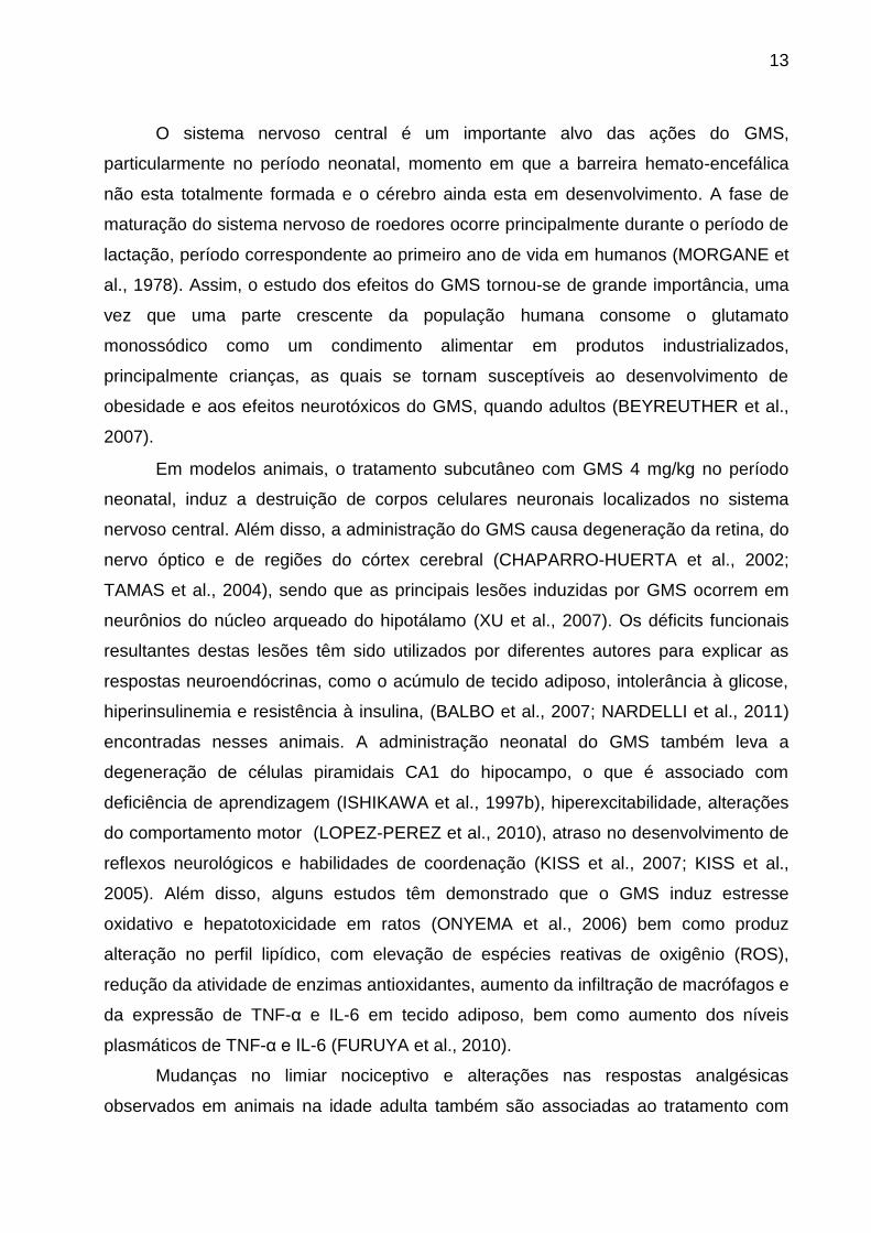
O sistema nervoso central é um importante alvo das ações do GMS,
particularmente no período neonatal, momento em que a barreira hemato-encefálica
não esta totalmente formada e o cérebro ainda esta em desenvolvimento. A fase de
maturação do sistema nervoso de roedores ocorre principalmente durante o período de
lactação, período correspondente ao primeiro ano de vida em humanos (MORGANE et
al., 1978). Assim, o estudo dos efeitos do GMS tornou-se de grande importância, uma
vez que uma parte crescente da população humana consome o glutamato
monossódico como um condimento alimentar em produtos industrializados,
principalmente crianças, as quais se tornam susceptíveis ao desenvolvimento de
obesidade e aos efeitos neurotóxicos do GMS, quando adultos (BEYREUTHER et al.,
2007).
Em modelos animais, o tratamento subcutâneo com GMS 4 mg/kg no período
neonatal, induz a destruição de corpos celulares neuronais localizados no sistema
nervoso central. Além disso, a administração do GMS causa degeneração da retina, do
nervo óptico e de regiões do córtex cerebral (CHAPARRO-HUERTA et al., 2002;
TAMAS et al., 2004), sendo que as principais lesões induzidas por GMS ocorrem em
neurônios do núcleo arqueado do hipotálamo (XU et al., 2007). Os déficits funcionais
resultantes destas lesões têm sido utilizados por diferentes autores para explicar as
respostas neuroendócrinas, como o acúmulo de tecido adiposo, intolerância à glicose,
hiperinsulinemia e resistência à insulina, (BALBO et al., 2007; NARDELLI et al., 2011)
encontradas nesses animais. A administração neonatal do GMS também leva a
degeneração de células piramidais CA1 do hipocampo, o que é associado com
deficiência de aprendizagem (ISHIKAWA et al., 1997b), hiperexcitabilidade, alterações
do comportamento motor (LOPEZ-PEREZ et al., 2010), atraso no desenvolvimento de
reflexos neurológicos e habilidades de coordenação (KISS et al., 2007; KISS et al.,
2005). Além disso, alguns estudos têm demonstrado que o GMS induz estresse
oxidativo e hepatotoxicidade em ratos (ONYEMA et al., 2006) bem como produz
alteração no perfil lipídico, com elevação de espécies reativas de oxigênio (ROS),
redução da atividade de enzimas antioxidantes, aumento da infiltração de macrófagos e
da expressão de TNF-α e IL-6 em tecido adiposo, bem como aumento dos níveis
plasmáticos de TNF-α e IL-6 (FURUYA et al., 2010).
Mudanças no limiar nociceptivo e alterações nas respostas analgésicas
observados em animais na idade adulta também são associadas ao tratamento com
14
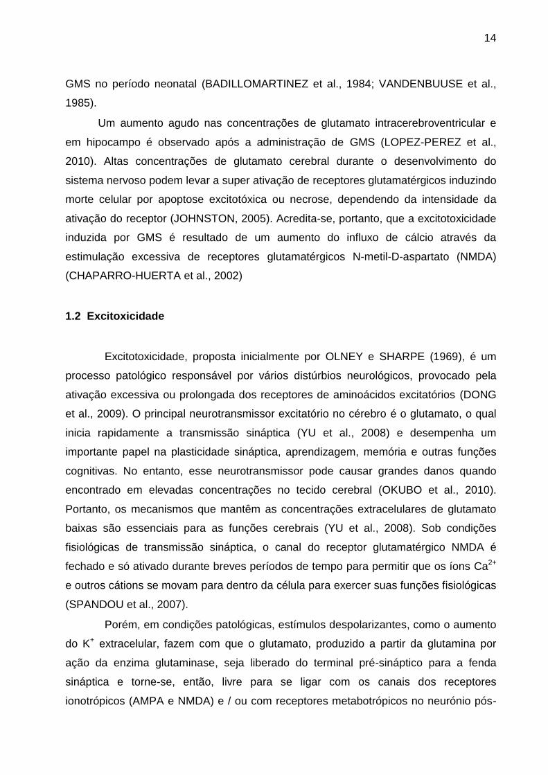
GMS no período neonatal (BADILLOMARTINEZ et al., 1984; VANDENBUUSE et al.,
1985).
Um aumento agudo nas concentrações de glutamato intracerebroventricular e
em hipocampo é observado após a administração de GMS (LOPEZ-PEREZ et al.,
2010). Altas concentrações de glutamato cerebral durante o desenvolvimento do
sistema nervoso podem levar a super ativação de receptores glutamatérgicos induzindo
morte celular por apoptose excitotóxica ou necrose, dependendo da intensidade da
ativação do receptor (JOHNSTON, 2005). Acredita-se, portanto, que a excitotoxicidade
induzida por GMS é resultado de um aumento do influxo de cálcio através da
estimulação excessiva de receptores glutamatérgicos N-metil-D-aspartato (NMDA)
(CHAPARRO-HUERTA et al., 2002)
1.2 Excitoxicidade
Excitotoxicidade, proposta inicialmente por OLNEY e SHARPE (1969), é um
processo patológico responsável por vários distúrbios neurológicos, provocado pela
ativação excessiva ou prolongada dos receptores de aminoácidos excitatórios (DONG
et al., 2009). O principal neurotransmissor excitatório no cérebro é o glutamato, o qual
inicia rapidamente a transmissão sináptica (YU et al., 2008) e desempenha um
importante papel na plasticidade sináptica, aprendizagem, memória e outras funções
cognitivas. No entanto, esse neurotransmissor pode causar grandes danos quando
encontrado em elevadas concentrações no tecido cerebral (OKUBO et al., 2010).
Portanto, os mecanismos que mantêm as concentrações extracelulares de glutamato
baixas são essenciais para as funções cerebrais (YU et al., 2008). Sob condições
fisiológicas de transmissão sináptica, o canal do receptor glutamatérgico NMDA é
fechado e só ativado durante breves períodos de tempo para permitir que os íons Ca2+
e outros cátions se movam para dentro da célula para exercer suas funções fisiológicas
(SPANDOU et al., 2007).
Porém, em condições patológicas, estímulos despolarizantes, como o aumento
do K+ extracelular, fazem com que o glutamato, produzido a partir da glutamina por
ação da enzima glutaminase, seja liberado do terminal pré-sináptico para a fenda
sináptica e torne-se, então, livre para se ligar com os canais dos receptores
ionotrópicos (AMPA e NMDA) e / ou com receptores metabotrópicos no neurónio pós-

15
sináptico. A ligação com os receptores AMPA permite a troca de Na+ e K+, enquanto a
ligação do glutamato com receptores NMDA permite influxo de Ca2+ no neurônio pós-
sináptico. Além disso, a interação do glutamato com os receptores metabotrópicos
provoca a hidrólise do fosfatidilinositol-4,5-bifosfato (PIP2) produzindo o inositol-1,4,5-
trifosfato (IP3), que, em seguida, estimula a liberação de Ca2+ armazenado no retículo
endoplasmático liso (SER). Esses eventos levam a um excesso de Ca2+ dentro da
célula nervosa, o qual estimula o início de vias de sinalização intracelular dependentes
de Ca2+ (CHEN e LIPTON, 2006), desencadeando processos catabólicos que resultam
em lesão neuronal (NDOUNTSE e CHAN, 2009) e que caracterizam a excitotoxicidade
(Figura 2).
Os mecanismos responsáveis pelo aumento dos níveis de glutamato
extracelular incluem maior liberação e menor captação de glutamato (GILGUN-SHERKI
et al., 2002a). Além disso, a homeostase do glutamato extracelular no sistema nervoso
central é regulada pela atividade de transportadores de aminoácidos excitatórios
(EAATs). Os transportadores de glutamato são dependentes do íon sódio e
necessitam, portanto da funcionalidade da enzima Na+, K+ - ATPase para regular as
concentrações intracelulares de sódio e potássio e conduzir a captação do
neurotransmissor (ROSE et al., 2009b). Existem evidências de que os transportadores
de glutamato e Na+, K+ - ATPase são parte dos mesmos complexos macromoleculares
e operam juntamente para regular a neurotransmissão glutamatérgica nos astrócitos e
neurônios (ROSE et al., 2009b; ZHANG et al., 2009). Portanto, alterações na atividade
da Na+, K+ - ATPase podem influenciar diretamente a sinalização do glutamato, a
atividade neural e o comportamento animal.
Além disso, íons Ca2+ são mediadores chave de danos excitotóxicos. Sob
condições fisiológicas estes íons ativam enzimas as quais influenciam uma ampla
variedade de componentes celulares que regulam processos celulares (NICHOLLS e
CHALMERS, 2004), incluindo o crescimento e diferenciação celular e atividade
sináptica. Porém, em eventos excitotóxicos a liberação sináptica excessiva de
glutamato pode levar à desregulação da homeostase do Ca2+ e consequentes danos
neuronais. Portanto, existem mecanismos homeostáticos para manter as
concentrações intracelulares de Ca2+ baixas e espacialmente e temporalmente
localizadas (LIPTON, 2008). Assim, além da Na+, K+ - ATPase, a enzima Ca2+ -
ATPase também desempenha um papel importante nos eventos de excitotoxicidade,

16
uma vez que regula a homeostase do Ca2+, o qual desempenha um papel primordial na
excitotoxicidade induzida por glutamato (TYMIANSKI et al., 1993b)
Além do envolvimento dos receptores glutamatérgicos com a excitotoxicidade e
danos ao sistema nervoso central (SNC), estudos têm demonstrado que a super
ativação destes receptores, especialmente do receptor do NMDA esta envolvida no
desenvolvimento de sensibilidade a dor inflamatória (FUNDYTUS, 2001; LI e
NEUGEBAUER, 2004; PETRENKO et al., 2003)
Figura 2- Sinapse glutamatérgica e excitotoxicidade. Adaptado de (MESSING, 2006)
1.3 Dor
A dor aguda é decorrente da ativação térmica, mecânica ou química de
subconjuntos de neurônios aferentes primários (nociceptores) que transmitem a
informação de dor para a medula espinhal, de onde é retransmitida a níveis supra-
espinhais. Assim, o processo nociceptivo engloba desde eventos de detecção do
estímulo, que pode ter diferentes origens, até a condução deste ao SNC, onde esse
estímulo é processado e respondido (BASBAUM et al., 2009a). Além disso, os
nociceptores aferentes podem ser modulados de forma que eles não só sinalizam a dor
aguda, mas também contribuem para condições de dor persistente, a qual está

17
associada à produção e liberação de múltiplos mediadores inflamatórios, incluindo
neurotransmissores e neuromoduladores (GRIFFIS et al., 2006).
Diversos fatores estão associados com a gênese e a transmissão do estímulo
nociceptivo, entre eles, os neurotransmissores excitatórios, principalmente o glutamato
(JULIUS e BASBAUM, 2001) óxido nítrico, peptídeos, como a substância P
(HARRISON e GEPPETTI, 2001), bradicinina (CALIXTO et al., 2000), prótons,
prostaglandinas e leucotrienos (FERREIRA, 1972). Além disso, alguns mediadores
podem ativar direta ou indiretamente canais iônicos voltagem-dependente e cascatas
de proteínas quinase, levando a alterações na permeabilidade da membrana o que
favorece a transmissão de impulsos elétricos ao longo das fibras (MILLAN, 1999;
PARADA et al., 2003; WOOLF e SALTER, 2000). Assim estes agem em conjunto, não
só para manter a atividade dos nociceptores aferentes primários e sustentar a dor, mas
também para aumentar a sensibilidade nociceptiva de tal forma que estímulos inócuos
produzam dor (JULIUS e BASBAUM, 2001).
A nocicepção envolve diferentes estruturas através da interação entre várias vias
anatômicas e neurofisiológicas. Estudos têm demonstrado que a formação do
hipocampo pode estar envolvida no surgimento da nocicepção (KLAMT e PRADO,
1991; MCKENNA e MELZACK, 1992), bem como a intensidade de estímulos
nociceptivos esta positivamente relacionada com a amplitude do potencial excitatório
pós-sináptico em células piramidais CA1 do hipocampo (WEI et al., 2000). Além disso,
esta estrutura do cérebro é vulnerável a elevadas concentrações de GMS no início da
vida (BEAS-ZARATE et al., 2001), quando estímulos nocivos podem alterar a atividade
neuronal no hipocampo (LORENZ et al., 2008).
Existem vários relatos de que o glutamato provoca hiperalgesia através da
excitação direta de fibras aferentes terminais (ANDERSON e SWANSON, 2000;
GEGELASHVILI et al., 2000). Além disso, a sinalização glutamatérgica pode definir o
início, duração e intensidade dos estímulos nocivos da periferia. Após a comunicação
sináptica, há um aumento na capacidade de resposta dos neurônios a estímulos
subsequentes, conhecido como sensibilização central, processo pelo qual um estado
de hiperexcitabilidade do SNC leva a um maior processamento de mensagens
nociceptivas (WOOLF, 1983).
Na dor aguda, a liberação de glutamato de terminais nociceptores centrais gera
correntes excitatórias pós-sinápticas em outros neurônios do corno dorsal. Isto ocorre
principalmente por meio da ativação de receptores de glutamato ionotrópicos pós-

18
sinápticos (AMPA e Cainato). A soma das correntes excitatórias geradas no neurônio
pós-sináptico resulta na transmissão da mensagem de dor para neurónios de ordem
superior. Nestas condições, os receptores de glutamato NMDA não estão ativados,
porém, em situações mais crônicas, a ativação de fibras C e nociceptores leva a
ativação de canais iônicos voltagem-dependentes, VGCCs. Os VGCCs estão
associados com as proteínas vesiculares Ca2+-dependentes as quais podem levar ao
influxo de Ca2+, despolarização de neurônios pós-sinápticos e um consequente
aumento na liberação de neurotransmissores, como o glutamato, que pode ativar
receptores NMDA.
O aumento do influxo de cálcio pode fortalecer as conexões sinápticas entre os
nociceptores e neurônios de transmissão de dor e ativar uma série de cascatas de
sinalização que irão aumentar a excitabilidade neuronal e facilitar a transmissão de
mensagens de dor para o cérebro (LATREMOLIERE e WOOLF, 2009) (Figura 3). A dor
pode resultar de duas condições diferentes: aumento da capacidade de resposta de
neurônios de transmissão da dor na medula espinhal (sensibilização central), ou
redução dos limites de ativação de nociceptores (sensibilização periférica). Na
sensibilização central, a dor é produzida pela atividade de fibras sensoriais primárias
não-nociceptivas, enquanto a sensibilização periférica é produzida quando
nociceptores terminais ficam expostos a produtos de dano tecidual e inflamação.
Figura 3 – Nocicepção mediada por receptor glutamatérgico NMDA. Adaptado de
(SCHMIDTKO et al., 2010)

19
1.4 Inflamação
A inflamação é uma reação fisiológica complexa e mediada por diferentes
eventos moleculares e celulares. Apesar de possuir caráter essencialmente protetor na
eliminação do agente agressor, em alguns casos o processo inflamatório pode tornar-se
muito intenso prejudicando o próprio organismo (LIBBY, 2007; MOALEM e TRACEY,
2006). Estímulos inflamatórios desencadeiam a síntese e a liberação de substâncias
capazes de causar alterações morfológicas e bioquímicas no local da lesão e nos tecidos
adjacentes. Essas modificações instalam-se gradualmente, moduladas por fatores
celulares específicos e que também caracterizam a fase do processo inflamatório (PAUL,
1998).
Dor e inflamação estão associadas com várias condições fisiopatológicas.
Lesões teciduais podem iniciar respostas inflamatórias acompanhadas pela dor. As
células danificadas liberam o conteúdo intracelular e ativam células do sistema
imunológico levando a produção de mediadores inflamatórios (JULIUS e BASBAUM,
2001). Embora os sinais de dor sejam processados no SNC, mediadores inflamatórios
e citocinas produzidas a partir de células imunes ativadas também podem estimular
nociceptores terminais no tecido periférico e aumentar a sensibilidade à dor (JULIUS e
BASBAUM, 2001). Estes mediadores agem diretamente sobre nociceptores terminais
para ativar e produzir dor ou para sensibilizar terminais periféricos, levando à
hipersensibilidade a estímulos subsequentes (REN e DUBNER, 2010).
As citocinas inflamatórias representam elementos-chave na comunicação entre
as células do sistema imunológico, regulando respostas teciduais à infecção,
inflamação e estresse. Quando encontradas no SNC, as citocinas podem ter duas
origens: os órgãos imunes periféricos e/ou o próprio SNC. Apesar da existência da
barreira hematoencefálica (BHE), citocinas produzidas perifericamente podem entrar no
SNC por difusão passiva nos órgãos circunventriculares, por transporte ativo através da
BHE (BANKS, 2006; BANKS et al., 2001; MAIER, 2003), ou ainda podem ser
sintetizadas por células imunitárias residentes ou periféricas que invadem o SNC
(RANSOHOFF e ENGELHARDT, 2012). Além disso, macrófagos, monócitos e
linfócitos perivasculares podem ser recrutados dentro do SNC para ampliar a resposta
inflamatória (RANSOHOFF et al., 2003).
Concentrações excessivas de glutamato extracelular agem sobre os receptores
glutamatérgicos microgliais e podem contribuir para a propagação dos processos

20
inflamatórios (POCOCK e KETTENMANN, 2007), assim como, mediadores
inflamatórios podem aumentar a liberação de glutamato, podendo causar um círculo
vicioso (Figura 4). Além disso, citocinas como o TNF-α podem potenciar a
excitotoxicidade mediada por glutamato por dois mecanismos complementares:
indiretamente, pela inibição do transporte de glutamato, e diretamente, através do
aumento na expressão de receptores de glutamato ionotrópicos localizados nas
sinapses. A IL-1β (MANDOLESI et al., 2013) e o TNF-α (CARMEN et al., 2009;
TOLOSA et al., 2011) também estão envolvidos na redução na expressão de
transportadores de glutamato durante a neuroinflamação (TAKAKI et al., 2012). Neste
sentido, já foi demonstrado que subunidades NR1 de receptores glutamatérgicos
NMDA são ativadas durante inflamação periférica (PENG et al., 2011), sugerindo um
papel do sistema glutamatérgico na inflamação.
Assim, a regulação dos níveis de glutamato não é apenas crucial no SNC, mas
também a nível periférico, para evitar perturbações na transmissão sensorial. Dessa
forma, a nocicepção pode estar sujeita a múltiplos níveis de controle bioquímico e
farmacológico, envolvendo uma diversidade de tipos de células e mediadores solúveis
(JULIUS e BASBAUM, 2001). Tendo isto em vista, substâncias capazes de neutralizar
vias de sinalização nociceptiva, tanto a nível periférico quanto central podem ser
promissoras no controle da dor.
Figura 4- Interação bidirecional entre mediadores inflamatórios e neurotoxicidade
glutamatérgica. Adaptado de MCNALLY et al. (2008)

21
1.5 Compostos orgânicos de Selênio
O selênio (Se) é um elemento químico não metálico da família dos calcogênios
da tabela periódica o qual foi descoberto pelo químico sueco Jöns Jacob Berzelius em
1817 (COMASSETO, 2010). Este elemento traço é nutricionalmente essencial para
mamíferos, com papéis fisiológicos como, por exemplo, como componente estrutural de
enzimas antioxidantes (RAYMAN, 2000). Portanto, a Organização Mundial de Saúde
(OMS) recomenda uma ingestão diária de 34-35 μg para adultos (FAO/OMS, 2002),
seja através da ingestão de alimentos comuns, de origem animal e vegetal, ou por
suplementação (DUMONT et al., 2006; RAYMAN, 2008). Além disso, a
biodisponibilidade do selênio varia de acordo com a fonte e estado nutricional do
indivíduo, sendo significativamente maior para as formas orgânicas de selênio (KIM e
MAHAN, 2001; YOUNG et al., 1982)
Compostos orgânicos de selênio tem sido bastante estudados, uma vez que
trabalhos já demonstraram as diversas atividades farmacológicas que eles apresentam
(NOGUEIRA et al., 2004b). Além das suas propriedades antioxidantes (BORTOLATTO
et al., 2013b; MEOTTI et al., 2004; MULLER et al., 1984), os compostos de selênio
possuem atividade neuroprotetora (PORCIUNCULA et al., 2001; ROSSATO et al.,
2002), anti-hipertensiva, anticâncer, antiviral, imunossupressora, antimicrobiana,
ansiolítica, antidepressiva e anti-inflamatória (BRUNING et al., 2009; NOGUEIRA et al.,
2003; SAVEGNAGO et al., 2008; ZASSO et al., 2005b). As propriedades
antinociceptivas desses compostos também estão descritas e tem sido amplamente
estudadas demonstrando que compostos orgânicos de selênio podem ser drogas
relevantes para a gestão da dor.
Estudos anteriores realizados em nosso laboratório demonstraram que o
composto orgânico de selênio, disseleneto de difenila (PhSe)2 (Figura 5), um
disseleneto de diarila de síntese simples, possui ampla distribuição nos tecidos
(PRIGOL et al., 2012a) e produz efeitos antinociceptivos, dependentes da dose, em
modelos químicos e térmicos de nocicepção em roedores (DA ROCHA et al., 2013b;
NOGUEIRA et al., 2003; ZASSO et al., 2005b). Além disso, já foi relatado que o
(PhSe)2 reduz a alodínia causada por injeção intraplantar de adjuvante completo de
Freund (CFA), bem como a alodinia induzida pela constrição parcial do nervo ciático,
além de atenuar a hiperalgesia aguda induzida pelo glutamato, bradicinina e
prostaglandina E2 em ratos (JESSE et al., 2008; SAVEGNAGO et al., 2007a).

22
Os mecanismos envolvidos na ação antinociceptiva deste composto incluem
interação com receptores serotoninérgicos, glutamatérgicos e vias nitrérgicas, bem
como sistemas peptidérgicos e vanilóide (SAVEGNAGO et al., 2007c; ZASSO et al.,
2005b). Assim, compostos orgânicos de selênio, como o (PhSe)2, são potenciais
fontes de novas substâncias químicas relevantes para o tratamento da dor e outras
aplicações terapêuticas. Sob este ponto de vista, considerando o uso crescente e
precoce de GMS na dieta da população e a necessidade de busca por drogas capazes
de atenuar os danos resultantes do uso deste aditivo alimentar, o estudo do efeito
antinociceptivo do (PhSe)2 na dor induzida por GMS torna-se de grande importância.
Figura 5– Estrutura química do composto (PhSe)2

23
2. OBJETIVOS
2.1 Objetivo geral
Considerando os aspectos mencionados, o objetivo desse estudo foi avaliar a
ação do composto disseleneto de difenila (PhSe)2 na nocicepção induzida por
glutamato monossódico administrado (MSG) no período neonatal em ratos, bem como
investigar os mecanismos pelos quais o (PhSe)2 age neste modelo.
2.2 Objetivos específicos
Avaliar o efeito antinociceptivo do tratamento com (PhSe)2 na hiperalgesia térmica
e alodínia mecânica induzida por GMS, em ratos;
Avaliar o envolvimento das enzimas Na+ K+- ATPase e Ca2+- ATPase na
nocicepção induzida por GMS e na ação antinociceptiva do (PhSe)2;
Pesquisar se a nocicepção induzida por GMS está acompanhada de um quadro
inflamatório, e avaliar o possível efeito antiinflamatório do (PhSe)2 neste modelo;
Investigar o envolvimento do sistema glutamatérgico na nocicepção induzida por
GMS e na ação antinociceptiva do (PhSe)2;

24
3. Resultados Os resultados que fazem parte dessa dissertação estão apresentados na forma
de um manuscrito. Os itens materiais e métodos, resultados, discussão e referências
bibliográficas do manuscrito estão dispostos de acordo com a recomendação do
periódico científico no qual está submetido.

25
3.1 Manuscrito
Antinociceptive action of diphenyl diselenide in the nociception induced by
neonatal administration of monosodium glutamate in rats
Suzan G. Rosa1; Caroline B. Quines1; Juliana T. Da Rocha1; Cristiani F. Bortolatto1;
Thiago Duarte2; Cristina W. Nogueira1,*
1Laboratório de Síntese, Reatividade e Avaliação Farmacológica e Toxicológica de
Organocalcogênios, Centro de Ciências Naturais e Exatas, Universidade Federal de
Santa Maria, Santa Maria, CEP 97105-900, RS, Brazil
2Programa de Pós-graduação em Farmacologia, Universidade Federal de Santa Maria,
Santa Maria, CEP 97105-900, RS, Brazil
Correspondence should be sent to:
Cristina Nogueira
Departamento de Química, Centro de Ciências Naturais e Exatas, Universidade
Federal de Santa Maria, 97105-900, Santa Maria, RS, Brazil.
Phone: 55-55-3220-8140
FAX: 55-55-3220-8978
E-mail: [email protected]

26
Abstract
Monosodium glutamate (MSG) is a neuroexcitatory amino acid commonly used as
flavoring of foods and its neonatal administration in animals leading to behavioral and
physiological disorders in adulthood, including increased pain sensitivity. However, little
is known about the mechanism of action by which MSG induces nociception. This study
evaluated the effect of diphenyl diselenide (PhSe)2, an organoselenium compound with
pharmacological properties already documented, on nociception induced by MSG.
Newborn Wistar rats received ten subcutaneous injections of MSG at a dose of 4.0 g/kg
or saline, once a day. At the 60th day of life, rats were treated daily with (PhSe)2 (1
mg/kg) or vehicle (canola oil) by intragastric route for 7 days. The behavioral tests
(locomotor activity, hot plate, tail-immersion and mechanical allodynia) were carried out.
In addition, hippocampal ex vivo assays were performed to determine Na+, K+-ATPase
and Ca2+-ATPase activities, cytokines levels and [3H]glutamate uptake. The results
demonstrated that MSG increased nociception in the hot plate test, but not in the tail
immersion test, and in the mechanical allodynia stimulated by Von-Frey Hair. (PhSe)2
decreased all nociceptive behaviors induced by MSG. MSG increased hippocampal
Na+,K+-ATPase and Ca2+-ATPase activities and pro-inflammatory cytokines levels.
MSG decreased the anti-inflammatory cytokine and the [3H]glutamate uptake in
hippocampi of rats and (PhSe)2 protected against these alterations. The results
indicated some mechanisms of action that contribute to nociception induced by MSG in
rats. This study also demonstrated that (PhSe)2 protected against MSG- induced
nociception.
Key words: Monosodium glutamate; excitotoxicity; nociception; selenium; diphenyl
diselenide, ATPases.
27
Introduction
The human diet has changed greatly during the past decades and with this the
introduction of industrialized foods in diet of children widely increased, since, processed
foods are more palatable by the use of food additives to preserve flavor and enhance
taste. One of the most commonly consumed food additives is monosodium glutamate
(MSG), a neuroexcitatory amino acid used as a flavoring agent (MCCABE e ROLLS,
2007).
Earlier studies have demonstrated that MSG has some adverse effects in
humans and experimental animals. In rodents, the administration of high doses of MSG
during early stages of brain development induces destruction of sites in the
hypothalamus (ABE et al., 1990), which provokes neuroendocrine abnormalities. These
abnormalities can result in animal functional and behavioral disorders in adulthood,
including obesity, hyperexcitability, impairment of memory, anxiogenic, depressive-like
behaviors, pain-sensitivity and changes in analgesic responses (BADILLO-MARTINEZ
et al., 1984; COLLISON et al., 2010; LOPEZ-PEREZ et al., 2010; QUINES et al., 2014;
VAN DEN BUUSE et al., 1985).
Even though little is known about the mechanism of action by which MSG
induces nociception, studies have demonstrated that MSG neonatal exposure leads to
degeneration in hippocampal CA1 pyramidal cells (BEAS-ZARATE et al., 2002;
ISHIKAWA et al., 1997a). In addition, previous data indicate that the hippocampal
formation is involved in emergence of nociception (KLAMT e PRADO, 1991; MCKENNA
e MELZACK, 1992) as well as the intensity of nociceptive stimulation is positively
related to amplitude of excitatory postsynaptic potentials in hippocampal CA1 pyramidal
cells (WEI et al., 2000). Thus, the study deals with the involvement of hippocampus in
nociception induced by MSG and compounds effective in blocking nociceptive signaling
pathways implicated in this process are of great importance.
Organoselenium compounds have roused interest due to their biological and
pharmacological activities (NOGUEIRA et al., 2004a). Besides to their antioxidant
properties (BORTOLATTO et al., 2013a; BRUNING et al., 2012), these compounds
have neuroprotective (ABDEL-HAFEZ e ABDEL-WAHAB, 2008), anti-inflammatory and
antinociceptive properties (CHAGAS et al., 2014; MARCONDES SARI et al., 2014). Of
particular importance, diphenyl diselenide (PhSe)2, an organoselenium compound, has
been documented as promising pharmacological agent in a number of experimental

28
models, which showed that this compound has antinociceptive and anti-inflammatory
properties (DA ROCHA et al., 2013a; LUCHESE et al., 2012). The mechanism of action
by which (PhSe)2 elicits antinociceptive action involves the modulation of serotonergic,
nitrergic and glutamatergic systems (SAVEGNAGO et al., 2007b; ZASSO et al., 2005a).
Regarding the pharmacokinetic properties, (PhSe)2 shows a wide tissue distribution
profile (PRIGOL et al., 2012b)
Based on the above considerations, the present study was designed to
investigate the effect of (PhSe)2 in nociception induced by MSG in rats. The possible
mechanisms related to the (PhSe)2 antinociceptive action were also investigated.
Materials and Methods
Animals
Newborn Wistar rats from our own breeding colony were used. The animals
were kept on a 12 h light/dark cycle with lights on at 7:00 a.m., at room temperature (22
± 10C) with free access to water and food. The experiments were performed according
to the guidelines of the Committee on Care and Use of Experimental Animal Resources,
the Federal University of Santa Maria, Brazil. All efforts were made to minimize animal
suffering and to reduce the number of animals used in the experiments.
Drugs
Diphenyl diselenide (PhSe)2 (Fig. 1) was prepared in our laboratory according to
the method described by (PAULMIER, 1986) and the chemical purity (99.9%) was
determined by gas chromatography–mass spectrometry (GC/MS). Analysis of 1H and
13C nuclear magnetic resonance (NMR) spectra showed analytical and spectroscopic
data in full agreement with its assigned structure. (PhSe)2 was diluted in canola oil.
Monosodium glutamate (MSG) was purchased from Sigma–Aldrich (St. Louis,
MO, USA). All other chemicals were of analytical grade and obtained from standard
commercial suppliers.

29
Experimental design
Newborn Wistar rats received ten subcutaneous injections of MSG at a dose of
4.0 g/kg or saline (0.9%) in a similar volume (1 ml/kg), once a day starting at day one
postnatal (KLINGBERG et al., 1987). Pups were weaned at the 21st day of life. At the
60th day of life, the female rats were divided into four groups. MSG and control groups
were treated with (PhSe)2 (1 mg/kg) or vehicle (canola oil, 1ml/kg) by the intragastric
(i.g.) route once a day, for 7 days. Thirty minutes after last treatment, the animals were
submitted to locomotor activity monitor and nociceptive tests: hot plate, tail-immersion
and mechanical allodynia tests. After the behavioral tests, rats were killed by cervical
dislocation and hippocampi, a target structure for neonatal MSG action (BEAS-ZARATE
et al., 2002), were quickly removed for ex vivo assays.
Behavioral tests
Spontaneous locomotor activity
With the purpose of excluding sedative or motor abnormality, spontaneous
locomotor activity of rats was performed in the locomotor activity monitor. The locomotor
activity monitor is a Plexiglas box (45 × 45 × 45 cm) surrounded by a frame consisting
of 32 photocells mounted on opposite walls (16 L × 16 W, spaced 2 cm apart) that
continuously tracks the animal's movement. Animals were placed in the center of the
apparatus and allowed to freely explore the arena during 4 min. Motor activity was
monitored with the Insight® Monitor Activity System and the rat position in the chamber
are detected by breaks of the photocell beams. Number of crossings and rearings,
average velocity (mm/s) and total distance traveled (dm) were recorded.
Hot plate test
The hot-plate test was carried out according to the method described previously
(WOOLFE e MACDONALD, 1944). In this test, the animals were placed in a glass
cylinder on a heated metal plate maintained at 55 ± 1 0C. The latency of nociceptive
responses such as licking or shaking one of the paws or jumping was recorded as the

30
reaction time. In order to avoid damage to the paws of the animals, the time standing on
the plate was limited to 60 s.
Tail-immersion test
The tail-immersion test was conducted as described previously (JANSSEN et
al., 1963). The test was performed by immersing the lower 3.5 cm of the tail into a cup
freshly filled with water from a large constant-temperature (55 ºC) bath until the typical
tail withdrawal response was observed. A 7 s cut-off was imposed.
Mechanical Allodynia test
The mechanical allodynia was measured as described before (BORTALANZA
et al., 2002). Rats were further acclimatized in individual clear Plexiglas boxes
(9 × 7 × 11 cm) on an elevated wire mesh platform to allow access to the ventral surface
of the hind paw. The withdrawal response frequency of the left hind paw was measured
following 10 applications (duration of 1-2 s each) of 2 g von Frey hairs (VFH; Stoelting,
Chicago, IL, USA).
Ex vivo assays
Tissue Preparation
The hippocampus samples of all animals were homogenized in 50 mM Tris/HCl
at pH 7.4. The homogenate was centrifuged at 2.500 g for 10 min at 4 0C to yield a low-
speed supernatant fraction (S1). Freshly prepared S1 was used for the determination of
Na+, K+-ATPase and Ca2+- ATPase activities and for the measurement of cytokine
levels.
Na+, K+-ATPase activity
The Na+, K+-ATPase activity was measured according to (WYSE et al., 2000).
An aliquot of S1 was added to the reaction mixture for Na+, K+-ATPase activity assay
containing 3 mM MgCl, 125 mM NaCl, 20 mM KCl, and 50 mM Tris-HCl, pH 7.4, in a
final volume of 500 μl. The reaction was initiated by the addition of ATP to a final

31
concentration of 3.0 mM. Control samples were carried out under the same conditions
with the addition of 0.1 mM ouabain. The samples were incubated for 30 min at 37 0C,
and the incubation was stopped by adding trichloroacetic acid solution (10% TCA) with
10 mM HgCl2. The Na+, K+-ATPase activity was calculated by the difference between
the two assays. Released inorganic phosphate (Pi) was measured by the method of
FISKE e SUBBAROW (1925). The enzymatic activity was expressed as nmol Pi/min/mg
protein.
Ca2+- ATPase activity
The Ca2+- ATPase activity was measured as previously described by ROHN et
al. (1993) with minor modifications (TREVISAN et al., 2009). Briefly, the assay medium
consisted of 30 mM Tris-HCl buffer (pH 7.4), 0.1 mM EGTA, 3 mM MgCl2 and 100 μg of
protein in the presence or absence of 0.4 mM CaCl2, in a final volume of 200 μl. The
reaction was initiated by the addition of ATP to a final concentration of 3.0 mM. After
incubation for 60 min at 37 0C, the reaction was stopped by adding trichloroacetic acid
solution (50% TCA). The released inorganic phosphate (Pi) was measured by the
method of FISKE e SUBBAROW (1925) . The Ca2+- ATPase activity was calculated by
subtracting the activity measured in the presence of Ca2+ from that determined in the
absence of Ca2+ ( no added Ca2+ plus 0.1 mM EGTA). The enzymatic activity was
expressed as nmol Pi/min/mg protein.
Determination of cytokines
The levels of IL-1, IL-6, TNFα, INFγ and IL-10 in the hippocampus was measured
using commercial ELISA kits for rat as described by the manufacturer
(eBIOSCIENCE®, San Diego, USA). The results are expressed in pg/mg of protein.
[3H]Glutamate uptake assay
The [3H] glutamate uptake assay was carried out in slices (0.4 mm) of
hippocampus obtained using a McIllwain chopper. The glutamate uptake was performed
according to the method described by THOMAZI et al. (2004). The slices were
transferred to multiwell dishes and washed with 1.0 ml Hank's buffered salt solution

32
(HBSS). After 10 min of pre-incubation, the uptake assay was performed by adding 13.3
μM [3H] glutamate in 300 μl HBSS at 37 0C. Incubation was terminated after 5 min by
three ice-cold washes with 1 ml HBSS immediately followed by the addition of 0.5 M
NaOH, which was kept overnight. An aliquot of 10 μl was removed to protein
determination. Unspecific uptake was measured using the same protocol described
above, with differences in the temperature (4 0C) and medium composition (choline
chloride instead of sodium chloride). Na+-dependent uptake was considered as the
difference between the total uptake and the unspecific uptake. Both uptakes were
performed in triplicate. Incorporated radioactivity was measured using a liquid
scintillation counter. Results were expressed as pmol of [3H] glutamate uptake /mg
protein/min.
Protein determination
The protein concentration was measured by the Coomassie blue method
according to BRADFORD (1976) using bovine serum albumin (1 mg/ml) as standard.
Statistical Analysis
The normality of data was analyzed using a D’Agostino and Pearson omnibus
normality test. Data were analyzed by two-way analysis of variance (ANOVA) followed
by the Newman–Keuls test. All data of experiments were expressed as means ± SEM.
Probability p values less than 0.05 (p < 0.05) were considered as statistically significant.
Results
Behavioral tests
Spontaneous locomotor activity
Neither the injection of MSG nor (PhSe)2 treatment altered the number of
crossings and rearings, velocity and total distance traveled (p > 0.05) by rats in the
locomotor activity monitor (Table 1).

33
Hot-plate test
The two-way ANOVA of hot-plate data revealed a significant MSG × (PhSe)2
interaction (F1,27=23.2061, p<0.001). The administration of MSG to rats decreased the
response latency to thermal stimuli as compared to the control group (p<0.001). (PhSe)2
caused a significant blocked of thermal hyperalgesia induced by MSG in rats (p<0.001)
(Fig. 2A)
Tail immersion test
The results of Fig. 2B show a significant main effect of (PhSe)2 in the tail
immersion test (F1,25=14.0488, p<0.001). Although the subcutaneous injection of MSG
did not change the tail-immersion response latency, the animals exposed to MSG and
treated with (PhSe)2 had an increase in the tail withdrawal response compared to the
control group (p<0.01).
Mechanical allodynia test
The two-way ANOVA of mechanical allodynia data demonstrated a significant
MSG × (PhSe)2 interaction (F1,24= 14.0713, p<0.001). Post hoc analyses indicated a
significant increase in the mechanical sensitivity on the left hind paws of rats
administered with MSG when compared to those of the control (p<0.001). Treatment
with (PhSe)2 was effective to decrease the mechanical allodynia induced by MSG
injection (p<0.001) (Fig. 3)
Ex vivo assays
Na+, K+-ATPase activity
Two-way ANOVA of hippocampal Na+, K+-ATPase activity revealed a
significant MSG X (PhSe)2 interaction (F1,16=5.440, p<0.05). Post hoc comparisons
demonstrated an increase in Na+, K+-ATPase activity induce by MSG administration
when compared to with the control group (p<0.05). The (PhSe)2 treatment totally
34
protected against alterations in Na+, K+-ATPase activity induced by MSG injections
(p<0.01). (Fig. 4)
Ca2+-ATPase activity
The two-way ANOVA of Ca2+-ATPase activity data revealed a significant MSG X
(PhSe)2 interaction (F1,18= 5.009, p<0.05) (Fig.5). Post hoc analysis showed a
significant stimulation of Ca2++-ATPase activity in hippocampi of rats exposed to MSG
(p<0.05) when compared to that of the control group. (PhSe)2 treatment was effective
against the increase in Ca2+-ATPase activity caused by MSG (p<0.05).
Determination of cytokines
The two-way ANOVA of the pro-inflammatory cytokine levels demonstrated a
significant MSG X (PhSe)2 interaction in IL-1β, IL-6, TNFα and INFγ levels (F1,12=9.584,
p<0.01; F1,11=7.780, p<0.05; F1,12=5.475, p<0.05; F1,12=10.622, p<0.01, respectively).
Post hoc comparisons revealed an increase at all the pro-inflammatory cytokines levels
in hippocampus of animals that received MSG (p<0.001) when compared to the control.
The treatment with (PhSe)2 was effective in protecting against the increased in
cytokines levels induce by MSG (p<0.01). In addition, two-way ANOVA demonstrated a
main effect of MSG in IL-10 levels (F1,12= 7.411, p<0.05). The MSG administrations
significantly decreased IL-10 levels (p<0.05) when compared to the control group. The
treatment with (PhSe)2 resulted in a tendency (but not statistically significant) to
increase cytokine anti-inflammatory (IL-10) levels in rats MSG-treated (p=0.07) (Fig.6)
[3H]Glutamate uptake assay
The Two-way ANOVA of these data revealed a significant MSG X (PhSe)2
interaction (F1,8=35.6137, p<0.001). Post hoc analyses indicated a significant decrease
in [3H] glutamate uptake by MSG and (PhSe)2 groups when compared to the control
group (p<0.05). The (PhSe)2 treatment was effective to protect against the decrease in
[3H] glutamate uptake levels in hippocampus slices induced by MSG group (p<0.01)
(Fig.7)

35
Discussion
In the present study, we investigated the effect of (PhSe)2 in nociception induced
by neonatal administration of MSG in rats using a behavioral tests battery. The
mechanisms of action by which (PhSe)2 attenuated MSG-induced nociception were
evaluated in this study. Our results demonstrated that the MSG administration to rats
reduced the thermal withdrawal latency in the hot plate test, but not in the tail immersion
test, and increased the response frequency of VFH stimulation.
The tail-immersion and hot-plate tests have been widely used as experimental
models to measure nociception, especially for the screening of analgesic drugs
(DEWEY et al., 1969). Tail-immersion is regarded as a spinal reflex, but the mechanism
of response could also involve higher brain structures (JENSEN e YAKSH, 1986), while
the hot-plate test produces two kinds of behavioral response, which are paw licking and
jumping. Both of these are considered to be supraspinally integrated responses
(CHAPMAN et al., 1985). In addition, mechanical hypersensitivity is stimulated by
traditional methods such as Von-Frey hairs. Mechanical allodynia is defined as a pain
sensation generated by physiological stimulation of low-threshold sensitive and occurs
due to changes of central processing of impulse activity in primary afferent neurons
which feed into central nociceptive pathways (LAMBERT et al., 2009). Considering that
MSG treatment altered mechanical allodynia and the withdrawal latency in the hot plate,
but did not change the response latency in tail immersion test, we can suppose that
nociception induced by MSG involves supraspinal reflexes rather than spinal ones.
The hippocampus contributes to several major functions of the brain, including
learning and memory, energy-intake regulation, reward related mechanisms and pain
(DAVIDSON et al., 2007; ERFANPARAST et al., 2010; KARAMI et al., 2002; KENNEY
e GOULD, 2008; KHANNA et al., 2004). This brain structure is vulnerable to exposure
to high concentrations of MSG early in life (BEAS-ZARATE et al., 2001), when
excessive activation of glutamate receptors induces both cell death and changes in the
cytoarchitecture of the surviving pyramidal neurons of the hippocampal CA1 field
(BEAS-ZARATE et al., 2002). In this study, we investigated the possible alterations
caused by MSG in hippocampus since this brain structure plays an important role in
nociception (CECCARELLI et al., 1999; SCHNEIDER et al., 2001; WEI et al., 2000) and
hippocampal neuronal activity can be modulated by noxious stimuli (LORENZ et al.,
2008).

36
Glutamate is a critical neurotransmitter for excitatory synaptic transmission and
for the generation and maintenance of pain hypersensitivity via activation of glutamate
receptors (BASBAUM et al., 2009b). The mechanisms responsible for the elevation of
extracellular glutamate levels include enhanced release of glutamate and the reduction
of glutamate uptake (GILGUN-SHERKI et al., 2002b). Previous findings demonstrated
that the neurotoxic effects of MSG treatment early in life induce nociceptive alterations
of these animals in adulthood (BADILLO-MARTINEZ et al., 1984; VAN DEN BUUSE et
al., 1985). Our results for the first time showed a decrease in [3H]glutamate uptake in
hippocampi of rats administered with MSG in the early life, which could account for the
accumulation of glutamate in the synaptic cleft and consequently cause excitotoxicity.
Assuming this hypothesis, the increase of glutamate in the synaptic cleft could be
related to the decrease of nociception threshold caused by MSG in rats. In addition,
(PhSe)2 treatment protected against the decrease of [3H]glutamate uptake caused by
MSG, suggesting that regulation of glutamate uptake contributes to antinociceptive
action of this compound. Accordingly, an interaction with the glutamatergic system is
one of mechanisms of action by which (PhSe)2 has antinociceptive action in acute
models of nociception (SAVEGNAGO et al., 2007d).
The increase of extracellular glutamate involves sustained elevations of
intracellular calcium levels through glutamate transporters, leading to an imbalance of
sodium ions across plasma membranes (GILGUN-SHERKI et al., 2002b). Glutamate
transporters are sodium-dependent proteins that putatively rely indirectly on Na+, K+-
ATPase to generate ion gradients that drive transmitter uptake (ROSE et al., 2009a).
This enzyme is present at high concentrations in the brain and other nervous tissue,
where it plays several roles in the maintenance of the electrochemical gradient across
the plasma membrane underlying resting and action potentials besides modulate the
neurotransmitter release and uptake (STAHL e HARRIS, 1986). Previous studies have
demonstrated that glutamate transporters and Na+, K+- ATPase are part of the same
macromolecular complexes and operate as a functional unit to regulate glutamatergic
neurotransmission in astrocytes and neurons (ROSE et al., 2009a; ZHANG et al.,
2009). As a consequence, alterations in Na+, K+- ATPase activity directly affects
neurotransmitter signaling, neural activity and animal behavior.
In addition to Na+, K+- ATPase activity be closely related to neuronal
excitotoxicity through its function in maintaining the ionic equilibrium cell, Ca2+- ATPase
plays an important role also in excitotoxic events. Intracellular Ca2+ signaling is
37
fundamental in neuronal functioning and disruption of Ca2+ homeostasis plays a primary
role in the glutamate evoked excitotoxicity (TYMIANSKI et al., 1993a). In this study,
neonatal MSG treatment stimulated the activities of Na+, K+- ATPase and Ca2+ ATPase.
These data suggest a cellular ionic imbalance that corroborates with the decrease of
glutamate uptake induced by MSG, resulting in an excitotoxic event. The stimulation of
both ATPases caused by MSG may compensate the decrease of glutamate uptake.
Moreover, (PhSe)2 protected against the stimulation of Na+, K+- ATPase and Ca2+
ATPase activities. This result suggests that (PhSe)2 antinociceptive action may be
related to the cellular ionic concentration maintenance, protecting against excitotoxicity
caused by MSG.
Additionally, the current study demonstrated that MSG increased pro-
inflammatory cytokine levels such as TNF-α, IL-1β, IL-6 and INF-γ and decreased IL-10
levels, an anti-inflammatory cytokine. Cytokines are barely detectable in the CNS under
physiological conditions, but they become rapidly upregulated by pathological events
like neuroinflammation and excitotoxicity (MINAMI et al., 1991). Studies indicate that
pro-inflammatory cytokines, particularly IL-1β, increase neuronal excitability
(BERNARDINO et al., 2005; VEZZANI et al., 1999). Considering that IL-1β can also
inhibit glutamate uptake in astrocytes (HU et al., 2000) it is plausible to propose that the
increase of pro-inflammatory cytokines results in elevated extracellular glutamate levels.
Moreover, TNF-α, IL-1β, IL-6 and INF-γ can also release pro-inflammatory mediators
such as: nitric oxide, bradykinin, histamine and/or substance P at the site of the
inflammatory process (MORIOKA et al., 2002; O'SHAUGHNESSY et al., 2006),
contributing to the emergence and maintenance of pain.
Our results demonstrated that the administration of (PhSe)2 had an anti-
inflammatory action, characterized by the reduction of pro-inflammatory cytokine levels
(TNF-α, IL-1β, IL-6 and INF-γ) and a tendency (but not statistically significant) to
increase the anti-inflammatory cytokine (IL-10) in rats treated with MSG. This is in
agreement with previous studies of our research group that demonstrated a protection
of (PhSe)2 against the increase of pro-inflammatory cytokines in other experimental
model (LUCHESE et al., 2012). Other important consideration is that the effect of
(PhSe)2 in decreasing IL-1 β levels induced by MSG can be one of the mechanisms by
which this compound normalized the glutamate uptake in rats treated with MSG. This
hypothesis can be supported by the study of HU et al. (2000) that demonstrated a link
between IL-1β levels and the inhibition of glutamate uptake. These findings suggest that

38
the anti-inflammatory action of (PhSe)2 contributes to its antinociceptive property
induced by the neonatal administration of MSG in rats.
In conclusion, the results of the present study demonstrated that the
administration of MSG induced alterations in [3H] glutamate uptake, Na+, K+-ATPase
and Ca2+- ATPase activities, and in the levels of cytokines in hippocampus. These
results contribute to clarify the mechanisms of action involved in nociception induced by
MSG, suggesting targets for treatment of this worrying pathology since the use of MSG
as a food additive is wide spreading all over the world. In addition, this study
demonstrated the antinociceptive action of (PhSe)2 after neonatal injections of MSG in
rats. (PhSe)2 was effective against excitotoxicity and neuroinflammation associated to
the administration of MSG in rats, indicating a new role of (PhSe)2 as an antinociceptive
compound. Considering the results demonstrated in this study the concern about the
consumption of MSG should increase. Nevertheless, further studies are needed to
better understand the toxicological mechanisms by which MSG-induced behavioral
alterations in rats.
Acknowledgements
The financial support by the Universidade Federal de Santa Maria (UFSM),
Coordenação de Aperfeiçoamento de Pessoal de Nível Superior (CAPES), Conselho
Nacional de Desenvolvimento Científico e Tecnológico (CNPq), and Fundação de
Amparo à Pesquisa do Estado do Rio Grande do Sul (FAPERGS is gratefully
acknowledged. C.W.N. is recipient of CNPq fellowship. J.T.R. is recipient of
FAPERGS/CAPES fellowship SPI Process # 2793-25.51/12-4.
Conflict of interest
The authors declare they have no conflicts of interest to disclose.

39
References
Abdel-Hafez, A.A., Abdel-Wahab, B.A., 2008. 5-(4-Chlorophenyl)-5,6-dihydro-1,3-
oxazepin-7(4H)-one derivatives as lipophilic cyclic analogues of baclofen: design,
synthesis, and neuropharmacological evaluation. Bioorganic & medicinal chemistry 16,
7983-7991.
Abe, M., Saito, M., Shimazu, T., 1990. Neuropeptide-Y in the Specific Hypothalamic
Nuclei of Rats Treated Neonatally with Monosodium Glutamate. Brain Res Bull 24, 289-
291.
Badillo-Martinez, D., Nicotera, N., Bodnar, R.J., 1984. Onset of pain threshold changes
induced by neonatal monosodium glutamate. Int J Neurosci 24, 275-279.
Basbaum, A.I., Bautista, D.M., Scherrer, G., Julius, D., 2009. Cellular and Molecular
Mechanisms of Pain. Cell 139, 267-284.
Beas-Zarate, C., Perez-Vega, M.I., Gonzalez-Burgos, I., 2002. Neonatal exposure to
monosodium L-glutamate induces loss of neurons and cytoarchitectural alterations in
hippocampal CA1 pyramidal neurons of adult rats. Brain Res 952, 275-281.
Beas-Zarate, C., Rivera-Huizar, S.V., Martinez-Contreras, A., Feria-Velasco, A.,
Armendariz-Borunda, J., 2001. Changes in NMDA-receptor gene expression are
associated with neurotoxicity induced neonatally by glutamate in the rat brain.
Neurochem Int 39, 1-10.
Bernardino, L., Xapelli, S., Silva, A.P., Jakobsen, B., Poulsen, F.R., Oliveira, C.R.,
Vezzani, A., Malva, J.O., Zimmer, J., 2005. Modulator effects of interleukin-1beta and
tumor necrosis factor-alpha on AMPA-induced excitotoxicity in mouse organotypic
hippocampal slice cultures. The Journal of neuroscience : the official journal of the
Society for Neuroscience 25, 6734-6744.
Bortalanza, L.B., Ferreira, J., Hess, S.C., Delle Monache, F., Yunes, R.A., Calixto, J.B.,
2002. Anti-allodynic action of the tormentic acid, a triterpene isolated from plant, against
neuropathic and inflammatory persistent pain in mice. European journal of
pharmacology 453, 203-208.
Bortolatto, C.F., Chagas, P.M., Wilhelm, E.A., Zeni, G., Nogueira, C.W., 2013. 2,2'-
dithienyl diselenide, an organoselenium compound, elicits antioxidant action and inhibits
monoamine oxidase activity in vitro. Journal of enzyme inhibition and medicinal
chemistry 28, 677-684.

40
Bradford, M.M., 1976. Rapid and Sensitive Method for Quantitation of Microgram
Quantities of Protein Utilizing Principle of Protein-Dye Binding. Anal Biochem 72, 248-
254.
Bruning, C.A., Prigol, M., Luchese, C., Jesse, C.R., Duarte, M.M., Roman, S.S.,
Nogueira, C.W., 2012. Protective effect of diphenyl diselenide on ischemia and
reperfusion-induced cerebral injury: involvement of oxidative stress and pro-
inflammatory cytokines. Neurochemical research 37, 2249-2258.
Ceccarelli, I., Scaramuzzino, A., Aloisi, A.M., 1999. Effects of formalin pain on
hippocampal c-Fos expression in male and female rats. Pharmacol Biochem Be 64,
797-802.
Chagas, P.M., Rosa, S.G., Sari, M.H., Oliveira, C.E., Canto, R.F., da Luz, S.C., Braga,
A.L., Nogueira, C.W., 2014. Evaluation of the pharmacological properties of salicylic
acid-derivative organoselenium: 2-hydroxy-5-selenocyanatobenzoic acid as an anti-
inflammatory and antinociceptive compound. Pharmacology, biochemistry, and behavior
118, 87-95.
Chapman, C.R., Casey, K.L., Dubner, R., Foley, K.M., Gracely, R.H., Reading, A.E.,
1985. Pain measurement: an overview. Pain 22, 1-31.
Collison, K.S., Makhoul, N.J., Inglis, A., Al-Johi, M., Zaidi, M.Z., Maqbool, Z., Saleh,
S.M., Bakheet, R., Mondreal, R., Al-Rabiah, R., Shoukri, M., Milgram, N.W., Al-
Mohanna, F.A., 2010. Dietary trans-fat combined with monosodium glutamate induces
dyslipidemia and impairs spatial memory. Physiol Behav 99, 334-342.
da Rocha, J.T., Pinton, S., Gai, B.M., Nogueira, C.W., 2013. Diphenyl diselenide
reduces mechanical and thermal nociceptive behavioral responses after unilateral
intrastriatal administration of 6-hydroxydopamine in rats. Biological trace element
research 154, 372-378.
Davidson, T.L., Kanoski, S.E., Schier, L.A., Clegg, D.J., Benoit, S.C., 2007. A potential
role for the hippocampus in energy intake and body weight regulation. Curr Opin
Pharmacol 7, 613-616.
Dewey, W.L., Snyder, J.W., Harris, L.S., Howes, J.F., 1969. The effect of narcotics and
narcotic antagonists on the tail-flick response in spinal mice. The Journal of pharmacy
and pharmacology 21, 548-550.
Erfanparast, A., Tamaddonfard, E., Farshid, A.A., Khalilzadeh, E., 2010. Effect of
microinjection of histamine into the dorsal hippocampus on the orofacial formalin-
induced pain in rats. Eur J Pharmacol 627, 119-123.

41
Fiske, C.H., Subbarow, Y., 1925. The calorimetric determination of phosphorus. Biol
Chem 66, 375-381.
Gilgun-Sherki, Y., Rosenbaum, Z., Melamed, E., Offen, D., 2002. Antioxidant therapy in
acute central nervous system injury: current state. Pharmacological reviews 54, 271-
284.
Hu, S., Sheng, W.S., Ehrlich, L.C., Peterson, P.K., Chao, C.C., 2000. Cytokine effects
on glutamate uptake by human astrocytes. Neuroimmunomodulation 7, 153-159.
Ishikawa, K., Kubo, T., Shibanoki, S., Matsumoto, A., Hata, H., Asai, S., 1997.
Hippocampal degeneration inducing impairment of learning in rats: model of dementia?
Behavioural brain research 83, 39-44.
Janssen, P.A., Niemegeers, C.J., Dony, J.G., 1963. The inhibitory effect of fentanyl and
other morphine-like analgesics on the warm water induced tail withdrawl reflex in rats.
Arzneimittel-Forschung 13, 502-507.
Jensen, T.S., Yaksh, T.L., 1986. Examination of spinal monoamine receptors through
which brainstem opiate-sensitive systems act in the rat. Brain research 363, 114-127.
Karami, M., Zarrindast, M.R., Sepehri, H., Sahraei, H., 2002. Role of nitric oxide in the
rat hippocampal CA1 area on morphine-induced conditioned place preference. Eur J
Pharmacol 449, 113-119.
Kenney, J.W., Gould, T.J., 2008. Modulation of hippocampus-dependent learning and
synaptic plasticity by nicotine. Mol Neurobiol 38, 101-121.
Khanna, S., Chang, L.S., Jiang, F.L., Koh, H.C., 2004. Nociception-driven decreased
induction of Fos protein in ventral hippocampus field CA1 of the rat. Brain Res 1004,
167-176.
Klamt, J.G., Prado, W.A., 1991. Antinociception and Behavioral-Changes Induced by
Carbachol Microinjected into Identified Sites of the Rat-Brain. Brain Res 549, 9-18.
Klingberg, H., Brankack, J., Klingberg, F., 1987. Long-term effects on behaviour after
postnatal treatment with monosodium-L-glutamate. Biomedica biochimica acta 46, 705-
711.
Lambert, G.A., Mallos, G., Zagami, A.S., 2009. Von Frey's hairs--a review of their
technology and use--a novel automated von Frey device for improved testing for
hyperalgesia. Journal of neuroscience methods 177, 420-426.
Lopez-Perez, S.J., Urena-Guerrero, M.E., Morales-Villagran, A., 2010. Monosodium
glutamate neonatal treatment as a seizure and excitotoxic model. Brain Res 1317, 246-
256.

42
Lorenz, I.H., Egger, K., Schubert, H., Schnurer, C., Tiefenthaler, W., Hohlrieder, M.,
Schocke, M.F., Kremser, C., Esterhammer, R., Ischebeck, A., Moser, P.L., Kolbitsch,
C., 2008. Lornoxicam characteristically modulates cerebral pain-processing in human
volunteers: a functional magnetic resonance imaging study. Brit J Anaesth 100, 827-
833.
Luchese, C., Prigol, M., Duarte, M.M., Nogueira, C.W., 2012. Diphenyl diselenide
reduces inflammation in the mouse model of pleurisy induced by carrageenan:
reduction of pro-inflammatory markers and reactive species levels. Inflammation
research : official journal of the European Histamine Research Society ... [et al.] 61,
1117-1124.
Marcondes Sari, M.H., Guerra Souza, A.C., Goncalves Rosa, S., Souza, D., Dorneles
Rodrigues, O.E., Wayne Nogueira, C., 2014. Contribution of dopaminergic and
adenosinergic systems in the antinociceptive effect of p-chloro-selenosteroid. European
journal of pharmacology 725, 79-86.
McCabe, C., Rolls, E.T., 2007. Umami: a delicious flavor formed by convergence of
taste and olfactory pathways in the human brain. The European journal of neuroscience
25, 1855-1864.
Mckenna, J.E., Melzack, R., 1992. Analgesia Produced by Lidocaine Microinjection into
the Dentate Gyrus. Pain 49, 105-112.
Minami, M., Kuraishi, Y., Satoh, M., 1991. Effects of kainic acid on messenger RNA
levels of IL-1 beta, IL-6, TNF alpha and LIF in the rat brain. Biochemical and biophysical
research communications 176, 593-598.
Morioka, N., Inoue, A., Hanada, T., Kumagai, K., Takeda, K., Ikoma, K., Hide, I.,
Tamura, Y., Shiomi, H., Dohi, T., Nakata, Y., 2002. Nitric oxide synergistically
potentiates interleukin-1 beta-induced increase of cyclooxygenase-2 mRNA levels,
resulting in the facilitation of substance P release from primary afferent neurons:
involvement of cGMP-independent mechanisms. Neuropharmacology 43, 868-876.
Nogueira, C.W., Zeni, G., Rocha, J.B., 2004. Organoselenium and organotellurium
compounds: toxicology and pharmacology. Chemical reviews 104, 6255-6285.
O'Shaughnessy, M.C., Vetsika, E.K., Inglis, J.J., Carleson, J., Haigh, R., Kidd, B.L.,
Winyard, P.G., 2006. The effect of substance P on nitric oxide release in a rheumatoid
arthritis model. Inflammation research : official journal of the European Histamine
Research Society ... [et al.] 55, 236-240.

43
Paulmier, C., 1986. Selenoorganic functional groups. Selenium reagents and
intermediates in organic synthesis 1, 25-51.
Prigol, M., Bruning, C.A., Martini, F., Nogueira, C.W., 2012. Comparative excretion and
tissue distribution of selenium in mice and rats following treatment with diphenyl
diselenide. Biological trace element research 150, 272-277.
Quines, C.B., Rosa, S.G., Da Rocha, J.T., Gai, B.M., Bortolatto, C.F., Duarte, M.M.M.F.,
Nogueira, C.W., 2014. Monosodium glutamate, a food additive, induces depressive-like
and anxiogenic-like behaviors in young Rats. Life Sci 107, 27-31.
Rohn, T.T., Hinds, T.R., Vincenzi, F.F., 1993. Ion transport ATPases as targets for free
radical damage. Protection by an aminosteroid of the Ca2+ pump ATPase and Na+/K+
pump ATPase of human red blood cell membranes. Biochemical pharmacology 46,
525-534.
Rose, E.M., Koo, J.C., Antflick, J.E., Ahmed, S.M., Angers, S., Hampson, D.R., 2009.
Glutamate transporter coupling to Na,K-ATPase. The Journal of neuroscience : the
official journal of the Society for Neuroscience 29, 8143-8155.
Savegnago, L., Pinto, L.G., Jesse, C.R., Alves, D., Rocha, J.B., Nogueira, C.W., Zeni,
G., 2007a. Antinociceptive properties of diphenyl diselenide: evidences for the
mechanism of action. European journal of pharmacology 555, 129-138.
Savegnago, L., Pinto, L.G., Jesse, C.R., Rocha, J.B., Nogueira, C.W., Zeni, G., 2007b.
Spinal mechanisms of antinociceptive action caused by diphenyl diselenide. Brain
research 1162, 32-37.
Schneider, F., Habel, U., Holthusen, H., Kessler, C., Posse, S., Muller-Gartner, H.W.,
Arndt, J.O., 2001. Subjective ratings of pain correlate with subcortical-limbic blood flow:
An fMRI study. Neuropsychobiology 43, 175-185.
Stahl, W.L., Harris, W.E., 1986. Na+,K+-ATPase: structure, function, and interactions
with drugs. Advances in neurology 44, 681-693.
Thomazi, A.P., Godinho, G.F., Rodrigues, J.M., Schwalm, F.D., Frizzo, M.E., Moriguchi,
E., Souza, D.O., Wofchuk, S.T., 2004. Ontogenetic profile of glutamate uptake in brain
structures slices from rats: sensitivity to guanosine. Mechanisms of ageing and
development 125, 475-481.
Trevisan, G., Maldaner, G., Velloso, N.A., Sant'Anna Gda, S., Ilha, V., Velho Gewehr
Cde, C., Rubin, M.A., Morel, A.F., Ferreira, J., 2009. Antinociceptive effects of 14-
membered cyclopeptide alkaloids. Journal of natural products 72, 608-612.

44
Tymianski, M., Charlton, M.P., Carlen, P.L., Tator, C.H., 1993. Source Specificity of
Early Calcium Neurotoxicity in Cultured Embryonic Spinal Neurons. Journal of
Neuroscience 13, 2085-2104.
van den Buuse, M., Versteeg, D.H., de Jong, W., 1985. Effects of neonatal treatment
with monosodium-glutamate in spontaneously hypertensive rats. Brain research 351,
135-138.
Vezzani, A., Conti, M., De Luigi, A., Ravizza, T., Moneta, D., Marchesi, F., De Simoni,
M.G., 1999. Interleukin-1beta immunoreactivity and microglia are enhanced in the rat
hippocampus by focal kainate application: functional evidence for enhancement of
electrographic seizures. The Journal of neuroscience : the official journal of the Society
for Neuroscience 19, 5054-5065.
Wei, F., Xu, Z.C., Qu, Z., Milbrandt, J., Zhuo, M., 2000. Role of EGR1 in hippocampal
synaptic enhancement induced by tetanic stimulation and amputation. J Cell Biol 149,
1325-1333.
Woolfe, G., MacDonald, A.D., 1944. The evaluation of the analgesic
action of pethidine hydrochloride. J Pharmacol Exp Ther 80, 300-307.
Wyse, A.T., Streck, E.L., Barros, S.V., Brusque, A.M., Zugno, A.I., Wajner, M., 2000.
Methylmalonate administration decreases Na+,K+-ATPase activity in cerebral cortex of
rats. Neuroreport 11, 2331-2334.
Zasso, F.B., Goncales, C.E., Jung, E.A., Araldi, D., Zeni, G., Rocha, J.B., Nogueira,
C.W., 2005. On the mechanisms involved in antinociception induced by diphenyl
diselenide. Environmental toxicology and pharmacology 19, 283-289.
Zhang, D.W., Hou, Q.M., Wang, M., Lin, A., Jarzylo, L., Navis, A., Raissi, A., Liu, F.,
Man, H.Y., 2009. Na,K-ATPase Activity Regulates AMPA Receptor Turnover through
Proteasome-Mediated Proteolysis. J Neurosci 29, 4498-4511.

45
Figure Captions
Fig. 1. Chemical structure of diphenyl diselenide (PhSe)2.
Fig.2. Effect of subcutaneous MSG-injection and intragastric treatment with (PhSe)2 on
the response latency to thermal stimuli in the hot plate test (A) and the tail immersion
test (B). Data are reported as the means±SEM for seven to nine animals per group.
**p<0.01, ***p<0.001 when compared to the control; ##p<0.01, ###p<0.001 when
compared to the MSG group (two-way ANOVA followed by Newman–Keuls test).
Fig.3. Effect of subcutaneous MSG-injection and intragastric treatment with (PhSe)2 on
the percent response to mechanical stimulation in the left hind paw with 2g of von Frey
hair filaments. Data are reported as the means ± SEM for six to eight animals per group.
***p<0.001 when compared to the control group; ###p<0.001 when compared to the
MSG group (two-way ANOVA followed by Newman–Keuls test).
Fig.4. Effect of subcutaneous MSG-injection and intragastric treatment with (PhSe)2 on
Na+, K+-ATPase activity in rat hippocampal homogenates. Results are expressed as
nanomole Pi per milligram of protein per minute. Data are reported as the means ±
SEM for four to six animals per group. *p<0.05 when compared to the control group;
##p<0.01 when compared to the MSG group (two-way ANOVA followed by Newman–
Keuls test).
Fig.5. Effect of subcutaneous MSG-injection and intragastric treatment with (PhSe)2 on
Ca2+-ATPase activity in rat hippocampal homogenates. Results are expressed as
nanomole Pi per milligram of protein per minute. Data are reported as the means ± SEM
for five to six animals per group. *p<0.05 when compared to the control group; #p<0.05
when compared to the MSG group (two-way ANOVA followed by Newman–Keuls test).
Fig.6. Effect of subcutaneous MSG-injection and intragastric treatment with (PhSe)2 on
pro-inflammatory cytokines IL-1β (A) ,IL-6 (B), TNFα (C) and INFγ (D) and anti-
inflammatory cytokines (IL-10) levels in rat hippocampal homogenates. Data are
reported as the means ± SEM for four animals per group. *p<0.05 ***p<0.001 when

46
compared to the control group; ##p<0.01; ###p<0.001 when compared to the MSG group
(two-way ANOVA followed by Newman–Keuls test).
Fig.7. Effect of subcutaneous MSG-injection and intragastric treatment with (PhSe)2 on
[3H]glutamate uptake levels in hippocampus slices of rats. Data are reported as the
means ± SEM for three animals per group. *p<0.05 when compared to the control
group; ##p<0.01 when compared to the MSG group (two-way ANOVA followed by
Newman–Keuls test).
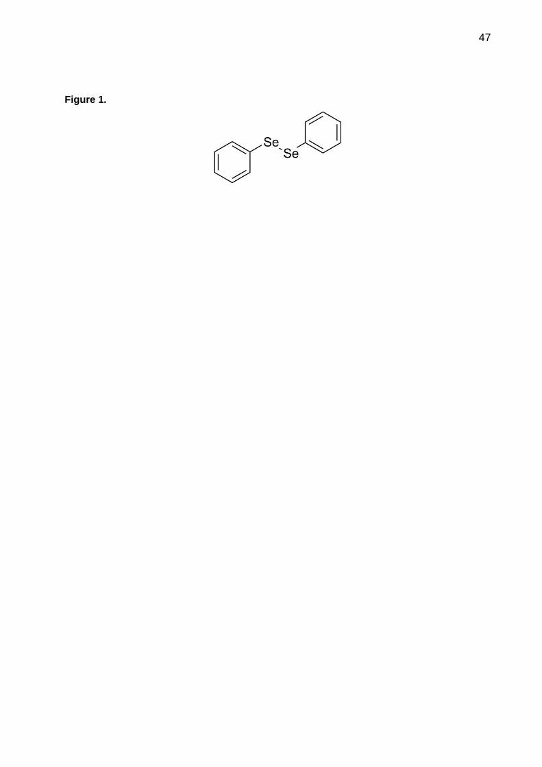
47
Figure 1.
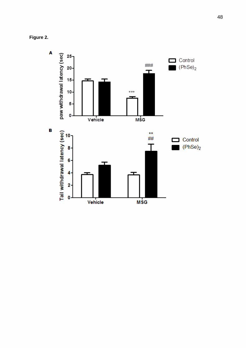
48
Figure 2.

49
Figure 3.
Vehicle MSG0
20
40
60Vehicle
(PhSe)2
***
###
Resp
on
se f
req
uen
ce o
f
VF
H s
tim
ula
tio
n(%
)

50
Figure 4.
Vehicle MSG0
20
40
60
80Control
(PhSe)2
##
*
Na
+,
K+-A
TP
ase a
cti
vit
y
(nm
ol
Pi/
mg
pro
tein
/ m
in )

51
Figure 5.
Vehicle MSG0.0
0.5
1.0
1.5Control
(PhSe)2*
#
Ca
2+-
AT
Pase a
cti
vit
y
(nm
ol
Pi/
mg
pro
tein
/min
)

52
Figure 6.

53
Figure 7.
Vehicle MSG0
5
10
15Vehicle
(PhSe)2
*
##
*
pm
ol
of
[3H
] g
luta
ma
te
up
tak
e/m
g p
rote
in/m
in

54
4. CONCLUSÃO
Os resultados apresentados nesta dissertação nos permitem concluir que:
Administração neonatal de GMS induz hiperalgesia e alodínia em ratos aos 60
dias de vida a qual é revertida pelo (PhSe)2;
O GMS induz alterações na atividade das enzimas Na+, K+-ATPase e Ca2+-
ATPase e o tratamento com(PhSe)2 reverte essas alterações;
A nocicepção induzida por GMS esta relacionada com o aumento de respostas
inflamatórias. O (PhSe)2 reverte a inflamação induzida por GMS em hipocampo de
ratos;
A administração neonatal de GMS leva a diminuição da captação de glutamato
em hipocampo e o (PhSe)2 reverte esta alteração;
Em conjunto os dados deste estudo sugerem que a nocicepção induzida por
GMS envolve eventos excitotóxicos e inflamatórios e a ação do (PhSe)2 no modelo
estudado esta relacionada com sua ação neuroprotetora e anti-inflamatória;
Em conjunto os resultados desta dissertação contribuem para esclarecer os
mecanismos de ação envolvidos na nocicepção induzida pelo MSG, sugerindo que a
nocicepção induzida por GMS está relacionado com a excitotoxicidade e inflamação
causados por este aditivo alimentar. Além disso, este estudo demonstrou a ação
antinociceptiva (PhSe)2 após injeções neonatais de MSG em ratos, a qual parece estar
relacionado com sei efeito neuuprotetor e anti-inflamatório.Embora mais estudos sejam
necessários para melhor elucidar os mecanismos toxicológicos pelos quais o GMS
induz alterações comportamentais em ratos, os dados deste estudo contribuem para a
compreensão dos mecanismos envolvidos na nocicepção induzida por GMS, bem
como sugerem novos alvos da ação nociceptiva do composto (PhSe)2.

55
5. REFERÊNCIAS
ABDEL-HAFEZ, A. A.; ABDEL-WAHAB, B. A. 5-(4-Chlorophenyl)-5,6-dihydro-1,3-oxazepin-7(4H)-one derivatives as lipophilic cyclic analogues of baclofen: design, synthesis, and neuropharmacological evaluation. Bioorg Med Chem, v. 16, n. 17, p. 7983-91, Sep 1 2008. ABE, M. et al. Neuropeptide-Y in the Specific Hypothalamic Nuclei of Rats Treated Neonatally with Monosodium Glutamate. Brain Research Bulletin, v. 24, n. 2, p. 289-291, Feb 1990. ANDERSON, C. M.; SWANSON, R. A. Astrocyte glutamate transport: Review of properties, regulation, and physiological functions. Glia, v. 32, n. 1, p. 1-14, Oct 2000. BADILLO-MARTINEZ, D. et al. Onset of pain threshold changes induced by neonatal monosodium glutamate. Int J Neurosci, v. 24, n. 3-4, p. 275-9, Nov 1984. BADILLOMARTINEZ, D. et al. Onset of Pain Threshold Changes Induced by Neonatal Monosodium Glutamate. International Journal of Neuroscience, v. 24, n. 3-4, p. 275-279, 1984. BALBO, S. L. et al. Fat storage is partially dependent on vagal activity and insulin secretion of hypothalamic obese rat. Endocrine, v. 31, n. 2, p. 142-148, Apr 2007. BANKS, W. A. The blood-brain barrier in psychoneuroimmunology. Neurologic Clinics, v. 24, n. 3, p. 413-+, Aug 2006. BANKS, W. A. et al. Intravenous human interleukin-1 alpha impairs memory processing in mice: Dependence on blood-brain barrier transport into posterior division of the septum. Journal of Pharmacology and Experimental Therapeutics, v. 299, n. 2, p. 536-541, Nov 2001. BASBAUM, A. I. et al. Cellular and molecular mechanisms of pain. Cell, v. 139, n. 2, p. 267-84, Oct 16 2009a. ______. Cellular and Molecular Mechanisms of Pain. Cell, v. 139, n. 2, p. 267-284, Oct 16 2009b. BEAS-ZARATE, C. et al. Neonatal exposure to monosodium L-glutamate induces loss of neurons and cytoarchitectural alterations in hippocampal CA1 pyramidal neurons of adult rats. Brain Research, v. 952, n. 2, p. 275-281, Oct 18 2002. BEAS-ZARATE, C. et al. Changes in NMDA-receptor gene expression are associated with neurotoxicity induced neonatally by glutamate in the rat brain. Neurochemistry International, v. 39, n. 1, p. 1-10, Jul 2001. BERNARDINO, L. et al. Modulator effects of interleukin-1beta and tumor necrosis factor-alpha on AMPA-induced excitotoxicity in mouse organotypic hippocampal slice cultures. J Neurosci, v. 25, n. 29, p. 6734-44, Jul 20 2005.

56
BEYREUTHER, K. et al. Consensus meeting: monosodium glutamate - an update. European Journal of Clinical Nutrition, v. 61, n. 7, p. 928-928, Jul 2007. BORTALANZA, L. B. et al. Anti-allodynic action of the tormentic acid, a triterpene isolated from plant, against neuropathic and inflammatory persistent pain in mice. Eur J Pharmacol, v. 453, n. 2-3, p. 203-8, Oct 25 2002. BORTOLATTO, C. F. et al. 2,2'-dithienyl diselenide, an organoselenium compound, elicits antioxidant action and inhibits monoamine oxidase activity in vitro. J Enzyme Inhib Med Chem, v. 28, n. 4, p. 677-84, Aug 2013a. ______. 2,2 '-dithienyl diselenide, an organoselenium compound, elicits antioxidant action and inhibits monoamine oxidase activity in vitro. Journal of Enzyme Inhibition and Medicinal Chemistry, v. 28, n. 4, p. 677-684, Aug 2013b. BRADFORD, M. M. Rapid and Sensitive Method for Quantitation of Microgram Quantities of Protein Utilizing Principle of Protein-Dye Binding. Anal Biochem, v. 72, n. 1-2, p. 248-254, 1976. BRUNING, C. A. et al. Protective effect of diphenyl diselenide on ischemia and reperfusion-induced cerebral injury: involvement of oxidative stress and pro-inflammatory cytokines. Neurochem Res, v. 37, n. 10, p. 2249-58, Oct 2012. BRUNING, C. A. et al. Involvement of the serotonergic system in the anxiolytic-like effect caused by m-trifluoromethyl-diphenyl diselenide in mice. Behavioural Brain Research, v. 205, n. 2, p. 511-517, Dec 28 2009. CALIXTO, J. B. et al. Kinins in pain and inflammation. Pain, v. 87, n. 1, p. 1-5, Jul 2000. CARMEN, J. et al. Tumor necrosis factor-alpha modulates glutamate transport in the CNS and is a critical determinant of outcome from viral encephalomyelitis. Brain Research, v. 1263, p. 143-154, Mar 31 2009. CECCARELLI, I. et al. Effects of formalin pain on hippocampal c-Fos expression in male and female rats. Pharmacology Biochemistry and Behavior, v. 64, n. 4, p. 797-802, Dec 1999. CHAGAS, P. M. et al. Evaluation of the pharmacological properties of salicylic acid-derivative organoselenium: 2-hydroxy-5-selenocyanatobenzoic acid as an anti-inflammatory and antinociceptive compound. Pharmacol Biochem Behav, v. 118, p. 87-95, Mar 2014. CHAPARRO-HUERTA, V. et al. Neuronal death and tumor necrosis factor-alpha response to glutamate-induced excitotoxicity in the cerebral cortex of neonatal rats. Neuroscience Letters, v. 333, n. 2, p. 95-98, Nov 22 2002. CHAPMAN, C. R. et al. Pain measurement: an overview. Pain, v. 22, n. 1, p. 1-31, May 1985.

57
CHEN, H. S. V.; LIPTON, S. A. The chemical biology of clinically tolerated NMDA receptor antagonists. Journal of Neurochemistry, v. 97, n. 6, p. 1611-1626, Jun 2006. COLLISON, K. S. et al. Dietary trans-fat combined with monosodium glutamate induces dyslipidemia and impairs spatial memory. Physiology & Behavior, v. 99, n. 3, p. 334-342, Mar 3 2010. COMASSETO, J. V. Selenium and Tellurium Chemistry: Historical Background. Journal of the Brazilian Chemical Society, v. 21, n. 11, p. 2027-2031, 2010. DA ROCHA, J. T. et al. Diphenyl diselenide reduces mechanical and thermal nociceptive behavioral responses after unilateral intrastriatal administration of 6-hydroxydopamine in rats. Biol Trace Elem Res, v. 154, n. 3, p. 372-8, Sep 2013a. ______. Diphenyl Diselenide Reduces Mechanical and Thermal Nociceptive Behavioral Responses After Unilateral Intrastriatal Administration of 6-Hydroxydopamine in Rats. Biological Trace Element Research, v. 154, n. 3, p. 372-378, Sep 2013b. DAVIDSON, T. L. et al. A potential role for the hippocampus in energy intake and body weight regulation. Current Opinion in Pharmacology, v. 7, n. 6, p. 613-616, Dec 2007. DEWEY, W. L. et al. The effect of narcotics and narcotic antagonists on the tail-flick response in spinal mice. J Pharm Pharmacol, v. 21, n. 8, p. 548-50, Aug 1969. DINIZ, Y. S. et al. Toxicity of hypercaloric diet and monosodium glutamate: oxidative stress and metabolic shifting in hepatic tissue. Food and Chemical Toxicology, v. 42, n. 2, p. 313-319, Feb 2004. DONG, X. X. et al. Molecular mechanisms of excitotoxicity and their relevance to pathogenesis of neurodegenerative diseases. Acta Pharmacologica Sinica, v. 30, n. 4, p. 379-387, Apr 2009. DUMONT, E. et al. Selenium speciation from food source to metabolites: a critical review. Analytical and Bioanalytical Chemistry, v. 385, n. 7, p. 1304-1323, Aug 2006. ERFANPARAST, A. et al. Effect of microinjection of histamine into the dorsal hippocampus on the orofacial formalin-induced pain in rats. European Journal of Pharmacology, v. 627, n. 1-3, p. 119-123, Feb 10 2010. FERREIRA, S. H. Prostaglandins, Aspirin-Like Drugs and Analgesia. Nature-New Biology, v. 240, n. 102, p. 200-&, 1972. FISKE, C. H.; SUBBAROW, Y. The calorimetric determination of phosphorus. Biol Chem, v. 66, p. 375-381, 1925. FUNDYTUS, M. E. Glutamate receptors and nociception - Implications for the drug treatment of pain. Cns Drugs, v. 15, n. 1, p. 29-58, 2001.

58
FURUYA, D. T. et al. Anti-inflammatory effect of atorvastatin ameliorates insulin resistance in monosodium glutamate-treated obese mice. Metabolism-Clinical and Experimental, v. 59, n. 3, p. 395-399, Mar 2010. GEGELASHVILI, G. et al. The high-affinity glutamate transporters GLT1, GLAST, and EAAT4 are regulated via different signalling mechanisms. Neurochemistry International, v. 37, n. 2-3, p. 163-170, Aug-Sep 2000. GILGUN-SHERKI, Y. et al. Antioxidant therapy in acute central nervous system injury: Current state. Pharmacological Reviews, v. 54, n. 2, p. 271-284, Jun 2002a. ______. Antioxidant therapy in acute central nervous system injury: current state. Pharmacol Rev, v. 54, n. 2, p. 271-84, Jun 2002b. GRIFFIS, C. A. et al. The effect of pain on leukocyte cellular adhesion molecules. Biological Research for Nursing, v. 7, n. 4, p. 297-312, Apr 2006. HARRISON, S.; GEPPETTI, P. Substance P. International Journal of Biochemistry & Cell Biology, v. 33, n. 6, p. 555-576, Jun 2001. HU, S. et al. Cytokine effects on glutamate uptake by human astrocytes. Neuroimmunomodulation, v. 7, n. 3, p. 153-9, 2000. INSAWANG, T. et al. Monosodium glutamate (MSG) intake is associated with the prevalence of metabolic syndrome in a rural Thai population. Nutrition & Metabolism, v. 9, Jun 8 2012. ISHIKAWA, K. et al. Hippocampal degeneration inducing impairment of learning in rats: model of dementia? Behav Brain Res, v. 83, n. 1-2, p. 39-44, Feb 1997a. ______. Hippocampal degeneration inducing impairment of learning in rats: Model of dementia? Behavioural Brain Research, v. 83, n. 1-2, p. 39-44, Feb 1997b. JANSSEN, P. A. et al. The inhibitory effect of fentanyl and other morphine-like analgesics on the warm water induced tail withdrawl reflex in rats. Arzneimittelforschung, v. 13, p. 502-7, Jun 1963. JENSEN, T. S.; YAKSH, T. L. Examination of spinal monoamine receptors through which brainstem opiate-sensitive systems act in the rat. Brain Res, v. 363, n. 1, p. 114-27, Jan 15 1986. JESSE, C. R. et al. Effect of a metabotropic glutamate receptor 5 antagonist, MPEP, on the nociceptive response induced by intrathecal injection of excitatory aminoacids, substance P, bradykinin or cytokines in mice. Pharmacology Biochemistry and Behavior, v. 90, n. 4, p. 608-613, Oct 2008. JOHNSTON, M. V. Excitotoxicity in perinatal brain injury. Brain Pathology, v. 15, n. 3, p. 234-240, Jul 2005.

59
JULIUS, D.; BASBAUM, A. I. Molecular mechanisms of nociception. Nature, v. 413, n. 6852, p. 203-210, Sep 13 2001. KARAMI, M. et al. Role of nitric oxide in the rat hippocampal CA1 area on morphine-induced conditioned place preference. European Journal of Pharmacology, v. 449, n. 1-2, p. 113-119, Aug 2 2002. KENNEY, J. W.; GOULD, T. J. Modulation of hippocampus-dependent learning and synaptic plasticity by nicotine. Molecular Neurobiology, v. 38, n. 1, p. 101-121, Aug 2008. KHANNA, S. et al. Nociception-driven decreased induction of Fos protein in ventral hippocampus field CA1 of the rat. Brain Research, v. 1004, n. 1-2, p. 167-176, Apr 9 2004. KIM, Y. Y.; MAHAN, D. C. Comparative effects of high dietary levels of organic and inorganic selenium on selenium toxicity of growing-finishing pigs. Journal of Animal Science, v. 79, n. 4, p. 942-948, Apr 2001. KISS, P. et al. Changes in open-field activity and novelty-seeking behavior in periadolescent rats neonatally treated with monosodium glutamate. Neurotoxicity Research, v. 12, n. 2, p. 85-93, Sep 2007. KISS, P. et al. Development of neurological reflexes and motor coordination in rats neonatally treated with monosodium glutamate. Neurotoxicity Research, v. 8, n. 3-4, p. 235-244, Nov 2005. KLAMT, J. G.; PRADO, W. A. Antinociception and Behavioral-Changes Induced by Carbachol Microinjected into Identified Sites of the Rat-Brain. Brain Research, v. 549, n. 1, p. 9-18, May 17 1991. KLINGBERG, H. et al. Long-term effects on behaviour after postnatal treatment with monosodium-L-glutamate. Biomed Biochim Acta, v. 46, n. 10, p. 705-11, 1987. LAMBERT, G. A. et al. Von Frey's hairs--a review of their technology and use--a novel automated von Frey device for improved testing for hyperalgesia. J Neurosci Methods, v. 177, n. 2, p. 420-6, Mar 15 2009. LATREMOLIERE, A.; WOOLF, C. J. Central Sensitization: A Generator of Pain Hypersensitivity by Central Neural Plasticity. Journal of Pain, v. 10, n. 9, p. 895-926, Sep 2009. LI, W. D.; NEUGEBAUER, V. Differential roles of mGluR1 and mGluR5 in brief and prolonged nociceptive processing in central amygdala neurons. Journal of Neurophysiology, v. 91, n. 1, p. 13-24, Jan 2004. LIBBY, P. Inflammatory mechanisms: The molecular basis of inflammation and disease. Nutrition Reviews, v. 65, n. 12, p. S140-S146, Dec 2007.

60
LIPTON, S. A. NMDA receptor activity regulates transcription of antioxidant pathways. Nature Neuroscience, v. 11, n. 4, p. 381-382, Apr 2008. LOPEZ-PEREZ, S. J. et al. Monosodium glutamate neonatal treatment as a seizure and excitotoxic model. Brain Research, v. 1317, p. 246-256, Mar 4 2010. LORENZ, I. H. et al. Lornoxicam characteristically modulates cerebral pain-processing in human volunteers: a functional magnetic resonance imaging study. British Journal of Anaesthesia, v. 100, n. 6, p. 827-833, Jun 2008. LUCHESE, C. et al. Diphenyl diselenide reduces inflammation in the mouse model of pleurisy induced by carrageenan: reduction of pro-inflammatory markers and reactive species levels. Inflamm Res, v. 61, n. 10, p. 1117-24, Oct 2012. MAIER, S. F. Bi-directional immune-brain communication: Implications for understanding stress, pain, and cognition. Brain Behavior and Immunity, v. 17, n. 2, p. 69-85, Apr 2003. MANDOLESI, G. et al. Interleukin-1 beta Alters Glutamate Transmission at Purkinje Cell Synapses in a Mouse Model of Multiple Sclerosis. Journal of Neuroscience, v. 33, n. 29, p. 12105-12121, Jul 17 2013. MARCONDES SARI, M. H. et al. Contribution of dopaminergic and adenosinergic systems in the antinociceptive effect of p-chloro-selenosteroid. Eur J Pharmacol, v. 725, p. 79-86, Feb 15 2014. MCCABE, C.; ROLLS, E. T. Umami: a delicious flavor formed by convergence of taste and olfactory pathways in the human brain. Eur J Neurosci, v. 25, n. 6, p. 1855-64, Mar 2007. MCKENNA, J. E.; MELZACK, R. Analgesia Produced by Lidocaine Microinjection into the Dentate Gyrus. Pain, v. 49, n. 1, p. 105-112, Apr 1992. MCNALLY, L. et al. Inflammation, glutamate, and glia in depression: A literature review. Cns Spectrums, v. 13, n. 6, p. 501-510, Jun 2008. MEOTTI, F. C. et al. Protective role of aryl and alkyl diselenides on lipid peroxidation. Environmental Research, v. 94, n. 3, p. 276-282, Mar 2004. MESSING, R. O. Nervous System Disorders. Pathophysiology of Disease (5th ed.), 2006. MILLAN, M. J. The induction of pain: An integrative review. Progress in Neurobiology, v. 57, n. 1, p. 1-164, Jan 1999. MINAMI, M. et al. Effects of kainic acid on messenger RNA levels of IL-1 beta, IL-6, TNF alpha and LIF in the rat brain. Biochem Biophys Res Commun, v. 176, n. 2, p. 593-8, Apr 30 1991.

61
MOALEM, G.; TRACEY, D. J. Immune and inflammatory mechanisms in neuropathic pain. Brain Research Reviews, v. 51, n. 2, p. 240-264, Aug 2006. MORGANE, P. J. et al. Effects of Protein-Malnutrition on Developing Central Nervous-System in Rat. Neuroscience and Biobehavioral Reviews, v. 2, n. 3, p. 137-230, 1978. MORIOKA, N. et al. Nitric oxide synergistically potentiates interleukin-1 beta-induced increase of cyclooxygenase-2 mRNA levels, resulting in the facilitation of substance P release from primary afferent neurons: involvement of cGMP-independent mechanisms. Neuropharmacology, v. 43, n. 5, p. 868-76, Oct 2002. MULLER, A. et al. A Novel Biologically-Active Organoselenium Compound .1. Glutathione Peroxidase-Like Activity Invitro and Antioxidant Capacity of Pz-51 (Ebselen). Biochemical Pharmacology, v. 33, n. 20, p. 3235-3239, 1984. NARDELLI, T. R. et al. Taurine prevents fat deposition and ameliorates plasma lipid profile in monosodium glutamate-obese rats. Amino Acids, v. 41, n. 4, p. 901-908, Oct 2011. NDOUNTSE, L. T.; CHAN, H. M. Role of N-methyl-D-aspartate receptors in polychlorinated biphenyl mediated neurotoxicity. Toxicology Letters, v. 184, n. 1, p. 50-55, Jan 10 2009. NICHOLLS, D. G.; CHALMERS, S. The integration of mitochondrial calcium transport and storage. Journal of Bioenergetics and Biomembranes, v. 36, n. 4, p. 277-281, Aug 2004. NOGUEIRA, C. W. et al. Anti-inflammatory and antinociceptive activity of diphenyl diselenide. Inflammation Research, v. 52, n. 2, p. 56-63, Feb 2003. NOGUEIRA, C. W. et al. Organoselenium and organotellurium compounds: toxicology and pharmacology. Chem Rev, v. 104, n. 12, p. 6255-85, Dec 2004a. NOGUEIRA, C. W. et al. Organoselenium and organotellurium compounds: Toxicology and pharmacology. Chemical Reviews, v. 104, n. 12, p. 6255-6285, Dec 2004b. O'SHAUGHNESSY, M. C. et al. The effect of substance P on nitric oxide release in a rheumatoid arthritis model. Inflamm Res, v. 55, n. 6, p. 236-40, Jun 2006. OKUBO, Y. et al. Imaging extrasynaptic glutamate dynamics in the brain. Proceedings of the National Academy of Sciences of the United States of America, v. 107, n. 14, p. 6526-6531, Apr 6 2010. OLNEY, J. W.; SHARPE, L. G. Brain Lesions in an Infant Rhesus Monkey Treated with Monosodium Glutamate. Science, v. 166, n. 3903, p. 386-&, 1969. ONYEMA, O. O. et al. Effect of vitamin E on monosodium glutamate induced hepatotoxicity and oxidative stress in rats. Indian Journal of Biochemistry & Biophysics, v. 43, n. 1, p. 20-24, Feb 2006.
62
PARADA, C. A. et al. Transient attenuation of protein kinase C epsilon can terminate a chronic hyperalgesic state in the rat. Neuroscience, v. 120, n. 1, p. 219-226, 2003. PAULMIER, C. Selenoorganic functional groups. Selenium reagents and intermediates in organic synthesis, v. 1, p. 25-51, 1986. PENG, J. M. et al. Enhanced NMDA receptor NR1 phosphorylation and neuronal activity in the arcuate nucleus of hypothalamus following peripheral inflammation. Acta Pharmacologica Sinica, v. 32, n. 2, p. 160-166, Feb 2011. PETRENKO, A. B. et al. The role of N-methyl-D-aspartate (NMDA) receptors in pain: A review. Anesthesia and Analgesia, v. 97, n. 4, p. 1108-1116, Oct 2003. POCOCK, J. M.; KETTENMANN, H. Neurotransmitter receptors on microglia. Trends in Neurosciences, v. 30, n. 10, p. 527-535, Oct 2007. PORCIUNCULA, L. O. et al. Ebselen prevents excitotoxicity provoked by glutamate in rat cerebellar granule neurons. Neuroscience Letters, v. 299, n. 3, p. 217-220, Feb 23 2001. PRIGOL, M. et al. Comparative Excretion and Tissue Distribution of Selenium in Mice and Rats Following Treatment with Diphenyl Diselenide. Biological Trace Element Research, v. 150, n. 1-3, p. 272-277, Dec 2012a. ______. Comparative excretion and tissue distribution of selenium in mice and rats following treatment with diphenyl diselenide. Biol Trace Elem Res, v. 150, n. 1-3, p. 272-7, Dec 2012b. QUINES, C. B. et al. Monosodium glutamate, a food additive, induces depressive-like and anxiogenic-like behaviors in young Rats. Life Sciences, v. 107, n. 1-2, p. 27-31, Jun 27 2014. RANSOHOFF, R. M.; ENGELHARDT, B. The anatomical and cellular basis of immune surveillance in the central nervous system. Nature Reviews Immunology, v. 12, n. 9, p. 623-635, Sep 2012. RANSOHOFF, R. M. et al. Three or more routes for leukocyte migration into the central nervous system. Nature Reviews Immunology, v. 3, n. 7, p. 569-581, Jul 2003. RAYMAN, M. P. The importance of selenium to human health. Lancet, v. 356, n. 9225, p. 233-241, Jul 15 2000. ______. Food-chain selenium and human health: emphasis on intake. British Journal of Nutrition, v. 100, n. 2, p. 254-268, Aug 2008. REN, K.; DUBNER, R. Interactions between the immune and nervous systems in pain. Nature Medicine, v. 16, n. 11, p. 1267-1276, Nov 2010.

63
ROHN, T. T. et al. Ion transport ATPases as targets for free radical damage. Protection by an aminosteroid of the Ca2+ pump ATPase and Na+/K+ pump ATPase of human red blood cell membranes. Biochem Pharmacol, v. 46, n. 3, p. 525-34, Aug 3 1993. ROSE, E. M. et al. Glutamate transporter coupling to Na,K-ATPase. J Neurosci, v. 29, n. 25, p. 8143-55, Jun 24 2009a. ROSE, E. M. et al. Glutamate Transporter Coupling to Na,K-ATPase. Journal of Neuroscience, v. 29, n. 25, p. 8143-8155, Jun 24 2009b. ROSSATO, J. I. et al. Ebselen blocks the quinolinic acid-induced production of thiobarbituric acid reactive species but does not prevent the behavioral alterations produced by intra-striatal quinolinic acid administration in the rat. Neuroscience Letters, v. 318, n. 3, p. 137-140, Feb 1 2002. ROTIMI, O. A. et al. Effects of fibre-enriched diets on tissue lipid profiles of MSG obese rats. Food and Chemical Toxicology, v. 50, n. 11, p. 4062-4067, Nov 2012. SAVEGNAGO, L. et al. Diphenyl diselenide attenuates acute thermal hyperalgesia and persistent inflammatory and neuropathic pain behavior in mice. Brain Research, v. 1175, p. 54-59, Oct 17 2007a. SAVEGNAGO, L. et al. Mechanisms involved in the antinociceptive effect caused by diphenyl diselenide in the formalin test. Journal of Pharmacy and Pharmacology, v. 60, n. 12, p. 1679-1686, Dec 2008. SAVEGNAGO, L. et al. Antinociceptive properties of diphenyl diselenide: evidences for the mechanism of action. Eur J Pharmacol, v. 555, n. 2-3, p. 129-38, Jan 26 2007b. SAVEGNAGO, L. et al. Antinociceptive properties of diphenyl diselenide: Evidences for the mechanism of action. European Journal of Pharmacology, v. 555, n. 2-3, p. 129-138, Jan 26 2007c. SAVEGNAGO, L. et al. Spinal mechanisms of antinociceptive action caused by diphenyl diselenide. Brain Res, v. 1162, p. 32-7, Aug 8 2007d. SCHMIDTKO, A. et al. Ziconotide for treatment of severe chronic pain. Lancet, v. 375, n. 9725, p. 1569-1577, May 1 2010. SCHNEIDER, F. et al. Subjective ratings of pain correlate with subcortical-limbic blood flow: An fMRI study. Neuropsychobiology, v. 43, n. 3, p. 175-185, 2001. SPANDOU, E. et al. Neuroprotective effect of long-term MgSO4 administration after cerebral hypoxia-ischemia in newborn rats is related to the severity of brain damage. Reproductive Sciences, v. 14, n. 7, p. 667-677, Oct 2007. STAHL, W. L.; HARRIS, W. E. Na+,K+-ATPase: structure, function, and interactions with drugs. Adv Neurol, v. 44, p. 681-93, 1986.
64
TAKAKI, J. et al. L-glutamate released from activated microglia downregulates astrocytic L-glutamate transporter expression in neuroinflammation: the 'collusion' hypothesis for increased extracellular L-glutamate concentration in neuroinflammation. Journal of Neuroinflammation, v. 9, Dec 23 2012. TAMAS, A. et al. Effects of pituitary adenylate cyclase activating polypeptide in retinal degeneration induced by monosodium-glutamate. Neuroscience Letters, v. 372, n. 1-2, p. 110-113, Nov 30 2004. THOMAZI, A. P. et al. Ontogenetic profile of glutamate uptake in brain structures slices from rats: sensitivity to guanosine. Mech Ageing Dev, v. 125, n. 7, p. 475-81, Jul 2004. TOLOSA, L. et al. TNF-alpha potentiates glutamate-induced spinal cord motoneuron death via NF-kappa B. Molecular and Cellular Neuroscience, v. 46, n. 1, p. 176-186, Jan 11 2011. TREVISAN, G. et al. Antinociceptive effects of 14-membered cyclopeptide alkaloids. J Nat Prod, v. 72, n. 4, p. 608-12, Apr 2009. TYMIANSKI, M. et al. Source Specificity of Early Calcium Neurotoxicity in Cultured Embryonic Spinal Neurons. Journal of Neuroscience, v. 13, n. 5, p. 2085-2104, May 1993a. TYMIANSKI, M. et al. Cell-Permeant Ca2+ Chelators Reduce Early Excitotoxic and Ischemic Neuronal Injury in-Vitro and in-Vivo. Neuron, v. 11, n. 2, p. 221-235, Aug 1993b. VAN DEN BUUSE, M. et al. Effects of neonatal treatment with monosodium-glutamate in spontaneously hypertensive rats. Brain Res, v. 351, n. 1, p. 135-8, Mar 1985. VANDENBUUSE, M. et al. Effects of Neonatal Treatment with Monosodium-Glutamate in Spontaneously Hypertensive Rats. Developmental Brain Research, v. 19, n. 1, p. 135-138, 1985. VEZZANI, A. et al. Interleukin-1beta immunoreactivity and microglia are enhanced in the rat hippocampus by focal kainate application: functional evidence for enhancement of electrographic seizures. J Neurosci, v. 19, n. 12, p. 5054-65, Jun 15 1999. WEI, F. et al. Role of EGR1 in hippocampal synaptic enhancement induced by tetanic stimulation and amputation. Journal of Cell Biology, v. 149, n. 7, p. 1325-1333, Jun 26 2000. WOOLF, C. J. Evidence for a Central Component of Post-Injury Pain Hypersensitivity. Nature, v. 306, n. 5944, p. 686-688, 1983. WOOLF, C. J.; SALTER, M. W. Neuroscience - Neuronal plasticity: Increasing the gain in pain. Science, v. 288, n. 5472, p. 1765-1768, Jun 9 2000. WOOLFE, G.; MACDONALD, A. D. The evaluation of the analgesic action of pethidine hydrochloride. J Pharmacol Exp Ther, v. 80, p. 300-307, 1944.

65
WYSE, A. T. et al. Methylmalonate administration decreases Na+,K+-ATPase activity in cerebral cortex of rats. Neuroreport, v. 11, n. 10, p. 2331-4, Jul 14 2000. XU, L. et al. Temporal and spatial expression of preprotachykinin a mRNA in the developing filial mice brain after maternal administration of monosodium glutamate at a late stage of pregnancy. Neuroscience, v. 145, n. 3, p. 974-980, Mar 30 2007. YOUNG, V. R. et al. Selenium Bioavailability with Reference to Human-Nutrition. American Journal of Clinical Nutrition, v. 35, n. 5, p. 1076-1088, 1982. YU, S. Y. et al. Role of AMPA receptor trafficking in NMDA receptor-dependent synaptic plasticity in the rat lateral amygdala. Journal of Neurochemistry, v. 106, n. 2, p. 889-899, Jul 2008. ZASSO, F. B. et al. On the mechanisms involved in antinociception induced by diphenyl diselenide. Environ Toxicol Pharmacol, v. 19, n. 2, p. 283-9, Feb 2005a. ZASSO, F. B. et al. On the mechanisms involved in antinociception induced by diphenyl diselenide. Environmental Toxicology and Pharmacology, v. 19, n. 2, p. 283-289, Feb 2005b. ZHANG, D. W. et al. Na,K-ATPase Activity Regulates AMPA Receptor Turnover through Proteasome-Mediated Proteolysis. Journal of Neuroscience, v. 29, n. 14, p. 4498-4511, Apr 8 2009.