(brn-3c) transcription factor in the inner ear - UCL Discovery
RESEARCH ARTICLE Open Access
Proliferation-associated POU4F2/Brn-3btranscription factor expression is regulated byoestrogen through ERa and growth factors viaMAPK pathwaySamir Ounzain1, Samantha Bowen1, Chandrakant Patel1, Rieko Fujita1, Richard J Heads2 andVishwanie S Budhram-Mahadeo1*
Abstract
Introduction: In cancer cells, elevated transcription factor-related Brn-3a regulator isolated from brain cDNA (Brn-3b) transcription factor enhances proliferation in vitro and increases tumour growth in vivo whilst conferring drugresistance and migratory potential, whereas reducing Brn-3b slows growth both in vitro and in vivo. Brn-3bregulates distinct groups of key target genes that control cell growth and behaviour. Brn-3b is elevated in >65% ofbreast cancer biopsies, but mechanisms controlling its expression in these cells are not known.
Methods: Bioinformatics analysis was used to identify the regulatory promoter region and map transcription startsite as well as transcription factor binding sites. Polymerase chain reaction (PCR) cloning was used to generatepromoter constructs for reporter assays. Chromatin immunoprecipitation and site-directed mutagenesis were usedto confirm the transcription start site and autoregulation. MCF-7 and Cos-7 breast cancer cells were used. Cellsgrown in culture were transfected with Brn-3b promoter and treated with growth factors or estradiol to test foreffects on promoter activity. Quantitative reverse transcriptase PCR assays and immunoblotting were used toconfirm changes in gene and protein expression.
Results: We cloned the Brn-3b promoter, mapped the transcription start site and showed stimulation by estradioland growth factors, nerve growth factor and epidermal growth factor, which are implicated in breast cancerinitiation and/or progression. The effects of growth factors are mediated through the mitogen-activated proteinkinase pathway, whereas hormone effects act via oestrogen receptor a (ERa). Brn-3b also autoregulates itsexpression and cooperates with ERa to further enhance levels.
Conclusions: Key regulators of growth in cancer cells, for example, oestrogens and growth factors, can stimulateBrn-3b expression, and autoregulation also contributes to increasing Brn-3b in breast cancers. Since increasing Brn-3b profoundly enhances growth in these cells, understanding how Brn-3b is increased in breast cancers will helpto identify strategies for reducing its expression and thus its effects on target genes, thereby reversing its effects inbreast cancer cells.
* Correspondence: [email protected] Molecular Biology Unit, UCL Institute of Child Health, 30 GuilfordStreet, London WC1N 1EH, UKFull list of author information is available at the end of the article
Ounzain et al. Breast Cancer Research 2011, 13:R5http://breast-cancer-research.com/content/13/1/R5
© 2011 Ounzain et al.; licensee BioMed Central Ltd. This is an open access article distributed under the terms of the Creative CommonsAttribution License (http://creativecommons.org/licenses/by/2.0), which permits unrestricted use, distribution, and reproduction inany medium, provided the original work is properly cited.
IntroductionThe class 4 POU (Pit-Oct-Unc) transcription factor-2-related to Brn-3 (POU4F2/Brn-3b), is referred to asBrn-3b because of homology in the DNA bindingdomain to the related Brn-3a transcription factor. Brn-3b is highly expressed in a significant proportion (>65%) of breast tumour biopsies analyzed [1]. Over-expression of Brn-3b in cancer cells is strongly asso-ciated with increased proliferation, in vitro, andenhanced tumour growth, in-vivo, whereas reducingBrn-3b (by antisense) decreases proliferation in-vitroand results in smaller, slower growing tumours in-vivo[2,3]. Brn-3b also confers resistance to growth inhibitoryor apoptosis inducing chemotherapeutic drugs but alsoincreases migratory potential of cancer cells [2]. Recentstudies also showed that Brn3b is increased in doxorubi-cin resistant breast cancer cells (R Fujita and V Budh-ram-Mahadeo, unpublished data).As a transcription factor, Brn-3b regulates the expres-
sion of critical genes that control different cellular pro-cesses. For example, increased proliferation by Brn-3bmay be associated with its ability to transactivate thepromoters of genes required for cell cycle progressionsuch as cyclin-dependent kinase 4 (CDK4) [4] and itsregulatory partner cyclin D1 [5], which are required,whilst repressing breast cancer susceptibility gene 1(BRCA1) [6], which is associated with cell cycle arrest inbreast cancer cells. Invasiveness and drug resistanceassociated with Brn-3b in cancer cells are linked with itsability to transactivate genes such as the small heatshock protein 27 (HSP27) [7] whilst repressing promo-ters of genes encoding adhesion molecules, for example,g-catenin/plakoglobin [8].However, whilst the effects of increased Brn-3b in
cancer cells have been characterised and many of its tar-get genes have been studied, we do not know which fac-tors contribute to the elevated Brn-3b mRNA andprotein levels observed in breast cancer. In this study,we have cloned and analysed the regulatory region thatcontrols Brn-3b gene expression in MCF-7 breast cancercells. The results presented herein identify a proximalpromoter present in the 5’ sequences upstream of theBrn-3b gene which drives expression in MCF-7 cells.This promoter is transactivated by the growth factorsnerve growth factor (NGF) and epidermal growth factor(EGF) and the hormone estradiol, all of which areknown to promote the proliferation and/or survival ofbreast cancer cells. NGF and EGF increase promoteractivity by signalling through the p42/p44-(ERK) mito-gen-activated protein kinase (MAPK) pathway, whereasthe effects of oestrogen are mediated via oestrogenreceptor a (ERa) but not oestrogen receptor b (ERb).We also show autoregulation by Brn-3b to increase itsown expression. These findings suggest that increased
transcription of Brn-3b in breast cancer cells is stimu-lated by growth factors and hormones that enhance pro-liferation and propagate through autoregulation.
Materials and methodsMaterialsGeneral laboratory reagents were purchased from Merck(Nottingham, UK) and Sigma (Dorset, UK) unless other-wise stated. Primary antibodies were used at dilutions of1: 1000-1500 and included Brn-3b-rabbit pAb (Abcam-Cambridge, UK); Brn-3b-goat pAb (Santa Cruz Biotech-nology Inc, USA); actin - goat pAb (I-19, Santa CruzBiotechnology). HRP-conjugated secondary Ab fromDako (Cambridgeshire, UK) was used for immunoblot-ting 1:2000). Estradiol (E4389), cyclic adenosine mono-phosphate (cAMP), phorbol 12,13-dibutyrate (PDBu)and 4-hydroxytamoxifen (tamoxifen) were from Sigma(Dorset, UK); epidermal growth factor (EGF), transform-ing growth factor-b (TGF-b); insulin-like growth-1 (IGF1) and nerve growth factor (NGF) were from RocheDiagnostics GmbH (Welwyn Garden City, Hertfordshire,UK). Signalling pathway inhibitors PD-98059 (MEK),SB-203580 kinase (p38), Genistein (tyrosine kinase), andWortmannin (PI3-kinase) were from Calbiochem (Not-tingham, UK). The MCF7 breast cancer cell line wasobtained from ATCC. Expression vectors, Brn-3b(l);Brn-3b(s); ER were previously described [9]. Dominantnegative and constitutively active MEK expression vec-tors were kind gift from D.S. Latchman[10].
In silico analysis of Brn-3b promoterHomo sapiens chromosome 4 contig was analysed usingthe Basic Local Alignment Search Tool, or BLAST [11],to identify a region containing the Brn-3b gene consist-ing of approximately 10 kb sequence (in bacterial artifi-cial chromosome clone AC093887). Further analysisusing Bioinformatics and Molecular Analysis Section(BIMAS) ProScan software [12] was used to identifyputative promoter sequences in this region of DNA.The VISTA Genome Browser [11] was used to generatehomology plots, whereas analysis using GenomatixTRANSFAC software analysis (Genomatix, Munich,Germany) identified binding sites for transcription fac-tors in the putative promoter sequences.
Brn-3b reporter constructsBrn-3b reporter constructs were generated so that theregulatory promoter region drove expression of a fireflyluciferase reporter gene in the pGL2 plasmid. The initialBrn-3b reporter construct was generated by amplifying~1,400 bp regions upstream of the Brn-3b genesequence and incorporating part of exon 1 (Figure 1b).The resultant construct was designated BstX1/Stu1/Xho1 (BSX) because it included sequences that can be
Ounzain et al. Breast Cancer Research 2011, 13:R5http://breast-cancer-research.com/content/13/1/R5
Page 2 of 15
isolated using restriction BstX1, Xho1 site (exon 1) andStu1 site and were used for diagnostic digestion. TheBSX exon-intron-exon (BSXEIE) construct was subse-quently generated by cloning the gene encodingsequence (exon 1, intron and exon 2) upstream of thisputative regulatory region, thus allowing Brn-3b promo-ter to drive its own gene expression.
Chromatin immunoprecipitation assayThe chromatin immunoprecipitation (ChIP) assay was car-ried out as described by Lee et al. [7]. In studies to identifythe transcription start sites, anti-TATA box binding protein(anti-TBP) Ab (Abcam) was used to immunoprecipitateregions of promoter bound by TBP in the transcriptioninitiation complex. Later studies to confirm Brn-3b binding
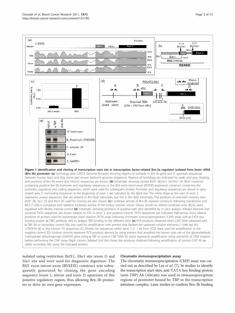
Figure 1 Identification and cloning of transcription start site in transcription factor-related Brn-3a regulator isolated from brain cDNA(Brn-3b) promoter. (a) Homology plots (VISTA Genome Browser) showing regions of similarity in Brn-3b gene and 5’ upstream sequencesbetween human (top) and dog, horse and mouse (bottom) genome sequences. Regions of homology are indicated by peaks and grey shading,and positions of Brn-3b exons and intronic sequences are shown. (b) Schematic showing cloned BstX1 (B)/Stu1 (S)/Xho1 (X) (BSX) constructcontaining putative Brn-3b promoter and regulatory sequences or the BSX exon-intron-exon (BSXEIE) expression construct containing thepromoter, regulatory and coding sequences, which were used for subsequent studies. Promoter and regulatory sequences are shown in grey-striped area. 5’ noncoding sequences at the beginning of exon 1 are indicated by the black bar. The white stripe at the start of exon 2represents unique sequences that are present in Brn-3b(s) transcripts, but not in Brn-3b(l) transcripts. The positions of restriction enzyme sitesBstX1 (B), Stu1 (S) and Xho1 (X) used for cloning are also shown. (c) Luciferase activity of Brn-3b reporter constructs following transfection intoMCF-7 cells is compared with baseline luciferase activity of the empty reporter vector. Values, shown as relative luciferase units (RLU),, wereequalised with Renilla internal control (d) Schematic showing positions of putative start sites identified by in silico analysis. Initiator element andproximal TATA sequences are shown relative to ATG in exon 1, and putative intronic TATA sequences are indicated. Half-arrows show relativepositions of primers used for polymerase chain reaction (PCR) assay following chromatin immunoprecipitation (ChIP) assay with a-TATA boxbinding protein (a-TBP) antibody (Ab) to analyse TBP binding to the different sites. (e) PCR products obtained when ChIP DNA (obtained witha-TBP Ab or secondary control Ab) was used for amplification with primers that flanked the upstream initiator elements (-1,048 bp) (A),-278TATA (B) or the intronic TA sequences (C). Primers for sequences within exon 2 (> 1 kb from ATG) were used for amplification in thenegative control (D). Positive controls represent PCR products derived by using primers that amplified the known start site of the glyceraldehyde3-phosphate dehydrogenase (GAPDH) gene using a-TBP or control ChIP DNA (E). Input represents amplification using one-tenth of DNA isolatedbefore performing the ChIP assay. Right column (labeled 2nd Ab) shows the products obtained following amplification of control ChIP Ab (a-rabbit secondary Ab) using the indicated primers.
Ounzain et al. Breast Cancer Research 2011, 13:R5http://breast-cancer-research.com/content/13/1/R5
Page 3 of 15
in its own promoter was done using antigoat Brn-3b Ab(Santa Cruz Biotechnology) to immunoprecipitate Brn-3bbound to chromatin in intact cells. Negative control ChIPassay was performed using antibody to glyceraldehyde 3-phosphate dehydrogenase (anti-GAPDH) (Abcam) or sec-ondary Ab (Dako) only. The shear size of DNA followingChIP assay and sonication was ~200 to 600 bp as deter-mined for agarose gel electrophoresis. The PCR assay forthe transcriptional start site was performed on ChIP DNAusing primers designed to amplify different regions of theputative Brn-3b promoter as follows: Upstream initiator:forward 5’-CTTGGGCCGCAACTTTATT-3’ and reverse5’-TACCTAAGGACCAGCCTCCA-3’; -278TATA: for-ward 5’-CGGGGAGAGGGGAGTATAAC-3’ and reverse5’-GATGCCTGACTCCGCTTG-3’; intronic TA: forward5’-TTGACAGCCCCCTTTATCTG-3’ and reverse 5’-AGG-CAACATCCCAGGTCATA-3’; and negative controlprimers which amplified the exon 2 sequence:forward 5’-ACCATGAACCCCATGCAC-3’ and reverse 5’-CTTGATGCCTCGCTGCTT-3’. The distance between theintronic site and the exonic sequences amplified was ~1 kb.As a positive control, the following primers were used toamplify the GAPDH promoter start site: forward 5’-TGAG-CAGACCGGTGTCAC-3’ and reverse 5’-AGGACTTTGG-GAACGACTGA-3’. Primers used to amplify the promoterregion containing the Brn-3b site were as follows: forward5’-GCCCCTTCTTCCTTTGATTG-3’ and reverse 5’-ACA-CACACACGCTCCTCTTG-3’. Standard conditions forPCR amplification included 2.5 mM MgCl2 and the follow-ing cycling parameters: 1 cycle at 94°C for 15 minutes fol-lowed by 40 cycles of amplification for each experimentsusing 95°C for 30 seconds, at 58°C for 30 seconds, and at72°C for 30 seconds. A final cycle was undertaken at 72°Cfor 5 minutes, the complete elongation steps and the PCRproducts were then resolved on a 2.5% agarose/Tris-borate-ethylenediaminetetraacetic acid gel.
Site-directed mutagenesisSite-directed mutagenesis was carried out to test theeffects of altering alter key bases in either the differentputative transcriptional start sites or transcription factorbinding sites, such as Brn-3b site or oestrogen responseelement (ERE), in the Brn-3b promoter. This was achievedusing the QuickChange Site-Directed Mutagenesis Kit(Stratagene, Stockport, Cheshire, UK) and experimentswere carried out in accordance with the manufacturer pro-tocol. Primers used to mutate the Brn-3 site (located at-1,324 bp to -1,312 bp from ATG) were forward 5’-CCCTTCTTCCTTTGATTGTGGCTAATGAAGAAG-GATCCATCCAGGGG CAGGGTTT-3’ and reverse 5’AAACCCTGCCCCTGGATGGATCCTTCTTCATTAGCCACAATCAAAGGAAGAAGGG-3’. Primers used tomutate ERE (located at -1,263 bp to -1,255 bp from ATG)were forward 5’-CATATGCGCTGTGTAATTT CTGG
AATTCCCTC TCCCTGTCAGTTG-3’ and reverse 5’-CAACTGACAGGGAGAGGGAATTCCAGAAAT TACACAGCGCATATG-3’. Primers used to alter the upstreaminitiator (located at -1,048 bp to -1,042 bp) were forward 5’CCAACGCTGGCTTGGGCCGCAACTCTAGATGG-GAGTTTTCTTTTTC-3’ and reverse 5’-GAAAAA-GAAACTCCCATCTAGAGTTGCGGCCCAAGCCAGCGTTGG-3’. To mutate the proximal TATA (located at-278 bp to -271 bp from ATG), we used the following pri-mers: forward 5’-GCGGGGAGAGGGGGGTACCCCTCGCCGG CCGCG-3’, reverse 5’-CGCGGCCGGCGAGGGGTACCCCCCTCTCCCC GC-3’. To mutate intronicTATA (located at +293bp to +297 bp from ATG), weused the following primers: forward 5’-GTCTTCCAACCCCACCGGTGGGTACCCCTGCATAATCACCGCTTA AA-3’ and reverse 5’-TTTAAG CGGTGATTATG-CAGGGGTACCCACCGGTGGGGTT GGAAGAC-3’.Consecutive rounds of mutagenesis were performed to gen-erate double or triple mutants. Restriction analysis, togetherwith DNA sequencing, confirmed the resulting mutations.
Western blot analysisTotal cellular protein preparation and immunoblottingwere undertaken as previously described [6] with 1 hourblock in phosphate-buffered saline Tween-20, primaryAb incubation for 1 to 3 hours and secondary Ab incu-bation for 45 to 60 minutes. Signals were developedusing enhanced chemiluminescence reagent (Amersham,Buckinghamshire, UK).
Cell culture, transient transfections and reporter assaysMCF-7 breast cancer cells were maintained in Dulbec-co’s modified Eagle’s medium (DMEM) supplementedwith 10% fetal calf serum, 1% nonessential amino acidsand 1% penicillin/streptomycin. Cells were plated ontosix-well plates (5 × 10⁴ cells/well) 24 hours before trans-fection with reporter and expression vectors usingFuGENE HD Transfection Reagent (Roche, WelwynGarden City, Hertfordshire, UK) or GeneJuice Transfec-tion Reagent (Merck Biosciences, Nottingham, UK).Transfection was undertaken according to the manufac-turer’s protocol. To reduce the activity of endogenousER, cells were grown in oestrogen-depleted medium,that is, phenol red minus DMEM supplemented withcharcoal-stripped FCS [9], for up to 72 hours beforetransfection and subsequent analysis. Forty-eight hoursfollowing transfection promoter activity was measuredusing the Dual-Luciferase Reporter Assay System (Pro-mega, Southampton, UK) according to the manufac-turer’s protocol using a TD 20/20 luminometer (TurnerDesigns, Promega, Southampton, UK). Internal controlRenilla luciferase reporter activity was used to controlfor variations in transfection efficiency, and values areexpressed as percentages of empty vector control.
Ounzain et al. Breast Cancer Research 2011, 13:R5http://breast-cancer-research.com/content/13/1/R5
Page 4 of 15
ResultsIdentification of the Brn-3b promoterBioinformatics analysis of 5’ sequences upstream of theBrn-3b coding sequence using the VISTA GenomeBrowser [13,14] revealed regions of high conservationacross different species (Figure 1a). Such sequencehomology often indicates key functions [12], so in silicoanalysis was undertaken for regulatory sequences in thisnoncoding region. Using BIMAS ProScan software [12],we identified putative transcription initiation sequences(for example, TATA or initiator elements) within theproximal sequences (Figure 1a), which can be indicativeof promoters. Furthermore, analysis of the sequenceusing MatInspector Transcription Factor Analysis Toolsoftware (TransFac, Genomatix) led to the identificationof putative binding sites for transcription factors thatare known to regulate the growth of cancer cells, forexample, estrogen receptor element (ERE), epidermalgrowth factor response element (EGRF) and serumresponse element (SRE). Because of the high conserva-tion across species, we examined whether polymorphismin these sequences might contribute to elevated Brn-3bexpression in breast cancer biopsies by sequencing andcomparing genomic DNA from 15 primary breast biop-sies (including normal breast and breast cancer biopsies)with the breast cancer cell lines HB4a and MCF-7. Nosignificant polymorphisms were observed, except inmicrosatellite sequences (data not shown), suggestingthat the increased Brn-3b mRNA observed in breasttumours might result from activation of its promoter byupstream growth effectors and/or signalling pathwaysthat stimulate gene transcription.
Cloning of promoter and mapping transcription start siteTo identify factors that stimulate Brn-3b promoter activ-ity and therefore gene expression in breast cancer cells,the BSX reporter construct, containing the putative Brn-3b promoter and regulatory sequences cloned into pGL2basic reporter vector (see Materials and methods, andFigure 1b) was used in transfection studies. Figure 1cshows high basal activity from the Brn-3b promoter con-struct compared with empty pGL empty vector control,thereby confirming that these sequences were sufficientto promote reporter gene expression. The BSXEIE con-struct containing additional sequences, including theintron region, give rise to similar results (not shown).To identify sites from which transcription may be
initiated on this promoter, an in vivo ChIP assay wasundertaken using an antibody to the TBP component ofthe basal transcriptional complex [13]. Primers weredesigned to amplify regions that flanked putative tran-scription start sites, as shown in Figure 1d (position inreference to first ATG), and referred to as upstreaminitiator sequence (at position -1048 to -1042) or
proximal TATA-like sequence (located at -278 to -272).The primers used to amplify an intronic region withTA-like elements were also tested because this regionwas found to have an alternative promoter in the relatedBrn-3a gene, which has a genomic arrangement similarto that of Brn-3b. The primers for sequences in exon 2were used as negative controls.Figure 1e shows the PCR products obtained following
amplification of a-TBP ChIP DNA using primers fordifferent putative start sites in the promoter. Figure 1e(lane B) shows that primers flanking the putative proxi-mal TATA site at -278 (-278TATA) produced a strongband that was not seen when these primers were usedto amplify control ChIP DNA (secondary Ab). This pro-duct was comparable to the positive control PCR pro-duct obtained using primers that amplified the knownstart site in the GAPDH gene (Figure 1e, lane E),suggesting significant TBP binding to this proximalTATA-containing region of the promoter. In contrast,amplification of sequences spanning the putative upstreaminitiator element (Figure 1e, lane A) or intronic regions(Figure 1e, lane C) gave rise to faint bands. This may resulteither from weak binding of TBP to these regions or fromvariability in shear size of ChIP DNA. No bands wereseen with primers amplifying exon 2 (negative control)(Figure 1e, lane D), indicating the specificity of the assay.The data therefore suggest significant binding of TBP toproximal TATA and possibly weak binding to initiator ele-ments and sequences within the intron.To confirm which of these sites was required for tran-
scription initiation, site-directed mutagenesis was usedto alter bases at the proximal -278TATA site, theupstream site (-1,042) or within the intronic TAsequences either alone or in different combinations(Figure 2a). Mutated constructs were used for similartransfection assays, and the results, shown in Figure 2b,demonstrate that mutation of -278TATA alone(Δ-278TATA column) resulted in significantly reducedpromoter activity (by 70% to 80%) compared with theWT promoter (set at 100% - Figure 2a, right, row 1).Furthermore, when proximal -278TATA was mutated inany combination (with initiator-like elements at -1,042or intronic TA), a similar loss of promoter activity wasobserved (Figure 2a, right, rows 7 and 9). However,mutation of upstream initiator-like elements (-1,048 to-1,042) alone (Figure 2a, right, row 2) or intronicTATA-like elements alone or in combination (Figure 2a,right, rows 4 to 6) did not reduce promoter activity if-278TATA was intact. These results suggest that theproximal TATA element (-278 from ATG) is essentialfor the formation of basal promoter complex requiredto drive expression from the Brn-3b promoter andhence will mark the vicinity of the transcriptional startsite. The intronic TA and distal initiator element did
Ounzain et al. Breast Cancer Research 2011, 13:R5http://breast-cancer-research.com/content/13/1/R5
Page 5 of 15
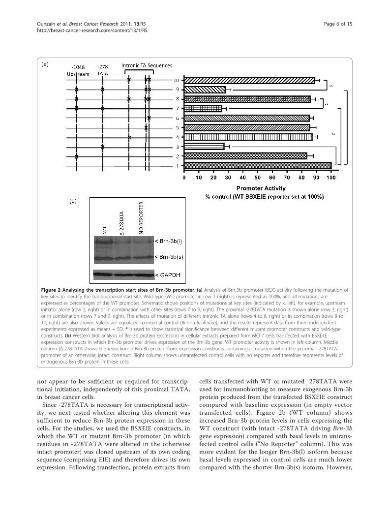
not appear to be sufficient or required for transcrip-tional initiation, independently of this proximal TATA,in breast cancer cells.Since -278TATA is necessary for transcriptional activ-
ity, we next tested whether altering this element wassufficient to reduce Brn-3b protein expression in thesecells. For the studies, we used the BSXEIE constructs, inwhich the WT or mutant Brn-3b promoter (in whichresidues in -278TATA were altered in the otherwiseintact promoter) was cloned upstream of its own codingsequence (comprising EIE) and therefore drives its ownexpression. Following transfection, protein extracts from
cells transfected with WT or mutated -278TATA wereused for immunoblotting to measure exogenous Brn-3bprotein produced from the transfected BSXEIE constructcompared with baseline expression (in empty vectortransfected cells). Figure 2b (WT column) showsincreased Brn-3b protein levels in cells expressing theWT construct (with intact -278TATA driving Brn-3bgene expression) compared with basal levels in untrans-fected control cells ("No Reporter” column). This wasmore evident for the longer Brn-3b(l) isoform becausebasal levels expressed in control cells are much lowercompared with the shorter Brn-3b(s) isoform. However,
Figure 2 Analysing the transcription start sites of Brn-3b promoter. (a) Analysis of Brn-3b promoter (BSX) activity following the mutation ofkey sites to identify the transcriptional start site. Wild-type (WT) promoter in row 1 (right) is represented as 100%, and all mutations areexpressed as percentages of the WT promoter. Schematic shows positions of mutations at key sites (indicated by x, left), for example, upstreaminitiator alone (row 2, right) or in combination with other sites (rows 7 to 9, right). The proximal -278TATA mutation is shown alone (row 3, right)or in combination (rows 7 and 9, right). The effects of mutation of different intronic TA alone (rows 4 to 6, right) or in combination (rows 8 to10, right) are also shown. Values are equalised to internal control (Renilla luciferase), and the results represent data from three independentexperiments expressed as means + SD. * is used to show statistical significance between different mutant promoter constructs and wild-typeconstructs. (b) Western blot analysis of Brn-3b protein expression in cellular extracts prepared from MCF7 cells transfected with BSXE1Eexpression constructs in which Brn-3b promoter drives expression of the Brn-3b gene. WT promoter activity is shown in left column. Middlecolumn (Δ-278TATA shows the reduction in Brn-3b protein from expression constructs containing a mutation within the proximal -278TATApromoter of an otherwise, intact construct. Right column shows untransfected control cells with no reporter and therefore represents levels ofendogenous Brn-3b protein in these cells.
Ounzain et al. Breast Cancer Research 2011, 13:R5http://breast-cancer-research.com/content/13/1/R5
Page 6 of 15
mutation of -278TATA (in the otherwise intact con-struct) resulted in loss of this induction of Brn-3b pro-tein (Δ-278TATA column) since levels were similar toendogenous expression in control cells. On the basis ofthe results of these different studies, we concluded thatthe proximal TATA located at position -278 from ATG(-278TATA) marks the transcription start site for Brn-3b transcription breast cancer cells.
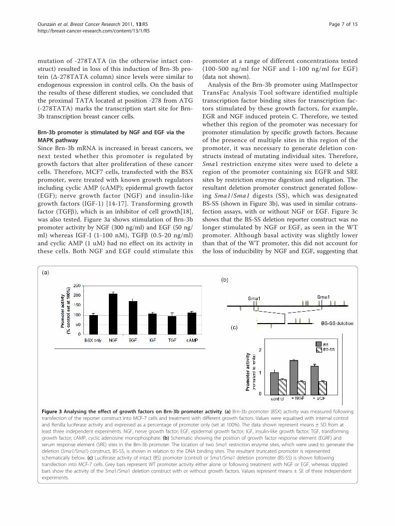
Brn-3b promoter is stimulated by NGF and EGF via theMAPK pathwaySince Brn-3b mRNA is increased in breast cancers, wenext tested whether this promoter is regulated bygrowth factors that alter proliferation of these cancercells. Therefore, MCF7 cells, transfected with the BSXpromoter, were treated with known growth regulatorsincluding cyclic AMP (cAMP); epidermal growth factor(EGF); nerve growth factor (NGF) and insulin-likegrowth factors (IGF-1) [14-17]. Transforming growthfactor (TGFb), which is an inhibitor of cell growth[18],was also tested. Figure 3a shows stimulation of Brn-3bpromoter activity by NGF (300 ng/ml) and EGF (50 ng/ml) whereas IGF-I (1-100 nM), TGFb (0.5-20 ng/ml)and cyclic AMP (1 uM) had no effect on its activity inthese cells. Both NGF and EGF could stimulate this
promoter at a range of different concentrations tested(100-500 ng/ml for NGF and 1-100 ng/ml for EGF)(data not shown).Analysis of the Brn-3b promoter using MatInspector
TransFac Analysis Tool software identified multipletranscription factor binding sites for transcription fac-tors stimulated by these growth factors, for example,EGR and NGF induced protein C. Therefore, we testedwhether this region of the promoter was necessary forpromoter stimulation by specific growth factors. Becauseof the presence of multiple sites in this region of thepromoter, it was necessary to generate deletion con-structs instead of mutating individual sites. Therefore,Sma1 restriction enzyme sites were used to delete aregion of the promoter containing six EGFR and SREsites by restriction enzyme digestion and religation. Theresultant deletion promoter construct generated follow-ing Sma1/Sma1 digests (SS), which was designatedBS-SS (shown in Figure 3b), was used in similar cotrans-fection assays, with or without NGF or EGF. Figure 3cshows that the BS-SS deletion reporter construct was nolonger stimulated by NGF or EGF, as seen in the WTpromoter. Although basal activity was slightly lowerthan that of the WT promoter, this did not account forthe loss of inducibility by NGF and EGF, suggesting that
Figure 3 Analysing the effect of growth factors on Brn-3b promoter activity. (a) Brn-3b promoter (BSX) activity was measured followingtransfection of the reporter construct into MCF-7 cells and treatment with different growth factors. Values were equalised with internal controland Renilla luciferase activity and expressed as a percentage of promoter only (set at 100%). The data shown represent means ± SD from atleast three independent experiments. NGF, nerve growth factor; EGF, epidermal growth factor; IGF, insulin-like growth factor; TGF, transforminggrowth factor; cAMP, cyclic adenosine monophosphate. (b) Schematic showing the position of growth factor response element (EGRF) andserum response element (SRE) sites in the Brn-3b promoter. The location of two Sma1 restriction enzyme sites, which were used to generate thedeletion (Sma1/Sma1) construct, BS-SS, is shown in relation to the DNA binding sites. The resultant truncated promoter is representedschematically below. (c) Luciferase activity of intact (BS) promoter (control) or Sma1/Sma1 deletion promoter (BS-SS) is shown followingtransfection into MCF-7 cells. Grey bars represent WT promoter activity either alone or following treatment with NGF or EGF, whereas stippledbars show the activity of the Sma1/Sma1 deletion construct with or without growth factors. Values represent means ± SE of three independentexperiments.
Ounzain et al. Breast Cancer Research 2011, 13:R5http://breast-cancer-research.com/content/13/1/R5
Page 7 of 15
key DNA binding sites present in this region are essen-tial for increasing promoter activity in breast cancercells.NGF and EGF act as ligands, which, when bound to
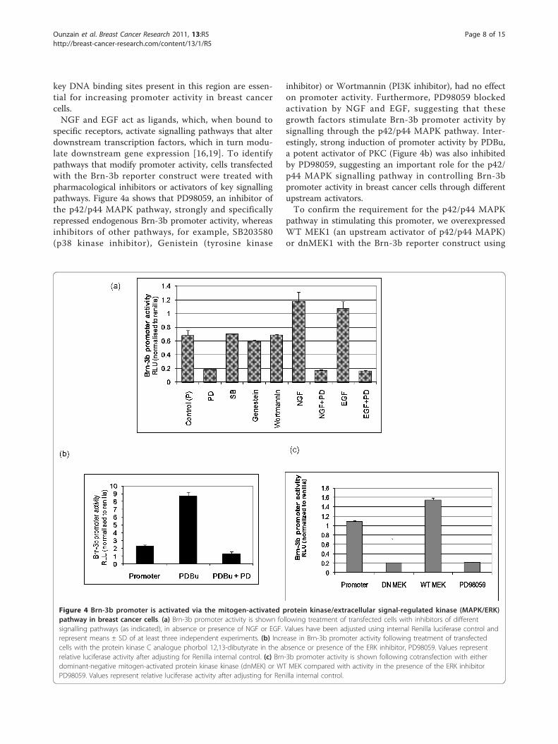
specific receptors, activate signalling pathways that alterdownstream transcription factors, which in turn modu-late downstream gene expression [16,19]. To identifypathways that modify promoter activity, cells transfectedwith the Brn-3b reporter construct were treated withpharmacological inhibitors or activators of key signallingpathways. Figure 4a shows that PD98059, an inhibitor ofthe p42/p44 MAPK pathway, strongly and specificallyrepressed endogenous Brn-3b promoter activity, whereasinhibitors of other pathways, for example, SB203580(p38 kinase inhibitor), Genistein (tyrosine kinase
inhibitor) or Wortmannin (PI3K inhibitor), had no effecton promoter activity. Furthermore, PD98059 blockedactivation by NGF and EGF, suggesting that thesegrowth factors stimulate Brn-3b promoter activity bysignalling through the p42/p44 MAPK pathway. Inter-estingly, strong induction of promoter activity by PDBu,a potent activator of PKC (Figure 4b) was also inhibitedby PD98059, suggesting an important role for the p42/p44 MAPK signalling pathway in controlling Brn-3bpromoter activity in breast cancer cells through differentupstream activators.To confirm the requirement for the p42/p44 MAPK
pathway in stimulating this promoter, we overexpressedWT MEK1 (an upstream activator of p42/p44 MAPK)or dnMEK1 with the Brn-3b reporter construct using
Figure 4 Brn-3b promoter is activated via the mitogen-activated protein kinase/extracellular signal-regulated kinase (MAPK/ERK)pathway in breast cancer cells. (a) Brn-3b promoter activity is shown following treatment of transfected cells with inhibitors of differentsignalling pathways (as indicated), in absence or presence of NGF or EGF. Values have been adjusted using internal Renilla luciferase control andrepresent means ± SD of at least three independent experiments. (b) Increase in Brn-3b promoter activity following treatment of transfectedcells with the protein kinase C analogue phorbol 12,13-dibutyrate in the absence or presence of the ERK inhibitor, PD98059. Values representrelative luciferase activity after adjusting for Renilla internal control. (c) Brn-3b promoter activity is shown following cotransfection with eitherdominant-negative mitogen-activated protein kinase kinase (dnMEK) or WT MEK compared with activity in the presence of the ERK inhibitorPD98059. Values represent relative luciferase activity after adjusting for Renilla internal control.
Ounzain et al. Breast Cancer Research 2011, 13:R5http://breast-cancer-research.com/content/13/1/R5
Page 8 of 15
cotransfection protocols. Figure 4c shows that increasingWT MEK1 could stimulate endogenous promoter activ-ity, whereas the dnMEK1 construct reduced basal pro-moter activity to levels seen with PD98059 treatment.Thus, Brn-3b promoter activity can be inhibited byblocking the MAPK/extracellular signal-regulated kinase(ERK) pathway by using either pharmacological inhibi-tors or dnMEK, thereby identifying the MAPK/ERKpathway as a pivotal regulator of Brn-3b expression inbreast cancer cells.
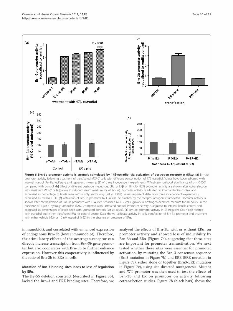
Activation of Brn-3b promoter by the hormone 17b-estradiol occurs via ERa but not ERbThe hormone oestrogen plays a critical role in the initia-tion and progression of many breast cancers becausebreast epithelial cells are highly responsive to its prolif-erative effects. Therefore, we tested whether active oes-trogen (17b-estradiol) could stimulate Brn-3b promoteractivity using MCF-7 cells sensitized to estradiol bygrowth in stripped-serum, phenol-red less DMEM [9].Cells transfected with the Brn-3b promoter constructwere either untreated or treated with different concen-trations of 17b-estradiol. Figure 5a shows that 17b-estra-diol significantly increased promoter activity comparedwith untreated cells, suggesting that this hormone canstimulate Brn-3b transcription in breast cancer cells,thereby contributing to downstream oestrogenic growtheffects.Estradiol can act through one of two receptors: ERa
or ERb. Of these, increased ERa is implicated in theetiology of breast cancers and is often targeted for treat-ment. We therefore tested the effects of coexpressingeither ERa or ERb on Brn-3b promoter activity.Figure 5b shows that the promoter was strongly stimu-lated by ERa, whereas ERb did not alter its activity, sug-gesting that the effects of oestrogen in breast cancercells are likely to be mediated via ERa. As expected, theaddition of the ER antagonist tamoxifen prevented acti-vation of the Brn-3b promoter by oestrogen (Figure 5c),thus confirming that this receptor is required for stimu-lation of Brn-3b promoter activity in MCF-7 cells. Thisfinding was further supported by studies carried out inER-negative Cos-7 cells, which showed that estradiol didnot activate the Brn-3b promoter unless exogenous ERwas introduced following transfection (Figure 5d). Theseresults suggest that ERa is necessary to mediate theeffects of oestrogens in MCF-7 breast cancer cells butcan also act independently of oestrogen to increase Brn-3b transcription.
Autoregulation by Brn-3b and cooperation with ERa alsoincreases promoter activityTRANSFAC software analysis revealed binding sites forBrn-3 proteins, suggesting that Brn-3b and/or a related
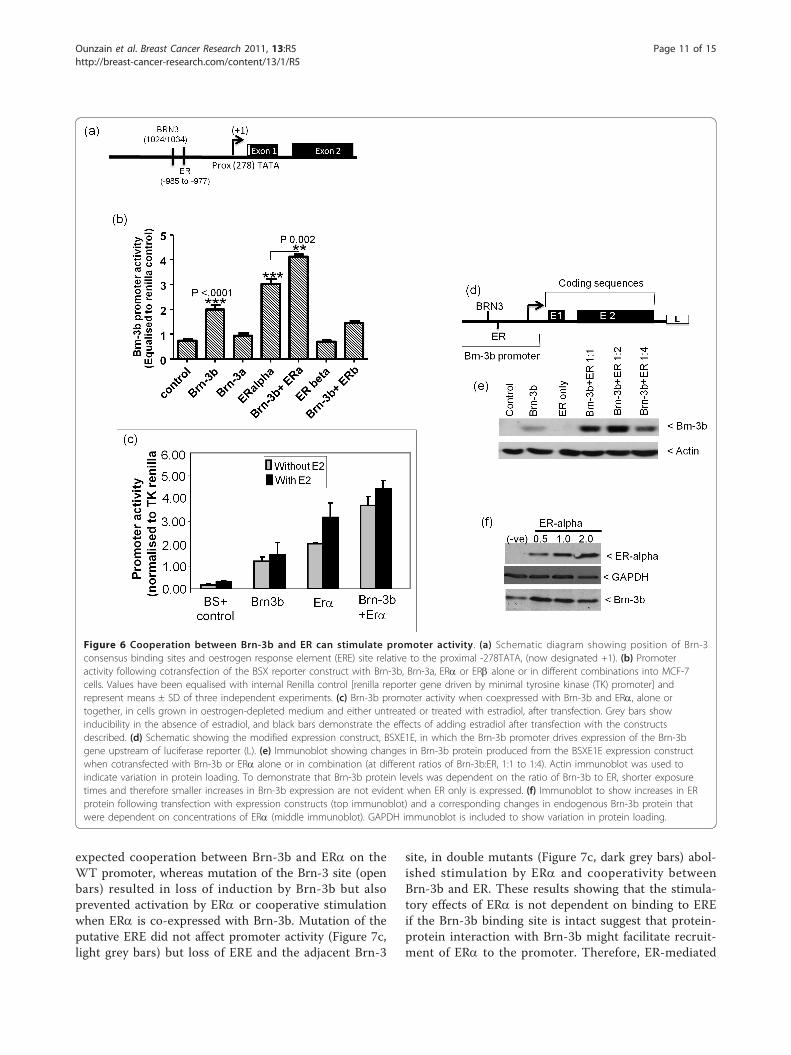
family member, Brn-3a, may also regulate promoteractivity. A putative ERE site was identified within proxi-mity to this site (Figure 6a), and since previous studiesdemonstrated physical interaction between Brn-3b andERa that could stimulate transcription of ERE-contain-ing target genes, we tested whether Brn-3b could regu-late its own promoter activity and cooperate with ERato increase its own expression.Figure 6b shows that Brn-3b could weakly transacti-
vate its own promoter, whereas the related Brn-3a pro-tein had no effect on promoter activity in these cells.Although ERa alone stimulated promoter activity, coex-pression of this receptor with Brn-3b resulted in moresignificant increases. ERb did not affect promoter activ-ity with or without Brn-3b, suggesting that a specificand unique cooperation occurs between ERa and Brn-3b to stimulate the Brn-3b promoter in breast cancercells. Studies carried out in sensitised MCF7 cells grownin “phenol red less” DMEM, containing stripped serum,to deplete oestrogenic activity, shows that exogenous(transfected) ERa could to stimulate Brn-3b promoterin the absence or presence of estradiol and also coop-erated with Brn-3b to further enhance promoter activity(Figure 6c). These results suggest that stimulation ofBrn-3b promoter by ERa can occur independently ofestradiol stimulation. We also tested whether increasedpromoter activation caused by the coexpression of Brn-3b and ERa could also result in enhanced proteinexpression. For this study, we used the modifiedBSXE1E construct (Figure 6d), in which the Brn-3b pro-moter, (containing Brn-3 and ERE elements), drivesexpression of its own coding sequence. This BSXEIEconstruct was cotransfected with Brn-3b or ERa expres-sion vectors, alone or together, into MCF-7 cells. Pro-teins extracted from transfected cells after 48 hourswere used for immunoblotting to detect Brn-3b protein.Figure 6e shows that transfected cells coexpressing exo-genous Brn-3b and ERa produced higher levels of Brn-3b protein than basal levels in control cells (column 1)or in cells transfected with Brn-3b alone (column 3),where the band represent exogenous as well as endo-genous Brn-3b proteins. Thus, coexpression of Brn-3bwith ERa at ratios of 1:1 and 1:2 (Brn-3b:ERa) resultedin increased Brn-3b protein, but further increases inERa (1:4 ratio) resulted in reduced protein levels, whichis suggestive of squelching. To demonstrate thissquelching effect, we needed to show reduction of Brn-3b protein expression at the higher ratio and this wasachieved by reducing exposure times. However, underthose conditions, the increases in endogenous Brn-3b fol-lowing transfection with ERa only were not evident inFigure 6e but can be seen in Figure 6f. Thus, transfectingincreasing amounts of ERa expression vector (0.5 -2.0μg) resulted in increased ERa protein (Figure 6f - top
Ounzain et al. Breast Cancer Research 2011, 13:R5http://breast-cancer-research.com/content/13/1/R5
Page 9 of 15
immunoblot), and correlated with enhanced expressionof endogenous Brn-3b (lower immunoblot). Therefore,the stimulatory effects of the oestrogen receptor candirectly increase transcription from Brn-3b gene promo-ter but also cooperates with Brn-3b to further enhanceexpression. However this cooperativity is influenced bythe ratio of Brn-3b to ERa in cells.
Mutation of Brn-3 binding sites leads to loss of regulationby ERaThe BS-SS deletion construct (described in Figure 3b),lacked the Brn-3 and ERE binding sites. Therefore, we
analysed the effects of Brn-3b, with or without ERa, onpromoter activity and showed loss of inducibility byBrn-3b and ERa (Figure 7a), suggesting that these sitesare important for promoter transactivation. We nexttested whether these sites were essential for promoteractivation, by mutating the Brn-3 consensus sequence(Brn3 mutation in Figure 7b) and ERE (ERE mutation inFigure 7c), either alone or together (Brn3-ERE mutationin Figure 7c), using site-directed mutagenesis. Mutantand WT promoter was then used to test the effects ofBrn-3b and ER on promoter on activity followingcotransfection studies. Figure 7b (black bars) shows the
Figure 5 Brn-3b promoter activity is strongly stimulated by 17b-estradiol via activation of oestrogen receptor a (ERa). (a) Brn-3bpromoter activity following treatment of transfected MCF-7 cells with different concentration of 17b-estradiol. Values have been adjusted withinternal control, Renilla luciferase and represent means ± SD of three independent experiments ***indicate statistical significance of p < 0.0001compared with control. (b) Effect of different oestrogen receptors, ERa or ERb on Brn-3b (BSX) promoter activity are shown after cotransfectioninto sensitised MCF-7 cells (grown in stripped serum medium for 48 hours). Promoter activity is adjusted to internal Renilla control andexpressed as percentage of levels seen with empty vector only (set at 100%). Values represent data from three independent experiments,expressed as means ± SD. (c) Activation of Brn-3b promoter by ERa can be blocked by the receptor antagonist tamoxifen. Promoter activity isshown after cotransfection of Brn-3b promoter with ERa into sensitised MCF-7 cells (grown in oestrogen-depleted medium for 48 hours) in thepresence of 1 μM 4 hydroxy tamoxifen (TAM) compared with untreated control. Promoter activity is adjusted to internal Renilla control andexpressed as percentages of levels seen with untreated controls (set at 100%). (d) Brn-3b promoter activity in ER-negative Cos-7 cells treatedwith estradiol and either transfected ERa or control vector. Data shows luciferase activity in cells transfection of Brn-3b promoter and treatmentwith either vehicle (-E2) or 10 nM estradiol (+E2) in the absence or presence of ERa.
Ounzain et al. Breast Cancer Research 2011, 13:R5http://breast-cancer-research.com/content/13/1/R5
Page 10 of 15
expected cooperation between Brn-3b and ERa on theWT promoter, whereas mutation of the Brn-3 site (openbars) resulted in loss of induction by Brn-3b but alsoprevented activation by ERa or cooperative stimulationwhen ERa is co-expressed with Brn-3b. Mutation of theputative ERE did not affect promoter activity (Figure 7c,light grey bars) but loss of ERE and the adjacent Brn-3
site, in double mutants (Figure 7c, dark grey bars) abol-ished stimulation by ERa and cooperativity betweenBrn-3b and ER. These results showing that the stimula-tory effects of ERa is not dependent on binding to EREif the Brn-3b binding site is intact suggest that protein-protein interaction with Brn-3b might facilitate recruit-ment of ERa to the promoter. Therefore, ER-mediated
Figure 6 Cooperation between Brn-3b and ER can stimulate promoter activity. (a) Schematic diagram showing position of Brn-3consensus binding sites and oestrogen response element (ERE) site relative to the proximal -278TATA, (now designated +1). (b) Promoteractivity following cotransfection of the BSX reporter construct with Brn-3b, Brn-3a, ERa or ERb alone or in different combinations into MCF-7cells. Values have been equalised with internal Renilla control [renilla reporter gene driven by minimal tyrosine kinase (TK) promoter] andrepresent means ± SD of three independent experiments. (c) Brn-3b promoter activity when coexpressed with Brn-3b and ERa, alone ortogether, in cells grown in oestrogen-depleted medium and either untreated or treated with estradiol, after transfection. Grey bars showinducibility in the absence of estradiol, and black bars demonstrate the effects of adding estradiol after transfection with the constructsdescribed. (d) Schematic showing the modified expression construct, BSXE1E, in which the Brn-3b promoter drives expression of the Brn-3bgene upstream of luciferase reporter (L). (e) Immunoblot showing changes in Brn-3b protein produced from the BSXE1E expression constructwhen cotransfected with Brn-3b or ERa alone or in combination (at different ratios of Brn-3b:ER, 1:1 to 1:4). Actin immunoblot was used toindicate variation in protein loading. To demonstrate that Brn-3b protein levels was dependent on the ratio of Brn-3b to ER, shorter exposuretimes and therefore smaller increases in Brn-3b expression are not evident when ER only is expressed. (f) Immunoblot to show increases in ERprotein following transfection with expression constructs (top immunoblot) and a corresponding changes in endogenous Brn-3b protein thatwere dependent on concentrations of ERa (middle immunoblot). GAPDH immunoblot is included to show variation in protein loading.
Ounzain et al. Breast Cancer Research 2011, 13:R5http://breast-cancer-research.com/content/13/1/R5
Page 11 of 15
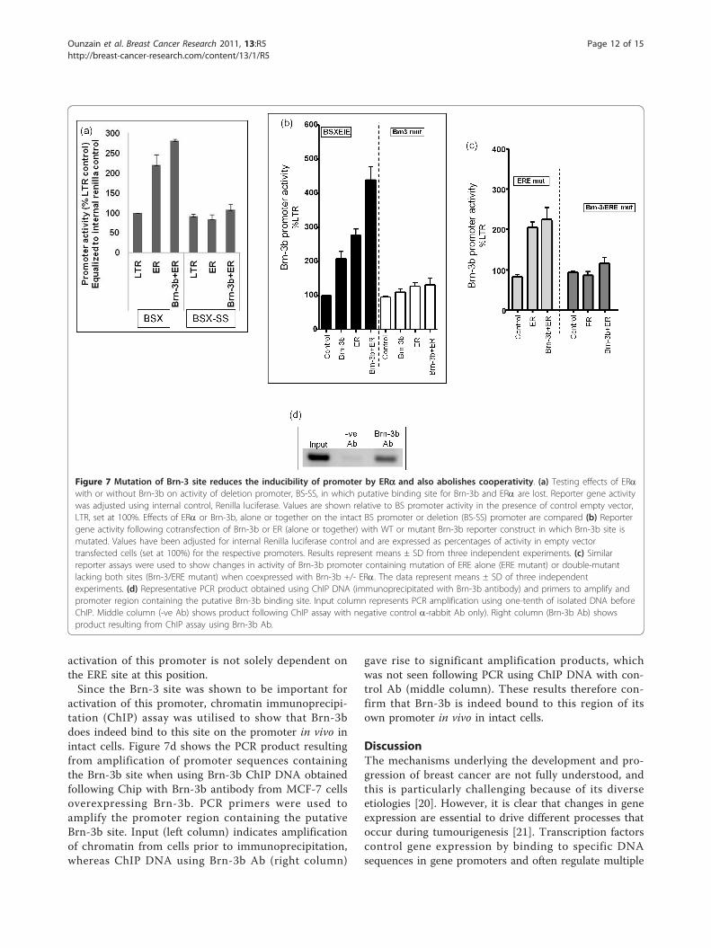
activation of this promoter is not solely dependent onthe ERE site at this position.Since the Brn-3 site was shown to be important for
activation of this promoter, chromatin immunoprecipi-tation (ChIP) assay was utilised to show that Brn-3bdoes indeed bind to this site on the promoter in vivo inintact cells. Figure 7d shows the PCR product resultingfrom amplification of promoter sequences containingthe Brn-3b site when using Brn-3b ChIP DNA obtainedfollowing Chip with Brn-3b antibody from MCF-7 cellsoverexpressing Brn-3b. PCR primers were used toamplify the promoter region containing the putativeBrn-3b site. Input (left column) indicates amplificationof chromatin from cells prior to immunoprecipitation,whereas ChIP DNA using Brn-3b Ab (right column)
gave rise to significant amplification products, whichwas not seen following PCR using ChIP DNA with con-trol Ab (middle column). These results therefore con-firm that Brn-3b is indeed bound to this region of itsown promoter in vivo in intact cells.
DiscussionThe mechanisms underlying the development and pro-gression of breast cancer are not fully understood, andthis is particularly challenging because of its diverseetiologies [20]. However, it is clear that changes in geneexpression are essential to drive different processes thatoccur during tumourigenesis [21]. Transcription factorscontrol gene expression by binding to specific DNAsequences in gene promoters and often regulate multiple
Figure 7 Mutation of Brn-3 site reduces the inducibility of promoter by ERa and also abolishes cooperativity. (a) Testing effects of ERawith or without Brn-3b on activity of deletion promoter, BS-SS, in which putative binding site for Brn-3b and ERa are lost. Reporter gene activitywas adjusted using internal control, Renilla luciferase. Values are shown relative to BS promoter activity in the presence of control empty vector,LTR, set at 100%. Effects of ERa or Brn-3b, alone or together on the intact BS promoter or deletion (BS-SS) promoter are compared (b) Reportergene activity following cotransfection of Brn-3b or ER (alone or together) with WT or mutant Brn-3b reporter construct in which Brn-3b site ismutated. Values have been adjusted for internal Renilla luciferase control and are expressed as percentages of activity in empty vectortransfected cells (set at 100%) for the respective promoters. Results represent means ± SD from three independent experiments. (c) Similarreporter assays were used to show changes in activity of Brn-3b promoter containing mutation of ERE alone (ERE mutant) or double-mutantlacking both sites (Brn-3/ERE mutant) when coexpressed with Brn-3b +/- ERa. The data represent means ± SD of three independentexperiments. (d) Representative PCR product obtained using ChIP DNA (immunoprecipitated with Brn-3b antibody) and primers to amplify andpromoter region containing the putative Brn-3b binding site. Input column represents PCR amplification using one-tenth of isolated DNA beforeChIP. Middle column (-ve Ab) shows product following ChIP assay with negative control a-rabbit Ab only). Right column (Brn-3b Ab) showsproduct resulting from ChIP assay using Brn-3b Ab.
Ounzain et al. Breast Cancer Research 2011, 13:R5http://breast-cancer-research.com/content/13/1/R5
Page 12 of 15
target genes. Because of this ability to control differenttarget genes, deregulation of transcription factors candrive events associated with the initiation and progres-sion of diseases such as cancer [22]. Previous studieshave shown that the Brn-3b transcription factor is ele-vated in >60% of primary breast cancers [1], and whenincreased, it significantly enhances proliferation andanchorage-independent growth in vitro and tumourgrowth in vivo [2,3]. Elevated Brn-3b also confers resis-tance to growth-inhibitory stimuli and increases themigratory potential of cancer cells [2], suggesting thatthis transcription factor acts through complex mechan-isms in cancer cells. More recent studies have shownincreases in Brn-3b in drug-resistant, migratory breastcancer cells (unpublished data, R. Fujita and V. Budhram-Mahadeo). The Brn-3b can give rise to such diverse effectsbecause it regulates different subsets of target genes thatcontrol distinct aspects of cellular growth and behavior.For example, Brn-3b might contribute to cellular prolifera-tion by transactivating the promoters of cell cycle regula-tors, CDK4 [4] and cyclin D1 [5] whilst repressing thetumour suppressor, BRCA1 [6]. However, its effects ondrug resistance and migration are likely to be associatedwith the ability of Brn-3b to regulate other genes, forexample, to transactivate Hsp27 [7] whilst repressing adhe-sion molecules, for example, g-catenin [8].Interestingly, reducing Brn-3b was sufficient to change
gene expression and reverse many growth effects [1].Therefore, Brn-3b can act as a master regulator whoseexpression profoundly alters the growth of cancer cells.In this regard, Brn-3b might represent an importanttherapeutic target whose reduction could alter theexpression of multiple downstream target genes andthereby reverse their effects on cancer cells. However, toidentify strategies for reducing Brn-3b in these cells, wemust understand the mechanisms that lead to itsincreased expression in breast cancer cells.In this study, we utilised bioinformatics analysis to
identify the putative Brn-3b promoter and cloned thisregulatory region into a reporter construct for furtherexperimental analysis. By using ChIP assays and site-directed mutagenesis, we identified a key TATA tran-scriptional start site located at ~278 bp from ATG,which is primarily associated with the expression ofBrn-3b mRNA in breast cancer cells. Although theupstream initiation site and TA-like elements in theintronic sequence were weakly immunoprecipitated byTBP Ab, these do not appear to be candidates for tran-scriptional start sites, since the mutation of any or allintronic TA sequences or upstream sequences did notreduce promoter activity, if the start site at -278TATAwas intact. This is interesting because an intronic pro-moter is thought to be important to drive isoform-speci-fic expression of the related Brn-3a gene, which has a
genomic arrangement similar to that of Brn-3b. How-ever, our results suggest that Brn-3b promoter activityin breast cancer cells is driven primarily from the proxi-mal -278TATA site, which is now used to define thetranscription start site from this promoter.Further analysis showed that the Brn-3b promoter can
be stimulated by specific growth factors, NGF and EGF,but not by IGF-1, cAMP or TGFb, and these stimula-tory effects require a region of promoter that containsmultiple EGFR and SRE sites. The ability of growth fac-tors such as NGF to increase transcription from theBrn-3b promoter is significant because NGF is knownto enhance the growth and drive proliferation of breastcancer cells but not of normal breast epithelial cells.Moreover, blocking NGF can inhibit tumour growthand metastasis [16,23], suggesting a key role for NGF incontrolling the growth of cancer but not of normal cells.NGF is produced in an autocrine manner by breast can-cer cells, and its mitogenic effects in these cells aremediated through the p42/p44 MAPK signalling path-way, since these effects can be blocked by the pharma-cological inhibitor PD98059, which targets MEK1 in thispathway [24]. In this study, we showed that stimulationof the Brn-3b promoter by NGF is blocked by PD98059,suggesting that the mitogenic effects of NGF in breastcancer cells may result in part from its ability toincrease the expression of regulators such as Brn-3b.The PKC analogue PDBu is also a potent activator ofthe Brn-3b promoter, and its effects can also be blockedby PD98059, suggesting that this activator converges onthe p42/p44 MAPK/ERK1 pathway to stimulate Brn-3bpromoter activity. Dominant negative (Dn) MEK alsoblocked endogenous Brn-3b promoter activity, in a man-ner that is similar to the ERK1 inhibitor, PD98059. Thusit would appear that the p42/p44 MAPK/ERK pathwayis pivotal for activating the Brn-3b promoter and henceexpression in breast cancer cells.In addition to stimulation by growth factors, the Brn-
3b promoter is strongly activated by the hormone estra-diol, which regulates the growth and proliferation ofnormal breast epithelium as well as breast cancer cellsand is important in the etiology of breast cancer [25].Oestrogens can regulate gene transcription by actingthrough one of two receptors: ERa or ERb. Our resultsshow that overexpression of ERa but not ERb couldstrongly stimulate Brn-3b promoter activity. ERa is par-ticularly relevant for the development and progressionof breast cancers because it is overexpressed in a signifi-cant proportion of breast cancers (> 60%). Furthermore,ER-positive breast cancers are often treated using recep-tor antagonists, for example, tamoxifen, as a first line oftherapy aimed at blocking ER-mediated proliferativeeffects [26]. Therefore, the ability of ERa to stimulateBrn-3b suggests that the proliferative effects of high ER
Ounzain et al. Breast Cancer Research 2011, 13:R5http://breast-cancer-research.com/content/13/1/R5
Page 13 of 15
levels may be associated with the ability of ERa to trans-activate other regulators, such as Brn-3b, which in turncan modulate genes associated with growth in thesecancer cells either alone or by cooperating with ERa.The complexity underlying the regulation of the Brn-
3b promoter is increased by autoregulation, wherebyBrn-3b can weakly stimulate its own expression by bind-ing to recognition sequences present in its promoter.However, cooperation between Brn-3b and ERa couldfurther enhance promoter activity. Such cooperationbetween Brn-3b and ERa to increase gene expressionwas previously observed on other ERE-containing targetpromoters, for example, HSP27, where Brn-3b stimu-lates expression directly by binding to specific sites inthe promoter or indirectly by interacting and cooperat-ing with ER to maximally activate this promoter [7].This ability of Brn-3b to cooperate with ERa to enhancegene expression [9], including its own, is clearly relevantto breast cancer because ER-expressing tumours thatare responsive to estradiol will stimulate Brn-3b, whichcan cooperate with ERa to further increase its ownexpression. Interestingly, mutation of the putative EREdid not prevent ER-mediated promoter activation whencoexpressed with Brn-3b, but mutation of the nearbyBrn-3 site abolished activation by ER and its cooperationwith Brn-3b. This indicates that ERa could stimulateBrn-3b promoter even if it is not bound to ERE, possiblybecause interaction with Brn-3b allows recruitment ofER to the promoter. Autoregulation of Brn-3b transcrip-tion, either alone or by cooperating with ER, is likely toincrease Brn-3b protein expression and subsequently, itstarget genes in these cells.Although stimulation of Brn-3b promoter activity by
the hormone oestrogen via ERa is likely to act indepen-dently and possibly, in parallel with growth factor-mediated promoter activation via the p42/p44 MAPKsignalling, there is also significant “cross-talk” betweenthese pathways in breast cancer cells. Thus, estradiolprimarily acts through its receptor, ERa, in breast can-cer cells, but it can also indirectly stimulate tyrosinekinase receptors, which are also relevant to breast can-cer cells. Similarly, transcriptional activity of oestrogenreceptor, ERa, is also modulated by p42/p44 MAPKpathway stimulation [27]. Evidence for cross-talkbetween NGF or EGF and the estradiol pathways hasalso been demonstrated [28], and in this regard, theanti-oestrogenic drug tamoxifen can inhibit proliferationby EGF or NGF on MCF-7 breast cancer cells [29].Therefore, diverse pathways, which are stimulated byeither hormone or growth factor may act in parallel orconverge to stimulate Brn-3b promoter activity andhence increase its expression in breast cancer cells. Evi-dence for autoregulation by Brn-3b and cooperationwith ERa to increase drive its own promoter activity,
would suggest that under such circumstances, this feed-back loop will maintain high Brn-3b expression. Whenelevated, Brn-3b is likely to alter the expression of mul-tiple downstream target genes, thereby affecting growthand behaviour in these cancer cells.
ConclusionsElevated Brn-3b profoundly enhances tumour growthand confers drug resistance in breast cancer cells, so itis important to identify which factors increase itsexpression in these cells. In the present studies, we havecloned and analysed the Brn-3b promoter. Furthermore,we have identified key pathways that converge on itspromoter to increase activity and hence gene and pro-tein expression in breast cancer cells. Thus, the hor-mone oestrogen and the growth factors NGF and EGFstimulate the activity of the Brn-3b promoter and subse-quently, Brn-3b mRNA and protein expression, suggest-ing that induction of Brn-3b by such factors will beimportant in changing the fate of these cells. IncreasedBrn-3b expression via growth factors such as NGF andEGF or the hormone, estradiol, which are implicated inenhancing the growth of breast cancer cells, are likely tobe are propagated by autoregulation. This will lead tochanges in multiple Brn-3b target genes which controlthe growth and behaviour of cancer cells. By elucidatingthe mechanisms through which regulators such as Brn-3b are increased in cancer cells, we will increase theunderstanding of how changes are brought about duringthe development and progression of this disease, and wemay also be able to identify strategies to reduce itsexpression and reverse its effects in breast cancer cells.
AbbreviationsBrn-3b: transcription factor-related Brn-3a regulator isolated from braincDNA; BSX-Brn-3b: promoter containing BstX1/Stu1/Xho1 fragment; ChIP:chromatin immunoprecipitation; EGF: epidermal growth factor; ER: oestrogenreceptor; ERα: oestrogen receptor α; ERβ: oestrogen receptor β; ERE:oestrogen response element; GF: growth factor; MAPK: mitogen-activatedprotein kinase; NGF: nerve growth factor; PI3K: phosphoinositide 3-kinase;POU4F2: member of class 4 subgroup of Pit-Oct-Unc transcription factors;qRT-PCR: quantitative reverse transcriptase polymerase chain reaction; SRE:serum response element; TF: transcription factor; WT: wild type.
AcknowledgementsThis work was supported by the British Heart Foundation, Breast CancerCampaign UK and the Association for International Cancer Research UK.Dominant-negative and constitutively active MEK expression vectors werekind gifts from DS Latchman.
Author details1Medical Molecular Biology Unit, UCL Institute of Child Health, 30 GuilfordStreet, London WC1N 1EH, UK. 2Cardiovascular Division, King’s CollegeLondon, Department of Cardiology, The Rayne Institute, St Thomas’sHospital, Lambeth Palace Road, London SE1 7EH, UK.
Authors’ contributionsSO mapped transcription start sites, used site-directed mutagenesis and ChIPassays to identify key transcription factor binding sites and helped toidentify signalling pathways associated with the regulation of promoter
Ounzain et al. Breast Cancer Research 2011, 13:R5http://breast-cancer-research.com/content/13/1/R5
Page 14 of 15
activity. SB, CP and RF were involved in cloning and initial characterizationof the promoter. RJH provided reagents required for aspects of the studyand invaluable contribution to aspects of these studies. VBM is the principalinvestigator of the study and was instrumental in designing the study,interpreting and processing the data and preparing the manuscript forpublication. All authors read and approved the final manuscript.
Competing interestsThe authors declare that they have no competing interests.
Received: 9 July 2010 Revised: 14 October 2010Accepted: 17 January 2011 Published: 17 January 2011
References1. Budhram-Mahadeo VS, Latchman DS: Targeting Brn-3b in breast cancer
therapy. Expert Opin Ther Targets 2006, 10:15-25.2. Irshad S, Pedley RB, Anderson J, Latchman DS, Budhram-Mahadeo V: The
Brn-3b transcription factor regulates the growth, behavior, andinvasiveness of human neuroblastoma cells in vitro and in vivo. J BiolChem 2004, 279:21617-21627.
3. Dennis JH, Budhram-Mahadeo V, Latchman DS: The Brn-3b POU familytranscription factor regulates the cellular growth, proliferation, andanchorage dependence of MCF7 human breast cancer cells. Oncogene2001, 20:4961-4971.
4. Samady L, Dennis J, Budhram-Mahadeo V, Latchman DS: Activation ofCDK4 Gene Expression in Human Breast Cancer Cells by the Brn-3b POUFamily Transcription Factor. Cancer Biol Ther 2004, 3:317-323.
5. Budhram-Mahadeo VS, Irshad S, Bowen S, Lee SA, Samady L, Tonini GP,Latchman DS: Proliferation-associated Brn-3b transcription factor canactivate cyclin D1 expression in neuroblastoma and breast cancer cells.Oncogene 2008, 27:145-154.
6. Budhram-Mahadeo V, Ndisang D, Ward T, Weber BL, Latchman DS: TheBrn-3b POU family transcription factor represses expression of theBRCA-1 anti-oncogene in breast cancer cells. Oncogene 1999,18:6684-6691.
7. Lee SA, Ndisang D, Patel C, Dennis JH, Faulkes DJ, D’Arrigo C, Samady L,Farooqui-Kabir S, Heads RJ, Latchman DS, Budhram-Mahadeo VS:Expression of the Brn-3b transcription factor correlates with expressionof HSP-27 in breast cancer biopsies and is required for maximalactivation of the HSP-27 promoter. Cancer Res 2005, 65:3072-3080.
8. Samady L, Faulkes DJ, Budhram-Mahadeo V, Ndisang D, Potter E, Brabant G,Latchman DS: The Brn-3b POU family transcription factor repressesplakoglobin gene expression in human breast cancer cells. Int J Cancer2006, 118:869-878.
9. Budhram-Mahadeo V, Parker M, Latchman DS: POU transcription factorsBrn-3a and Brn-3b interact with the estrogen receptor and differentiallyregulate transcriptional activity via an estrogen response element. MolCell Biol 1998, 18:1029-1041.
10. Liu YZ, Boxer LM, Latchman DS: Activation of the Bcl-2 promoter bynerve growth factor is mediated by the p42/p44 MAPK cascade. NucleicAcids Res 1999, 27:2086-2090.
11. Frazer KA, Pachter L, Poliakov A, Rubin EM, Dubchak I: VISTA:computational tools for comparative genomics. Nucleic Acids Res 2004, 32:W273-W279.
12. Loots GG: Genomic identification of regulatory elements by evolutionarysequence comparison and functional analysis. Adv Genet 2008,61:269-293.
13. Burley SK, Roeder RG: Biochemistry and structural biology of transcriptionfactor IID (TFIID). Annu Rev Biochem 1996, 65:769-799.
14. Lanzino M, Morelli C, Garofalo C, Panno ML, Mauro L, Ando S, Sisci D:Interaction between estrogen receptor alpha and insulin/IGF signaling inbreast cancer. Curr Cancer Drug Targets 2008, 8:597-610.
15. Lo HW, Hsu SC, Hung MC: EGFR signaling pathway in breast cancers:from traditional signal transduction to direct nuclear translocalization.Breast Cancer Res Treat 2006, 95:211-218.
16. Descamps S, Toillon RA, Adriaenssens E, Pawlowski V, Cool SM,Nurcombe V, Le Bourhis X, Boilly B, Peyrat JP, Hondermarck H: Nervegrowth factor stimulates proliferation and survival of human breastcancer cells through two distinct signaling pathways. J Biol Chem 2001,276:17864-17870.
17. Descamps S, Pawlowski V, Revillion F, Hornez L, Hebbar M, Boilly B,Hondermarck H, Peyrat JP: Expression of nerve growth factor receptorsand their prognostic value in human breast cancer. Cancer Res 2001,61:4337-4340.
18. Buck MB, Knabbe C: TGF-beta signaling in breast cancer. Ann N Y Acad Sci2006, 1089:119-126.
19. Tagliabue E, Castiglioni F, Ghirelli C, Modugno M, Asnaghi L, Somenzi G,Melani C, Menard S: Nerve growth factor cooperates with p185(HER2) inactivating growth of human breast carcinoma cells. J Biol Chem 2000,275:5388-5394.
20. Veronesi U, Boyle P, Goldhirsch A, Orecchia R, Viale G: Breast cancer. Lancet2005, 365:1727-1741.
21. Cleator S, Ashworth A: Molecular profiling of breast cancer: clinicalimplications. Br J Cancer 2004, 90:1120-1124.
22. Liu S, Dontu G, Wicha MS: Mammary stem cells, self-renewal pathways,and carcinogenesis. Breast Cancer Res 2005, 7:86-95.
23. Descamps S, Lebourhis X, Delehedde M, Boilly B, Hondermarck H: Nervegrowth factor is mitogenic for cancerous but not normal human breastepithelial cells. J Biol Chem 1998, 273:16659-16662.
24. Melck D, Rueda D, Galve-Roperh I, De PL, Guzman M, Di MV: Involvementof the cAMP/protein kinase A pathway and of mitogen-activated proteinkinase in the anti-proliferative effects of anandamide in human breastcancer cells. FEBS Lett 1999, 463:235-240.
25. Sommer S, Fuqua SA: Estrogen receptor and breast cancer. Semin CancerBiol 2001, 11:339-352.
26. Iwase H: Molecular action of the estrogen receptor and hormonedependency in breast cancer. Breast Cancer 2003, 10:89-96.
27. Hart LL, Davie JR: The estrogen receptor: more than the averagetranscription factor. Biochem Cell Biol 2002, 80:335-341.
28. Kato S: Estrogen receptor-mediated cross-talk with growth factorsignaling pathways. Breast Cancer 2001, 8:3-9.
29. Chiarenza A, Lazarovici P, Lempereur L, Cantarella G, Bianchi A, Bernardini R:Tamoxifen inhibits nerve growth factor-induced proliferation of thehuman breast cancerous cell line MCF-7. Cancer Res 2001, 61:3002-3008.
doi:10.1186/bcr2809Cite this article as: Ounzain et al.: Proliferation-associated POU4F2/Brn-3b transcription factor expression is regulated by oestrogen throughERa and growth factors via MAPK pathway. Breast Cancer Research 201113:R5.
Submit your next manuscript to BioMed Centraland take full advantage of:
• Convenient online submission
• Thorough peer review
• No space constraints or color figure charges
• Immediate publication on acceptance
• Inclusion in PubMed, CAS, Scopus and Google Scholar
• Research which is freely available for redistribution
Submit your manuscript at www.biomedcentral.com/submit
Ounzain et al. Breast Cancer Research 2011, 13:R5http://breast-cancer-research.com/content/13/1/R5
Page 15 of 15
Copyright © 2022 FDOKUMEN