PKA-induced phosphorylation of ERα at serine 305 and high PAK1 levels is associated with...
38
1 PKA-induced phosphorylation of ERα at serine 305 and high PAK1 levels are associated with sensitivity to tamoxifen in ER positive breast cancer. Marleen Kok 1 , Wilbert Zwart 2 , Caroline Holm 3,4 , Renske Fles 1 , Michael Hauptmann 5 , Laura J. Van ’t Veer 6 , Lodewyk F.A. Wessels 5 , Jacques Neefjes 2 , Olle Stål 7 , Sabine C. Linn 8,9 , Göran Landberg 3,4 and Rob Michalides 2 Departments of Experimental Therapy 1 , Cell Biology 2 , Bioinformatics and Statistics 5 , Pathology 6 , Molecular Biology 8 and Medical Oncology 9 , Netherlands Cancer Institute, Amsterdam, the Netherlands, Center for Molecular Pathology 3 , Lund University, Malmö University Hospital, Malmö, Sweden, Breakthrough Breast Cancer Research Unit 4 , Paterson Institute for Cancer Research, Manchester, UK. Division of Oncology 7 , Department of Clinical and Experimental Medicine, Linköping University, Linköping, Sweden. Running title Sensitivity to tamoxifen is influenced by PKA/ERα S305P and PAK1 Disclaimers None Corresponding author Rob Michalides, Netherlands Cancer Institute Plesmanlaan 121, 1066 CX Amsterdam, The Netherlands Phone 0031 20 5122022/Fax 0031 20 5122029/[email protected] ABSTRACT peer-00554994, version 1 - 12 Jan 2011 Author manuscript, published in "Breast Cancer Research and Treatment 125, 1 (2010) 1-12" DOI : 10.1007/s10549-010-0798-y
Transcript of PKA-induced phosphorylation of ERα at serine 305 and high PAK1 levels is associated with...

1
PKA-induced phosphorylation of ERα at serine 305 and high PAK1
levels are associated with sensitivity to tamoxifen in ER positive breast
cancer.
Marleen Kok1, Wilbert Zwart2, Caroline Holm3,4, Renske Fles1, Michael
Hauptmann5, Laura J. Van ’t Veer6, Lodewyk F.A. Wessels5, Jacques
Neefjes2, Olle Stål7, Sabine C. Linn8,9, Göran Landberg3,4 and Rob Michalides2
Departments of Experimental Therapy1, Cell Biology2, Bioinformatics and
Statistics5, Pathology6, Molecular Biology8 and Medical Oncology9,
Netherlands Cancer Institute, Amsterdam, the Netherlands, Center for
Molecular Pathology3, Lund University, Malmö University Hospital, Malmö,
Sweden, Breakthrough Breast Cancer Research Unit4, Paterson Institute for
Cancer Research, Manchester, UK. Division of Oncology7, Department of
Clinical and Experimental Medicine, Linköping University, Linköping, Sweden.
Running title Sensitivity to tamoxifen is influenced by PKA/ERαS305P and PAK1
Disclaimers None
Corresponding author Rob Michalides, Netherlands Cancer Institute
Plesmanlaan 121, 1066 CX Amsterdam, The Netherlands
Phone 0031 20 5122022/Fax 0031 20 5122029/[email protected]
ABSTRACT
peer
-005
5499
4, v
ersi
on 1
- 12
Jan
201
1Author manuscript, published in "Breast Cancer Research and Treatment 125, 1 (2010) 1-12"
DOI : 10.1007/s10549-010-0798-y

2
Purpose
Phosphorylation of estrogen receptor at serine 305 (ER S305-P) by protein
kinase A (PKA) or p21-activated kinase 1 (PAK1) has experimentally been
associated with tamoxifen sensitivity. Here, we investigated the clinical
application of this knowledge to predict tamoxifen resistance in ER-positive
breast cancer patients.
Methods
Using immunohistochemistry, a score including PAK1 and co-expression of
PKA and ER S305-P (PKA/ ER S305-P) was developed on a training set
consisting of 103 patients treated with tamoxifen for metastatic disease, and
validated on 231 patients randomized between adjuvant tamoxifen or no
treatment.
Results
In the training set, PAK1 levels were associated with tumor progression after
tamoxifen (HR 1.57, 95% CI 0.99-2.48), as was co-expression of PKA and
ER S305-P (HR 2.00, 95% CI 1.14-3.52). In the validation set, a significant
tamoxifen benefit was found among the 73% patients negative for PAK1 and
PKA/ER S305-P (HR 0.54, 95% CI 0.34-0.87), while others (27%) were likely
to have no benefit from tamoxifen (HR 0.88, 95% 0.42-1.82). The test for
interaction showed a significant difference in recurrence-free survival between
groups defined by PAK1 and PKA/ER S305-P (p=0.037). Elevated PAK1 and
PKA/ ER S305-P appeared to influence tamoxifen sensitivity.
Conclusion
peer
-005
5499
4, v
ersi
on 1
- 12
Jan
201
1
3
Both PAK1 and PKA/ER S305-P levels were associated with sensitivity to
tamoxifen in breast tumors and the combination of these variables should be
considered in predicting tamoxifen benefit.
Keywords PKA, PAK1, phosphorylation of ER, tamoxifen sensitivity, breast cancer
INTRODUCTION
Resistance to anti-estrogens is one of the major challenges in breast cancer
treatment. For more than 25 years, the golden standard for endocrine
treatment of breast cancer has been tamoxifen. However, approximately half
peer
-005
5499
4, v
ersi
on 1
- 12
Jan
201
1

4
of the patients with estrogen receptor alpha (ERα)-positive tumors do not
respond to tamoxifen [1]. More recently, novel classes of endocrine agents
have been introduced, including aromatase inhibitors and fulvestrant [2].
These drugs have a different mechanism of action as compared to tamoxifen
[3, 4], and consequently, a different spectrum of patients responds to these
novel agents [5-7]. To select an optimal adjuvant treatment [8], the
identification of additional biomarkers is essential to select patients who will
have no benefit from tamoxifen and should thereby be treated with alternative
anti-estrogens.
Various mechanisms can contribute to tamoxifen resistance [9], including
kinase activity that results in phosphorylation of ERα [10]. Phosphorylation of
ERα at serine 305 (ERαS305-P) by protein kinase A (PKA) leads to an
activation of ERα and to transcription of ERα-responsive genes in response to
tamoxifen treatment [11, 12], thus inducing resistance. In addition,
modification of ERα has been associated with p21-activated kinase 1 (PAK1)
[13-15], and nuclear expression and amplification of the PAK1 gene correlates
with tamoxifen resistance as well [16, 17].
Recently, we have presented the first clinical study showing that ER S305-P
may be a biomarker that, as suggested by the experimental studies, can
identify patients unlikely to respond to tamoxifen [18]. In order to gain insights
into the association between both PAK1 and active PKA with ERαS305-P and
to determine their clinical relevance, we studied these three putative markers
in a clinical setting (n=334). We developed and validated a score integrating
PKA activity, PAK1 and, ERαS305-P that is associated with benefit from
peer
-005
5499
4, v
ersi
on 1
- 12
Jan
201
1

5
tamoxifen benefit, enabling in more than 50% of the patients that will develop
tamoxifen resistance.
MATERIALS AND METHODS
NKI-AVL, Lund and Linköping Universities ethical boards approved the
studies. The data are presented according to the REMARK recommendations
[19].
peer
-005
5499
4, v
ersi
on 1
- 12
Jan
201
1

6
Patients training series
As previously described [18, 20], a consecutive series of breast cancer
patients who had been treated at the Netherlands Cancer Institute-Antoni van
Leeuwenhoekhuis hospital were selected according to the following criteria: 1)
invasive ER -positive breast carcinoma, 2) no adjuvant systemic treatment, 3)
development of relapse before 2002, for which first-line tamoxifen mono-
therapy had been given. Tamoxifen was administered according to national
guidelines of that time. Twenty nine patients were excluded due to insufficient
tumor material. Analyses presented here are based on 103 patients. The
clinico-pathological properties of the 103 patients were similar to those of the
whole group of patients (data not shown).
Patients validation series
As described before, 564 premenopausal breast cancer patients were
randomized to either 2 years of tamoxifen (n=276) or no systemic treatment
(control) (n=288) [17, 18, 21, 22]. Here, we analyzed ER -positive tumors of
231 patients of this series for whom PAK1, pPKA and, ERαS305-P
immunohistochemistry were available.
Clinical endpoints
peer
-005
5499
4, v
ersi
on 1
- 12
Jan
201
1
7
In the training series, time to tumor progression (TTP) was considered the
primary endpoint measured from the start of tamoxifen treatment until
treatment was ended because of progression of disease. In the validation
series, recurrence-free survival (RFS) was the primary endpoint measured
from surgery to either local, regional, or distant recurrence or breast cancer
specific death.
Immunohistochemistry (IHC)
Tissue microarrays (TMAs) were constructed using formalin-fixed paraffin-
embedded (FFPE) tumor blocks [23]. From both patient series, primary breast
tumors were examined by immunohistochemistry.
Details regarding IHC are presented in the Supplementary Data (Table A1).
ER was considered positive when > 10% of invasive cells showed nuclear
reactivity [22]. IHC and scoring with the monoclonal ERαS305-P antibody
(Millipore # 124-9-4) has been described previously [18]. PAK1 staining was
performed and scored as described before and nuclear expression was
assessed [17]. For antigen retrieval of phosphorylated PKA-catalytic subunit
(threonine 197) (pPKA) (Cell Signaling, #4781), citrate buffer (10 mM, pH 6.0)
was pre-heated; slides were added for 15 minutes (microwave 300W),
incubated overnight with the antibody (dilution 1:40) and detected using
diaminobenzidine. pPKA cytoplasmatic intensity was evaluated. As this is the
first report describing this antibody for IHC, we used a simple cut-off
comparing no pPKA with any pPKA expression. Scoring of the TMAs was
performed without any information on disease outcome of the patients. To
peer
-005
5499
4, v
ersi
on 1
- 12
Jan
201
1

8
control phospho-specificity, a sample expressing pPKA was
dephosphorylated with 1000 U lambda phosphatase (2 hours, 37°
C)(Millipore). TMA images are available (http://telepathology.nki.nl login:
ER305PAK1PKA, password:tamoxifen).
Cell culture, transfection, western blotting
MCF-7 cells were cultured in DMEM medium supplemented with 10% fetal
calf serum and standard antibiotics. Two days before transfection, cells were
kept in phenol red-free DMEM containing 5% charcoal-treated serum
(Hyclone). 3x 10⁶ Cells were transfected [12] with expression constructs
encoding PAK1 K423E [24], and/or the catalytic subunit of PKA [25] and after
two days prepared for western blotting [18]. To activate PKA, forskolin
(Sigma) was added 15 min prior to lysis at a concentration of 10 -5 M.
Antibodies against ER (Stressgen Biotechnologies Corp), ER S305P
(Millipore), PAK1 (Cell Signaling, #2602), PKA C-α (Cell Signaling, #4782),
and, -tubulin (Sigma) were used at the recommended concentrations.
Gene expression analysis
The gene expression dataset has been described previously and is part of the
training series described in this paper [20]. Agilent 44K expression data are
available at http://research.agendia.com.
peer
-005
5499
4, v
ersi
on 1
- 12
Jan
201
1

9
Analyses were performed using BRB Array Tools (version 3.6). First, using
the gene set expression comparison tool, 302 pathways as defined by
Biocarta were analyzed. The evaluation of pathways that are differentially
expressed between ER305-P positive and ER305-P negative samples was
done using a functional class scoring analysis as described by Xu et al. [26].
Fisher’s Least Square (LS) summary statistic (10,000 permutations) was used
to test which pathways were differentially expressed in ERαS305-P-positive
tumors. First, a p-value is computed for each gene in a pathway. Then the set
of p-values for a pathway is summarized by the LS summary statistics. For a
set of N genes, the LS summary statistic (LS = ∑i=1N(-log(pi) )/N) is defined as
the mean negative natural logarithm of the p-values of the appropriate single
gene univariate test [27].
Second, we related the pathways to either PKA or PAK1 using the Cancer
Genome Anatomy Project (http://cgap.nci.nih.gov/Genes/GeneFinder) and 27
and 12 pathways out of the 302 were found to be related to PKA and PAK1,
respectively. Next, we tested whether the list of significant pathways as
defined by the LS statistic (p<0.05)(see above) was enriched for PKA-related
pathways using Fisher’s Exact test [28].
PAK1-PKA/ ER S305-P Predictive Score
Tumors that express no PAK1 and show no co-expression of pPKA and
ER S305-P are classified as ‘negative’. A negative PAK1-PKA/ ER S305-P
Score corresponds to a high likelihood of being tamoxifen sensitive. Tumors
that express PAK1 or show co-expression of pPKA and ER S305-P are
peer
-005
5499
4, v
ersi
on 1
- 12
Jan
201
1

10
classified as ‘positive’. A positive PAK1-PKA/ ER S305-P Score corresponds
to a high likelihood of being tamoxifen resistant.
Statistics
Level of agreement between observers for pPKA staining was expressed by
means of kappa. Clinicopathological characteristics according to pPKA were
compared using Fisher’s exact or Mann-Whitney U tests. Hazard ratios (HRs)
were calculated using Cox regression analysis. Variables were entered into
the multivariable model in one single step and including the clinicopathological
variables used in previous studies for comparison [17, 18]. To evaluate
whether tamoxifen benefit was modified by marker level, we allowed the
tamoxifen-related HR to vary by marker level while adjusting for the main
effect of the marker. Homogeneity of the tamoxifen-related HRs across
marker levels was assessed by adding an interaction term between tamoxifen
and marker level to a model including main effects for both factors. Analyses
were performed using SPSS (version 15.0.1).
peer
-005
5499
4, v
ersi
on 1
- 12
Jan
201
1

11
RESULTS
In order to determine the clinical relevance of both PAK1, pPKA and
phosphorylation of ERα at serine 305, we evaluated the expression of these
three markers in a series of breast cancer patients (n=103) who had received
tamoxifen for metastatic disease (training series, clinical characteristics are
presented in Table 1).
Tests for PAK1 and ERαS305-P have been described before [17, 18]. Active
PKA was determined using an antibody detecting PKA phosphorylated at
threonine 197 of the catalytic subunit (pPKA) [29]. To ensure that the antibody
detects the phosphorylated form of PKA in FFPE tissues, sections were
dephosphorylated prior to IHC. After treatment with lambda-phosphatase, no
pPKA signal was detected (Supplementary Data Figure A1). Scoring of the
intensity of pPKA staining by two observers resulted in a kappa of 0·84
(p<0·0001). Clinico-pathological parameters according to pPKA are shown in
the Supplementary Data (Table A2), and in Figure 1, which showed the
proportion of tumors that express PAK1, pPKA or both. In addition, the co-
expression of PAK1 and PKA with ERαS305-P was summarized.
Previously, we have shown that ERαS305-P status alters tamoxifen response
in the adjuvant setting [18]. The association between ERαS305-P status and
outcome after tamoxifen in the metastatic disease setting was less
pronounced (Figure 2A) [18]. pPKA positivity alone was not associated with
TTP after tamoxifen treatment (Figure 2B). However, patients with a tumor
that co-expressed pPKA and ERαS305-P had a significantly worse outcome
after tamoxifen treatment (Figure 2C, univariate HR=2.00, p=0.017). In
peer
-005
5499
4, v
ersi
on 1
- 12
Jan
201
1

12
addition, PAK1 was related to tumor progression after tamoxifen (Figure 2D,
univariate HR 1.57, p=0.055, which is in line with our previous results [17].
Although PAK1 has been linked to ERαS305-P in vitro [13], the tumors that co-
expressed PAK1 and ERαS305-P did not show a significantly increased risk
for tumor progression compared to tumors that expressed PAK1 alone
(Figure 2E).
These data suggested that PKA activity is linked to ERαS305-P, since tumors
expressing both pPKA and ERαS305-P have a poor outcome after tamoxifen,
while activation of PAK1 seems to be related to reduced tamoxifen sensitivity
independent of ERαS305-P. To verify the connection between pPKA and
ERαS305-P in a direct manner, we activated PKA in the breast tumor cell line
MCF7 by forskolin (an activator of PKA via cAMP induction) treatment. The
levels of ERαS305-P were increased upon PKA activation (Figure 3, lane 2).
Over-expression of the catalytic subunit of PKA also induced ERαS305-P
(Figure 3, lane 5). Over-expressed PAK1 (PAK1 T423E, a constitutive
kinase-active construct), however, did not affect ERαS305-P levels in MCF7
cells (Figure 3, lane 3). These in vitro results confirmed the clinical data
indicating that ERα is phosphorylated by PKA and not by PAK-1.
To further test whether ERαS305-P is indeed associated with PKA in human
breast tumors, we evaluated gene expression of 11 tumors known to have a
phosphorylated ER at serine 305 and 47 ERαS305-P-negative tumors.
These 58 tumors were selected because for these both IHC and gene
expression data were available. The ERαS305-P-positive tumors were not
significantly different from the rest of the trainingset presented above with
peer
-005
5499
4, v
ersi
on 1
- 12
Jan
201
1

13
respect to the proportion of PAK1 and pPKA expression (data not shown). Of
the 302 pathways tested, 19 were differentially expressed in ERαS305-P-
positive tumors (permutated p<0.05, Table 2). The enrichment for pathways
(5/19=26%) that include one or more PKA subunits was significant (p=0.019),
while none of the 12 PAK1-related pathways was involved.
The in vitro experiments, the gene expression analysis as well as the clinical
data in the training series, indicated that ERαS305-P is associated with pPKA
but not with PAK1, suggesting that PAK1 is associated with sensitivity to
tamoxifen via a mechanism independent of ERαS305-P. Indeed, upon
adjustment for PAK1, co-expression of pPKA and ERαS305-P (PKA/ERαS305-
P) was still significantly associated with TTP (multivariable HR 1.37, 95% CI
1.05-1.79, p=0.022). In addition, we did not see a significant overlap in
between the PAK1 positive and PKA/ERαS305-P positive tumors (p=0.25,
Fisher’s Exact test). In order to capture both resistance mechanisms for
predicting tamoxifen sensitivity, we combined PAK1 with pPKA-associated
ERαS305-P which classified 38% (39/103) of the patients in the training series
(Figure 4A and 4B) with an increased risk for progression after tamoxifen
(Figure 2F). In particular, the number of patients was increased by the
combination of the two independent predictive markers. Multivariable Cox
regression analysis revealed that the PAK1-PKA/ ER S305-P Score identified
a group of breast cancer patients who have a poor outcome after tamoxifen
treatment independent of traditional factors (Table 3A).
Next, we validated the PAK1-PKA/ ER S305-P Score in an independent
series (n=231), which was a subgroup of patients from an adjuvant tamoxifen
peer
-005
5499
4, v
ersi
on 1
- 12
Jan
201
1

14
trial for whom sufficient material was available in order to assess the markers.
They were not significantly different from the remaining group of patients with
an ERα-positive tumor (Supplementary Data Figure A2, Table A3). In
addition, the benefit from tamoxifen in the subset of 231 cases was similar to
the tamoxifen benefit in the entire trial (Supplementary Data Figure A3). The
proportions of tumors expressing ERαS305-P, pPKA or PAK1 in the validation
series were comparable to those found in the training series (Table 1 and
Figure 1). 27% of the tumors in the validation series had either PAK1
expression and/or pPKA-associated ERαS305-P (Figure 4C). These patients
had no significant benefit from adjuvant tamoxifen (Figure 5B, HR=0.88, 95%
CI 0.42-1.82). whereas patients who were negative according to the PAK1-
PKA/ ER S305-P Score did benefit from tamoxifen (Figure 5A, HR=0.54,
95% CI 0.34-0.87).
This difference was statistically significant in a multivariable analysis
(interaction, p=0.037, Table 3B). Notably, the PAK1-PKA/ ER S305-P Score
was not significantly associated with RFS in patients not treated with
tamoxifen (Figure 5C, HR=0.88, 95% CI 0.49-1.56) or in the tamoxifen
treated subpopulation (Figure 5D, HR=1.41, 95% CI 0.74-2.70).. Table A4
presents the distribution of prognostic factors in the subgroup of patients who
were negative according to the PAK1-PKA/ ER S305-P Score. No statistical
differences in prognostic factors were seen between the treated arm versus
the control group. The patients who were positive for the PAK1-PKA/
ER S305-P Score had a poorer survival after tamoxifen than the Score-
peer
-005
5499
4, v
ersi
on 1
- 12
Jan
201
1

15
negative group of patients (Figure 5D). This difference was, however, not
statistically significant (p=0.30, HR=1.41, 95% CI 0.74-2.70).
DISCUSSION
Here, we developed a score for tamoxifen sensitivity in metastatic breast
cancer patients based on a combination of PKA-induced phosphorylation of
ERα at serine 305 and levels of PAK1 in the primary tumor of the patient. The
score was validated in an independent series of patients who were
randomized between tamoxifen and no systemic treatment and identified
breast cancer patients who benefit less from tamoxifen and who could be
offered alternative treatment options. Besides assessment of the modification
of the drug target (ERα), the PAK1-PKA/ ER S305-P Score incorporates
information on kinase activities that can potentially modify the drug target. The
PAK1-PKA/ ER S305-P Score has several advantages over the use of a
single marker: Firstly, a combination of markers may capture several
resistance mechanisms, which is relevant for a heterogeneous disease such
as breast cancer. Secondly, involvement of a particular signaling pathway is
more reliably assessed by multiple measurements within that pathway, in
particular in archived material using semi-quantitative assays. Our data
validate this concept.
Although tamoxifen has reduced breast cancer mortality by 30%, half of the
treated patients at risk still develop a relapse despite adjuvant tamoxifen
treatment [30]. It has been cumbersome to identify the endocrine agent most
effective for an individual patient at high risk for recurrent disease. At present,
the only validated predictive biomarker for tamoxifen response used in the
peer
-005
5499
4, v
ersi
on 1
- 12
Jan
201
1

16
clinic is ERα expression, but this test has suboptimal positive predictive value.
Data on the predictive value of PR are conflicting [22, 31, 32].
We confirmed the findings of Holm et al. that PAK1 levels are correlated with
tamoxifen sensitivity [17]. Our study indicated no direct link between PAK1
and ERαS305-P, since outcome of the PAK1-positive group was not
significantly affected by implementing ERαS305-P for the identification of
tumors with poor outcome (see Figure 2E). In addition, our expression
analysis showed no clear link between ERαS305P and PAK1-related
pathways (see Table 2). Finally, in vitro PAK1 over-expression did not induce
ERαS305P (see Figure 3). Although our data indicated that PAK1 is not
directly involved in phosphorylation of ERαS305, PAK1 remained still an
important additional marker in the identification of tamoxifen non -responders.
Our gene expression analysis revealed that pathways including PKA activity
are overrepresented in tumors with an ERα phosphorylated at serine 305. This
mechanism of PKA activation was first described by Miller et al., who showed
a correlation between tamoxifen resistance and mRNA downregulation of a
negative regulator of PKA (PKA-RIα) [33], and was also supported by our
previous work in which we correlated mRNA levels of PKA-RIα to outcome
after adjuvant tamoxifen treatment in breast cancer patients. More
importantly, we showed that PKA activity induced a modification of ERα which
in turn is causal for tamoxifen resistance in vitro [11]. Although
phosphorylation of ERαS305 by PKA is clearly associated with tamoxifen
resistance in vitro, pPKA expression alone appeared not to be sufficient to
predict tamoxifen response (Figure 2B). The majority of breast tumors
peer
-005
5499
4, v
ersi
on 1
- 12
Jan
201
1

17
expressed pPKA, while approximately 20% of the tumors showed ERαS305-P.
This suggests that additional factors like phosphatases, may play a role in
causing detectable ERαS305-P. The weak association between the markers
may also be due to the application of antibodies detecting phospho-proteins in
archived samples. This may, in some cases, be complicated by fixation
procedures that might affect the stability of phospho-proteins. On the other
hand, the PAK1-PKA/ ER S305-P Score was validated in an independent
dataset from another hospital with stainings performed in a different
laboratory. This indicated that, though the designs and patient selections were
quite different between the training and validation series, the association of
the PAK1-PKA/ ER S305-P Score with RFS after tamoxifen treatment was
roughly similar in both patient series, although it was somewhat weaker and
no longer significant in the validation study. The differences between the
training set and validation set regarding design and patient selection may limit
the interpretation of our results. Lack of a placebo group in the training set did
not allow a data-driven definition of a marker combination so that we therefore
relied mainly on a biological-driven definition based on functional experiments.
However, the main effect of the PAK1-PKA/ ER S305-P Score in the
validation set can be calculated from Table 3B as (.71*1.22)/(1.0*.44)=1.97,
and is very similar to the 1.91 in Table 3A (training set).
Further research is needed to determine the diagnostic accuracy of the PAK1-
PKA/ ER S305-P Score, such as sensitivity and specificity. Since patients in
the validation series received only two years of tamoxifen treatment, further
validation of the PAK1-PKA/ ER S305-P Score has to be done for the
peer
-005
5499
4, v
ersi
on 1
- 12
Jan
201
1

18
currently prescribed five years of adjuvant endocrine treatment. In addition,
our data are based on a subgroup of patient that had predominantly lymph-
node positive disease, resulting in a relatively poor survival even in the
subgroup predicted as ‘tamoxifen sensitive’ by our Predictive Score.
Consequently, our study design allows the selection of patients who may have
sufficient benefit of tamoxifen monotherapy in the adjuvant setting. Finally, the
PAK1-PKA/ ER S305-P Score identified between 54 and 76% of the resistant
cases in the respective breast cancer series. The remainder fraction has yet
to be identified, but is still present in the tamoxifen-responsive subgroup of
patients (Figures 2F and 5D). The PAK1-PKA/ ER S305-P Score identifies
therefore, a subfraction of the patients who benefit less from tamoxifen
treatment.
Each of the markers, PAK1 and pPKA/ERαS305P, identified a separate group
of patients in both series of breast cancer patients that showed a reduced
response to tamoxifen. The combination of these markers, however, enabled
us to identify a major proportion of the less tamoxifen responsive cases. The
combined marker identified 38% and 27% of the total number of breast cancer
patients in the test and validation series, respectively, as tamoxifen resistant.
In each set of patients, approximately half of the patients are expected to
become resistant to tamoxifen. This implies that the combined marker
identified respectably 76% and 54% of all tamoxifen resistant cases in these
two breast cancer patient series.
In this study, we confirmed the correlation between PAK1 and tamoxifen
resistance and provided evidence for the relationship between pPKA and
peer
-005
5499
4, v
ersi
on 1
- 12
Jan
201
1

19
ERαS305-P that is relevant for tamoxifen response in patients. The proportion
of tumors that have both PAK1 as well as pPKA-associated ERαS305-P was
limited (9% in training, 0.4% in validation, Figure 4) suggesting that the three
markers reflect two different mechanisms. We have shown previously that the
effects of tamoxifen on RFS in subgroups defined by PAK1 alone were
different [17]. The current study provides evidence that pPKA/ERαS305-P is a
marker for tamoxifen sensitivity that is not related to PAK1. Using both
markers resulted in the identification of an increased proportion of patients
(27% based on the PAK1-PKA/ ER S305-P Score versus 14% based on
PAK1 alone) who are less sensitive to tamoxifen (Figure 4).
Only a few candidate biomarkers predicting drug response progress from
laboratory to the clinic. Accurate patient stratification into responders and non-
responders on the basis of one single biomarker is rare. The strength of the
predictive PAK1-PKA/ ER S305-P Score presented here is not only that it
combined three markers and consequently captures PKA/PAK1-pathway
activities at different levels in the signaling cascade, but that the implication of
all three markers in tamoxifen sensitivity is supported by extensive functional
experiments [11-15, 17]. This PAK1-PKA/ ER S305-P Score may provide an
important step towards personalized anti-estrogen therapy as patients who
have less benefit from tamoxifen have alternative treatment options such as
fulvestrant or aromatase inhibitors and thus may improve the outcome of
breast cancer.
peer
-005
5499
4, v
ersi
on 1
- 12
Jan
201
1

20
LEGENDS
Figure 1
Expression of PAK1, pPKA and ERαS305-P in human breast tumors.
Proportion of tumors expressing nuclear PAK1, pPKA and/or ERαS305-P.
Below, the co-expression of PAK1 and pPKA with ERαS305P is depicted.
Figure 2
Association of ERαS305-P, pPKA and PAK1 with outcome after
tamoxifen treatment for metastatic disease (training series)
Kaplan-Meier analysis according to ERαS305-P, pPKA and nuclear PAK1
expression in 103 patients. All HR and p-values are based on univariate Cox
regression analysis. A) Time to tumor progression (TTP) according to
ERαS305-P, B) TTP according to pPKA, C) TTP according to ERαS305-P and
pPKA. Red line represents patients with a tumor co-expressing ERαS305-P
and pPKA. Blue line represents patients with a tumor expressing pPKA but no
ERαS305-P. HR and p-value estimate the difference between the groups
depicted in blue and red, D) TTP according to nuclear PAK1, E) TTP
according to ERαS305-P and nuclear PAK1. Red line represents patients with
a tumor co-expressing ERαS305-P and PAK1. Blue line represents patients
with a tumor expressing PAK1 but no ERαS305-P. HR and p-value estimate
the difference between the groups depicted in blue and red. F) TTP according
to the ERαS305-P/pPKA and PAK1. Red line represents patients with a tumor
expressing pPKA-associated ERαS305-P, and/or nuclear PAK1. Green line
peer
-005
5499
4, v
ersi
on 1
- 12
Jan
201
1

21
represents patients with a tumor that is expressing neither nuclear PAK1 nor
pPKA-associated ERαS305-P.
Figure 3
PAK1 does not directly phosphorylate ER S305 in MCF-7 cells
Western blot analysis of MCF-7 breast cancer cells expressing kinase-active
PAK1-T423E (lanes 3,4,7,8) either or not in combination with the catalytic
subunit of PKA, PKA-cat, (lanes 5-8). In the even lanes, cells were treated for
30 minutes prior to lysis with 10 M forskolin for PKA activation, whereas cells
were untreated in uneven lanes. Protein was analyzed for expression of ER ,
ER S305-P, PAK1, PKA and -tubulin (loading control). While both PKA
activation and PKA-cat overexpression induced phosphorylation of ER S305-
P, this did not occur when overexpressing PAK1.
Figure 4
The PAK1-PKA/ ER S305-P Score affects outcome of tamoxifen
treatment
A) Categories defined by the PAK1-PKA/ ER S305-P Score. B) and C)
Illustration of the predictive PAK1-PKA/ ER S305-P Score and the proportion
of patients classified as less sensitive to tamoxifen.
peer
-005
5499
4, v
ersi
on 1
- 12
Jan
201
1

22
Figure 5
Association of the PAK1-PKA/ ER S305-P Score with outcome after
adjuvant tamoxifen treatment (validation series)
Kaplan-Meier analysis according to the PAK1-PKA/ ER S305-P Score in 231
patients. Recurrence-free survival (RFS) of patients who had been randomly
assigned to tamoxifen or no adjuvant systemic treatment. Tumors with no
PAK1 and no pPKA-associated ER S305-P expression (A), and tumors with
either PAK1 and/or pPKA-associated ER S305-P expression (B) were
analyzed separately. (C) RFS according to the PAK1-PKA/ ER S305-P Score
among patients who did not receive any adjuvant treatment (controls). (D)
RFS according to the PAK1-PKA/ ER S305-P Score among patients who did
receive adjuvant tamoxifen.
peer
-005
5499
4, v
ersi
on 1
- 12
Jan
201
1

23
ACKNOWLEDGEMENTS
The authors would like to thank Desiree Verwoerd for technical assistance
Guus Hart for statistical advice and Marieke Vollebergh, Stella Mook, Els
Berns and Stefan Sleijfer for critical reading. We thank dr Jonathan Chernoff
and dr M Zaccolo for the generous gift of the expression constructs. This
research was supported by Dutch Cancer Society, TI Pharma, A Sister’s
Hope, Swedish Cancer Society, Malmö University Hospital Research and
Cancer Funds and Astra Zeneca.
peer
-005
5499
4, v
ersi
on 1
- 12
Jan
201
1

24
Reference List
1. Pritchard KI (2003) Endocrine therapy of advanced disease: analysis and
implications of the existing data. Clin Cancer Res 9:460S-467S
2. O'Regan RM, Jordan VC (2002) The evolution of tamoxifen therapy in
breast cancer: selective oestrogen-receptor modulators and
downregulators. Lancet Oncol 3:207-214
3. Johnston SR, Dowsett M (2003) Aromatase inhibitors for breast cancer:
lessons from the laboratory. Nat Rev Cancer 3:821-831
4. Wakeling AE, Dukes M, Bowler J (1991) A potent specific pure
antiestrogen with clinical potential. Cancer Res 51:3867-3873
5. Osborne CK, Pippen J, Jones SE, Parker LM, Ellis M, Come S, Gertler
SZ, May JT, Burton G, Dimery I, Webster A, Morris C, Elledge R, Buzdar
A (2002) Double-blind, randomized trial comparing the efficacy and
tolerability of fulvestrant versus anastrozole in postmenopausal women
with advanced breast cancer progressing on prior endocrine therapy:
results of a North American trial. J Clin Oncol 20:3386-3395
6. Howell A, Robertson JF, Quaresma Albano J, Aschermannova A,
Mauriac L, Kleeberg UR, Vergote I, Erikstein B, Webster A, Morris C
(2002) Fulvestrant, formerly ICI 182,780, is as effective as anastrozole in
peer
-005
5499
4, v
ersi
on 1
- 12
Jan
201
1

25
postmenopausal women with advanced breast cancer progressing after
prior endocrine treatment. J Clin Oncol 20:3396-3403
7. Miller WR, Bartlett JM, Canney P, Verrill M (2007) Hormonal therapy for
postmenopausal breast cancer: the science of sequencing. Breast
Cancer Res Treat 103:149-160
8. Rabaglio M, Aebi S, Castiglione-Gertsch M (2007) Controversies of
adjuvant endocrine treatment for breast cancer and recommendations of
the 2007 St Gallen conference. Lancet Oncol 8:940-949
9. Jordan VC, O'Malley BW (2007) Selective estrogen-receptor modulators
and antihormanal resistance in breast cancer. J Clin Oncol 25:5815-5824
10. Ali S, Coombes RC (2002) Endocrine-responsive breast cancer and
strategies for combating resistance. Nat Rev Cancer 2:101-112
11. Michalides R, Griekspoor A, Balkenende A, Verwoerd D, Janssen L,
Jalink K, Floore A, Velds A, van't Veer L, Neefjes J (2004) Tamoxifen
resistance by a conformational arrest of the estrogen receptor alpha after
PKA activation in breast cancer. Cancer Cell 5:597-605
12. Zwart W, Griekspoor A, Berno V, Lakeman K, Jalink K, Mancini M,
Neefjes J, Michalides R (2007) PKA-induced resistance to tamoxifen is
associated with an altered orientation of ERalpha towards co-activator
SRC-1. EMBO J 26:3534-3544
peer
-005
5499
4, v
ersi
on 1
- 12
Jan
201
1

26
13. Wang RA, Mazumdar A, Vadlamudi RK, Kumar R (2002) P21-activated
kinase-1 phosphorylates and transactivates estrogen receptor-alpha and
promotes hyperplasia in mammary epithelium. EMBO J 21:5437-5447
14. Rayala SK, Talukder AH, Balasenthil S, Tharakan R, Barnes CJ, Wang
RA, Aldaz M, Khan S, Kumar R (2006) P21-activated kinase 1 regulation
of estrogen receptor-alpha activation involves serine 305 activation
linked with serine 118 phosphorylation. Cancer Res 66:1694-1701
15. Balasenthil S, Barnes CJ, Rayala SK, Kumar R (2004) Estrogen receptor
activation at serine 305 is sufficient to upregulate cyclin D1 in breast
cancer cells. FEBS Lett 567:243-247
16. Bostner J, Ahnstrom Waltersson M, Fornander T, Skoog L, Nordenskjold
B, Stal O (2007) Amplification of CCND1 and PAK1 as predictors of
recurrence and tamoxifen resistance in postmenopausal breast cancer.
Oncogene 26:6997-7005
17. Holm C, Rayala S, Jirstrom K, Stal O, Kumar R, Landberg G (2006)
Association between Pak1 expression and subcellular localization and
tamoxifen resistance in breast cancer patients. J Natl Cancer Inst
98:671-680
18. Holm C, Kok M, Michalides R, Fles R, Koornstra RH, Wesseling J,
Hauptmann M, Neefjes J, Peterse JL, Stal O, Landberg G, Linn SC
(2009) Phosphorylation of the oestrogen receptor alpha at serine 305
peer
-005
5499
4, v
ersi
on 1
- 12
Jan
201
1

27
and prediction of tamoxifen resistance in breast cancer. J Pathol
217:372-379
19. McShane LM, Altman DG, Sauerbrei W, Taube SE, Gion M, Clark GM
(2005) Reporting recommendations for tumor marker prognostic studies
(REMARK). J Natl Cancer Inst 97:1180-1184
20. Kok M, Linn SC, Van Laar RK, Jansen MP, van den Berg TM, Delahaye
LJ, Glas AM, Peterse JL, Hauptmann M, Foekens JA, Klijn JG, Wessels
LF, Van't Veer LJ, Berns EM (2009) Comparison of gene expression
profiles predicting progression in breast cancer patients treated with
tamoxifen. Breast Cancer Res Treat 113:275-283
21. Ryden L, Jirstrom K, Bendahl PO, Ferno M, Nordenskjold B, Stal O,
Thorstenson S, Jonsson PE, Landberg G (2005) Tumor-specific
expression of vascular endothelial growth factor receptor 2 but not
vascular endothelial growth factor or human epidermal growth factor
receptor 2 is associated with impaired response to adjuvant tamoxifen in
premenopausal breast cancer. J Clin Oncol 23:4695-4704
22. Ryden L, Jonsson PE, Chebil G, Dufmats M, Ferno M, Jirstrom K,
Kallstrom AC, Landberg G, Stal O, Thorstenson S, Nordenskjold B
(2005) Two years of adjuvant tamoxifen in premenopausal patients with
breast cancer: a randomised, controlled trial with long-term follow-up.
Eur J Cancer 41:256-264
peer
-005
5499
4, v
ersi
on 1
- 12
Jan
201
1

28
23. Liu CL, Montgomery KD, Natkunam Y, West RB, Nielsen TO, Cheang
MC, Turbin DA, Marinelli RJ, van de Rijn M, Higgins JP (2005) TMA-
Combiner, a simple software tool to permit analysis of replicate cores on
tissue microarrays. Mod Pathol 18:1641-1648
24. Beeser A, Jaffer ZM, Hofmann C, Chernoff J (2005) Role of group A p21-
activated kinases in activation of extracellular-regulated kinase by growth
factors. J Biol Chem 280:36609-36615
25. Zaccolo M, De Giorgi F, Cho CY, Feng L, Knapp T, Negulescu PA,
Taylor SS, Tsien RY, Pozzan T (2000) A genetically encoded,
fluorescent indicator for cyclic AMP in living cells. Nat Cell Biol 2:25-29
26. Xu X, Zhao Y, Simon R (2008) Gene set expression comparison kit for
BRB Array Tools. Bioinformatics 24: 137-139
27. BRB Array Tools Manual (Version 3.8), p. 70.
http://linus.nci.nih.gov/~brb/download_individual_new.html
28. Fisher R.A (1922) On the interpretation of χ2 from contingency tables,
and the calculation of P. Journal of the Royal Statistical Society 85: 87-
94
29. Moore MJ, Kanter JR, Jones KC, Taylor SS (2002) Phosphorylation of
the catalytic subunit of protein kinase A. Autophosphorylation versus
peer
-005
5499
4, v
ersi
on 1
- 12
Jan
201
1

29
phosphorylation by phosphoinositide-dependent kinase-1. J Biol Chem
277:47878-47884
30. Early Breast Cancer Trialists' Collaborative Group (2005) Effects of
chemotherapy and hormonal therapy for early breast cancer on
recurrence and 15-year survival: an overview of the randomised trials.
Lancet 365:1687-1717
31. Bezwoda WR, Esser JD, Dansey R, Kessel I, Lange M (1991) The value
of estrogen and progesterone receptor determinations in advanced
breast cancer. Estrogen receptor level but not progesterone receptor
level correlates with response to tamoxifen. Cancer 68:867-872
32. Viale G, Regan MM, Maiorano E, Mastropasqua MG, Dell'Orto P,
Rasmussen BB, Raffoul J, Neven P, Orosz Z, Braye S, Ohlschlegel C,
Thurlimann B, Gelber RD, Castiglione-Gertsch M, Price KN, Goldhirsch
A, Gusterson BA, Coates AS (2007) Prognostic and predictive value of
centrally reviewed expression of estrogen and progesterone receptors in
a randomized trial comparing letrozole and tamoxifen adjuvant therapy
for postmenopausal early breast cancer: BIG 1-98. J Clin Oncol 25:3846-
3852
33. Miller WR, Hulme MJ, Bartlett JM, MacCallum J, Dixon JM (1997)
Changes in messenger RNA expression of protein kinase A regulatory
subunit ialpha in breast cancer patients treated with tamoxifen. Clin
Cancer Res 3:2399-2404
peer
-005
5499
4, v
ersi
on 1
- 12
Jan
201
1

30
peer
-005
5499
4, v
ersi
on 1
- 12
Jan
201
1

Training (n=103) Validation (n=231)
Variable Category N % N %
Year of diagnosis Range 1977-1997 1986-1991
Time to tumor progression* Median in months (range) 14 (1-169)
Follow-Up Median in years (range) 12 (0-17)
Age at surgery Median in years (range) 60 (36-83) 45 (26-57)
Grade** I/IIIIIUnknown
7132
69%31%
15177
3
66%34%
Lymph Node Status NegativePositiveUnknown
6833
2
67%33%
55176
24%76%
WHO subtype Invasive Ductal CarcinomaInvasive Lobular CarcinomaUnknown or other subtype
8712
4
88%12%
1962312
89%11%
Table 1. Patient characteristics
Size ≤ 20 mm>20 mm
6142
59%41%
97134
42%58%
Progesterone Receptor (IHC) ≤ 10 %>10 %Unknown
3667
35%65%
22196
13
10%96%
ERαS305-P (IHC) NegativePositiveUnknown
8320
81%19%
1783518
84%16%
pPKA (IHC) NegativePositiveUnknown
3073
29%71%
25165
41
13%87%
PAK1 (IHC) NegativePositiveUnknown
7528
73%27%
19134
6
85%15%
* Measured from start until stop tamoxifen treatment. ** According Nottingham Grading system (Elston et al. Histopathology 1993).IHC= immunohistochemistry, ERαS305-P= phosphorylation of ER at serine 305, pPKA= phosphorylated PKA, PAK1= p21-activated kinase
peer
-005
5499
4, v
ersi
on 1
- 12
Jan
201
1
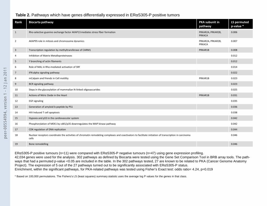
Table 2. Pathways which have genes differentially expressed in ERαS305-P positive tumors
Rank Biocarta pathway PKA subunit in
pathway
LS permuted
p-value *
1 Rho-selective guanine exchange factor AKAP13 mediates stress fiber formation PRKAR2A, PRKAR2B,
PRKACA
0.006
2 AKAP95 role in mitosis and chromosome dynamics PRKAR2A, PRKAR2B,
PRKACA
0.007
3 Transcription regulation by methyltransferase of CARM1 PRKAR1B 0.008
4 Inhibition of Matrix Metalloproteinases 0.012
5 Y branching of actin filaments 0.012
6 Role of MAL in Rho-mediated activation of SRF 0.014
7 IFN alpha signaling pathway 0.022
8 mCalpain and friends in Cell motility PRKAR1B 0.023
9 BCR signaling pathway 0.023
10 Steps in the glycosylation of mammalian N-linked oligosaccarides 0.025
11 Actions of Nitric Oxide in the Heart PRKAR1B 0.031
12 EGF signaling 0.035
13 Generation of amyloid b-peptide by PS1 0.036
14 HIV Induced T cell apoptosis 0.038
15 Hypoxia and p53 in the cardiovascular system 0.042
16 Phosphorylation of MEK1 by cdk5/p35 downregulates the MAP kinase pathway 0.042
17 CDK regulation of DNA replication 0.044
18 Nuclear receptors coordinate the activities of chromatin remodeling complexes and coactivators to facilitate initiation of transcription in carcinoma
cells
0.046
19 Bone remodelling 0.046
ERαS305-P positive tumours (n=11) were compared with ERαS305-P negative tumours (n=47) using gene expression profiling. 42,034 genes were used for the analysis. 302 pathways as defined by Biocarta were tested using the Gene Set Comparison Tool in BRB array tools. The path-ways that had a permuted p-value <0.05 are included in the table. In the 302 pathways tested, 27 are known to be related to PKA (Cancer Genome Anatomy Project). The expression of 5 out of the 27 pathways turned out to be significantly associated with ERαS305-P status. Enrichment, within the significant pathways, for PKA-related pathways was tested using Fisher’s Exact test: odds ratio= 4.24, p=0.019
* Based on 100,000 permutations. The Fishers’s LS (least squares) summary statistic uses the average log P values for the genes in that class.
peer
-005
5499
4, v
ersi
on 1
- 12
Jan
201
1

Table 3. Multivariable Cox regression analysis of the risk of progression after tamoxifen according to the PAK1-PKA/ ERαS305-P Score
A.Trainingset, NKI, tamoxifen for metastatic disease
Due to missing values in the factors used for adjustment, the analysis was based on 101 cases with 89 events. Variables included as previously described for this series (16,17). Including variables that performed significant in this trainingset in univariate analyses (progesterone receptor, HER2 and disease-free interval) did not substantially change the HR for the Algorithm. Ki67 was not available for this series. * Nottingham grading system
B. Validationset, Lund, adjuvant tamoxifen versus no systemic treatment in randomized trial
Variable Category HR 95% CI P-value
Variable Category HR 95% CI P-value
Grade* I/IIIII
11.38 1.09-1.76 0.008
Lymph node status NegativePositive
11.30 0.83-2.05 0.26
Age Continuous (per year) 1.00 0.98-1.02 0.68
PAK1-PKA/ ERαS305-P Score NegativePositive
11.91 1.23-2.95 0.004
Due to missing values in the factors used for adjustment, the analysis was based on 201 cases with 88 events. Variables included as previously described for this series (16,17)* Nottingham grading system** The interaction variable indicates whether there is a difference in treatment response in relation to the PAK1-PKA/ ERαS305-P Score.
Variable Category HR 95% CI P-value
Grade* I/IIIII
11.74 1.07-2.84 0.026
Lymph node status NegativePositive
10.94 0.58-1.55 0.82
Age Continuous (per year) 0.95 0.91-1.00 0.052
Ki67 ≤25 %> 25%
11.22 0.70-2.13 0.47
PAK1-PKA/ ERαS305-P Score NegativePositive
10.71 0.37-1.35 0.29
Tamoxifen
Interaction** (PAK1-PKA/ ERαS305-PScore and Tamoxifen)
Negative PAK1-PKA/ ERαS305-P Score -Control-Tamoxifen
Positive PAK1-PKA/ ERαS305-P Score -Control-Tamoxifen
10.4411.22
0.26-0.74
0.55-2.74
0.002
0.620.037
peer
-005
5499
4, v
ersi
on 1
- 12
Jan
201
1

NonepPKAPAK1pPKA+PAK1
N=18 17%N=57 55%N=12 12%N=16 16%
No ERS305-pERS305-p
N=83 81%N=20 19%
NonepPKAPAK1pPKA+PAK1Missing
N=22 12%N=137 74%N=3 2%N=22 12%N=47
No ERS305-PERS305-PMissing
N=178 84%N=35 16%N=18
A. Training series (n=103) B. Validation set (n=231)
Figure 1. Expression of PAK1, pPKA and ERαS305-P in human breast tumors
NonepPKAPAK1pPKA+PAK1
N=2 10%N=11 55%N=0 0%N=7 35%
N=20 19%
NonepPKAPAK1pPKA+PAK1Missing
N=2 8%N=22 85%N=1 4%N=1 4%N=9
ERS305-PMissing
N=35 16%N=18
peer
-005
5499
4, v
ersi
on 1
- 12
Jan
201
1

Figure 2
No. at risk83 62 46 36 30 24 1920 12 10 7 3 2 2
No. at risk30 23 17 14 11 9 773 51 39 29 22 17 14
No pPKApPKA
B. pPKAA. ERαS305-P
No ERαS305-P ERαS305-P
No pPKApPKApPKA-associated ERαS305-P
C. ERαS305-P and pPKA
No. at risk30 23 17 14 11 9 755 41 31 23 20 16 1318 10 8 6 2 1 1
Median TTP 14 monthsMedian TTP 17 monthsMedian TTP 9 months
*HR=2.0095% CI 1.14-3.52p=0.017
*HR=1.2295% CI 0.77-1.93p=0.40
HR=1.4195% CI 0.85-2.35p=0.19
Median TTP 16 monthsMedian TTP 9 months
Median TTP 14 monthsMedian TTP 15 months *
No PAK1PAK1PAK1-associated ERαS305-P
E. ERαS305-P and PAK1D. PAK1
No PAK1PAK1
No. at risk75 56 43 35 28 22 1828 18 13 8 5 4 3
No. at risk75 56 43 35 28 22 1821 14 10 6 5 4 37 4 3 2 0 0 0
Median TTP 18 monthsMedian TTP 9 monthsMedian TTP 9 months
No pPKA-associated ERαS305-P and no PAK1pPKA-associated ERαS305-P and/or PAK1
F. ERαS305-P, pPKA and PAK1
No. at risk64 50 38 31 26 21 1739 24 18 12 7 5 4
HR=1.7795% CI 1.16-2.71p=0.008
Median TTP 18 monthsMedian TTP 9 months
HR=1.4995% CI 0.61-3.66p=0.39
**HR=1.57
95% CI 0.99-2.48p=0.055
Median TTP 18 monthsMedian TTP 9 months
*
peer
-005
5499
4, v
ersi
on 1
- 12
Jan
201
1

Figure 3
peer
-005
5499
4, v
ersi
on 1
- 12
Jan
201
1

Figure 4
B. Training series (n=103) C. Validation set (n=231)
27%73%
A. PAK1-PKA/ ERαS305-P Score
PAK1 OR pPKA-associated ERS305-P positive = PAK1-PKA/ ERαS305-P Score positive = tumor classified as less sensitiveIF (No PAK1) AND (No pPKA-associated ERS305-P) = PAK1-PKA/ ERαS305-P Score negative = tumor classified as sensitive
less sensitivesensitive
less sensitive 38%sensitive 62%
Classified as less sensitive to tamoxifen
19%10%9%
Classified as less sensitive to tamoxifen
14%12%0.4%
peer
-005
5499
4, v
ersi
on 1
- 12
Jan
201
1

A. No pPKA-associated ERαS305-P and no PAK1 B. pPKA-associated ERαS305-P and/or PAK1(PAK1-PKA/ ERαS305-P Score negative) (PAK1-PKA/ ERαS305-P Score positive)
Figure5
HR=0.5495% CI 0.34-0.87p=0.012
HR=0.8895% CI 0.42-1.82p=0.73
No. at riskTAM 78 57 51 18Control 91 57 44 10
No. at riskTAM 31 22 17 4Control 31 20 17 6
10-yrs RFS 67.1 % (SE 0.05)
10-yrs RFS 50.9 % (SE 0.05)
10-yrs RFS 54.8 % (SE 0.09)
10-yrs RFS 54.8 % (SE 0.09)
C. Untreated patients D. Tamoxifen treated patients
No. at riskPAK1-PKA/ ERαS305-P Score positive31 20 17 6PAK1-PKA/ ERαS305-P Score negative91 57 44 10
HR=0.8895% CI 0.49-1.56p=0.65
No. at riskPAK1-PKA/ ERαS305-P Score positive31 22 17 4PAK1-PKA/ ERαS305-P Score negative78 57 51 18
HR=1.4195% CI 0.74-2.70p=0.30
peer
-005
5499
4, v
ersi
on 1
- 12
Jan
201
1