Identification of the Rac-GEF P-Rex1 as an essential mediator of ErbB signaling in breast cancer
Upload
independentCategory
view
6download
0
Induction of ErbB-3 Expression by a6b4 IntegrinContributes to Tamoxifen Resistance in ERb1-NegativeBreast CarcinomasValentina Folgiero1., Paolo Avetrani1., Giulia Bon1, Selene E. Di Carlo1, Alessandra Fabi2, Cecilia
Nistico2, Patrizia Vici2, Elisa Melucci3, Simonetta Buglioni3, Letizia Perracchio3, Isabella Sperduti4, Laura
Rosano1, Ada Sacchi1, Marcella Mottolese3, Rita Falcioni1*
1 Department of Experimental Oncology, Regina Elena Cancer Institute, Rome, Italy, 2 Department of Medical Oncology, Regina Elena Cancer Institute, Rome, Italy,
3 Department of Pathology, Regina Elena Cancer Institute, Rome, Italy, 4 Scientific Direction, Regina Elena Cancer Institute, Rome, Italy
Abstract
Background: Tamoxifen is still the most widely used drug in hormone therapy for the treatment of breast cancer. Itsbenefits in adjuvant treatment are well documented in controlled and randomized clinical studies, which havedemonstrated an increase in disease-free intervals of patients with positive hormonal receptors. However, the mechanismsinvolved in endocrine resistance are not clear. Laboratory and clinical data now indicate that bi-directional molecular cross-talk between nuclear or membrane ER and growth factor receptor pathways may be involved in endocrine resistance. Werecently found a functional interaction between a6b4 integrin and ErbB-3 receptor to maintain the PI3K/Akt survivalpathway of mammary tumour cells. We sought to improve understanding of this process in order to provide theinvolvement of both receptors insight into mechanism of Tamoxifen resistance.
Methods and Findings: Using human breast cancer cell lines displaying different levels of a6b4 and ErbB-3 receptors and aseries of 232 breast cancer biopsies from patients submitted to adjuvant Tamoxifen monotherapy for five years, weevaluated the functional interaction between both receptors in relationship to Tamoxifen responsiveness. In mammarycarcinoma cells, we evidenced that the a6b4 integrin strongly influence Akt phosphorylation through ErbB-3 proteinregulation. Moreover, the ErbB-3 inactivation inhibits Akt phosphorylation, induces apoptosis and inhibits in vitro invasionfavouring Tamoxifen responsiveness. The analysis of human tumors revealed a significant relationship between a6b4 andErbB-3 in P-Akt-positive and ERb1-negative breast cancers derived from patients with lower disease free survival.
Conclusions: We provided evidence that a strong relationship occurs between a6b4 and ErbB-3 positivity in ERb1-negativebreast cancers. We also found that the association between ErbB-3 and P-Akt positivity mainly occurs in ERb1-negativebreast cancer derived from patients with lower DFS indicating that both receptors are clinically relevant in predicting theresponse to Tamoxifen.
Citation: Folgiero V, Avetrani P, Bon G, Di Carlo SE, Fabi A, et al (2008) Induction of ErbB-3 Expression by a6b4 Integrin Contributes to Tamoxifen Resistance inERb1-Negative Breast Carcinomas. PLoS ONE 3(2): e1592. doi:10.1371/journal.pone.0001592
Editor: Dong-Yan Jin, University of Hong Kong, China
Received October 29, 2007; Accepted January 18, 2008; Published February 13, 2008
Copyright: � 2008 Folgiero et al. This is an open-access article distributed under the terms of the Creative Commons Attribution License, which permitsunrestricted use, distribution, and reproduction in any medium, provided the original author and source are credited.
Funding: This study was supported by Grant: Italian Association for Cancer Research and Ministero della Salute (R. Falcioni, M. Mottolese); Alleanza contro ilCancro (M. Mottolese); G. Bon is recipient of a fellowship from Federazione Italiana Ricerca sul Cancro (FIRC).
Competing Interests: The authors have declared that no competing interests exist.
*E-mail: [email protected]
.These authors contributed equally to this work.
Introduction
In many breast cancer (BC), activation of the phosphatidylino-
sitol 3-kinase (PI3K) pathway may deeply reduce the efficacy to
targeted therapies [1–3]. In the last few years, a strong activation
of the PI3-K/Akt signaling pathway was observed in tumor cells
that express high levels of integrin a6b4, a laminin receptor
implicated in tumor progression and invasion [4–9]. The
involvement of this integrin in tumor progression is supported
by large experimental evidence. In mammary and ovary
carcinoma cell lines, a6b4 integrin associates with ErbB-2
overexpression and co-operates to promote a PI3K-dependent
invasion and survival [10,6]. In MMTV-Neu mice, the introduc-
tion of a targeted deletion of the b4 cytoplasmic domain revealed
that b4 integrin promotes tumor progression cooperating with
ErbB-2 signaling [11]. Inactivation of a6b4 integrin by RNA
interference inhibits tumor growth both in vitro and in vivo [12–14]
and strongly reduces the activity of the PI3K pathway inducing
apoptosis upon hormone deprivation and TAM treatment in
MCF7 BC cells [12]. In addition, we have recently evidenced that
the a6b4-induced PI3K-dependent survival pathway of two
different BC cell lines is due to the capability of a6b4 integrin to
enhance ErbB-3 expression. This enhancement results in an
increase of ErbB-2/ErbB-3 heterodimerization and consequently
in the activation of the PI3K survival pathway [15]. Collectively,
these studies suggest a strong cooperation between a6b4 integrin
PLoS ONE | www.plosone.org 1 February 2008 | Volume 3 | Issue 2 | e1592

and EGFR family members in mammary tumors and highlight a
pathway by which this integrin might contribute to BC
tumorigenicity and responsiveness to treatments.
BC remains one of the most heterogeneous tumors in terms of
capability to give metastases, expression of hormone receptors and
responsiveness to therapies and is the first cause of death for
women aged 35–45 years [16]. Tamoxifen (TAM) is still the most
widely used drug in hormone therapy for the treatment of this
neoplasia. Its benefits in adjuvant treatment and metastatic disease
are well documented in controlled and randomized clinical studies,
which have demonstrated an increase in disease-free intervals and
overall survival of patients with positive hormonal receptors [17].
However, endocrine therapies do not always work in patients,
despite the presence of hormone receptors in their tumors [18].
Originally, only estrogen receptor (ER) a and progesterone
receptor (PgR) were thought to be involved in hormone signaling.
However, a second ER, termed ERb, was subsequently discov-
ered, adding another dimension of complexity to the regulation of
hormone response [19–20]. Insights into the mechanisms of
endocrine therapy resistance, although still cause for debate, have
come from several studies concerning the biology of ERs and the
various signaling pathways in the cell with which they commu-
nicate. Laboratory and clinical data now indicate that bidirec-
tional molecular cross-talk between nuclear or membrane ER and
growth factor receptor pathways may be involved in endocrine
resistance [21]. An understanding of these ER activities at the
molecular level may yield new strategies to prevent or overcome
resistance to TAM and other forms of treatment.
In the present work, using ER-positive human BC cell lines, we
investigated the functional interaction between a6b4 and ErbB-3
proteins in relationship to TAM responsiveness. In addition, with the
aim to translate our in vitro study to an in vivo model, we carried out
immunohistochemical (IHC) analysis to evaluate the functional
relationship between desease-free survival (DFS) and expression of
a6b4, ErbB-2, ErbB-3, P-Akt and ERb1 in a retrospective series of
232 ERa and/or PgR positive BCs derived from patients which had
been homogeneously submitted to adjuvant TAM monotherapy.
Combining our analyses, we provide evidence that a6b4 expression
is functionally associated with ErbB-3 and P-Akt molecules in vitro.
However, even though a6b4 expression in vivo is still strongly
associated with ErbB-3 positivity and ERb1 negativity, it does not
influence patient outcome. Interestingly, we report for the first time a
strong association of ErbB-3 and P-Akt positivity that mainly occurs
in ERb1 negative BC derived from patients with lower DFS. This
result suggests that both receptors are clinically relevant in predicting
the response to Tamoxifen treatment.
ResultsExpression of b4, ErbB-2, ErbB-3, ERa and ERb receptorsin mammary tumor cell lines
We first evaluated the expression level of ERa and ERb, b4
integrin subunit, ErbB2, and ErbB-3 in a series of human
mammary tumor cell lines including MDA-MB 231, MDA-MB
361, SKBr3, BT474, BT549, and T47D. Analysis of ERa by
Western blotting (Figure 1A) and ERb1 by RT-PCR, using
specific primers to detect ERb1 mRNA, (Figure 1B) showed that
BT549 cells were negative for both ERs whereas the other cell
lines were positive for at least one ER. Then, the expression of
ERb1 protein was evaluated by immunocytochemistry (Figure S1).
The data obtained confirmed the expression of ERb1 protein in
each cell line that resulted positive for ERb1 mRNA. As expected,
the analysis of the other receptors by cytofluorimetry showed that
MDA-MB 361, SKBr3 and BT474 and T47D cells express
considerable levels of ErbB-2 protein (Figure 1C) [22]. Moreover,
Figure 1. Expression of b4, ErbB-2, ErbB-3 and ERa and b1receptors in mammary tumor cell lines. A. The expression of ERawas evaluated by western blot analysis. The anti-actin Ab was used tovalidate equivalent loading protein. B. ERb1 expression was evaluatedby RT-PCR from total mRNA extracted from the indicated cell lines usingprimers specific for human ERb1 and the housekeeping aldolase genes.C. Mammary tumor cell lines MDA-MB 231, MDA-MB 361, SKBr3, BT474,BT549 and T47D were analyzed by FACS to reveal the expression levelof b4 integrin subunit, ErbB-2 and ErbB-3 receptors.doi:10.1371/journal.pone.0001592.g001
PI3K and TAM Resistance
PLoS ONE | www.plosone.org 2 February 2008 | Volume 3 | Issue 2 | e1592

the same cells express b4 and ErbB-3 proteins at comparable
levels, whereas BT549 and MDA-MB 231 cells displaying low
levels of ErbB-2 and b4 proteins were also ErbB-3 negative,
supporting our recent finding that b4 overexpression regulates
ErbB-3 protein at translational level [15].The regulation of ErbB-3 expression by a6b4 influences
AKT activation. Given that a6b4 integrin is the receptor for
laminin 5 (LM5) and, as we previously demonstrated, ligation of the
integrin to this substrate enhances PI3K signaling, we first verified
the level of Akt phosphorylation upon stimulation in the mammary
tumor cell lines. To this end, MDA-MB 361, BT474, SKBr3, BT549
and MDA-MB 231 cells were spread onto LM5 for 20 minutes and
the level of Akt activity was evaluated by Ser473 phosphorylation. As
reported in Figure 2A, a strong enhancement of Akt phosphorylation
was detectable in the cells expressing a6b4, ErbB-2 and ErbB-3
receptors (i.e., MDA-MB 361, BT474 and SKBr3 cells) while, it did
not occur in cells expressing low levels of b4, ErbB-2 and undectable
level of ErbB-3 (i.e., BT549 and MDA-MB 231) (Figure 2A). As
expected, after 60 minutes of LM5 stimulation, the phosphorylation
of Akt returned to the basal levels (data not shown).
To confirm the essential role of ErbB-3 protein in the activation
of Akt by a6b4, a b4 shRNA (b4si) or an ErbB-3 siRNA (B3si)
were expressed in MDA-MB 361, BT474 and SKBr3 cells, as
previously described [15]. As expected, depletion of b4 resulted in
a strong reduction of b4 compared to the levels found in scramble
(scr) control cells. Of interest, b4 depletion also caused a strong
reduction of ErbB-3 expression and Akt phosphorylation
(Figure 2B, upper panel). Moreover, ErbB-3 depletion resulted
in a strong reduction of ErbB-3 expression and, at the same time,
of Akt phosphorylation (Figure 2B, lower panel). Since a6b4
regulates ErbB-3 level and the depletion of either b4 or ErbB-3
proteins resulted in a strong inhibition of Akt activation, the data
confirm the essential role of ErbB-3 in the activation of Akt by
a6b4 integrin in mammary tumor cells (Figure 2B).
ErbB-3 depletion causes apoptosis and inhibits in vitro
invasion favoring TAM responsiveness. To further evaluate
the function of ErbB-3 in the PI3K survival pathway, we analyzed
cell death and apoptosis in the absence of hormones and under TAM
treatment of ErbB3 positive (SKBr3, MDA-MB 361, BT474 and
T47D) and ErbB3 negative (MDAMB231) cell lines. As shown in
Figure 3A and 3B, in the absence of hormones, depletion of ErbB-3
protein caused per se a strong cell death compared to scr cells
(SKBr3/B3i 32% vs SKBr3/scr 8%, p = 0.001; MDA MB361/B3i
42% vs MDA MB361/scr 6%, p,0.0001; BT474/B3i 35% vs
BT474/scr 11%, p = 0.04; T47D/B3i 39% vs T47D/scr 7%,
p = 0.04). Cell death was further increased by TAM treatment
(SKBr3/B3si/TAM 48% vs SKBr3/scr/TAM 19%, p,0.0001;
MDA MB361/B3si/TAM 55% vs MDA MB361/scr/TAM 21%,
p,0.0001; BT474/B3si/TAM 38% vs BT474/scr/TAM 18%,
p = 0.02; T47D/B3si/TAM 42% vs T47D/scr/TAM 10%,
p = 0.005) as also assessed by cleavage of PARP, a marker of
apoptotic death (Fig. 3B). The results we obtained on cell death and
apoptosis on T47D cells strongly reinforce our hypothesis that ErbB-
3 sustains the survival function of mammary tumor cells in the
absence of hormone stimuli. Indeed, this cell line is negative for
ERb1 expression, does not respond to TAM treatment, and
undergoes apoptosis only upon ErbB-3 depletion. MDAMB231
cells that are ErbB-3 negative, even if are ERb1 positive, do not
respond to TAM treatment and proliferate and survive as well as
untreated cells (p = 0.82) (Fig. 3A,B), indicating that these cells have
developed other survival pathway(s).
It is widely reported that TAM resistance and, as a consequence,
tumor progression may be also due to PI3K activation [23]. In order
to understand the role of ErbB-3 in the invasion process, we
evaluated the invasive capability of scr and ErbB-3-depleted SKBr3,
MDA-MB-361, BT474, T47D cells in the absence of hormones and
upon TAM treatment. As shown in Figure 3C, depletion of ErbB-3
protein caused per se a strong inhibition of the invasion compared to
scr cells (percent of invasion: SKBr3/B3si 50% vs SKBr3/scr 100%,
P = 0.001; MDA MB361/B3si 54% vs MDA MB361/scr 100%,
p,0.0001; BT474/B3si 70% vs BT474/scr 100%, p = 0.03; T47D/
B3si 62% vs T47D/scr 100%, p = 0.04). The inhibition of the
invasion in ErbB-3-depleted cells further increased following TAM
treatment compared to scr cells (percent of invasion: SKBr3/B3si/
TAM 35% vs SKBr3/scr/TAM 85%, p,0.0001; MDA MB361/
B3si/TAM 42% vs MDA MB361/scr/TAM 82%, p,0.0001;
BT474/B3si/TAM 61% vs BT474/scrTAM 83%, p = 0.04; T47D/
B3si/TAM 50% vs T47D/scr/TAM 90%, p,0.001). As expected,
TAM treatment does not alters the capability of MDAMB231 cells
to invade matrigel (p = 0.06) (Figure 3C). Representative invading
stained cells are showed on Figure S2. Collectively, these data
Figure 2. The a6b4 influence Akt activation by ErbB-3. A. BT549,MDA-MB 231, MDA-MB 361, BT474 and SKBr3 cells were serum-starvedfor 24 hrs and then the cells were spread onto LM5 and extracted indetergent. Equivalent amounts of protein were separated by SDS-PAGEand analyzed by immunobloting to evaluate the relative expression ofb4 and phospho-Akt. Total-Akt Ab was used to validate equivalentloading of protein in each lane. B. MDA-MB 361, BT474 and SKBr3 cellswere transiently transfected for 48 hrs with either scrambled or specificb4-shRNA and ErbB-3 siRNA. The cells were then serum-starved for24 hrs and extracted in detergent. Equivalent amounts of protein wereseparated by SDS-PAGE and analyzed by immunobloting to evaluatethe relative expression of b4, ErbB-3 and phospho-Akt. Hsp70 Ab wasused to validate equivalent loading of protein in each lane.doi:10.1371/journal.pone.0001592.g002
PI3K and TAM Resistance
PLoS ONE | www.plosone.org 3 February 2008 | Volume 3 | Issue 2 | e1592

indicate a role of ErbB-3 protein in the mechanisms that regulate the
invasion of mammary tumor cells. Since we have previously
demonstrated that b4 depletion reduces the responsiveness of
mammary tumor cells to TAM treatment, our data also suggest
that a cooperative signaling between ErbB-3 and a6b4 integrin could
influence resistance to hormone therapy in vivo.
Immunohistochemical analysis of b4 integrin subunit,ErbB-3, ErbB-2, P-Akt, and ERb1 in human primary BC
To verify whether the functional interaction between a6b4
integrin and ErbB-3 receptor also occurred in vivo, we studied 232
biopsies of BC patients surgically treated at our Institute and
submitted to adjuvant TAM therapy. The detailed clinicopatho-
logical characteristics of the patients are described in Table 1.
These tumors were first analyzed by IHC for the expression of b4
integrin subunit, ErbB-3, ErbB-2, ERb1 and P-Akt expression. As
summarized in Figure 4A, of the 232 cases analyzed, b4 exhibited
a strong homogeneous (score 2) or heterogeneous (score 1)
immunoreaction in 170 BC (73,3%). 77 BC (33,2%) overexpressed
ErbB-3 and 158 (68,1%) were ERb1 positive. Moreover, we found
that 136 BC (59%) were P-Akt positive, while 59 (25,4%) were
positive for ErbB-2. Representative immunohistochemically pos-
itive cases for b4, ErbB-3, P-Akt, ERb1 and ErbB-2 and control
tissue sections are shown in Figure 4B.
Figure 3. ErbB-3 expression influences survival and invasion of mammary tumor cells treated under TAM treatment. A. SKBr3, MDA-MB 361, BT474, T47D and MDAMB231 cells after three days of hormone deprivation were transiently transfected with either scrambled or specificErbB-3 siRNA. Where specified, 24 hrs after transfection scrambled and ErbB3 interfered cells were pre-incubated for 24 hours at 37uC with TAM2.5 mM. 48 hours following transfection, the cell death was evaluated by Trypan-blue exclusion. Statistical differences were evaluated by T test(p,0.05). B. Equivalent amounts of total cell lysate derived from the cell lines described in A were separated by SDS-PAGE and analyzed byimmunobloting to evaluate the expression level of PARP cleavage. Hsp70 Ab was used to validate equivalent loading of protein in each lane. C.SKBr3, MDA-MB 361, BT474, T47D and MDAMB231 cells transfected as described in A were assayed for their ability to invade matrigel in the absenceof hormone and under TAM treatment. Statistical differences were evaluated by T test (p,0.05).doi:10.1371/journal.pone.0001592.g003
PI3K and TAM Resistance
PLoS ONE | www.plosone.org 4 February 2008 | Volume 3 | Issue 2 | e1592

Relationship among b4 integrin subunit, pathologicaland biological parameters
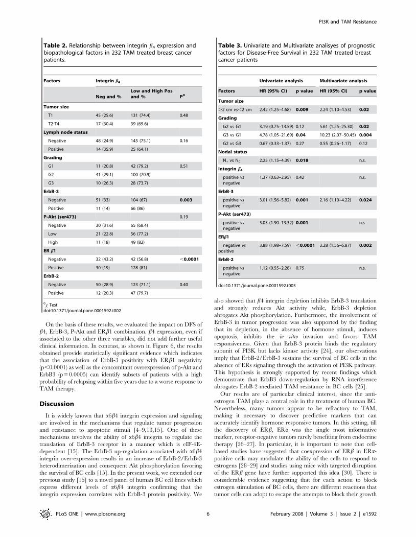
Table 2 summarizes the associations between b4 expression and
biopathological factors in our series of 232 BC patients. We found
that all tumors, which were positive for ErbB-3 receptor, showed a
higher score in b4 expression, b4 immunoreaction being signifi-
cantly associated to ErbB-3 (p = 0.003). Interestingly, we also
found that the majority of high b4-positive tumors were ERb1-
negative (p,0.0001). In contrast, b4 was not significantly related
to P-Akt, ErbB-2 protein and any conventional pathological
parameters, namely tumor size, grading and nodal status.
Impact of biopathological parameters on disease freesurvival
At a median follow up of 58 months (range 1–179 months), a
total of 36 patients (15%) showed progressive disease.
The results of the univariate and multivariate analyses for DFS
in the 232 patients included in this study are summarized in
Table 3. Univariate analysis (Cox model) identified tumor size
(HR 2.42, C.I. 1.25–4.68, p = 0.009), grading (G3, HR 4.78, C.I.
1.05–21.69, p = 0.04), nodal status (HR 2.25, C.I. 1.15–4.39,
p = 0.018), ErbB-3 (HR 3.01, C.I. 1.56–5.82, p = 0.001), P-Akt
overexpression (HR 5.03, C.I. 1.90–13.32, p = 0.001) and lack of
ER b1 (HR 3.88, C.I. 1.98–7.59, p,0.0001) as significant
predictors of DFS.
Each variable that significantly affected DFS in the univariate
analyses were introduced into a Cox proportional risk model.
Multivariate analyses revealed that tumor size (HR 2.24, C.I.
1.10–4.53, p = 0.02), grading (G2 vs G1, HR 5.61, C.I. 1.25–
25.30, p = 0.02 and G3 vs G1, HR 10.23, C.I. 2.07–50.45,
p = 0.004), ErbB-3 expression (HR 2.16, C.I. 1.10–4.22, p = 0.024)
and lack of ERb1 (HR 3.28, C.I. 1.56–6.87, p = 0.002) were
independent prognostic variables influencing DFS. ERb1 nega-
tivity appears to be the most powerful prognostic indicator of a
reduced DFS, indicating that ERb1 positive tumors are more
likely to be responsive to TAM therapy.
Kaplan-Meier curves (Figure 5), stratified, respectively, for b4,
ErbB-3, P-Akt and ERb1 expression in all valuable cases, indicate
that a significantly longer DFS can be observed in patients with
ErbB-3 negative (p = 0.0006), P-Akt negative (p = 0.005) and
ERb1 positive (p,0.0001) tumors. b4 expression, considered as a
single factor, did not influence the patient outcome.
Figure 4. Immunohistochemical analysis of b4, ErbB-3, P-Akt,ERb1 and ErbB-2 in 232 primary BC. A. Distribution (%) of the bio-pathological factors b4 integrin subunit, ErbB-3, P-Akt(ser473), ERb1 andErbB-2 in 232 TAM treated breast cancers. B. Representative immuno-histochemically positive cases for b4, ErbB-3, P-Akt(ser473), ERb1 andErbB-2 protein detection and control tissue sections.doi:10.1371/journal.pone.0001592.g004
Table 1. Clinicopathological characteristics of 232 invasivebreast carcinomas TAM treated
CHARACTERISTIC %
Number of patients 232
Mean age 63
Menopausal
Pre 25 10.8
Post 207 89.2
Histotype
Invasive ductal carcinoma 193 83.2
Invasive lobular carcinoma 28 12.1
Tubular carcinoma 7 3
Papillary carcinoma 4 1.7
Tumor size
T1 176 79.9
T2 51 22
T3,T4 5 2.1
Lymph node status
Negative 193 83.2
Positive 39 16.8
Grading
G1 53 22.8
G2 141 60.8
G3 38 16.4
ERa
Negative (#10%) 25 10.8
Positive (.10%) 207 89.2
PgR
Negative (#10%) 55 23.7
Positive (.10%) 177 76.3
*range 39–95doi:10.1371/journal.pone.0001592.t001
PI3K and TAM Resistance
PLoS ONE | www.plosone.org 5 February 2008 | Volume 3 | Issue 2 | e1592
On the basis of these results, we evaluated the impact on DFS of
b4, ErbB-3, P-Akt and ERb1 combination. b4 expression, even if
associated to the other three variables, did not add further useful
clinical information. In contrast, as shown in Figure 6, the results
obtained provide statistically significant evidence which indicates
that the association of ErbB-3 positivity with ERb1 negativity
(p,0.0001) as well as the concomitant overexpression of p-Akt and
ErbB3 (p = 0.0005) can identify subsets of patients with a high
probability of relapsing within five years due to a worse response to
TAM therapy.
Discussion
It is widely known that a6b4 integrin expression and signaling
are involved in the mechanisms that regulate tumor progression
and resistance to apoptotic stimuli [4–9,13,15]. One of these
mechanisms involves the ability of a6b4 integrin to regulate the
translation of ErbB-3 receptor in a manner which is eIF-4E-
dependent [15]. The ErbB-3 up-regulation associated with a6b4
integrin over-expression results in an increase of ErbB-2/ErbB-3
heterodimerization and consequent Akt phosphorylation favoring
the survival of BC cells [15]. In the present work, we extended our
previous study [15] to a novel panel of human BC cell lines which
express different levels of a6b4 integrin confirming that the
integrin expression correlates with ErbB-3 protein positivity. We
also showed that b4 integrin depletion inhibits ErbB-3 translation
and strongly reduces Akt activity while, ErbB-3 depletion
abrogates Akt phosphorylation. Furthermore, the involvement of
ErbB-3 in tumor progression was also supported by the finding
that its depletion, in the absence of hormone stimuli, induces
apoptosis, inhibits the in vitro invasion and favors TAM
responsiveness. Given that ErbB-3 protein binds the regulatory
subunit of PI3K but lacks kinase activity [24], our observations
imply that ErbB-2/ErbB-3 sustains the survival of BC cells in the
absence of ERs signaling through the activation of PI3K pathway.
This hypothesis is strongly supported by recent findings which
demonstrate that ErbB3 down-regulation by RNA interference
abrogates ErbB-2-mediated TAM resistance in BC cells [25].
Our results are of particular clinical interest, since the anti-
estrogen TAM plays a central role in the treatment of human BC.
Nevertheless, many tumors appear to be refractory to TAM,
making it necessary to discover predictive markers that can
accurately identify hormone responsive tumors. In this setting, till
the discovery of ERb, ERa was the single most informative
marker, receptor-negative tumors rarely benefiting from endocrine
therapy [26–27]. In particular, it is important to note that cell-
based studies have suggested that coexpression of ERb in ERa-
positive cells may modulate the ability of the cells to respond to
estrogens [28–29] and studies using mice with targeted disruption
of the ERb gene have further supported this idea [30]. There is
considerable evidence suggesting that for each action to block
estrogen stimulation of BC cells, there are different reactions that
tumor cells can adopt to escape the attempts to block their growth
Table 2. Relationship between integrin b4 expression andbiopathological factors in 232 TAM treated breast cancerpatients.
Factors Integrin b4
Neg and %Low and High Posand % P0
Tumor size
T1 45 (25.6) 131 (74.4) 0.48
T2-T4 17 (30.4) 39 (69.6)
Lymph node status
Negative 48 (24.9) 145 (75.1) 0.16
Positive 14 (35.9) 25 (64.1)
Grading
G1 11 (20.8) 42 (79.2) 0.51
G2 41 (29.1) 100 (70.9)
G3 10 (26.3) 28 (73.7)
ErbB-3
Negative 51 (33) 104 (67) 0.003
Positive 11 (14) 66 (86)
P-Akt (ser473) 0.19
Negative 30 (31.6) 65 (68.4)
Low 21 (22.8) 56 (77.2)
High 11 (18) 49 (82)
ER b1
Negative 32 (43.2) 42 (56.8) ,0.0001
Positive 30 (19) 128 (81)
ErbB-2
Negative 50 (28.9) 123 (71.1) 0.40
Positive 12 (20.3) 47 (79.7)
0x Testdoi:10.1371/journal.pone.0001592.t002
Table 3. Univariate and Multivariate analisyes of prognosticfactors for Disease-Free Survival in 232 TAM treated breastcancer patients
Univariate analysis Multivariate analysis
Factors HR (95% CI) p value HR (95% CI) p value
Tumor size
.2 cm vs,2 cm 2.42 (1.25–4.68) 0.009 2.24 (1.10–4.53) 0.02
Grading
G2 vs G1 3.19 (0.75–13.59) 0.12 5.61 (1.25–25.30) 0.02
G3 vs G1 4.78 (1.05–21.69) 0.04 10.23 (2.07–50.45) 0.004
G2 vs G3 0.67 (0.33–1.37) 0.27 0.55 (0.26–1.17) 0.12
Nodal status
N+ vs N0 2.25 (1.15–4.39) 0.018 n.s.
Integrin b4
positive vsnegative
1.37 (0.63–2.95) 0.42 n.s.
ErbB-3
positive vsnegative
3.01 (1.56–5.82) 0.001 2.16 (1.10–4.22) 0.024
P-Akt (ser473)
positive vsnegative
5.03 (1.90–13.32) 0.001 n.s
ERb1
negative vspositive
3.88 (1.98–7.59) ,0.0001 3.28 (1.56–6.87) 0.002
ErbB-2
positive vsnegative
1.12 (0.55–2.28) 0.75 n.s.
doi:10.1371/journal.pone.0001592.t003
PI3K and TAM Resistance
PLoS ONE | www.plosone.org 6 February 2008 | Volume 3 | Issue 2 | e1592

[31]. The activation of growth factor signaling is involved in the
mechanism of resistance to endocrine therapy and it has been
hypothesized that it may substitute estrogen in sustaining the
growth and survival of BC cells [31].
Aimed at translating our in vitro results to human BC, we
evaluated, by IHC, a6b4, ERb1, ErbB3, and P-AKT expression in
232 primary mammary tumors derived from patients submitted to
adjuvant TAM monotherapy. Even though we found a significant
correlation between b4 and ErbB-3 expression and ERb1
negativity, in the BCs we analyzed, the expression of the integrin
did not influence the patient outcome.
ErbB-3 proteins mainly occurred in the P-Akt-positive and
ERb1-negative BC derived from patients with lower DFS.
Although previous experimental studies have implied that a6b4
integrin facilitates tumor progression by regulating growth factor
receptors signaling [15], to our knowledge this is the first study
demonstrating an in vivo correlation between b4 and ErbB-3
expression suggesting that b4 can regulate ErbB-3 protein in vivo
and favor indirectly tumor progression.
The high percentage of mammary tumors we analyzed which
over-express b4 integrin subunit is consistent with previous
findings [8]. Although in vivo a6b4 integrin expression has not
been extensively evaluated, there are two separate studies
reporting that 90% of advanced BC expressed a6 subunit [32]
and that high level of a6b4 expression in mammary tumor has
prognostic value [33]. Furthermore, the over-expression of b4
integrin subunit in the ERb1-negative tumors we have analyzed is
also in agreement with a recent study which demonstrates that
laminin-binding integrins and especially b4 integrin subunit is
elevated in ER-negative BC [34].
These and numerous other studies conducted in vivo in a smaller
number of tumors clearly indicate that b4 molecule mediates the
signaling events which play a role in tumor progression [8]. This
hypothesis is based on the capability of this integrin to enhance not
Figure 5. DFS (232 cases) for TAM treated patients with BC categorised on the basis of (A) b4, (B) ErbB-3, (C) P-Akt (ser473), (D) ERb1expression. Survival curves were generated according to the Kaplan-Meier method; statistical comparisons were made using the log-rank test.doi:10.1371/journal.pone.0001592.g005
PI3K and TAM Resistance
PLoS ONE | www.plosone.org 7 February 2008 | Volume 3 | Issue 2 | e1592

only the translation of growth factor receptor but also of key
growth factor such as VEGF [13,35]. It has been observed that
ablation of a6b4 expression by shRNA in BC cells impaired the
ability of these cells to form xenograft tumors and to produce
VEGF [13]. Moreover, the finding that the depletion of a6b4
integrin in mammary cells inhibits the PI3K pathway and
facilitates the responsiveness to TAM treatment [12] correlates
with the capability of a6b4 integrin to regulate ErbB-3 translation
and subsequent Akt activation [15]. We can hypothesize that a6b4
integrin controls the translation of key molecules whose functions
are strictly related to carcinoma survival. The ability of a6b4
integrin to control ErbB-3 expression in vitro [15] and the strong
relationship between b4 and ErbB-3 receptor (P = 0.003, see
Table 2) we observed in vivo confirms this hypothesis. Although in
the BC we analyzed, b4 does not directly influence the patient
outcome, its expression may influence a different regulation of ErbB-
3 and consequently, as suggested by our analyses, PI3K activation
through its heterodimerization with ErbB-2. Collectively, these
phenotypic alterations may have a significant impact on DFS.
From our data it is evident that ErbB-3 may represent a key
molecule involved in the mechanisms of TAM resistance in ERb1-
negative BC. This finding is in agreement with a recent report
demonstrating that ErbB-3 modulates ErbB-2-mediated prolifer-
ation, colony formation and resistance to TAM treatment [25].
Figure 6. DFS (232 cases) for TAM treated patients with breast carcinomas categorised according to the combinations in allevaluated cases of (A) ErbB-3 and ERb1 expression, (B) ErbB-3 and P-Akt expression. Survival curves were generated according to theKaplan-Meier method; statistical comparisons were made using the log-rank test.doi:10.1371/journal.pone.0001592.g006
PI3K and TAM Resistance
PLoS ONE | www.plosone.org 8 February 2008 | Volume 3 | Issue 2 | e1592

Even though there are many studies on the role of ErbB-2 in BC
prognosis and therapeutic response, little is known regarding the
role of ErbB-3 protein in these processes [18]. However, in
agreement with our data, it has been found that the DFS is shorter
in patients with ErbB-3 overexpression and that the level of ErbB-
3 expression in primary BC seems to be involved in tumor
progression from non-invasive to invasive tumors [36]. Moreover,
it has also been shown that ErbB1-3 positive tumors had
significantly poorer survival [37]. The strong relationship we
found between ErbB-3 and P-Akt positivity and low DFS relative
to patients with ERb1-negativity reinforces the hypothesis that
growth factor signaling is involved in the mechanism of resistance
to endocrine therapy. However, from our study, it is clear that
ERb1 negativity appears to be the most powerful prognostic factor
influencing DFS in response to TAM treatment and this data is in
agreement with previous observations showing that low level of
ERb predict resistance to TAM treatment [38]. Together these
studies provide strong evidence that ERb1 is a predictor of
response to TAM treatment in BC.
We can conclude that, even though the regulation of mammary
tumor growth and survival by ERs and EGFR family members and
the biology of b4 integrin in tumors are not completely known, our in
vitro and in vivo results provide strong evidence of a functional
cooperation among these factors in supporting the survival of
mammary tumors and this cooperation in ERb1-negative tumors
may result in a decreased responsiveness to TAM therapy.
Materials and MethodsCell lines
The human mammary carcinoma cell lines, MDA-MB361,
BT474, SKBr3, MDA-MB231, T47D and BT549 were obtained
from the ATCC and maintained in DMEM medium supplement-
ed with 10% FCS (INVITROGEN, Milan, Italy). Rat bladder
epithelial cell line 804G was cultured in minimum essential
medium supplemented with 10% FCS and employed for LM5 rich
matrix preparation [39].
Antibody and matrix proteinsThe rat anti-hum b4 subunit (Clone 439-9B) was prepared as
previously described and used in immunoprecipitation, and
immunofluorescence (FACS) analysis experiments [6]. The mouse
anti-human b4 subunit 450-11A was used in western blotting and
immunohistochemistry experiments [12]. The rabbit anti-ErbB-3
[Ab (C17), Ab6 (2B5)] and the mouse anti-ErbB-3 Ab (Ab-4 Clone
H3.90.6) were used in western blot, immunoprecipitation and in
immunofluorescence (FACS) analysis experiments, respectively.
Clones C17 and 2B5 were purchased from Santa Cruz
Biotechnologioes (Milan, IT) and clone Ab4 was purchased from
NeoMarkers (Fremont, CA). The rabbit anti total and phospho-
AKT (Ser473) antibodies were purchased from Cell Signaling
(Milan, IT). The rabbit anti ERa and the mouse anti ERb Abs
were purchased from Santa Cruz Biotechnologioes (Milan, IT)
and UCS Diagnostic (Rome, IT) respectively. The hsp70 (N27F3-
4) Ab was purchased from Stressgen (Milan, IT). The mouse anti-
PARP (Clone C2-10) was purchased from Pharmingen (Milan,
Italy). FITC and Peroxidase-conjugated anti-IgGs were purchased
from Cappel and BioRad (Milan, IT).The laminin-5-rich matrix
from 804G cells was prepared as described previously [39]. In
brief, 804G cells were plated onto 100 mm dishes or 96 well plates
and allowed to reach confluence. The cells were washed in sterile
PBS and were removed from their matrix by treatment for 10 min
in 20 mM NH4OH at 4uC. The remaining cells were removed by
washing three times with sterile PBS. The Poly-L-lysine was from
SIGMA (Milan, Italy).
Flow cytometry analysisThe expression level of b4, ErbB2 and ErbB-3 in MDA-MB
231, MDA-MB 361, BT474, SKBr3, BT549 and T47D cells was
detected by flow cytometry analysis of stained cells. In brief, cells
harvested using citrate saline buffer (0.134 M KCl, 0.015 M Na
citrate) were washed twice with cold PBS containing 0.002%
EDTA and 10 mM NaN3 (washing buffer). Samples of 16106 cells
were incubated for 1 h at 4uC with saturating concentrations of
primary antibodies diluted in PBS containing 0.5% bovine serum
albumin (BSA). Cells were then washed three times with washing
buffer (PBS containing 0.5% BSA) and incubated for 1 h at 4uCwith 50 l of FITC-conjugated secondary antibodies [F(ab’)2
(Cappel, West Chester, PA, U.S.A.)] diluted 1:20 in PBS/BSA.
After three washes, the cells were suspended in 1 ml of washing
buffer. Cell suspensions were analyzed by a flow-cytometer (Epics
XL analyzer, Coulter Corporation, Miami, FL) after addition of 5l
of a 1mg/ml solution of propidium iodide to exclude non-viable
cells. At least 16104 cells per sample were analyzed.
Western Blot analysisTo analyze ER-a, b4 and ErbB-3 protein expression, the cells
were lysed with RIPA buffer (50 mM Tris (pH 8), 150 mM NaCl,
1% Nonidet P40, 0,1% deoxycholate, 0,1% SDS, 1mM PMSF,
5 mM Na3VO4, 50 mM protease inhibitors (SIGMA-Aldrich,
Milan, IT) for 30 minutes at 4uC. Total cell lysates were clarified
by centrifugation at 14,000 rpm for 30 minutes. Aliquots of cell
extracts containing an equivalent amount of proteins were
resolved by SDS-polyacrilamide gel electrophoresis 10% (SDS-
PAGE) and transferred to nitrocellulose. To analyze Akt activation
after stimulation by LM5, MDA-MB 361, BT474 and SKBr3
(16106) cell lines, after serum starvation for 24 hours, were seeded
onto 100 mm tissue culture dishes coated with LM5-rich matrix
preparation from 804G cells. The cells were washed three times
with ice cold PBS and lysed with NP40 buffer (1% Nonidet P40,
10% glycerol, 137 mM NaCl, 20 mM Tris HCl (pH 7,4), 50 mM
NaF, 1 mM PMSF, 5mM Na3VO4, 50 mM protease inhibitors
(SIGMA-Aldrich, Milan, IT) for 30 minutes at 4uC. Total cell
lysates were clarified by centrifugation at 14,000 rpm for
30 minutes. Aliquots of cell extracts containing equivalent
amounts of proteins were resolved by SDS-PAGE, transferred to
nitrocellulose and probed with the rabbit polyclonal Ab directs to
P-Akt. As secondary Abs, the horseradish peroxidase-coniugated
goat anti-mouse or rabbit were used. The chemiluminescence was
resolved by an enhanced chemiluminescence ECL kit (Amersham,
Milan, IT). Total proteins were normalized by anti-actin, anti-
Hsp70 and total-Akt Abs, respectively.
RT-PCRTotal RNA was prepared using RNAzol B according to the
manufacturer’s procedure (Invitrogen, Milan, IT). Human ERb1
mRNA for RT-PCR analysis was carried out using specific
primers as previously described [40]. The oligonucleotides we use
to amplify ERb1 mRNA were as follow:
hERb1 sense: 59TGCTTTGGTTTGGGTGATTGC39;
hERb1 anti-sense: 59TTTGCTTTTACTGTCCTCTGC39.
The housekeeping aldolase mRNA was used as an internal
control.
ImmunocytochemistryFor the detection of ERb by immunocytochemistry, 56105 cells
of each cell line (MDA-MB 231, MDA-MB 361, SKBr3, BT474,
BT549 and T47D) were centrifuged onto glass slides (cytospin) and
PI3K and TAM Resistance
PLoS ONE | www.plosone.org 9 February 2008 | Volume 3 | Issue 2 | e1592

fixed in 2% formaldehyde for 10 minutes. Endogenous peroxidase
was blocked by incubating in 3% H2O2 in PBS for 10 minutes.
After two rinses in PBS, nonspecific binding was blocked by a 10-
minute incubation with normal serum (ScyTek Laboratories,
Logan, UT). Samples were then incubated in mouse anti-ERb1
antibody (1:20 dilution) in 0,5% bovine serum albumine with PBS
overnight, in a humidified atmosphere. Detection steps were done
using the UltraTek HRP kit according to the manufacturer’s
procedure (ScyTek Laboratories), and peroxidase activity was
localized with DAB (diamino-benzidine) substrate. Slides were
counterstained by Hematoxilin and mounted under a coverslip in
glycerol.
RNA InterferenceThe inactivation of b4 was obtained by the LipofectAMINE
PLUSTM method (INVITROGEN) using pSUPER.retro vector
containing b4-shRNA or scramble RNA (scr-shRNA) sequences. To
inactivate ErbB-3 expression, cells were transiently transfected with
Transit-TKO reagent (MIRUS, Medison, Wisconsin) following the
manufacturer procedures with the ErbB-3 anti-sense double strand
siRNA as previously described [15]. The cells were harvested
48 hours post transfection with RIPA buffer for the detection of b4
and ErbB3 expression and with NP-40 buffer for the detection of P-
AKT. Total proteins were separated by 8% and 10% SDS-PAGE
respectively and transferred to nitrocellulose. The proteins were
detected by western blot analysis as described above.
Cell death and apoptosisSKBr3, MDA-MB 361, BT474, TD47D and MDA-MB 231
cells (36105) were plated onto 60mm dishes in hormone-
deprivation conditions for three days. The following day, the cells
were trasiently transfected with a scrambled or ErbB-3 siRNA
sequence and 24 hours after transfection the cells were treated
with 2.5 mM TAM or ethanol as a control for 24 hrs. The viability
of the cells was evaluated by Trypan blue exclusion. Each assay
was repeated at least three times. Following the same procedure,
the cells were lysed in Triton buffer (20 mM Tris pH 7.5, 150 mM
NaCl, 1 mM EDTA, 1 mM EGTA, 1% Triton x-100, 0.5%
NP40, 2.5% sodium pyrophosphatate, 1 mM Na3VO4, 50 mM
protease inhibitors) and sonicated for 15 seconds. Samples were
boiled for 5 minutes at 95uC, resolved by SDS-polyacrilamide gel
electrophoresis (8%), transferred to nitrocellulose and probed with
a mouse anti-PARP Ab.
Chemoinvasion assayChemoinvasion was assessed using a 48-well modified Boyden’s
chamber (NeuroProbe, Pleasanton, CA) and 8-mm pore polyvinyl
pyrrolidone–free polycarbonate Nucleopore filters (Costar, New
York, NY). The filters were coated with an even layer of 3 mg/mL
Cultrex (Trevigen, Gaithersburg, MD). The lower compartment of
the chamber was filled with 24 hours conditioned serum free
medium produced from NIH3T3 fibroblasts. SKBr3, MDA
MB361, BT474, TD47D and MDA-MB 231 cells, after 3 days
of hormone deprivation, were plated (1.56106 cells) onto 100 mm
dishes. The following day, the cells were transfected with
scrambled or ErbB-3 siRNA. Where specified, 24 hrs after
transfection scrambled and ErbB-3 interferred cells were pre-
incubated for 24 hours at 37uC with TAM 2.5 mM. The cells
were, then, harvested (26106 cells/ml) and placed in the upper
compartment (45 ml/well) of the Boyden’s chamber. After 8 hours
of incubation at 37uC, the cells migrated on the lower surface of
the filters were fixed and stained with DiffQuick (Merz-Dade,
Dudingen, Switzerland). Then, the migrated cells in 12 high-
power fields were counted. Each assay was carried out in
quadruplicate and repeated at least three times. The ability of
the cells to adhere to the filters was verified by staining the upper
side of the filter for each cell line.
PatientsWe studied a cohort of 232 hormonal receptor positive breast
cancer patients surgically treated at the Regina Elena Cancer
Institute (Rome, Italy) between 1986 and 2002, who had received
an up-front adjuvant hormonal monotherapy with TAM at the dose
of 20 mg per day for a maximum of 5 years. Invasive breast cancers
were classified according to the World Health Organization
Classification of Tumors [41] and were graded according to Bloom
and Richardson. The information recorded for each patient
consisted of: age at surgery, menopausal status, tumor size, axillary
node status, histotype, and histologic grade. Patients selected for the
study presented complete follow-up data and uniform methodology
for hormone receptor content determination.
The study was reviewed and approved by the ethical committee
of Regina Elena National Cancer Institute, and written informed
consent was obtained from all patients.
Immunohistochemistryb4 integrin, ERb1, P-AKT(ser473), ErbB-2 and ErbB-3
expression were assessed by indirect immunoperoxidase staining.
Immunohistochemical staining was carried out on 5-mm-thick
paraffin-embedded tissues. Sections were harvested on SuperFrost
Plus slides (Menzel-Glaser, Braunschweig, Germany).
The deparaffinized and rehydrated sections were pretreated by
microwave in 1mM citrate buffer (pH6.0) at 430 W (two 109 cycles
followed by a 59 one) for ERb1 and at 760 W (three 59 cycles) for
p-AKT(ser473), ErbB-2, ErbB-3 and b4 antigen.
Sections were incubated overnight with the anti-ERb1 (clone
PPG5/10, Biogenex,Space, Milan, Italy), the anti-b4 integrin (clone
450-11A directed to the cytoplasmic tail of the subunit) [12], the anti
p-AKT(ser473) (Cell Signaling Technology, Sial, Rome, IT) and the
anti-c-ErbB-3 (ErbB-3, clone RTJ-1, Novocastra Menarini, Flor-
ence, IT). ERb1 was considered positive when more than 20% of
neoplastic cells showed a nuclear immunoreactivity. ErbB-2 and
ErbB-3 overexpression was determined as defined in the HercepTest
kit guide (0 or 1+ negative, 2+ and 3+ positive).
The integrin b4 subunit was evidenced both in the membrane
and in the cytoplasm of neoplastic cells and was scored considering
both intensity and frequency from 0 to 2 according to the
following criteria: 0. No Reaction, 1. Low Reaction (1–10% of
positive cells with score +/++/+++ or .10–50% with score +), 2.
High Reaction (.10–50% of positive cells with score ++/+++ or
.50% with score +/++/+++). The P-AKT(ser473) immunostain-
ing was scored as described for b4 protein.
The immunoreactions were revealed by a streptavidin-biotin-
peroxidase system (Super Sensitive Link-Label IHC Detection
System, Biogenex) using 3-amino-9-ethylcarbazole (Dako, Milan,
IT) as a chromogenic substrate. All sections were slightly
counterstained with Mayer’s hematoxylin and mounted in aqueous
mounting medium (UCS Diagnostics, Rome, IT). Evaluation of the
immunohistochemical results was done independently and in
blinded manner by two investigators (M.M, and P.A.).
Statistical analysisThe correlation between b4 integrin expression and the
biopathological characteristic variables was tested by the Pearson
Chi-Square test. For the purpose of our study, disease-free survival
(DFS) was considered as a measure of poor outcome. The disease
free survival was calculated from the date of tumor diagnosis to the
date of first recurrence or metastasis. Patients without recurrence
PI3K and TAM Resistance
PLoS ONE | www.plosone.org 10 February 2008 | Volume 3 | Issue 2 | e1592

were censored at the time of last follow-up or death. The Hazard
risk and the confidence limits were estimated for each variable
using the Cox univariate model and adopting the most suitable
prognostic category as the referent group. The DFS curves were
estimated by the Kaplan-Meier product-limit method. The log-
rank test was used to assess differences between subgroups, and
significance was defined as p,0.05.
A multivariate Cox proportional hazard model was also
developed using stepwise regression (forward selection) with
predictive variables which were significant in the univariate
analyses. The enter limit and remove limit were p = 0.10 and
p = 0.15, respectively. The SPSS (11.0) statistical program was
used for analysis.
Supporting Information
Figure S1 The expression of ERbeta1 protein was evaluated by
immunocytochemistry. 56105 MDA-MB 231, MDA-MB 361,
SKBr3, BT474, BT549 and T47D cells were centrifuged onto
glass slides and fixed in 2% formaldehyde for the dectection of
ERb1 expression.
Found at: doi:10.1371/journal.pone.0001592.s001 (10.39 MB
TIF)
Figure S2 Representative invading stained cells. Chemoinvasion
was assessed using a 48-well modified Boyden’s chamber and 8-
mm pore polyvinyl pyrrolidone-free polycarbonate filters. SKBr3,
MDA MB361, BT474, TD47D and MDA-MB 231 (src, scr/
TAM, B3si, B3si/TAM) cells migrated on the lower surface of the
filters were fixed and stained. Then, the migrated cells in 12 high-
power fields were counted. Each assay was carried out in
quadruplicate and repeated at least three times.
Found at: doi:10.1371/journal.pone.0001592.s002 (10.34 MB
TIF)
Acknowledgments
We would like to thank Silvia Soddu for helpful discussions and critical
reading of the manuscript, Simona Nanni for helpful discussion in ERs
field. Maria Pia Gentileschi for technical assistance, Maria Assunta Fonsi
for the secretarial assistance and Michael Kenyon for his formal revision of
the manuscript.
Author Contributions
Conceived and designed the experiments: RF. Performed the experiments:
MM VF PA GB SD SB. Analyzed the data: MM IS. Contributed
reagents/materials/analysis tools: AF CN PV EM LP LR. Wrote the
paper: RF. Other: Critical discussion: MM AS.
References
1. Hynes NE, Lane HA (2005) ERBB receptors and cancer: the complexity of
targeted inhibitors. Nat Rev Cancer 5: 341–354.
2. Yarden Y, Sliwkowski MX (2001) Untangling the ErbB signaling network. Nat.
Rev Mol Cell Biol 2: 127–137.
3. Harrari D, Yarden Y (2000) Molecular Mechanisms underlying ErbB2/HER2action in breast cancer. Oncogene 19: 6102–6114.
4. Shaw LM, Rabinovitz I, Wang HH, Toker A, Mercurio AM (1997) Activationof phosphoinositide 3-OH kinase by the a6b4 integrin promotes carcinoma
invasion. Cell 91: 949–960.
5. Shaw LM (2001) Identification of insulin receptor substrate 1 (IRS-1) and IRS-2
as signaling intermediates in the alphbeta4 integrin-dependent activation ofphosphoinositide 3-OH kinase and promotion of invasion. Mol Cell Biol 21:
5082–5093.
6. Gambaletta D, Marchetti A, Benedetti L, Mercurio AM, Sacchi A, et al. (2000)
Cooperative signaling between alpha(6)beta(4) integrin and ErbB-2 receptor is
required to promote phosphatidylinositol 3-kinase-dependent invasion. J BiolChem 275: 10604–10610.
7. Lipscomb EA, Mercurio AM (2005) Mobilization and activation of a signalingcompetent a6b4 integrin underlines its contribution to carcinoma progression.
Cancer and Metastasis Rev 24: 413–423.
8. Bon G, Folgiero V, Di Carlo S, Sacchi A, Falcioni R (2007) The involvement of
a6b4 integrin in the mechanisms that regulate Breast Cancer Progression. Breast
Cancer Res 9: 1–5.
9. Bachelder RE, Ribick MJ, Marchetti A, Falcioni R, Soddu S, et al. (1999) p53
inhibits a6b4 integrin survival signaling by promoting the caspase 3-dependentcleavage of AKT/PKB. J Cell Biol 147: 1063–1072.
10. Falcioni R, Antonimi A, Nistico P, Di Stefano S, Crescenzi M, et al. (1997) a6b4and a6b1 integrins associate with ErbB-2 in human carcinoma cell lines. Exp.
Cell Res. 236: 76–85.
11. Guo W, Pylayeva Y, Pepe A, Yoshioka T, Muller WJ, et al. (2006) Beta 4
integrin amplifies ErbB2 signaling to promote mammary tumorigenesis. Cell
126: 489–502.
12. Bon G, Folgiero V, Bossi G, Felicioni L, Marchetti A, et al. (2006) The loss of b4
integrin subunit reduces the tumorigenicity of MCF7 mammary cells and causesapoptosis upon hormone deprivation. Clin Cancer Res 12: 3280–3287.
13. Lipscomb EA, Simpson KJ, Lyle SR, Ring JE, Dugan AS, et al. (2005) Thealpha6beta4 integrin maintains the survival of human breast carcinoma cells in
vivo. Cancer Res 65: 10970–10976.
14. Bertotti A, Comoglio PM, Trusolino L (2005) Beta4 integrin is a transforming
molecule that unleashes Met tyrosine kinase tumorigenesis. Cancer Res 65:10674–10679.
15. Folgiero V, Bachelder ER, Bon G, Sacchi A, Falcioni R, et al. (2007) The a6b4
integrin can regulate ErbB-3 expression: Implications for a6b4 signaling andfunction. Cancer Res 67: 1645–1652.
16. Jemal A, Murray T, Ward E, Samuels A, Tiwari RC, et al. (2005) Cancerstatistics 2005. CA Cancer J. Clin 55: 10–30.
17. Ryden L, Jonsson PE, Chebil G, Dufmats M, Ferno M, et al. (2005) Two yearsof adjuvant tamoxifen in premenopausal patients with breast cancer: a
randomised, controlled trial with long-term follow-up. Eur. J. Cancer 41:256–264.
18. Osborne CK (1998) Tamoxifen in the treatment of breast cancer. N Engl J Med
339: 1609–1618.
19. Kuiper GG, Carlsson B, Grandien K, Enmark E, Haggblad J, et al. (1997)
Comparison of the ligand binding specificity and transcript tissue distribution of
estrogen receptors alpha and beta. Endocrinology 138: 863–870.
20. Schiff R, Osborne CK (2005) Endocrinology and hormone therapy in breast
cancer New insight into estrogen receptor-a function and its implication for
endocrine therapy resistance in breast cancer. Breast Cancer Research 7:
205–211.
21. Grunt TW, Saceda M, Martin MB (1995) Bidirectional interactions between the
estrogen receptor and the cerbB-2 signaling pathways: heregulin inhibits
estrogenic effects in breast cancer cells. Int J Cancer 63: 560–567.
22. Pasleau F, Grooteclaes M, Gol-Winkler R (1993) Expression of the c-erbB2 gene
in the BT474 human mammary tumor cell line: measurement of c-erbB2
mRNA half-life. Oncogene 8: 849–854.
23. Fresno Vara JA, Casado E, de Castro J, Cejas P, Belda-Iniesta C, et al. (2004)
PI3K/AKT signaling pathway and cancer. Cancer Treat Review 30: 193–204.
24. Hellyer NJ, Cheng K, Koland JG (1998) ErbB3 (HER3) interaction with the p85
regulatory subunit of phosphoinositide 3-kinase. Biochem J 333: 757–763.
25. Liu B, Ordonez-Ercan D, Fan Z, Edgerton SM, Yang XH, et al. (2007)
Downregulation of erbB3 abrogates erbB2-mediated tamoxifen resistance in
breast cancer cells. In J Cancer 129: 1874–1882.
26. Miller WR (1996) Prediction of estrogen sensitivity/dependence. In: Millar MR,
ed. Estrogen and Breast Cancer. Austin: RG Landes Co. pp 151–169.
27. Miller WR, Anderson TJ, Dixon JM, Saunders PT (2006) Oestrogen receptor
beta and neoadjuvant therapy with tamoxifen: prediction of response and effects
of treatment. Br J Cancer 94: 1333–1338.
28. Strom A, Hartman J, Foster JS, Kietz S, Wimalasena J, et al. (2004) Estrogen
receptor beta inhibits 17beta-estradiol-stimulated proliferation of the breast
cancer cell line T47D. Proc Natl Acad Sci USA 101: 1566–1571.
29. Barnes DM, Millis RR, Gillett CE, Ryder K, Skilton D, et al. (2004) The
interaction of oestrogen receptor status and pathological features with adjuvant
treatment in relation to survival in patients with operable breast cancer: a
retrospective study of 2600 patiens. Endocrine-Related Cancer 11: 85–96.
30. Lindberg MK, Moverare S, Skrtic S, Gao H, Dahlman-Wright K, et al. (2003)
Estrogen receptor (ER)-beta reduces ERalpha regulated gene transcription,
supporting a ‘Ying Yang’ relationship between ERalpha and ERbeta in mice.
Mol Endocrinol 17: 203–208.
31. Nicholson RI, Staka C, Boyns F, Hutcheson IR, Gee JM (2004) Growth factor-
driven mechanisms associated with resistance to estrogen deprivation in breast
cancer: new opportunities for therapy. Endocrine-Related Cancer 11: 623–629.
32. Friedrichs K, Ruiz P, Franke F, Gille I, Terpe HJ, et al. (1995) High expression
level of a6 integrin in human breast carcinoma is correlated with reduced
survival. Cancer Res 55: 901–906.
33. Tagliabue E, Ghirelli C, Squicciarini P, Aiello P, Colnaghi MI, et al. (1998)
Prognostic value of a6b4 integrin expression in breast carcinomas is affected by
laminin production from tumor cells. Clin Cancer Res 4: 407–410.
34. Perou CM, Sorlie T, Elsen MB, van de Rijn M, Jeffrey SS, et al. (2000)
Molecular portraits of human breast tumors. Nature 406: 747–752.
PI3K and TAM Resistance
PLoS ONE | www.plosone.org 11 February 2008 | Volume 3 | Issue 2 | e1592

35. Chung J, Bachelder RE, Lipscomb EA, Shaw LM, Mercurio AM (2002) Integrin
(alpha6beta4) regulation of eIF-4E activity and VEGF translation: a survivalmechanism for carcinoma cells. J Cell Biol 158: 165–174.
36. Naidu R, Yadav M, Nair S, Kutty MK (1998) Expression of c-erbB3 protein in
primary breast carcinomas. Br. J. Cancer 78: 1385–1390.37. Witton CJ, Reeves JR, Going JJ, Cooke TG, Bartlett JM (2003) Expression of
HER1-4 family receptor tyrosine kinases in breast cancer. J Pathol 200:279–287.
38. Hopp TA, Weiss HL, Parra IS, Cui Y, Osborne CK, et al. (2004) Low levels of
estrogen receptor b protein. Clin Cancer Res 10: 7490–7499.
39. Langhofer M, Hopkinson SB, Jones JC (1993) The matrix secreted by 804G cells
contains laminin-related components that participate in hemidesmosome
assembly in vitro. J Cell Sci 105: 753–764.
40. Campbell-Thompson M, Lynch IJ, Bhardwaj B (2001) Expression of estrogen
receptor (ER) subtypes and ERb1 isoforms in colon cancer. Cancer Res. 61:
632–40.
41. Travasoli FA (2005) Breast pathology: rationale for adopting the ductal
intraepithelial neoplasia (DIN) classification. Nature Clin Practice Oncol 2:
116–117.
PI3K and TAM Resistance
PLoS ONE | www.plosone.org 12 February 2008 | Volume 3 | Issue 2 | e1592
Copyright © 2022 FDOKUMEN