Tamoxifen Pharmacogenomics: The Role of CYP2D6 as a Predictor of Drug Response

The Effects of Tamoxifen and Toremifene on Bone Cells Involve Changesin Plasma Membrane Ion Conductance
PETRI LEHENKARI,1 VILHELMIINA PARIKKA,2,3 TIMO J RAUTIALA,1 MATTI WECKSTROM,4
JOHANNA DAHLLUND,2 PIRKKO L HARKONEN,2 and H KALERVO VAANANEN2
ABSTRACT
Selective estrogen receptor modulators (SERMs), tamoxifen (Tam) and toremifene (Tor), are widely used inthe treatment of breast cancer. In addition, they have been demonstrated to prevent estrogen deficiency–induced bone loss in postmenopausal women. These effects are thought to be caused by the interaction of theSERMs with the estrogen receptor, although SERMs have also been shown to conduct non–receptor-mediatedeffects such as rapid changes in membrane functions. We compared the effects of Tam, Tor, and 17�-estradiol(E2) on the viability of rat osteoclasts and osteoblasts. Both Tam and Tor were found to cause osteoclastapoptosis in in vitro cultures, which was reversed by E2. In addition, at higher concentration (10 �M), bothSERMs had an estrogen receptor–independent effect, which involved interaction with the plasma membraneas demonstrated with UMR-108 osteosarcoma cells by Tam and Tor, but not E2. A leak of protons leading tochanges in intracellular pH was shown both in medullary bone derived membrane vesicles and in intact cells.These effects were followed by a rapid loss of cell viability and subsequent cell lysis. Our results show that bothTam and Tor have an ionophoric effect on the plasma membranes of bone cells and that these SERMs differedin this ability: Tor induced rapid membrane depolarization only in the presence of high concentration ofpotassium. These non–receptor-mediated effects may be involved in therapeutic responses and explain someclinical side effects associated with the treatment of patients with these SERMs. (J Bone Miner Res 2003;18:473–481)
Key words: estrogen, tamoxifen, toremifene, osteoclast, plasma membrane
INTRODUCTION
ESTROGEN HAS AN important role in the development andgrowth of bones and later in the maintenance of the
bone mass. Postmenopausal estrogen deficiency causesbone loss and leads to increased risk of fractures. Althoughthe importance of estrogen is evident, the mechanisms be-hind the effects are not fully understood. It is well demon-strated that 17�-estradiol (E2) decreases differentiation ofosteoclasts from the mononuclear precursors.(1,2) We haverecently shown that E2 affects the resorption activity ofmature osteoclasts, especially by reducing the depth ofresorption pits.(3) E2 has also been suggested to inhibit boneresorption by inducing osteoclast apoptosis,(4,5) althoughcontradictory data have also been reported.(6,7)
Selective estrogen receptor modulators (SERMs) arecompounds that have both estrogen agonist and antagonistproperties. They can function in the same way as estrogen insome tissues (e.g., bone) and like antiestrogen in othertissues (e.g., breast).(8) The nonsteroidal SERM tamoxifen(Tam) and its chlorinated derivative toremifene (Tor) areused for the treatment of breast cancer,(9) and their role inthe prevention of breast cancer has also been surveyed.(10)
In addition, both Tam and Tor have been shown to have aneffect on bone mineral density (BMD) in postmenopausalwomen.(11) Both SERMs bind to the estrogen receptor (ER),the interaction of which mediates many of the biologicaland chemotherapeutic effects of these compounds. How-ever, additional non–ER-mediated mechanisms exist.(12)
The anti-cancer efficacy is partly based on the ability ofthese compounds to decrease proliferation of breast cancercells. Tor(13) and Tam(14) have also been shown to induceThe authors have no conflict of interest.
1Department of Surgery and Anatomy, University of Oulu, Oulu, Finland.2Department of Anatomy and Medicity Research Laboratory, University of Turku, Turku, Finland.3Turku Graduate School of Biomedical Sciences, Turku, Finland.4Department of Physiology, University of Oulu, Oulu, Finland.
JOURNAL OF BONE AND MINERAL RESEARCHVolume 18, Number 3, 2003© 2003 American Society for Bone and Mineral Research
473
programmed cell death, the latter even in ER-negativebreast cancer cells.(15)
Several mechanisms of action for the ER -independent ef-fects of the SERMs have been suggested. For instance, Tamhas been shown to induce fast changes in intracellular calciummovements,(16,17) and there is evidence of a direct interactionof Tam with protein kinase C,(18) calmodulin(19) transforminggrowth factor (TGF)-�,(20) and P-glycoprotein.(21) The inter-action with P-glycoprotein is probably associated with theimportant ability of Tam and Tor to reverse the multidrugresistance syndrome.(21,22) Recent studies have implicated therole of caspases(23) and mitogen-activated protein kinases.(24)
Also oxidative stress(25) and changes in mitochondria func-tion(26) may play important roles in TAM-induced apoptosis.Some earlier results have demonstrated that Tam can interactdirectly with membrane lipids(27) and change membrane prop-erties such as fluidity.(28) This is important because mostSERMs are highly lipophilic compounds that can accumulatein biomembranes and produce even high local concentrations.This is in agreement with the measurement of much higherconcentrations of Tam in tissues than in serum.(29,30) Specialinterest has been focused on the possibility that some of theside effects, such as cataract, in Tam treated patients(31) arecaused by other than ER -related effects. As shown by Zhanget al.,(32) Tam is able to block particular chloride channels,which might impair the maintenance of normal ionic concen-trations and hydration of the lens. Accordingly, reduction intransmittance of bovine lenses have been observed in tissueculture in the presence of high concentration of Tam.(33)
In this work, we have studied the effect of E2, Tam, andTor in different bone cell models. We show that both Tamand Tor, but not E2, induced apoptosis in osteoclasts. Ad-ditionally, both Tam and Tor decreased the number ofosteoblasts at high concentrations. This was studied in detailto characterize the possibility of direct, non–receptor-mediated effects of E2 and the SERMs in vitro. We showhere that both Tam and Tor could directly interact with theplasma membrane and make it permeable to protons, al-though this effect was different in Tam and Tor. Thechanges in membrane permeability caused drastic changesin intracellular ion concentrations and membrane potential,which, at high Tam or Tor concentration, eventually led tocell death caused by the disintegration of the cell membrane.
MATERIALS AND METHODS
Reagents
All cell culture reagents, if not otherwise mentioned, wereobtained from Life Technologies, Inc. (Grand Island, NY,USA). E2 (Sigma Chemical Co., St. Louis, MO, USA),Tam, and Tor (both from Orion Corp., Oulu, Finland) weredissolved either in ethanol or methanol as a 10 mM stocksolution. The maximal amount of vehicle in cell cultureswas 0.1% ethanol/methanol. Hoechst 33258 and leukocyteacid phosphatase kit 386-A were purchased from Sigma.
Osteoclast culture and apoptosis analysis
Mixed rat bone cell populations were cultured on bovinebone slices as previously described.(34) Osteoclasts weremechanically harvested from the long bones of 1- to 2-day-
old rats. Cells were cultured in phenol red-free Dulbeccomodified Eagle�s medium (DMEM) buffered with 20 mMHEPES and containing 0.84 g sodium bicarbonate/liter,2 mM L-glutamine, 100 IU of penicillin, 100 �gstreptomycin/ml, and 10% heat-inactivated FCS for 24 h.Cells were stained with TRACP (Kit 386-A) and DNA-binding fluorochrome Hoechst 33258. Osteoclasts withfragmented nuclei and shrunken cytoplasm were counted asapoptotic.(35)
Cell culture
UMR-108 rat osteosarcoma cells (ATCC CRL 1663;American Tissue Culture Collection, Rockville, MD, USA)were cultured in a phenol red-free RPMI-1640 medium,supplemented with 5% heat-inactivated FCS. The cells weregrown in plastic tissue culture dishes (Nunc, Roskilde,Denmark) and subcultured twice a week. For experimentsinvolving hormone or antihormone treatment, the growthmedium was supplemented with FCS stripped twice withdextran charcoal (2� DC-FCS).
Determination of the cell number
UMR-108 cells were plated at a density of 1 � 105
cells/well in a 6-well plates (Nunc) and allowed to grow for24 h in a phenol red-free RPMI-1640 medium supplementedwith 5% 2� DC-FCS. Media were changed, and 1 and 10�M E2, Tam, or Tor were added to three parallel wells.Control cells received the vehicle only. The cells werecounted in a Coulter Counter particle counter (CoulterCounter Ltd., Harpenden, UK) on days 1, 2, 3, and 4.
Preparation of the microsomal vesicle fraction
For the source of vacuolar H�-ATPase, we usedosteoclast-enriched medullary bone. Bones were removedfrom regularly laying hens and homogenized in an Ultra-Turrax homogenizer in a homogenizing buffer containing20 mM Tris-HCl pH 7.4, 250 mM sucrose, 1 mM phenyl-methylsulfonylfluoride (PMSF), 1 mM EGTA, and 1 mMdithriothreitol (DTT). The homogenate was centrifuged at1000g for 15 minutes, and the pellet was rehomogenizedand recentrifuged. Combined supernatants were centrifugedat 10,000g for 15 minutes, and the resulting supernatant wascentrifuged at 100,000g for 50 minutes. The final pellet(PIII) was suspended in the homogenizing buffer using aglass-Teflon homogenizer, frozen with liquid nitrogen, andstored for later use. Only frozen pellets were used in exper-iments.
Purification of phospholipid liposomes
About 5 g of bovine kidney was cut into small pieces, and50 ml of chloroform-methanol-water (10:5:1) was added.The mixture was kept on ice for 60 minutes, shaken vigor-ously every 5 minutes. It was then centrifuged for 30minutes at 5000g at 4°C. The chloroform layer was takenand evaporated. The solid lipids were dissolved in 3 ml of20 mM Tris-HCl, pH 7.4, containing 200 mMn-octylglucoside. The mixture was then dialyzed overnightagainst 3000 ml of 20 mM Tris-HCl, pH 7.4.
474 LEHENKARI ET AL.

Measurement of cytosolic pH
The UMR-108 cells cultured on glass coverslips wereloaded for 30 minutes at 37°C with 0.5 mM 2�7�-bis-(2-carboxyethyl)-5-carboxyfluorescein-tetracetoxy methyl es-ter (BCECF-AM) in one of the following buffers. Thenormal buffer contained 127 mM NaCl, 5 mM KCl, 2 mMMgCl2, 0.5 mM NaH2PO4, 2 mM CaCl2, 5 mM NaHCO3, 10mM glucose, and 10 mM HEPES, pH 7.2, whereas the highK� buffer contained 127 mM KCl, 2 mM MgCl2, 2 mMCaCl2, 10 mM glucose, and 10 mM HEPES, pH 7.2, and theK�-free buffer contained 127 mM choline chloride, 2 mMMgCl2, 2 mM CaCl2, 10 mM glucose, and 10 mM HEPES,pH 7.2. Pluronic acid (0.02%; Molecular Probes, Inc., Eu-gene, OR, USA) was added to improve the loading. All dyeswere dissolved in DMSO, which had no effect in any of theexperimental conditions. After several washes, the cover-slips were placed in a chamber on the stage of a Nikon TMDDiaphot inverted microscope surrounded by a incubationhood (Nikon, Tokyo, Japan), which was kept at 37°C. Theexchange of buffer was performed using perfusion equip-ment providing gradual addition of different solutions. Formeasuring, the pH was adjusted to 7.8. The camera andimage intensifier used were Sony CCD 72E (Dage-MTIInc., Michigan City, MI, USA) and Videoscope KS-1381(Videoscope International Ltd., Washington, DC, USA),respectively. Cells were excited at 495 and 440 nm for pHmeasurements by a computer-driven filter wheel (MAC2000; Ludl Electronic Products Ltd., Hawthorne, NY, USA)and emitted light was obtained through a dichroid mirrorand interference filters at 510 nm. The 495/440 ratios wereobtained on a pixel-by-pixel basis and converted to pHvalues based on a calibration table for UMR-108 cells.
Intracellular recording of resting potential
The cells were placed in PBS in a shallow Petri dish.Membrane potentials were recorded by conventional tech-niques(36) using glass microelectrodes filled with 2 M po-tassium acetate and 5 mM KCl (pH 7.0 adjusted with PBS,KOH, and HCl). Input resistances of the electrodes were40–70 M�. We used an electrode holder with a stringattachment on the electrode side, made of chloriodizedsilver wire (diameter, 0.3 mm), to soften the electrodeimpact on the cells. The cells were impaled either by smallmovements by a micromanipulator or light tapping of thevibration isolated table. All recorded electrical signals wereamplified (Dagan 8100–1; Dagan Co., Minneapolis, MN,USA) and stored on DAT tapes (Biological DTR-1800;Biological Ltd., Claix, France). All recordings were moni-tored during recordings and reproduced using a digital os-cilloscope with thermal printer facility (DL-1100;Yokogawa, Tokyo, Japan). The input resistance of the cellswas monitored by passing current pulses through the record-ing electrode either in bridge or in switched-clamp mode(37)
and recording the resulting voltage change.
Proton transport experiments
Proton transport by isolated membrane vesicles was mea-sured as previously described.(38) When measuring the in-hibitory effect, the compound studied was preincubated for
5 minutes with vesicles before adding the ATP. Protontransport activity was calculated from original curves bymeasuring the proton load for the first 2 minutes. Whenmeasuring the proton leak, protons were allowed to accu-mulate for 4 minutes, and subsequently the compound beingtested was added to a cuvette. The affectivity of leakage wascalculated from the original curves by measuring the protonleak for the first minute and comparing it to the total protonaccumulation. The interaction of SERMs with phospholipidwas determined by incubating them with liposomes for 1minute before adding PIII vesicles and ATP. Tam, Tor, andE2 were dissolved in methanol just before measuring.
Statistical analysis
Differences between groups were assessed by ANOVAand Student�s t-test. A p value �0.05 was considered sig-nificant. All columns describe the mean � SE.
RESULTS
The effect of E2, Tam, and Tor on bone cells
We first studied the effect of E2 and the two SERMs onthe viability of the mature osteoclasts using 24-h ratosteoclast cultures. At the concentrations used, E2 had noeffect on the number of osteoclasts, on the number ofapoptotic osteoclasts, or on the apoptosis index (Fig. 1A).An apoptosis index was counted as the number of apoptoticosteoclast per total number of osteoclasts � 100. Becauseserum withdrawal is known to induce apoptosis inosteoclasts, we next studied the effect of E2 on osteoclastapoptosis under serum-free conditions (results not shown).Serum deprivation as such caused an approximately 70%increase in the apoptosis index, and E2 partially protectedosteoclasts from serum starvation-induced apoptosis. Asshown in Fig. 1B, addition of Tam to culture medium at alow concentration of 1 nM induced a marked increase inosteoclast apoptosis. The effect of Tam was completely, anddose dependently, blocked by E2 (Fig. 1C) at low Tamconcentration (1 nM). Tor also induced osteoclast apoptosis(Fig. 1D), but the effect was not as drastic as with Tam. Athigh concentration (10 �M), however, both Tam and Tor(Figs. 1B and 1D) dramatically reduced the number ofosteoclasts, and at these high concentrations, E2 was notable to reduce the apoptosis rate (data not shown). Serumwas present in all experiments shown in Fig. 1.
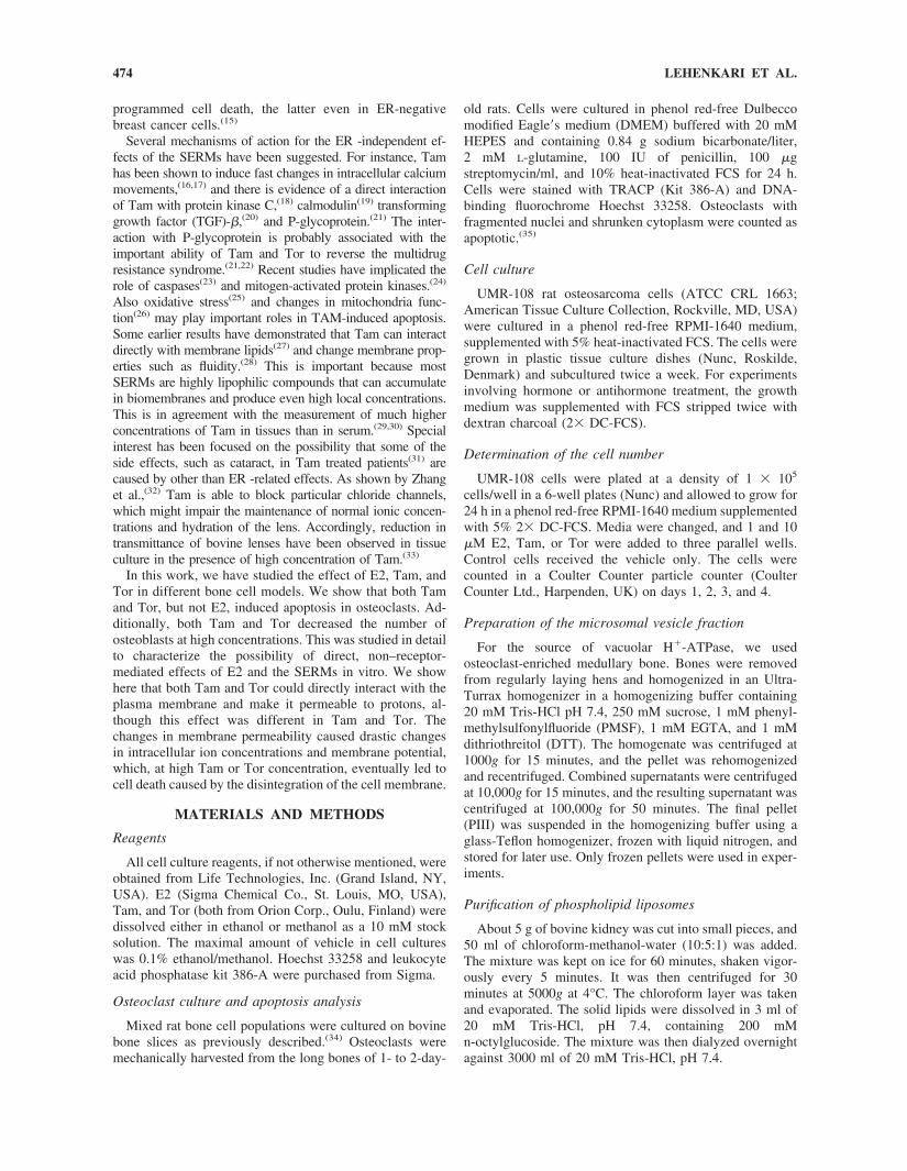
We next studied whether osteoblastic cells were similarlyaffected by E2 and the SERM compounds. Rat osteosar-coma cell line (UMR-108) cells were cultured for 4 days inthe presence of E2, Tam, or Tor (Fig. 2A). E2, even at 10�M concentration, did not disturb cell growth. Tam and Torat 1 �M concentration slightly retarded the rate of cellgrowth. The addition of 10 �M Tam or Tor to the culturemedium almost completely prevented cell proliferation. Wenext studied the effects of different Tam concentrationsfurther. Figure 2B shows the effect of Tam on the nonos-teoclastic cells included in the rat osteoclast-enriched bonemarrow cell cultures. The number of mononuclear cells/1000 �m2 from 10 separate locations at each bone slice wascounted. We found that 10 �M Tam drastically reduced thecell number, but lower concentrations had no effect (except
475TAMOXIFEN AND TOREMIFENE INTERACT WITH PLASMA MEMBRANE

100 fM Tam, which slightly increased the number of mono-nuclear cells). It is notable that this 10 �M concentration is10,000-fold higher than that which increased apoptosis ofosteoclasts in our model.
Intracellular recording of resting potential
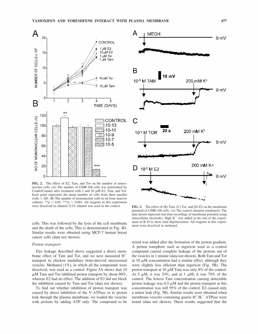
Because both Tam, and to some extent Tor, seemed toinfluence cell viability, this phenomenon was further stud-ied using UMR-108 cells. We found that resting membranepotentials, when recorded with glass capillary microelec-trodes, were in the range of �15 to �40 mV, which istypical for many epithelial cells. The recorded input resis-tances ranged from 5 to 10 M�. The application of Tamresulted in a rapid depolarization of the membrane potential,which stabilized after a few seconds to �10 to �15 mV(Fig. 3B). Application of high K� buffer rapidly abolishedthe membrane potential. In contrast, addition of Tor did notchange the resting membrane potential (Fig. 3C). Changingto the high K� buffer again abolished the resting potential,which proceeded more rapidly than in the case of Tam.Application of the vehicle (Fig. 3A) or E2 (Fig. 3D) slightlyhyperpolarized the resting potential. The input resistance ofthe cells was clearly reduced both by Tam and Tor appli-cation (data not shown), but because of the low initial inputresistance of the cells, the exact magnitude of the decreasecould not be quantified.
Measurement of cytosolic pH
To study if the effect on membrane potential was accom-panied by changes in pH balance, we studied the intracel-
lular pH of the UMR-108 cells. We first measured the pH ina normal buffer, in the presence of physiological concen-trations of ions, under which conditions the cells have goodopportunities to compensate for abnormal ion fluxes. Underthese conditions, the addition of both SERMs caused asignificant pH rise, although the effect of Tam on theintracellular pH was much stronger than that of Tor (Fig.4A). Methanol and E2 had no effect, although, at the be-ginning of measuring, methanol caused a transient rise ofpH. We next tested these compounds in a buffer that did notcontain any monovalent cations (Fig. 4B)—cells were firstbalanced and also loaded in these changed buffers, as de-scribed in the Materials and Methods section. We observedno membrane leakage or accompanying cell death over a20-minute incubation with the SERMs. However, the intra-cellular pH rose to the level of the external pH in thepresence of Tam, whereas Tor, E2, and methanol did nothave this effect. In all cases, the pH rise was at leastpartially compensated by the cells over a longer period oftime.
Finally, we used a high K� buffer without carbonate andsodium to disable the compensation of possible pH changes.The cells were shown to survive although the membranepotential must have been reversed by high K�. Figure 4Cshows the pH changes caused by the addition of the com-pounds investigated. The addition of methanol did not causeany changes in the internal pH, whereas E2 significantlylowered the pH. In the presence of 50 �M Tam or Tor, thedye freely leaked out of the cells, and this was obviouslycaused by the disruption of the outer cell membrane of the
FIG. 1. The effect of (A) E2, (B) Tam, (C) Tam � E2, and (D) Tor on the number of normal and apoptotic osteoclasts after 24 h in culture.Cell numbers are presented as percentage of control. Apoptosis index was counted as number of apoptotic osteoclasts/total number of osteoclasts.Absolute numbers for control groups are 165.25 � 16.72, 165.00 � 10.21, 50.25 � 4.94, and 150.70 � 29.20 osteoclasts for E2-, Tam-, Tam� E2-, and Tor-treated groups, respectively. The average number of apoptotic osteoclasts/bone slice in control groups was 21.77 � 3.47 and theaverage apoptosis index in control groups was 17.88 � 2.90. The p values from Student’s t-test between control and other groups are shown asasterisks (*p � 0.05, **p � 0.01, ***p � 0.001). In C, the asterisks showing the statistical significance between the control and Tam-treated grouphave been underlined. Other asterisks in C show the comparison between Tam- and Tam � E2-treated groups. The concentration of Tam usedin this experiment was 1 nM. All reagents in this experiment were dissolved in ethanol; 0.1% ethanol was used in the control.
476 LEHENKARI ET AL.
cells. This was followed by the lysis of the cell membraneand the death of the cells. This is demonstrated in Fig. 4D.Similar results were obtained using MCF-7 human breastcancer cells (data not shown).
Proton transport
Dye leakage described above suggested a direct mem-brane effect of Tam and Tor, and we next measured H�
transport in chicken medullary bone-derived microsomalvesicles. Methanol (1%), in which all the compounds weredissolved, was used as a control. Figure 5A shows that 10�M Tam and Tor inhibited proton transport by about 80%,whereas E2 had no effect. The addition of E2 did not blockthe inhibition caused by Tam and Tor (data not shown).
To find out whether inhibition of proton transport wascaused by direct inhibition of the V-ATPase or to protonleak through the plasma membrane, we loaded the vesicleswith protons by adding ATP only. The compound to be
tested was added after the formation of the proton gradient.A proton ionophore such as nigericin used as a controlcompound caused complete leakage of the protons out ofthe vesicles in 1 minute (data not shown). Both Tam and Torat 10 �M concentration had a similar effect, although theywere slightly less efficient than nigericin (Fig. 5B). Theproton transport at 10 �M Tam was only 8% of the control.At 5 �M, it was 24%, and at 1 �M, it was 79% of thecontrol. The lowest Tam concentration causing detectableproton leakage was 0.5 �M and the proton transport at thisconcentration was still 95% of the control. E2 caused onlya minor leak (Fig. 5B). Similar results were obtained whenmembrane vesicles containing gastric H�/K� ATPase weretested (data not shown). These results suggested that the
FIG. 2. The effect of E2, Tam, and Tor on the number of nonos-teoclast cells. (A) The number of UMR-108 cells was determined byCoulterCounter after treatment with 1 and 10 �M E2, Tam, and Tor.Each point represents the mean number of cells from three parallelwells � SD. (B) The number of mononuclear cells in rat bone marrowcultures. **p � 0.01, ***p � 0.001. All reagents in this experimentwere dissolved in ethanol; 0.1% ethanol was used in the control. FIG. 3. The effect of (B) Tam, (C) Tor, and (D) E2 on the membrane
potential of UMR-108 cells. (A) The control situation (methanol). Thedata shown represent real-time recordings of membrane potential usingintracellular electrodes. High K� was added at the end of the experi-ment in B–D to show total depolarization. All reagents in this experi-ment were dissolved in methanol.
477TAMOXIFEN AND TOREMIFENE INTERACT WITH PLASMA MEMBRANE

H�-transport inhibition by Tam and Tor could be caused bytheir ability to cause proton leak and not because of thedirect inhibition of the proton pump. The compounds eitherreacted directly with the membrane and formed an ionchannel for protons or they bound to an existing membranechannel, opening it for protons. Finally, they possibly inter-acted with the membrane itself and changed its properties,
which may have induced the opening of some voltage-dependent channels.
To test these hypotheses, we studied the effect of addedprotein-free phospholipid liposomes. As shown in Fig. 6A,the addition of liposomes could block the proton leakagecaused by 10 �M Tam. A 10-fold excess of protein-freemembrane fraction compared with plasma membrane vesi-cles blocked the major part of inhibition from 86% to 5%.Similar results were obtained with 10 �M Tor, suggestingthat these compounds bind directly to the lipid membranes,which leads to the formation of channels allowing protontransition at least. When choline chloride was used insteadof K� (or Na�) in the assay buffer, Tor lost its ability tocause proton leakage at tested concentrations. Tam acted aseffectively in both high and low K� buffers (Fig. 6B).
DISCUSSION
Our data show that the effect of Tam was associated withan increased rate of apoptosis, which obviously led to a
FIG. 4. The effect of E2, Tam, and Tor on the intracellular pH ofUMR-108 cells. (D) Six image analyzer pictures of a single UMR-108cell at different time points when Tam was present at high K� buffer.Numbers presented under each cell picture correspond to time points inC. All compounds were added to samples at 100 s. The measurementtime was 16 minutes. The final concentration of each molecule was 50�M and the same amount of methanol was present in all groups. (A)Cells were loaded in normal buffer (pH 7.2) and measured in normalbuffer (pH 7.8). Intracellular pH was followed for 33 minutes insteadof 16 minutes. (C) Cells were loaded in high K� buffer (pH 7.2) andmeasured in high K� buffer (pH 7.8). (B) Cells were loaded in K�-freebuffer (pH 7.2) and measured in K�-free buffer (pH 7.8).
FIG. 5. The effect of E2 and SERMs on H� transport in (A) chickenmedullary bone microsomal vesicles and (B) proton leakage fromH�-loaded chicken medullary bone microsomal vesicles. (A) Eachcompound was added to the cuvette 5 minutes before measuring for afinal concentration of 10 �M. Curves represent the effects of (a) Tor,(b) Tam, (c) E2, and (d) methanol control. (B) Vesicles were incubatedwith ATP for 4 minutes, and the tested compound was added to thefinal concentration of 10 �M (arrow). Curves in B represent the effectsof (a) Tor, (b) Tam, (c) methanol control, and (d) E2.
478 LEHENKARI ET AL.

shortened life span of osteoclasts. In this study, a similareffect could not be demonstrated with E2. In contrast, E2was able to protect osteoclasts from apoptosis induced byTam or serum withdrawal. E2 protection against Tam-induced apoptosis may be caused by the specific receptor-mediated mechanisms, but it is also possible that it is causedby E2’s independent survival effect, as suggested by E2opposition of serum withdrawal-induced apoptosis. Our os-teoclast data are in line with the previous studies in whichTam, but not E2, affected the viability(7) or membrane HCL
transport(39) of osteoclasts. The viability of osteoblasticcells was affected only in the presence of 10 �M or higherconcentration of Tam. In contrast, even nanomolar Taminduced apoptosis in osteoclasts, the effect of which may beER mediated. We suggest that Tam has general cytotoxicityat higher concentrations.
These non–receptor-mediated effects of Tam were furthercharacterized in electrophysiological experiments. In thisexperimental setting, a depolarization to near zero after theapplication of high K� buffer is to be expected, as theprocedure changes the K�-equilibrium potential (the Nernstpotential) from about �90 mV to about 0 mV. The rela-tively rapid depolarization reflects the high resting perme-ability of the plasma membrane to potassium. The Cl�
equilibrium potential in the normal buffer is very likely tobe near zero. Typical of cells of epithelial origin,(36,37) thecell membrane is apparently quite permeable to Cl� as well,because the resting potential was recorded to be between thepotassium and chloride equilibrium potentials. The rapiddepolarization caused by application of Tam must havebeen caused by increased membrane permeability to an ion(or ions) having equilibrium potential positive from theresting membrane voltage. In principle this change could bespecific for Cl� permeability. However, it is more likelythat Tam induces a nonspecific increase of permeability thatallows leakage of many types of ions through the plasmamembrane. In our experiments, the effect of Tam and Tormay be independent on binding to any specific target pro-tein, because the compounds were able to affect protein-freephospholipid liposomes as demonstrated in proton transportexperiments.
Our results suggest that Tam and Tor have a detergent-like effect both on isolated membrane vesicles and themembranes of intact cells. However, there was a differencein the response times between proton transport experimentswith membrane vesicles and intracellular pH measurementsin intact cells. This was likely caused by the compensatorymechanisms by which living cells control the intracellularion concentrations. Isolated vesicles do not have such acapacity. Depolarization of the plasma membrane obviouslydecreased the ability of the cells to react to ion fluxes, thusspeeding up cell lysis.
The serum concentration of Tam is approximately 0.1–1.0 �M at normal therapeutical doses.(40,41) In our experi-ments, a micromolar concentration of Tam or Tor (1–50�M) in the absence of serum produced ionophoric effects invitro in osteoblasts. The addition of serum reduced thiseffect, which is probably caused by the binding of theseSERMs to the protein components of serum and the conse-quent reduction in the concentration of free, available drug.A considerable effect was, however, still observed in vitroin the presence of 5–20% serum. It is thus conceivable thatdirect membrane effects of Tam and Tor might also play arole in vivo. In addition, the data available show that thesecompounds accumulate in certain tissues, which can lead tohigh local concentrations for example in the lung and theliver. In these tissues, up to 60–70 times the serum concen-tration of Tam can be found.(29) Thus, it seems likely thatTam and Tor concentrations in vivo can reach the levelsrequired to produce direct membrane effects.
FIG. 6. The effect of phospholipid liposomes on the proton leakinduced by (A) Tam and the effect of K� removal on the protontransport inhibition by (B) Tam and Tor. In both A and B, the finalconcentration of antiestrogen was 10 �M. (A) Curve a represents theeffect of Tam on proton accumulation, and curve b represents themethanol control when no phospholipid liposomes were present; curvec represents Tam, and curve d represents methanol control in thepresence of phospholipid liposomes. (B) Curve a represents the effectof Tam and curve b represents the effect of Tor in proton leakage in theabsence of K�.
479TAMOXIFEN AND TOREMIFENE INTERACT WITH PLASMA MEMBRANE
It is not known, however, whether these effects wouldexist in bone. The local concentrations of Tam or Tor inbone during therapy have not yet been reported. It is likelythat osteoblasts and osteocytes are protected, because Tamor Tor has not been reported to cause bone loss. Their effecton resorbing osteoclasts could, however, also involve areceptor-independent component, because the resorptionprocess as such requires an intact and effective ion balanc-ing apparatus.(42)
Tam and Tor are structurally similar except for one chlo-rinated ethyl side-chain present in Tor. The difference in themolecular structure has proved, however, important at leastfor the capacity of the two compounds to generate hepato-toxic metabolites.(43) The chlorine atom also makes Tor lesspolar than Tam, which may make the interaction of Tor withthe membranes more difficult. This is in agreement with ourresults of membrane potential measurements. Depolariza-tion of the membranes in the presence of high potassiumconcentration enhanced the ionophoric effect of Tor to thelevel of Tam. According to these results, it can be expectedthat also in vivo Tor is less potent in inducing ion leakagethrough the plasma membrane.
Our results differ from some studies(4,5) in which E2 wasreported to induce apoptosis of osteoclasts directly. How-ever, in those studies, the experiments were carried out withpurified rabbit osteoclasts(5) or with murine osteoclasts(4) ina culture medium containing either 0.1% bovine serumalbumin (BSA) or 10% charcoal stripped FCS. Our exper-iments were conducted in a medium containing non-stripped FCS, and we used nonpurified primary rat bonecells, most of which were nonosteoclasts. It should be noted,however, that although we used a mixed cell population inour cultures, we could still measure different responses onresorption activity(3) and apoptosis in osteoclasts.(35) Wefind these conditions satisfactory in modeling the in vivoeffects of E2, because the sum effect of E2 always dependsnot only on its direct effect on osteoclasts but also on thenumerous other cells present in the bone tissue.
In conclusion, our results show that Tam, and in someconditions Tor, has an ionophoric effect on the plasmamembrane. It is possible that this effect is involved inchemotherapeutic effects associated with the treatment ofthe patients with these compounds. It is further possible thatthe distinction between the two SERMs in their ability toaffect membranes could at least partly explain the differ-ences in the spectrum of clinical side effects elicited bythese drugs. Additionally, we await with great interest thedetailed comparative studies between Tam and Tor effectson bone.
ACKNOWLEDGMENTS
The authors thank the Academy of Finland, the NationalTechnology Agency (Finland), and the Turku GraduateSchool of Biomedical Sciences for supporting this study.
REFERENCES
1. Jilka RL, Hangoc G, Girasole G, Passeri G, Williams DC,Abrams JS, Boyce B, Broxmeyer H, Manolagas SC 1992 In-creased osteoclast development after estrogen loss: Mediationby interleukin-6. Science 257:88 –91.
2. Roggia C, Gao Y, Cenci S, Weitzmann MN, Toraldo G, GiancarloI, Pacifici R 2001 Up-regulation of TNF-producing T cells in thebone marrow: A key mechanism by which estrogen deficiencyinduces bone loss in vivo. Proc Natl Acad Sci USA 98:13960–13965.
3. Parikka V, Lehenkari P, Sassi M-L, Halleen J, Risteli J, HarkonenP, Vaananen HK 2001 Estrogen reduces the depth of resorptionpits by disturbing the organic bone matrix degradation activity ofmature osteoclasts. Endocrinology 142:5371–5378.
4. Hughes DE, Dai A, Tiffee JC, Li HH, Mundy GR, Boyce BF 1996Estrogen promotes apoptosis of murine osteoclasts mediated byTGF-beta. Nat Med 2:1132–1136.
5. Kameda T, Mano H, Yuasa T 1997 Estrogen inhibits boneresorption by directly inducing apoptosis of the bone-resorbingosteoclasts. J Exp Med 186:489 – 495.
6. Dempster DW, Stein LS, Kilb JM, Moonga BS, Arnett TR, Lind-say R 1997 Estrogen does not promote apoptosis in differentiatedrat osteoclasts. Bone 20:109S.
7. Arnett TR, Lindsay R, Kilb JM, Moonga BS, Spowage M, Demp-ster DW 1996 Selective toxic effects of tamoxifen on osteoclasts:Comparison with the effects of oestrogen. J Endocrinol 149:503–508.
8. Burger HG 2000 Selective oestrogen receptor modulators. HormRes 53:25–29.
9. Pyrhonen S, Ellmen J, Vuorinen J, Gershanovich M, Tominaga T,Kaufmann M, Hayes DF 1999 Meta-analysis of trials comparingtoremifene and tamoxifen and factors predicting outcome of an-tiestrogen therapy in postmenopausal women with breast cancer.Breast Cancer Res Treat 56:133–143.
10. Marshall E 1998 Tamoxifen. ‘A big deal’, but a complex hand toplay. Science 280:196.
11. Marttunen MB, Hietanen P, Titinen A, Roth HJ, Viinikka L,Ylikorkala O 1999 Effects of tamoxifen and toremifene on urinaryexcretion of pyridinoline and deoxypyridinoline and bone densityin postmenopausal patients with breast cancer. Calcif Tissue Int65:365–368.
12. Mandlekar S, Kong A-NT 2001 Mechanisms of tamoxifen-inducedapoptosis. Apoptosis 6:469–477.
13. Warri AM, Huovinen RL, Laine AM, Martikainen PM, HarkonenPL 1993 Apoptosis in toremifene-induced growth inhibition ofhuman breast cancer cells in vivo and in vitro. J Natl Cancer Inst85:1412–1418.
14. Budtz PE 1999 Role of proliferation and apoptosis in net growthrates of human breast cancer cells (MCF-7) treated with oestradioland/or tamoxifen. Cell Prolif 32:289–302.
15. Mandlekar S, Yu R, Tan TH, Kong AN 2000 Activation ofcaspase-3 and c-Jun NH2-terminal kinase-1 signaling pathways intamoxifen-induced apoptosis of human breast cancer cells. CancerRes 60:5995–6000.
16. Chang HT, Huang JK, Wang JL, Cheng JS, Lee KC, Lo YK, LinMC, Tang KY, Jan CR 2001 Tamoxifen-induced Ca2� mobiliza-tion in bladder female transitional carcinoma cells. Arch Toxicol75:184–188.
17. Morley P, Whitfield JF 1994 Effect of tamoxifen on carbachol-triggered intracellular calcium responses in chicken granulosacells. Cancer Res 54:69–74.
18. Schwartz Z, Sylvia VL, Guinee T, Dean DD, Boyan BD 2002Tamoxifen elicits its anti-estrogen effects in growth plate chon-drocytes by inhibiting protein kinase C. J Steroid Biochem MolBiol 80:401–410.
19. Williams JP, McKenna MA, Thames AM III, McDonald JM 2000Tamoxifen inhibits phorbol ester stimulated osteoclastic bone re-sorption: An effect mediated by calmodulin. Biochem Cell Biol78:715–723.
20. Chau D, Mancoll JS, Lee S, Zhao J, Phillips LG, Gittes GK,Longaker MT 1998 Tamoxifen downregulates TGF-beta produc-tion in keloid fibroblasts. Ann Plast Surg 40:490–493.
21. Callaghan R, Higgins CF 1995 Interaction of tamoxifen with themultidrug resistance P-glycoprotein. Br J Cancer 71:294–299.
22. Braybrooke JP, Vallis KA, Houlbrook S, Rockett H, Ellmen J,Anttila M, Ganesan TS, Harris AL, Talbot DC 2000 Evaluation oftoremifene for reversal of multidrug resistance in renal cell cancerpatients treated with vinblastine. Cancer Chemother Pharmacol46:27–34.
23. Mandlekar S, Hebbar V, Christov K, Kong AN 2000 Pharmaco-dynamics of tamoxifen and its 4-hydroxy and N-desmethyl metab-olites: Activation of caspases and induction of apoptosis in rat
480 LEHENKARI ET AL.

mammary tumors and in human breast cancer cell lines. CancerRes 60:6601–6606.
24. Rabenoelina F, Semlali A, Duchesne MJ, Freiss G, Pons M, BadiaE 2002 Effect of prolonged hydroxytamoxifen treatment of MCF-7cells on mitogen activated kinase cascade. Int J Cancer 98:698–706.
25. Ferlini C, Scambia G, Marone M, Distefano M, Gaggini C, Fer-randina G, Fattorossi A, Isola G, Benedetti Panici P, Mancuso S1999 Tamoxifen induces oxidative stress and apoptosis in oestro-gen receptor-negative human cancer cell lines. Br J Cancer 79:257–263.
26. Cardoso CM, Custodio JB, Almeida LM, Moreno AJ 2001 Mech-anisms of the deleterious effects of tamoxifen on mitochondrialrespiration rate and phosphorylation efficiency. Toxicol Appl Phar-macol 176:145–152.
27. Custodio JB, Almeida LM, Madeira VM 1993 The anticancer drugtamoxifen induces changes in the physical properties of model andnative membranes. Biochim Biophys Acta 1150:123–129.
28. Severcan F, Kazanci N, Zorlu F 2000 Tamoxifen increases mem-brane fluidity at high concentrations. Biosci Rep 20:177–84.
29. Lien EA, Solheim E, Ueland PM 1991 Distribution of tamoxifenand its metabolites in rat and human tissues during steady-statetreatment. Cancer Res 51:4837–4844.
30. Lien EA, Wester K, Lonning PE, Solheim E, Ueland PM 1991Distribution of tamoxifen and metabolites into brain tissue andbrain metastases in breast cancer patients. Br J Cancer 63:641–645.
31. Paganini-Hill A, Clark LJ 2000 Eye problems in breast cancerpatients treated with tamoxifen. Breast Cancer Res Treat 60:167–172.
32. Zhang JJ, Jacob TJ, Valverde MA, Hardy SP, Mintenig GM,Sepulveda FV, Gill DR, Hyde SC, Trezise AE, Higgins CF 1994Tamoxifen blocks chloride channels. A possible mechanism forcataract formation. J Clin Invest 94:1690–1697.
33. Zhang JJ, Jacob TJC 1996 Volume regulation in the bovine lensand cataract. The involvement of chloride channels. J Clin Invest97:971–978.
34. Lakkakorpi P, Tuukkanen J, Hentunen T, Jarvelin K, VaananenHK 1989 Organization of osteoclast microfilaments during theattachment to bone surface in vitro. J Bone Miner Res 4:817–825.
35. Selander KS, Monkkonen J, Karhukorpi EK, Harkonen P, Han-nuniemi R, Vaananen HK 1996 Characteristics of clodronate-induced apoptosis in osteoclasts and macrophages. Mol Pharmacol50:1127–1138.
36. Weckstrom M, Kouvalainen E, Juusola M 1992 Measurement ofcell impedance in frequency domain using discontinuous currentclamp and white-noise-modulated current injection. Pflugers Arch421:469–72.
37. Finkel AS, Redman S 1984 Theory and operation of a singlemicroelectrode voltage clamp. J Neurosci Methods 11:101–127.
38. Sundquist K, Lakkakorpi P, Wallmark B, Vaananen K 1990 Inhi-bition of osteoclast proton transport by bafilomycin A1 abolishesbone resorption. Biochem Biophys Res Commun 168:309–313.
39. Williams JP, Blair HC, McKenna MA, Jordan E, McDonald JM1996 Regulation of avian osteoclastic H�-ATPase and bone re-sorption by tamoxifen and calmodulin antagonists. J Biol Chem271:12488–12495.
40. Langan-Fahey SM, Tormey DC, Jordan VC 1990 Tamoxifen me-tabolites in patients on long-term adjuvant therapy for breast can-cer. Eur J Cancer 26:883–888.
41. Daniel CP, Gaskell SJ, Bishop H, Nicholson RI 1979 Determina-tion of tamoxifen and an hydroxylated metabolite in plasma frompatients with advanced breast cancer using gas chromatography-mass spectrometry. J Endocrinol 83:401–408.
42. Lehenkari P, Hentunen TA, Laitala-Leinonen T, Tuukkanen J,Vaananen HK 1998 Carbonic anhydrase II plays a major role inosteoclast differentiation and bone resorption by effecting thesteady state intracellular pH and Ca2�. Exp Cell Res 242:128–137.
43. Montandon F, Williams GM 1994 Comparison of DNA reactivityof the polyphenylethylene hormonal agents diethylstilbestrol, ta-moxifen and toremifene in rat and hamster liver. Arch Toxicol68:272–275.
Address reprint requests to:Petri Lehenkari, PhD
University of OuluDepartment of Anatomy
KajaanintieFIN-90220 Oulu, FinlandE-mail: [email protected]
Received in original form April 16, 2002; in revised form Septem-ber 6, 2002; accepted October 1, 2002.
481TAMOXIFEN AND TOREMIFENE INTERACT WITH PLASMA MEMBRANE
Copyright © 2022 FDOKUMEN