FGF signalling restricts haematopoietic stem cell specification ...
Upload
independentCategory
view
1download
0
Introduction
Macrophages derived from the differentiation of monocytesexpress steroid hormone receptors and are therefore sensitive tothe hormones present in their microenvironment. The study ofsteroid hormone action on modulation of human macrophagefunction is of significant interest because these versatile cells areinvolved in the regulation of immune response and consequentlyare relevant to pathogenesis of many diseases. The ovarian steroidoestrogen is able to exert pleiotropic effect on macrophages,including modulation of the death pathway, for example, it exertsparadoxical effects on human U937 macrophages where cell deathis induced by oestrogen [1], but the same hormone accords pro-tection to these cells from TNF-� induced apoptosis [2]. Similar
effect is exerted on murine osteoclasts, where oestrogen exposureleads to caspase-dependent apoptosis [3, 4]. A previous studyfrom this laboratory demonstrated 17�-oestradiol (E2) inducedapoptosis in macrophages derived from human peripheral bloodmonocytes and THP-1 acute monocytic leukaemia cell line, whenBcl-2 was down-regulated [5]. It is well established that the sur-vival of a cell in response to certain apoptotic stimuli depends onthe critical ratio of mitochondrial Bcl-2 and Bax. Consequent tosensing of apoptotic stimuli, Bax, which exists as an inactivemonomer in the cell cytoplasm, migrates to the mitochondria tointeract with the existing mitochondrial Bcl-2 and the resultinginteraction determines the fate of the cell, higher or lower ratio ofBcl-2/Bax being anti-apoptotic or pro-apoptotic [6], respectively.For the change of location of Bax, a conformational alterationoccurs in the Bax protein, leading to exposure of the mitochondr-ial targeting sequence resulting in its translocation to the outermitochondrial membrane. Multiple mechanisms have been pro-posed as instrumental in initiating Bax translocation, whichinclude neutralization of several pro-survival proteins by BH3 onlymembers of the Bcl-2 family such as Noxa and Puma [7],
Oestrogen modulates human macrophage apoptosis via
differential signalling through oestrogen receptor-� and �
Manikandan Subramanian, Chandrima Shaha *
Cell Death and Differentiation Research Laboratory, National Institute of Immunology, New Delhi, India
Received: October 15, 2008; Accepted: January 6, 2009
Abstract
Human macrophages express oestrogen receptors and are therefore competent to respond to the hormone present in their microenvi-ronment, which is implicated in sexual dimorphism observed in several immune and autoimmune phenomena. An earlier study fromthis laboratory demonstrated 17�-oestradiol (E2) induced apoptosis in macrophages derived from human peripheral blood monocytesand THP-1 acute monocytic leukaemia cell line when Bcl-2 was down-regulated; however, the involvement of E2 receptor subtypes inthe modulation of death pathways in these cells remain unknown. Using macrophages derived from THP-1 human acute monocyticleukaemia cells as a model, we demonstrate that plasma membrane associated oestrogen receptor (ER) -� participate in E2 inducedBcl-2 increase, through activation of the mitogen activated protein kinase (MAPK) pathway whereas cytosolic ER-� transmits signalsfor the pro-apoptotic event of Bax translocation. The mechanistic basis of Bax translocation comprised of ER-� mediated increase inintracellular pH, facilitated by activation of the Na�–H� exchanger. Intracellular alkalinization accompanied by concomitant Bcl-2increase and Bax migration does not cause cellular apoptosis; however, siRNA mediated down-regulation of ER-� during E2 exposureleads to inhibition of Bcl-2 increase and consequently apoptosis due to the unopposed action of mitochondrial Bax. In summary, thisstudy underscores the importance of integrative signalling modality from multiple oestrogen receptor pools in modulating oestrogeneffects on human monocyte-derived macrophage apoptotic signalling pathway, which opens new vistas to explore the use of selectiveoestrogen receptor modulators in apoptosis-based therapies.
Keywords: oestrogen • macrophage • oestrogen receptor • Bcl-2 • Bax • apoptosis • alkalinization
J. Cell. Mol. Med. Vol 13, No 8B, 2009 pp. 2317-2329
*Correspondence to: Dr. Chandrima SHAHA,Cell Death and Differentiation Research Laboratory,National Institute of Immunology,Aruna Asaf Ali Marg, New Delhi-110067, India.Tel:. 91-11-26703627Fax: 91-11-26742125E-mail: [email protected]
© 2009 The AuthorsJournal compilation © 2009 Foundation for Cellular and Molecular Medicine/Blackwell Publishing Ltd
doi:10.1111/j.1582-4934.2008.00679.x
Small Molecules: Molecular Bullets

2318
phosphorylation events mediated by kinases [8, 9], down-modu-lation of clusterin [10], intracellular acidification [11] or alkaliniza-tion [12] and caspase-dependent cleavage of Bax [13]. Becausemodulation of Bcl-2 and Bax could form part of a strategy formanipulation of cell survival, it is important to establish the asso-ciated processes that lead to such changes.
Although it is known that E2 can influence cell death pathwaysin human macrophages, the involvement of oestrogen receptor-�(ER-�) and -� (ER-�) expressed at multiple subcellular locations[14] in these cells in mediating death or survival signals is notknown. It is important to understand the relative contribution ofthe two receptors on effects elicited by oestrogen because sig-nalling through separate subtypes could have diverse outcome[15]. This is evident from the distinct phenotypes obtained withER-� and ER-� knockout mice [16], and in addition, evidence fordifferential receptor activity come from studies showing overlap-ping but exclusive sets of downstream target genes for the twosubtypes [17]. The classical model of ER action is where ligandbound ER interacts with oestrogen response elements in targetgenes and initiates transcription by modulating co-repressors andco-activators. Conversely, ERs can also interact with other tran-scription factors like activating protein-1 and stimulating protein-1to initiate transcription [18, 19]. In addition to the above mode of actions, oestrogen may elicit effects through genomic or non-genomic mechanisms by binding to oestrogen receptors localizedon the plasma membrane of target cells [20] and activate mitogenactivated protein kinase (MAPK) signalling [21] or induce intracel-lular Ca2� fluxes [22]. Elicitation of a particular event in responseto E2 could be dependent on the relative concentrations of the twoER subtypes, for example, U937 monocytes expressing mostlyER-� are sensitive to oestrogen induced apoptosis; however, afterdifferentiation to macrophages, receptor population expressed ispredominantly ER-�, as a result of which the apoptosis inducingeffect of oestrogen becomes ineffective [23].
The purpose of this study was to investigate the involvement ofthe two ER subtypes in mediating apoptosis associated events inhuman macrophages using THP-1 monocyte derivedmacrophages as a model system. We show that THP-1 humanmacrophage survival is compromised in the presence of E2 if ER-� but not ER-� receptor levels are down-regulated. This isbecause E2 signalling via ER-� mediates the anti-apoptotic eventof Bcl-2 up-regulation, whereas ER-� signals for the pro-apoptoticevent of Bax translocation to the mitochondria via Na�-H�
exchanger mediated intracellular alkalinization.
Materials and methods
Cell lines and culture
THP-1, a human acute monocytic leukaemia cell line, and MCF-7, a humanbreast carcinoma cell line (ATCC, Manassas, VA, USA), were maintained inRoswell Park Memorial Institute (RPMI)-1640 (Biological Industries,Kibbutz Beit Haemek, Israel), supplemented with 10% FCS (Biological
Industries, Kibbutz Beit Haemek, Israel). Differentiation of THP-1 mono-cytes to macrophages was induced by treatment with 10 ng/mL PMA for 36 hrs. Forty eight hours prior to experimentation, the cells were transferredto phenol-red free RPMI-1640 supplemented with 10% dextran-coatedcharcoal stripped FCS to remove all extraneous sources of oestrogen.
Reagents
E2 (cyclodextrin encapsulated), E2 cojugated to BSA (E2-BSA), E2-BSAconjugated to FITC (E2-BSA-FITC), PD98,059, nigericin, amiloride and pro-pidium iodide (PI) were obtained from Sigma-Aldrich (St. Louis, MO,USA). ICI 182,780 was obtained from Tocris Cookson (Bristol, UK).Negative control siRNA was purchased from Ambion (Austin, TX, USA),whereas Bcl-2, ER-� and ER-� siRNAs were obtained from Dharmacon(Lafayette, CO, USA). The siRNA transfection reagent Transpass R2 waspurchased from New England Biolabs (Ipswich, MA, USA). All reagents forWestern blotting and ECL development were from Amersham Biosciences(Piscataway, NJ, USA). SNARF (5-(and-6)-carboxy SNARF®1-AM),Sodium Green™ tetra-acetate, secondary antimouse IgG conjugated toAlexa fluor 488 and Hoechst 33342 nuclear dye were purchased fromMolecular Probes (Eugene, OR, USA). Antibodies for oestrogen receptor�/�, oestrogen receptor-� and actin were procured from Calbiochem(Darmstadt, Germany), whereas antibodies against phospo-ERK andwhole-ERK were from StressGen Biotechnologies (Victoria, BC, Canada).Anti-Bcl-2, anti-Bax and anti-cytochrome c antibodies were from SantaCruz Biotechnology (Santa Cruz, CA, USA), anti-GAPDH antibody was fromAmbion (Austin, TX, USA), whereas anti-histone dimethyl lysine antibodywas purchased from Upstate (VA, USA). Secondary antimouse and anti-rabbit IgG conjugated to horseradish peroxidase were procured fromJackson Immunoresearch (Cambridgeshire, UK). The Vybrant apoptosisdetection system was purchased from Promega (Madison, WI, USA). Anti-clusterin antibody was a kind gift from Dr. C. Yan Cheng of the PopulationCouncil, NY, USA. All other chemicals used were purchased from Sigma-Aldrich (St. Louis, MO, USA) unless mentioned otherwise.
Intracellular pH measurement
Intracellular pH measurement was performed with the long-wavelength fluorescent pH indicator carboxy SNARF-1 AM following manufacturer’s pro-tocol. Briefly, the cells (106 /ml) were resuspended in serum-free RPMI-1640and incubated with a final concentration of 1 �M SNARF-1 AM, diluted froma stock solution of 1 mM in DMSO for 15 min. at room temperature. Cellswere washed and incubated for 20 min. at room temperature for completede-esterification of AM esters. In situ calibration of SNARF-1 AM was per-formed with the ionophore nigericin at 10 �M concentration in a high-K�
buffer to equilibrate intracellular pH with that of the controlled extracellularmedium. Appropriate groups were subjected to different treatments, and flu-orescence measurements were commenced in a spectrofluorometer (PerkinElmer, Waltham, MA, USA), followed by kinetic analysis. The pH was calcu-lated from the fluorescence measurements using the following formula:
pH � pKa � log [{(R � RB)/(RA � R)} � (FB(2) /FA(2))],
where pKa of carboxy SNARF-1 AM is 7.5. R is the ratio of fluorescentintensities (F) measured at two emission wavelengths, 580 (1) and 640 nm (2), with fixed excitation at 514 nm. The subscripts A and B rep-resent the limiting values at the acidic and basic end-points of the titration,respectively. Na�-free and HCO3
�-free buffer were prepared as describedby Khaled et al. [24].
© 2009 The AuthorsJournal compilation © 2009 Foundation for Cellular and Molecular Medicine/Blackwell Publishing Ltd

J. Cell. Mol. Med. Vol 13, No 8B, 2009
2319
Intracellular Na� measurement
For intracellular Na� measurement, cells (106 /mL) were labelled for 20 min. at room temperature with the cell permeable fluorescent Na� indi-cator Sodium Green™ tetra-acetate, diluted to 1 �M in RPMI-1640 from a5 mM stock solution made in DMSO. After washing the cells to removeexcess probe, kinetic fluorescent measurements were carried out with aspectrofluorometer at an excitation of 480 nm and emission of 520 nm(BMG Fluostar Optima, BMG technologies, Offenburg, Germany). In situCalibration was accomplished by using the indicator in solutions of pre-cisely known free Na� concentration in the presence of the pore formingantibiotic gramicidin (10 �M). Intracellular Na� was calculated using thefollowing equation:
[Na�]free � Kd (F – Fmin/Fmax – F),
where Kd of the dye is 5.7 mM at 37C, F is the fluorescence of the exper-imental sample, Fmin is fluorescence in the absence of Na� and Fmax is flu-orescence in the presence of saturating concentrations of Na�.
siRNA transfection
THP-1 macrophages were transfected with specific siRNAs usingTranspass R2 transfection reagent as described previously [5]. Briefly, Bcl-2 siRNA (15 pmol), ER-� and ER-� siRNA (100 pmol) or negative controlsiRNA (pre-designed siRNA with no known target genes) at similar con-centrations were added to transfection reagent TranspassR2, diluted inserum-free medium, and incubated for 20 min. to allow the formation of transfection complexes. The formed complexes were added to 105 cells/well grown in 24-well plates and incubated for 6 hrs, followingwhich fresh complete medium was added. Transfection efficiency was esti-mated by observing Cy3 fluorescence of the negative control siRNA with aNikon TE2000-E fluorescence microscope using a tetramethyl rhodaminefilter (530–580 nm). For all transfections, target protein knockdown wasassessed 24 hrs after transfection by probing extracts of transfected cellson Western blots with anti-Bcl-2 and anti ER-�/� antibody.
Subcellular fractionation
THP-1 macrophages were allowed to swell for 10 min. in hypotonic buffer(10 mM NaCl, 1.5 mM MgCl2, 10 mM Tris-HCl, pH 7.5) followed byhomogenization with a Dounce homogenizer (50 strokes). Immediatelyafter cell lysis, the mitochondria were stabilized by addition of mitochondr-ial stabilization buffer (525 mM mannitol, 175 mM sucrose, 12.5 mM Tris-HCl, pH 7.5; 2.5 mM EDTA, pH 7.5), and the homogenate was centrifugedat 1300 � g for 15 min. to isolate the nuclear fraction. The post-nuclearsupernatant was further centrifuged at 17,000 � g for 15 min. in an ultra-centrifuge (Optima XL-100K, Beckman, Fullerton, CA, USA) to isolate themitochondria. The post-mitochondrial supernatant was centrifuged at100,000 � g for 1 hr to obtain the membranous fraction as a pellet and thesupernatant as the cytosol. The homogeneity of the obtained fractions wasdetermined by Western blotting with probes specific for each fraction.
Cell viability assay
To assess cell viability, PI dye exclusion assay was performed by incubat-ing the cells with 1 �g/ml PI for 5 min. at 37C, followed by one wash with
ice-cold PBS. The cells were analysed under a Nikon TE2000-E fluores-cence microscope using Nikon G2A filter cube. The percentage cell deathwas calculated as the number of cells with PI positive nuclei as against thetotal number of cells. Annexin-V-PI staining was performed as describedpreviously [5], and data acquisition was performed on a BD-LSR flowcytometer equipped with a 488 nm air-cooled argon ion laser. The acquireddata was analysed using WinMDI software (Microsoft v.2.9).
Reverse transcription-polymerase chain reaction
Total RNA was isolated using TRIzol reagent (GIBCO, CA, USA), and cDNAwas synthesized as described previously [5]. The specific primers used were
ER-� (sense) GTGGGAATGATGAAAGGTGG; ER-� (antisense) TCCAGA-GACTTCAGGGTGCT
ER-� (sense) TGAAAAGGAAGGTTAGTGGGAACC; ER-� (antisense)TGGTCAGGGACATCATCATGG
Actin(sense),5�-GTGGGGCGCCCCAGGCACCA-3�; Actin(antisense),5’CTCCTTAATGTCACGCACGATTTC-3�.
PCR was performed after determining the cycle number in which a lin-ear amplification of serially diluted template could be achieved. The PCRproducts were then resolved on 1.5% agarose gel and visualized by ethid-ium bromide staining and quantitated by densitometry.
Immunocyotochemistry
Cells were fixed with 4% formaldehyde for 20 min., followed by severalwashes with ice-cold PBS. Saponin (0.1%) was used for cell permeabiliza-tion, and 3% normal goat serum was used as a blocking reagent to reducenon-specific binding. The permeabilized cells were incubated with primaryantibody recognizing ER-�/� (1 : 100) followed by incubation with second-ary antimouse IgG conjugated to Alexa fluor 488 (1 : 200). For live-cellstaining, all incubations were performed at 4C with anti-ER-� antibody inaddition to reagents as described above. For nuclear labelling of cells,Hoechst 33342 was used. All stainings were visualized using a NikonTE2000-E fluorescence microscope using appropriate filter blocks, and theimage acquisition was carried out with a high-resolution Retiga Exi camera(Q-imaging, Surrey, BC, Canada), the mask of co-localization was createdand the co-efficient of colocalization was calculated using Image-Pro Plussoftware (Media cybernetics, Silver Spring, MD, USA).
SDS-PAGE and Western blot
Whole cell extracts were prepared by treating the cells with lysis buffer(0.125 M Tris, 4% SDS, 20% glycerol and 10% 2-ME), and protein estima-tion was performed with CBX protein assay kit (G-Biosciences, St. Louis,MO, USA). Lysates were resolved on 12% SDS-PAGE gel, following whichthey were transferred onto nitrocellulose membrane as described before[5]. Non-specific binding sites were blocked by incubating the blots in 5%non-fat skimmed milk with 0.05% PBS-Tween 20 for 1 hr. Primary (1:5000) and secondary antibody (1 : 10,000) incubations were carried outfor 1 hr each, and immunoreactivity was visualized by enhanced chemilu-minescence using ECL reagent, as described previously [5].
Densitometry and statistical analysis
Quantitative assessment of reaction intensity in Western blots was per-formed using a UVP gel-documentation instrument, and the data were
© 2009 The AuthorsJournal compilation © 2009 Foundation for Cellular and Molecular Medicine/Blackwell Publishing Ltd

2320
analysed with the LabWorks image analysis and acquisition software(v4.0.0.8; UVP, Upland, CA, USA). At least three Western blots per experi-ment were quantitated to arrive at the average value of the signal. All meas-urements were normalized to internal loading controls. Data are expressedas mean � standard error (SE) unless mentioned. Comparisons weremade between different groups using unpaired Student’s t-test. The valueswere considered to be significantly different at P 0.05. All analysis wasperformed on data acquired from three or more independent experiments.
Results
Human macrophages express both oestrogenreceptor-� and � at multiple subcellular locations
E2 initiates cellular signalling pathways via interaction with itsreceptors expressed primarily as two subtypes – the ER-� and
ER-� [25] – found in the nucleus, plasma membrane or cytosol,the distribution varying with different cell types [26]. Humanmacrophages are known to express both ER-� and ER-� [27, 28],however, the subcellular localization of these receptors is notknown. In the current study, membrane bound E2 receptors weredetected on viable differentiated THP-1 macrophages by labellinglive cells at 4C with membrane impermeable E2-BSA linked toFITC (E2-BSA-FITC), and flow cytometric analysis showed anobvious shift in fluorescence labelling intensity in these cells com-pared with those labelled with only BSA-FITC used as a control(Fig. 1A). Decreased fluorescence readings obtained with cellsincubated with E2-BSA-FITC in the presence of unconjugated E2compared with cells exposed to only E2-BSA-FITC confirmedspecificity of this binding (Fig. 1A).
Further analysis of presence of ERs in different subcellularlocations in these cells showed surface labelling of ERs on livecells (Fig. 1B, a, i–iv) with an anti-E2 receptor antibody raisedagainst common epitopes on ER-� and ER-� receptor proteins,thus corroborating the above data obtained with E2-BSA-FITC.
© 2009 The AuthorsJournal compilation © 2009 Foundation for Cellular and Molecular Medicine/Blackwell Publishing Ltd
Fig. 1 Human macrophages express oestro-gen receptor � and � on the plasma mem-brane and cytoplasm. (A) The histogram rep-resents flow-cytometric analysis of live THP-1macrophages incubated with E2 conjugated toBSA-FITC (red line) or co-incubation with dif-ferent concentrations of E2 (blue, 10 nM E2and pink, 100 nM E2 lines) under similar con-ditions. The area shaded grey represents unla-belled cells, and the black line represents cellslabelled with only BSA-FITC. Note the distinctshift in staining of the cells treated with E2-BSA-FITC demonstrating recognition of cellsurface localization of ERs. (B) Indirectimmunofluorescence on live as well asformaldehyde-fixed cells stained with anti-ER-�/� antibody. (a) THP-1 live; (b), THP-1 fixed;(c), secondary antibody control, (d) fixed MCF-7 cells – (i), nomarski image; (ii), ER-�/�staining; (iii), nuclear staining with Hoechst33342; (iv), overlap of (ii) and (iii). The MCF-7cells (d, i–iv) used as positive controls showpresence of nuclear receptors. The bar repre-sents 10 �m. All data are representative of atleast three independent experiments. (C)Western blots of subcellular fractions of THP-1 cells probed with anti-ER-�/� showing pres-ence of both forms in the cytoplasm (b, Cyto,lane 2), predominantly ER-� in membranefraction (b, Memb, lane 1) and absence ofreceptors in nuclear fraction (b, Nuc, lane 3).(a) MCF-7 cell extracts show the presence ofboth ER-� and ER-�. Western blot for histoneand GAPDH was performed to assess thehomogeneity of the obtained nuclear and cyto-plasmic fractions, respectively.

J. Cell. Mol. Med. Vol 13, No 8B, 2009
2321
Intracellular receptors were detected by staining fixed and perme-abilized cells with the same antibody that demonstrated the pres-ence of receptors within the cytosol and the membrane but not inthe nucleus (Fig. 1B, b, i–iv). Figure 1B, c, i–iv shows absence offluorescence in secondary antibody controls. MCF-7 cells, whereE2 receptors are predominantly nuclear [29], were used as posi-tive controls and showed distinct nuclear staining with the sameantibody (Fig. 1B, d, i–iv). Cytosolic ERs dimerize and migrate tothe nuclei upon ligand engagement [18]; this phenomenon wasused as a control for cytosolic receptors, and Figure S1 showsthat upon E2 exposure the cytosolic ERs migrate to the nuclei (E2,b and d) compared with control (Control, b and d) and E2-BSAtreatment (E2-BSA, b and d). Measurement of the mask of colo-calization clearly shows significant colocalization of ER receptorstaining with nuclear staining after E2 treatment (E2, e) comparedwith vehicle treated controls (Control, e) and after E2-BSA treat-ment (E2-BSA, e).
To determine the subtype specific distribution of receptors,Western blots of subcellular fractions of THP-1 cells were probedwith the same antibody as above, and it recognized both receptortypes in the cellular cytosolic fraction (Fig. 1C, b, lane 2) whereasthe membranous fraction primarily showed reactivity for ER-�subtype, the expression of ER-� being low (Fig. 1C, b, lane 1). Thenuclear fraction did not show any reactivity, suggesting theabsence of nuclear oestrogen receptors in this particular cell type(Fig. 1C, b, lane 3). Total extract of MCF-7 cells known to expressboth ER-� and ER-� was used as positive control (Fig. 1C, a).Collectively, the above data demonstrated the presence of ERs onthe cell surface as well as in the cytosol of THP-1 monocytederived macrophages, nucleus being devoid of such receptors.Both ER-� and ER-� are present intracellularly, whereas theplasma membrane appears to be primarily populated by ER-�.
E2 modulates Bcl-2 and Bax via different sets of receptors in THP-1 cells
To investigate if the receptors at different subcellular locationstransmit similar or different signals for modulation of themitochondrial apoptotic pathway, both membrane permeableand impermeable E2 were used. E2-BSA, where E2 is conju-gated to BSA through a six atom hydrocarbon tether [30]restricting its diffusion through the plasma membrane, wasused to distinguish signals originating from membrane boundreceptors only [20, 30].
Equivalent increase in Bcl-2 levels was obtained with mem-brane permeable E2 as well as membrane impermeable E2-BSA at6 hrs (Fig. 2A, lanes 2 and 4). Pure E2 receptor antagonist ICI182,780 inhibited this increase (Fig. 2A, lanes 3 and 5), confirm-ing that in both cases, E2 receptors were involved. In contrast toits ability to modulate Bcl-2, E2-BSA was unable to exert any effect on subcellular localization of Bax (Fig. 2B, lanes 5 and 6),whereas unconjugated E2 was able to stimulate Bax translocation(Fig. 2B, lanes 3 and 4). The inability of E2-BSA to induce migra-tion of Bax from cytosol to the mitochondria unlike free E2
suggested that Bax migration was independent of membranereceptor mediated signalling.
Based on the above data, attempt was made to identify recep-tor subtypes involved in mediating the above responses. ER-� andER-� mRNA and protein were selectively down-regulated withsiRNA to test the effect of this down-regulation on Bcl-2 and Baxand, eventually, cell death. Figure 3A shows the RT-PCR for ER-�mRNA performed with primers specific for ER-�, where lane 2
© 2009 The AuthorsJournal compilation © 2009 Foundation for Cellular and Molecular Medicine/Blackwell Publishing Ltd
Fig. 2 Regulation of Bax and Bcl-2 is mediated by E2 receptors localizedat different sub-cellular locations. (A) Extracts of THP-1 cells treated withE2 (10 nM)(lane 2) and E2-BSA for 6 hrs (10 nM)(lane 4) show Bcl-2 up-regulation. Presence of ICI 182,780 (1 �M) during E2 (lane 3) and E2 BSA(lane 5) treatment inhibited Bcl-2 up-regulation. Same blots were strippedand reprobed for actin, which was used as a loading control. The bargraph represents densitometric measurements of Bcl-2 expression rela-tive to control and normalized to actin (n � 3). The error bars represent� SEM. * P 0.05 compared with control. # P 0.05 compared withE2 or E2-BSA treated group. (B) THP-1 macrophages were exposed for 6hrs to E2 (lanes 3 and 4) or E2 BSA (lanes 5 and 6) and subcellular frac-tions of mitochondria (M) and cytosol (C) were analysed for the localiza-tion of Bax. Note the absence of Bax translocation in the E2-BSA treatedgroups. The blot was stripped and reprobed for cytochrome c to determinethe homogeneity of mitochondrial fractions. Note the heavy presence ofcytochrome c in the mitochondrial fraction. All data are representative ofat least three independent experiments. Cyt C: cytochrome c.

2322 © 2009 The AuthorsJournal compilation © 2009 Foundation for Cellular and Molecular Medicine/Blackwell Publishing Ltd

J. Cell. Mol. Med. Vol 13, No 8B, 2009
2323
shows decrease in ER-� mRNA when siRNA to ER-� was used butno decrease with ER-� siRNA (Fig. 3A, lane 3). Figure 3B, showsthe RT-PCR for ER-� mRNA performed with ER-� specific primers,where lane 3 shows down-regulation of ER-� mRNA levels withER-� siRNA but not with ER-� siRNA (lane 2). Negative controlsiRNA did not show any interference with RNA levels of eitherreceptors (Fig. 3A and B, lanes 1). Status of protein levels of ER-�and ER-� after ER-� siRNA (Fig. 3C, lane 3) and ER-� siRNA (Fig. 3C, lane 2) transfection shows that protein levels of both recep-tors were significantly down-regulated with respective siRNA treat-ment. A distinct reduction of surface receptor population wasobserved with siRNA for ER-� (Fig. 3D, b, i-iii) compared with a neg-ative control siRNA (Fig. 3D, a, i-iii), as visualized by immunostain-ing of treated and untreated cells with a specific anti-ER-� antibody.
Next, the effects of ER-� and ER-� down-regulation on Bcl-2and Bax modulation were examined. E2-BSA was able to increaseBcl-2 in the presence of negative control siRNA (Fig. 3E, lane 2)but not when the cells were transfected with ER-� siRNA (Fig. 3E,lane 4), thus linking the involvement of ER-� in Bcl-2 increase.This treatment with ER-� siRNA, therefore, creates a conditionwithin the cell, which is pro-apoptotic in nature because presenceof E2 will induce Bax translocation, and in the absence of Bcl-2,Bax will induce apoptosis. In contrast, the knockdown of ER-�during E2-BSA exposure did not affect Bcl-2 increase (Fig. 3E, lane6), showing the absence of any effect of membrane associatedER-� on Bcl-2 levels. Knockdown of ER-� (Fig. 3F, lanes 3 and 4)but not of ER-� (Fig. 3F, lanes 5 and 6) in the presence of E2resulted in inhibition of Bax migration, thereby creating an anti-apoptotic condition because Bcl-2 will not have to counteract theeffect of translocated Bax to the mitochondria. The translocationwas complete in cells transfected with negative control siRNA
(Fig. 3F, lanes 1 and 2). Together, the above data demonstrate adichotomous effect of E2 on the components of the mitochondrialcell death pathway mediated through the two ER subtypes, ER-�mediating Bcl-2 increase and ER-� arbitrating Bax translocation.
Arguably, the modulation of the two receptor levels leading tochanges in the pro-apoptotic Bax and anti-apoptotic Bcl-2 wouldaffect cell survival. Annexin-V/PI staining showed that ER-�knockdown in the presence of E2 did not induce any apoptoticdeath (Fig. 3G, ii), whereas ER-� knockdown resulted in about40% late apoptotic cells and 17% early apoptotic cells at 6 hrsafter E2 exposure (Fig. 3G, iii). This corroborated our findings thatER-� but not ER-� was involved with the survival pathway, andinterference with this pathway resulted in increased cell death inthe presence of E2.
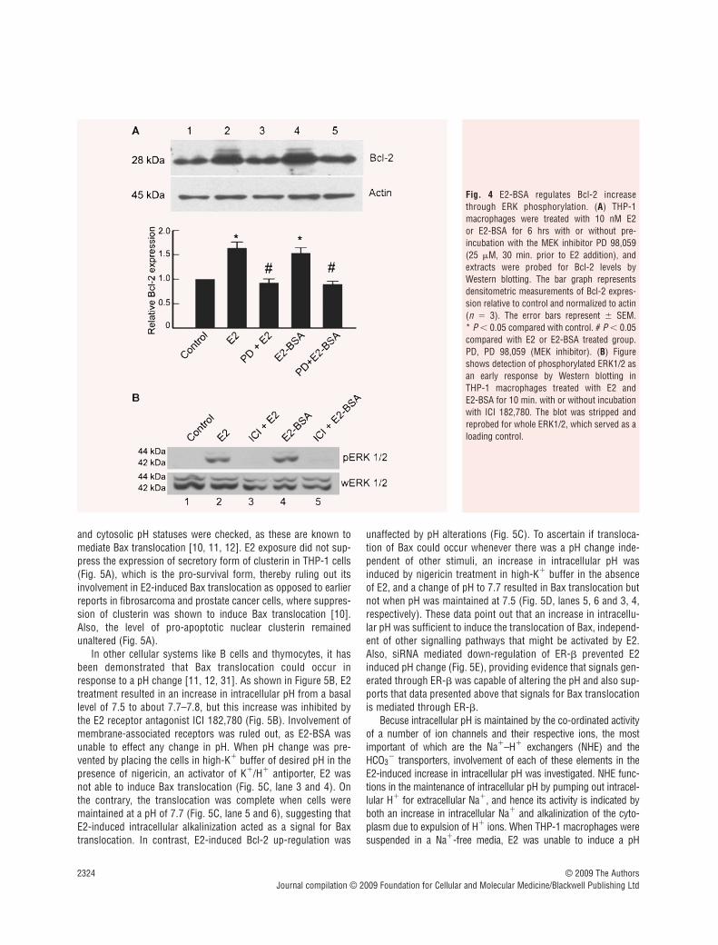
Bcl-2 modulation is mediated through ERK phosphorylation whereas Bax translocation isdependent upon intracellular alkalinization
Downstream to ER engagement by E2-BSA and E2, inhibition ofERK phosphorylation by MEK inhibitor PD 98,059 resulted inabrogation of Bcl-2 up-regulation (Fig. 4A, lane 5 and 3).Furthermore, E2-BSA was able to induce phosphorylation of ERKin as early as 10 min. (Fig. 4B, lane 4), which could be inhibited byICI 182,780 (Fig. 4B, lane 5). Therefore, this indicated the compe-tence of the membrane associated ERs to transmit Bcl-2 up-regulation signals. Following the observation that cytosolic ER-�was involved in Bax translocation, the mechanisms that lead tothis change in subcellular localization was investigated. Clusterin
© 2009 The AuthorsJournal compilation © 2009 Foundation for Cellular and Molecular Medicine/Blackwell Publishing Ltd
Fig. 3 Bcl-2 increase and Bax translocation are regulated by distinct oestrogen receptor subtypes. (A) RT-PCR for ER-� mRNA. Down-regulation of ER-� mRNA at 24 hrs of treatment by siRNA against ER-� (lane 2) in THP-1 cells as visible by amplification of mRNA for ER-� by RT-PCR. Note thatthere is no down-regulation of ER-� mRNA in cells transfected with ER-� siRNA (lane 3). (B) RT-PCR for ER-� mRNA. Down-regulation of ER-� mRNAby siRNA against ER-� (lane 3) at 24 hrs of treatment as visible by amplification of mRNA for ER-� by RT-PCR. Note that there is no down-regulationof ER-� mRNA (lane 2) in cells transfected with ER-� siRNA. (C) Western blot analysis on whole cell lysates for expression of ER-� and ER-� in THP-1cells transfected with negative control siRNA (lane 1), ER-� siRNA (lane 2) or ER-� siRNA (lane 3), 24 hrs after transfection. Note the down-regulationof ER-� and ER-� after treatment with respective siRNAs. The blot was stripped and reprobed for actin, which was used as an endogenous loading con-trol. (D) Live cell fluorescence microscopic analysis of ER-� expression on the plasma membrane by immunostaining with an ER-� specific antibodyin negative control siRNA transfected (a, i–iii) and ER-� siRNA (b, i-iii) transfected THP-1 macrophages at 24 hrs. The blue stain (ii) is labelling withthe nuclear dye Hoechst 33342. Note the loss of membrane ER-� expression in ER-� siRNA transfected cells (b, i-iii). The bar represents 10 �m. (E) Analysis of Bcl-2 expression by Western blotting in THP-1 macrophages transfected with negative control siRNA (Neg con siRNA), ER-� siRNA (ER-� si)or ER-� siRNA (ER-� si) for 24 hrs and subsequently treated with or without 10 nM E2-BSA for 6 hrs. Note the reduction in Bcl-2 when ER-� siRNAand E2-BSA (lane 4) was used compared with ER-� siRNA and E2-BSA, respectively (lane 6). Western blot for actin was used as loading control. Bargraph is densitometric representation of the relative Bcl-2 expression compared with negative control siRNA transfected cells. * P 0.05 comparedwith the respective control groups. # P 0.05 compared with negative control siRNA transfected cells treated with E2-BSA. (F) Western blot analysisfor subcellular localization of Bax was performed in cells transfected with negative control siRNA (lanes 1, 2), ER-� siRNA (lanes 3, 4) or ER-� siRNA(lanes 5, 6) for 24 hrs, followed by treatment with 10 nM E2 for 6 hrs. Note the decrease in Bax translocation in lanes 3 and 4. The blots were strippedand reprobed for cytochrome c, which served as a control to determine the homogeneity of the obtained mitochondrial and cytosolic fractions. All dataare representative of at least three independent experiments. (G) Flow-cytometric analysis of Annexin-V-PI staining in ER-� (iii) or ER-� (ii) knockdowncells, treated with respective siRNAs for 24 hrs, followed by exposure to 10 nM E2 for 6 hrs. Note that ER-� down-regulation in the presence of E2shows high number of apoptotic cells. Cells in the lower left quadrant represent viable cells. Neg Con si: negative control siRNA; siER-�: ER-� siRNA;si ER-�: ER-� siRNA.
2324 © 2009 The AuthorsJournal compilation © 2009 Foundation for Cellular and Molecular Medicine/Blackwell Publishing Ltd
Fig. 4 E2-BSA regulates Bcl-2 increasethrough ERK phosphorylation. (A) THP-1macrophages were treated with 10 nM E2 or E2-BSA for 6 hrs with or without pre-incubation with the MEK inhibitor PD 98,059(25 �M, 30 min. prior to E2 addition), andextracts were probed for Bcl-2 levels byWestern blotting. The bar graph representsdensitometric measurements of Bcl-2 expres-sion relative to control and normalized to actin(n � 3). The error bars represent � SEM. * P 0.05 compared with control. # P 0.05compared with E2 or E2-BSA treated group.PD, PD 98,059 (MEK inhibitor). (B) Figureshows detection of phosphorylated ERK1/2 asan early response by Western blotting in THP-1 macrophages treated with E2 and E2-BSA for 10 min. with or without incubationwith ICI 182,780. The blot was stripped andreprobed for whole ERK1/2, which served as aloading control.
and cytosolic pH statuses were checked, as these are known tomediate Bax translocation [10, 11, 12]. E2 exposure did not sup-press the expression of secretory form of clusterin in THP-1 cells(Fig. 5A), which is the pro-survival form, thereby ruling out itsinvolvement in E2-induced Bax translocation as opposed to earlierreports in fibrosarcoma and prostate cancer cells, where suppres-sion of clusterin was shown to induce Bax translocation [10].Also, the level of pro-apoptotic nuclear clusterin remained unaltered (Fig. 5A).
In other cellular systems like B cells and thymocytes, it hasbeen demonstrated that Bax translocation could occur inresponse to a pH change [11, 12, 31]. As shown in Figure 5B, E2treatment resulted in an increase in intracellular pH from a basallevel of 7.5 to about 7.7–7.8, but this increase was inhibited bythe E2 receptor antagonist ICI 182,780 (Fig. 5B). Involvement ofmembrane-associated receptors was ruled out, as E2-BSA wasunable to effect any change in pH. When pH change was pre-vented by placing the cells in high-K� buffer of desired pH in thepresence of nigericin, an activator of K�/H� antiporter, E2 wasnot able to induce Bax translocation (Fig. 5C, lane 3 and 4). Onthe contrary, the translocation was complete when cells weremaintained at a pH of 7.7 (Fig. 5C, lane 5 and 6), suggesting thatE2-induced intracellular alkalinization acted as a signal for Baxtranslocation. In contrast, E2-induced Bcl-2 up-regulation was
unaffected by pH alterations (Fig. 5C). To ascertain if transloca-tion of Bax could occur whenever there was a pH change inde-pendent of other stimuli, an increase in intracellular pH wasinduced by nigericin treatment in high-K� buffer in the absenceof E2, and a change of pH to 7.7 resulted in Bax translocation butnot when pH was maintained at 7.5 (Fig. 5D, lanes 5, 6 and 3, 4,respectively). These data point out that an increase in intracellu-lar pH was sufficient to induce the translocation of Bax, independ-ent of other signalling pathways that might be activated by E2.Also, siRNA mediated down-regulation of ER-� prevented E2induced pH change (Fig. 5E), providing evidence that signals gen-erated through ER-� was capable of altering the pH and also sup-ports that data presented above that signals for Bax translocationis mediated through ER-�.
Becuse intracellular pH is maintained by the co-ordinated activityof a number of ion channels and their respective ions, the mostimportant of which are the Na�–H� exchangers (NHE) and theHCO3
� transporters, involvement of each of these elements in theE2-induced increase in intracellular pH was investigated. NHE func-tions in the maintenance of intracellular pH by pumping out intracel-lular H� for extracellular Na�, and hence its activity is indicated byboth an increase in intracellular Na� and alkalinization of the cyto-plasm due to expulsion of H� ions. When THP-1 macrophages weresuspended in a Na�-free media, E2 was unable to induce a pH

J. Cell. Mol. Med. Vol 13, No 8B, 2009
2325© 2009 The AuthorsJournal compilation © 2009 Foundation for Cellular and Molecular Medicine/Blackwell Publishing Ltd
Fig. 5 Bax translocation is dependent on E2-induced change in intracellular pH mediated via ER-�. (A) Western blot analysis of clusterin expressionafter E2 treatment indicates no change in the expression of the different isoform levels. Pre-sClu: precursor to secretory clusterin; nClu: nuclear clus-terin; sClu: secretory clusterin. (B) Intracellular pH measurement was performed with the pH sensitive dye SNARF-1 AM. The graph represents the cal-culated intracellular pH of THP-1 macrophages exposed to various drug treatments as indicated over a time-course of 40 min. Note that although E2increased the intracellular pH, this was prevented in the presence of antioestrogen ICI 182,780 but E2-BSA was unable to alter intracellular pH. ICI: ICI182,780. (C) Appropriate groups of THP-1 macrophages were resuspended in high-K� buffer of pH 7.5 or 7.7. The control group was left untreatedwhereas the other two groups were treated with 1 �M nigericin and 10 nM E2 for 6 hrs. The cytosolic (C) and mitochondrial (M) fractions of the appro-priate groups were probed for Bax and Bcl-2 expression by immunoblotting. Western blotting for cytochrome c was performed to determine the homo-geneity of the obtained fractions. Note the lack of Bax migration in cells maintained at basal pH (lanes 3 and 4). Cyt C: cytochrome c. (D) Appropriategroups of THP-1 macrophages were resuspended in high-K� buffer of pH 7.5 or 7.7 and permeabilized with 1 �M nigericin to maintain the intracellu-lar pH the same as that of the extracellular medium. Lysates of cytosolic (C) and mitochondrial (M) fractions were probed for Bax and cytochrome c byWestern blotting. Note the migration of Bax at pH 7.7. Cyt C: cytochrome c. (E) Intracellular pH measurement in cells transfected with negative controlsiRNA and ER-� siRNA following treatment with 10 nM E2. Note the inhibition of increase in pH with knockdown of ER-�. Neg Con si: negative controlsiRNA; ER-� si: ER-� siRNA.

change indicating that influx of Na� was essential for alkalinization(Fig. 6A). HCO3
� was not required for E2-induced intracellular alka-linization because E2 was able to induce a pH change in a HCO3
� freemedia (Fig. 6B). A possible role for NHE in mediating cellular alkalin-ization was indicated by an increase in intracellular Na� in responseto E2 as observed by an increase in Sodium Green™ fluorescence(Fig. 6C). This was further confirmed when amiloride, an NHEinhibitor lowered Na� levels and also prevented alkalinization of thecytoplasm (Fig. 6C and D). If amiloride could prevent intracellular
alkalinization, ideally then amiloride should be able to preventtranslocation of Bax if pH increase and Bax translocation were linked.Western blots of cytosolic and mitochondrial fraction obtained fromcells treated with E2 in the presence of amiloride showed absence ofBax translocation to mitochondria (Fig. 6E, lanes 5 and 6) comparedwith cells treated with E2 only (Fig. 6E, lanes 3 and 4). In summary,the above data suggest that E2 signals, through ER-�, to induceintracellular alkalinization via activation of NHE, which results in Bax translocation.
2326 © 2009 The AuthorsJournal compilation © 2009 Foundation for Cellular and Molecular Medicine/Blackwell Publishing Ltd
Fig. 6 Oestrogen increases intracellular pHby activation of Na�–H� exchanger. THP-1macrophages were resuspended in Na�-freebuffer (A) or HCO3
�-free buffer (B), andintracellular pH was measured usingSNARF-1 AM dye after treatment with 10 nME2 over a time period of 40 min. (C)Intracellular Na� was monitored in THP-1macrophages using the fluorescent dyeSodium Green™ that shows a distinctincrease in Na� in response to E2.Amiloride, a Na�–H� exchanger inhibitor,was used at a concentration of 2 �M 10 min.prior to E2 treatment, which was able toinhibit the increase in intracellular Na� lev-els. (D) Intracellular pH measurement inTHP-1 macrophages exposed to E2 and pre-incubated with or without the Na�–H�
exchanger inhibitor amiloride (2 �M) showsan abrogation of E2 induced increase in pHin the presence of amiloride. (E) Lysates ofcytosolic (C) and mitochondrial (M) frac-tions prepared from the above groups after6 hrs of incubation, and probed for Bax andcytochrome c on Western blots show theabsence of Bax translocation to the mito-chondria in the presence of amiloride (lanes5 and 6). Cyt C: cytochrome c. (F) Cell via-bility. THP-1 macrophages were transfectedwith negative control siRNA or Bcl-2 siRNA,and 24 hrs post-transfection cells were sub-jected to appropriate treatments with E2 asindicated. All cells were resuspended inhigh-K� buffer and appropriate groups,where intracellular pH was to be maintainedat 7.5 or 7.7, were treated with 1 �Mnigericin. E2 treatment was given for 6 hrsand viability was analysed by propidiumiodide dye exclusion method performed withfluorescence microscopy. The bar graphrepresents the percentage cell death in thevarious treatment groups. * P 0.05, com-pared with cells transfected with negativecontrol siRNA and treated with E2. # P
0.05 compared with Bcl-2 siRNA transfectedcells treated with E2. Neg Con si: negativecontrol siRNA; siBcl: Bcl-2 siRNA.

J. Cell. Mol. Med. Vol 13, No 8B, 2009
2327
To confirm that alkalinization-induced change in Baxtranslocation is the major pro-apoptotic event induced by E2,Bcl-2 knockdown macrophages were treated with E2, a situa-tion that normally precipitates cell death (Fig. 6F, siBcl � E2),but when these cells are maintained at pH of 7.5, the apoptosis inducing effect of E2 was abrogated (Fig. 6F, siBcl �E2, pH 7.5).
Discussion
Insights into the role of oestrogen in macrophage survival andassociated mechanisms are of great relevance because the find-ings would have direct bearing on the development of tumour tar-geting therapies [32]. Our earlier study showed that E2 was ableto induce apoptosis in human macrophages when Bcl-2 wasdown-regulated [5]. The work described in this manuscriptexplores the involvement of E2 receptor subtypes localized in dis-tinct subcellular compartments in the regulation of mitochondrialdeath pathway in human THP-1 macrophages. We demonstratethat (i) signals for Bcl-2 increase is primarily mediated throughmembrane associated ER-�; (ii) the translocation of Bax to mito-chondria is mediated via signalling through intracellular ER-�receptors and (iii) the E2-induced Bax translocation is dependenton intracellular alkalinization mediated through activation ofNa�/H� exchangers.
Ratio of Bcl-2/Bax is crucial in maintaining cell viability undercertain conditions, and therefore the relative involvement of theERs in regulating this ratio was important to examine.Recognition of ER-� binding sites on live cells by specific anti-ER-� antibody and knockdown of surface ER-� by siRNA for ER-� clearly confirmed the presence of surface localized ER-� inTHP-1 cells, which is in agreement with a growing body of evi-dence showing the presence of membrane associated ERs invarious cell types [33, 34]. Interestingly, the surface located ER-�emerges as the major transducer of survival signal during E2treatment, as demonstrated by the ability of E2-BSA, the mem-brane impermeable form of E2, to up-regulate Bcl-2, as well asabrogation of this effect upon siRNA mediated knockdown ofsurface localized ER-� resulting in cell death. Although we showthat membrane associated ER-� is sufficient to transduce thesurvival signal, the relative contribution of cytosolic ER-� in thesurvival response could not be determined due to the non-avail-ability of specific inhibitors to intracellular versus the membranereceptors. The anti-apoptotic role of ER-� as noted in our stud-ies is in concurrence with other reports that implicate ER-� inmediating E2 induced protective role during H2O2 induced apop-tosis in murine skeletal muscle C2C12 cells [35] or in humanosteosarcoma cell line [36]. The downstream events afterengagement of E2 on membrane associated ER-� involved theactivation of MAPK pathway for an induction of Bcl-2 increasebecause E2-BSA was competent to phosphorylate ERK, and MEKinhibition could inhibit Bcl-2 up-regulation. The functional role of
surface receptors in mediating survival signals was of interestbecause surface receptors are amenable to selective manipula-tion by cell impermeable agonists, providing opportunities toexploit the surface receptors for induction or inhibition of spe-cific cellular functions.
In contrast to E2-induced Bcl-2 increase, Bax translocationwas independent of membrane bound receptors because E2-BSA, which interacted with surface receptors, was unable toinduce Bax migration. Because knockdown of ER-� but not ofER-� resulted in abrogation of Bax translocation, it indicated theimportance of ER-� in death inducing arm of the mitochondrialapoptotic pathway. This particular function has not been demon-strated in cells of monocytic origin, but mediation of pro-apoptoticevents by ER-� is known in cells of non-myeloid lineage likebreast and colon cancer cells [37, 38]. As Bax translocation isthe primary event that initiates changes pertaining to cell death,the mechanism of translocation consequent to ER- � mediatedsignalling by E2 is of importance. It is known that translocationof Bax to the mitochondria is linked to alteration in its conforma-tion resulting in the exposure of its N-terminal or BH-3 domain[39, 40], which is under the control of various physiological fac-tors, possibly of different natures [41]. Prior knowledge that achange in pH could trigger Bax movement [11, 12, 31] promptedus to focus on the possibility of E2 inducing a pH change in THP-1 cells. Because Bax translocation could be initiated uponintracellular alkalinization in the presence or in the absence ofE2, movement of Bax was likely to be facilitated by any stimuluscapable of altering cellular pH. Importantly, the consequence ofBcl-2/Bax ratio changes would affect cell survival, and a sub-stantial decrease in cell death was observed after E2-induced pHchange was blocked at the time of Bcl-2 knockdown, presumablydue to lack of Bax translocation to the mitochondria, thus vali-dating the observation that Bax migration to mitochondria due topH change in the absence of concomitant Bcl-2 up-regulation isresponsible for increased apoptosis.
Therefore, intracellular alkalinization was an importantevent, and this appeared to be mediated by NHE because theprocess was Na�-dependent and could be inhibited byamiloride, a NHE inhibitor. A number of studies show that E2can alter NHE functions [42] through a NHE regulatory factor(NHE-RF), which is a primary response gene under ER control[43]. However, the rapid increase in pH in response to E2observed in our system makes transcriptional regulationthrough NHE-RF unlikely. The mechanism of NHE involvementremains unknown.
In summary, this study highlights the importance of oestro-gen signalling through distinct ER subtypes in modulating themitochondrial death pathway of human monocyte derivedmacrophages. The observations raise interesting possibilities ofexploring the use of selective oestrogen receptor modulatorsspecific for ER-� or ER-� or those which could signal exclu-sively through the membranous or cytoplasmic pool of recep-tors to manipulate death pathway in human macrophages. Forexample, estren, which is an oestrogen agonist signalling selec-tively on the membranous ER with no known transcriptional
© 2009 The AuthorsJournal compilation © 2009 Foundation for Cellular and Molecular Medicine/Blackwell Publishing Ltd

2328
effects via the classical ER mechanism [44], could be used forgenerating anti-apoptotic effects. The development and use ofsuch agonists and antagonists could be utilized to target spe-cific receptor population in target cells to achieve desired ther-apeutic effects like manipulation of death pathways in favour oragainst cell survival.
Acknowledgements
This work was supported by grants from the Department of Biotechnology,New Delhi, India. Anti-clusterin antibody was a kind gift from Dr. C. YanCheng of the Population Council, NY, USA. Technical assistance of Mr.Neelaram is appreciated.
Supporting Information
Additional Supporting Information may be found in the online ver-sion of this article.
Fig. S1 Translocation of oestrogen receptors into the nucleusupon E2 treatment. Sub-cellular distribution of ERs consequentto treatment with 10 nM E2 or E2-BSA for 2 h is shown byimmunofluorescence using an antibody, which recognizes bothER-� and ER-�. The panel stained green represents the ER-�/�staining (b), the blue staining represents nuclear staining withHoecsht 33342 nuclear dye (c). The merge of images in panelb and c is shown in panel d. The panel “coloc mask” (e) repre-sents the area within the cell showing colocalization of estro-gen receptors with the nucleus. The value within the panel“coloc mask” represents the coefficient of colocalization of ER-�/� staining with the nuclear staining. Panel a representsphase contrast images. The bar represents 10 �m.
This material is available as part of the online article from:http://www.blackwell-synergy.com/doi/abs/10.1111/j.1582-4934.2009.00679.x(This link will take you to the article abstract).
Please note: Wiley-Blackwell are not responsible for the content orfunctionality of any supporting materials supplied by the authors.Any queries (other than missing material) should be directed tothe corresponding author for the article.
© 2009 The AuthorsJournal compilation © 2009 Foundation for Cellular and Molecular Medicine/Blackwell Publishing Ltd
References
1. Carruba G, D’Agostino P, Miele M, et al.Estrogen regulates cytokine productionand apoptosis in PMA-differentiated,macrophage-like U937 cells. J CellBiochem. 2003; 90: 187–96.
2. Vegeto E, Pollio G, Pellicciari C, et al.Estrogen and progesterone induction ofsurvival of monoblastoid cells undergoingTNF-alpha-induced apoptosis. FASEB J.1999; 13: 793–803.
3. Saintier D, Khanine V, Uzan B, et al.Estradiol inhibits adhesion and promotesapoptosis in murine osteoclasts in vitro. J Steroid Biochem Mol Biol. 2006; 99:165–73.
4. Zecchi-Orlandini S, Formigli L, Tani A, et al. 17beta-estradiol induces apoptosis in the preosteoclastic FLG 29.1 cell line.Biochem Biophys Res Commun. 1999;255: 680–5.
5. Subramanian M, Shaha C. Up-regulationof Bcl-2 through ERK phosphorylation is associated with human macrophage survival in an estrogen microenvironment.J Immunol. 2007; 179: 2337–8.
6. Martinou JC, Green DR. Breaking themitochondrial barrier. Nat Rev Mol CellBiol. 2001; 2: 63–7.
7. Adams JM, Cory S. The Bcl-2 apoptoticswitch in cancer development and therapy.Oncogene. 2007; 26: 1324–37.
8. Ghatan S, Larner S, Kinoshita Y, et al. p38 MAP kinase mediates baxtranslocation in nitric oxide-induced apop-tosis in neurons. J Cell Biol. 2000; 150:335–47.
9. Linseman DA, Butts BD, Precht TA, et al.Glycogen synthase kinase-3beta phospho-rylates Bax and promotes its mitochondriallocalization during neuronal apoptosis. J Neurosci. 2004; 24: 9993–10002.
10. Zhang H, Kim JK, Edwards CA, et al.Clusterin inhibits apoptosis by interactingwith activated Bax. Nat Cell Biol. 2005; 7:909–15.
11. Ahmad KA, Iskandar KB, Hirpara JL, et al. Hydrogen peroxide-mediatedcytosolic acidification is a signal for mito-chondrial translocation of Bax during drug-induced apoptosis of tumor cells. CancerRes. 2004; 64: 7867–78.
12. Khaled AR, Kim K, Hofmeister R, et al.Withdrawal of IL-7 induces Bax transloca-tion from cytosol to mitochondria througha rise in intracellular pH. Proc Natl AcadSci USA. 1999; 96: 14476–81.
13. Choi WS, Lee EH, Chung CW, et al.Cleavage of Bax is mediated by caspase-dependent or -independent calpain activa-tion in dopaminergic neuronal cells: pro-tective role of Bcl-2. J Neurochem. 2001;77: 1531–41.
14. Beato M. Gene regulation by steroid hor-mones. Cell. 1989; 56: 335–44.
15. Zhao C, Dahlman-Wright K, GustafssonJA. Estrogen receptor beta: an overviewand update. Nucl Recept Signal. 2008; 6:e003.
16. Couse JF, Korach KS. Estrogen receptornull mice: what have we learned and wherewill they lead us? Endocr Rev. 1999; 20:358–417.
17. Kian TM, Rogatsky I, Tzagarakis-FosterC, et al. Estradiol and selective estrogen receptor modulators differentiallyregulate target genes with estrogen recep-tors alpha and beta. Mol Biol Cell. 2004;15: 1262–72.
18. Bjornstrom L, Sjoberg M. Mechanisms ofestrogen receptor signaling: convergenceof genomic and nongenomic actions ontarget genes. Mol Endocrinol. 2005; 19:833–42.
19. Kushner PJ, Agard DA, Greene GL, et al.Estrogen receptor pathways to AP-1. J Steroid Biochem Mol Biol. 2000; 74:311–7.
20. Razandi M, Pedram A, Merchenthaler I,et al. Plasma membrane estrogen recep-tors exist and functions as dimers. MolEndocrinol. 2004; 18: 2854–65.
21. Pedram A, Razandi M, Levin ER. Natureof functional estrogen receptors at the

J. Cell. Mol. Med. Vol 13, No 8B, 2009
2329
plasma membrane. Mol Endocrinol. 2006;20: 1996–2009.
22. Stefano GB, Prevot V, Beauvillain JC, et al. Estradiol coupling to human mono-cyte nitric oxide release is dependent onintracellular calcium transients: evidencefor an estrogen surface receptor. J Immunol. 1999; 163: 3758–63.
23. Mor G, Sapi E, Abrahams VM, et al.Interaction of the estrogen receptors withthe Fas ligand promoter in human mono-cytes. J Immunol. 2003; 170: 114–22.
24. Khaled AR, Moor AN, Li A, et al. Trophicfactor withdrawal: p38 mitogen-activatedprotein kinase activates NHE1, whichinduces intracellular alkalinization. MolCell Biol. 2001; 21: 7545–57.
25. Matthews J, Gustafsson JA. Estrogen sig-naling: a subtle balance between ER alphaand ER beta. Mol Interv. 2003; 3: 281–92.
26. Levin ER. Integration of the extranuclearand nuclear actions of estrogen. MolEndocrinol. 2005; 19: 1951–9.
27. Kramer PR, Wray S. 17-Beta-estradiolregulates expression of genes that func-tion in macrophage activation and choles-terol homeostasis. J Steroid Biochem MolBiol. 2002; 81: 203–16.
28. Phiel KL, Henderson RA, Adelman SJ, et al. Differential estrogen receptor geneexpression in human peripheral bloodmononuclear cell populations. ImmunolLett. 2005; 97: 107–13.
29. Monsma FJ Jr, Katzenellenbogen BS,Miller MA, et al. Characterization of theestrogen receptor and its dynamics inMCF-7 human breast cancer cells using a
covalently attaching antiestrogen.Endocrinology 1984; 115: 143–53.
30. Taguchi Y, Koslowski M, Bodenner DL.Binding of estrogen receptor with estrogenconjugated to bovine serum albumin(BSA). Nucl Recept. 2004; 2: 5.
31. Belaud-Rotureau MA, Leducq N,Macouillard Poulletier dG, et al. Earlytransitory rise in intracellular pH leads toBax conformation change duringceramide-induced apoptosis. Apoptosis.2000; 5: 551–60.
32. Lewis CE, Pollard JW. Distinct role ofmacrophages in different tumor microenvi-ronments. Cancer Res. 2006; 66: 605–12.
33. Prevot V, Croix D, Rialas CM, et al.Estradiol coupling to endothelial nitricoxide stimulates gonadotropin-releasinghormone release from rat median emi-nence via a membrane receptor.Endocrinology. 1999; 140: 652–9.
34. Benten WP, Lieberherr M, Giese G, et al.Estradiol binding to cell surface raisescytosolic free calcium in T cells. FEBS Lett.1998; 422: 349–53.
35. Vasconsuelo A, Milanesi L, Boland R.17Beta-estradiol abrogates apoptosis inmurine skeletal muscle cells throughestrogen receptors: role of the phos-phatidylinositol 3-kinase/Akt pathway. JEndocrinol. 2008; 196: 385–97.
36. Kallio A, Guo T, Lamminen E, et al.Estrogen and the selective estrogen recep-tor modulator (SERM) protection againstcell death in estrogen receptor alpha andbeta expressing U2OS cells. Mol CellEndocrinol. 2008; 289: 38–48.
37. Hodges-Gallagher L, Valentine CD,Bader SE, et al. Estrogen receptor betaincreases the efficacy of antiestrogens byeffects on apoptosis and cell cycling inbreast cancer cells. Breast Cancer ResTreat. 2008; 109: 241–50.
38. Qiu Y, Waters CE, Lewis AE, et al.Oestrogen-induced apoptosis in colono-cytes expressing oestrogen receptor beta.J Endocrinol. 2002; 174: 369–77.
39. Schinzel A, Kaufmann T, Schuler M, et al. Conformational control of Bax localization and apoptotic activity byPro168. J Cell Biol. 2004; 164: 1021–32.
40. Cartron PF, Oliver L, Mayat E, et al.Impact of pH on Bax alpha conformation,oligomerisation and mitochondrial integra-tion. FEBS Lett. 2004; 578: 41–6.
41. Tafani M, Cohn JA, Karpinich NO, et al.Regulation of intracellular pH mediatesBax activation in HeLa cells treated withstaurosporine or tumor necrosis factor-alpha. J Biol Chem. 2002; 277:49569–76.
42. Hillebrand U, Hausberg M, Stock C, et al.17beta-estradiol increases volume, apicalsurface and elasticity of human endothe-lium mediated by Na�/H� exchange.Cardiovasc Res. 2006; 69: 916–24.
43. Ediger TR, Kraus WL, Weinman EJ, et al. Estrogen receptor regulation of the Na�/H� exchange regulatory factor.Endocrinology. 1999; 140: 2976–82.
44. Kousteni S, Chen JR, Bellido T, et al.Reversal of bone loss in mice bynongenotropic signaling of sex steroids.Science. 2002; 298: 843–6.
© 2009 The AuthorsJournal compilation © 2009 Foundation for Cellular and Molecular Medicine/Blackwell Publishing Ltd
Copyright © 2022 FDOKUMEN