Mouse BRN-3 family of POU transcription factors - NCBI
9
Nucleic Acids Research, 1993, Vol. 21, No. 25 5921-5929 Mouse BRN-3 family of POU transcription factors: a new aminoterminal domain is crucial for the oncogenic activity of BRN-3A Thomas Theil, Susan McLean-Hunter+, Martin Zornig and Tarik Moroy* Institut fur Molekularbiologie und Tumorforschung (IMT), Philipps Universitat Marburg, Emil-Mannkopff- Strasse 2, D-35037 Marburg, Germany Received September 7, 1993; Revised and Accepted November 20, 1993 ABSTRACT The class IV POU domain genes Brn-3a, -b and -c are differentially expressed during neural development and at least Brn-3a also in neuroectodermal tumors. In contrast to Brn-3b and Brn-3c, Brn-3a encodes two protein variants: Brn-3a(I) and BM-3a(s). Brn3a(s) lacks 84 aminoterminal residues but is otherwise identical to Brn-3a(I). Outside the well conserved carboxyterminal POU domains all three Brn-3 proteins (-a, -b and -c) diverge until the aminoterminal end where a new domain of about 100 amino acids is identified. This domain is conserved only between Brn-3 proteins and other class IV POU factors. Brn-3a(I) that contains the complete domain but not Brn-3a(s) that lacks 84 amino acids of it is able to tumorigenically transform primary fibroblasts. Brn-3b that lacks 40 amino acids of the new domain does itself not transform, but abolishes the oncogenic potential of Brn-3a(I) when transfected together. This demonstrates not only that Brn3-a(U) is a proto-oncogene and may well be causally involved in the generation of neuroectodermal tumors but also suggests that the intactness of the new aminoterminal domain described here is crucial for oncogenic activity. In addition, our data indicate that Brn-3b acts as an inhibitor of Brn-3a(I) activiy. INTRODUCTION The group of POU domain proteins was originally defined by the sequence homology of four related transcription factors: Pit-i, Oct-i, Oct-2 and Unc 86 (1-5). The POU domain is 150-160 amino acids long and contains a bipartite DNA binding domain, a highly conserved amino tenninal region of about 80 amino acids and a carboxy terminal stretch of about 60 conserved amino acids. These stretches represent the POU specific domain and the POU homeodomain, respectively (for a review see 6). Both subdomains are separated by a less conserved linker region of 15-25 amino acids. The regions adjacent to the POU domain are highly divergent between the family members. Via PCR, a number of additional POU domain containing genes related to the four original genes have been isolated. Subsequently, the growing number of POU domain transcription factors were grouped by sequence homology into six classes: POU I-VI (for a review see 6). The POU domain mediates specific DNA binding in a cooperative manner. While the classic homeodomain binds to short AT rich sequences, the POU domain recognizes longer stretches as for example the classic octamer motif ATGCAAAT and depends in addition on flanking sequences. The POU homeodomain alone binds only weakly to DNA and needs the POU specifc domain for efficient binding. The interaction of both subdomains leads to cooperative binding to an asymmetrical DNA motif with high affinity (for review see 6). The POU domain does not only bind DNA but is also important for interaction with other proteins. The POU factor Oct-i for example (see below) binds to the promotor of Herpes simplex virus (HSV) immediate early genes as part of a complex of proteins in which Oct-i contacts the HSV transactivator VP16 directly (7). This interaction occurs between the first two helices of the POU homeodomain and the specificity seems to depend on a single amino acid residue in the first helix of the Oct-i POU homeodomain (8,9). This example shows that the interaction of POU domain proteins with co-transactivators that are like the POU proteins themselves only present in certain cells and at different developmental stages represents one mechanism to regulate the activity of POU factors. The human Brn-3a gene was originally isolated from a human genomic library based on a fortuitous DNA sequence homology to N-myc (10,11) and was then dubbed RDC-I. The human RDC-1 cDNA was found to encode a typical bipartite POU domain that shows strong homology to the POU domain of the rat Brn-3 gene and the C.elegans Unc 86 gene (5,8,12). The POU domains of rat Brn-3a and human RDC-I are almost identical (11 -14). Most recently, partial cDNA clones for the human and rat Brn-3b (13,15) and for the rat Brn-3c (14) have been described. The rat Brn-3a gene is differentially expressed during neural development (13); in humans, expression of Brn-3 genes * To whom correspondence should be addressed + Present address: Scion Health Ltd, University of Cambridge, 307 Huntingdon Road, Cambridge CB3 OJQ, UK .=/ 1993 Oxford University Press
-
Upload
khangminh22 -
Category
Documents
-
view
0 -
download
0
Transcript of Mouse BRN-3 family of POU transcription factors - NCBI

Nucleic Acids Research, 1993, Vol. 21, No. 25 5921-5929
Mouse BRN-3 family of POU transcription factors: a newaminoterminal domain is crucial for the oncogenic activityof BRN-3A
Thomas Theil, Susan McLean-Hunter+, Martin Zornig and Tarik Moroy*Institut fur Molekularbiologie und Tumorforschung (IMT), Philipps Universitat Marburg, Emil-Mannkopff-Strasse 2, D-35037 Marburg, Germany
Received September 7, 1993; Revised and Accepted November 20, 1993
ABSTRACT
The class IV POU domain genes Brn-3a, -b and -c aredifferentially expressed during neural development andat least Brn-3a also in neuroectodermal tumors. Incontrast to Brn-3b and Brn-3c, Brn-3a encodes twoprotein variants: Brn-3a(I) and BM-3a(s). Brn3a(s) lacks84 aminoterminal residues but is otherwise identical toBrn-3a(I). Outside the well conserved carboxyterminalPOU domains all three Brn-3 proteins (-a, -b and -c)diverge until the aminoterminal end where a newdomain of about 100 amino acids is identified. Thisdomain is conserved only between Brn-3 proteins andother class IV POU factors. Brn-3a(I) that contains thecomplete domain but not Brn-3a(s) that lacks 84 aminoacids of it is able to tumorigenically transform primaryfibroblasts. Brn-3b that lacks 40 amino acids of the newdomain does itself not transform, but abolishes theoncogenic potential of Brn-3a(I) when transfectedtogether. This demonstrates not only that Brn3-a(U) isa proto-oncogene and may well be causally involvedin the generation of neuroectodermal tumors but alsosuggests that the intactness of the new aminoterminaldomain described here is crucial for oncogenic activity.In addition, our data indicate that Brn-3b acts as aninhibitor of Brn-3a(I) activiy.
INTRODUCTION
The group of POU domain proteins was originally defined bythe sequence homology of four related transcription factors: Pit-i,Oct-i, Oct-2 and Unc 86 (1-5). The POU domain is 150-160amino acids long and contains a bipartite DNA binding domain,a highly conserved amino tenninal region of about 80 amino acidsand a carboxy terminal stretch of about 60 conserved amino acids.These stretches represent the POU specific domain and the POUhomeodomain, respectively (for a review see 6). Both subdomainsare separated by a less conserved linker region of 15-25 aminoacids. The regions adjacent to the POU domain are highlydivergent between the family members. Via PCR, a number of
additional POU domain containing genes related to the fouroriginal genes have been isolated. Subsequently, the growingnumber of POU domain transcription factors were grouped bysequence homology into six classes: POU I-VI (for a reviewsee 6).The POU domain mediates specific DNA binding in a
cooperative manner. While the classic homeodomain binds toshort AT rich sequences, the POU domain recognizes longerstretches as for example the classic octamer motifATGCAAATand depends in addition on flanking sequences. The POUhomeodomain alone binds only weakly to DNA and needs the
POU specifc domain for efficient binding. The interaction of bothsubdomains leads to cooperative binding to an asymmetrical DNAmotif with high affinity (for review see 6). The POU domaindoes not only bind DNA but is also important for interaction withother proteins. The POU factor Oct-i for example (see below)binds to the promotor of Herpes simplex virus (HSV) immediateearly genes as part of a complex of proteins in which Oct-icontacts the HSV transactivator VP16 directly (7). Thisinteraction occurs between the first two helices of the POUhomeodomain and the specificity seems to depend on a singleamino acid residue in the first helix of the Oct-i POUhomeodomain (8,9). This example shows that the interaction ofPOU domain proteins with co-transactivators that are like thePOU proteins themselves only present in certain cells and atdifferent developmental stages represents one mechanism toregulate the activity of POU factors.The human Brn-3a gene was originally isolated from a human
genomic library based on a fortuitous DNA sequence homologyto N-myc (10,11) and was then dubbed RDC-I. The humanRDC-1 cDNA was found to encode a typical bipartite POUdomain that shows strong homology to the POU domain of therat Brn-3 gene and the C.elegans Unc 86 gene (5,8,12). The POUdomains of rat Brn-3a and human RDC-I are almost identical(11-14). Most recently, partial cDNA clones for the human andrat Brn-3b (13,15) and for the rat Brn-3c (14) have beendescribed. The rat Brn-3a gene is differentially expressed duringneural development (13); in humans, expression of Brn-3 genes
* To whom correspondence should be addressed
+ Present address: Scion Health Ltd, University of Cambridge, 307 Huntingdon Road, Cambridge CB3 OJQ, UK
.=/ 1993 Oxford University Press

5922 Nucleic Acids Research, 1993, Vol. 21, No. 25
in general is readily detected in brain, eye and spinal cord duringthe second and third trimester of gestation, but ceases at laterstages (11). Similarly, in the mouse, expression was measuredwith a probe detecting transcripts of all three Bm-3 familiymembers and was found to occur in brain and spinal cord witha peak at day 13 of embryonal development (11). However, thisprobe could not distinguish the expression of individual familymembers.The human Brn-3a homologue RDC-J is expressed in
neuroepitheliomas and Ewing's sarcomas (11). Both aremalignant neoplasias derived from neuroectodermal tissue andresemble a distinct type of round cell tumors that were groupedwith other neoplasias into one class termed 'PNET' (peripheralneuroectodermal tumors). It has been speculated that this specificexpression could reflect a role of Brn-3a in the generation ofthese tumors (10,11).As a prerequisite to determine their possible involvement in
tumorigenesis we studied the gene structure and developmentalexpression of all members of the Brn-3 family. We report herethe genomic structure and the full sequences of the mouse Bm-3a,-b and 3c genes. We demonstrate that they are differentiallyexpressed during neural development and that the conserved POUdomains of Bm-3a, -3b and -3c bind cooperatively to theheptamer/octamer DNA recognition site. We finally show thata newly identified aminoterminal domain is crucial for Bm-3ato aquire oncogenic potential and that Bmn-b can interfere withthe activity of Brn-3a.
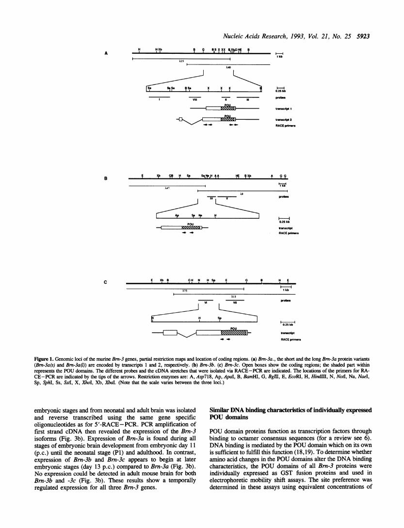
RESULTSStructure of murine Brn-3 family genesA mouse genomic DNA library was screened with a probe thatcovered part of the human Brn-3a/RDC-1 POU domain and 3'untranslated region (11) to isolate genomic clones representingall members of the Brn-3 gene family . Twenty independent 1phage clones were characterised by restriction mapping andhybridisation analysis. Sequence analysis of subgenomicfragments containing the POU domain and comparison to alreadyknown sequence portions from rat and human showed that themouse Brn-3a, -3b and -3c genes had been isolated (Fig. la-c).Fragments derived from genomic clones that cover either thePOU domain or are located outside of this region and arerepresentative and specific for the respective Brn-3 genes wereisolated. These fragments were used as probes in Southernanalyses (probes I, V, VI, Fig. la-c) with mouse genomic DNAto proove the integrity of the isolated phage clones and thecompleteness of the screening procedure (data not shown).
Characterization of cDNAs and identification of a newaminoterminal domaincDNA molecules of the corresponding genes were isolated withRACE-PCR techniques using poly A+ RNA from mouseembryonal day 13 (p.c.) spinal cord where all three Brn-3 genesare expressed (see below). Genomic fragments as well as cDNAclones were sequenced to gain complete information about thegenomic structure of the three Bm-3 loci (Fig. la-c):Transcription of the murine Brn-3a locus yields two transcriptsof about the same length (see below) that could be translated intoa long and a short Brn-3a protein variant. These proteins differin 84 amino terminal residues but are otherwise identical. Thelong and short open reading frames predict proteins withtheoretical molecular weights of 33.5 and 43 kDa, respectively.
Our analysis of the human Brn-3a locus revealed an analogousstructure (data not shown).
Transcription of the Brn-3b locus gives rise to one transcript(Fig. lb) with the capacity to encode a protein of 322 amino acids.Comparison of a partial human Bm-3b protein sequence (15) withthe murine homologue described here show 98% homology onamino acid level and an extension of the open reading frame of58 amino acids for the murine clone.For murine Brn-3c , a 700 bp cDNA was isolated via RA-
CE-PCR (Fig. lc). Sequence data derived from genomic clonesand cDNA clones predict a protein of 338 amino acids with anMr of about 35 kDa. Similar to Brn-3a the Brn-3c coding regionis interrupted by an intron (Fig. la,c). Interestingly, this intronoccupies the same position in the Bm-3a and Bm-3c gene so thatthe overall structure of both genes is remarkably similar (Fig.la, c). Exon one of Brn-3a and Brn-3c have the capacity toencode 41 and 40 amino acids, respectively.Within the POU domain the three Brn-3 proteins show almost
complete amino acid identity, only 12 residues out of 160 beingexchanged (Fig. 2a). The third helical regions of thehomeodomain and of the POU specific domain, which by analogywith the Antennapedia homeodomain and the Oct-I POU specificdomain are involved in DNA binding, are entirely conserved(16,17). Interestingly, one of the amino acid differences refersto the same position (Fig. 2a, arrowhead) that distinguishes Oct-iand Oct-2 in their ability to interact with the HSV transactivatorVP16 (8,9,14).The N-terminal region represents the second conserved part
of the Brn-3 proteins (Fig. 2b). Homology is not only confinedto the members of the Brn-3 family but also extends to theDrosophila melanogaster I-POU and C.elegans Unc 86 proteins.This region of about 100 amino acids can distinguish class IVPOU domain proteins from members of other classes andtherefore represents a new domain. For Brn-3a and Brn-3c thehomolgy in the N-terminus covers the whole 100 amino acidstretch further emphasizing their similarity in structure. Brn-3bis shorter and shows homology over 60 amino acids in this region(Fig. 2b). 84 amino acids of this domain are almost entirelyconserved between Bm-3a, Bm-3c but are not found in the shortBm-3a protein (Brn-3a(s)) (Fig.2b).
Murine Bm-3 genes are differentiafly expressed during neuraldevelopmentExpression ofBrn-3 genes was examined first by using a generalmurine Brn-3 probe coding for the conserved POU domain (probeII, Fig. la). Northern transfer hybridisation experiments withtotal RNA from day 13 (p.c.) mouse embryonal spinal cordrevealed transcripts of 3.5 and 2.5 kb (Fig. 3a). In a second step,specific probes originating from the 3' untranslated region ofBrn-3a and Brn-3b (probes HI and IV, respectively, see Fig. la,b) or from the coding region upstream of the POU domain ofBrn-3c (probe VI, see Fig. lc ) were used for sequentialhybridisation to the same filter and revealed 3.5 kb transcriptsfor Brn-3a and Brn-3b and a 2.5 kb transcript for Brn-3c (Fig.3a). Rehybridisation of the filter with a probe specific fortranscript 1 of Brn-3a (probe VIII, Fig. 1) showed the presenceof a 3.5 kb transcript indicating that this transcript is indeedpresent in spinal cord (Fig. 3a). Thus, the Brn-3a transcripts 1and 2 (Fig. la) and the Brn-3b transcript (Fig. lb) are very similarin length.To determine expression of the individual Brn-3 genes during
neural development, total RNA from head parts of various
Nucleic Acids Research, 1993, Vol. 21, No. 25 5923
125).40
x x xI II t1X III
p
I kb
0.25kb
probes
transcrpt 1
tIrnscript 2
RACE primer
1 kb
-IVv
D1p Sr I
POUi0.25kb
trnscriptRACE pimers
E Xb 8 GH N HS E G ,8 H E
| I kb
V 13
Sp
11~~~~~~~~~~~~~~~~~~~~~~~~~
probes
0.25 kb
tIrnecript
RACE prime
Figure 1. Genomic loci of the murine Brn-3 genes, partial restriction maps and location of coding regions. (a) Bm-3a., the short and the long Brn-3a protein variants(Bm-3a(s) and Bm-3aq)) are encoded by transcripts 1 and 2, respectively. (b) Brn-3b. (c) Brn-3c. Open boxes show the coding regions; the shaded part withinrepresents the POU domains. The different probes and the cDNA stretches that were isolated via RACE-PCR are indicated. The locations of the primers for RA-CE-PCR are indicated by the tips of the arrows. Restriction enzymes are: A, Asp718, Ap, ApaI, B, BamHI, G, Bgl, E, EcoRI, H, Hindlll, N, Notl, Na, NaeI,Sp, SphI, Ss, SstI, X, XhoI, Xb, Xba. (Note that the scale varies between the three loci.)
embryonic stages and from neonatal and adult brain was isolatedand reverse transcribed using the same gene specificoligonucleotides as for 5'-RACE-PCR. PCR amplification offirst strand cDNA then revealed the expression of the Brn-3isoforms (Fig. 3b). Expression of Brn-3a is found during allstages of embryonic brain development from embryonic day 11(p.c.) until the neonatal stage (P1) and adulthood. In contrast,expression of Brn-3b and Brn-3c appears to begin at laterembryonic stages (day 13 p.c.) compared to Brn-3a (Fig. 3b).No expression could be detected in adult mouse brain for bothBrn-3b and -3c (Fig. 3b). These results show a temporallyregulated expression for all three Brn-3 genes.
Similar DNA binding characteristics of individually expressedPOU domains
POU domain proteins function as transcription factors throughbinding to octamer consensus sequences (for a review see 6).DNA binding is mediated by the POU domain which on its ownis sufficient to fulfill this function (18,19). To determine whetheramino acid changes in the POU domains alter the DNA bindingcharacteristics, the POU domains of all Brn-3 proteins were
individually expressed as GST fusion proteins and used inelectrophoretic mobility shift assays. The site preference was
determined in these assays using equivalent concentrations of
AH H Xb
IS. S.ll
I Vill
B f Xsb 9 1 Sp Sq,Ia)41
C
rl%ju
8 lp OS IX IXX IlX,p; HE ?
lie

5924 Nucleic Acids Research, 1993, Vol. 21, No. 25
6
!
F,
SIx S'E MRF:~~~~~~~~~~..::::.:.IANVD.1 F.KJ ~ $I- YP iSaAXRRli
MNMN*-J(cQ-F EHP .. :St~5 .SAMRS..C..............P....
.......:....a....sV.. V .GKS.........
-uVDIVS-XW5Tu /I vs: KSHJ{ H SPTt. PC,S'*
LQ.NLFAS,LDE5 - ARAE*::: ...:.:........
*LQSSNUGG DS LL ARELG'D IL AFRA
ITs LA IHHIHHEHHRI
sw't
Figure 2. Comparison of amino acid sequences between Brn-3a, -3b and 3c. and other POU factors. (a) Comparison of the POU domains of Brn-3a, -3b and -3c.(b) New aminoterminal domain of class IV POU transcription factors. Amino acid sequence comparison of Brn-3 proteins, Unc 86 and I-POU. The position ofthe aminoterminal methionine of the short Brn-3a variant (Bm-3a(s)) is marked by an asterisk.
various radiolabeled oligonucleotide binding sites correspondingto naturally occuring octamer motifs. All Brn-3 POU domainsshowed similar DNA binding characteristics: The IgHepoligonucleotide (see Materials and Methods) that contains adjacentoctamer and heptamer motifs was bound with highest affinitycompared to the single octamer motifs HSV-Oct and wt-Oct (Fig.4a). No binding could be observed to recognition elements ofthe Drosophila homeodomain gene product Ubx (data not shown).Binding experiments with the IgHep oligomer showed two bandsindicating the existence of two DNA-protein complexes (Figs.4a, b) Both complexes disappeared in competition experimentswith unlabelled IgHep but not with a random competitoroligonucleotide (RCO) (Fig. 4b). The lower mobility complex(upper band) disappeared with less amounts of unlabelled IgHepcompetitor oligonucleotide indicating a lower binding affinity ofthis complex. In this complex two Brn-3 POU domain moleculesmay have bound to IgHep similar to the interaction of Brn-4 withits recognition site (20). The slight residual binding seen withGST-Brn-3 at a 1:1000 excess of unlabelled IgHep (Fig. 4b) isprobably due to an excess of Brn-3a fusion protein.The IgHep oligonucleotide in the normal (Oct+Hep+) and in
altered versions with either a mutated octamer (Oct-Hep+) ora mutated heptamer motif (Oct+Hep-) was used to testcooperative binding of the Brn-3a POU domain in analogy tothe POU domain of Oct-I (21,22). Mutation of the heptamer(Oct+Hep-) dramatically diminishes binding of the POUdomain (Fig. 4c). In addition, the binding appears to be weakercompared to wt-octamer or HSV octamer sequences (Fig. 4a).Mutations in the octamer (Oct-Hep+) leads to almost completeloss of both DNA-protein complexes (Fig. 4c). Bindingexperiments with Bm-3b and -3c POU domains yielded similar
Flgure 3. Expression ofBm-3 genes. (a) 30 tg of total RNA from mouse embryoday 13 (p.c.) spinal cord was analysed by Northern blotting with the probesindicated. For the exact location of the gene specific probes see Fig. la-c. Theprobes used were entirely sequenced to ensure their specificity and exclude crosshybridisation between Bm-3 family members. The specific transcripts for Brn-3a,-b and -c were detected (arrows). A 2 kb tanscript (small arrow) was only detectedwith the general POU probe (probe II) and probe VIII that is specific for Brn-3atranscript 1 and may represent yet another Brn-3a transcript. (b) Differentialexpression of individual Bm-3 genes during neural development was assayed withRT-PCR using RNA from the indicated sources (E, embryonal stages; P,newborn).
results (data not shown). This indicates that the binding of isolatedBrn-3 POU domains to octamer sequences is enhanced by anadjacent heptamer motif probably due to cooperative effects.
Brn-3a shows oncogenic potentialThe human Brn-3a is expressed in cell lines representative ofa subset of neuronal tumors such as neuroepitheliomas andEwing's sarcomas (10,11), suggesting a role of Bm-3a in the
i;
.0

Nucleic Acids Research, 1993, Vol. 21, No. 25 5925
A
GST- GST- GST- GSTBrn-3a Brn-3b Brn-3c -
11Z 1 11 _
>~ I 9 X I 0 I: X X~I 0=w >w'o WOxxpvxx_~~~c (/)
S
-_*-f-ree probe
BGST-Brn-3a
Ig-Hep RCO
a oo o o oo o~ o o~
GST-Brn-3b
Ig-Hep RCO
o oo o a oo o~ o o_
GST-Brn-3c
ig-Hep RCO
55Q8 8 0 0r
-
competitor
*; w
.*
_ free probe
C
Oct+Hep- Ocl-Hep+ Oct+Hep+
cs cs) cn
m m m
En cc cc cn
U probe
Figure 4. DNA binding activity of the POU domains of Bm-3a, -3b and -3cexpressed as GST fusion proteins. (a) Specificity of binding to the wildtype (wt)octamer, the Herpes simplex virus (HSV) octamer or to the IgHep oligonucleotidecontaining adjacent octamer and heptamer sequences (GST alone did not showany binding). (b) Competition of specific DNA binding to IgHep with an excessof unlabeled IgHep oligonucleotide or with a nonspecific random competitoroligonucleotide (RCO). (c) Cooperative binding of GST-Brn3a to IgHep(Oct+Hep+) and mutated versions (Oct+Hep-, Oct-Hep+).
development of these malignancies. As both human and murineBm-3a genes code for different forms of Brn-3a protein, weaimed to clarify whether these forms have different biologicalproperties and different abilities to induce malignanttransformation. A human genomic clone capable to produce boththe long and the short form of Brn-3a was cloned into themodified eukaryotic expression vector pLTRpoly (23). Theresulting construct (p3aLS, see Material and Methods) was thenused alone and in combination with a plasmid carrying theactivated Ha-ras gene (construct pEJ6.6ras, (24)) to transfectprimary fibroblasts (REFs) derived from day 14 rat embryos.Foci of transformed cells were observed in the control c-myc/lHa-ras cotransfection (constructs pKo-myc and pEJ6.6ras, (24)) andin cells that received both Ha-ras and p3aLS (Fig. 5a, b, Table1). A construct capable of encoding the long and short form ofBrn-3a from the mouse showed a similar transforming activityas the human p3aLS (data not shown). No induction of foci wasseen when p3aLS and c-mryc were transfected separately (Table1) or in combination (not shown). In contrast, a construct witha mouse genomic clone encoding only the short Brn-3a variant(construct p3aS) or Brn-3b (construct p3b) in the same expressionvector showed little or no oncogenic activity when cotransfectedwith activated Ha-ras (Table 1). This suggests that the intactnessof the newly identified aminoterminal domain is crucial for fulltransforming activity but also points to functional differencesbetween Brn-3a (1) on one hand and Brn-3a(s) and Brn-3b on theother hand. Interestingly, when p3aLS and p3b are transfectedtogether with pEJ6.6ras, no or little foci formation is seen (Table1), suggesting that Brn-3b inhibits the oncogenic activity ofBrn-3a().
Independent foci from c-myclHa-ras and p3aLS/Ha-rascotransfections were selected, grown in medium (DMEM)containing 10% serum and established as stable lines. Todemonstrate anchorage independence and tumorigenicity,p3aLSIHa-ras transformed cells were tested for growth insemisolid medium and for the formation of tumors in nude mice.All cell lines derived from p3aLS/Ha-ras cotransfections wereable to form colonies in soft agar after an average of 13 days(Fig. Sc, d, Table 2). Cell lines derived from foci after c-myclHa-ras cotransfection were used as a control and showed morecolonies that were also larger in size than colonies derived fromp3aLS/Ha-ras lines (Fig. 5c, d, Table 2). In addition, all c-mryclHa-ras and all p3aLS/Ha-ras transformed cell lines gaverise to tumors in nude mice after an average latency period of13 days (Table 2). In contrast, untransfected REFs were incapableof anchorage independant growth (not shown) and tumorformation in nude mice (Table 2).
Northern blot analysis of RNA from cell lines established fromfoci after transfection with c-myclHa-ras (lines 19 and 21), withp3aLS/Ha-ras (lines 9 and 24) and with p3aS/Ha-ras (line 40)showed the presence of the expected transcripts for c-myc andHa-ras (Fig. 5e). Hybridisation with a POU specific proberevealed a 3.6 kb transcript in the lines 9 and 24 and a 3.5 kbtranscript in the line 40 (Fig. 5e). Gel shift experiments withwhole cell extracts from line 40 and an octamer containingologonucleotide indicated that a functional short Brn-3a proteinvariant was produced from construct p3aS (not shown). This alsodemonstrates that the low or absent transforming activity of theshort Brn-3a variant is an intrinsic effect and not due to the lackof expression in REFs. The same can be assumed for Bmn-3bas the same vector system was used for all constructs. PCRanalysis with primers located in exon one and exon two of Brn-3a

5926 Nucleic Acids Research, 1993, Vol. 21, No. 25
A
0e
'-i d_* _ A;-
.- :I-
AB-..1. -. .~~~~~~~~~Oo'i.,t *;%Figure 5. (a) Transformed cells after cotransfection of primary REFs with the plasmids pKo-myc and pEJ6.6 ras that contain the c-myc gene and the activated Ha-rasgene, respectively (see also Materials and Methods). (b) Transformed cells after cotransfection with p3aLS and pEJ6.6ras. (c) Colony grown in soft agar from cellscotransfected with pKo-myc and pEJ6.6ras. (d) Colony grown in soft agar from cells cotransfected with p3aLS and pEJ6.6ras. (e) Expression of Bmn-3a, c-nyc,Ha-ras and GAPDH in REFs and cell lines established from transformed foci. Cell lines 19 and 21 had been established after transformation of REFs with the constructspKo-myc and pEJ6.6ras. The lines 9 and 24 were obtained from foci after transformation with p3aLS and pEJ6.6ras, line 40 after transfection of p3aS and pEJ6.6ras.Expression of Bm-3a resulting from the constructs p3aLS and p3aS is seen in lines 9, 24 and 40, respectively. Rehybridisation of the same filter with probe VIIIthat was derived from the untranslated 5' end of Brn-3a transcript 1 and is specific for this transcript (Fig. 1) showed that line 24 but not line 9 expresses this Brn-3amRNA molecule. The construct used to establish line 40 did not contain this region (see Fig. 1 and Material and Methods). The exogenous c-myc transcript presentin lines 19 and 21 stems from transfected pKo-myc and is shorter (lower arrow) than the endogenous transcript in lines 9 and 24 (upper arrow) because the pKo-mycplasmid contains only exon 2 and 3 but not exon 1 of the murine c-myc gene. The endogenous c-myc transcript is absent (40) in pKo-myc/pEJ6.6ras transfectants(lines 9 and 21) and present in very low amounts in REFs and the lines 9, 24 and 40 (upper arrow).
confirmed that transcript 2 (Fig. la) capable of encoding the longform of the Bm-3a protein is present in the transformed cell lines9 and 24 (not shown). Hybridisation with probe VIII (Fig. la)indicated that transcript 1 is present in line 24 but not in line9 suggesting that expression of the smaller Brn-3a protein is notcrucial for transformation (Fig. Se). The nature of the smallertranscript seen in cell line 9 remains to be determined. The celllines established from foci represent stable transfectants.Therefore, the varying relative levels of expression of thetransfected constructs (Fig. 5e) is probably due to different copynumbers.
DISCUSSIONThe Brn-3 family of developmental control genes consists of threemembers in rat and probably two members in humans (11-15).We describe here the characterisation of all three members ofthe Bm-3 family in the mouse (Brn-3a, -3b and 3c) and presentthe full aminotenninal ends of all family members. All three areencoded by single copy genes that are located on differentgenomic loci. Their expression appears to be regulateddifferentially during neural development very much like the ratand human homologues (11-14). Consistent with findings in therat, Brn-3a is expressed at earlier and later stages in neuraldevelopment compared to Brn-3b and -3c. This suggests that
expression of Brn-3a is more generalised during neuronaldevelopment, whereas Bm-3b and -3c expression may be morerestricted.Brn-3a and -3c contain at least two exons but Brn-3b appears
to represent another example of a POU domain gene with anintronless coding region. The Bm-3b form decribed hereresembles in that respect the genes Bmn-1, -2, -4 and scip (20,25).It is possible that these genes arose from a POU domain ancestorgene by duplication via a reverse transcribed RNA.Retrotransposition of genes might therefore be a way to duplicategenes or single exons and place them under differenttranscriptional control elements to achieve differential expressionof similar genes. However, we cannot completely rule out thatother Bmn-3b transcripts exist in tissues other than spinal cordor in other developmental stages and might contain aminoterminalexons similar to Brn-3a and -3c. The genomic loci encodingBmn-3a and Brn-3c appear very similar in structure as both containan amino terminal intron. Interestingly however, the Bmn-3c locusproduces only one specific mRNA species whereas the Bmn-3agene gives rise to two transcripts of similar length capable ofencoding a long and a short form of the Brn-3a protein. Thetranscription start points for the two Bm-3a transcripts as far asthey can be inferred from the 5' ends of PCR-RACE clonesare located about 500 bp apart on the genomic sequence. Thissuggest that two different promotors are used for the transcription
4m --II*
N,
Ai-:..,". ;, s, ':
.N! :,;T,';f Y.
.- 1..,,

Nucleic Acids Research, 1993, Vol. 21, No. 25 5927
Table 1. REF transfection experiments
The number of foci per individual experiment obtained after cotransfection withthe indicated DNA constructs is given per 106 cells. 5 independent experimentswere carried out. The last column shows the average number of foci. For thedescription of the constructs see Materials and Methods.
of the Brn-3a locus implicating a mechanism of differentialregulation of Brn-3a expression and activity.
Specific activity of the individual Brn-3 isoforms in a distinctcell type or at a certain developmental stage might also dependon the interactions with other proteins through the POU domainitself. Indeed, the high degree of homology between the threeBrn-3 POU domains and their similar DNA bindingcharacteristics do not preclude the occurrence of single aminoacid changes particularly at the end of helix 1 of the POUhomeodomain. Here, Brn-3a contains a valine whereas Brn-3band -3c both show isoleucine residues as in the correspondingrat genes (14). The analogous amino acid position has been shownto be critical for the interaction between the POU domain of Oct-Iand the Herpes simplex virus transactivator VP16 (8,9). Thechange at this position suggests that Brn-3a, -3b and -3c mightbe differentially modulated in their activity through interactionwith other cellular transactivators very much like Oct-i.An alternative mechanism of maintaining specificity is the
expression of inhibitors: the example of the two class IV POUfactors I-POU and tI-POU shows that a change in two aminoacids in otherwise identical products from the same genedistinguishes a transcriptional activator from a repressor (26,27).A similar mechanism of specifiying the activity of POU factorsmight be the presence of a new aminoterminal domain describedhere. This domain shows a remarkable degree of homologyexclusively confined to three Brn-3 proteins and to Unc 86 andI-POU that all belong to the same class of POU factors. Theregion between this conserved aminoterminal domain and theconserved carboxyterminal POU domains diverges between allthree Brm-3 proteins, Unc-86 and I-POU, suggesting strongly thatthis new aminoterminal domain is of critical functionalimportance. It is conceivable that the length of this domainregulates specifc biological properties of class IV POU factors,may be through interaction with other co-transactivators. Indeed,only the intact aminoterminal homology domain provides the long
Table 2. Cell lines were established from foci obtained after cotransfecting REFswith the indicated constructs
constructs cell soft agar colonies tumors inline nude mice
col./104cells col./l0Scells (days)
pKo-myc+ #19 400 2000 2/2 (11)pEJ6.6(ras)
pKo-myc+ #21 300 1000 2/2 (13)pEJ6.6(ras)
p3aLS+pEJ6.6(ras) #9 200 500 2/2 (16)
p3aLS+pEJ6.6(ras) #24 150 500 2/2 (13)
REFs 0 0 0./2 (25)
Single cells from these lines were seeded out in soft agar to test their ability ofanchorage independent growth. The colonies were scored after about 10 days,but at the same time for a given line. Tumorigenicity was tested by injectionof about 5 x 10⁶ cells from the established lines intraperitoneally into nude mice.Two animals per cell line were used. Tumors appeared after the latency timegiven in parentheses.
Brn-3a variant (Brn-3a(l)), with transforming and tumorigenicactivity. These findings not only establish the POU factorBrn-3a(l) as an onco-protein and suggest that Brn-3a(l) mayindeed be implicated in the generation of neuroepitheliomas andEwing's sarcomas as has been speculated (11), but may alsoreflect a general mechanism to regulate Brn-3a activity.Interestingly, Bm-3b or Brn-3a(s) that lack 39 and 84 amino acidsin their aminoterminal domains show no or little oncogenicactivity. Moreover, Bm-3b can inhibit the oncogenic activity ofBrn-3a(1) suggesting either direct interaction of both proteins orcompetition of necessary cofactors or DNA recognition sequencesas possible mechanism of regulating the activity ofBm-3 proteins.However, further experiments have to elucidate the exact natureof the inhibition of Brn3a(l) transforming activity.
Other POU factors as Oct-I or Oct-3/4 are not able to transformestablished fibroblasts as for example 3T3 cells or RATI cells.In contrast, homeobox and paired box containing genes have beenshown to transform mouse 3T3 or RAT-I fibroblasts (28-30).In addition, the homeobox containing genes PBX-1 and Hox 11(tcl-3) are activated through chromosomal translocations indifferent forms of leukemia and are prime candidates to beclassified as oncogenes (31-33). Although the oncogenicpotential of all these genes has not been evaluated yet in primarycells or in transgenic mice, a common picture emerges that linksdevelopment and tumorigenesis in that both require the regulationof cell growth, proliferation and invasion. It is thereforeconceivable that deregulated expression of some but not alltranscriptional transactivators involved in developmental controlcan lead to the tumorigenic conversion of a cell. It appears nowmost interesting what target genes are regulated by these newonco-proteins. It is possible that these target genes represent anew class of downstream regulators that are functionally differentfrom targets of non oncogenic developmental control genes.
MATERIALS AND METHODSIsolation of genomic clonesA genomic library prepared from mouse liver DNA (mouse strain129, Stratagene, Lambda FixTM II) was screened using a SnaIfragment from the human RDC-1 cDNA that covered the POUdomain and part of the 3' untranslated sequence. 20 independent
DNA transfected No. of foci/106 cells Average number of foci(constructs)
(S indep. experiments)
pKo-myc 0 / 0 / 0 /0/0 0
pEJ6.6(ras) 0 / I / 0 / 0 / 1 0.4
pKo-myc+pEJ6.6(ras) 100/54 /51/40/1 17 72.4
p3aLS 0 / 0 / O /0 /n.t. 0
p3aLS+pEJ6.6(ras) 6/ 9/ 17 /13 /45 18
p3aS 0/0/0/0/0 0
p3aS+pEJ6.6(ras) 0/ 2 / 2 /5 /I 2
p3b n.tjn.tj 0 / 0 /0 0
p3b+pE16.6(ras) n.t./ n.t. 3 / 1 / 3 2.3
p3aLS+p3b+pEJ6.6(ras) n.t./ n.t./ n.t./ 1 / 4 2.5

5928 Nucleic Acids Research, 1993, Vol. 21, No. 25
clones were isolated and divided into 3 groups according torestriction mapping. Fragments of each group containing the POUhomeodomain were subcloned into the bluescript SK vector andpartially sequenced by the Sanger dideoxy chain terminationmethod. These fragments included a 4.4 kb Bgll fragment, a7.5 kb XbaI fragment and a 4.5 kb NotI fragment for Brn-3a,Brn-3b and Brn-3c, respectively.
Isolation of cDNA clones by RACE-PCRTo isolate 5' end cDNA clones the 5' RACE system from GIBCOBRL was used and the manufacturers instructions were followed.Poly A+ RNA from spinal cord derived from mice atembryonal day 13 (p.c.) was prepared as described (35) andreverse transcribed using gene specific oligonucleotides residingwithin the POU domain except for Bm-3a (see Fig. 1). Aftertailing of the first strand cDNA, primary and secondaryamplification was performed with primers residing upstream ofthe forementioned oligonucleotides used for cDNA synthesis.Finally, cDNA molecules for each of the three genes wereisolated, subcloned into bluescript vector and entirely sequenced.Control reactions without reverse transcriptase in the first strandsynthesis were used in amplification reactions under the sameconditions and did not yield any products.
Oligonucleotides for first strand cDNA synthesis included forBrn-3a, Bm-3b and Brn-3c, respectively:5'-GCACGGTGGACGTGGAC-3'5'-GCGATCATGTTGTTGTGTGACAG-3'5'-GGGATCTTAAGATTGGCTAAAG-3'.Primary amplification was performed with:5'-CTATTCATCGTGTGGTACGTG-3'5'-GCGTGAGAGACTCAAACCTG-3'5'-TTGATGCGCCTCTGCTTGAAG-3'.The following oligonucleotides were used in a secondamplification step:5'-AATGAATTCGCTTGAAAGGGTGGCTCTTG-3'5'-AATGAATTCATCCACGTCGCTCATGCAG-3'5'-AATGAATTCGGGTGACTCATGCCCATG-3'.A cDNA clone from the 3' region of the murine Brn-3a gene
was isolated using a described protocol (34). Oligonucleotidesfor primary and secondary amplification of cDNA are:5'-TTCTCTGCCACTTACTGAG-3'5'-GAGGATCCGAATTCCCAGCCTAGAGAACAGG-3'.
After amplification the obtained PCR products were subclonedinto bluescript SK vector and entirely sequenced by the Sangerdideoxy chain termination method.
DNA and RNA analysesMouse genomic DNA was isolated according to standardprocedures (35) and digested with restriction enzymes tocompletion, separated on a 0.8% agarose gel, denatured andtransferred to nitrocellulose or nylon filter membranes usingstandard protocols. Hybridisations were carried out in buffer with50% formamide at 42°C. All probes were double stranded DNAfragments and were radiolabeled with [32P] dCTP and a randomhexamer primer mix using the large fragment of DNApolymerase I. Filters were washed under stringent conditions with0.2xSSC, 0.1% SDS at 650C and exposed at -700C to KodakXAR 5 films with enhancing screens. RNA was isolated usingthe AGPC method (36), fractionated on 0.8% agaroseformaldehyde gels and directly transferred onto nylon filters.
RT-PCR experiments were as described (35). RNA from headparts, neonatal and adult stages was reverse trancribed using thesame gene specific oigonucleotides that were used in RACE PCR.5' oligonucleotides for the amplification of first strand moleculeswere as follows: for Brn-3a: CACTTTGCCGCGACTTTG; forBm3-b: CTGAGCGTAATGTGTGCCTTC; for Brn-3c: GCAA-GAACCCAAATTCTC. 3' oligonucleotides were the same as
in the RACE-PCR reactions. The PCR products were separatedon a gel, transferred to a filter and hybridised to oligonucleotideprobes specific for the individual Bm-3 genes. Control reactionswithout reverse transcriptase in the first strand synthesis were
used in amplification reactions under the same conditions. Neitherspecific bands nor hybridisation signals could be detected in thesereactions (not shown).
Expression and purification of glutathione-S-transferase(GST) fusion proteinsDNA fragments containing the complete POU domain codingsequence were generated by PCR amplification from plasmidDNA using the following oligonucleotide pairs for Brn-3a,Brn-3b and Brn-3c, respectively.5'-AATGGATCCTGCGACTCGGACACGGACCCG-3'5'-GAGGATCCGAATTCCCTCAGTAAGTGGCAGAG-3'5'-AATGGATCCCACATGGGCTGCATGAGCGAC-3'5'-AATGGATCCCTTCACTGCGAAACCGGTTCAA-3'5'-AATGGATCCGCGTGTCTCAGCGATGTGGAG-3'5'-AATGGATCCCTGGAGTGTCCCGTAAGCTC-3'
After digestion with BamHI these fragments were subclonedinto the bacterial expression vector pGEX-2T and expressed as
a fusion protein with gluthathione-S-transferase. The fusionprotein was isolated via gluthathione-S-affinity chromatographyaccording to the suppliers instructions (SIGMA). The eluates fromthe affmity collumns showed one single band on SDS -PAGEanalysis after Coomassie staining indicating successful purificationof the fusion proteins.
DNA binding studiesThe probes used for bandshift assays were oligonucleotides, 5'endlabelled with T4 polynucleotide kinase and purified bygelfiltration. The oligonucleotides represent the following DNAbinding sites (sense sequences shown): the octamer binding sitefrom the Herpes simplex virus immediate-early gene promoter(HSV-Oct, 37), 5'-GCATGCTAATGATATTCTTT-3'; Theoctamer binding site from the mouse Ig heavy chain enhancercontaining adjacent octamer and heptamer motifs (IgHep, 38),5'-CTGATTTGCATATTCATGAGAC-3'; the wild typeoctamer which is identical to the octamer motif containing regionof the mouse heavy chain enhancer (25,39) 5'-GGTAATTTG-CATTTCTAA-3'.To demonstrate specificity and cooperativity of binding to the
IgHep DNA binding site either the octamer (Oct-Hep+) or theheptamer motif (Oct+Hep-) were mutated. The oligonucleotideswere as described (20). Binding reactions were performed on
ice in 20 i1 binding buffer (10 mM Hepes-KOH, pH 7,9; 60mM KCI; 1 mM EDTA; 1 mM DTT; 4% Ficoll) in the presenceof 1 ,ug poly(dIdC). About 50 ng of purified fusion protein wasused in each binding experiment. Free DNA and protein-DNAcomplexes were resolved on a native 6% polyacryamide gel runin 0.5 xTBE. After electrophoresis, the gel was dried andexposed. Competition assays were done under the same
Hybridisation and washing conditions were as described above. conditions but with unlabefled oligonucleotides added.

Nucleic Acids Research, 1993, Vol. 21, No. 25 5929
REF assay and constructsThe construct p3aLS contains a 5 kb BamHIIEcoRI humangenomic DNA fragment (1 1) that is capable to encode the longand the short Brn-3a protein and as backbone the modifiedeukaryotic expression vector pLTRpoly (23) with an LTR fromMoloney murine leukemia virus at the 5' end and splice andpolyadenylation sequences from SV40 at the 3' end. A 2.2 kbPvullIBamHI mouse genomic fragment only capable of encodingthe short form of Brn-3a (Brn-3a(s)) and a 1.6 kb SalIIPvuHmouse genomic fragment capable of encoding Brn-3b weresubcloned into the same vector and gave rise to the constructsp3aS and p3b, respectively.
Pregnant Fisher rats were killed at day 14, the embryos wereremoved and placed in sterile PBS. Cells from the torso weredispersed using normal trypsin/EDTA+ 1% chicken serum. Theywere separated from debris by low speed centrifugation. The finalcell pellet was resuspended in DMEM/10% serum (FCS) andplated at 1 million cells per 10 cm petri dish. After the cells hadreached confluency, they were replated at 1 million again andused for transfections the following day (1 million cells pertransfection). Transfections were done by the conventionalcalcium phosphate precipitation method overnight with 20 ,g ofDNA. This included 10 ,g of pEJ6.6ras (containing the activatedHa-ras gene from a human bladder carcinoma, 24) and 10 tgpKo-myc (containing exon 2 and 3 of the mouse c-myc gene underthe control of the SV40 early promoter/enhancer) or an equalamount of DNA of the different Brn-3a or Brn-3b constructstogether with either c-myc or Ha-ras plasmids. DNA from pUCvector was used to reach a total of 20 sg ofDNA in transfectionswith a single oncogene. 24 h after transfection the cells wereshocked with 25% glycerol in Medium for 1-2 min and another24 h later were split into four plates and assayed for focusformation. The test for anchorage independance was done in 6cm petri dishes containing 0.5% agar and 10% FCS in DMEM.This basal layer was then overlaid with 1.5 ml of 0.33% agarand 10% FCS in DMEM containing 104 and 105 REFs or thesame number of cells from c-myclHa-ras or p3aLS/Ha-rascotransfection and incubated. Colonies were scored after about10 days.
ACKNOWLEDGEMENTSWe thank Robert Collum and Fred Alt for the human RDC-Jprobe, and for pKoMyc, Tomi Maikela and Kari Alitalo forpLTRpoly and pEJ6.6ras. We are indebted to David Latchmanand John Wood for communicating results prior to publicationand to Rolf Miiller and Renate Renkawitz-Pohl for criticallyreading the manuscript. We thank Heike Lovec for help withtransfection assays. This work was supported by grants from theDeutsche Forschungsgemeinschaft (SFB 215/D1O) and theMildred Scheel Stiftung fur Krebsforschung to T.M., T.T. andM.Z. were supported by a fellowship from the Graduiertenkolleg'Zell- und Tumorbiologie'.
Note addedWhile this paper was in press Xiang et aL (Neuron 11, 689-701,1993) isolated an aminoterminally spliced Brn-3b mRNA fromrat retina and Gerrereo et al. (PNAS, 90, 10841-10845, 1993)reported a conserved domain (termed POU IV box) in the aminoterminus of the Brn-3a protein very similar to the domaindescribed here.
REFERENCES
1. Herr, W., Sturm, R.A, Clerc, R.G., Corcoran, L.M., Baltimore, D., Sharp,P.A., Ingraham, H.A., Rosenfeld, M.G., Finney, M., Ruvkun, G. andHorvitz, H.R. (1988) Genes Dev. 2: 1513-1516
2. Ingraham, H.A., Chen, R., Mangalam, H.J., Elsholtz, H.P., Flynn, S.E.,Lin, C.R., Simmons, D.M., Swanson, L. and Rosenfeld, M.G. (1988) Cell55: 519-529
3. Clerc, R.G., Corcoran, L.M., Lebowitz, J.H., Baltimore, D. and Sharp,P.A. (1988) Genes Dev. 2: 1570-1581
4. Sturm, R.A. and Herr, W. (1988) Nature 336: 601-6045. Finney, M., Ruvkun, G and Horvitz, H.R. (1988) Cell 55: 757-7696. Wegner, M., Drolet D.W. and Rosenfeld, M.G. (1993) Curr. Opin.Cell
Biol. 5: 488-4987. Stem, S. and Herr, W. (1991) Genes Dev. 5: 2555-25668. Lai J.S., Cleary, M.A. and Herr, W. (1992) Genes Dev 6: 2058-20659. Pomerantz, J.L., Kristie, T.M. and Sharp, P.A. (1992) Genes Dev. 6:
2047-205710. Collum, R.G., DePinho, R., Mellis, S., Thiele, C.J., Israel, M.A. and Alt,
F.W. (1989) Cancer Cells 7: 113-11611. Collum, R.G., Fisher, P.E., Datta, M., Mellis, S., Thiele, C.J., Huebner,
K., Croce, C.M., Israel, M.A., Theil, T., Moroy, T., DePinho, R. andAlt, F.W. (1992) Nuci. Acids Res. 20: 4919-4925
12. He, X., Treacy, M.N., Simmons, D.M., Ingraham, H.A., Swanson, L.W.and Rosenfeld, M.G. (1989) Nature 340: 35-42
13. Lillycrop, K.A., Budrahan, V.S., Lakin, N.D., Terrenghi, G., Wood, J.N.,Polak, J.M. and Latchman, D.S. (1992) Nucl. Acids Res. 20: 5093-5096
14. Ninldna, N.N., Stevens, G.E.M., Wood, J.N. and Richardson, W.D. (1993)Nucl. Acids Res. 21: 3175 -3182
15. Ring, C.J.A. and Latchman, D.S. (1993) Nucl. Acids Res. 21: 294616. Assa-Munt, N., Mortishire-Smith, R.J., Aurora, R., Herr, W. and Wright,
P.E. (1993) Cell 73: 193-20517. Kissinger, C.R., Liu, B., Martin-Blanco, E., Komberg, T.B. and Pabo, C.O.
(1990) Cell 63: 579-59018. Ingraham, H.A., Flynn, S.E., Voss, J.W., Albert, V.R., Kapiloff, M.S.,
Wilson, L. and Rosenfeld, M.G. (1990) Cell 61: 1021-103319. Verrijzer, C.P., Alkema, M.J., van Weperen, W.W., van Leeuwen, H.C.,
Strating, M.J.J. and van der Vliet, P.C. (1992) EMBO J. 11: 4993-500320. Mathis, J.M., Simmons, D.M., He, X., Swanson, L.W. and Rosenfeld,
M.G. (1992) EMBO J. 11: 2551-256121. LeBowitz, J.H., Clerc, R.G., Brenowitz, M. and Sharp, P.A. (1989) Genes
Dev. 3: 1625-163822. Poellinger L., Yoza, B.K. and Roeder, R.G. (1989) Nature 337: 573 -57623. Mikeli, T.P., Koskinen, P.J. Vastrik, I. and Alitalo, K. (1992) Science 256:
373-37724. Land, H., Parada, L.F. and Weinberg, R.A. (1983) Nature 304: 596-60225. Kuhn, R., Monuki, E.S. and Lemke, G. (1991) Mol. Cell. Biol. 11:
4642-465026. Treacy, M.N., He, X. and Rosenfeld, M.G. (1991) Nature 350: 577-58427. Treacy, M.N., Neilson, L.I., Turner, E.E., He, X. and Rosenfeld, M.G.
(1992) Cell 68: 491-50528. Aberdam, D., Negreanu, V., Sachs, L. and Blatt, C. (1991) Mol. Cell. BioL
11: 554-55729. Maulbecker, C.C. and Gruss, P (1993) EMBO J. 12: 2361-236730. Maulbecker, C.C. and Gruss, P. (1993) Cell Growth Diff 4: 431-44131. Hatano, M., Roberts, C.W.M., Minden, M., Crist, W.M. and Korsmeyer,
S.J. (1991) Science 253: 79-8232. Kamps, M.P., Murre, C., Sun, X. and Baltimore, D. (1990) Cell 60:
547-55533. Lu, M., Gong, Z. Shen, W. and Ho, A.D. (1991) EMBO J. 10: 2905-291034. Frohman, M.A., Dush, M.K. and Martin, G.R. (1988) Proc. Natl. Acad.
Sci. 85: 8998-900235. Sambrook, J., Fritsch, T. and Maniatis, T. (1989) Molecular Cloning, A
Laboratory Manual, Cold Spring Harbor, USA36. Chomczynski, P. and Sacchi, N. (1987) Anal. Biochem. 162: 156-15937. Kristie, T.M., LeBowitz, J.H. and Sharp, P.A. (1989) EMBO J. 8:
4229-423838. Wirth, T., Staudt, L. and Baltimore, D. (1987) Nature, 329: 174-17839. Ephrussi, A., Church, G.M., Tonegawa, S. and Gilbert, W. (1985) Science
227: 134-14040. Zimmerman, K. and Alt, F.W (1990) Critical Rev. Oncol. 2: 75-95