COMMUNICATIONS - NCBI
28
Brit. J. Ophthal., 35, 61. COMMUNICATIONS REACTION OF CORNEAL NERVE FIBRES TO INJURY* BY E. ZANDERt AND G. WEDDELL From the Department of Anatomy, University of Oxford IN a recent paper (Zander and Weddell, 1951) a description of the innervation of the normal cornea is given so far as it can be determined by direct observation. In the present paper the results are described of a number of experimental procedures designed to test the validity of observations made by the direct method and also to add facts not obtainable by that method. Apart from information on the general pattern of corneal innervation, a study of degeneration and regeneration of nerve fibres in this tissue provides other data of considerable interest. Corneas were examined after the following procedures had been carried out: (1) corneal autografting; (2) keratotomy; (3) section or avulsion of the infra-orbital nerve; (4) section of the ciliary nerves; (5) removal of the superior cervical sympathetic ganglion; (6) destruction of the Gasserian ganglion. MATERIAL AND METHODS The animals used were rabbits weighing between 2 and 3 kg. and two rhesus monkeys. For the operative procedures they were anisthetized with nembutal given intravenously. At the end of the experimental period the animals were killed with an overdose of nembutal. The comeal nerve fibres were either stained with methylene blue or impregnated with silver. Details of the methods used have already been published (Weddell and Zander, 1950). Briefly, staining is carried out by the introduction of a dilute solution of dye into the anterior chamber of the eye, where it is allowed to remain for a specified time. After staining, the cornea is very carefully removed with sharp scissors and prepared for microscopical examination after fixation of the dye with a mixture of ammonium molybdate and ammonium picrate. The details of the fixation process are complicated * Received for publication October 18. 1950. + In receipt of a grant from the Swiss " Stiftung fur biologisch-medizinische Stipendien " and on leave from the Neurosurgical Clinic, University of Zurich. 61
-
Upload
khangminh22 -
Category
Documents
-
view
1 -
download
0
Transcript of COMMUNICATIONS - NCBI
Brit. J. Ophthal., 35, 61.
COMMUNICATIONS
REACTION OF CORNEAL NERVE FIBRESTO INJURY*
BY
E. ZANDERt AND G. WEDDELLFrom the Department of Anatomy, University of Oxford
IN a recent paper (Zander and Weddell, 1951) a description of theinnervation of the normal cornea is given so far as it can bedetermined by direct observation. In the present paper the resultsare described of a number of experimental procedures designed totest the validity of observations made by the direct method and alsoto add facts not obtainable by that method.
Apart from information on the general pattern of cornealinnervation, a study of degeneration and regeneration of nerve fibresin this tissue provides other data of considerable interest.
Corneas were examined after the following procedures had beencarried out:
(1) corneal autografting;(2) keratotomy;(3) section or avulsion of the infra-orbital nerve;(4) section of the ciliary nerves;(5) removal of the superior cervical sympathetic ganglion;(6) destruction of the Gasserian ganglion.
MATERIAL AND METHODS
The animals used were rabbits weighing between 2 and 3 kg. and two rhesus monkeys.For the operative procedures they were anisthetized with nembutal given intravenously.At the end of the experimental period the animals were killed with an overdose ofnembutal.The comeal nerve fibres were either stained with methylene blue or impregnated with
silver. Details of the methods used have already been published (Weddell and Zander,1950). Briefly, staining is carried out by the introduction of a dilute solution of dyeinto the anterior chamber of the eye, where it is allowed to remain for a specified time.After staining, the cornea is very carefully removed with sharp scissors and preparedfor microscopical examination after fixation of the dye with a mixture of ammoniummolybdate and ammonium picrate. The details of the fixation process are complicated
* Received for publication October 18. 1950.
+ In receipt of a grant from the Swiss " Stiftung fur biologisch-medizinische Stipendien " and onleave from the Neurosurgical Clinic, University of Zurich.
61
E. ZANDER AND G. WEDDELLand must be carried out with extreme care. The silver-impregnation method has itsbasis in the procedure described by Glees (1946), but a number of modifications havebeen introduced to make it suitable for the cornea.
EXPERIMENTAL PROCEDURES(1) CORNEAL AUTOGRAFTING.The primary purpose of these experiments was to study the
process of degeneration of corneal nerve fibres.It has been claimed by a number of authors that the process in the
cornea is much slower than elsewhere in the body (e.g., Leoz Ortin,1915, 1917; Escapini, 1948), and autografting some two-thirds ofthe cornea seemed the most suitable method of testing this statement.In addition, it was hoped that some information on the relationbetween opacity in grafts and their nerve supply might be obtained.
Little work seems to have been done on the behaviour of nervefibres in corneal grafts. Franceschetti and Babel (1947) havedescribed the nerve supply of a corneal heterograft in man 6 yearsafter it had been transplanted. The graft had remained transparentsince it was applied. Following the removal of the cornea they usedsilver-impregnation methods to demonstrate the nerve fibres. Theyfound that no nerve bundles entered the graft from the host's cornea;the bundles either took a recurrent course or ran round the graft.A few single nerve fibres penetrated the scar and entered the graft,where they gave rise to a profuse plexiform network of nerve fibressituated just beneath the epithelium. The authors concluded thatthe presence of these nerve fibres was responsible for the transparencyof the graft, for no nerve fibres were found in opaque grafts whichthey examined. Escapini (1948) studied the course of degenerationand regeneration of nerve fibres in relation to corneal transplants inrabbits and concluded that the processes of nerve degeneration andregeneration in the cornea resemble those in other tissue except thatthey are much slower. He finds, in contradistinction toFranceschetti and Babel, that opaque grafts appear to be as wellinnervated and as sensitive as clear ones.Operation.-In all, fifteen rabbits were used. The animals were anaesthetized with
nembutal, and sufficient eserine solution was placed in the conjunctival sac to obtaina constricted pupil. A small circumferential incision was made right through thecornea with a keratome, one-third of the distance from the limbus to the centre ofthe pupil, and this was immediately resutured with a No. 1 nylon thread. Thisprocedure was continued until two-thirds of the central part of the cornea had beencompletely excised and resutured into its original position with about ten stitches.If there was an iris prolapse which could not be reduced, the protruding portion wasexcised. The lids were subsequently sewn together and remained closed for14 days, after which the stitches were removed. After removal of the sutures sensorytests were commenced. They were carried out with a No. 1 nylon suture and cotton-wool, attempts being made to elicit a blink reflex.
62
REACTION OF CORNEAL NERVE FIBRES 63
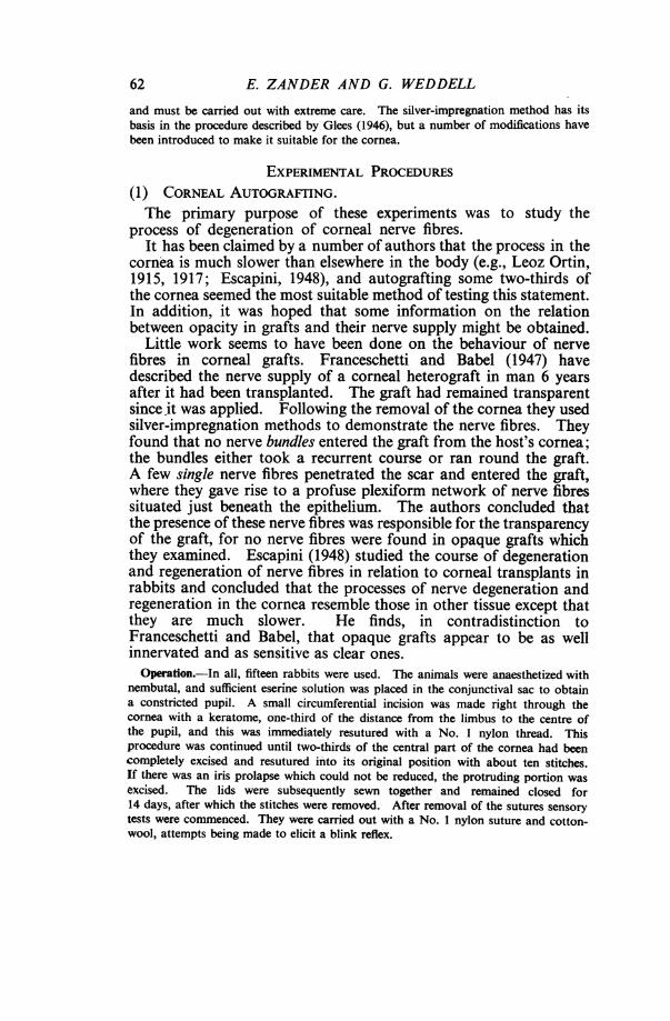
Observations.-The results of the operations and the fate of thegrafts are shown in Table I, further observations being groupedunder five heads, as follows:
(a) degeneration of nerve fibres;(b) return of sensibility;(c) regeneration of nerve fibres;
(d) reaction of the corneal corpuscles;(e) reaction of the Schwann cells.
TABLE IANALYSIS OF CORNEAL AUTOGRAFT OPERATIONS
Time betweenRabbit operation and, State of graft at end ofNo. ementaleperiod experimental period
(days)
Blink reflex aftergrafting
1 2 3 atendweek weeks weekt prod
2 whole cornea slightly _ - - 0 silver impregnationopaque
2 2 whole cornea slightly - 0 methylene blueopaque
3 7 whole cornea slightly 0 _ - 0 silver impregnationopaque
4 7 whole cornea slightly 0 - - 0 methylene blueopaque
5 14 lear 0 0 - 0 silver impregnation
6 14 whole cornea slightly 0 0 - 0 methylene blueopaque
7 21
8 21
Q0
clear 0
upper half of cornea 0opaque
~ _
10 50 centre opaque
11 80 centre opaque
12 1100 clear
13 110
14 220
15 220
clear
0
0
o
upper half of cornea 0slightly opaque
clear 0 0
0 -- + silver impregnation
o 0 0 methylene blue
0
0
-t -' silver impregnation
1- + methylene blue
o 0
0 0
> + silver impregnation
+ + silver impregnation
methylene blue
0 +- + + + silver impregnation
+ + + methylene blue
0 - no reflex
-- doubtful response
+ - weak response
+ -- brisk response++++ = response equivalent to that
obtained in unoperated eye- - reflex not tested for
Neurohistologicalmethod used
_
..i
clear
E. ZANDER AND G. WEDDELL
FIG. 2.
FIG. 5.
FIG. 3.
FIG. 6.-;.fi'I;
FIG. 4.
:. .. .O..s,
FIG. 7.
FIG. 8. FIG. 9.
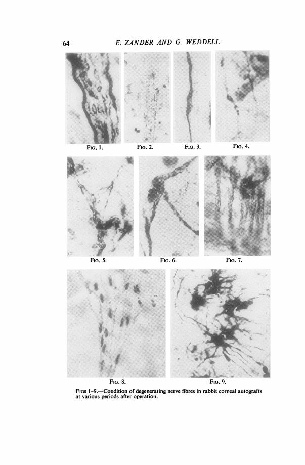
FIGS 1-9.-Condition of degenerating nerve fibres in rabbit corneal autograftsat various periods after operation.
64
FIG. 1.
REACTION OF CORNEAL NERVE FIBRES
FIG. 1.-Silver-impregnated preparation 2 days after operation. It showsa nerve bundle in the substantia propria. Some of the nerve fibres areseverely affected; even the smooth axis cylinder is very swollen. (x 463)FIG. 2.-Methylene-blue stained preparation 2 days after operation. Itshows a nerve bundle in the substantia propria in which all the axons areshowing signs of degenerative change. (x 193)FIG. 3.-Silver-impregnated preparation 2 days after operation. The axonin the substantia propria is swollen and "neurofibrillae" are developing. (x 463)FIG. 4.-Silver-impregnated preparation 2 days after operation. The axonsin the substantia propria show typical signs of degenerative change. (x 1130)FIG. 5.-Silver-impregnated preparation 7 days after operation. Theaxons in the substantia propria are in an advanced state of degeneration. (x 595)FIG. 6.-Silver-impregnated preparation 14 days after operation. It showsaxon fragments lying on the surface of a Schwann cell in the substantiapropria. (x 655)FIG. 7. Silver-impregnated preparation 21 days after operation. It showsSchwann-cell pathways in the substantia propria. The cytoplasm of the cellscontains beaded threads. No axon fragments can be seen. (x 655)FIG. 8.-Methylene-blue stained preparation 21 days after operation. Italso shows Schwann-cell pathways in the substantia propria, devoid of axons,but containing beaded intra-cytoplasmic threads. (x 519)FIG. 9.-Silver-impregnated preparation 2 days after operation. It showsthe peculiar reaction of the corneal corpuscles. (x 575)
(a) DEGENERATION OF NERVE FIBRES
Two days after Grafting.-The process of degeneration was marked, and generally-similar in type and degree to that seen among non-myelinated nerve fibres in the skinof the rabbit's ear by Weddell and Glees (1941), the signs of degeneration taking theform of a breaking up of the axis cylinder into " neurofibrillae " or of the formationof vacuoles within the axis cylinder (Figs 1, 2, 3, 4). At this early stage (as in skin)some fibres appear to suffer less than others although all nerve fibres are affected tosome extent (Fig. 1) in some part of their course. Sections taken to include the incisionand tissue on either side of it show that nerve fibres proximal to the incision were
similarly affected from some 2 mm. More proximally, the fibres appeared to be quitenormal.
Seven days after Grafting.-The degenerative processes are again similar to thoseseen in the skin of the rabbit's ear, all the axis cylinders being in a disintegratingcondition (Fig. 5) except for a few single nerve fibres in the substantia propria at thecentre of the cornea, which appeared to be more " resistant
Fourteen days after Grafting.-The changes are still more severe. All nerve fibresare seen to exhibit extensive degenerative changes (Fig. 6). The rate and nature of thedegenerative process of nerve fibres in the cornea are thus similar to that undergone bynon-myelinated nerve fibres in skin from the rabbit's ear.
Twenty-one days after Grafting.-Only traces of the original nerve fibres can be seen
as fragments lying along the original Schwann-cell pathways, the process ofdegeneration being virtually complete (Figs 7 and 8). However, regenerating nerve
fibres can now be seen coursing along a certain number of Schwann-cell pathways.They arise from the severed axons proximal to the scar.
(b) RETURN OF SENSIBILITY (Table I)This was tested as follows: either a No. 1 nylon suture or a wisp
.of cotton-wool was lightly applied at right angles to the surface .of
65
E. ZANDER AND G. WEDDELL
FIG. 14. FIG. 15.
FIG. 16. FIG. 17.
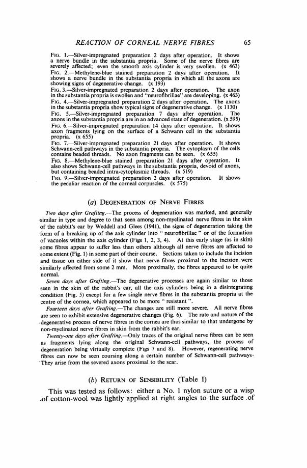
Figs 10-17.-Condition of regenerating nerve fibres in rabbit cornealautografts at various periods after operation.
66

REACTION OF CORNEAL NERVE FIBRES
FIG. 10.-Silver-impregnated preparation 21 days after operation. Itshows single regenerating axons in the substantia propria which have piercedthe scar and entered the cornea. They are not related to Schwann-cellpathways. (x 670)FIG. 1 1.-Methylene-blue stained preparation 50 days after operation.It shows a bundle of immature axons passing along a Schwann-cell pathwayin the substantia propria. (x 390)FIG. 12.-Silver-impregnated preparation 50 days after operation. Itshows a regenerating axon making its way between the cells of the basalepithelial layer. (x 545)FIG. 13.-Methylene-blue stained preparation 80 days after operation.It shows regenerating beaded axons lying among the epithelial cells. (x 465)
FIG. 14.-Silver-impregnated preparation 110 days after operation. Itshows mature regenerated axons in the substantia propria. The number ofbranches to which one of the axons gives rise is greater than that seen innormal specimens. One daughter axon in particular has divided into anumber of branches on its way round a cell, the nucleus of which can be seen.(x 254)FIG. 15.-Silver-impregnated preparation 110 days after operation. Itshows an axon taking a very complicated course in the substantia propria.(x 420)FIG. 16.-Silver-impregnated preparation 220 days after operation. It showsa nerve bundle containing relatively fewer axons than normal, some ofwhich are still immature, in the substantia propria. (x 673)
FIG. 17.-Methylene-blue stained preparation 220 days after operation.It shows immature axons ramifying among the epithelial cells. (x 635)
the graft, these procedures having been found to elicit a briskcorneal reflex from a normal cornea at each contact.
Fourteen days after Grafting.-No corneal reflex could be obtained, whether thegraft was clear or opaque.
Twenty-one days after Grafting.-A feeble blink reflex could occasionally be obtainedin the periphery of the graft, but it tired quickly. No response could be elicited withcotton-wool, however. The reflex was obtained from animals with opaque as well as
clear grafts.Seven weeks after Grafting.-A response could be obtained at every stimulation in
the periphery of the graft, but the reflex was not a strong one. Only an occasionalresponse was obtained from the centre of the graft. Cotton-wool still elicited no
response.Twelve weeks after Grafting.-A brisk and moderately strong response was obtained
at each stimulus throughout the graft. Cotton-wool now elicited a feeble responseon some occasions in the periphery of the graft only.
Sixteen weeks after Grafting.-The response to the nylon suture was stronger stilland cotton-wool now elicited a moderate response each time it was applied to the centreof the graft.
Twenty-eight weeks after Grafting.-The response from the grafted side was
indistinguishable from that obtained from the unoperated side.
In every case recovery of sensibility in opaque grafts followed acourse similar to that in clear grafts.The observations tabulated above broadly determined the times
following operation at which the grafts were examined histologically.
67

E. ZANDER AND G. WEDDELL
(c) REGENERATION OF NERvE FIBREsTwenty-one days after Grafting.-A few fine isolated nerve fibres can be seen
coursing throughout the substantia propria. They can be recognized as regeneratingnerve fibres by reason of their origin from nerve bundles proximal to the scar and bytheir morphological features (Fig. 10). The fibres are fine, but not of uniformdiameter; they are made up of a series of interconnected elongated swellings joined bysegments which are only half as broad. They are quite similar in appearance to thoseseen regenerating in the skin of the rabbit's ear in the early stages except that theyseldom appear to terminate in growth cones; nor do they necessarily grow alongSchwann-cell pathways: in fact many lie freely among the connective tissue elementsof the substantia propria. Individual fibres can be traced for relatively long distancesand many have covered a distance of not less than 0.5 cm. from one side of the graftto the other.
Fifty days after Grafting.-Many nerve fibres have now invaded the graft, but theyonly penetrate the scar singly or in pairs, not in the form of closely packed bundles.Those fibres which penetrate the scar in the region of a pre-existing Schwann-cellpathway follow the latter; but many fibres appear to have forged their own pathwaysand to have progressed as far as those associated with Schwann cells. Thus at thisstage there are more nerve fibres unassociated with Schwann pathways than in thenormal cornea. The bundles of nerve fibres situated just beneath the epithelium,which are so striking a feature in the normal cornea, are starting to re-form; but thecourse pursued by individual axons composing the bundles is far more complex thanin the normal cornea. Some axons have reached the epithelium and are givingrise to finely beaded fibres (Figs 12 and 13). A proportion of the nerve fibres nowappears smooth in outline, i.e., they have presumably reached maturity. Many fibresare still in a state of immaturity, however; particularly those entering the plexiformlyarranged bundles (Fig. 11).
Eighty days after Grafting.-The graft is full of mature nerve fibres, which, however,are all derived from relatively few axons that have penetrated the scar. There isstill a large number of immature fibres passing towards the epithelium, and the numberof small beaded fibres in the epithelium is far below that in a normal cornea. Thepeculiar paths taken by, and the complex twistings of, many mature axons are evidenceof the fact that they are regenerated fibres. In some cases, just as in the skin of therabbit's ear, fibres appear to have avoided cells in their pathway, splitting and rejoiningaround them (Fig. 14). In contrast with the ear skin, however, growth cones areseldom seen (except in the region of the scar); nor are there large swellings on thecourse of the regenerated nerve fibres, suggestive of obstruction. There is still anumber of Schwann-cell pathways devoid of nerve fibres.One hundred and ten days after Grafting.-Apart from the fact that the axons
penetrating the scar are not grouped into bundles consisting of up to 30 axons, themass of nerve fibres present in the graft makes it hardly possible to distinguish thesespecimens from normal corneas. Only examination of individual axons, by reason oftheir complex and bizarre course, shows the true state of affairs (Figs 14 and 15). Themajority of nerve fibres appear to have matured.Two hundred and twenty days after Grafting.-Many axons are seen in both the
substantia propria and the epithelium. It is again difficult to distinguish thesespecimens from normal corneas except by examination of individual axons under highmagnification. There are still immature axons present (Figs 16 and 17). In theregion of the incision, more axons are now seen to have penetrated into the cornea,some in the form of bundles containing ten to fifteen nerve fibres.
68
REACTION OF CORNEAL NERVE FIBRES
(d) REACTION OF THE CORNEAL CORPUSCLESTwo days after Grafting.-Many corneal corpuscles have undergone some change
which makes them visible when the cornea is stained or impregnated by methylene blueor silver, using the procedures that are optimal for revealing nerve fibres. (In thenormal cornea, under these conditions, they remain invisible.). They have assumed astellate form, and their processes have multiplied and become thicker, smoother andlonger than normal (Fig. 9). These changes are most marked in the region of thescar, where every visible corneal corpuscle seems to have been affected in this manner.Towards the centre of the graft, whilst the majority of corpuscles are rendered visibleby techniques optimal for nerve fibres, the morphological changes in many are of lessdegree than in the region of the scar and the shape of the cells resembles more nearlythose of normal corneal corpuscles. In thin silver preparations it is on occasionalmost impossible to distinguish processes of altered corneal corpuscles from normalnerve fibres. However, in thicker silver sections and methylene-blue stained specimensthey can be traced from origin to destination, and may thus easily be distinguishedfrom nerve fibres (which at this stage all show clear signs of degeneration).
Three weeks after Grafting.-The changes are similar to those seen two days aftergrafting.Seven weeks after Grafting.-The corneal corpuscles have returned to their normal
state except in the region of the scar. They are now only rendered visible in largenumbers by unspecific neurohistological techniques (that is by methods which stainother tissue elements as well as nerve fibres), and under such conditions are seenmorphologically to resemble normal corpuscles. In the region of the scar the cellprocesses lie parallel to the incision, and the cells and their numerous processes are stillmore easily stained or impregnated than usual.
Twelve, Eighteen, Twenty-four, and Thirty-one weeks after Grafting.-No furtherchange in the state of the corneal corpuscles has occurred.
(e) REACTION OF THE SCHWANN CELLSTwo days after Grafting.-The Schwann cells are also rendered visible by techniques
optimal for the demonstration of nerve fibres. They now appear as a series of solid,interconnected strands, containing elongated nuclei and beaded intra-cytoplasmicthreads. Whereas in the normal state it is only possible to render visible the beadedintra-cytoplasmic threads by overstaining or impregnating, and no other portion of thecytoplasm, after denervation all the cytoplasmic content of the cells becomes visible.
Three weeks after Grafting.-The Schwann cells react in the same way as they do twodays after the operation. In a few cases some of the cell columns are seen to beassociated with regenerating nerve fibres, but this does not alter their staining or
impregnation reaction (Figs 7 and 8).Seven weeks after Grafting.-No significant change can be observed.Twelve weeks after Grafting.-The Schwann-cell pathways associated with mature
regenerated nerve fibres are now less readily stained or impregnated. The Schwann-cellpathways not associated with nerve fibres still show no significant change.
Sixteen weeks after Grafting.-It is now difficult to render visible any Schwann-cellpathways unless unspecific neurohistological methods are used. No Schwann-cellpathways not associated with nerve fibres can be seen, but throughout the graft it isoccasionally possible to find short elongated columns of cells which are not seen inthe normal cornea or in grafts before this length of time has elapsed since the operation.
69
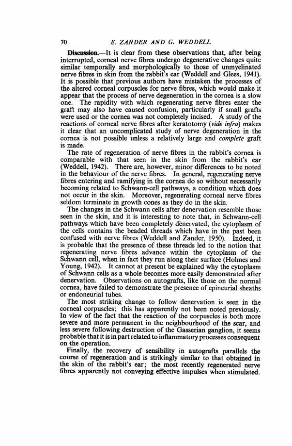
E. ZANDER AND G. WEDDELLDiscussion.-It is clear from these observations that, after being
interrupted, corneal nerve fibres undergo degenerative changes quitesimilar temporally and morphologically to those of unmyelinatednerve fibres in skin from the rabbit's ear (Weddell and Glees, 1941).It is possible that previous authors have mistaken the processes ofthe altered corneal corpuscles for nerve fibres, which would make itappear that the process of nerve degeneration in the cornea is a slowone. The rapidity with which regenerating nerve fibres enter thegraft may also have caused confusion, particularly if small graftswere used or the cornea was not completely incised. A study of thereactions of corneal nerve fibres after keratotomy (vide infra) makesit clear that an uncomplicated study of nerve degeneration in thecornea is not possible unless a relatively large and complete graftis made.The rate of regeneration of nerve fibres in the rabbit's cornea is
comparable with that seen in the skin from the rabbit's ear(Weddell, 1942). There are, however, minor differences to be notedin the behaviour of the nerve fibres. In general, regenerating nervefibres entering and ramifying in the cornea do so without necessarilybecoming related to Schwann-cell pathways, a condition which doesnot occur in the skin. Moreover, regenerating corneal nerve fibresseldom terminate in growth cones as they do in the skin.The changes in the Schwann cells after denervation resemble those
seen in the skin, and it is interesting to note that, in Schwann-cellpathways which have been completely denervated, the cytoplasm ofthe cells contains the beaded threads which have in the past beenconfused with nerve fibres (Weddell and Zander, 1950). Indeed, itis probable that the presence of these threads led to the notion thatregenerating nerve fibres advance within the cytoplasm of theSchwann cell, when in fact they run along their surface (Holmes andYoung, 1942). It cannot at present be explained why the cytoplasmof Schwann cells as a whole becomes more easily demonstrated afterdenervation. Observations on autografts, like those on the normalcornea, have failed to demonstrate the presence of epineurial sheathsor endoneurial tubes.The most striking change to follow denervation is seen in the
corneal corpuscles; this has apparently not been noted previously.In view of the fact that the reaction of the corpuscles is both moresevere and more permanent in the neighbourhood of the scar, andless severe following destruction of the Gasserian ganglion, it seemsprobable that it is in part related to inflammatory processes consequenton the operation.
Finally, the recovery of sensibility in autografts parallels thecourse of regeneration and is strikingly similar to that obtained inthe skin of the rabbit's ear; the most recently regenerated nervefibres apparently not conveying effective impulses when stimulated.
70

REACTION OF CORNEAL NERVE FIBRES
Maturation of some degree is a pre-requisite for this function. It isquite clear that both the regeneration of nerve fibres and sensoryrecovery take place equally well in transparent and opaque transplants.
(2) KERATOTOMY.Few workers have reported observations on the neurohistological
changes after keratotomy. However, the operation has beenperformed, in connexion with other studies, and much work hasbeen done on the ensuing sensory changes (Schroeder, 1923).
In 1878 Tizzoni examined corneas from rabbits 41 monthsafter keratotomy, using a gold-chloride impregnation method. Hegives a brief description of the process of degeneration andregeneration of nerve fibres, and remarks that degeneration is slowin the cornea in view of the presence of degenerating fibres at thislongperiodfollowingoperation. Theprocessofregeneration,hesays,takes the form of a replacement of the old nerve fibres by new nervefibres, the growing ends of which are not surmounted by growthcones. Schultz (1881), using frogs and a gold-chloride technique,states that 3 weeks after keratotomy he was unable to determineexactly what was happening, for both degeneration and regenerationprocesses appeared to be taking place simultaneously. Ranvier(1881) performed keratotomies in rabbits, only extending as deep asDescemet's membrane, and impregnated them 7 days later withgold-chloride. He states that after this period the sector of thecornea related to the lesion shows a complete absence of nerve fibresin the epithelium, and is sharply distinguished from the surroundingcornea in this respect. However, the nerve fibres in the " plexusfundamentalis" of the substantia propria are all intact. The mostrecent publication on the subject is by Jent (1948) ,who used rabbits,staining the corneal nerve fibres with methylene blue. He performedhis keratotomies in the same way as Ranvier; that is, extending onlyas far as Descemet's membrane but completely encircling the cornea1 mm. from the limbus. He correlated his histological observationswith sensory tests, and observed that 24 to 48 hours after operationthe nerve fibres did not stain as well as normally. This heattributed to degenerative changes. He believed degeneration to becomplete after 5 days, for at later periods he could no longerdemonstrate nerve fibres with methylene blue; moreover, he couldobtain no blink reflex by stimulation of any part of the cornea.He could elicit a feeble, sluggish corneal reflex 28 days after operationand observed that nerve fibres could once more be seen after stainingwith methylene blue, thus indicating that regeneration was takingplace.
It is thus not clear from the literature how much of the cornea isdenervated as the result of keratotomy; nor is it clear how, or fromwhere, the denervated portion is re-innervated. In view of this, and
71
E. ZANDER AND G. WEDDELL
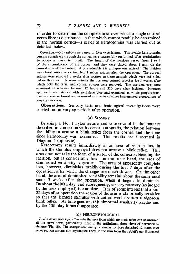
in order to determine the complete area over which a single cornealnerve fibre is distributed-a fact which cannot readily be determinedin the normal cornea-a series of keratotomies was carried out asdetailed below.Operation.-Only rabbits were used in these experiments. Thirty-eight keratotomies
passing completely through the cornea were successfully performed, after eserinizationto obtain a constricted pupil. The length of the incisions varied from J toof the circumference of the cornea, and they were placed about 1 mm. on thecorneal side of the limbus. Any irreducible iris prolapse was excised. The incisionwas closed with one or two No. 1 nylon sutures after the operation. The cornealsutures were removed 3 weeks after incision in those animals which were not killedbefore this time. In some animals the lids were sutured together for 3 weeks, afterwhich both the tarsal and corneal sutures were removed. The operated eyes wereexamined at intervals between 12 hours and 220 days after incision. Nineteenspecimens were stained with methylene blue and examined as whole preparations;nineteen were sectioned and examined as a series of silver-impregnated preparations ofvarying thickness.
Observations.-Sensory tests and histological investigations werecarried out at varying periods after operation.
(a) SENSORYBy using a No. 1 nylon suture and cotton-wool in the manner
described in connexion with corneal autografts, the relation betweenthe ability to arouse a blink reflex from the cornea and the timesince keratotomy was examined. The results are illustrated inDiagram 1 (opposite).Keratotomy results immediately in an area of sensory loss in
which the stimulus employed does not arouse a blink reflex. Thisarea does not take the form of a sector of the cornea subtending theincision, but is considerably less; on the other hand, the area ofdiminished sensibility is greater. The area of apparently completeloss, however, diminishes rapidly during the first 7 days after theoperation, after which the changes are much slower. On the otherhand, the area of diminished sensibility remains about the same untilsome 3 weeks after the operation, when it begins to diminish.By about the 90th day, and subsequently, sensory recovery (as judgedby the tests employed) is complete. It is of some interest that about28 days after operation the region of the scar is abnormally sensiti.veso that the lightest stimulus with cotton-wool arouses a vigorousblink reflex. As time goes on, this abnormal sensitivity recedes andby the 50th day it has disappeared.
(b) NEUROHISTOLOGICALTwelve hours after Operation.-In the area from which no blink reflex can be aroused,
all the nerve fibres, particularly those in the epithelium, show signs of degenerativechanges (Fig. 18). The changes seen are quite similar to those described 12 hours afternerve section among non-myelinated fibres in the skin from the rabbit's ear illustrated
72

REACTION OF CORNEAL NERVE FIBRES
2 DAYS 7 DAYS 2IDAYS
28DAYS 50DAYS 8ODAYS. + 44++ .
DIAGRAM 1.-Results of sensory tests at intervals after operation.Black area complete sensory loss.Hatched area partial sensory loss.Crosses hypersensitivity.Arrows extent of incision.
by Weddell and Glees (1941). There are also some signs of early degenerative changesamong nerve fibres beyond the sector subtending the incision.Two days after Operation.-The condition in various parts of the cornea is
illustrated in Diagram 2. There are apparently no normal nerve fibres in the regionof the cornea more than halfway from the scar towards the centre of the cornea.There are, however, degenerating fibres to be seen more than halfway from the centreof the cornea to the limbus opposite to the site of the operation. The degenerative
ak bDIAGRAM 2.-Zone of (a) epithelium (b) substantia propria over which cornealnerve fibres are affected 2 days after operation.
Black area degenerating nerve fibres.Hatched area both degenerating and normal nerve fibres.Unshaded area occasional degenerating nerve fibres.Arrows extent of incision.
73
E. ZANDER AND G. WEDDELL
FIG. 16.
FIG. 20. FIG. 21.
FIG. 19.
FIG. 22. FIG. 2i.
FIG. 24. FIG. 25. FIG. 26. FIG. 27.
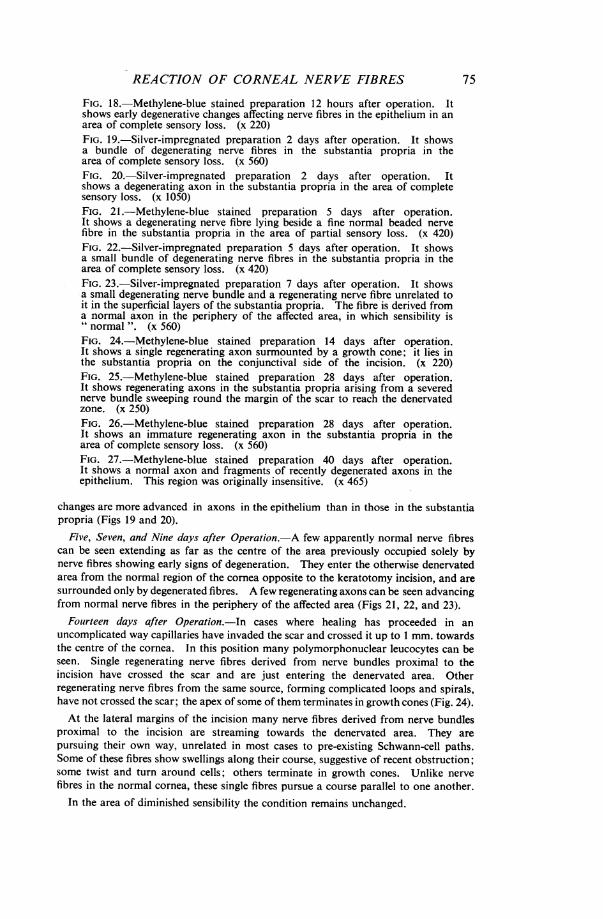
Figs 18-27.-Condition of nerve fibres in affected zones of rabbit corneasat various periods after keratotomy.
74
REACTION OF CORNEAL NERVE FIBRES
FIG. 18.-Methylene-blue stained preparation 12 hours after operation. Itshows early degenerative changes affecting nerve fibres in the epithelium in anarea of complete sensory loss. (x 220)FIG. 19.-Silver-impregnated preparation 2 days after operation. It showsa bundle of degenerating nerve fibres in the substantia propria in thearea of complete sensory loss. (x 560)FIG. 20. Silver-impregnated preparation 2 days after operation. Itshows a degenerating axon in the substantia propria in the area of completesensory loss. (x 1050)FIG. 21.-Methylene-blue stained preparation 5 days after operation.It shows a degenerating nerve fibre lying beside a fine normal beaded nervefibre in the substantia propria in the area of partial sensory loss. (x 420)FIG. 22.-Silver-impregnated preparation 5 days after operation. It showsa small bundle of degenerating nerve fibres in the substantia propria in thearea of complete sensory loss. (x 420)FIG. 23.-Silver-impregnated preparation 7 days after operation. It showsa small degenerating nerve bundle and a regenerating nerve fibre unrelated toit in the superficial layers of the substantia propria. The fibre is derived froma normal axon in the periphery of the affected area, in which sensibility is" normal ". (x 560)FIG. 24.-Methylene-blue stained preparation 14 days after operation.It shows a single regenerating axon surmounted by a growth cone; it lies inthe substantia propria on the conjunctival side of the incision. (x 220)FIG. 25.-Methylene-blue stained preparation 28 days after operation.It shows regenerating axons in the substantia propria arising from a severednerve bundle sweeping round the margin of the scar to reach the denervatedzone. (x 250)FIG. 26. Methylene-blue stained preparation 28 days after operation.It shows an immature regenerating axon in the substantia propria in thearea of complete sensory loss. (x 560)FIG. 27.-Methylene-blue stained preparation 40 days after operation.It shows a normal axon and fragments of recently degenerated axons in theepithelium. This region was originally insensitive. (x 465)
changes are more advanced in axons in the epithelium than in those in the substantiapropria (Figs 19 and 20).
Five, Seven, and Nine days after Operation.-A few apparently normal nerve fibrescan be seen extending as far as the centre of the area previously occupied solely bynerve fibres showing early signs of degeneration. They enter the otherwise denervatedarea from the normal region of the cornea opposite to the keratotomy incision, and aresurrounded only by degenerated fibres. A few regenerating axons can be seen advancingfrom normal nerve fibres in the periphery of the affected area (Figs 21, 22, and 23).
Fourteen days after Operation.-In cases where healing has proceeded in anuncomplicated way capillaries have invaded the scar and crossed it up to 1 mm. towardsthe centre of the cornea. In this position many polymorphonuclear leucocytes can beseen. Single regenerating nerve fibres derived from nerve bundles proximal to theincision have crossed the scar and are just entering the denervated area. Otherregenerating nerve fibres from the same source, forming complicated loops and spirals,have not crossed the scar; the apex of some of them terminates in growth cones (Fig. 24).At the lateral margins of the incision many nerve fibres derived from nerve bundles
proximal to the incision are streaming towards the denervated area. They arepursuing their own way, unrelated in most cases to pre-existing Schwann-cell paths.Some of these fibres show swellings along their course, suggestive of recent obstruction;some twist and turn around cells; others terminate in growth cones. Unlike nervefibres in the normal cornea, these single fibres pursue a course parallel to one another.
In the area of diminished sensibility the condition remains unchanged.
75
76 E. ZANDER AND G. WEDDELL
FIG. 33.
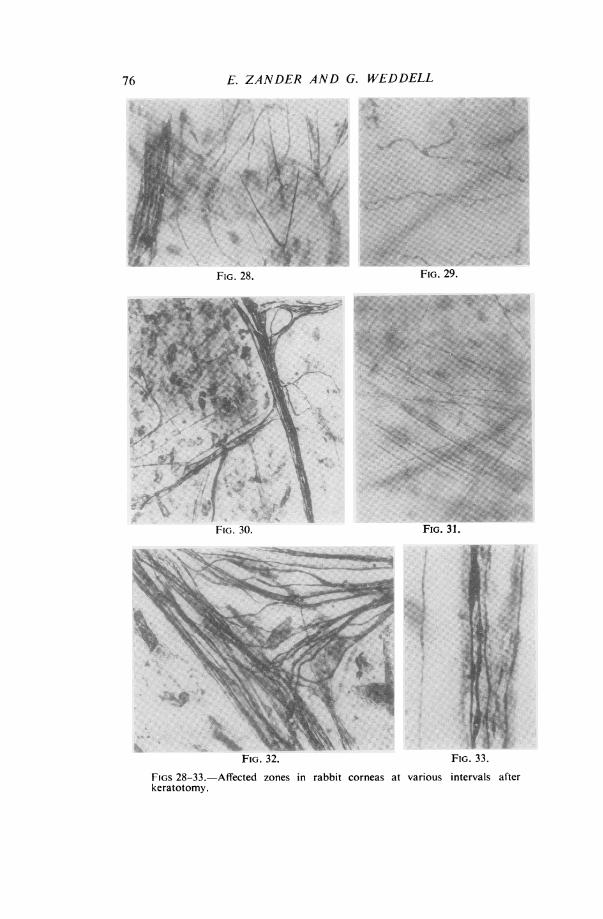
FIGS 28-33.-Affected zones in rabbitkeratotomy.
corneas at various intervals after

REACTION OF CORNEAL NERVE FIBRES
FIG. 28.-Silver-impregnated preparation from region of scar 50 daysafter operation. It shows single axons in the substantia propria passingtowards the scar and then turning away again. Empty Schwann-cell pathwaysare seen close to these axons; they are on the left of the picture. (x 235)FIG. 29.-Methylene-blue stained preparation 50 days after operation.It shows immature regenerating nerve fibres in the basal layer of epitheliumin the area of complete sensory loss. (x 425)FIG. 30.-Silver-impregnated preparation 110 days after operation. Itshows mature regenerated nerve fibres in the substantia propria in a zone ofnormal sensibility. This region was previously at the centre of the zoneof complete sensory loss. (x 164)FIG. 31.-Methylene-blue stained preparation 110 days after operation.It shows immature regenerating axons in the substantia propria sweepingaround the lateral margin of the scar. (x 215)FIG. 32.-Silver-impregnated preparation 220 days after operation. Itshows bundles of mature axons in the substantia propria in a zone of" normal "sensibility. This zone was originally completely insensitive. (x 510)FIG. 33.-Silver-impregnated preparation 220 days after operation. Itshows a bundle of immature axons coursing along a Schwann-cell pathwayin the substantia propria, lying beside a mature axon which in unrelated toSchwann cells. This specimen came from a zone of " normal" sensibilitywhich was originally completely insensitive. (x 1050)
Twenty-eight days after Operation.-In the region of the incision more nerve fibreshave penetrated the scar, and many more fibres have entered the denervated area afterpassing around its lateral margins (Fig. 25).
In the area of diminished sensibility regenerating nerve fibres derived from normaland apparently pre-existing nerve bundles are advancing towards the denervated areafrom a position opposite to that of the incision (Fig. 26). About this time it is possibleto trace almost the complete course of single nerve fibres entering the cornea lateral tothe keratotomy and terminating in the region of apparently complete sensory loss.Diagrams 3(a) and 3(b) show the course of two such fibres which could be so analysed.
DIAGRAM 3(a) and (b).-Camera lucida drawings showing approximatedistribution of single axons ending in substantia propria of cornea, made28 and 100 days respectively after keratotomy.
Position and extent of incision indicated by arc.Solid lines continuous axon.Interrupted lines beaded terminals.
77
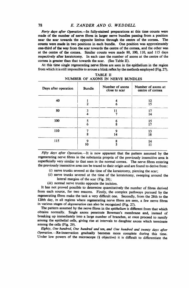
78 E. ZANDER AND G. WEDDELLForty days after Operation.-In fully-stained preparations at this time counts were
made of the number of nerve fibres in larger nerve bundles passing from a positionnear the scar towardls the opposite limbus through the centre of the cornea. Thecounts were made in two positions in each bundle. One position was approximatelyone-third of the way from the scar towards the centre of the cornea, and the other wasat the centre of the cornea. Similar counts were made 80, 100, 110, and 115 daysrespectively after keratotomy. In each case the number of axons at the centre of thecornea is greater than that towards the scar. (See Table II).At this time single regenerating nerve fibres are seen in the epithelium in the region
from which it is still impossible to arouse a blink reflex by the methods employed (Fig. 27).TABLE II
NUMBER OF AXONS IN NERVE BUNDLES
Days after operation Bundle Numbr of axons centre of cornea
40 1 4 122 6 15
80 3 11 174 7 14
100 5 6 156 8 17
110 7 9 138 14 18
115 9 8 1410 5 11
Fifty days after Operation. It is now apparent that the pattern assumed by theregenerating nerve fibres in the substantia propria of the previously insensitive area issuperficially very similar to that seen in the normal cornea. The nerve fibres enteringthe previously insensitive area can be traced to their origin and are found to derive from:
(i) nerve trunks severed at the time of the keratotomy, piercing the scar;(ii) nerve trunks severed at the time of the keratotomy, sweeping around the
lateral margins of the scar (Fig. 28);(iii) normal nerve trunks opposite the incision.
It has not proved possible to determine quantitatively the number of fibres derivedfrom each source, for two reasons. Firstly, the complex pathways pursued by theregenerating fibres make the task a very difficult one. Secondly, from the 28th to the120th day, in all regions where regenerating nerve fibres are seen, a few nerve fibresin various stages of degeneration can also be recognized (Fig. 27).The pattern assumed by the nerve fibres in the epithelium is different from that which
obtains normally. Single axons penetrate Bowman's membrane and, instead ofbreaking up immediately into a large number of branches, at once proceed to ramifyamong the epithelial cells, giving rise at intervals to daughter axons which terminateamong the cells (Fig. 29).
Eighty, One hundred, One hundred and ten, and One hundred and twenty days afterOperation.-Re-innervation gradually becomes more complete during this time.Under low powers of the microscope (4 objective) it is difficult to differentiate the
REACTION OF CORNEAL NERVE FIBRESaffected region from the surrounding normal cornea. Under higher powers, however,regenerating nerve fibres can still be recognized, but they gradually become fewer innumber. In addition, the nerve fibres in the affected regions are seen to take morecomplex pathways, and the pattern assumed by the epithelial nerve fibres is stillabnormal although the number of fibres seen in any given area approaches normal(Figs 30 and 31). It is remarkable that a number of nerve fibres in all states ofdegeneration is still seen throughout the affected area.Two hundred and twenty days after Operation.-More nerve fibres have penetrated the
scar, and in one specimen only ten single nerve fibres were seen sweeping round itslateral margin. More nerve fibres penetrating the scar are grouped into bundles.The appearance and number of nerve fibres reaching the cornea through the scar are,in fact, almost comparable with that seen in an unoperated sector of the eye (Fig. 32).Owing to the normal variation in numbers, however, counts of nerve fibres were notmade, for they would have been misleading. Despite the superficial appearance ofnormality at low magnification, a number of immature nerve fibres can still be seenunder higher magnification, although no degenerating nerve fibres are evident (Fig. 33).
Discussion.-It is now clear that the changes following keratotomyare complex and cannot be clearly understood unless specimens areexamined at successive stages following operation.
Fourteen hours after keratotomy early signs of nerve degenerationare apparent among all the nerve fibres in the area from which noblink reflex can be aroused. This is in accordance with theobservations made concerning nerve degeneration after autoplasticgrafting, but the changes were seen at an earlier stage than in anyspecimen examined in that series.The fact that no normal nerve fibres were seen among the
degenerating fibres was surprising, for a few degenerating nerve fibreswere seen among normal nerve fibres at a considerable distance fromthe lesion towards the opposite limbus. The appearance of anoccasional normal nerve fibre 5 days and subsequently afteroperation in the insensitive area was also surprising. These findingsare most easily explained on the basis that the few normal nervefibres present amongst the many degenerating fibres had undergonea reversible degenerative change of a non-Wallerian type (Denny-Brown and Brenner, 1944a and b).
It is unfortunate that, owing to the mass of degenerating nervefragments present, the few normal axons cannot be traced throughouttheir course until some 2 to 4 weeks after the lesion, for one objectof these experiments is thereby defeated. It was hoped as the resultof keratotomy (i.e. partial denervation) to obtain an accuratemeasure of the area over which single corneal nerve fibres aredistributed; but, by the time their distribution can be mapped out,nerve fibres from surrounding normal nerves are invading thedenervated area. The invading fibres, it is true, can be classedmorphologically as regenerating fibres, whereas those present in theinsensitive area appear quite normal. Thus the picture given of thedistribution of a single corneal nerve fibre is probably very similar to
79
E. ZANDER AND G. WEDDELLthat which it occupies in the normal cornea, although it may besomewhat exaggerated by extension.The denervated area is re-innervated by fibres from three sources:(i) nerve fibres arising from severed nerve bundles proximal to the incision, which
pierce the scar;(ii) nerve fibres from the same source which pass round the lateral margins of the scar;(iii) nerve fibres which invade the denervated area and which are derived from the
surrounding normal nerve bundles.Unfortunately, the pattern formed by the nerve fibres entering the
denervated area is so complex that it is not possible to determine therelative proportion of nerve fibres derived from each source at anyparticular stage after the operation. Nevertheless, it is clear fromTable II that fewer nerve fibres enter the cornea by piercing the scar,until a very late stage after operation, than normally enter from theperiphery. Such an assessment would, in any case, be of doubtfulvalue, for the reparative process is a long and continuous one, lastingmore than 120 days after the operation. Moreover, throughout thistime nerve fibres in early stages of degeneration can be seen, a findingin marked contrast to that seen after autoplastic grafting.
It is now clear why workers who examined corneas afterkeratotomy believed that nerve degeneration was such a slow processin this tissue.The abnormal sensitivity of the scar about 28 days after operation
is explained on the basis that at this time the number of axonssurmounted by growth cones at this site reaches a maximum(Weddell, Sinclair, and Feindel, 1948).The return of sensation as judged by the presence of a blink reflex
shows that the last region to recover is that closest to the scar.These observations are in accordance with those reported bySchroeder (1923) in a careful clinical study after keratotomy. Theysuggest that the invading nerve fibres play a not unimportant role inthe re-innervation process. However, in the specimens taken after220 days the number of nerve fibres entering the originallydenervated area by piercing the scar appears to be approaching thatwhich would have entered the cornea in such a position undernormal conditions. The nerve fibres have re-grouped themselvesinto bundles, and in one case only ten nerve fibres were seen sweepinground one lateral- margin of the scar to reach the originallydenervated area. Moreover, at this stage no nerve fibres in earlystages of degeneration could be seen.
These observations are explained on the basis that the denervatedarea is at first re-innervated by invasion from surrounding normalnerves which advance to meet the nerye fibres regenerating throughand around the scar. As those regenerating through the scar becomemore numerous, so those invading the territory from other regionsdegenerate, the process continuing until stabilization is reached.
80

REACTION OF CORNEAL NERVE FIBRES
The stage at which stabilization takes place clearly depends upontime, and presumably upon the nature of the scar. It is possiblethat in some cases the affected area may even become fullyre-innervated by nerve fibres from severed nerve bundles piercing thescar. In the two cases examined this had all but occurred at theend of 220 days; however, despite the fact that no degeneratingnerve fibres were seen, it may be presumed that stabilization had notbeen attained, for immature nerve fibres could still be seen. It is,therefore, possible that complete restitution might have occurred ata still later date.
Regenerating nerve fibres apparently grow into the denervated zoneof the cornea between the layers of the stroma as easily as they doalong Schwann-cell pathways. This is in marked contrast to whattakes place in the skin. However, the mere presence of the requisitequota of nerve fibres in the affected zone does not constitute the endof the process. There are apparently some factors which influencethe process sufficiently to determine that, by degrees, the originalpattern of innervation is restored at the expense of the fibres whichhave taken new pathways to arrive at their destination.The processes of nerve repair after keratotomy in mammals are
of interest in that they are in broad agreement with the hypothesesconcerning nerve patterns and the mechanics of nerve growthexpressed by Weiss (1941) as the result of work on amphibians andthe examination of the growth of neuroblasts in tissue culture.
Finally, keratotomy demonstrates clearly how labile the peripheralnervous system is, and illustrates the danger of assuming that partialdenervation experiments will necessarily provide a picture whichrepresents the pattern normally assumed by the unoperated nervefibres.
(3) SECTION AND AVULSION OF THE INFRA-ORBITAL NERVE.
As Vonderahe (1928) suggested that the infra-orbital nerve suppliesthe lower half of the cornea in man, it was thought advisableto determine whether this was also the case in the rabbit. Althoughexamination of normal material had excluded the possibility thatmany nerve fibres reach the cornea from the infra-orbital nerve, thepossibility that some may do so cannot be excluded by this method.
Operation.,The infra-orbital nerve was approached by the method described byKrause (1884). After identification, it was divided and then avulsed from the infra-
orbital canal on the left side in four rabbits. Periods of 7 days (two rabbits) and20 days (two rabbits) respectively were left for nerve degeneration to occur. Thecorneas from two of the rabbits were examined after methylene-blue staining, andfrom two after silver impregnation.
Observations.-No differences were found in the blink reflex onstimulation of the cornea with a wisp of cotton-wool between the
81

E. ZANDER AND G. WEDDELLnormal and operated side or between the upper and lower half,periphery, and centre of the cornea on the operated side, althoughthere was apparently complete anaesthesia of the skin supplied bythe infra-orbital nerve. No " trophic" changes were observed inthe corneas on the operated side and no changes in the conjunctivalvessels. The corneas were well stained or impregnated throughouttheir extent. Careful examination failed to show any clear-cutdifferences ia the appearance of the nerve fibres between the normaland operated sides.
Discussion.-Vonderahe has suggested that in man the infra-orbital nerve (and thus the second division of the fifth nerve) suppliesthe cornea, for he found in a case of traumatic injury involving thesecond division of the fifth nerve that sensibility in the lower half ofthe cornea of the affected side was impaired. In addition, Boucheron(1890, 1891) suggests that the periphery of the cornea may be suppliedby nerve fibres which reach it from other than the ciliary nerves.This implies that the infra-orbital nerve may be implicated.
Vonderahe's observations and Boucheron's statements have givenrise to the suggestion that in testing for corneal sensitivity in theclinic reactions from the upper and lower halves of the cornearespectively should be compared.
In the rabbit it seems improbable that the cornea receives sensorynerves from the infra-orbital nerve. In view of the fact thatVonderahe's case was not an uncomplicated section of the infra-orbital nerve, it is clear that observations of this kind in man shouldbe re-examined, for it is possible that fibres from the maxillarydivision of the fifth nerve may reach the cornea by routes other thanby way of the infra-orbital nerve, as suggested by Boucheron(1890, 1891).(4) SECTION OF THE CILIARY NERVES.The purpose of these experiments was to see whether any nerve
fibres reach the cornea by routes other than the ciliary nerves. Asalready noted, Boucheron (1890, 1891) believed that the periphery ofthe cornea is innervated by nerve fibres which do not travel with theciliary nerves, and Vonderahe (1928) has suggested that in man thelower half of the cornea is innervated by the fibres travelling in theinfra-orbital nerve.Operation.-The corneas from three rabbits were examined on the side on which all
the ciliary nerves, together with the optic nerve, had been severed, close to their entryinto the eyeball, 3 days previously. Care was taken to cause as little damage aspossible to the ciliary vessels.
Observations.-Unfortunately, the operated eye in every casebecame so rapidly and so seriously affected that it was not considereduseful to keep any animal longer than 3 days after operation. Ornthe fourth day after the section of the nerves, the cornea in almost
82
REACTION OF CORNEAL NERVE FIBRES
every case becomes opaque and is grossly distorted; in addition,some degree of keratitis is present (a condition recalling earlyphthisisbulbi in man). In the corneas examined 3 days afteroperation only degenerating nerve fibres were seen. No normalnerve fibres whatever were encountered. The corneal corpuscles areaffected in the same way as after autoplastic grafting.
Discussion.-These experiments are inconclusive in view of theobservations made after keratotomy, for there may have been afew nerve fibres in these corneas undergoing reversible degenerativechanges. Nevertheless, the experiments do indicate that the majorityof nerve fibres in the rabbit reach the cornea via the ciliary nerves:thus confirming what appears to be the case in normal material(Zander and Weddell, 1951).(5) ExcISION OF THE SUPERIOR CERVICAL SYMPATHETIC GANGLION.
Although Boeke (1935) stated that few, if any, sympathetic nervefibres supply the cornea, Rodger (1950) mentioned their presence.In view of this divergence of opinion, and of the fact that the methodof direct observation in normal material cannot solve the problem,it was decided to determine experimentally whether post-ganglionicsympathetic fibres leave the superior cervical ganglion to supply thecornea.Operation.-The superior cervical sympathetic ganglion was excised on the left side
in each of five rabbits. The operation was carried out in accordance with theinstructions of Krause (1884) and resulted in a constriction of the pupil, dilation of theconjunctival blood vessels, and an increase in temperature and flushing of the ear onthe affected side. Periods of 7, 14, 21, and 50 days were allowed for degeneration tooccur. The corneas from three rabbits were examined after methylene-blue staining,and from two after silver impregnation.
Observations.-After operation no differences in the cornealsensitivity of the two sides could be detected by the use of a wisp ofcotton-wool to arouse a blink reflex. There were no obvious trophicchanges or differences in transparency between the two sides. Thestaining and impregnation of the corneas was complete and uniform.No obvious differences in the nerve supply between the normal andoperated corneas was observed. A count of the nerve fibres in thenerve bundles in the periphery of the corneas was carried out in themethylene-blue stained specimens of one of the animals, and nosignificant differences were noted between the normal and operatedsides (see Table in Zander and Weddell, 1951, which indicates thenormal variation in the numbers to be expected).
Discussion.-These observations suggest that few post-ganglionicnerve fibres, if any, pass directly from the superior cervical ganglionto the cornea. It is possible that fibres reach the cornea viaganglion-cell relays situated between the superior cervical ganglionand the cornea, but it appears unlikely, from these observations, that
83

84 E. ZANDER AND G. WEDDELL
FIG. 34.
FIG. 36.
FIG. 35.
PIG. 37.
FIG. 38. FIG. 39. FIG. 40.
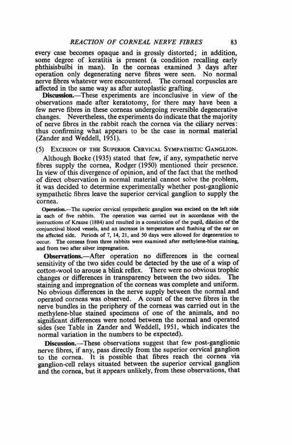
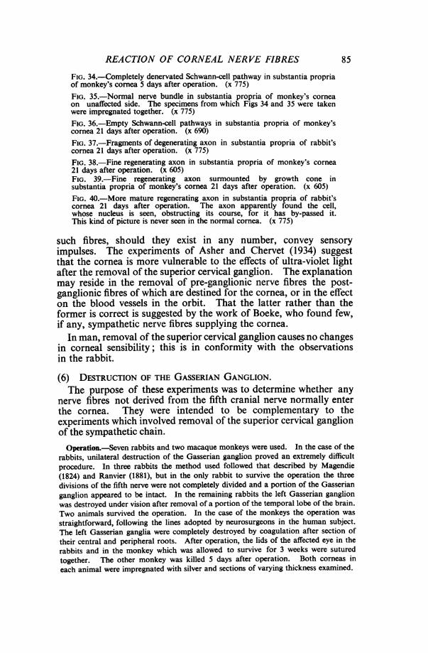
FIGS 34-40.-Silver-impregnated preparations from normal and affectedcorneas 5 days and 21 days after destruction of Gasserian ganglion.
REACTION OF CORNEAL NERVE FIBRES
FIG. 34.-Completely denervated Schwann-cell pathway in substantia propriaof monkey's cornea 5 days after operation. (x 775)FIG. 35.-Normal nerve bundle in substantia propria of monkey's corneaon unaffected side. The specimens from which Figs 34 and 35 were takenwere impregnated together. (x 775)FIG. 36.-Empty Schwann-cell pathways in substantia propria of monkey'scornea 21 days after operation. (x 690)FIG. 37.-Fragments of degenerating axon in substantia propria of rabbit'scornea 21 days after operation. (x 775)FIG. 38.-Fine regenerating axon in substantia propria of monkey's cornea21 days after operation. (x 605)FIG. 39.-Fine regenerating axon surmounted by growth cone insubstantia propria of monkey's cornea 21 days after operation. (x 605)
FIG. 40.-More mature regenerating axon in substantia propria of rabbit'scornea 21 days after operation. The axon apparently found the cell,whose nucleus is seen, obstructing its course, for it has by-passed it.This kind of picture is never seen in the normal cornea. (x 775)
such fibres, should they exist in any number, convey sensoryimpulses. The experiments of Asher and Chervet (1934) suggestthat the cornea is more vulnerable to the effects of ultra-violet lightafter the removal of the superior cervical ganglion. The explanationmay reside in the removal of pre-ganglionic nerve fibres the post-ganglionic fibres of which are destined for the cornea, or in the effecton the blood vessels in the orbit. That the latter rather than theformer is correct is suggested by the work of Boeke, who found few,if any, sympathetic nerve fibres supplying the cornea.
In man, removal of the superior cervical ganglion causes no changesin corneal sensibility; this is in conformity with the observationsin the rabbit.
(6) DESTRUCTION OF THE GASSERIAN GANGLION.The purpose of these experiments was to determine whether any
nerve fibres not derived from the fifth cranial nerve normally enterthe cornea. They were intended to be complementary to theexperiments which involved removal of the superior cervical ganglionof the sympathetic chain.Operation.-Seven rabbits and two macaque monkeys were used. In the case of the
rabbits, unilateral destruction of the Gasserian ganglion proved an extremely difficultprocedure. In three rabbits the method used followed that described by Magendie(1824) and Ranvier (1881), but in the only rabbit to survive the operation the threedivisions of the fifth nerve were not completely divided and a portion of the Gasserianganglion appeared to be intact. In the remaining rabbits the left Gasserian ganglionwas destroyed under vision after removal of a portion of the temporal lobe of the brain.Two animals survived the operation. In the case of the monkeys the operation was
straightforward, following the lines adopted by neurosurgeons in the human subject.The left Gasserian ganglia were completely destroyed by coagulation after section oftheir central and peripheral roots. After operation, the lids of the affected eye in therabbits and in the monkey which was allowed to survive for 3 weeks were suturedtogether. The other monkey was killed 5 days after operation. Both corneas ineach animal were impregnated with silver and sections of varying thickness examined.
85
E. ZANDER AND G. WEDDELL
Observations.-Sensory tests and histological investigations werecarried out with the following results.
(a) SENSORY
In every case the cornea was completely insensitive, no blink reflexbeing aroused by the application of cotton-wool or a No. 1 nylonsuture.
(b) HISTOLOGICALThe cornea from the monkey killed 5 days after operation showed
no macroscopical evidence of keratitis, and few polymorphonuclearcells were seen; in addition, the corneal corpuscles appeared to bevery little affected. No normal nerve fibres were seen, but theSchwann-cell pathways were clearly outlined and seen to havefragments of degenerating axons lying along their surface (Fig. 34).In the conjunctiva of the operated eye there were blood vessels withnerve fibres beside them at the margin of the cornea. The unoperatedeye, which was also prepared for histological examination, appearedto be normally innervated (Fig. 35). The corneas from the animalskilled 3 weeks after operation showed some macroscopic evidence ofkeratitis and the corneas were infiltrated by a number of poly-morphonuclear cells. The corneal corpuscles showed a reactionsimilar to that seen 3 weeks after grafting. Numerous Schwann-cellpathways, having only an occasional axis-cylinder fragment lying ontheir surface, were seen (Figs 36 and 37). However, there werethroughout the cornea a few isolated regenerating nerve fibres(Figs 38, 39, and 40). The ultimate source of these fibres was notdetermined, but they entered singly from the conjunctiva. The eyesfrom the unoperated side were also prepared for microscopicalexamination and appeared to be normally innervated.
Discussion.-These experiments show clearly that normally fewnerve fibres reach the cornea other than by way of the fifth cranialnerve, and thus we are able to confirm Boeke's statement that if thecornea does contain any sympathetic nerve fibres at all, these must bevery few. They also show once again how easily nerve fibres grow intothe cornea, the source of the regenerating nerve fibres which wereseen 3 weeks after operation almost certainly being from the vascularnerves in the conjunctiva by a process of extension. This observationsuggests that great care needs to be exercised when interpretingresults following experimental nerve section. It is clearly not safeto conclude that, following removal of one set of nerve fibres, theremaining axons will remain morphologically quiescent. Ofparticular interest in connexion with these experiments are those ofClark (1942, 1943) who showed that the work of Tello (1911)concerning the regeneration of nerve fibres in the central nervoussystem was explicable on the basis of vascular nerves invading the
86

REACTION OF CORNEAL NERVE FIBRES
region of the lesion, and that in fact no regeneration of centralnervous system axons takes place after injury.The observation that the corneal corpuscles show so few signs
of reaction 5 days after destruction of the Gasserian ganglion inthe monkey is interesting. It may be due to the fact that the lidswere not sutured in this case, thereby preventing surface abrasion ofthe cornea.
Reiser (1936, 1937) stated that 2 days after destruction of theGasserian ganglion, although degenerative changes affected all thenerve fibres of the main nerve bundles, the preterminal networkshowed only slight evidence of degenerative change and the terminalreticulum had not changed at all. His statement can be criticized ontwo accounts. Firstly, using thin silver sections and the Bielschowskymethod, 2 days is too short a time to be certain of seeing degenerativechanges in every isolated axon segment; this may account for hisstatement that the pre-terminal network is only slightly affected.Secondly, the so-called terminal reticulum has been shown to be anartefact (Weddell and Zander, 1951).
SUMMARY
(1) The rate and character of nerve degeneration in the rabbit'scornea are similar to that in the skin of the rabbit's ear.
(2) The rate of regeneration of nerve fibres in the cornea iscomparable with that in the skin of the rabbit's ear.
(3) Regenerating nerve fibres within the cornea seldom terminatein growth cones; they apparently pursue their way as easily whenunrelated to Schwann cells as when related to them.
(4) The area over which a single corneal nerve fibre is distributedis at least as large as a quadrant of the cornea.
(5) After denervation, both Schwann cells and corneal corpusclesundergo characteristic changes.
(6) After keratotomy, the denervated area is re-innervated fromthree sources:
(i) regenerating nerve fibres which penetrate the scar,(ii) regenerating fibres which pass around the lateral margins of
the scar,(iii) invading fibres which sprout from surrounding normal nerve
fibres and invade the denervated area.
(7) From the 50th to the 120th day after keratotomy there aresigns of active nerve degeneration as well as nerve regeneration.
(8) Recovery is histologically nearly complete 220 days afterkeratotomy. It appears that the nerve fibres which originallyinvade the denervated zone from normal surrounding nerves, and bysweeping around the incision, have retreated in favour of regenerated
87

E. ZANDER AND G. WEDDELL
nerve fibres originating from those severed at the time of keratotomywhich traverse the scar to reach their destination.
(9) As the result ofsection or destruction ofthe following structures:the infra-orbital nerve, the superior cervical sympathetic ganglion,the ciliary nerves and the Gasserian ganglion, it is clear that therabbit's cornea is probably supplied solely by axons from the fifthcranial nerve reaching the cornea by way of the ciliary nerves.
(10) Five days after extirpation of the Gasserian ganglion onlydegenerating nerve fibres can be seen throughout the cornea.
(11) Three weeks after extirpation of the Gasserian ganglionregenerating nerve fibres can be seen in the cornea, which iscompletely insensitive. The source of these fibres is unknown, butit is likely that they arise from vascular nerves in the conjunctivaby a process of extension.
This investigation was made possible by a grant from the Rockefeller Foundationfor which we wish to express our gratitude. We should also like to thank Mr. ArthurDent, Miss Jean Gurden, and Mr. Patrick Selwood for their skilful technical assistance.At the outset of these investigations we had the invaluable surgical assistance ofMr. J. P. F. Lloyd, F.R.C.S., to whom we offer our sincere thanks.
REFERENCESASHER, L., and CHERVET, N. (1934). Klin. Wschr., 13, 1688.BOEKE, J. (1935). Z. mikr.-anat. Forsch., 38, 594.BOUCHERON, A. (1890). C.R. Soc. Biol. Paris, 9 ser., 2, 71.
(1891). Ibid., 3, 59.CLARK, W. E. LE GROS (1942). J. Anat., Lond., 77, 20.
(1943). Ibid., 77, 251.DENNY-BROWN, D., and BRENNER, C. (1944a). Arch. Neurol. Psychiat., Chicago, 51, 1.
(1944b). Ibid., 52, 1.ESCAPINI, H. (1948). Arch. Ophthal., Chicago, 39, 135.FRANCESCHETrI, A., and BABEL, J. (1947). Ann. Oculist., Paris, 180, 142.GLEES, P. (1946). J. Neuropath. exp. Neurol., 5, 54.HOLMES, W., and YOUNG, J. Z. (1942). J. Anat., Lond., 77, 63.JENT, M. (1945). Helv. physiol. pharmacol. Acta, 3, 65.KRAUSE, W. (1884). " Die Anatomie des Kaninchens ". 2nd ed. Engelmann,
Leipzig.LEOZ ORTIN, G. (1915). Arch. oftal. hisp.-amer., 15, 225.
(1917). Ibid., 17, 615.MAGENDIE, F. (1824). J. Physiol. exp. path., Paris, 4, 176.RANVIER, L. (1881). " Le;ons d'anatomie generales faites au CollEge de France par
L. Ranvier; annee 1878-1879". Baillitre, Paris.REISER, K. A. (1937). Arch. Augenheilk., 110, 253.RODGER, F. C. (1950). British Journal of Ophthalmology, 34, 107.SCHROEDER, E. (1923). Graefes Arch. Ophthal., 111, 17.SCHULTZ, F. (1881). " Experimentelle Studien uber Degeneration und Regeneration
der Cornealnerven ". Laakman, Dorpat.TELLO, F. (1911). Trab. Lab. Invest. biol. Univ. Madr., 9, 123.TIZZONI, G. (1878). Zbl. med. Wiss., 16, 225.VONDERAHE, A. R. (1928). Arch. Neurol. Psychiat., Chicago, 20, 836.WEDDELL, G. (1942). J. Anat., Lond., 77, 49.
and GLEES, P. (1941). Ibid., 76, 65.and ZANDER, E. (1950). Ibid., 84, 168.SINCLAIR, D. C., and FEINDEL, W. H. (1948). J. Neurophysiol., 11, 99.
WEISS, P. (1941). Growth, 5. 3rd Growth Symposium, p. 163.ZANDER, E., and WEDDELL, G. (1951). J. Anat., Lond., 85, 68.
88