
Studies on Hexokinase - NCBI
9
Vol. 49 Studies on Hexokinase 1. THE HEXOKINASE ACTIVITY OF RAT-BRAIN EXTRACTS BY H. WEIL-MALHERBE AND A. D. BONE Research Department, Runwell Hospital, Wickford, Essex (Received 13 November 1950) This and the two following papers are part of an investigation which was originally undertaken to study the possibility of using the hexokinase reaction for the assay of hormonal changes in human blood. For this purpose it seemed inadvisable to use purified hexokinase preparations, since the inter- action with hormones might conceivably depend on the association of the enzyme with other tissue con- stituents. Preliminary experiments had shown that aqueous extracts of rat brain possessed a hexokinase activity which was higher, more stable and more reproducible than similar extracts of rat muscle. Moreover, these extracts were practically free of carbohydrate and of codehydrogenases and their apyrase activity was low. Undesirable side reactions were therefore less likely to occur. For these reasons most experiments were performed with an aqueous extract of rat brain as a source of hexokinase. In this paper the properties of the enzyme system used, together with some new observations on brain hexokinase, will be described. METHODS AND MATERIALS Abbreviations ATP = adenosinetriphosphate; ADP = adenosinediphos. phate; P7 mi.. =P of acid-hydrolysable phosphate groups of ATP and ADP; terminal P =terminal (myosin-labile) P of ATP; HMP =hexosemonophosphate; HDP =hexosediphos- phate. Preparation of brain extracts A rat brain was homogenized for 3 min. in a small glass homogenizer of close fit (Potter & Elvehjem, 1936) with a total volume of 7 ml. glass-distilled water, added in portions, at 0°. After centrifuging for 5 min. in an angle centrifuge at 3500 rev./min. the clear supernatant was taken off with a rubber-teat pipette, care being taken not to disturb the precipitate. The residue was suspended in 7 ml. water and again centrifuged. The combined extracts (approx. 11 ml.) contained about 6 mg. protein/ml. In the initial stages of the investigation the protein content of the extract was deter- mined with Folin & Ciocalteu's (1927) reagent and the volume adjusted to the desired protein concentration. The colour formation from brain extracts with the phenol re- agent had previously been calibrated by simultaneous Kjeldahl estimations. Since the protein content of the brain extracts was found to vary little, this estimation was later omitted. Extracts prepared in this way were found to retain their hexokinase activity for 2-3 days at 0°. Composition of test solution In all experiments inorganic constituents were added to give the following concentrations (M): NaHCO3, 0-02; MgCI2, 0-0067; NaF, 0-0238. The concentrations of brain extract, phosphate acceptor and phosphate donor were varied to suit the requirements of the experiment, but, unless otherwise stated, the standard concentrations were: brain extract, 0-2 ml. (approx. 1-2 mg. protein)/3 ml.; glucose, 1-85 mM; ATP, 3-33 mm. The usual volume of the test solution was 3 ml., but for some experiments it was increased to 6 or 10 ml. Experiments were carried out at 30° in an atmosphere of nitrogen containing 5 % (by vol.) CO. Measurement of reaction rates Manometric experiments. These were carried out in Warburg manometers. The reaction was started by tipping in the phosphate acceptor (glucose or HMP) from the side bulb; any pressure changes due to the reaction of ATP with preformed phosphate acceptors in the brain extract were thus avoided. With the small amounts of protein used, retention was found to be too small to be measured with accuracy. It was therefore neglected. Likewise, the 'esterifi- cation correction' (Meyerhof & Kiessling, 1933) was neg- ligible since the K. values of the disappearing phosphate esters did not significantly differ from those of the newly formed esters. Chemical estimations. The measurement of reaction rates by chemical methods involved the analysis of an initial and subsequent samples. The reaction was started by adding the brain extract as last component. The initial sample was with- drawn immediately afterwards with a pipette and delivered into a solution of the appropriate deproteinizing agent. Analytical procedures Glucose. Glucose was estimated in 0-5-1-0 ml. samples by the method of Nelson (1944) after Ba(OH)2-ZnSO4 precipi- tation. All estimations were carried out in duplicate. Phosphatefractions. Inorganic phosphate was determined by the method of Fiske & Subbarow (1925) or, in later experiments, by a modification of the method of Martin & Doty (1949; see Weil-Malherbe & Green, 1951). The acid- labile phosphate groups of ATP and ADP were determined after hydrolysis in N-HCl at 1000 for 7 min. and corrected for the phosphate fraction hydrolysed between 7 and 30 min. under the same conditions (Lohmann, 1928). Owing to the high yield of hexosediphosphate in these experiments serious errors were incurred if this correction was omitted. ATP was determined from the phosphate hydrolysed by purified (four times precipitated) myosin; this was carried out as previously described (Weil-Malherbe, 1950) and 22-2 339
-
Upload
khangminh22 -
Category
Documents
-
view
3 -
download
0
Transcript of Studies on Hexokinase - NCBI

Vol. 49
Studies on Hexokinase1. THE HEXOKINASE ACTIVITY OF RAT-BRAIN EXTRACTS
BY H. WEIL-MALHERBE AND A. D. BONEResearch Department, Runwell Hospital, Wickford, Essex
(Received 13 November 1950)
This and the two following papers are part of aninvestigation which was originally undertaken tostudy the possibility ofusing the hexokinase reactionfor the assay of hormonal changes in human blood.For this purpose it seemed inadvisable to usepurified hexokinase preparations, since the inter-action with hormones might conceivably depend onthe association of the enzyme with other tissue con-stituents. Preliminary experiments had shown thataqueous extracts ofrat brain possessed a hexokinaseactivity which was higher, more stable and morereproducible than similar extracts of rat muscle.Moreover, these extracts were practically free ofcarbohydrate and of codehydrogenases and theirapyrase activity was low. Undesirable side reactionswere therefore less likely to occur. For these reasonsmost experiments were performed with an aqueousextract ofrat brain as a source ofhexokinase. In thispaper the properties of the enzyme system used,together with some new observations on brainhexokinase, will be described.
METHODS AND MATERIALS
AbbreviationsATP = adenosinetriphosphate; ADP = adenosinediphos.phate; P7 mi.. =P of acid-hydrolysable phosphate groups ofATP and ADP; terminal P =terminal (myosin-labile) P ofATP; HMP =hexosemonophosphate; HDP =hexosediphos-phate.
Preparation of brain extractsA rat brain was homogenized for 3 min. in a small glass
homogenizer of close fit (Potter & Elvehjem, 1936) with atotal volume of 7 ml. glass-distilled water, added in portions,at 0°. After centrifuging for 5 min. in an angle centrifuge at3500 rev./min. the clear supernatant was taken off with arubber-teat pipette, care being taken not to disturb theprecipitate. The residue was suspended in 7 ml. water andagain centrifuged. The combined extracts (approx. 11 ml.)contained about 6 mg. protein/ml. In the initial stages oftheinvestigation the protein content of the extract was deter-mined with Folin & Ciocalteu's (1927) reagent and thevolume adjusted to the desired protein concentration. Thecolour formation from brain extracts with the phenol re-agent had previously been calibrated by simultaneousKjeldahl estimations. Since the protein content of the brainextracts was found to vary little, this estimation was lateromitted. Extracts prepared in this way were found to retaintheir hexokinase activity for 2-3 days at 0°.
Composition of test solutionIn all experiments inorganic constituents were added
to give the following concentrations (M): NaHCO3, 0-02;MgCI2, 0-0067; NaF, 0-0238. The concentrations of brainextract, phosphate acceptor and phosphate donor werevaried to suit the requirements ofthe experiment, but, unlessotherwise stated, the standard concentrations were: brainextract, 0-2 ml. (approx. 1-2 mg. protein)/3 ml.; glucose,1-85 mM; ATP, 3-33 mm.The usual volume of the test solution was 3 ml., but for
some experiments it was increased to 6 or 10 ml.Experiments were carried out at 30° in an atmosphere of
nitrogen containing 5 % (by vol.) CO.
Measurement of reaction ratesManometric experiments. These were carried out in
Warburg manometers. The reaction was started by tippingin the phosphate acceptor (glucose or HMP) from the sidebulb; any pressure changes due to the reaction ofATP withpreformed phosphate acceptors in the brain extract werethus avoided. With the small amounts of protein used,retention was found to be too small to be measured withaccuracy. It was therefore neglected. Likewise, the 'esterifi-cation correction' (Meyerhof & Kiessling, 1933) was neg-ligible since the K. values of the disappearing phosphateesters did not significantly differ from those of the newlyformed esters.
Chemical estimations. The measurement of reaction ratesby chemical methods involved the analysis of an initial andsubsequent samples. The reaction was started by adding thebrain extract as last component. The initial sample was with-drawn immediately afterwards with a pipette and deliveredinto a solution of the appropriate deproteinizing agent.
Analytical proceduresGlucose. Glucose was estimated in 0-5-1-0 ml. samples by
the method of Nelson (1944) after Ba(OH)2-ZnSO4 precipi-tation. All estimations were carried out in duplicate.
Phosphatefractions. Inorganic phosphate was determinedby the method of Fiske & Subbarow (1925) or, in laterexperiments, by a modification of the method of Martin &Doty (1949; see Weil-Malherbe & Green, 1951). The acid-labile phosphate groups of ATP and ADP were determinedafter hydrolysis in N-HCl at 1000 for 7 min. and corrected forthe phosphate fraction hydrolysed between 7 and 30 min.under the same conditions (Lohmann, 1928). Owing to thehigh yield of hexosediphosphate in these experiments seriouserrors were incurred if this correction was omitted.ATP was determined from the phosphate hydrolysed by
purified (four times precipitated) myosin; this was carriedout as previously described (Weil-Malherbe, 1950) and
22-2
339

H. WEIL-MALHERBE AND A. D. BONEADP was calculated by the difference: (P7 mill) - 2(myosin-labile P).For the simultaneous estimation of inorganic, acid-labile
and myosin-labile P, 4 ml. of test solution were added to16 ml. 10% (w/v) trichloroacetic acid. The filtrate wasmeasured and 1 ml. x-HCI was added. The solution wasextracted four times with 10 ml. portions of peroxide-freeredistilled ether to remove excess trichloroacetic acid. Etherwas removed in vacuo from the warmed solution which wasthen neutralized and made up to 20 ml. Inorganic P wasdetermined in a 4 ml. sample and acid-labile P in two 1 ml.samples hydrolysed for 7 and 30 min. respectively. Twosamples of 6 ml. were used for the estimation of myosin-labile P. Care was taken to avoid an excess ofmyosin, as thismight have resulted in a partial hydrolysis of the secondphosphate group ofATP. The correct amount ofmyosin wasfound in preliminary experiments with ATP solutions ofknown concentration.
HeXosediphosphate. HDP was determined by the estima-tion of alkali-labile phosphate after incubation with aldolasein presence of KCN (Meyerhof, Ohlmeyer & Mohle, 1938;Meyerhof&Junowicz-Kocholaty, 1942). Crystalline aldolasewas prepared from rabbit muscle by the method of Taylor,Green & Cori (1948). The crystalline enzyme was dried invacuo and 3 mg. of the powder, which contained some(NH,)2SO4, were dissolved and added to a neutralized tri-chloroacetic acid filtrate. A calibration curve was deter-mined with a solution of pure HDP.The method estimates the sum of HDP and triosephos-
phates. All analytical results referring to 'HDP' represent inreality the sum of these compounds.Ammonia. NH, was distilled in the apparatus of Parnas &
Heller (1924) and determined by nesslerization.
Chemical preparationsATP was prepared from rabbit muscle by the method of
Dounce, Rothstein, Beyer, Meier & Freer (1948). A numberof consecutive specimens showed a satisfactory ratio of totalorganic to acid-labile P, though the figures were higher thanthose calculated for Ba2(Cj0H3L013N5P8).4H2O (P, 10-92;P7mit, 7.3 %), possibly due to the presence of some acid Basalt. A specimen analysis was as follows: total organic P,12-60; P7ml,, 8-36 %. The myosin-labile P usually amountedto 29-33% of the total organic P.ADP. After several unsatisfactory attempts with myosin
a pure specimen of ADP was prepared with purified yeasthexokinase by incubating 30 mg. of enzyme-protein with750 mg. glucose and 11-4 ml. 0.1 m-ATP in bicarbonate-CO2buffer (pH 7.4) for 30 min. at 300. The volume ofthe solution,which also contained Mg++ in the usual concentration, was150 ml. The ADP formed was purified through its mercuricand Ba salts in the usual way. Yield: 390 mg. Ba salt (totalorganic P, 8.7 %; P7ml,,, 4-49 %).The yeast hexokinase was obtained by the procedure of
Berger, Slein, Colowick & Cori (1946) and corresponded totheir fraction 3 a. The bulk of the preparation which was notused for the preparation ofADP was dried after precipitationwith (NHJ)2SO4 (0.7 saturation). The enzyme preparationobtained on solution and dialysis was highly active.
Hexo8emonopho8phate8. A specimen of the neutral Ba saltof Robison ester mixture was given by Dr M. G. Macfarlane.Organic P: 8-08% (on air-dried material).A specimen of greater purity was isolated after the ex-
periment in which ADP was prepared by adding an equal
volume of ethanol to the supernatant from the first Ba pre-cipitation. The air-dried Ba salt (340 mg.) contained 7-08%organic P. Estimation of fructose (Roe, 1934) and aldosetitration (Macleod & Robison, 1929) revealed a content of45% ketose and 55% aldose.
Fructose-6-phosphate was prepared from HDP as the Basalt according to Neuberg, Lustig & Rothenberg (1943).Organic P: 6-43% (on air-dried material).
Glucose-6-phosphate was synthesized by the method ofLardy & Fischer (1946). The Ca salt, dried in vacuo at 780,contained 10 69 % organic P.
Hexosedipho8phate. The acid Ba salt was prepared from acommercial sample (Schwarz Laboratories, New York)according to Neuberg et al. (1943). It contained 12-80%organic P.
All Ba salts were converted to the K salts for use. Caglucose-6-phosphate was converted to the Na salt by passagethrough a column ofZeo-Karb 215 (Permutit Company Ltd.,London), pretreated with Na+.
Adenylic acid was prepared by alkaline hydrolysis ofATP(Lohmann, 1932). It contained 9-3% organic P.
Cozymase, ofpurity50-60 %, was prepared bythe methodof LePage (1947).
Insulin hydrochloride oflow Zn content was obtained fromBritish Drug Houses Ltd. and had: Zn, 0 035 %; activity,20-21 units/mg.
Experimental errorA measure of the experimental error was obtained by the
statistical analysis of fifty pairs of duplicate experiments inwhich hexokinase activity was determined by the rate ofglucose disappearance. A standard deviation of 6-55% wasfound. Deviations > ± 17% from the control experimentmay therefore be regarded as significant (P6 0-01).
RESULTS
The effect of substrate concentrationGlucose. The affinity between brain hexokinase and glucose
is so great that the initial reaction rate (observed mano-metrically) is independent of the concentration of glucoseabove values of0-1 mm. To obtainreliable resultswith smaller
20
Ln
10
U
'U 0-1 02 03Glucose concn. (mM)
0-4
Fig. 1. Effect of glucose concentration on the initial rate ofthe hexokinase reaction. Manometric experiment: 0.1 ml.brain extract in 6 ml. Other components in standardconcentration.
concentrations it was necessary to double the standardvolume of solution per vessel and to reduce the standardconcentration ofbrain extract to one-fourth. In this way theMichaelis constant, Km,, was estimated to be approximately5 x 10-m (Fig. 1). This value is not very different from thatof 1 x 10⁻⁴M reported by Harpur & Quastel (1949).
I95I
100
340

RAT-BRAIN HEXOKINASEHexo8emonophoaphate. The Michaelis constant for the
phosphorylation of hexosemonophosphate is appreciablygreater than that for glucose. Values of Ki,, obtained inseveral experiments, lay in the range 4-10 x 10-4M (Fig. 2)and are thus ofan order similar to those obtained for fructoseand D-glucosamine (Harpur & Quastel, 1949).
100 X
c 80 -
ELvl60
40
20
0 10 20 30Hexosemonophosphate (mM)
Fig. 2. Effect of hexosemonophosphate concentration on
the initial activity of phosphohexokinase. ATP added:20 ,umol./3 ml. Other components in standard concentra-tion.
80
> 60
040
1-20
0 4 8 12 16 20 24 28ATP (mM)
Fig. 3. Effect of ATP concentration on the initial rateof hexokinase and phosphohexokinase reactions. HMPadded: 11 1 ILmol./3 ml. Other components in standardconcentration. Curve A, with glucose as substrate.Curve B, with HMP as substrate.
ATP. The maximum initial velocity of phosphorylationwas reached with a concentration of 1-5-2 mM-ATP,with either glucose or hexosemonophosphate as phosphateacceptor (Fig. 3). This, however, was true only for the first5-10 min. and, as the reaction progressed, the rate was
maintained better if ATP was present in excess. Under thestandard conditions of enzyme and glucose concentration,a concentration of 3-33 mm-ATP maintained the reaction atabout its maximum rate for at least 20 min.
Reaction product8Manometric experiment8. The manometric measurement of
hexokinase activity in brain extracts is complicated by someside reactions, but the necessary corrections are small. Theactivity of apyrase was identical in the presence and in theabsence of phosphate acceptors, as shown by the increase ofinorganic phosphate (Table 1). A correction for the pressurechanges due to apyrase activity can therefore be based on a
control experiment without phosphate acceptors.After the completion of the phosphorylation reaction
a small negative pressure was usually observed. Similarpressure changes were observed when ADP or adenylic acidwas added to brain extract. It was therefore attributed tothe activity of adenylic deaminase.
0 10 20 30 40 50 60 70 80Time (min.)
Fig. 4. Acid formation during hexokinase reaction. Brainextract, 0-4 ml.; ATP, 20,imol.; glucose, 5-55,umol. in3 ml. Curve A, Co, evolution in presence of brain extractonly. Curve B, addition of 1 mg. adenylic deaminase(fraction 3P, Weil-Malherbe & Bone, 1951 b) after 40 mi.Curve C, addition of 1 mg. adenylic deaminase at begin-ning of experiment.
The final yield of acid formation could be corrected for theactivity of adenylic deaminase either by extrapolating thedescending part of the curve back to zero time (see Figs. 4
Table 1. End product8 of hexokina8e and pho&phohexokinase reactions
(Brain extract 0 3 ml.; ATP, 20,umol. in 3 ml. Duration of experiment, 50 min.)
Reaction products (,umol.)Substrate added
(Kmol.)No additionGlucose (5.55)HMP (11.1)
CO2 Inorganic P0*9 1-310-0 1.19.8 1.2
NH30-72-62*7
HDP C02* +NH3*0.
5-69.3
* Corrected for control value without substrate.
11-010-9
A
Vol. 49 341

3H. WEIL-MALHERBE AND A. D. BONEand 7) or by the estimation of NH, formation, oorrected fortheNH$ formationina controlexperimentwithout phosphateacceptors (Table 1). With either method the result usuallycorresponded closely to the theoretical yield of 2 mol. acid inthe case of glucose and of 1 mol. acid in the case of hexose-monophosphate.
Addition ofa muscle fractionwith high adenylic deaminasea¢tivity (fraction 3P, Weil-Malherbe & Bone, 1951 b) aftercompletion of the phosphorylation reaction resulted in an
immediate rapid gas absorption. When the reaction wasterminated the final acid formation amounted to 1 mol./mol.glucose (Fig. 4, curve B) in agreement with the reaction:
Glucose +ATP-+HDP +NH3 +inosinic acid.
The same end value was reached when the muscle fractionwas present from the beginning of the reaction (Fig. 4,curve C).The CO evolution was usually linear until the reaction was
almost completed (Fig. 4, curve A), but an initial lag period,lasting for 10-20 min., was repeatedly observed, so that theresulting curvewas slightlysigmoidinshape (Fig. 7, curveA).
If the reaction was stopped after 30-50% of the glucosehad disappeared, the ratio of acid formation to glucosedisappearance was often significantly smaller than 2,probablyindicating an intermediate accumulationof hexose-monophosphate. This decrease of the C02/glucose ratiowas the more marked the greater the dilution of the brainextract (Table 2).
Brainextractprotein
(mg./3 ml.)
to a similar end value (Table 3) showing that isomeraseactivity was not a limiting factor in the brain extracts.Formation of hexosediphosphate. Under the conditions of
our experiments added hexosediphosphate was quantita-tively recovered after incubation with brain extract. Therapiddestruction of coenzymeiby the powerful nucleotidaseof brain (Mcflwain & Rodnight, 1949) effectively blockedglycolysis at the triosephosphate stage. The use of iodo-acetate, advocated by Wiebelhaus & Lardy (1949), is there-fore unnecessary.The measurement of hexosediphosphate formation after
the completion of the phosphorylation reaction indicated aquantitative conversion of both glucose and hexosemono-phosphate to hexosediphosphate (Table 1), thus confirmingthe manometric experiments. On the other hand, loweryields were sometimes found when the reaction was stoppedbefore it had gone to completion, especially when dilutebrain extracts were employed (Table 2).
Phosphate utilization. The increase of inorganic phosphatein brain extracts was practically nil in the absence of ATP,even if hexosemono- or hexosediphosphate were added. Inthe presence of ATP, therefore, formation of inorganicphosphate may be attributed to apyrase activity. (The termapyrase is used to indicate the enzymic breakdown ofATP toadenylic acid and inorganic phosphate, even though it ispossible that 'apyrase' activity is due to the combinedaction of ATPase and myokinase.) Accordingly the utiliza-tion of phosphate in the hexokinase reaction is obtained
Table 2. Effect of enzyme concentration on ratios of carbon dioxide evolvedand hexosediphosphate formed to glucose disappearing
(ATP concn. 6-67 mm.)
Glucose added
0-4 2-771*2 5.553-6 11 1
1-2 5.553*6 11 1
1-23-6
5.5511-1
A Glucoi
-0-83-2-2-6-4
-4288- 6 55
- 555
-11.1
pmoL
A COs A Inorganic P A HDPExp. 1. Duration 20 min.
1.0 0*12 02-9 0-35 0-8
10-8 0 50 4*2
Exp. 2. Changes in first 20 min.
1-62 0 778'15 0-97
After completion of reaction (80 min.).90 1B6818-5 3.80
- A COs/ -A HDP/A glucose A glucose
1-201-321.69
1-061B67
1-621B67
0-634.9
4-811-0
0-360B66
0-411.0
0-870.99
Table 3. Acidformation during phosphorylationof hexosemonophosphate
(Brain extract, 0-3 ml.; ATP, 201&mol. in 3 ml.)CO, formed (!&mol.)
!4mol. In Afteradded 10 min. completion
Hexosemonophosphate* 10-8 4.5 8-0Glucose-6-phosphate 10-9 4.5 8-7Fructose-6-phosphate 10-6 4-7 8-5
* Yeast-hexokinase preparation.
Equivalent amounts of hexosemonophosphate (yeasthexokinase preparation), fructose-6-phosphate and glucose-6-phosphate caused acid production at a similar rate and up
from (decrease of P7 ,i,. fraction) - (increase of inorganic P)and the utilization of ATP from (decrease of terminal Pfraction) -0 5 (increase of inorganic P). It must be em-phasized again that it is essential to correct the phosphatehydrolysed after 7 min. by the increment of phosphatehydrolysis occurring between 7 and 30 min. as described.The ratio of P utilized to glucose disappearance was close
to the theoretical value of 2 in most experiments (Tables 4and 5). It was of interest to know what proportion of thephosphate utilized originated from the terminal and from theintermediate phosphate group of ATP respectively. Thecalculation of the ADP formed during the reaction showedthat in some experiments practically no ADP accumulated,but in others significant amounts were formed (Table 4). Thebrain extracts thus contain myokinase, whose activity is,however, not always sufficient to keep pace with the forma-
'95I342
Be

RAT-BRAIN HEXOKINASE
Table 4. The phosphate balance of the hexokina8e reaction in brain extracts
(Brain extract, 0 9 ml.; glucose, 16-67 j.mol.; ATP, 40,umol. in 10 ml. Duration of experiment, 20 min.)
,umol.
Exp.no.123
AGlucose-7-2-9.9- 8*4
AInorganic P
1-083-207
AP7 min.
- 15-6- 21-8-18-3
putilized*
14-518-617-6
A ATP- 8-5-11.0-11*8
* Calculated as described in text.
ATPutilized*
8-09.4
11-4
A ADP1-50-25-3
P utilized/A glucose
2-011-882-09
Table 5. Activity of ATP and ADP as phosphate donor8 in the hexokinaBe reaction
(Brain extract, 0-2 ml.; glucose, 5-551&mol.; ATP, 10,umol. or ADP, 20,umol. in 3 ml. Duration of experiment, 20 min.)
Fmol.Exp.no.1
2
3
PhosphatedonorATPADPATPADPATPADP
A Glucose-2-7-2-0-3-3-2-0-3-8-2*7
A Inorganic P1*61-0
A P7 min.-740- 4-6
P utilized5.43-6
NH3*1-06212
* In 60 mn.
tion of ADP by hexokinase. If ATP is replaced by ADP asphosphate donor the rate of the reaction is reduced by about30% (Table 5). This may be due to an inhibitory effect ofADP or to the fact that, in absence ofATP, the reaction rateis limited by the activity of myokinase. It is, however, un-likely that myokinase activity was limiting in presence ofATP, as the latter was always added in considerable excess.Moreover, at the end of the experiment, about 70% of theamount initially added could be recovered (Table 4).Ammonia formation. In absence of phosphate acceptors
about 1 mol. of NHE was formed for 2 mol. of inorganic P.NH, formation in presence of phosphate acceptors was onlyslightly higher (Tables 1 and 5), indicating a fairly weakactivity of adenylic deaminase. Similar amounts of NH3were formed from adenylic acid and from ADP (Table 6);myokinase activity was therefore not a limiting factor forammonia formation.
Table 6. Ammoniaformationfrom ADPand adenylic acid in brain extract
(Brain extract, 0-2 ml.; ADP or adenylic acid, 10,hmol. in3 ml. Duration of experiment, 80 min.)
A NH3(tmol.)
ADPAdenylic acid
2-72-9
The activities of apyrase, myokinase and adenylic de-aminase were somewhat variable in different extracts. More-over, theyseemed to varyin parallel. According to Meyerhof& Geliazkowa (1947) brain apyrase is largely bound to in.soluble tissue particles. If the same is true for myokinaseand adenylic deaminase the observed variations may beexplained by the varying efficiency of comminution, centri-fugation and the separation of the supernatant. Though theprocedure was standardized as far as possible, a completecontrol of these factors was apparently not achieved.
Inhibition by reaction produetsAccording to Colowick & Kalckar (1943) yeast hexokinase
is not inhibited by either ADP, adenylic acid or hexose-monophosphate, while myokinase is inhibited by adenylicacid. We have fully confirmed these observations. But incontrast to yeast hexokinase the hexokinase ofbrain extractsis strongly inhibited by hexosemonophosphate in low con-centrations. In spite ofthe presence ofisomerase it was foundin experiments of short duration that glucose-6-phosphate ismore inhibitory than fructose-6-phosphate and that theinhibitory effect of the natural equilibrium mixture is abouthalf-way between those of the pure components (Table 7),
Table 7. Effect8 of hexo8emonophosphate8and adenylicacid on the hexokinase activ.ity of brain extracts
(Brain extract, glucose and ATP in standard concn.;duration of experiment, 20 min.)
ConcnAddition (mM)
NoneHexosemonophosphate* 0.55
1-83Fructose-6-phosphate 0 55
1-83Glucose-6-phosphate 0.55
1-83NoneAdenylic acid 3-33
Decrease in1. glucose
(mg.)0 4730-3980-2630*4300-3600-3470-1400-1800-203
Inhibition(%)
1644.59
2426-572
0* Yeast-hexokinase preparation.
suggestingthattheeffectisduesolelytoglucose-6-phosphate.As regards the different response of brain and yeast hexo-kinase, it may be recalled that adrenochrome inhibits theformer but not the latter (Meyerhof & Randall, 1948).
343

H. WEIL-MALHERBE AND A. D. BONE
6
5
4
2
0 0-2 0-4 0-6 0 8 1-0 1-2 14
Hexosemonophosphate (mM)
1-6 1-8 20 2-2
Fig. 5. Effect of glucose concentration on the inhibition of brain-hexokinase activity by increasing concentrations ofhexosemonophosphates. Brain extract, 0-2 ml.; ATP, 10lmol. in 3 ml. The figure includes the results of severalexperiments. Exp. 1 (unenclosed symbols): hexosemonophosphate. Exp. 2 (symbols in circles): hexosemono-phosphate. Exps. 3-6 (symbols in rectangles): fructose-6-phosphate. V,,=reaction rate of control, V,=reactionrate in presence of inhibitor. 0, 0 370 mm-glucose; A, 0-925 mm-glucose; x, 1-85 mm-glucose; *, 3 70 mM-glucose.
8
7
6
5
3
2
0
x
101
~J
x
IIO ! A
_*
I I II I I
0 0-2 0 4 0-6 0-8 1 0 1-2
Hexosemonophosphate (mM)
0
0
xA
X
r-A'
1.I0*L . -;
1-4 1-6 18 2-0
Fig. 6. Effect of ATP concentration on the inhibition of brain-hexokinase activity by increasing concentrations of
hexosemonophosphates. Brain extract, 0-2 ml.; glucose, 5-55 umol. in 3 ml. Exp. 1 (unenclosed symbols): hexosemono-phosphate. Exp. 2 (symbols in full-line rectangle): fructose-6-phosphate. Exp. 3 (symbols in dotted-line rectangle):fructose-6-phosphate. Vi,, VI as in Fig. 5. 0, 1-67 mM-ATP; A, 3.33 mM-ATP; x, 6-67 mM-ATP; *, 10 0 mM-ATP.
344 I95I
a_~~~~~~~~~~~~~~~~~~~~~9x
I~IX (93
_~ ~~~~~~~~~~~ LAA(X) I.,9)
L-.J
I I A I I I I I I I I
3

RAT-BRAIN HEXOKINASEA graphical analysis according to the method of Ebersole,
Guttentag & Wilson (1944) showed that the inhibition is non-competitive with respect to either glucose or ATP (Figs. 5and 6). With concentrations of ATP < 2 mm an increase ofinhibition was sometimes found compared with higher con-centrations ofATP, but above this level the inhibitory effectdid not consistently vary with ATP concentration. At thelower levels of ATP concentration the saturation of hexo-kinase and phosphohexokinase was presumably incompleteresulting in a slower rate of hexosemonophosphate dis-appearance.
There was no inhibition of hexokinase by adenylicacid (Table 7), or of hexokinase or phosphohexokinase byhexosediphosphate. The absence of an inhibitory effect ofadenylic acid strengthens the conclusion that, under theconditions employed, the reaction rate is not limited bymyokinase activity.
Summation experimentsWhen the phosphorylation of glucose, hexosemonophos-
phate and of both together was measured by the manometricmethod, the initial reaction rates were about equal (Fig. 7).
360 -...
320 -
280 - C
240-
800
0 10 30 50 70 90 11020 40 60 80 100 120
Time (min.)Fig. 7. Summation experiment, manometric. Brain extract,0 3 ml.; ATP, 20 pmol. in 3 ml. Curve A, acid formationduring phosphorylation of 5-55mol. glucose. Curve B,acid formation during phosphorylation of 7-2 ,umol.glucose-6-phosphate. Curve C, acid formation in presenceof both substrates.
There was thus no summation. Two explanations arepossible: either the two phosphorylation reactions arelimited by the activity of a common factor or the phos-phorylation of glucose is virtually suspended until most ofthe inhibitory glucose-6-phosphate has been removed. Theonly factor known to be common to both hexokinase andphosphohexokinase is ATP, which was present in sufficient
excess to saturate both systems. That the second explanationis probably correct is indicated by the course of glucose dis-appearance as determined chemically in a parallel experi-ment; it shows that the reaction was strongly inhibited solong as hexosemonophosphate was present (Fig. 8).
10 20 30 40cTime (min.)
Fig. 8. Summation experiment; chemical estimation ofglucose disappearance. Conditions as in experiment ofFig. 7. Curve A, glucose disappearance in absenceof glucose-6-phosphate. Curve B, glucose disappearancein presence of 7*2 ,umol. glucose-6-phosphate. The arrowindicates the completion of glucose-6-phosphate phos-phorylation in the simultaneous manometric experiment.
The phosphorylation of hexosemonophosphate in prefer-ence to glucose may seem in disagreement with the fact thatthe affinity of glucose for the phosphorylating enzyme ishigher than that of hexosemonophosphate. In reality, nosuch contradiction exists, as the inhibition by hexosemono-phosphate was found to be non-competitive and thereforeindependent of the relative affinities of the two substrates.Furthermore, while the inhibitory compound, glucose-6-phosphate, reacts with hexokinase, the substrate of phos-phorylation, fructose-6-phosphate, reacts with a differentenzyme, phosphohexokinase.
Activity of rat-brain hexokina8e inpresence of various 8ubstances
In view of the activation of hexokinase by proteins fromerythrocytes and muscle tissue (Weil-Malherbe & Bone,1951 a, b) it was necessary to investigate the specificity oftheeffect. Anumber ofsubstances were therefore tested for theireffect on hexokinase activity. Those selected were known tohave a protective effect on enzymes in general, either by theircolloidal nature, by their combination with heavy metals orby their reducing properties. The first group included glyco-gen, gum arabic and several proteins, such as casein, horseserum (dialysed against distilled water and inactivated at560 for 4 hr.), and ovalbumin, both before and after heatdenaturation; the second group consisted of cyanide, pyro-phosphate and the amino-acids histidine and glycine; andthe third group of the thiol compounds cysteine and gluta-thione, and ascorbic acid. None of these substances causedany signficant activation.
In all experiments ATP was added in the form of theK salt. The concentration ofK+ was therefore 0-0132 M. Anyfurther increase of the K concentration, or complete re-placement of Na by K was without effect on hexokinaseactivity.
VoI. 49 345

H. WEIL-MALHERBE AND A. D. BONE
In addition, certain substances were tested, which werereported, or may be suspected, to have activating effects onhexokinase. But in our experiments no activation wasobserved with cozymase or reduced cozymase (Colowick &Price, 1945 a), both in presence and absence of nicotinamide,with guanine (Colowick & Price, 1945 b), with insulin, withacetylcholine or with various combinations of these com-pounds. In view ofthe negative results ofthese experiments,the actual figures are omitted.
DISCUSSION
The brain extracts used in these experiments wereshown to convert glucose quantitatively to hexose-diphosphate. In addition to hexokinase andphospho-hexokinase they contain small and somewhatvariable amounts of 'apyrase', myokinase andadenylic deaminase resulting in the formation ofadenylic and inosinic acids from ATP and ADP. Themyokinase activity is usually sufficiently great toensure at least a partial utilization of the inter-mediate phosphate group of ATP, but, except per-haps with ADP as phosphate donor, the reactionrate was not limited by the activity of myokinaseowing to the presence of a sufficient excess of ATP.There are indications that in the initial phases
of the reaction, and especially with more diluteextracts, some hexosemonophosphate accumulates.But this accumulation is unlikely to go far as itwould soon cause inhibition of hexokinase activityand a preferential phosphorylation of hexosemono-phosphate.
These results differ in some respects from thoseobtained by Meyerhof and his colleagues in similarexperiments. Meyerhof & Geliazkowa (1947) foundapyrase activity to be greater in the absence than inthe presence of phosphate acceptors, whereas we
0 X
N O-C N
1 3 2 1
found it to be the same in both cases. The differenceis probably due to the fact that we used an excess ofATP (10-20 ,umol.) over glucose (5-5 ,umol.), while inMeyerhof & Geliazkowa's experiments glucose
(22,umol.) was present in excess (initial concentra-tion of ATP 3-4 btmol.). Meyerhof & Geliazkowaalso claim, on indirect evidence, that ADP waspractically stable in their brain extracts owing tothe absence of myokinase. Yet in later experiments(Meyerhof & Wilson, 1948) it was shown that ADPwas utilized as phosphate donor with the sameefficiency as ATP. Finally, Meyerhof & Randall(1948) assume that 'nothing but HMP is formed atfirst with an excess of glucose'. This conclusion wasbased on the observation ofa ratio ofP utilization toglucose disappearance of 1, but the figures for Putilization were not corrected for the simultaneoushydrolysis of hexosediphosphate by subtracting theincrement ofphosphate hydrolysis occurring between7 and 30 min. from the 7 min. hydrolysis. Relying onthis statement we also at first omitted the correctionand sometimes found ratios close to 1. When thecorrection was applied, however, . substantiallyhigher ratios were obtained and we are thereforeinclined to regard the earlier observations asfortuitous.
Since the inhibition ofhexokinase by hexosemono-phosphate is, on the whole, independent of theconcentration of either glucose or ATP, it is of anon-competitive nature. According to current inter-pretations of the mechanism of enzyme action thismeans that hexosemonophosphate combines withthe enzyme at a site different from those whichreact with either glucose or ATP, but which isnevertheless essential for the reaction. If we desig-nate this site by the number 3, and the sites reactingwith ATP and glucose by 1 and 2, respectively, themechanism of action of brain hexokinase mayperhaps be envisaged as taking place in the followingstages:
)~~~~~
0 ;o D
3 2 1 3 2
ATP is assumed to react with site 1 in such a waythat its terminal phosphate group is free; similarly,glucose is a#sumed to react with site 2 in such a waythat the C-6 group is free. The free groups of both
346 I95I
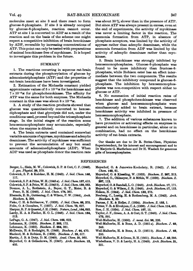
Vol. 49 RAT-BRAIN HEXOKINASE 347molecules meet at site 3 and there react to formglucose-6-phosphate. If site 3 is already occupiedby glucose-6-phosphate, reaction cannot occur.ATP at site 1 is converted to ADP as a result of thereaction and on the basis of the scheme one mightexpect a competitive inhibition of brain hexokinaseby ADP, reversible by increasing concentrations ofATP. This point can only be tested with preparationsof animal hexokinase free of myokinase. It is hopedto investigate this problem in the future.
SUMMARY1. The reactions occurring in aqueous rat-brain
extracts during the phosphorylation of glucose byadenosinetriphosphate (ATP) and the properties ofrat-brain hexokinase have been investigated.
2. Estimation of the Michaelis constant yieldedapproximate values of 5 x 10⁻⁶m for hexokinase and7 x 1O-4M for phosphohexokinase. The affinity forATP was the same for both enzymes. The Michaelisconstant in this case was about 8 x 1O-4M.
3. A study of the reaction products showed thatglucose was quantitatively converted to hexose-diphosphate and that glycolysis did not, under theconditions used, proceed beyondthe triosephosphatestage. In the initial stages of the reaction somehexosemonophosphate may accumulate, especiallywhen the enzyme is diluted.
4. The brain extracts used contained somewhatvariable amounts ofapyrase, myokinase and adenylicdeaminase. The activity of myokinase was sufficientto prevent the accumulation of any but smallamounts of adenosinediphosphate (ADP). WhenADP was used as phosphate donor the reaction rate
was about 30% slower than in the presence of ATP.But since ATP was always present in excess, even atthe end of experiments, the activity of myokinasewas never a limiting factor in the reaction. Theammonia formation from ATP, in absence ofphosphate acceptors, was limited by the activity ofapyrase rather than adenylic deaminase, while theammonia formation from ADP was limited by theactivity of adenylic deaminase rather than myo-kinase.
5. Brain hexokinase was strongly inhibited byhexosemonophosphates. Glucose-6-phosphate wasfound to be more inhibitory than fructose-6-phosphate, while Robison ester has an effect inter-mediate between the two components. The resultssuggest that the inhibitory compound is glucose-6-phosphate. The inhibition by hexosemonophos-phates was non-competitive with respect either toglucose or ATP.
6. No summation of initial reaction rates ofhexokinase and phosphohexokinase was observedwhen glucose and hexosemonophosphate weresimultaneously added to brain extract, becausehexokinase activity was inhibited in presence ofhexosemonophosphate.
7. The addition of various substances known tohave protective or activating effects on enzymes ingeneral and on hexokinase in particular, alone or incombination, had no effect on the hexokinaseactivity of rat-brain extracts.
Our thanks are due to Dr R. Strom-Olsen, PhysicianSuperintendent, for his interest and encouragement and toDr Mariorie G. Macfarlane and Dr H. Waelsch for generousgifts of hexosephosphates.
REFERENCESBerger, L., Slein, M. W., Colowick, S. P. & Coni, C. F. (1946).
J. gen. Phy8iol. 29, 379.Colowick, S. P. & Kalckar, H. M. (1943). J. biol. Chem. 148,
117.Colowick, S. P. & Price,W. H. (1945 a). J. biol. Chem. 157,415.Colowick, S. P. & Price,W. H. (1945 b). J. biol. Chem. 159,563.Dounce, A. L., Rothstein, A., Beyer, G. T., Meier, R. &
Freer, R. M. (1948). J. biol. Chem. 174, 361.Ebersole, E. R., Guttentag, C. & Wilson, P. W. (1944). Arch.
Biochem. 8, 399.Fiske, C. H. & Subbarow, Y. (1925). J. biol. Chem. 68, 375.Folin, 0. & Ciocalteu, V. (1927). J. biol. Chem. 73, 627.Harpur,R. P. & Quastel,J.H. (1949). Nature,Lond.,164,693.Lardy, H. A. & Fischer, H. 0. L. (1946). J. biol. Chem. 164,
513.LePage, G. A. (1947). J. biol. Chem. 168, 623.Lohmann, K. (1928). Biochem. Z. 203, 172.Lohmann, K. (1932). Biochem. Z. 254, 381.McIlwain, H. & Rodnight, R. (1949). Biochem. J. 44,470.Macleod, M. & Robison, R. (1929). Biochem. J. 28, 517.Martin, J. B. & Doty, D. M. (1949). Anal. Chem. 21, 965.Meyerhof, 0. & Geliazkowa, N. (1947). Arch. Biochem. 12,
405.
Meyerhof, 0. & Junowicz-Kocholaty, R. (1942). J. biol.Chem. 149, 61.
Meyerhof, 0. & Kiesling, W. (1933). Biochem. Z. 267, 313.Meyerhof, O., Ohlmeyer, P. & MShle, W. (1938). Biochem. Z.
297, 113.Meyerhof, 0. &Randall, L. 0. (1948). Arch. Biochem. 17,171.Meyerhof, 0. & Wilson, J. R. (1948). Arch. Biochem.17, 153.Nelson, N. (1944). J. biol. Chem. 153, 375.Neuberg, C., Lustig, H. & Rothenberg, M. A. (1943). Arch.
Biochem. 8, 33.Parnas, J. K. & HeIfer, J. (1924). Biochem. Z. 162, 1.Potter, V. R.&Elvehjem, C. A. (1936). J. biol. Chem.114,495.Roe, J. H. (1934). J. biol. Chem. 107, 15.Taylor, J. F., Green, A. A. & Cori G. T. (1948). J. biol. Chem.
173, 591.Weil-Malherbe, H. (1950). J. ment. Sci. 96, 226.Weil-Malherbe, H. & Bone, A. D. (1951 a). Biochem. J. 49,
348.Weil-Malherbe, H. & Bone, A. D. (1951 b). Biochem. J. 49,
355.Weil-Malherbe, H. & Green, R. H. (1951). Biochem. J. 49,286.Wiebelhaus, V. D. & Lardy, H. A. (1949). Arch. Biochem. 21,
321.


