Estrogens in males: what have we learned in the last 10 years?
18
Asian J Androl 2005; 7 (1): 3-20 . 3 . Estrogens in males: what have we learned in the last 10 years? Vincenzo Rochira, Antonio R. M. Granata, Bruno Madeo, Lucia Zirilli, Giuseppina Rossi, Cesare Carani Integrated Department of Medicine and Medical Specialties, University of Modena and Reggio Emilia, Modena 41100, Italy Abstract This review focuses on the role of estrogen in men, mainly in male reproduction. The continuing increase in data obtained, and recent discoveries in this area will enable a better understanding of male physiology; these, in turn, will have important clinical implications. (Asian J Androl 2005 Mar; 7: 3–20) Keywords: estrogen; male sexual behavior; male infertility; gonadotropin feedback . Review . Correspondence to: Vincenzo Rochira, Cattedra di Endocrinologia, Dipartimento Integrato di Medicine e Specialità Mediche, Via del Pozzo, 71, 41100 Modena, Italy. Tel: +39-59-422-224, Fax: +39-59-363-114 E-mail: [email protected] Received 2004-08-02 Accepted 2005-01-04 1 Historical background Traditionally the adult male reproductive function was considered to be controlled by both gonadotropins and androgens, and it was testosterone [1], which for many years was considered the “male hormone”. In 1934 the conversion of the male hormone into the female one in male stallions was postulated, leading to the first step in considering testosterone as a prohormone for estrogens [2, 3]. This may be considered the ancestor for the de- velopment of a field of research focusing on the biologi- cal significance of estrogens in men. Nevertheless, tes- tosterone was not considered a prohormone for estra- diol with certainty until the 1950s [4–8] and detailed stud- ies on circulating sex steroids were provided later in the 1960s [9]. Well-established evidence for testosterone conversion into estradiol in the human male was not pro- vided till the 1970s, when MacDonald et al. described estrogens synthesis in peripheral tissues of normal men [10]. A major step forward was also made in the 1970s when it was demonstrated that the testes are a source of estrogens in men [11, 12]. In the meantime, further re- search on this issue has demonstrated that both imma- ture and mature germ cells and spermatozoa, are all able to produce estrogens [13–16]. The development of testicular paracrinology between 1980s and 1990s played a significant role in the study of estrogens in the male reproductive function [17–19]: the interest in understanding in detail the regulatory role of each paracrine substance in the complicated hormonal network in the testicular milieu, as well as the “mystery” of cell-to-cell interaction in the testis, provided strong stimuli to researchers. In this view, starting from bio- chemical studies on aromatization of androgens to estro- gens [20–29], the following immunocytochemical stud- ies on both aromatase [30–33] and estrogen receptors (ER) [34–39] directed the interest of biologists to the significance of estrogens signaling the pathway in the male reproductive system. Progress in molecular biol- ogy leaded to gene cloning for aromatase [40–42], ERα [43–45] and ERβ [46–48] gene cloning, this being a cru- cial step for the following deep investigation of estro- gens physiology in the male. In fact the studies on aromatase [42] and ER functions [49, 50], together with corresponding gene-expression studies on aromatase [51–54], on ERα [55] and on ERβ [55, 56] have pro- gressively clarified the relationships between estrogens and male reproductive function. For a long time, data on the effects of estrogens on © 2005, Asian Journal of Andrology, Shanghai Institute of Materia Medica, Chinese Academy of Sciences. All rights reserved. DOI: 10.1111/j.1745-7262.2005.00018.x
-
Upload
independent -
Category
Documents
-
view
3 -
download
0
Transcript of Estrogens in males: what have we learned in the last 10 years?

Asian J Androl 2005; 7 (1): 3-20
.3.
Estrogens in males: what have we learned in the last 10 years?
Vincenzo Rochira, Antonio R. M. Granata, Bruno Madeo, Lucia Zirilli, Giuseppina Rossi, Cesare Carani
Integrated Department of Medicine and Medical Specialties, University of Modena and Reggio Emilia, Modena 41100, Italy
Abstract
This review focuses on the role of estrogen in men, mainly in male reproduction. The continuing increase in dataobtained, and recent discoveries in this area will enable a better understanding of male physiology; these, in turn, willhave important clinical implications. (Asian J Androl 2005 Mar; 7: 3–20)
Keywords: estrogen; male sexual behavior; male infertility; gonadotropin feedback
.Review .
Correspondence to: Vincenzo Rochira, Cattedra di Endocrinologia,Dipartimento Integrato di Medicine e Specialità Mediche, Via delPozzo, 71, 41100 Modena, Italy.Tel: +39-59-422-224, Fax: +39-59-363-114E-mail: [email protected] 2004-08-02 Accepted 2005-01-04
1 Historical background
Traditionally the adult male reproductive function wasconsidered to be controlled by both gonadotropins andandrogens, and it was testosterone [1], which for manyyears was considered the “male hormone”. In 1934 theconversion of the male hormone into the female one inmale stallions was postulated, leading to the first step inconsidering testosterone as a prohormone for estrogens[2, 3]. This may be considered the ancestor for the de-velopment of a field of research focusing on the biologi-cal significance of estrogens in men. Nevertheless, tes-tosterone was not considered a prohormone for estra-diol with certainty until the 1950s [4–8] and detailed stud-ies on circulating sex steroids were provided later in the1960s [9]. Well-established evidence for testosteroneconversion into estradiol in the human male was not pro-vided till the 1970s, when MacDonald et al. describedestrogens synthesis in peripheral tissues of normal men[10]. A major step forward was also made in the 1970swhen it was demonstrated that the testes are a source of
estrogens in men [11, 12]. In the meantime, further re-search on this issue has demonstrated that both imma-ture and mature germ cells and spermatozoa, are all ableto produce estrogens [13–16].
The development of testicular paracrinology between1980s and 1990s played a significant role in the study ofestrogens in the male reproductive function [17–19]: theinterest in understanding in detail the regulatory role ofeach paracrine substance in the complicated hormonalnetwork in the testicular milieu, as well as the “mystery”of cell-to-cell interaction in the testis, provided strongstimuli to researchers. In this view, starting from bio-chemical studies on aromatization of androgens to estro-gens [20–29], the following immunocytochemical stud-ies on both aromatase [30–33] and estrogen receptors(ER) [34–39] directed the interest of biologists to thesignificance of estrogens signaling the pathway in themale reproductive system. Progress in molecular biol-ogy leaded to gene cloning for aromatase [40–42], ERα[43–45] and ERβ [46–48] gene cloning, this being a cru-cial step for the following deep investigation of estro-gens physiology in the male. In fact the studies onaromatase [42] and ER functions [49, 50], together withcorresponding gene-expression studies on aromatase[51–54], on ERα [55] and on ERβ [55, 56] have pro-gressively clarified the relationships between estrogensand male reproductive function.
For a long time, data on the effects of estrogens on
© 2005, Asian Journal of Andrology, Shanghai Institute of Materia Medica, Chinese Academy of Sciences. All rights reserved.
DOI: 10.1111/j.1745-7262.2005.00018.x

.4.
Estrogens in the human male
the male reproductive system have been limited to theprenatal period, as the developing testis was consideredto be responsive to estrogens [57, 58]. A direct cause-effect relationship between the exposure to high dosesof estrogens or diethylstilbestrol (DES) and malforma-tion of male reproductive structures was known in bothanimals [59] and humans [60–62] from the 1950sonwards. However, the role played by estrogens in malereproduction became known only in the late 1990s[63, 64]. The revelations started with the developmentof lines of male transgenic mice lacking functional ERα[65, 66] or β [67] or a functional aromatase enzyme [68].This shed new light on the role of estrogens in male re-production [69]. Concomitantly, the discovery of muta-tions in both the human ERα [70] and aromatase [71]genes have reinforced the idea that estrogens play a keyrole in the human male reproductive system.Accordingly, since the 1980s it was known that seminalfluid contains both sex steroids: testosterone and estro-gens [72–75].
Previously a role for estrogen action in the male re-productive system had being proposed based on scat-tered data [61, 62], but recent advances came out fromin vitro, in vivo and immunohistochemical studies thathave begun to elucidate the mechanisms of estrogen ac-tion on the male reproductive tract [16, 76, 77].
2 Introduction
Today the concept that estrogens are essential forbone maturation and mineralization in both men andwomen is well established [71, 78]; however, physiciansstill do not accept the idea that estrogens may also regu-late human male reproduction.
It seems paradoxical that estrogen, the "femalehormone", may play a critical role in human malereproduction, even though a clear demonstration of theneed of estrogen for normal fertility has been obtained inrodents [69, 79, 80]. Accordingly, the discovery ofmutations in both the human ERα [70] and aromatase[71] genes fits largely with data obtained from severallines of estrogen deficient mice. Anyhow, at present, acertain cause-effect relationship between congenital es-trogen deficiency and abnormal fertility in men has notyet been determined, and it remains a hard issue to trans-pose to the human male what we have learned from theanimal. Surely in the future, increasing cases of con-genital estrogen deficiency in men will help to elucidatewhether or not the congenital lack of estrogenss is re-lated to reduced fertility in men. At the moment, the
increasing body of evidence on the importance of estro-gens on male reproductive function have led to the ap-pearance of comprehensive chapters about estrogensand male reproduction in some textbook of endocrinol-ogy [81, 82], underlining the impact of this issue on bothexperimental studies [80] and clinical practice [77].
3 Estrogens and the male reproductive system
The immunocytochemical studies performed on themale reproductive structures of rodents and men revealedthe areas in which ERs and aromatase enzymes areexpressed and are functionally active. A different patternexists between rodents and human males [69, 80, 81].
The site of ERs and aromatase expression varieswidely during development in rodents and both aromataseand ERs are expressed at a very early stage [81, 83].ERα is abundant in the developing efferent ductules, aswell as in the mature ones [15, 80, 81], leading to the ideathat ERα may be crucial for lifelong male reproductionin rodents. Even in the rodent testes, ERα is expressedearly by Leydig cells when the androgen receptor is notyet expressed. In contrast, ERβ expression prevails duringfetal life, suggesting a major role for ERβ in the develop-ment and function of male reproductive structures untilbirth [83]. However β-ERKO mice display normal fer-tility and reproductive structures nothwistanding non-functioning ERβ, thus leaving partially unknown the sig-nificance of ERβ during the development of male repro-ductive system [67, 69]. Probably ERβ may be the tar-get when the fetus is exposed to a supraphysiologicalamount of estrogens and may be enrolled leading to nega-tive effects on reproductive structures. Accordingly, itwas known as early as the 1930s that prenatal exposureto estrogens interferes with the normal development oftestes and the reproductive structures of some species[57]. In the seminiferous epithelium (Sertoli cells and afew germ cells) and in the epididymis of the male fetus,ERβ expression is higher than ERα, the latter being ab-sent or very low [69, 84].
ERβ is involved in estrogen-related apoptosis of germcells and as a consequence in the blockade of germ celllineage growth during fetal and neonatal life [85]. Thus,ERβ may take part in the process through which expo-sure to environmental estrogens produce negative effectson male reproduction. Finally, the finding of botharomatase and ERs in the developing fetal testes impliesa possible involvement of estrogens in the process ofdifferentiation and maturation of rodent testes duringprenatal life starting from an early stage of morpho-
Asian J Androl 2005; 7 (1): 3-20
.5.
genesis, albeit ERβ playing a more significant role thanERα.
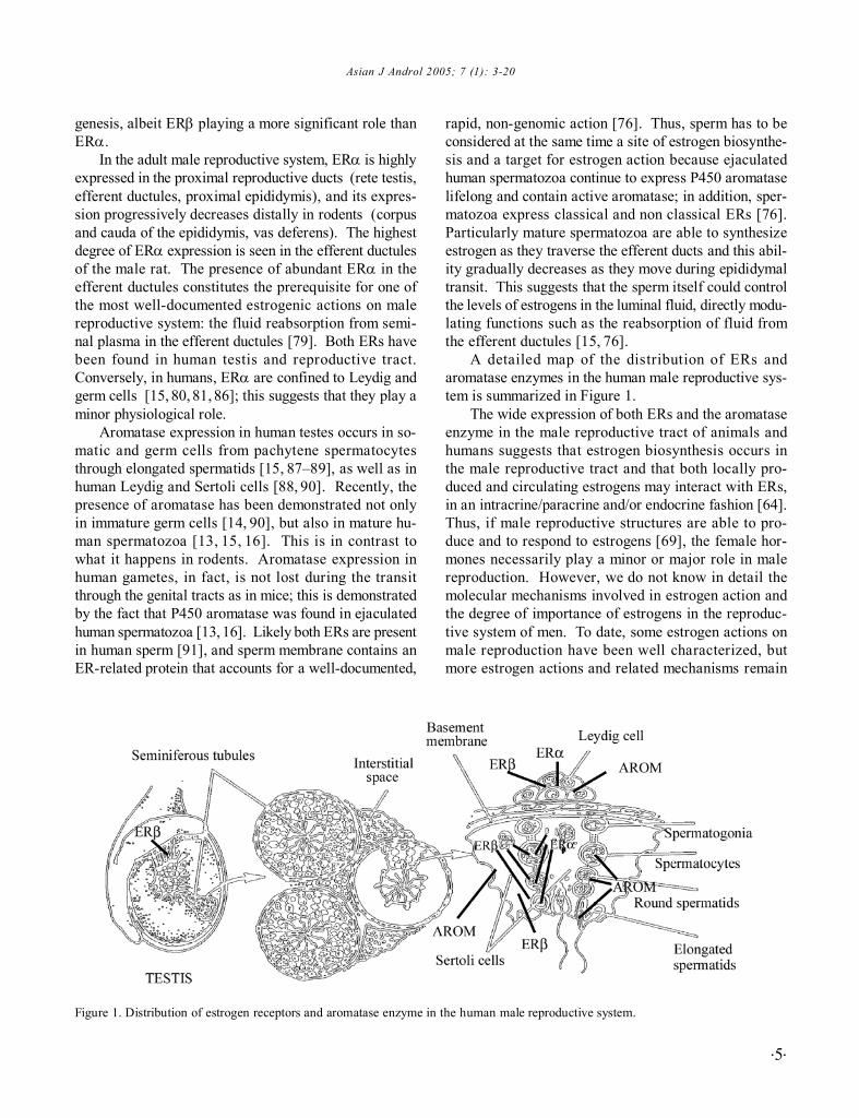
In the adult male reproductive system, ERα is highlyexpressed in the proximal reproductive ducts (rete testis,efferent ductules, proximal epididymis), and its expres-sion progressively decreases distally in rodents (corpusand cauda of the epididymis, vas deferens). The highestdegree of ERα expression is seen in the efferent ductulesof the male rat. The presence of abundant ERα in theefferent ductules constitutes the prerequisite for one ofthe most well-documented estrogenic actions on malereproductive system: the fluid reabsorption from semi-nal plasma in the efferent ductules [79]. Both ERs havebeen found in human testis and reproductive tract.Conversely, in humans, ERα are confined to Leydig andgerm cells [15, 80, 81, 86]; this suggests that they play aminor physiological role.
Aromatase expression in human testes occurs in so-matic and germ cells from pachytene spermatocytesthrough elongated spermatids [15, 87–89], as well as inhuman Leydig and Sertoli cells [88, 90]. Recently, thepresence of aromatase has been demonstrated not onlyin immature germ cells [14, 90], but also in mature hu-man spermatozoa [13, 15, 16]. This is in contrast towhat it happens in rodents. Aromatase expression inhuman gametes, in fact, is not lost during the transitthrough the genital tracts as in mice; this is demonstratedby the fact that P450 aromatase was found in ejaculatedhuman spermatozoa [13, 16]. Likely both ERs are presentin human sperm [91], and sperm membrane contains anER-related protein that accounts for a well-documented,
rapid, non-genomic action [76]. Thus, sperm has to beconsidered at the same time a site of estrogen biosynthe-sis and a target for estrogen action because ejaculatedhuman spermatozoa continue to express P450 aromataselifelong and contain active aromatase; in addition, sper-matozoa express classical and non classical ERs [76].Particularly mature spermatozoa are able to synthesizeestrogen as they traverse the efferent ducts and this abil-ity gradually decreases as they move during epididymaltransit. This suggests that the sperm itself could controlthe levels of estrogens in the luminal fluid, directly modu-lating functions such as the reabsorption of fluid fromthe efferent ductules [15, 76].
A detailed map of the distribution of ERs andaromatase enzymes in the human male reproductive sys-tem is summarized in Figure 1.
The wide expression of both ERs and the aromataseenzyme in the male reproductive tract of animals andhumans suggests that estrogen biosynthesis occurs inthe male reproductive tract and that both locally pro-duced and circulating estrogens may interact with ERs,in an intracrine/paracrine and/or endocrine fashion [64].Thus, if male reproductive structures are able to pro-duce and to respond to estrogens [69], the female hor-mones necessarily play a minor or major role in malereproduction. However, we do not know in detail themolecular mechanisms involved in estrogen action andthe degree of importance of estrogens in the reproduc-tive system of men. To date, some estrogen actions onmale reproduction have been well characterized, butmore estrogen actions and related mechanisms remain
Figure 1. Distribution of estrogen receptors and aromatase enzyme in the human male reproductive system.

.6.
Estrogens in the human male
to be elucidated in detail.
4 The role of estrogens on male reproduction
The previously unsuspected physiological role ofestrogens in the testicular function of animals was re-vealed by the creation of estrogen-deficient mice. Linesof estrogen-deficient mice represent a useful experimen-tal model obtained by genetic manipulation which con-sists in the knock-out of a single gene, resulting in a nonfunctioning product (enzyme or receptor) in the offspring.Gene inactivation generated four different lines of estro-gen-deficient knock-out mice (Table 1). The knock-outof genes encoding for ERs led to the following lines ofestrogen-resistant mice: 1) the α-estrogen receptor knockout (ERKO) mice, in which the gene encoding for theERα is disrupted; 2) the β-ERKO mice, with an inacti-vated ERβ; and 3) the αβ-ERKO mice, in which bothreceptors α and β are non-functioning. The α-ERKO,β-ERKO and αβ-ERKO mice provide helpful informa-tion regarding the loss of ERs function (estrogenresistance). The fourth line is that of aromatase knock-out (ArKO) mice in which the gene encoding for thearomatase enzyme is knocked-out with undetectable cir-culating estrogens from birth, provides an experimentalanimol model useful for studying the effects of the con-genital lack of both circulating and locally producedestrogens. The reproductive phenotype of estrogen-de-ficient knock-out mice is summarized in Table 1.
Adult, sexually mature, male α-ERKO mice are in-
fertile even though the development of the male repro-ductive tract is mainly unaffected [69]. Seminiferousepithelium is atrophic and degenerating, while tubules andrete testes are both dilated [92]. Testicular histology isnormal at birth and starts to degenerate when the mouseis 20–30 days old. At 40–60 days, testicular histologyshows very dilated tubules, an increase in testicular vol-ume and atrophy of the seminiferous epithelium [69]. Inα-ERKO mice, fluid absorption is reduced at the level ofthe efferent ducts [79] and a defect partially mimickedalso by the administration of anti-estrogens in wild-typemice with a similar effect on testicular histology [79,93–95]. In the male genital tract, the highest concentra-tion of ERα is found in the efferent ducts [94], and theestrogen-dependent fluid reabsorption in this site prob-ably results from estrogen interaction with the ERα dur-ing prenatal development [79, 92, 96]. The lack of fluidreabsorption in the efferent ductules of α-ERKO malemice and the consequent dilatation of these ductules in-duces a retroactive progressive swelling of the seminif-erous tubules. The damage of seminiferous tubule isdue to increased fluid back-pressure and it leads to se-verely impaired spermatogenesis, coupled with testicu-lar atrophy, as clearly seen at the age of 150 days [69,79] (Table 1). In addition, the pattern of reproductivehormone profiles is peculiar in α-ERKO male mice: se-rum Luteinizing hormone (LH) is increased and as a result,serum testosterone is higher and Leydig cells hyperpla-sia is present, but with normal Follicle-stimulating hor-mone (FSH) [65, 66, 69] (Table 2).
Table 1. Reproductive phenotypes of ERKO and ArKO mice.Estrogen deficientknock-out male miceα-ERKO
β-ERKO
α-βERKOArKO
Fertility pattern
Infertility starting at 30 days ofage
Normal fertility
Similar to αERKO MouseNormal fertility until 7 months,fertility decreases with advanc-ing age.Infertility at the age of 1 year.
Testicular histology
At birth: normal in adult mice:germ cell deprivation with dilatedseminiferous tubules and atrophyof the seminiferous epithelium.Normal testicular histology at birthand in adult mice. Increased num-ber of germ cells at birth.Similar to αERKO miceAt birth: normal until 14 wk.In adult mice (age > 1 year): im-paired spermatogenesis with arrestof spermatogenesis
Mechanism of induction of infer-tilityReduced fluid absorption at levelof the efferent ducts.Impaired expression of the Na+
transported NHER–
–- Failure of germ cell differentiation;- Need of estrogens for sperm maturation through the reproductivetract;- Temporary compensatory effectof estrogens in diet.
ERKO: estrogen receptor knock-out; ArKO: aromatase knock-out.

Asian J Androl 2005; 7 (1): 3-20
.7.
Studies performed on α-ERKO mice established notonly the role of estrogens on male reproduction, but alsohighlighted a previously unknown physiological functionof efferent ductules. Thus, efferent ductules, other thanproviding an anatomic connection between rete testesand the epididymis, which is useful for sperm transitthey constitute also a functional structure in which about90 % of sperm fluid is reabsorbed. In this view, efferentductules regulate sperm concentration, which becomeshigher in the ducts prior to entry into the epididymis [97].As a matter of fact, the histology of the efferent duct isvery close to that of the proximal tubules of the kidney[98]. It is likely that the fluid reabsorption in the efferentductules is mediated through the Na+ transporter, namedNHE3; the disruption of ERα or the use of anti-estro-gens resulted in decreased expression of NHE3 mRNA,as well as in a decrease of other proteins involved inwater reabsorption, such as acquaporin I [99, 100].
Data from the study of ArKO [68] and β-ERKO [67]on male mice supports the idea that estrogen actions onthe male reproductive tract are more complex than pre-viously thought on the basis of the only knowledge of α-ERKO mice physiology [69]. In fact, unlike α-ERKOmice, male ArKO mice are initially fully fertile [68], butfertility decreases with advancing age [101], conversely,β-ERKO mice are fully fertile and apparently have nor-mal reproductivity also in adulthood [67] (Table 1). ArKOmice show an abnormal pattern of circulating gonadot-ropins according to the absence of the estrogen-depen-dent inhibitory effect at the pituitary level (Table 3), whilehormonal pattern in β-ERKO mice is less clear (Table 2).The reproductive phenotype of αβ-ERKO mice is veryclose to that of α-ERKO mice and it is characterized byinfertility and enlarged seminiferous tubules [69] (Table
1). The modification of the testicular histology of thetestes in male ArKO starts at 7 months and, after 1 year,a complete arrest of spermatogenesis is evident at thelevel of early spermatid and Leydig cell hyperplasia, with-out significant changes in the volume of seminiferoustubule lumen [101] (Table 1). Surely, the mechanisminvolved in the development of infertility is different inArKO if compared with α-ERKO male mice, becausethe early arrest of spermatogenesis suggests a failure ingerm cell differentiation, probably due to the lack of es-trogens in the testicular environment in the first, whilereduced fluid reabsorbion occurs in the second. Thesefindings, together with the observation that β-ERKO malemice are fully fertile [67], lead to the hypothesis thatestrogen activity in the male reproductive tract differs,with regard to both the types of ERs involved in the path-way of estrogenic action, and the site of action throughthe male reproductive tract [69]. Accordingly, in ArKOmale mice, the failure of germ cell differentiation that isprobably related to the lack of estrogen action on semin-iferous epithelium while αER disruption and related ar-rest of fluid reabsorption take place in the efferent ductulesof α-ERKO mice [102]. In very young ArKO mice sper-matogenesis is preserved because a small amount ofestrogens, such as those introduced with the diet, prob-ably is sufficient to promote germ cell maturation for abrief period. Thus, the degree of infertility is less severein ArKO mice than in α-ERKO; since ligand independentER pathways remains functionally active [69] in ArKOmice. Later, the continuous lack of estrogens causessperm abnormalities with advancing age in ArKO mice,since estradiol is probably necessary to maintain sper-matogenesis and promote normal sperm maturation, bothin the seminiferous epithelium and through the repro-
Table 2. Reproductive phenotype in estrogen-receptor disruption: a comparative analysis between mice and men.
Testis
Germ cells
Sperm characteristics
FertilityHormonal Pattern
α-ERKOGerm cells loss; enlargedseminiferous tubuleNormal development of germ cellswhen transplanted in the WTReduced number; motility andfertilizing capacityInfertileLH ↑FSH =T ↑E2 ↑
β-ERKO miceNormal
Not described
Normal sperm count
FertileLH =FSH =T =E2 =
Estrogen resistance in men (ERα)Normal volume (20–25 mL)
Not described
Normal sperm count (25×106.mL–1)Reduced viability (18 %)Fertile?LH ↑FSH ↑T ↓=
E2 ↑T = testosterone; E2 = estradiol

.8.
Estrogens in the human male
ductive tract [15, 90, 101]. Accordingly, recent findingsfrom in vitro studies on human germ cells treated withestrogens, suggest that estradiol may serve as a survivalfactor for round spermatids and that the lack of estradiolmay promote apoptosis with a resulting failure in elon-gated spermatid differentiation [103]. Again, ERα is notpresent in the seminiferous epithelium, and the presenceof ERα in Sertoli cells does not impair normaldevelopment, as shown in β-ERKO mice which are fullyfertile [69].
The above studies support the concept that a func-tional ERα, but not ERβ, is needed for the developmentand the maintenance of a normal fertility in male mice[67, 69, 79, 92]. Recently, another estrogen function hasbeen postulated on the basis of the finding that estrogensprobably regulate cell-to-cell adhesion in the testis andmay play a role in the establishment of Sertoli-germ cellstructural connection [104]. Clearly, further studies areneeded to fully understand the precise role of estrogensand their receptors on both spermatogenesis and func-tion of seminal way as well as the importance of intracrineand paracrine pathways for these effects. It has to beremarked, however, that results from mice lacking func-tional ERs or aromatase enzyme point to an importantrole for estrogens in the maintenance of mating behaviorin male mice, and that infertility in α-ERKO, αβ-ERKOand ArKO mice are, at least partially, due to weekness ofvarious aspects of mating behavior just at early age[69, 80]. Sexual behavior, in fact, is strictly linked toreproductive functions, and if estrogens modulate mat-ing behavior, they also necessarily affect reproductive
outcomes in an indirect way.Many studies involving rodents suggest that inap-
propriate exposure to estrogens in utero and during theneonatal period impairs testicular descent, efferent ductulefunction, the hypothalamic-pituitary-gonadal axis, andtesticular function [57, 60, 62, 64, 69]. Hence, a role forestrogens in the development of male reproductive struc-tures has been largely supported by several studies. Inboth rodents and various animal species, prenatal expo-sure to diethylstilbestrol, a synthetic potent estrogeniccompound, led to an abnormal development of male re-productive structures. It seems that both a delay inMüllerian duct formation or an incomplete Müllerian ductregression, with a female-like differentiation of the non-regressed caudal part may account for abnormal sexstructures at birth, after estrogens excess in prenatal life[105]. Accordingly, an increase in the expression of anti-Müllerian-hormone (AMH) mRNA, which is not accom-panied by a regression of the ducts, may be involved inmale mice fetuses exposed to diethylstilbestrol (DES).Certainly, the timing of DES exposure is crucial for theinduction of abnormalities of Müllerian duct developmentand regression [57, 105]. In animals, exposure to estro-gens excess in the neonatal period leads topermanentchanges in testis function and spermatogenesis, with re-sulting reduced fertility into adulthood [64, 80]. Theconcept that estrogens excess may impair fertility hasbeen extended also to men, and an excess of environ-mental estrogens has been related to impaired fertility[61, 106]. A decline in the sperm count of men in West-ern countries has coincided with a progressive increase
Table 3. Reproductive phenotype in aromatase deficiency, a comparative analysis between mice and men.
Testis
Germ cells
Sperm characteristics
FertilityHormonal pattern
ARKO miceSimpson et al.
1997[53]Normal at14 wkDisruption of sper-matogenesis at 1 of ageReduced sperm countand decreased viabilityat 8 month of ageInfertile at 1 yearLH ↑FSH =T ↑E2 Undetect.
Morishima et al.1995[117]
Increased Volume34 mL
Not described
Not studied
Not studiedLH ↑FSH ↑T ↑E2 Undetect.
Carani et al.1997[118]
Normal Volume8 mLGerm cell arrest atspermatocyte levelSevere oligozoosper-mia absent motility
InfertileLH = ↑FSH ↑T =E2 Undetect.
Herrmann et al.2002[120]
Normal Volume14 mLNot described
Oligo-astenoZoospermia
Fertile?LH =FSH ↑T ↑E2 Undetect.
Maffei et al.2004[116]
Normal Volume 10-11mL CryptorchidismComplete germ celldepletionNot studied
InfertileLH =FSH ↑T =E2 Undetect.
Aromatase deficiency in men
T = testosterone; E2 = estradiol; Undetect. = undetectable
Asian J Androl 2005; 7 (1): 3-20
.9.
in environmental estrogens [61, 106–108]. In the past,uncorrect clinical use of DES by pregnant women in-creased the incidence of genital malformations at birth[107]. The most frequent structural and functional ab-normalities reported were: epididymal cysts, meatalstenosis, hypospadias, cryptorchidism and microphal-lus [106-111]. The frequency of abnormalities was de-pendent on the timing of estrogen exposure, and it washigher when DES was taken before the 11th week ofgestation (i.e. the time of Müllerian duct formation)[109–111]. These data support the previously discussedhypothesis that the asynchrony between formation andregression of embryonal reproductive structures is de-termined by estrogen exposure (e.g. the presence ofMüllerian duct remnants) [105].
In the past, various reports have also demonstratedthat the quality of the semen of men exposed to DES inutero is significantly worse than that of unexposed con-trols [110, 111], although no clear condition of subfertilityor clinical infertility has been evident [62]. While vari-ous studies suggest that environmental estrogens affectmale fertility in animal models, the implications for hu-man spermatogenesis are less clear [112]. Exogenousestrogens could interfere with the development of thegenita l structures if administered during ear lyorganogenesis, by both leading to an impairment of go-nadotropin secretion and an imbalance in the androgen-tos to estrogens ratio, which may account for impairedandrogen receptor stimulation or inhibition, according tothe dose, the cell type and the age [107, 108, 113, 114].
The role of estrogens in male reproductive structuredevelopment remains conflicting. Animal studies sug-gest that exposure to excessive amounts of estrogen maynegatively affect the development of male reproductiveorgans. However,these effects are considered to be theresult of an impaired hypothalamic-pituitary function, asa consequence of estrogen excess and of the concomi-tant androgen deficiency [113, 114]. Much of the knowl-edge on estrogen overexposure and human fertility isinferred from animal data, and the validity of these con-cepts has not been established in men.
The negative effects of estrogen excess during fetallife are well documented, but we do not know if con-genital estrogen deprivation may affect the developmentof male reproductive structures. Mouse models of con-genital estrogen deficiency show a normal male repro-ductive structure, suggesting that congenital lack of es-trogen activity does not affect the development of malereproductive organs in animals [69, 80]. Anyhow, somedefects in the development of the efferent ductules in α-
ERKO mice are thought to be a consequence of a con-genital absence of estrogen action [93], such as a defectin cremaster muscle development [115]. Bilateral cryp-torchidism was present in one patient with aromatase de-ficiency [116], suggesting a possible role of estrogens intestis descent, although this was not seen in the transgenicmice models. The presence of a unique case of cryp-torchidism among men with aromatase deficiency, doesnot permit to draw any conclusions about a possible rela-tionship between estrogen deficiency and the occurrenceof abnormalities in testis development and descent.
Congenital estrogen deficiency in men is the result ofnaturally occurring inactivating mutations of both thearomatase gene (aromatase deficiency) [71] or of the ERαgene [70]. To date, five subjects with aromatase defi-ciency have been described (four adult men and one maleinfant) (Table 4) [116–120], and only a unique case ofestrogen resistance is still known [70]. Many clinicalaspects are shared by both the estrogen-resistant manand the four adult men with aromatase deficiency, butthe possible occurrence of infertility has not been reportedin all of them [71, 81, 116–118, 120–121]. The demon-stration of abundant ERs in human efferent ducts andaromatase activity in human sperm, indicates the involve-ment of estrogens in the reproductive function of men.On the other hand, data from human subjects with con-genital estrogen deficiencies have provided conflicting andsomewhat confusing results. Even though it is hard totransfer what we have learned from models of estrogen-deficient mice to men, the comparison of rodents andhuman reproductive phenotypes (Tables 2, 3) may behelpful in resolving questions regarding the role of estro-gens in male reproductive systems. Again the reproduc-tive phenotype of rodents seems to resemble, at least inpart, that of patients with naturally occurring mutationsin their ERα or aromatase gene (Tables 2, 3).
The only man know to be estrogen resistant had re-duced sperm motility [70], but normal sperm count;however, α-ERKO mice show an impairment of bothsperm count and sperm viability (Table 2). The fouradult men affected by congenital aromatase deficiencyshowed a variable degree of impaired spermatogenesis[121]. A severely reduced sperm count and an impair-ment of sperm viability with germ cell arrest at the levelof primary spermatocytes, was found in one subject [71].A complete germ cell arrest was shown at the testicularbiopsy in a second subject, whose semen analysis wasunfortunately not available: the patient refused the analysis,according to his religious believe [116] (Table 3). A thirdpatient had a slightly reduced sperm count and reduced

.10.
Estrogens in the human male
sperm viability [120] (Table 3). There is no relevant dataconcerning the fourth patient described because thesperm count was not obtained [117]. It should be notedthat impaired sperm motility is the main feature in bothα-estrogen resistant man and mice, and that germ cellarrest is the main feature in both aromatase-deficient menand mice (Table 2, 3). Thus, the possible associationbetween the lack of estrogen activity and infertility inmen–which is suggested by the constant finding of ab-normal spermatogenesis in men with congenital estro-gen deficiency, together with reproductive abnormalitiesin estrogen-deficient mice – discloses the important roleplayed by estrogens on male reproductive function.
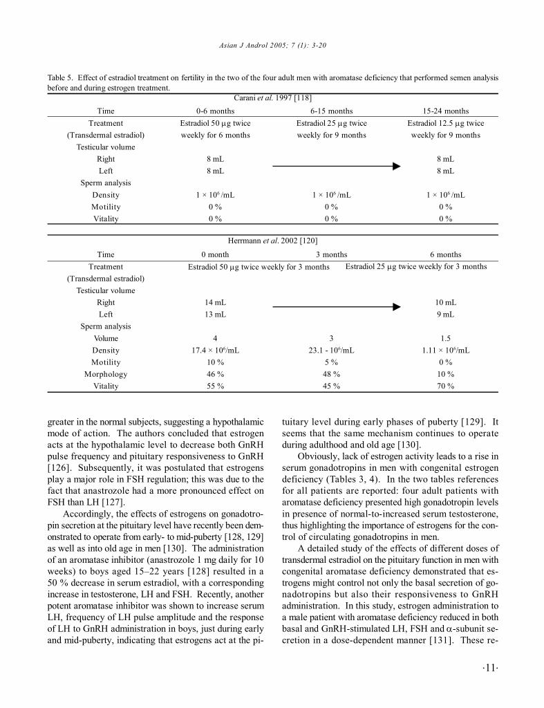
The effects of estrogen replacement treatment onspermatogenesis are available only in two of the fouradult men with aromatase deficiency. In both, estrogenadministration did not improve neither sperm count normotility (Table 5) [71, 118, 120]. In the patient describedby Herrmann et al., estrogen treatment resulted in a de-cline in the sperm count and a decrease in the testisvolume, probably as a consequence of LH and FSH inhi-bition [120], but these data should be interpreted withcaution.
The variable degree of fertility impairment in menwith congenital deficiencies of estrogen action or syn-thesis means that there is uncertainly as to whether thesefeatures are a consequence of a lack of estrogen actionor only epiphenomena, even though a possible role ofestrogens on human spermatogenesis is suggested byrodent studies. Our knowledge on the role of estrogensin human male reproduction is far from complete, andthe issue is more complex, if we consider that excessiveexposure to environmental estrogens is a possible causeof impaired fertility [61, 106]. Thus, it is difficult toreconcile existing data about effects of both estrogendeficiency and excess on male reproductive function [61,63, 106, 122].
The recent discovery of an inactivating mutation ofthe aromatase gene in a male infant [118], together withnew cases of infant or adult men with congenital estro-
gen deficiency will shed new light on this controversialissue. Certainly, better comprehension of the natural his-tory of human estrogen deficiency will improve ourknowledge about the role of estrogens in male fertility.
5 Regulation of gonadotropin feedback
Animal and human models of congenital estrogendeficiency provide further evidence on the role of estro-gens on gonadotropin secretion [71, 78], thus confirm-ing that aromatization of testosterone is required for thenormal functioning of the hypothalamic-pituitary testicularaxis.
Previous data obtained from gonadotrophin-releas-ing hormone (GnRH)-deficient males treated with test-osterone alone, testosterone plus testolactone (anaromatase inhibitor), or estradiol [123, 124], are available.Since longtime, these studies showed that the addition ofthe aromatase inhibitor completely prevented the sup-pression of gonadotropin secretion classically inducedby testosterone, in both normal and GnRH-deficient men,thus revealing a direct and an indirect effect (througharomatization) of androgens. These studies demonstratedan important direct inhibitory effect of estradiol on go-nadotropin secretion in both the GnRH-deficient and nor-mal men, and support the concept that at least part of theinhibitory effect on gonadotropin secretion is mediatedby the conversion of testosterone to estradiol at the pitu-itary level [123, 124]. In contrast, it seems that the 5α-reduction of testosterone in DHT does not play an im-portant role in the pituitary secretion of gonadotropins[125].
More recently, a hypothalamic site of estrogen ac-tion has been demonstrated in men. In order to clarifythe role of estrogen on the feedback regulation of gona-dotropin secretion at the hypothalamic level, Hayes et al.[126] showed that the aromatase inhibitor, anastrozole,led to an increase in the mean gonadotropin levels, inboth normal men and men affected by idiopathichypogonadotropic hypogonadism (IHH); the increase was
Table 4. Human aromatase deficiency: summary of the 5 described cases.Author
Morishima et al., 1995[117]Carani et al., 1997 [118]
Deladoey et al., 1999 [119]Maffei et al., 2001 [116]
Herrmann et al., 2002 [120]
Age (years)2431<128
27
LocationNew YorkModena
BernBuenos Aires
Essen
Affected exonIXIXVV
VI
MutationArg375-CysArg365-Gln
Leu157XDC-stopNucleotide 628 G to A
insertion of 10 aa and stopC to A in splicing acceptor site
Asian J Androl 2005; 7 (1): 3-20
.11.
greater in the normal subjects, suggesting a hypothalamicmode of action. The authors concluded that estrogenacts at the hypothalamic level to decrease both GnRHpulse frequency and pituitary responsiveness to GnRH[126]. Subsequently, it was postulated that estrogensplay a major role in FSH regulation; this was due to thefact that anastrozole had a more pronounced effect onFSH than LH [127].
Accordingly, the effects of estrogens on gonadotro-pin secretion at the pituitary level have recently been dem-onstrated to operate from early- to mid-puberty [128, 129]as well as into old age in men [130]. The administrationof an aromatase inhibitor (anastrozole 1 mg daily for 10weeks) to boys aged 15–22 years [128] resulted in a50 % decrease in serum estradiol, with a correspondingincrease in testosterone, LH and FSH. Recently, anotherpotent aromatase inhibitor was shown to increase serumLH, frequency of LH pulse amplitude and the responseof LH to GnRH administration in boys, just during earlyand mid-puberty, indicating that estrogens act at the pi-
tuitary level during early phases of puberty [129]. Itseems that the same mechanism continues to operateduring adulthood and old age [130].
Obviously, lack of estrogen activity leads to a rise inserum gonadotropins in men with congenital estrogendeficiency (Tables 3, 4). In the two tables referencesfor all patients are reported: four adult patients witharomatase deficiency presented high gonadotropin levelsin presence of normal-to-increased serum testosterone,thus highlighting the importance of estrogens for the con-trol of circulating gonadotropins in men.
A detailed study of the effects of different doses oftransdermal estradiol on the pituitary function in men withcongenital aromatase deficiency demonstrated that es-trogens might control not only the basal secretion of go-nadotropins but also their responsiveness to GnRHadministration. In this study, estrogen administration toa male patient with aromatase deficiency reduced in bothbasal and GnRH-stimulated LH, FSH and α-subunit se-cretion in a dose-dependent manner [131]. These re-
Table 5. Effect of estradiol treatment on fertility in the two of the four adult men with aromatase deficiency that performed semen analysisbefore and during estrogen treatment.
0-6 monthsEstradiol 50 µg twiceweekly for 6 months
8 mL8 mL
1 × 10⁶ /mL0 %0 %
0 month
14 mL13 mL
417.4 × 10⁶/mL
10 %46 %55 %
6-15 monthsEstradiol 25 µg twiceweekly for 9 months
1 × 10⁶ /mL0 %0 %
3 months
323.1 - 106/mL
5 %48 %45 %
15-24 monthsEstradiol 12.5 µg twiceweekly for 9 months
8 mL8 mL
1 × 10⁶ /mL0 %0 %
6 months
10 mL9 mL
1.51.11 × 106/mL
0 %10 %70 %
Estradiol 50 µg twice weekly for 3 months Estradiol 25 µg twice weekly for 3 months
Carani et al. 1997 [118]Time
Treatment(Transdermal estradiol)
Testicular volumeRightLeft
Sperm analysisDensityMotilityVitality
TimeTreatment
(Transdermal estradiol)Testicular volume
RightLeft
Sperm analysisVolumeDensityMotility
MorphologyVitality
Herrmann et al. 2002 [120]
.12.
Estrogens in the human male
sults have been recently confirmed in the last describedcase of aromatase deficiency described [120]. However,a complete normalization of serum FSH during estradioltreatment was not achieved in presence of physiologicallevels of circulating estradiol and supraphysiological lev-els of estrogens were necessary to obtain FSH normal-ization [117, 132]; Higher serum FSH was undoubtelyrelated to the concomitant severe impairment of patient’sspermatogenesis [118, 132].
Some dilemmas still remain when we interprete thesedata from men with congenital estrogen deficiency. Forexample, in the infant with congenital aromatasedeficiency, no abnormalities were found in either gona-dotropin secretion or testes size [119]. The presence ofnormal levels of gonadotropins raises the possibility thatthe role of estrogens in the hypothalamo-pituitary-tes-ticular axis becomes relevant post-infancy, probably inthe peripubertal period [128, 129]. Thus, the control ofgonadotropin feedback exerted by sex steroids, duringearly infancy and childhood, remains a matter of debate.
The precise mechanism of estrogen action at boththe hypothalamic and pituitary levels in men remains un-clear [109, 133–135]. Whether the respective role ofeach ER at these two sites and/or whether non-genomicestrogen actions play a role in the control of the gona-dotropin feedback remains to be determined.
Further studies are needed to establish the contribu-tion of both circulating and locally produced estrogensto gonadotropin feedback, as well as the target cells in-volved in estrogen action within the hypothalamus.Nevertheless, it is now well established that some andro-gens need to be converted to estrogens in order to en-sure the integrity of the gonadotropin feedback mecha-nism in men, having testosterone itself a less significantrole than previously thought.
6 Clinical and therapeutical implication
On the basis of an undisputed role of estrogens ongonadotropic feedback inhibition, some clinical insightson the management of male infertility have been made[109, 134, 135].
Since the 1960s antiestrogen agents have been usedas an empirical treatment of male infertility [136], whichwas based on their effect of modulation of the hypotha-lamic-pituitary testicular axis. The blockade of the nega-tive feedback on gonadotrophins by the inhibiting estro-gen action at the hypothalamic and pituitary levels, stimu-lates LH and FSH secretions with a consequent improve-ment of spermatogenesis, in the absence of clear evi-
dences of direct effect of antiestrogens on testicular sper-matogenesis [135, 137–139]. Accordingly, aromataseinhibitors administration improves fertility rate in infer-tile men with an impaired testosterone to estradiol ratio[77].
Clomiphene or tamoxifen have been the most usedantiestrogen agents for the treatment of male infertility[138–143]; on the contrary the new generation of selec-tive ER modulators does not show significant changes inmale fertility [147, 148]. Tamoxifen represents the firstline treatment for men affected by idiopathic oligozo-ospermia as recommended by the World Health Organi-zation (WHO) (2000) [137]. However, the real efficacyof antiestrogens is far from being elucidated yet sinceother published reports [140, 141] desceibed oppositeconflicting results [138, 142]. Also, it is a matter ofdebate whether the increase of sperm density inducedby antiestrogens is actually related to a real improvementin both sperm fertility and pregnancy rate [139, 143]. Apossible explanation of uncertain results for what con-cerns antiestrogen efficacy in the treatment of male in-fertility is that patients with idiopathic oligozoospermicconstitute an heterogeneous group, of which only a sub-group responds positively to therapy [144, 145]. How-ever till now all the studies failed to identify the charac-teristics of this subgroup and now physicians still do notknow in advance who will improve sperm count duringtreatment and differences between responder and non-responder [144, 145].
Tamoxifen (20 mg.day–1) has been also used withtestosterone undecanoate (120 mg.day–1) in men affectedby idiopathic oligozoospermia. This combined treatmentwas efficacious in improving not only the sperm param-eters (total sperm number, sperm morphology andmotility) [142, 146], but also the pregnancy rate [146].
In conclusion, as indicated by WHO, anti-estrogens,alone or in combination with testosterone, may repre-sent a first line therapy for idiopathic oligozoospermia,to produce the use of assisted reproduction techniques.However, further studies will be necessary to detect thereal efficacy of antiestrogens treatment in improving thepregnancy rate or to identify the features of the respond-ers to treatment.
7 Estrogens and male sexual behavior
7.1 Gender-identity and sexual orientationSex steroids, mainly testosterone, modulate adult male
sexual behavior in mammals [149]. In non-primatemammals, sexual dimorphism of the central nervous sys-
Asian J Androl 2005; 7 (1): 3-20
.13.
tem (CNS) has classically been ascribed to androgenexposure in male during late fetal and early neonataldevelopment; thus testosterone aromatization to estra-diol was considered to be the key step towards the es-tablishment of a masculine brain [150–155]. Accordingto Dorner’s hypothesis [155], a prenatal and perinatalestrogen exposure of the brain may be responsible forthe establishment of a male brain [156], an event thatparadoxically occurs in the male brain rather than thefemale one. In rodents, ovaries release less estrogenthan testes at this stage of development, and estrogensare inactivated in the female fetus by various biochemi-cal mechanisms, such as binding to alpha-fetoprotein[157].
The role of prenatal sex steroids in the determinationof both the volume of some hypothalamic structures andadult sexual preference has been suggested in severalstudies on different species [153, 154, 156, 159] and ithas been applied also to humans [150–152, 160, 161].Recently, the role of local hypothalamic aromatase ac-tivity and expression in partner preference has been con-firmed in rams [162, 163]. In this study, sexual partnerpreferences were strictly linked to the volume of the ovinesexually dimorphic nucleus (oSDN) (i.e. a larger oSDNfor female-oriented rams, a smaller oSDN for male-ori-ented rams) and the oSDN was associated with a differ-ent pattern of aromatase expression (a higher aromataseexpression for female-oriented rams having a biggeroSDN) [162, 163]. This study demonstrated for thefirst time that aromatase expression, brain structure andpartner preference may be all involved in the determina-tion of adult sexual behavior [163], and behavioral as-pects of partner preference. If we consider also thatdifferences in aromatase expression in the brain betweenmale and female rodents develop early [164], and thatmales have more neurons containing aromatase mRNAthan females at birth [165], it is possible that estrogen-related precocious changes in brain structures will de-termine sexual behavior during adulthood.
All these studies indicate that it is reasonable to pos-tulate that aromatization in the CNS may be a prerequi-site for the development of a male brain (female-orientedmales) in animals. However, a clear cause-effect rela-tionship has not been established and different patternsof aromatase expression in the hypothalamus are onlyassociated with differences in the volume of hypotha-lamic structures and partner preferences [162–165]. Inaddition, it should also be noted that a different patternof hormonal status or differences in volume of brain struc-ture may be the results of a different behavior rather
than a condition which precedes behavioral features.In the last two years a lot of data highlighted the
importance of non genomic actions of estrogens in thebrain. In this regard it seems that not only aromataseexpression but also aromatase activity may be modu-lated by estrogens, via a rapid non genomic pathway,through plasma membrane receptors [159, 166]. Thus,non genomic actions of estrogens in the brain may beinvolved in the control of sexual behavior [166] and inthe regulation of hypotalamic-pituitary axis [167].
Sexual dimorphism of hypothalamic structures de-velops in rodents as a consequence of early estrogenexposure in males or early lack of estrogens in females,and the same mechanism seems to occur in men andwomen [150, 155, 161, 168]. Particularly, it was thoughtthat testosterone deficiency, and the lack of its estro-genic metabolites during the early phases of developmentcould affect sexual orientation [155, 161, 168]. Anyhow,the lack of a clear demonstration that sexual orientationdepends on both early estrogen exposure and the volumeof oSDN, makes it difficult to establish if the samemechanisms operate in humans too.
In humans, the relationship between anatomic struc-tures of the brain, sex steroid exposure and sexual orien-tation are more complex. LeVay suggested that the thirdinterstitial nucleus of the anterior hypothalamus (INAH3),the human analog of the sexually dimorphic nucleus inthe preoptic area (oSDN-POA), is smaller in womenand homosexual men than in heterosexual men [161].Previously, INAH3 resulted larger in men than in women[150, 160]; but there have been conflicting results re-garding the link between INAH3 and sexual preferencein humans [169] – other studies have found no differ-ence in the INAH3 volume of homosexual and hetero-sexual men [151, 152, 169].
Starting from a proven association between earlyestrogen exposure and brain structure on one hand, andbrain structure and partner preference on the other hand,the role of sex steroids and of testosterone aromatizationon sexual preference has been considered of primaryimportance for the determination of both adult sexualorientation and sexual behavior in both animals and hu-mans [150, 151, 155, 168, 170, 171].
Recently, a detailed study of a man with aromatasedeficiency did not reveal any abnormalities in gender iden-tity and sexual orientation [172]. Based on this study,the patient was categorized as masculine, his gender iden-tity was male and the psychosexual orientation washeterosexual. Data obtained from the other men withestrogen resistance or aromatase deficiency, confirmed

.14.
Estrogens in the human male
the absence of changes in gender identity or sexual ori-entation in men with a congenital lack of estrogen activ-ity [70, 81, 116, 117, 120]. These results contribute newand important information regarding the effects of es-trogen deprivation on human male psychosexuality; theseresults conflict with the data obtained from animal studies.
Surely, aromatase deficient patients would be sub-jected to maternal estrogens in utero, and it is also pos-sible that such estrogen exposure would be sufficientfor normal sexual behavior development, but the fact thatcongenital estrogen aromatase deficiency in men doesnot affect psychosexual orientation and gender identity,suggests that estrogen does not have a significant role inthe establishment of some aspects of sexual behaviorduring early prenatal and perinatal life in men. Thus, inhumans psychological and social factors probably re-main the most relevant determinants of gender-role be-havior [78, 170–172], with hormones playing a minorrole. Evidence does exist that a man with complete an-drogen insensitivity syndrome presents female genderidentity and female heterosexual orientation, notwith-standing normal early estrogen exposure and a male karyo-type [114]. Obviously, hormones may affect sexual dif-ferentiation and sex assignment at birth and, onlyindirectly, psychosexual development in men [114].
Rare syndromes of congenital deficiency of sex ste-roid synthesis or function in men disclose some impor-tant differences in the sexual behavior of males of differ-ent species and reinforce the complexity of the relation-ship between anatomic correlates and behavior in hu-mans [173].
However, the cause-effect relationships among sexsteroid exposure, brain structures and partner preferencesremain to be established.
7.2 Sexual behaviorIn mammals, adult male sexual behavior is at least
partially dependent on the presence of testosterone, whichis the main hormone involved in male sexuality [174–176].In men testosterone deficiency frequently produces lossof libido and erectile dysfunction [175, 176]. At the sametime, testosterone replacement therapy increases sexualinterest and improves sexual behavior [149, 175]. Incontrast, the role of aromatization in the establishmentand maintenance of male sexual behavior has been char-acterized only recently. In rodents estrogens are neces-sary for normal male sexual behavior. Congenitalaromatase deficiency and estrogen action blockade re-sult in a severe impairment of sexual behavior in rodents.ArKO mice [177], αβ−ERKO male mice and α-ERKO
mice exhibit a significant reduction in mounting frequencyand a significantly prolonged latency to mount whencompared with heterozygous and wild-type animals [69,80, 177]. On the contrary, β-ERKO mice did not showany defect in the components of sexual behavior, includ-ing ejaculation. These findings suggest that at least oneof the ERs [ERα] is required for the expression of simplemounting behavior in male mice and, as a consequence,that activation of the androgen receptor alone is not suf-ficient for a fully normal sexual behavior in rodents, con-firming thus that aromatization of androgens is alsorequired.
However, novel evidence suggests that this issue maybe more complex than expected. Genetic backgroundmay affect sexual behavior in some lines of imbreeded a-knock out mice. Accordingly, some selected genetic back-grounds restored sexual behavior (particularly intromis-sion and ejaculation) in α-ERKO mice offspring [178].
Much less is known about the role of estrogens insexual behavior in the human male, particularly the de-gree to which the effects of testosterone may be as-cribed to its conversion into estradiol. Some data speakin favour of a possible role of estrogens on the sexualbehavior in men [179, 180], but other studies did notshow estrogen to have any positive effects on male sexu-ality [181, 182]. A detailed sexual investigation of a manwith aromatase deficiency, before and during testoster-one or transdermal estradiol treatment, showed an in-crease in all the parameters of sexual activity (frequencyof masturbation, sexual intercourse, erotic fantasies andlibido), without significant changes during testosteronetreatment [172].
In men with congenital estrogen deficiency it seemsthat estrogens may play a role in adult sexual behavior,even if it is not possible to exclude the possibility thatimprovements observed were the result of an adjustmentsin well being and mood, which were produced by theestrogen replacement therapy.
These findings from transgenic mice and aromatase-deficient men suggest that the physiological levels of es-trogen could be required for completely normal sexualbehavior, although androgens are the main sex steroidinvolved in controlling male sexual behavior [149].
Recently, ERs have been detected in the penile tissueof corpora cavernosa [183, 184] and increasing evidencesuggests that estrogens play an important role in the en-dothelial function also in men [185]. Thus, it will be notsurprising if in the future it is revealed that estrogens hasa role in erectile function in men[186].

Asian J Androl 2005; 7 (1): 3-20
.15.
8 Conclusion
Sex steroids account for sexual dimorphism becausethey are responsible for the establishment of primary andsecondary sexual characteristics, which are under thecontrol of androgens and estrogens in men and women,respectively. A previously unsuspected role of estro-gens on male reproduction changed our knowledge thatreproductive functions of estrogen were confined tofemales. Table 6 summarizes the role of estrogens onmale reproduction system in animals and humans. Re-cent studies on the role of estrogens in humans [71, 78,88, 118] showed that a great number of estrogen actionsare preserved in both sexes [131, 132], such as estro-gens effects on bone and growth arrest [71, 118, 132].From a biological perspective, this field of research dis-closes a new mechanism of parsimony, which has beenselected by nature, according to a general conservativeprinciple.
Surprisingly, data obtained from animals point theattentions of researchers to the role of estrogens on re-production in men, a concept that, in the past was con-fined only to female reproduction.
Finally, differences on estrogen actions among spe-cies indicate how difficult it is to apply what we havelearned from animal studies to human physiology, espe-cially for what concerns behavioral aspects.
Table 6. Summary of both well-established and supposed estrogen actions on male reproductive system. FunctionSpermatogenesis
Gonadotropin secrection
Sexual behavior
Well-established
Supposed
Well-established
Supposed
Well-established
Supposed
AnimalsFluid reabsorption in the efferent ductules(ERα)Sperm concentration (ERα)Abnormal development of male reproductivestructures after exposure to estrogen excessGrowth control of germ cells proliferation dur-ing fetal life (ERβ)Germ cell differentiationInhibition of germ cell apoptosis (ERβ)Control of cell adhesion (particularlyon Sertoli cells)Inhibition of gonadotropin secretion at pitu-itary levelInhibition of gonadotropin secretion at hypo-thalamic levelPromotion of mating copulative behavior
Determinant for partner preference
HumansAbnormal development of male repro-ductive structures after exposure to es-trogen excess
Control of spermatogenesis and spermmaturation
Inhibition of gonadotropin secretion atboth pituitary and hypothalamic level
–
No effects on gender identity and sexualorientationPossible positive role on male sexualbehavior
The importance of variation among species [187],the evidences that estrogens are the major sex steroidacting on some physiological functions in men [71], theemerging minor role on others physiological functions[81], the demonstration that at the same time some con-servative biological estrogen actions are preserved amongspecies [69] and between sexes [118] seem to be in con-trast with the concept that estrogens ensure sexualdimorphism. Nevertheless it simply display a multiplic-ity of actions which demonstrates again the wonderfulway in which Nature operates in assuring the unique-ness and variety of biological processes.
References
1 Weinbauer GF, Nieschlag E. The role of testosterone inspermatogenesis. In: Nieschlag E, Behre HM, editors. Test-osterone action deficiency substitution. Berlin Heidelberg:Springer-Verlag; 1990. p23–50.
2 Zondek B. Mass excretion of oestrogenic hormone in the urineof the stallion. Nature 1934; 33: 209–10.
3 Zondek B. Oestrogenic hormone in the urine of stallion. Na-ture 1934; 133: 494.
4 Heard RD, Jellinck PH, O’Donnell VJ. Biogenesis of theestrogens: the conversion of testosterone–4–C14 to estrone inthe pregnant mare. Endocrinology 1955; 57: 200–4.
5 West CD, Damast BL, Sarro SD, Pearson OH. Conversion oftestosterone to estrogens in castrated, adrenalectomized hu-man females. J Biol Chem 1956; 218: 409–18.
6 Baggett B, Dorfman RI, Engel LL, Savard K. The conversionof testosterone–3–C14 to C14–estradiol–17beta by human

.16.
Estrogens in the human male
ovarian tissue. J Biol Chem 1956; 221: 931–41.7 Baggett B, Engel LL, Balderas L, Lanman G. Conversion of
C14–androgens to C14–estrogenic steroids by endocrinetissues. Endocrinology 1959; 64: 600–8.
8 Davis JW, Gut M, Lemon HM, Wotiz HH. Studies in steroidmetabolism. V. The conversion of testosterone–4–C14 to es-trogens by human ovarian tissue. J Biol Chem 1956; 222: 487–95.
9 Longcope C, Kato T, Horton R. Conversion of blood andro-gens to estrogens in normal adult men and women. J ClinInvest 1969; 48: 2191–201.
10 MacDonald PC, Madden JD, Brenner PF, Wilson JD, SiiteriPK. Origin of estrogen in normal men and in women withtesticular feminization. J Clin Endocrinol Metab 1979; 49:905–16.
11 Kelch RP, Jenner MR, Weinstein R, Kaplan SL, GrumbachMM. Estradiol and testosterone secretion by human, simianand canine testes in males with hypogonadism and in malepseudohermafrodites with the feminizing testes syndrome. JClin Invest 1972; 51: 824–30.
12 Payne AH, Kelch RP, Musich SS, Halpern ME. Intratesticularsite of aromatization in the human. J Clin Endocrinol Metab1976; 42: 1081–7.
13 Lambard S, Galeraud-Denis I, Carreau S. Mise en évidence destranscrits du cytochrome P450 aromatase dans lesspermatozoïdes humains éjaculés. Andrologie 2001; 11: 36–44.
14 Carreau S, Bourguiba S, Lambard S, Galeraud-Denis I, GenisselC, Bilinska B, et al. Aromatase expression in male germ cells. JSteroid Bioch Mol Biol 2001; 79: 203–8.
15 Carreau S, Bourguiba S, Lambard S, Galeraud-Denis I, GenisselC, Levallet J. Reproductive system: aromatase and estrogens.Mol Cell Endocrinol 2002; 193: 137–43.
16 Aquila S, Sisci D, Gentile M, Middea E, Siciliano L, Ando S.Human ejaculated spermatozoa contain active P450 aromatase.J Clin Endocrinol Metab 2002; 87: 3385–90.
17 Labrie F. Intracrinology. Mol Cell Endocrinol 1991; 78: C113–8.
18 Skinner MK. Cell-cell interactions in the testis. Endocr Rev1991; 12: 45–77.
19 Saez JM. Leydig cells: endocrine, paracrine, and autocrineregulation. Endocr Rev 1994; 15: 574–626.
20 Valladares LE, Payne AH. Induction of testicular aromatiza-tion by luteinizing hormone in mature rats. Endocrinology1979; 105: 431–6.
21 Rommerts FF, Brinkman AO. Modulation of steroidogenicactivities in testis Leydig cells. Mol Cell Endocrinol 1981; 21:15–28.
22 Rommerts FF, de Jong FH, Brinkmann AO, van der MolenHJ. Development and cellular localization of rat testiculararomatase activity. J Reprod Fertil 1982; 65: 281–8.
23 Tsai-Morris CH, Aquilano DR, Dufau ML. Gonadotropic regu-lation of aromatase activity in the adult rat testis. Ann NYAcad Sci 1984; 438: 666–9.
24 Tsai-Morris CH, Aquilano DR, Dufau ML. Gonadotropic regu-lation of aromatase activity in the adult rat testis. Endocrinol-ogy 1985; 116: 31–7.
25 Tsai-Morris CH, Aquilano DR, Dufau ML. Cellular localiza-tion of rat testicular aromatase activity during development.Endocrinology 1985; 116: 38–46.
26 Papadopoulos V, Carreau S, Szerman-Joly E, DrosdowskyMA, Dehennin L, Scholler R. Rat testis 17 beta–estradiol:
identification by gas chromatography-mass spectrometry andage related cellular distribution. J Steroid Biochem 1986; 24:1211–6.
27 Payne AH, Perkins LM, Georgiou M, Quinn PG. Intratesticularsite of aromatase activity and possible function of testicularestradiol. Steroids 1987; 50: 435–48.
28 Tsai-Morris CH, Knox GF, Dufau ML. Acquisition of hor-mone mediated mechanisms regulating testicular steroidogen-esis during development. Ann NY Acad Sci 1987; 513: 40–57.
29 Carreau S, Papadopoulos V, Drosdowsky MA. Stimulationof adult rat Leydig cell aromatase activity by a Sertoli cellfactor. Endocrinology 1988; 122: 1103–9.
30 Kurosumi M, Ishimura K, Fujita H, Osawa Y. Immunocy-tochemical localization of aromatase in rat testis. Histochem-istry 1985; 83: 401–4.
31 Brodie A, Inkster S. Aromatase in the human testis. J SteroidBiochem Mol Biol 1993; 44: 549–55.
32 Inkster S, Yue W, Brodie A. Human testicular aromatase: im-munocytochemical and biochemical studies. J Clin EndocrinolMetab 1995; 80: 1941–7.
33 Almadhidi J, Seralini GE, Fresnel J, Silberzahn P, Gaillard JL.Immunohistochemical localization of cytochrome P450aromatase in equine gonads. J Histochem Cytochem 1995; 43:571–7.
34 Gorski J, Toft D, Shyamala G. Hormone receptors: studies onthe interaction of estrogen with the uterus. Recent Prog HormRes 1968; 24: 45–80.
35 Jensen EV, De Sombre ER. Mechanism of action of the femalesex hormones. Ann Rev Biochem 1972; 41: 203–30.
36 Danzo BJ, Eller BC. The presence of a cytoplasmic estrogenreceptor in sexually mature rabbit epididymides: comparisonwith the estrogen receptor in immature rabbit epididymalcytosol. Endocrinology 1979; 105: 1128–34.
37 Danzo BJ, Eller BC, Hendry WJ 3rd. Identification of cyto-plasmic estrogen receptors in the accessory sex organs of therabbit and their comparison to the cytoplasmic estrogen re-ceptor in the epididymis. Mol Cell Endocrinol 1983; 33: 197–209.
38 Greco TL, Furlow JD, Duello TM, Gorski J. Immunodetectionof estrogen receptors in fetal and neonatal female mouse re-productive tracts. Endocrinology 1991; 129: 1326–32.
39 Greco TL, Furlow JD, Duello TM, Gorski J. Immunodetectionof estrogen receptors in fetal and neonatal male mouse repro-ductive tracts. Endocrinology 1992; 130: 421–9.
40 Means GD, Mahendroo MS, Corbin CJ, Mathis JM, PowellFE, Mendelson CR, et al. Structural analysis of the gene en-coding human aromatase cytochrome P-450, the enzyme re-sponsible for estrogen biosynthesis. J Biol Chem 1989; 264:19385–91.
41 Harada N, Yamada K, Saito K, Kibe N, Dohmae S, Takagi Y.Structural characterization of the human estrogen synthetase(aromatase) gene. Biochem Biophys Res Commun 1990; 166:365–72.
42 Toda K, Terashima M, Kawamoto T, Sumimoto H, YokoyamaY, Kuribayashi I, et al. Structural and functional characteriza-tion of human aromatase P-450 gene. Eur J Biochem 1990;193: 559–65.
43 Walter P, Green S, Greene G, Krust A, Bornert JM, JeltschJM, et al. Cloning of the human estrogen receptor cDNA. ProcNatl Acad Sci U S A 1985; 82: 7889–93.
44 Green S, Walter P, Kumar V, Krust A, Bornert JM, Argos P, etal. Human oestrogen receptor cDNA: sequence, expression

Asian J Androl 2005; 7 (1): 3-20
.17.
and homology to v-erb-A. Nature 1986; 320: 134–9.45 Greene GL, Gilna P, Waterfield M, Baker A, Hort Y, Shine J.
Sequence and expression of human estrogen receptor comple-mentary DNA. Science 1986; 231: 1150–4.
46 Kuiper GG, Enmark E, Pelto-Huikko M, Nilsson S, GustafssonJA. Cloning of a novel estrogen receptor expressed in rat pros-tate and ovary. Proc Natl Acad Sci U S A 1996; 93: 5925–30.
47 Mosselman S, Polman J, Dijkema R. ER beta: identificationand characterization of a novel human estrogen receptor. FEBSLett 1996; 392: 49–53.
48 Kuiper GG, Carlsson B, Grandien K, Enmark E, Haggblad J,Nilsson S, et al. Comparison of the ligand binding specificityand transcript tissue distribution of estrogen receptors alphaand beta. Endocrinology 1997; 138: 863–70.
49 Evans RM. The steroid and thyroid hormone receptorsuperfamily. Science 1988; 240: 889–95.
50 Katzenellenbogen BS. Estrogen receptors: bioactivities andinteractions with cell signaling pathways. Biol Reprod 1996;54: 287–93.
51 Nitta H, Bunick D, Hess RA, Janulis L, Newton SC, MilletteCF, et al. Germ cells of the mouse testis express P450aromatase. Endocrinology 1993; 132: 1396–401.
52 Janulis L, Hess RA, Bunick D, Nitta H, Janssen S, Asawa Y,et al. Mouse epididymal sperm contains active P450 aromatasewhich decreases as sperm traverse the epididymis. J Androl1996; 17: 111–6.
53 Simpson ER, Zhao Y, Agarwal VR, Michael MD, Bulun SE,Hinshelwood MM, et al. Aromatase expression in health anddisease. Recent Prog Horm Res 1997; 52: 185–213.
54 Brodie A, Inkster S, Yue W. Aromatase expression in the hu-man male. Mol Cell Endocrinol 2001; 78: 23–8.
55 Couse JF, Lindzey J, Grandien K, Gustafsson JA, KorachKS. Tissue distribution and quantitative analysis of estrogenreceptor-alpha (ERalpha) and estrogen receptor-beta (ERbeta)messenger ribonucleic acid in the wild-type and ERalpha-knockout mouse. Endocrinology 1997; 138: 4613–21.
56 Enmark E, Pelto-Huikko M, Grandien K, Lagercrantz S,Lagercrantz J, Fried G, et al. Human estrogen receptor beta–gene structure, chromosomal localization, and expressionpattern. J Clin Endocrinol Metab 1997; 82: 4258–65.
57 Wolff E, Ginglinger A. Sur la transformation des Poulets malesen intersexues par injection d’hormone femelle (folliculine)aux embryons. Archs Anat Histol Embryol 1935; 20: 219–78.
58 Weniger JP. Aromatase activity in fetal gonads of mammals. JDev Physiol 1990; 14: 303–6.
59 Greco TL, Duello TM, Gorski J. Estrogen receptors, estradiol,and diethylstilbestrol in early development: the mouse as amodel for the study of estrogen receptors and estrogen sensi-tivity in embryonic development of male and female repro-ductive tracts. Endocr Rev 1993; 14: 59–71.
60 Herbst AL, Poskanzer DC, Robboy SJ, Friedlander L, ScullyRE. Prenatal exposure to stilbestrol: a prospective compari-son of exposed female offspring with unexposed controls. NEngl J Med 1975; 292: 334–9.
61 Sharpe RM, Skakkebaek NE. Are oestrogens involved in fall-ing sperm counts and disorders of the male reproductive tract?Lancet 1993; 341: 1392–5.
62 Wilcox AJ, Baird DD, Weinberg CR, Hornsby PP, Herbst AL.Fertility in men exposed prenatally to diethylstilbestrol. NEngl J Med 1995; 332: 1411–6.
63 Sharpe RM. Do males rely on female hormones? Nature 1997;390: 447–8.
64 Sharpe RM. The roles of oestrogen in the male. TrendsEndocrinol Metab 1998; 9: 371–7.
65 Lubahn DB, Moyer JS, Golding TS, Couse JF, Korach KS,Smithies O. Alteration of reproductive function but not prena-tal sexual development after insertional disruption of the mouseestrogen receptor gene. Proc Natl Acad Sci U S A 1993; 90:11162–6.
66 Korach KS. Insights from the study of animals lacking func-tional estrogen receptor. Science 1994; 266: 1524–7.
67 Krege JH, Hodgin JB, Couse JF, Enmark E, Warner M, MahlerJF, et al. Generation and reproductive phenotypes of micelacking estrogen receptor beta. Proc Natl Acad Sci U S A 1998;95: 15677–82.
68 Fisher CR, Graves KH, Parlow AF, Simpson ER. Character-ization of mice deficient in aromatase (ArKO) because oftargeted disruption of the cyp19 gene. Proc Natl Acad Sci U SA 1998; 95: 6965–70.
69 Couse JF, Korach KS. Estrogen receptor null mice: what havewe learned and where will they lead us? Endocr Rev 1999; 20:358–417.
70 Smith EP, Boyd J, Frank GR, Takahashi H, Cohen RM, SpeckerB, et al. Estrogen resistance caused by a mutation in the estro-gen-receptor gene in a man. N Engl J Med 1994; 331: 1056–61.
71 Faustini-Fustini M, Rochira V, Carani C. Oestrogen deficiencyin men: where are we today? Eur J Endocrinol 1999; 140: 111–29.
72 Ronnberg L, Kivinen S, Yikorkala O. Gonadotropins, prolactin,testosterone and estradiol in seminal plasma: effect of clomi-phene treatment. Andrologia 1981; 13: 406–11.
73 Ying W, Hedman M, de la Torre B, Jensen F, Pedersen PH,Diczfalusy E. Effect of vasectomy on the steroid profile ofhuman seminal plasma. Int J Androl 1983; 6: 116–24.
74 Bujan L, Mieusset R, Audran F, Lumbroso S, Sultan C. In-creased oestradiol level in seminal plasma in infertile men.Hum Reprod 1993; 8: 74–7.
75 Zalata A, Hafez T, Verdonck L, Vermeulen L, Comhaire F.Androgens in seminal plasma: markers of the surface epithe-lium of the male reproductive tract. Int J Androl 1995; 18:271–7.
76 Luconi M, Muratori M, Forti G, Baldi E. Identification andcharacterization of a novel functional estrogen receptor onhuman sperm membrane which interferes with progesteroneeffects. J Clin Endocrinol Metab 1999; 84: 1670–8.
77 Raman JD, Schlegel PN. Aromatase inhibitor for male infertility.J Urol 2002; 167: 624–9.
78 Grumbach MM, Auchus RJ. Estrogen: consequences and im-plications of human mutations in synthesis and action. J ClinEndocrinol Metab 1999; 84: 4677–94.
79 Hess RA, Bunick D, Lee KH, Bahr J, Taylor JA, Korach KS,et al. A role for oestrogens in the male reproductive system.Nature 1997; 390: 509–12.
80 O’Donnell L, Robertson KM, Jones ME, Simpson ER. Estro-gen and spermatogenesis. Endocr Rev 2001; 22: 289–318.
81 Rochira V, Valassi E, Fabbi M, Carani C. Estrogens in malereproduction. Section of male reproductive endocrinology. In:McLachlan R, De Groot LJ, editors. On-line Endotext Text-book of Endocrinology. www.endotext.org.
82 Lindzey GJ, Korach KS. Estrogen action in males insightsthrough mutations in aromatase and estrogen-receptor. Series:Contemporary Endocrinology. In: Bagatell C, Bremner WJ,editors. Androgens in Health and Disease. Totowa, NJ: HumanaPress; 2003. p89–102.

.18.
Estrogens in the human male
83 Takeyama J, Suzuki T, Inoue S, Kaneko C, Nagura H, HaradaN, et al. Expression and cellular localization of estrogen recep-tors alpha and beta in the human fetus. J Clin EndocrinolMetab 2001; 86: 2258–62.
84 Jefferson WN, Couse JF, Banks EP, Korach KS, NewboldRR. Expression of estrogen receptor beta is developmentallyregulated in reproductive tissues of male and female mice. BiolReprod 2000; 62: 310–7.
85 Delbes G, Levacher C, Pairault C, Racine C, Duquenne C,Krust A, et al. Estrogen receptor beta-mediated inhibition ofmale germ cell line development in mice by endogenous estro-gens during perinatal life. Endocrinology 2004; 145: 3395–403.
86 Pelletier G, El-Alfy M. Immunocytochemical localization ofestrogen receptors alpha and beta in the human reproductiveorgans. J Clin Endocrinol Metab 2000; 85: 4835–40.
87 Janulis L, Bahr JM, Hess RA, Janssen S, Osawa Y, Bunick D.Rat testicular germ cells and epididymal sperm contain activeP450 aromatase. J Androl 1998; 19: 65–71.
88 Rochira V, Balestrieri A, Madeo B, Baraldi E, Faustini-FustiniM, Granata AR, et al. Congenital estrogen deficiency: in searchof the estrogen role in human male reproduction. Mol CellEndocrinol 2001; 178: 107–15.
89 Cooke PS, Young P, Hess RA, Cunha GR. Estrogen receptorexpression in developing epididymis, efferent ductules, andother male reproductive organs. Endocrinology 1991; 128:2874–9.
90 Carreau S. Germ cells: a new source of estrogens in the malegonad. Mol Cell Endocrinol 2001; 178: 65–72.
91 Durkee TJ, Mueller M, Zinaman M. Identification of estrogenreceptor protein and messenger ribonucleic acid in humanspermatozoa. Am J Obstet Gynecol 1998; 178: 1288–97.
92 Eddy EM, Washburn TF, Bunch DO, Goulding EH, GladenBC, Lubahn DB, et al. Targeted disruption of the estrogenreceptor gene in male mice causes alteration of spermatogen-esis and infertility. Endocrinology 1996; 137: 4796–805.
93 Lee KH, Hess RA, Bahr JM, Lubahn DB, Taylor J, Bunick D.Estrogen receptor alpha has a functional role in the mouse retetestis and efferent ductules. Biol Reprod 2000; 63: 1873–80.
94 Hess RA, Gist DH, Bunick D, Lubahn DB, Farrell A, Bahr J,et al. Estrogen receptor (alpha and beta) expression in theexcurrent ducts of the adult male rat reproductive tract. J Androl1997; 18: 602–11.
95 Cho HW, Nie R, Carnes K, Zhou Q, Sharief NA, Hess RA. Theantiestrogen ICI 182,780 induces early effects on the adultmale mouse reproductive tract and long-term decreased fertil-ity without testicular atrophy. Reprod Biol Endocrinol 2003;1: 57.
96 Mahato D, Goulding EH, Korach KS, Eddy EM. Estrogenreceptor-alpha is required by the supporting somatic cells forspermatogenesis. Mol Cell Endocrinol 2001; 178: 57–63.
97 Clulow J, Jones RC, Hansen LA, Man SY. Fluid and electro-lyte reabsorption in the ductuli efferentes testis. J ReprodFertil Suppl 1998; 53: 1–14.
98 Hermo L, Oko R, Morales CR. Secretion and endocytosis inthe male reproductive tract: a role in sperm maturation. IntRev Cytol 1994; 154: 106–89.
99 Zhou Q, Clarke L, Nie R, Carnes K, Lai LW, Lien YH, et al.Estrogen action and male fertility: roles of the sodium/hydro-gen exchanger-3 and fluid reabsorption in reproductive tractfunction. Proc Natl Acad Sci U S A 2001; 98: 14132–7.
100 Lee KH, Finnigan-Bunick C, Bahr J, Bunick D. Estrogen regu-
lation of Ion transporter messenger RNA levels in mouse ef-ferent ductules are mediated differentially through estrogenreceptor (ER) alpha and ERbeta. Biol Reprod 2001; 65: 1534–41.
101 Robertson KM, O’Donnell L, Jones ME, Meachem SJ, BoonWC, Fisher CR, et al. Impairment of spermatogenesis in micelacking a functional aromatase (cyp 19) gene. Proc Natl AcadSci U S A. 1999; 96: 7986–91.
102 Luconi M, Forti G, Baldi E. Genomic and nongenomic effectsof estrogens: molecular mechanisms of action and clinical im-plications for male reproduction. J Steroid Bioch Mol Biol2002; 80: 369–81.
103 Pentikäineh V, Erkkilä K, Suomaleinen L, Parvinen M, DunkelL. Estradiol acts as a germ cell survival factor in the humantestis in vitro. J Clin Endocrinol Metab 2000; 85: 2057–67.
104 MacCalman CD, Getsios S, Farookhi R, Blaschuk OW. Estro-gens potentiate the stimulatory effects of follicle-stimulatinghormone on N-cadherin messenger ribonucleic acid levels incultured mouse Sertoli cells. Endocrinology 1997; 138: 41–8.
105 Visser JA, McLuskey A, Verhoef-Post M, Kramer P,Grootegoed JA, Themmen AP. Effect of prenatal exposure todiethylstilbestrol on Mullerian duct development in fetal malemice. Endocrinology 1998; 139: 4244–51.
106 Toppari J, Larsen JC, Christiansen P, Giwercman A, GrandjeanP, Guillette LJ Jr, et al. Male reproductive health and environ-mental xenoestrogens. Environ Health Perspect 1996; 104:741–803.
107 Toppari J, Skakkebaek NE. Sexual differentiation and environ-mental endocrine disrupters. Baillieres Clin Endocrinol Metab1998; 12: 143–56.
108 Cheek AO, McLachlan JA. Environmental hormones and themale reproductive system. J Androl 1998; 19: 5–10.
109 Kaleva M, Toppari J. Genetics and hormones in testiculardescent. Hormones 2003; 2: 211–6.
110 Stillman RJ. In utero exposure to diethylstilbestrol: adverseeffects on the reproductive tract and reproductive performanceand male and female offspring. Am J Obstet Gynecol 1982;142: 905–21.
111 Norgil Damgaard I, Main KM, Toppari J, Skakkebaek NE.Impact of exposure to endocrine disrupters in utero and inchildhood on adult reproduction. Best Pract Res ClinEndocrinol Metab 2002; 16: 289–309.
112 Akingbemi BT, Hardy MP. Oestrogenic and antiandrogenicchemicals in the environment: effects on male reproductivehealth. Ann Med 2001; 33: 391–403.
113 McLachlan JA, Newbold RR, Bullock B. Reproductive tractlesions in male mice exposed prenatally to diethylstilbestrol.Science 1975; 190: 991–2.
114 Wilson JD. The role of androgens in male gender role behavior.Endocr Rev 1999; 20: 726–37.
115 Donaldson KM, Tong SY, Washburn T, Lubahn DB, EddyEM, Hutson JM, et al. Morphometric study of the guber-naculum in male estrogen receptor mutant mice. J Androl 1996;17: 91–5.
116 Maffei L, Murata Y, Rochira V, Tubert G, Aranda C, VazquezM, et al. Dysmetabolic syndrome in a man with a novel muta-tion of the aromatase gene: effects of testosterone, alendronate,and estradiol treatment. J Clin Endocrinol Metab 2004; 89:61–70.
117 Morishima A, Grumbach MM, Simpson ER, Fisher C, Qin K.Aromatase deficiency in male and female sibling caused by anovel mutation and the physiological role of estrogens. J Clin

Asian J Androl 2005; 7 (1): 3-20
.19.
Endocrinol Metab 1995; 80: 3689–99.118 Carani C, Qin K, Simoni M, Faustini-Fustini M, Serpente S,
Boyd J, et al. Effect of testosterone and estradiol in a manwith aromatase deficiency. N Engl J Med 1997; 337: 91–5.
119 Deladoey J, Fluck C, Bex M, Yoshimura N, Harada N, MullisPE. Aromatase deficiency caused by a novel P450arom genemutation: impact of absent estrogen production on serum go-nadotropin concentration in a boy. J Clin Endocrinol Metab1999; 84: 4050–4.
120 Herrmann BL, Saller B, Janssen OE, Gocke P, Bockisch A,Sperling H, et al. Impact of estrogen replacement therapy in amale with congenital aromatase deficiency caused by a novelmutation in the CYP19 gene. J Clin Endocrinol Metab 2002;87: 5476–84.
121 Rochira V, Balestrieri A, Madeo B, Spaggiari A, Carani C.Congenital estrogen deficiency in men: a new syndrome withdifferent phenotypes; clinical and therapeutic implications inmen. Mol Cell Endocrinol 2002; 193: 19–28.
122 Sharpe RM. Declining sperm counts in men – is there anendocrine cause? J Endocrinol 1993; 136: 357–60.
123 Finkelstein JS, Whitcomb RW, O'Dea LS, Longcope C,Schoenfeld DA, Crowley WF Jr. Sex steroid control of gona-dotropin secretion in the human male. I. Effects of testoster-one administration in normal and gonadotropin-releasing hor-mone-deficient men. J Clin Endocrinol Metab 1991; 73: 609–20.
124 Finkelstein JS, O'Dea LS, Whitcomb RW, Crowley WF Jr.Sex steroid control of gonadotropin secretion in the humanmale. II. Effects of estradiol administration in normal and go-nadotropin-releasing hormone-deficient men. J Clin EndocrinolMetab 1991; 73: 621–8.
125 Bagatell CJ, Dahl KD, Bremner WJ. The direct pituitary ef-fect of testosterone to inhibit gonadotropin secretion in menis partially mediated by aromatization to estradiol. J Androl1994; 15: 15–21.
126 Hayes FJ, Seminara SB, Decruz S, Boepple PA, Crowley WFJr. Aromatase inhibition in the human male reveals a hypotha-lamic site of estrogen feedback. J Clin Endocrinol Metab 2000;85: 3027–35.
127 Hayes FJ, DeCruz S, Seminara SB, Boepple PA, CrowleyWF Jr. Differential regulation of gonadotropin secretion bytestosterone in the human male: absence of a negative feed-back effect of testosterone on follicle-stimulating hormonesecretion. J Clin Endocrinol Metab 2001; 86: 53–8.
128 Mauras N, O’Brien KO, Klein KO, Hayes V. Estrogen sup-pression in males: metabolic effects. J Clin Endocrinol Metab2000; 85: 2370–7.
129 Wickman S, Dunkel L. Inhibition of P450 aromatase enhancesgonadotropin secretion in early and midpubertal boys: evi-dence for a pituitary site of action of endogenous E. J ClinEndocrinol Metab 2001; 86: 4887–94.
130 Taxel P, Kennedy DG, Fall PM, Willard AK, Clive JM, RaiszLG. The effect of aromatase inhibition on sex steroids,gonadotropins, and markers of bone turnover in older men. JClin Endocrinol Metab 2001; 86: 2869–74.
131 Rochira V, Balestrieri A, Faustini-Fustini M, Borgato S, Beck-Peccoz P, Carani C. Pituitary function in a man with congeni-tal aromatase deficiency: effect of different doses oftransdermal E2 on basal and stimulated pituitary hormones. JClin Endocrinol Metab 2002; 87: 2857–62.
132 Rochira V, Faustini-Fustini M, Balestrieri A, Carani C. Estro-gen replacement therapy in a man with congenital aromatase
deficiency: effects of different doses of transdermal estradiolon bone mineral density and hormonal parameters. J ClinEndocrinol Metab 2000; 85: 1841–5.
133 Vanderschueren D, Bouillon R. Editorial: estrogen deficiencyin men is a challenge for both the hypothalamus and pituitary.J Clin Endocrinol Metab 2000; 85: 3024–6.
134 McArdle CA, Schomerus E, Groner I, Poch A. Estradiol regu-lates gonadotropin-releasing hormone receptor number, growthand inositol phosphate production in a T3–1 cells. Mol CellEndocrinol 1992; 87: 95–103.
135 Sullivan KA, Witkin JW, Ferin M, Silverman AJ. Gonadotro-pin-releasing hormone neurons in the rhesus macaque are notimmunoreactive for the estrogen receptor. Brain Res 1995;685: 198–200.
136 Mellinger RC, Thompson RJ. The effect of clomiphene citratein male infertility. Fertil Steril 1966; 17: 94–103
137 World Health Organization 2000. In: Rowe P, Comhaire F,Hargreave B, Mahmoud A, editors. WHO Manual for the Stan-dardized Investigation, Diagnosis and Management of the In-fertile Male. Cambridge: Cambridge University Press; 2000.p37–60.
138 Kotoulas IG, Cardamakis E, Michopoulos J, Mitropoulos D,Dounis A. Tamoxifen treatment in male infertility. I. Effect onspermatozoa. Fertil Steril 1994; 61: 911–4.
139 Vandekerckhove P, Lilford R, Vail A, Hughes E. Clomipheneor tamoxifen for idiopathic oligo/asthenospermia. CochraneDatabase Syst Rev 2000; (2): CD000151.
140 AinMelk Y, Belisle S, Carmel M, Jean-Pierre T. Tamoxifencitrate therapy in male fertility. Fertil Steril 1987; 48: 113–7.
141 Krause W, Holland-Moritz H, Schramm P. Treatment of idio-pathic oligozoospermia with tamoxifen–a randomized controlledstudy. Int J Androl 1992; 15: 14–8.
142 Adamopoulos DA, Nicopoulou S, Kapolla N, KaramertzanisM, Andreou E. The combination of testosterone undecanoatewith tamoxifen citrate enhances the effects of each agent givenindependently on seminal parameters in men with idiopathicoligozoospermia. Fertil Steril 1997; 67: 756–62.
143 O'Donovan PA, Vandekerckhove P, Lilford RJ, Hughes E.Treatment of male infertility: is it effective? Review and meta-analyses of published randomized controlled trials. HumReprod 1993; 8: 1209–22.
144 Abel BJ, Carswell G, Elton R, Hargreave TB, Kyle K, Orr S, etal. Randomised trial of clomiphene citrate treatment and vita-min C for male infertility. Br J Urol 1982; 54: 780–4.
145 World Health Organization. A double-blind trial of clomiphenecitrate for the treatment of idiopathic male infertility. Int JAndrol 1992; 15: 299–307.
146 Adamopoulos DA, Pappa A, Billa E, Nicopoulou S, KoukkouE, Michopoulos J. Effectiveness of combined tamoxifen cit-rate and testosterone undecanoate treatment in men with idio-pathic oligozoospermia. Fertil Steril 2003; 80: 914–20.
147 Hoyt JA, Fisher LF, Swisher DK, Byrd RA, Francis PC. Theselective estrogen receptor modulator, raloxifene: reproduc-tive assessments in adult male rats. Reprod Toxicol 1998; 12:223–32.
148 Cappon GD, Horimoto M, Hurtt ME. Reproductive toxicityassessment of lasofoxifene, a selective estrogen receptor modu-lator (SERM), in male rats. Birth Defects Res B Dev ReprodToxicol 2004; 71: 142–9.
149 Robbins A. Androgens and male sexual behavior from mice tomen. Trends Endocrinol Metab 1996; 7: 345–50.
150 Swaab DF, Fliers E. A sexually dimorphic nucleus in the hu-
.20.
Estrogens in the human male
man brain. Science 1985; 228: 1112–5.151 Swaab DF, Hofman MA. Sexual differentiation of the human
hypothalamus: ontogeny of the sexually dimorphic nucleus ofthe preoptic area. Brain Res Dev Brain Res 1988; 44: 314–8.
152 Swaab DF, Gooren LJ, Hofman MA. Gender and sexual orien-tation in relation to hypothalamic structures. Horm Res 1992;38 Suppl: 51–61.
153 Pilgrim C, Reisert I. Differences between male and femalebrains – developmental mechanisms and implications. HormMetab Res. 1992; 24: 353–9.
154 Gorski RA. Sexual differentiation of the endocrine brain andits control. In: Motta M, editor. Brain endocrinology 2nd ed.New York: Raven Press Ltd; 1991. p71–104.
155 Dorner G. Neuroendocrine response to estrogen and braindifferentiation in heterosexuals, homosexuals, and transsexuals.Arch Sex Behav 1988: 17: 57–75.
156 MacLusky NJ, Naftolin F. Sexual differentiation of the centralnervous system. Science 1981; 211: 1294–302.
157 Baum MJ, Woutermen PJ, Slob AK. Sex difference in whole–body androgen content in rats on fetal days 18 and 19 withoutevidence that androgen passes from males to females. BiolReprod 1991; 44: 747–51.
158 Cooke B, Hegstrom CD, Villeneuve LS, Breedlove SM. Sexualdifferentiation of the vertebrate brain: principles andmechanisms. Front Neuroendocrinol 1998; 19: 323–62.
159 Balthazart J, Baillien M, Charlier TD, Cornil CA, Ball GF.Multiple mechanisms control brain aromatase activity at thegenomic and non-genomic level. J Steroid Biochem Mol Biol2003; 86: 367–79.
160 Allen LS, Hines M, Shryne JE, Gorski RA. Two sexuallydimorphic cell groups in the human brain. J Neurosci 1989; 9:497–506.
161 LeVay S. A difference in hypothalamic structure between het-erosexual and homosexual men. Science 1991; 253: 1034–7.
162 Reckon JA, Perkins A, Roselle CE, Fitzgerald JA, Choate JV,Stormshak F. Endocrine correlates of partner preference be-havior in rams. Biol Reprod 1996; 55: 120–6.
163 Roselle CE, Larkin K, Reckon JA, Stellflug JN, Stormshak F.The volume of a sexually dimorphic nucleus in the ovine me-dial preoptic area/anterior hypothalamus varies with sexualpartner preference. Endocrinology 2004; 145: 478–83.
164 Roselli CE, Klosterman SA. Sexual differentiation of aromataseactivity in the rat brain: effects of perinatal steroid exposure.Endocrinology 1998; 139: 3193–201.
165 Wagner CK, Morrell JI. Distribution and steroid hormone regu-lation of aromatase mRNA expression in the forebrain of adultmale and female rats: a cellular-level analysis using in situhybridization. J Comp Neurol 1996; 370: 71–84.
166 Cross E, Roselli CE. 17beta-estradiol rapidly facilitateschemoinvestigation and mounting in castrated male rats. Am JPhysiol 1999; 276: R1346–50.
167 Shupnik MA. Oestrogen receptors, receptor variants andoestrogen actions in the hypothalamic.pituitary axis. JNeuroendocrinol 2002;14: 85–94.
168 Meyer-Bahlburg HF. Psychoendocrine research on sexualorientation: current status and future options. Prog Brain Res1984; 61: 375–98.
169 Byne W, Lasco MS, Kemether E, Shinwari A, Edgar MA,Morgello S, et al. The interstitial nuclei of the human anteriorhypothalamus: an investigation of sexual variation in volumeand cell size, number and density. Brain Res 2000; 856: 254–
8.170 Collaer ML, Hines M. Human behavioral sex differences: a
role for gonadal hormones during early development? PsycholBull 1995; 118: 55–107.
171 Byne W, Parsons B. Human sexual orientation: the biologictheories reappraised. Arch Gen Psychiatry 1993; 50: 228–39.
172 Carani C, Rochira V, Faustini-Fustini M, Balestrieri A, GranataAR. Role of oestrogen in male sexual behaviour: insights fromthe natural model of aromatase deficiency. Clin Endocrinol(Oxf) 1999; 51: 517–24.
173 Morris JA, Gobrogge KL, Jordan CL, Breedlove SM. Brainaromatase: dyed-in-the-wool homosexuality. Endocrinology2004; 145: 475–7.
174 Cunningham GR, Hirshkowitz M, Korenman SG, Karacan I.Testosterone replacement therapy and sleep-related erectionsin hypogonadal men. J Clin Endocrinol Metab 1990; 70: 792–7.
175 Carani C, Granata AR, Bancroft J, Marrama P. The effects oftestosterone replacement on nocturnal penile tumescence andrigidity and erectile response to visual erotic stimuli inhypogonadal men. Psychoneuroendocrinology 1995; 20: 743–53.
176 Granata AR, Rochira V, Lerchl A, Marrama P, Carani C. Rela-tionship between sleep-related erections and testosterone lev-els in men. J Androl 1997; 18: 522–7.
177 Honda S, Harada N, Ito S, Takagi Y, Maeda S. Disruption ofsexual behavior in male aromatase-deficient mice lacking exons1 and 2 of the cyp19 gene. Biochem Biophys Res Comm 1998;252: 445–9.
178 Dominguez-Salazar E, Bateman HL, Rissman EF. Backgroundmatters: the effects of estrogen receptor alpha gene disruptionon male sexual behavior are modified by background strain.Horm Behav 2004; 46: 482-90.
179 Luisi M, Franchi F. Double-blind group comparative study oftestosterone undecanoate and mesterolone in hypogonadal malepatients. J Endocrinol Inv 1980; 3: 305–8.
180 Davidson JM, Camargo C, Smith ER, Kwan M. Maintenanceof sexual function in a castrated man treated with ovariansteroids. Arch Sex Behav 1983; 12: 263–74.
181 Gooren LJ. Human male sexual function do not require aroma-tization of testosterone: a study using tamoxifen, testolactone,and dihydrotestosterone. Arch Sex Behav 1985: 14: 539–48.
182 Bagatell CJ, Heiman JR, Rivier JE, Bremner WJ. Effects ofendogenous testosterone and estradiol on sexual behavior innormal young men. J Clin Endocrinol Metab 1994; 78: 711–6.
183 Srilatha B, Adaikan PG. Estrogen and phytoestrogen predis-pose to erectile dysfunction: do ER-alpha and ER-beta in thecavernosum play a role? Urology 2004; 63: 382–6.
184 Schultheiss D, Badalyan R, Pilatz A, Gabouev AI, Schlote N,Wefer J, et al. Androgen and estrogen receptors in the humancorpus cavernosum penis: immunohistochemical and cell cul-ture results. World J Urol 2003; 21: 320–4.
185 Lew R, Komesaroff P, Williams M, Dawood T, Sudhir K.Endogenous estrogens influence endothelial function in youngmen. Circ Res 2003; 93: 1127–33.
186 Srilatha B, Adaikan PG. Oestrogen-androgen crosstalk in thepathophysiology of erectile dysfunction. Asian J Androl 2003;5: 307–13.
187 Swaab DF. The complex effects of sex hormones on the brainit all depends on the species, sex, age and cell type. XX vs XY2003; 1: 3–4.