Non-polar interactions between cholesterol and phospholipids: a molecular dynamics simulation study
Upload
independentCategory
view
0download
0
Blockade of arachidonic acid incorporation into
phospholipids induces apoptosis in U937
promonocytic cells
Rebeca Perez,* Xavier Matabosch,† Amadeu Llebaria,† Marıa A. Balboa,* and Jesus Balsinde1,*
Institute of Molecular Biology and Genetics,* Spanish Research Council and University of Valladolid Schoolof Medicine, 47003 Valladolid, Spain, Research Unit on Bioactive Molecules,† Institute for Chemical andEnvironmental Research, Spanish Research Council, 08034 Barcelona, Spain
Abstract Arachidonic acid (AA) participates in a reacyla-tion/deacylation cycle of membrane phospholipids, the so-called Lands cycle, that serves to keep the concentration ofthis free fatty acid in cells at a very low level. To manipulatethe intracellular AA level in U937 phagocytes, we have usedseveral pharmacological strategies to interfere with theLands cycle. We used inhibitors of the AA reacylationpathway, namely thimerosal and triacsin C, which block theconversion of AA into arachidonoyl-CoA, and a CoA-independent transacylase inhibitor that blocks the move-ment of AA within phospholipids. In addition, we used cellsoverexpressing group VIA phospholipase A2, an enzymewith key roles in controlling basal fatty acid deacylationreactions in phagocytic cells. All of these different strategiesresulted in the expected increase of cellular free AA but alsoin the induction of cell death by apoptosis. Moreover, whenused in combination with any of the aforementioned drugs,AA itself was able to induce apoptosis at doses as low as10 MM. Blocking cyclooxygenase or lipoxygenases had noeffect on the induction of apoptosis by AA. Collectively,these results indicate that free AA levels within the cells mayprovide an important cellular signal for the onset ofapoptosis and that perturbations of the mechanismscontrolling AA reacylation, and hence free AA availability,may decisively affect cell survival.—Perez, R., X. Matabosch,A. Llebaria, M. A. Balboa, and J. Balsinde. Blockade ofarachidonic acid incorporation into phospholipids inducesapoptosis in U937 promonocytic cells. J. Lipid Res. 2006. 47:484–491.
Supplementary key words calcium-independent phospholipase A2 .
deacylation . reacylation . Lands cycle
The availability of free, unesterified arachidonic acid(AA) is known to be one of the limiting factors for theproduction of prostaglandins and leukotrienes by activatedinflammatory cells. AA is the intermediate in a deacyla-tion/reacylation cycle of membrane phospholipids, the so-
called Lands cycle, in which the fatty acid is hydrolyzedfrom phospholipids by phospholipase A2 (PLA2) andreincorporated by the concerted action of fatty acyl-CoAsynthetase and lysophospholipid acyltransferase (1–3). Inunstimulated cells, the reacylation pathway dominates overthe phospholipolytic step; hence, AA levels are kept verylow. However, given this dominance of AA reacylation overAA deacylation in cells of phagocytic origin, the accumu-lation of free fatty acid may also occur if the reacylation isinhibited (2). When cellular stimulation occurs, agonist-activated PLA2 shifts the cycle toward the accumulation offree AA, which is then available for eicosanoid synthesis.Nevertheless, AA reacylation is still very significant inactivated cells, as manifested by the fact that only a minorportion of the AA released by PLA2 is converted intooxygenated metabolites, the remainder being rapidlyincorporated back into phospholipids (2).
AA incorporation into phospholipids is critically de-pendent on the availability of certain lysophospholipidacceptors, particularly lysophosphatidylcholine [1-acyl-2-lyso-sn-glycero-3-phosphocholine (lysoPC)] and, to a lesserdegree, 2-lysophosphatidylinositol. In phagocytes, mainte-nance of the steady-state levels of lysoPC involves thecontinuing action of group VIA calcium-independentphospholipase A2 (iPLA2-VIA) on cellular phospholipids(3–5). Thus, inhibition of iPLA2 by different approachesresults in a decrease of the steady-state level of lysoPCand, hence, of AA incorporation into phospholipids (3, 6,7). Conversely, cellular overexpression of iPLA2 results inincreased lysoPC levels and the increased capacity of thecells to incorporate AA into phospholipids (8).
Manuscript received 6 September 2005 and in revised form 1 December 2005.
Published, JLR Papers in Press, December 2, 2005.DOI 10.1194/jlr.M500397-JLR200
Abbreviations: AA, arachidonic acid; DAPI, 4V,6-diamidino-2-phe-nylindole; IHP, diethyl 7-(3,4,5-triphenyl-2-oxo-2,3-dihydroimidazol-1-yl)hepatine phosphonate; iPLA2-VIA, group VIA calcium-independentphospholipase A2; lysoPC, lysophosphatidylcholine (1-acyl-2-lyso-sn-glycero-3-phosphocholine); PARP, poly(ADP-ribose) polymerase; PC,choline glycerophospholipid; PLA2, phospholipase A2; PS, phosphati-dylserine.
1 To whom correspondence should be addressed.e-mail: [email protected]
Copyright D 2006 by the American Society for Biochemistry and Molecular Biology, Inc.
This article is available online at http://www.jlr.org484 Journal of Lipid Research Volume 47, 2006
by guest, on June 2, 2013w
ww
.jlr.orgD
ownloaded from
Once the AA is initially incorporated into cholineglycerophospholipids (PCs), it is slowly transferred tocertain lysophospholipids, particularly 1-alkenyl-2-lyso-sn-glycero-3-phosphoethanolamine, in a reaction catalyzedby CoA-independent transacylase. The CoA-independenttransacylase-driven AA phospholipid-remodeling reactionsappear to be of the utmost importance for the cell to placethe AA in the appropriate phospholipid pools (2, 9).
Recently, several lines of evidence have suggested thatthe control of AA levels may be important in signalingapoptotic cell death. For example, inhibition of cytosolicPLA2a-mediated AA release has been shown to preventtumor necrosis factor-a-mediated apoptosis in colonocytes(10). On the other hand, overexpression of either long-chain fatty acyl-CoA synthetase or cyclooxygenase-2, whichscavenge free AA by converting it into the CoA thioesterderivative or prostaglandin, respectively, protects cellsfrom apoptosis (11).
To address the link between the generation of free AAand the induction of apoptosis in U937 promonocytes, wehave used several strategies to increase the cellular level ofthis fatty acid. These include the overexpression of iPLA2-VIA as well as the inhibition of the enzymatic mechanismscontrolling AA reacylation and remodeling at severalsteps, including arachidonoyl-CoA synthetase and CoA-independent transacylase. The results described in thiswork demonstrate that increases of unesterified AA inU937 cells attributable to a blockade of reacylation resultin apoptotic cell death.
EXPERIMENTAL PROCEDURES
Reagents
[5,6,8,9,11,12,14,15-3H]AA (200 Ci/mmol) was purchasedfrom Amersham Iberica (Madrid, Spain). pcDNA3.1 vectorcontaining the mouse iPLA2-VIA gene was kindly provided byDr. Suzanne Jackowski (St. Jude Children’s Research Hospital,Memphis, TN). Pyrrolidine-1 was synthesized in our laboratoryexactly as described elsewhere (12). Triacsin C was from BioMol(Plymouth Meeting, PA). All other reagents were from Sigma (St.Louis, MO).
Cell culture
U937 cells were maintained in RPMI 1640 medium supple-mented with 10% (v/v) fetal calf serum, 2 mM glutamine,penicillin (100 U/ml), and streptomycin (100 Ag/ml). Forexperiments, the cells were incubated at 37jC in a humidifiedatmosphere of CO2/air (1:19) at a cell density of 0.5–1 3 106
cells/ml in 12-well plastic culture dishes (Costar). In someexperiments, cells were maintained for at least 7 days in serum-free medium to obtain AA-depleted cells (13, 14).
Measurement of [3H]AA incorporation into phospholipids
U937 cells were placed in serum-free medium for 60 min be-fore exposure to exogenous [3H]AA (1 AM, 0.5 ACi/ml). At theindicated times, supernatants were removed and the cell mono-layers were scraped twice with 0.1% Triton X-100. Total lipids wereextracted and were separated by thin-layer chromatography withn-hexane-diethyl ether-acetic acid (70:30:1, v/v). For separation of
phospholipid classes, a mobile phase consisting of chloroform-methanol-acetic acid-water (25:20:3:0.3, v/v) was used. This systemallows a good resolution between major phospholipids, includingphosphatidylinositol and phosphatidylserine.
Measurement of [3H]AA phospholipid remodeling
For these experiments, the U937 cells were pulse-labeled with[3H]AA (1 AM, 0.5 ACi/ml) for 30 min at 37jC. The cells werethen washed four times with medium containing 1 mg/ml bovineserum albumin to remove the nonincorporated label. Afterward,the cells were placed in serum-free medium and incubated at37jC for the indicated periods of time. The lipids were extractedand separated as described above.
Measurement of apoptosis
Apoptosis was analyzed using three methods: chromatincondensation, poly(ADP-ribose) polymerase (PARP) cleavage,and externalization of phosphatidylserine (PS). Chromatin con-densation was analyzed by nuclear staining with the DNA bindingfluorophore 4V,6-diamidino-2-phenylindole (DAPI). Briefly, cellswere fixed with 4% paraformaldehyde in PBS, stained for 30 minwith DAPI (1 Ag/ml), and mounted in an antifading medium.Fluorescence was analyzed with a Nikon TE-2000U invertedmicroscope with an ultraviolet light filter. PARP hydrolysis wasdetected by immunoblot analysis. Cells were lysed in an ice-coldlysis buffer, and 50 Ag of cellular protein from each sample wasseparated by standard 10% SDS-PAGE and transferred tonitrocellulose membranes. Dilution of both primary and second-ary antibodies was made in PBS containing 5% defatted dry milkand 0.1% Tween 20. After 1 h of incubation with anti-PARPantibody (Santa Cruz Technologies) at 1:1,000 dilution, blotswere washed four times and the secondary peroxidase-conjugatedantibody was added for another 1 h. Immunoblots were de-veloped using the Amersham ECL system. PS externalization wasdetermined by labeling with the Annexin-V FITC ApoptosisDetection Kit (Pharmingen), which recognizes PS exposure onthe outer leaflet of the plasma membrane, according to themanufacturer’s instructions. The cells were analyzed by flowcytometry using a Coulter-Epics XL-MCL cytofluorimeter.
Data presentation
Assays were carried out in duplicate or triplicate. Each set ofexperiments was repeated at least three times with similar results.Paired Student’s t-tests were used to compare treated sampleswith controls. P values are given in the figure legends.
RESULTS
Effect of AA on apoptosis in U937 cells
In previous studies, we have reported that H2O2 rapidlyand efficiently induces apoptosis in U937 phagocytes (8)and promotes the liberation of unmetabolized free AA(15). Recent studies have suggested that AA or any of itsoxidative products may be involved in the induction ofapoptosis in certain cell types (11, 14). To begin testing thishypothesis in our system, we initially cultured the U937cells in medium containing different amounts of exoge-nous AA (0–20 AM, 24 h incubation). Treatment of the cellswith H2O2 resulted in the expected increased exposure ofPS on the cell surface (8); however, the extent of H2O2-induced PS externalization was not affected by the amount
AA-induced apoptosis in U937 cells 485
by guest, on June 2, 2013w
ww
.jlr.orgD
ownloaded from
of AA initially present in the medium. H2O2-inducedchromatin condensation, as analyzed by DAPI staining, wasalso not affected by the presence of AA (data not shown).
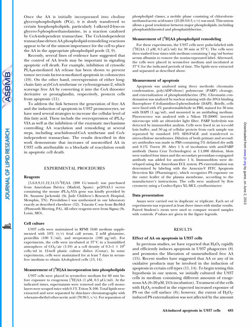
In the next set of experiments, we prepared cells de-pleted of AA by incubating them in serum-free, essentialfatty acid-deficient medium for at least 7 days. This pro-cedure results in cells containing far less AA in phospho-lipids than cells normally grown in serum (13, 14). Figure 1shows that AA-depleted cells were significantly less sensitiveto H2O2-induced apoptosis than cells maintained understandard culture conditions or cells supplemented with10 AM AA. Apoptosis induced by H2O2 was also lower inthe AA-depleted cells (Fig. 1).
Inhibitors of AA esterification induce apoptosisin U937 cells
The data shown in Fig. 1 suggested that the presence ofcellular AA is critical for the full induction of apoptosis.However, this observation appears to contrast with the re-sults indicating that supplementation with exogenous AAdoes not significantly influence the induction of apoptosis.
Cultured cell lines of monocytic origin are known toincorporate and remodel AA into phospholipids at ratesmanyfold greater than their primary counterparts (i.e.,blood leukocytes) (6, 16–19). In this regard, Fig. 2 showsthat U937 cells did indeed incorporate AA and remodeledit through phospholipids at a high rate. Approximately35–40% of the total AA added to the medium was found tobe esterified into phospholipids at 30 min, whereas neutrallipids incorporated lesser amounts (Fig. 2A). At earlyincubation times, PC incorporated the majority of AA. Atlonger times, the AA content in PC decreased gradually,paralleling the increased incorporation of AA in ethanol-amine glycerophospholipid. Overall, these changes are
consistent with data in other systems (6, 16, 20) and reflectthe remodeling action of CoA-independent transacylaseon cellular phospholipids.
The high rate of AA incorporation and remodeling inU937 cells may help reconcile the results reported inFig. 1. It is conceivable that the lack of a significant effecton apoptosis in cells exposed to exogenous AA is attrib-utable to an exceedingly high AA reacylation rate thatprevents the accumulation of unesterified AA within thecell, and hence the onset of apoptosis. If this hypothesis iscorrect, circumstances that prevent AA reacylation shouldincrease the extent of apoptosis in U937 cells.
To verify this hypothesis, we first used thimerosal, anorganometallic compound that is known to prevent fattyacid incorporation into phospholipids by blocking theenzyme long-chain fatty acid-CoA synthetase (21). Initialexperiments demonstrated that thimerosal at concentra-tions up to 3 AM effectively impeded the incorporation ofAA (10 AM) into phospholipids (3.61 6 0.42 nmol of AAincorporated per million cells in the absence of thimerosalversus 0.86 6 0.10 nmol of AA incorporated per millioncells in the presence of 3 AM thimerosal; means 6 SEM,n 5 4). Higher thimerosal concentrations led to substan-tial cell death by necrosis, as judged by the trypan blue
Fig. 1. Apoptosis of arachidonic acid (AA) depleted and non-depleted U937 cells. Cells were maintained in serum-free medium(AA depleted) or in serum-containing medium (standard condi-tions) supplemented with or without 10 AM AA (see abscissa). Cellswere incubated for 20 h in the presence (hatched bars) or absence(open bars) of 500 AM H2O2, and apoptosis was measured by theFITC-annexin V binding assay as described in ExperimentalProcedures. Data are shown as means 6 SEM of three independentexperiments carried out in duplicate. * P , 0.05.
Fig. 2. AA incorporation and remodeling within phospholipids inU937 cells. A: Cells were exposed to 1 AM AA (0.5 ACi/ml) for thetimes indicated. Afterward, AA incorporated into phospholipids(closed circles) or triacylglycerol (open circles) was determined. B:Cells, labeled with [3H]AA, were washed and then incubatedwithout label for different periods of time. Radioactivity in cholinephospholipids (open circles), ethanolamine phospholipids (closedcircles), and phosphatidylinositol (open triangles) was deter-mined. Values are given as percentages of total radioactivitypresent in phospholipids. Data are shown as means 6 SEM ofthree independent experiments carried out in duplicate.
486 Journal of Lipid Research Volume 47, 2006
by guest, on June 2, 2013w
ww
.jlr.orgD
ownloaded from

exclusion assay. Figure 3A shows that 3 AM thimerosal hada slight impact on its own on PS externalization but,importantly, also substantially increased PS externalizationin U937 cells incubated with 10 AM exogenous AA, which,on its own, was ineffective.
To ensure that the increase of PS externalization ob-served in Fig. 3A was indeed linked to apoptotic cell death,other indices of apoptosis, such as chromatin condensa-tion and the appearance of fragments of the caspasesubstrate PARP, were measured. Chromatin condensationwas analyzed by nuclear staining with the DNA bindingfluorophore DAPI. Figure 3B shows that U937 cells treatedwith AA plus thimerosal for 24 h demonstrated clear signsof chromatin condensation, indicating apoptosis. Cleavage
of PARP was evaluated by immunoblot analysis. Untreatedcells expressed the intact PARP 112 kDa polypeptide (Fig.3C), which was found to rapidly disappear in a time-dependent manner after exposure of the cells to AA plusthimerosal. Control experiments using cells treated withthimerosal alone or AA alone did not show any nuclearmorphology changes or PARP cleavage, confirming that itis the impairment of AA esterification that is associatedwith apoptosis.
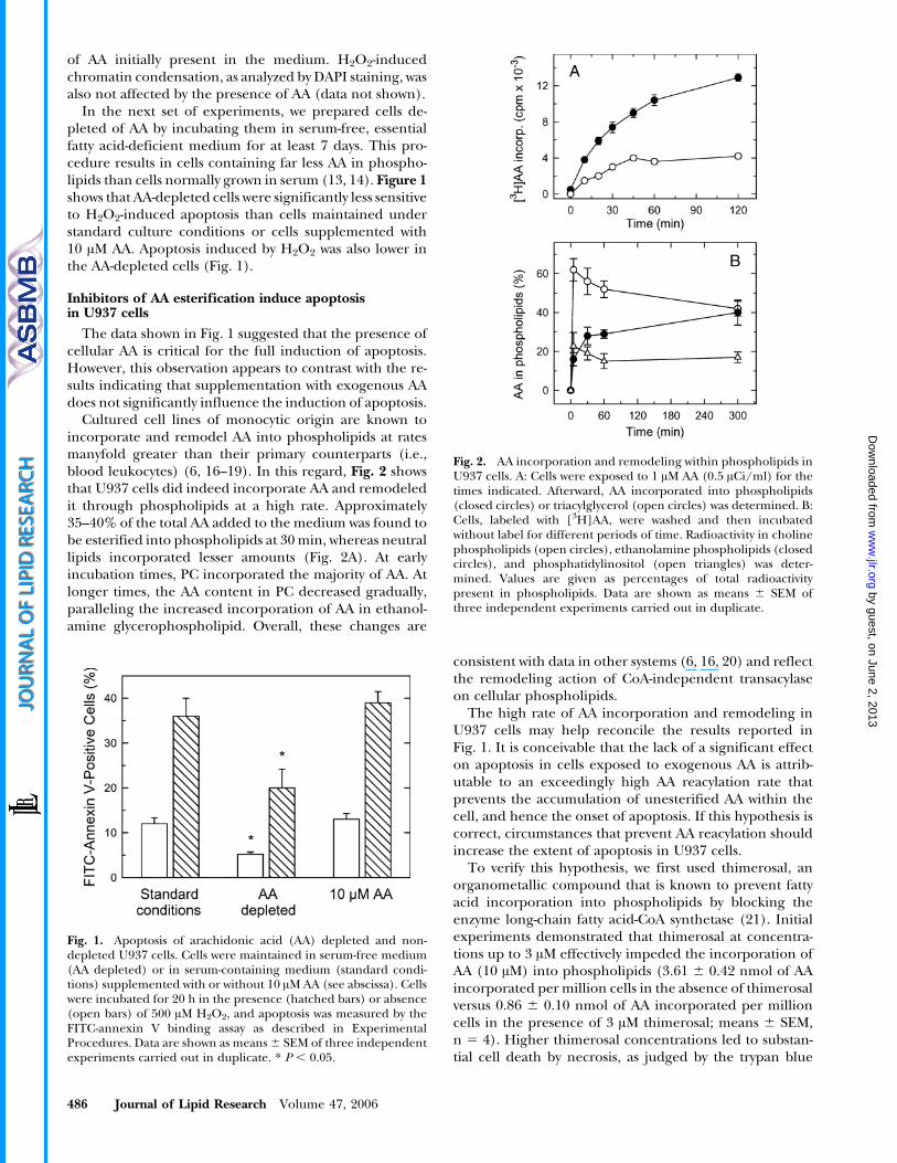
We next evaluated whether triacsin C, a well-establishedinhibitor of long-chain fatty acid-CoA synthetase (22),exerted a similar effect as thimerosal on the induction ofapoptosis. Figure 4 reveals that triacsin C inhibited AAincorporation into phospholipids and caused a concen-tration-dependent increase of PS externalization. Cotreat-ment of exogenous AA with triacsin C caused apoptoticcell death to a much greater extent (Fig. 4). Collectively,the experiments depicted in Figs. 3 and 4 suggest thatconditions that lead to increased levels of unesterified AAresult in apoptosis. Conversely, the data shown in Fig. 1indicate that AA depletion blunts apoptosis.
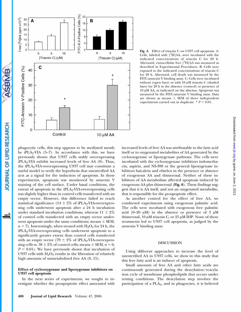
In the experiments described above, unesterified AAlevels were manipulated by inhibiting the first step of theAA incorporation route, the formation of the arachidonyl-CoA that is needed for direct incorporation into phospho-lipid. In the next series of experiments, we used thealternative strategy of blocking the AA phospholipid-remodeling step by inhibiting the activity of the CoA-independent transacylase. To this end, we used theselective CoA-independent transacylase inhibitor diethyl7-(3,4,5-triphenyl-2-oxo-2,3-dihydroimidazol-1-yl)hepatinephosphonate (IHP) (23). Long-term incubation with IHPresulted in significant alterations of the distribution of AAamong cellular lipids. The data shown in Fig. 5Ademonstrate that IHP reduced the level of AA inphospholipids and concomitantly increased the levels ofthis fatty acid in triacylglycerol. Given the blockade of AAphospholipid remodeling by IHP, the shift of AA fromphospholipid to triacylglycerol was to be expected andprobably arises from the direct acylation of diacylglycerolvia de novo synthesis (14, 24, 25). Importantly, theintracellular (cell-associated) level of unesterified AA wasalso noticeably increased after treatment with IHP (Fig.5A). Figure 5B shows that incubation of the U937 cells withIHP resulted in increased death by apoptosis, as judged bythe FITC-annexin V binding assay, and that cotreatmentwith exogenous AA augmented the extent of apoptosis.These results provide further evidence that conditions thatlead to the accumulation of unesterified AA induceapoptosis in U937 cells.
Overexpression of calcium-independent PLA2 augmentsapoptosis in U937 cells
In addition to blocking either AA incorporation or AAphospholipid remodeling, we used a third strategy tomanipulate the levels of unesterified AA in cells: the use ofcells transfected with the iPLA2-VIA gene. Small amountsof AA and other fatty acids are constantly being generatedunder resting conditions, and in certain cell types such as
Fig. 3. Effect of thimerosal on U937 cell apoptosis. A: Cells wereincubated without (open bars) or with 3 AM thimerosal (shadedbars) for 20 h in the absence (control) or presence of 10 AM AA, asindicated on the abscissa. Apoptosis was measured by the FITC-annexin V binding assay as described in Experimental Procedures.Data are shown as means 6 SEM of three independentexperiments carried out in duplicate. * P , 0.01. B: Cells wereuntreated (a, b) or treated with thimerosal plus AA (c, d) for 20 h,stained with 4V,6-diamidino-2-phenylindole, and examined byfluorescence microscopy. Panels a and c are Nomarski images,and panels b and d show the fluorescence of the same cells. C: Cellswere treated with thimerosal plus AA for the indicated times andanalyzed for poly(ADP-ribose) polymerase (PARP) expression byimmunoblotting. Mr, relative molecular mass.
AA-induced apoptosis in U937 cells 487
by guest, on June 2, 2013w
ww
.jlr.orgD
ownloaded from
phagocytic cells, this step appears to be mediated mostlyby iPLA2-VIA (5–7). In accordance with this, we havepreviously shown that U937 cells stably overexpressingiPLA2-VIA exhibit increased levels of free AA (8). Thus,the iPLA2-VIA-overexpressing U937 cell may constitute auseful model to verify the hypothesis that unesterified AAacts as a signal for the induction of apoptosis. In theseexperiments, apoptosis was monitored by annexin Vstaining of the cell surface. Under basal conditions, theextent of apoptosis in the iPLA2-VIA-overexpressing cellswas slightly higher than in control cells transfected with anempty vector. However, this difference failed to reachstatistical significance (14 6 2% of iPLA2-VIA-overexpres-sing cells underwent apoptosis after a 24 h incubationunder standard incubation conditions, whereas 11 6 2%of control cells transfected with an empty vector under-went apoptosis under the same conditions; means 6 SEM,n 5 7). Interestingly, when treated with H2O2 for 24 h, theiPLA2-VIA-overexpressing cells underwent apoptosis to asignificantly greater extent than control cells transfectedwith an empty vector (79 6 2% of iPLA2-VIA-overexpres-sing cells vs. 38 6 2% of control cells; means 6 SEM, n 5 6;P , 0.01). We have previously shown that incubation ofU937 cells with H2O2 results in the liberation of relativelyhigh amounts of unmetabolized free AA (8, 15).
Effect of cyclooxygenase and lipoxygenase inhibitors onU937 cell apoptosis
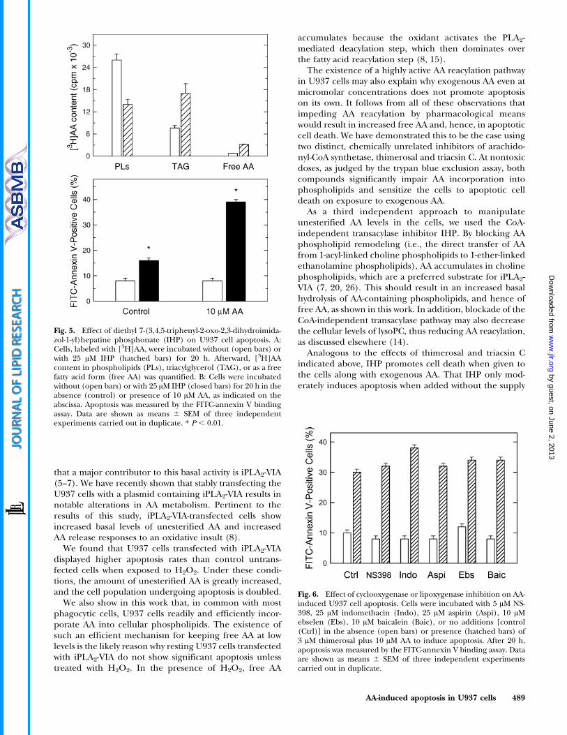
In the next series of experiments, we sought to in-vestigate whether the proapoptotic effect associated with
increased levels of free AA was attributable to the fatty aciditself or to oxygenated metabolites of AA generated by thecyclooxygenase or lipoxygenase pathways. The cells wereincubated with the cyclooxygenase inhibitors indometha-cin, aspirin, and NS-398 or the general lipoxygenase in-hibitors baicalein and ebselen in the presence or absenceof exogenous AA and thimerosal. Neither of these in-hibitors of AA metabolism affected apoptosis induced byexogenous AA plus thimerosal (Fig. 6). These findings sug-gest that it is AA itself, and not an oxygenated metabolite,that is responsible for the proapoptotic effect.
As another control for the effect of free AA, weconducted experiments using exogenous palmitic acid.The cells were incubated with exogenous free palmiticacid (0–20 AM) in the absence or presence of 3 AMthimerosal, 10 AM triacsin C, or 25 AM IHP. None of thesetreatments led to U937 cell apoptosis, as judged by theannexin V binding assay.
DISCUSSION
Using different approaches to increase the level ofunesterified AA in U937 cells, we show in this study thatthis free fatty acid is an inducer of apoptosis.
Small amounts of free AA and other fatty acids arecontinuously generated during the deacylation/reacyla-tion cycle of membrane phospholipids that occurs underresting conditions. The deacylation step involves theparticipation of a PLA2, and in phagocytes, it is believed
Fig. 4. Effect of triacsin C on U937 cell apoptosis. A:Cells, labeled with [3H]AA, were incubated with theindicated concentrations of triacsin C for 20 h.Afterward, extracellular free [3H]AA was measured asdescribed in Experimental Procedures. B: Cells wereexposed to the indicated concentrations of triacsin Cfor 20 h. Afterward, cell death was measured by theFITC-annexin V binding assay. C: Cells were incubatedwithout (open bars) or with 10 AM triacsin C (shadedbars) for 20 h in the absence (control) or presence of10 AM AA, as indicated on the abscissa. Apoptosis wasmeasured by the FITC-annexin V binding assay. Dataare shown as means 6 SEM of three independentexperiments carried out in duplicate. * P , 0.01.
488 Journal of Lipid Research Volume 47, 2006
by guest, on June 2, 2013w
ww
.jlr.orgD
ownloaded from
that a major contributor to this basal activity is iPLA2-VIA(5–7). We have recently shown that stably transfecting theU937 cells with a plasmid containing iPLA2-VIA results innotable alterations in AA metabolism. Pertinent to theresults of this study, iPLA2-VIA-transfected cells showincreased basal levels of unesterified AA and increasedAA release responses to an oxidative insult (8).
We found that U937 cells transfected with iPLA2-VIAdisplayed higher apoptosis rates than control untrans-fected cells when exposed to H2O2. Under these condi-tions, the amount of unesterified AA is greatly increased,and the cell population undergoing apoptosis is doubled.
We also show in this work that, in common with mostphagocytic cells, U937 cells readily and efficiently incor-porate AA into cellular phospholipids. The existence ofsuch an efficient mechanism for keeping free AA at lowlevels is the likely reason why resting U937 cells transfectedwith iPLA2-VIA do not show significant apoptosis unlesstreated with H2O2. In the presence of H2O2, free AA
accumulates because the oxidant activates the PLA2-mediated deacylation step, which then dominates overthe fatty acid reacylation step (8, 15).
The existence of a highly active AA reacylation pathwayin U937 cells may also explain why exogenous AA even atmicromolar concentrations does not promote apoptosison its own. It follows from all of these observations thatimpeding AA reacylation by pharmacological meanswould result in increased free AA and, hence, in apoptoticcell death. We have demonstrated this to be the case usingtwo distinct, chemically unrelated inhibitors of arachido-nyl-CoA synthetase, thimerosal and triacsin C. At nontoxicdoses, as judged by the trypan blue exclusion assay, bothcompounds significantly impair AA incorporation intophospholipids and sensitize the cells to apoptotic celldeath on exposure to exogenous AA.
As a third independent approach to manipulateunesterified AA levels in the cells, we used the CoA-independent transacylase inhibitor IHP. By blocking AAphospholipid remodeling (i.e., the direct transfer of AAfrom 1-acyl-linked choline phospholipids to 1-ether-linkedethanolamine phospholipids), AA accumulates in cholinephospholipids, which are a preferred substrate for iPLA2-VIA (7, 20, 26). This should result in an increased basalhydrolysis of AA-containing phospholipids, and hence offree AA, as shown in this work. In addition, blockade of theCoA-independent transacylase pathway may also decreasethe cellular levels of lysoPC, thus reducing AA reacylation,as discussed elsewhere (14).
Analogous to the effects of thimerosal and triacsin Cindicated above, IHP promotes cell death when given tothe cells along with exogenous AA. That IHP only mod-erately induces apoptosis when added without the supply
Fig. 5. Effect of diethyl 7-(3,4,5-triphenyl-2-oxo-2,3-dihydroimida-zol-1-yl)hepatine phosphonate (IHP) on U937 cell apoptosis. A:Cells, labeled with [3H]AA, were incubated without (open bars) orwith 25 AM IHP (hatched bars) for 20 h. Afterward, [3H]AAcontent in phospholipids (PLs), triacylglycerol (TAG), or as a freefatty acid form (free AA) was quantified. B: Cells were incubatedwithout (open bars) or with 25 AM IHP (closed bars) for 20 h in theabsence (control) or presence of 10 AM AA, as indicated on theabscissa. Apoptosis was measured by the FITC-annexin V bindingassay. Data are shown as means 6 SEM of three independentexperiments carried out in duplicate. * P , 0.01.
Fig. 6. Effect of cyclooxygenase or lipoxygenase inhibition on AA-induced U937 cell apoptosis. Cells were incubated with 5 AM NS-398, 25 AM indomethacin (Indo), 25 AM aspirin (Aspi), 10 AMebselen (Ebs), 10 AM baicalein (Baic), or no additions [control(Ctrl)] in the absence (open bars) or presence (hatched bars) of3 AM thimerosal plus 10 AM AA to induce apoptosis. After 20 h,apoptosis was measured by the FITC-annexin V binding assay. Dataare shown as means 6 SEM of three independent experimentscarried out in duplicate.
AA-induced apoptosis in U937 cells 489
by guest, on June 2, 2013w
ww
.jlr.orgD
ownloaded from

of exogenous AA can be explained by the participation ofalternative metabolic pathways to attenuate the level ofunesterified fatty acid under these conditions. In accordwith this, increased acylation of AA into the triacylglycerolfraction is detected in cells treated with IHP comparedwith untreated cells. This indicates that a significantportion of the free AA produced after CoA-indepen-dent transacylase inhibition is diverted to the low-affin-ity, high-capacity de novo route for triacylglycerol bio-synthesis, a route that is seldom used under normalconditions by cultured cell lines of the phagocytic lineage(2, 24, 26).
The concentrations of AA that were able to induceapoptosis when combined with drugs inhibiting fatty acidincorporation and remodeling are in the low micromolarrange. These concentrations are likely to be reachedlocally in vivo during an inflammatory process andtherefore may be of pathophysiological relevance. It isworth noting that palmitic acid fails to elicit an apoptoticresponse either alone or in combination with thimerosal,triacsin C, or IHP. This indicates that the proapoptoticeffect of AA described in this study is not a detergenteffect. Using well-established cyclooxygenase and lipoxy-genase inhibitors, we have also demonstrated that the AAeffects are attributable to the fatty acid itself and not toan oxidized metabolite. These studies, along with resultsby others (11, 14), raise the question of how increasingcellular free AA levels puts into motion the machinery thatleads to programmed cell death. Although much work isclearly needed to define the molecular steps involved inAA-induced apoptosis, it is interesting that AA at lowmicromolar concentrations is known to affect a number ofintracellular effectors and signaling pathways, includingthe generation of apoptosis-inducing ceramides (27–30).Overall, our work strongly suggests that the enzymaticmechanisms regulating free AA availability in cells must beregarded as potential candidates for the design of drugswith antitumoral potential (31, 32).
The authors are grateful to Montse Duque and Yolanda Saez forexpert technical assistance. This work was supported by theFundacio La Marato de TV3 (Grant 011232) and the SpanishMinistry of Education and Science (Grants BFU2004-01886/BMC and SAF2004-04676).
REFERENCES
1. Lands, W. E. M. 2000. Stories about acyl chains. Biochim. Biophys.Acta. 1483: 1–14.
2. Chilton, F. H., A. H. Fonteh, M. Surette, M. Triggiani, and J. D.Winkler. 1996. Control of arachidonate levels within inflammatorycells. Biochim. Biophys. Acta. 1299: 1–15.
3. Balsinde, J., and E. A. Dennis. 1997. Function and inhibition ofintracellular calcium-independent phospholipase A2. J. Biol. Chem.272: 16069–16072.
4. Winstead, M. V., J. Balsinde, and E. A. Dennis. 2000. Calcium-independent phospholipase A2: structure and function. Biochim.Biophys. Acta. 1488: 28–39.
5. Balsinde, J., and M. A. Balboa. 2005. Cellular regulation andproposed biological functions of group VIA calcium-independentphospholipase A2 in activated cells. Cell. Signal. 17: 1052–1062.
6. Balsinde, J., S. E. Barbour, I. D. Bianco, and E. A. Dennis. 1994.Arachidonic acid mobilization in P388D1 macrophages is con-trolled by two distinct Ca21-dependent phospholipase A2 enzymes.Proc. Natl. Acad. Sci. USA. 91: 11060–11064.
7. Balsinde, J., M. A. Balboa, and E. A. Dennis. 1997. Antisenseinhibition of group VI Ca2+-independent phospholipase A2 blocksphospholipid fatty acid remodeling in murine P388D1 macro-phages. J. Biol. Chem. 272: 29317–29321.
8. Perez, R., R. Melero, M. A. Balboa, and J. Balsinde. 2004. Role ofgroup VIA calcium-independent phospholipase A2 in arachidonicacid release, phospholipid fatty acid incorporation, and apoptosisin U937 cells responding to hydrogen peroxide. J. Biol. Chem. 279:40385–40391.
9. Yamashita, A., T. Sugiura, and K. Waku. 1997. Acyltransferases andtransacylases involved in fatty acid remodeling of phospholipidsand metabolism of bioactive lipids in mammalian cells. J. Biochem.(Tokyo). 122: 1–16.
10. Dong, M., K. Guda, P. R. Nambiar, A. Rezaie, G. S. Belinsky,G. Lambeau, C. Giardina, and D. W. Rosenberg. 2003. Inverseassociation between phospholipase A2 and COX-2 expressionduring mouse colon tumorigenesis. Carcinogenesis. 24: 307–315.
11. Cao, Y., A. T. Pearman, G. A. Zimmerman, T. M. McIntyre, andS. M. Prescott. 2000. Intracellular unesterified arachidonic acidsignals apoptosis. Proc. Natl. Acad. Sci. USA. 97: 11280–11285.
12. Seno, K., T. Okuno, K. Nishi, Y. Murakami, F. Watanabe,T. Matsuura, M. Wada, Y. Fujii, M. Yamada, T. Ogawa, et al. 2000.Pyrrolidine inhibitors of human cytosolic phospholipase A2. J. Med.Chem. 43: 1041–1044.
13. Blank, M. L., Z. L. Smith, and F. Snyder. 1992. Contributing factorsin the trafficking of [3H]arachidonate between phospholipids.Biochim. Biophys. Acta. 1124: 262–272.
14. Surette, M. E., A. N. Fonteh, C. Bernatchez, and F. H. Chilton.1999. Perturbations in the control of cellular arachidonic acidlevels block cell growth and induce apoptosis in HL-60 cells.Carcinogenesis. 20: 757–763.
15. Balboa, M. A., and J. Balsinde. 2002. Involvement of calcium-independent phospholipase A2 in hydrogen peroxide-inducedaccumulation of free fatty acids in human U937 cells. J. Biol. Chem.277: 40384–40389.
16. Balsinde, J., B. Fernandez, and J. A. Solıs-Herruzo. 1994. Increasedincorporation of arachidonic acid into phospholipids in zymosan-stimulated mouse peritoneal macrophages. Eur. J. Biochem. 221:1013–1018.
17. Surette, M. E., and F. H. Chilton. 1998. The distribution andmetabolism of arachidonate-containing phospholipids in cellularnuclei. Biochem. J. 330: 915–921.
18. Joly, F., M. Breton, C. Wolf, E. Ninio, and O. Colard. 1992.Heterogeneity of arachidonate and paf-acether precursor pools inmast cells. Biochim. Biophys. Acta. 1125: 305–312.
19. Boilard, E., and M. E. Surette. 2001. Anti-CD3 and concanavalin A-induced human T cell proliferation is associated with an increasedrate of arachidonate-phospholipid remodeling. Lack of involve-ment of group IV and group VI phospholipase A2 in remodelingand increased susceptibility of proliferating T cells to CoA-independent transacyclase inhibitor-induced apoptosis. J. Biol.Chem. 276: 17568–17575.
20. Balsinde, J. 2002. Roles of various phospholipases A2 in providinglysophospholipid acceptors for fatty acid phospholipid incorpora-tion and remodelling. Biochem. J. 364: 695–702.
21. Kaever, V., U. Firla, and K. Resch. 1988. Sulfhydryl reagents asmodel substances for eicosanoid research. Eicosanoids. 1: 49–57.
22. Hartman, E. J., S. Omura, and M. Laposata. 1989. Triacsin C: adifferential inhibitor of arachidonoyl-CoA synthetase and nonspe-cific long chain acyl-CoA synthetase. Prostaglandins. 37: 655–671.
23. Chilton, F. H., A. N. Fonteh, C. M. Sung, D. M. Hinkley, T. J.Torphy, R. J. Mayer, L. A. Marshall, J. D. Heravi, and J. D. Winkler.1995. Inhibitors of CoA-independent transacylase block themovement of arachidonate into 1-ether-linked phospholipids ofhuman neutrophils. Biochemistry. 34: 5403–5410.
24. Balsinde, J., and E. A. Dennis. 1996. The incorporation of ara-chidonic acid into triacylglycerol in P388D1 macrophage-like cells.Eur. J. Biochem. 235: 480–485.
25. Blank, M. L., Z. L. Smith, and F. Snyder. 1993. Arachidonate-containing triacylglycerols: biosynthesis and a lipolytic mechanismfor the release and transfer of arachidonate to phospholipids inHL-60 cells. Biochim. Biophys. Acta. 1170: 275–282.
26. Balsinde, J., I. D. Bianco, E. J. Ackermann, K. Conde-Frieboes, and
490 Journal of Lipid Research Volume 47, 2006
by guest, on June 2, 2013w
ww
.jlr.orgD
ownloaded from
E. A. Dennis. 1995. Inhibition of calcium-independent phospho-lipase A2 prevents arachidonic acid incorporation and phospho-lipid remodeling in P388D1 macrophages. Proc. Natl. Acad. Sci. USA.92: 8527–8531.
27. Chan, T. A., P. J. Morin, B. Vogelstein, and K. W. Winzler. 1998.Mechanisms underlying nonsteroidal antiinflammatory drug-me-diated apoptosis. Proc. Natl. Acad. Sci. USA. 95: 681–686.
28. Jayadev, S., C. M. Linardic, and Y. A. Hannun. 1994. Identification ofarachidonic acid as a mediator of sphingomyelin hydrolysis inresponse to tumor necrosis factor alpha. J. Biol. Chem. 269: 5757–5763.
29. Jayadev, S., H. L. Hayter, N. Andrieu, C. J. Gamard, B. Liu, R. Balu,M. Hayakawa, F. Ito, and Y. A. Hannun. 1997. Phospholipase A2 is
necessary for tumor necrosis factor alpha-induced ceramidegeneration in L929 cells. J. Biol. Chem. 272: 17196–17203.
30. Cai, Z., A. Bettaieb, N. E. Mahdani, L. G. Legres, R. Stancou, J.Masliah, and S. J. Chouaib. 1997. Alteration of the sphingomyelin/ceramide pathway is associated with resistance of human breastcarcinoma MCF7 cells to tumor necrosis factor-alpha-mediatedcytotoxicity. J. Biol. Chem. 272: 6918–6926.
31. Monjazeb, A. M., C. E. Clay, K. P. High, and F. H. Chilton. 2002.Antineoplastic properties of arachidonic acid and its metabolites.Prostaglandins Leukot. Essent. Fatty Acids. 66: 5–12.
32. Das, U. N. 1999. Essential fatty acids, lipid peroxidation andapoptosis. Prostaglandins Leukot. Essent. Fatty Acids. 61: 157–163.
AA-induced apoptosis in U937 cells 491
by guest, on June 2, 2013w
ww
.jlr.orgD
ownloaded from
Copyright © 2022 FDOKUMEN