Oxpholipin 11D: An Anti-Inflammatory Peptide That Binds Cholesterol and Oxidized Phospholipids
13
Oxpholipin 11D: An Anti-Inflammatory Peptide That Binds Cholesterol and Oxidized Phospholipids Piotr Ruchala 1 *, Mohamad Navab 1 , Chun-Ling Jung 1 , Susan Hama-Levy 1 , Ewa D. Micewicz 2 , Hai Luong 1¤ , Jonathan E. Reyles 3 , Shantanu Sharma 3 , Alan J. Waring 1 , Alan M. Fogelman 1 , Robert I. Lehrer 1 1 Department of Medicine, David Geffen School of Medicine, University of California Los Angeles, Los Angeles, California, United States of America, 2 Department of Radiation Oncology, David Geffen School of Medicine, University of California Los Angeles, Los Angeles, California, United States of America, 3 Department of Chemistry and Center for Macromolecular Modeling & Materials Design, California State Polytechnic University, Pomona, California, United States of America Abstract Background: Many Gram-positive bacteria produce pore-forming exotoxins that contain a highly conserved, 12-residue domain (ECTGLAWEWWRT) that binds cholesterol. This domain is usually flanked N-terminally by arginine and C-terminally by valine. We used this 14-residue sequence as a template to create a small library of peptides that bind cholesterol and other lipids. Methodology/Results: Several of these peptides manifested anti-inflammatory properties in a predictive in vitro monocyte chemotactic assay, and some also diminished the pro-inflammatory effects of low-density lipoprotein in apoE-deficient mice. The most potent analog, Oxpholipin-11D (OxP-11D), contained D-amino acids exclusively and was identical to the 14- residue design template except that diphenylalanine replaced cysteine-3. In surface plasmon resonance binding studies, OxP-11D bound oxidized (phospho)lipids and sterols in much the same manner as D-4F, a widely studied cardioprotective apoA-I-mimetic peptide with anti-inflammatory properties. In contrast to D-4F, which adopts a stable a-helical structure in solution, the OxP-11D structure was flexible and contained multiple turn-like features. Conclusion: Given the substantial evidence that oxidized phospholipids are pro-inflammatory in vivo, OxP-11D and other Oxpholipins may have therapeutic potential. Citation: Ruchala P, Navab M, Jung C-L, Hama-Levy S, Micewicz ED, et al. (2010) Oxpholipin 11D: An Anti-Inflammatory Peptide That Binds Cholesterol and Oxidized Phospholipids. PLoS ONE 5(4): e10181. doi:10.1371/journal.pone.0010181 Editor: Maxim Antopolsky, University of Helsinki, Finland Received January 29, 2010; Accepted March 24, 2010; Published April 14, 2010 Copyright: ß 2010 Ruchala et al. This is an open-access article distributed under the terms of the Creative Commons Attribution License, which permits unrestricted use, distribution, and reproduction in any medium, provided the original author and source are credited. Funding: These studies were supported in part by U.S. Public Health Service Grant HL-30568, the Laubisch, Castera, and M.K. Grey Funds at UCLA and funds from the Adams and Burnham endowments provided by the Dean’s Office of the David Geffen School of Medicine at UCLA (PR). The funders had no role in study design, data collection and analysis, decision to publish, or preparation of the manuscript. Competing Interests: MN and AMF are principals in Bruin Pharma and AMF is an officer in Bruin Pharma. Oxpholipins and their use are protected by patent rights (provisional application filed, PR and RIL co-inventors). This does not alter the authors’ adherence to all the PLoS ONE policies on sharing data and materials. * E-mail: [email protected] ¤ Current address: Astellas Pharma, Inc., Santa Monica, California, United States of America Introduction Oxysterols and oxidized lipids are important mediators in many signaling pathways [1,2]. Oxysterols may interact with various molecular targets, including liver X receptors-a and b [3], estrogen receptors a and b [3,4] cytoplasmic oxysterol-binding protein (OSBP) [5], OSBP-related proteins (ORPs) [6] or insulin- induced gene (Insig) proteins [3,7]. These interactions can not only affect sterol and lipid metabolism, they may also influence pathological processes as diverse as Niemann-Pick C disease [8] and atherosclerosis [2]. The signaling network of oxidized (phospho)lipids and their derivatives is complex. Many structurally related, biologically active compounds are generated from arachidonic acid via the eicosanoid synthesis pathway (also known as the arachidonic acid cascade) [9–11]. These compounds, collectively known as eicosanoids, include prostaglandins (PGs), thromboxanes (TXs), leukotrienes (LTs) and other oxidized derivatives/intermediates. Eicosanoids are key mediators and regulators of inflammatory responses [10,12,13] with implications for asthma [14–18], diabetes [19], cancer [20–23], colitis [24,25], rheumatoid arthritis [26,27], inflammatory bowel disease [28] and atherosclerosis [29,30]. Evidence gathered over past two decades has shown that inflammation is a key player in pathophysiology of atherogenic cardiovascular disease (CVD) [31–33]. Consequently, therapies targeting inflammatory response have already been implemented in clinical practice of CVD [34–36] and further strategies and anti- inflammatory treatment regimens are being investigated [32]. An emerging approach in treating cardiovascular disease is based on using apoA-I mimetic peptides [37–41] with anti-inflammatory properties to sequester oxysterols and oxidized lipids [42]. These peptides have properties similar to apoA-I Milano [17,43], a naturally occurring apoA-I mutant containing an extra cysteine disulfide bridge. ApoA-I Milano reduced atheromas by up to 30% [44] and ameliorated plaque build-up in arterial walls [45]. Whereas apoA-I Milano requires intravenous administration, D- 4F (Ac-DWFKAFYDKVAEKFKEAF-NH 2 ), a prominent apoA- I-mimetic peptide, can be taken orally [37,39,46–48]. PLoS ONE | www.plosone.org 1 April 2010 | Volume 5 | Issue 4 | e10181
-
Upload
independent -
Category
Documents
-
view
2 -
download
0
Transcript of Oxpholipin 11D: An Anti-Inflammatory Peptide That Binds Cholesterol and Oxidized Phospholipids
Oxpholipin 11D: An Anti-Inflammatory Peptide ThatBinds Cholesterol and Oxidized PhospholipidsPiotr Ruchala1*, Mohamad Navab1, Chun-Ling Jung1, Susan Hama-Levy1, Ewa D. Micewicz2, Hai Luong1¤,
Jonathan E. Reyles3, Shantanu Sharma3, Alan J. Waring1, Alan M. Fogelman1, Robert I. Lehrer1
1 Department of Medicine, David Geffen School of Medicine, University of California Los Angeles, Los Angeles, California, United States of America, 2 Department of
Radiation Oncology, David Geffen School of Medicine, University of California Los Angeles, Los Angeles, California, United States of America, 3 Department of Chemistry
and Center for Macromolecular Modeling & Materials Design, California State Polytechnic University, Pomona, California, United States of America
Abstract
Background: Many Gram-positive bacteria produce pore-forming exotoxins that contain a highly conserved, 12-residuedomain (ECTGLAWEWWRT) that binds cholesterol. This domain is usually flanked N-terminally by arginine and C-terminallyby valine. We used this 14-residue sequence as a template to create a small library of peptides that bind cholesterol andother lipids.
Methodology/Results: Several of these peptides manifested anti-inflammatory properties in a predictive in vitro monocytechemotactic assay, and some also diminished the pro-inflammatory effects of low-density lipoprotein in apoE-deficientmice. The most potent analog, Oxpholipin-11D (OxP-11D), contained D-amino acids exclusively and was identical to the 14-residue design template except that diphenylalanine replaced cysteine-3. In surface plasmon resonance binding studies,OxP-11D bound oxidized (phospho)lipids and sterols in much the same manner as D-4F, a widely studied cardioprotectiveapoA-I-mimetic peptide with anti-inflammatory properties. In contrast to D-4F, which adopts a stable a-helical structure insolution, the OxP-11D structure was flexible and contained multiple turn-like features.
Conclusion: Given the substantial evidence that oxidized phospholipids are pro-inflammatory in vivo, OxP-11D and otherOxpholipins may have therapeutic potential.
Citation: Ruchala P, Navab M, Jung C-L, Hama-Levy S, Micewicz ED, et al. (2010) Oxpholipin 11D: An Anti-Inflammatory Peptide That Binds Cholesterol andOxidized Phospholipids. PLoS ONE 5(4): e10181. doi:10.1371/journal.pone.0010181
Editor: Maxim Antopolsky, University of Helsinki, Finland
Received January 29, 2010; Accepted March 24, 2010; Published April 14, 2010
Copyright: � 2010 Ruchala et al. This is an open-access article distributed under the terms of the Creative Commons Attribution License, which permitsunrestricted use, distribution, and reproduction in any medium, provided the original author and source are credited.
Funding: These studies were supported in part by U.S. Public Health Service Grant HL-30568, the Laubisch, Castera, and M.K. Grey Funds at UCLA and funds fromthe Adams and Burnham endowments provided by the Dean’s Office of the David Geffen School of Medicine at UCLA (PR). The funders had no role in studydesign, data collection and analysis, decision to publish, or preparation of the manuscript.
Competing Interests: MN and AMF are principals in Bruin Pharma and AMF is an officer in Bruin Pharma. Oxpholipins and their use are protected by patentrights (provisional application filed, PR and RIL co-inventors). This does not alter the authors’ adherence to all the PLoS ONE policies on sharing data and materials.
* E-mail: [email protected]
¤ Current address: Astellas Pharma, Inc., Santa Monica, California, United States of America
Introduction
Oxysterols and oxidized lipids are important mediators in many
signaling pathways [1,2]. Oxysterols may interact with various
molecular targets, including liver X receptors-a and b [3],
estrogen receptors a and b [3,4] cytoplasmic oxysterol-binding
protein (OSBP) [5], OSBP-related proteins (ORPs) [6] or insulin-
induced gene (Insig) proteins [3,7]. These interactions can not only
affect sterol and lipid metabolism, they may also influence
pathological processes as diverse as Niemann-Pick C disease [8]
and atherosclerosis [2].
The signaling network of oxidized (phospho)lipids and their
derivatives is complex. Many structurally related, biologically active
compounds are generated from arachidonic acid via the eicosanoid
synthesis pathway (also known as the arachidonic acid cascade)
[9–11]. These compounds, collectively known as eicosanoids,
include prostaglandins (PGs), thromboxanes (TXs), leukotrienes
(LTs) and other oxidized derivatives/intermediates. Eicosanoids are
key mediators and regulators of inflammatory responses [10,12,13]
with implications for asthma [14–18], diabetes [19], cancer
[20–23], colitis [24,25], rheumatoid arthritis [26,27], inflammatory
bowel disease [28] and atherosclerosis [29,30].
Evidence gathered over past two decades has shown that
inflammation is a key player in pathophysiology of atherogenic
cardiovascular disease (CVD) [31–33]. Consequently, therapies
targeting inflammatory response have already been implemented
in clinical practice of CVD [34–36] and further strategies and anti-
inflammatory treatment regimens are being investigated [32]. An
emerging approach in treating cardiovascular disease is based on
using apoA-I mimetic peptides [37–41] with anti-inflammatory
properties to sequester oxysterols and oxidized lipids [42]. These
peptides have properties similar to apoA-I Milano [17,43], a
naturally occurring apoA-I mutant containing an extra cysteine
disulfide bridge. ApoA-I Milano reduced atheromas by up to 30%
[44] and ameliorated plaque build-up in arterial walls [45].
Whereas apoA-I Milano requires intravenous administration, D-
4F (Ac-DWFKAFYDKVAEKFKEAF-NH2), a prominent apoA-
I-mimetic peptide, can be taken orally [37,39,46–48].
PLoS ONE | www.plosone.org 1 April 2010 | Volume 5 | Issue 4 | e10181
This report describes OxP-11D, a novel 14-residue peptide
whose sequence closely resembles a cholesterol-binding domain
found in a family of pore-forming bacterial exotoxins. The ability
of OxP-11D to bind oxidized phospholipids and sterols resemble
those of D-4F [42]. In addition, OxP-11D reduced the release of
monocyte chemotactic factors from LDL-stimulated human aortic
endothelial cells, another property of D-4F. Although more
extensive development of OxP-11D and related oxpholipins
remains to be done, the current findings seem interesting enough
to warrant its description now.
Results
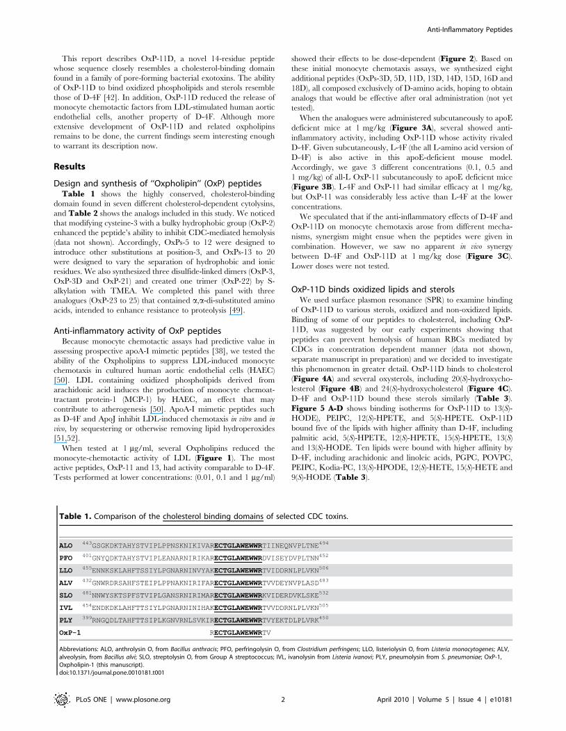
Design and synthesis of ‘‘Oxpholipin’’ (OxP) peptidesTable 1 shows the highly conserved, cholesterol-binding
domain found in seven different cholesterol-dependent cytolysins,
and Table 2 shows the analogs included in this study. We noticed
that modifying cysteine-3 with a bulky hydrophobic group (OxP-2)
enhanced the peptide’s ability to inhibit CDC-mediated hemolysis
(data not shown). Accordingly, OxPs-5 to 12 were designed to
introduce other substitutions at position-3, and OxPs-13 to 20
were designed to vary the separation of hydrophobic and ionic
residues. We also synthesized three disulfide-linked dimers (OxP-3,
OxP-3D and OxP-21) and created one trimer (OxP-22) by S-
alkylation with TMEA. We completed this panel with three
analogues (OxP-23 to 25) that contained a,a-di-substituted amino
acids, intended to enhance resistance to proteolysis [49].
Anti-inflammatory activity of OxP peptidesBecause monocyte chemotactic assays had predictive value in
assessing prospective apoA-I mimetic peptides [38], we tested the
ability of the Oxpholipins to suppress LDL-induced monocyte
chemotaxis in cultured human aortic endothelial cells (HAEC)
[50]. LDL containing oxidized phospholipids derived from
arachidonic acid induces the production of monocyte chemoat-
tractant protein-1 (MCP-1) by HAEC, an effect that may
contribute to atherogenesis [50]. ApoA-I mimetic peptides such
as D-4F and ApoJ inhibit LDL-induced chemotaxis in vitro and in
vivo, by sequestering or otherwise removing lipid hydroperoxides
[51,52].
When tested at 1 mg/ml, several Oxpholipins reduced the
monocyte-chemotactic activity of LDL (Figure 1). The most
active peptides, OxP-11 and 13, had activity comparable to D-4F.
Tests performed at lower concentrations: (0.01, 0.1 and 1 mg/ml)
showed their effects to be dose-dependent (Figure 2). Based on
these initial monocyte chemotaxis assays, we synthesized eight
additional peptides (OxPs-3D, 5D, 11D, 13D, 14D, 15D, 16D and
18D), all composed exclusively of D-amino acids, hoping to obtain
analogs that would be effective after oral administration (not yet
tested).
When the analogues were administered subcutaneously to apoE
deficient mice at 1 mg/kg (Figure 3A), several showed anti-
inflammatory activity, including OxP-11D whose activity rivaled
D-4F. Given subcutaneously, L-4F (the all L-amino acid version of
D-4F) is also active in this apoE-deficient mouse model.
Accordingly, we gave 3 different concentrations (0.1, 0.5 and
1 mg/kg) of all-L OxP-11 subcutaneously to apoE deficient mice
(Figure 3B). L-4F and OxP-11 had similar efficacy at 1 mg/kg,
but OxP-11 was considerably less active than L-4F at the lower
concentrations.
We speculated that if the anti-inflammatory effects of D-4F and
OxP-11D on monocyte chemotaxis arose from different mecha-
nisms, synergism might ensue when the peptides were given in
combination. However, we saw no apparent in vivo synergy
between D-4F and OxP-11D at 1 mg/kg dose (Figure 3C).
Lower doses were not tested.
OxP-11D binds oxidized lipids and sterolsWe used surface plasmon resonance (SPR) to examine binding
of OxP-11D to various sterols, oxidized and non-oxidized lipids.
Binding of some of our peptides to cholesterol, including OxP-
11D, was suggested by our early experiments showing that
peptides can prevent hemolysis of human RBCs mediated by
CDCs in concentration dependent manner (data not shown,
separate manuscript in preparation) and we decided to investigate
this phenomenon in greater detail. OxP-11D binds to cholesterol
(Figure 4A) and several oxysterols, including 20(S)-hydroxycho-
lesterol (Figure 4B) and 24(S)-hydroxycholesterol (Figure 4C).
D-4F and OxP-11D bound these sterols similarly (Table 3).
Figure 5 A-D shows binding isotherms for OxP-11D to 13(S)-
HODE), PEIPC, 12(S)-HPETE, and 5(S)-HPETE. OxP-11D
bound five of the lipids with higher affinity than D-4F, including
palmitic acid, 5(S)-HPETE, 12(S)-HPETE, 15(S)-HPETE, 13(S)
and 13(S)-HODE. Ten lipids were bound with higher affinity by
D-4F, including arachidonic and linoleic acids, PGPC, POVPC,
PEIPC, Kodia-PC, 13(S)-HPODE, 12(S)-HETE, 15(S)-HETE and
9(S)-HODE (Table 3).
Table 1. Comparison of the cholesterol binding domains of selected CDC toxins.
ALO 443GSGKDKTAHYSTVIPLPPNSKNIKIVARECTGLAWEWWRTIINEQNVPLTNE494
PFO 401GNYQDKTAHYSTVIPLEANARNIRIKARECTGLAWEWWRDVISEYDVPLTNN452
LLO 455ENNKSKLAHFTSSIYLPGNARNINVYAKECTGLAWEWWRTVIDDRNLPLVKN506
ALV 432GNWRDRSAHFSTEIPLPPNAKNIRIFARECTGLAWEWWRTVVDEYNVPLASD483
SLO 481NNWYSKTSPFSTVIPLGANSRNIRIMARECTGLAWEWWRKVIDERDVKLSKE532
IVL 454ENDKDKLAHFTTSIYLPGNARNINIHAKECTGLAWEWWRTVVDDRNLPLVKN505
PLY 399RNGQDLTAHFTTSIPLKGNVRNLSVKIRECTGLAWEWWRTVYEKTDLPLVRK450
OxP-1 RECTGLAWEWWRTV
Abbreviations: ALO, anthrolysin O, from Bacillus anthracis; PFO, perfringolysin O, from Clostridium perfringens; LLO, listeriolysin O, from Listeria monocytogenes; ALV,alveolysin, from Bacillus alvi; SLO, streptolysin O, from Group A streptococcus; IVL, ivanolysin from Listeria ivanovi; PLY, pneumolysin from S. pneumoniae; OxP-1,Oxpholipin-1 (this manuscript).doi:10.1371/journal.pone.0010181.t001
Anti-Inflammatory Peptides
PLoS ONE | www.plosone.org 2 April 2010 | Volume 5 | Issue 4 | e10181

Generally, D-4F seems to bind with higher affinity to oxidized
lipids containing palmitoyl moiety: PGPC, POVPC, PEIPC and
KOdiA-PC. OxP-11D on the other hand seems to be more
selective toward certain sterols: 20(S)-hydroxycholesterol, 22(S)-
hydroxycholesterol, 24(S)-hydroxycholesterol and certain arachi-
donic acid derivatives: 5(S)-HPETE, 12(S)-HPETE, 15(S)-HPETE
and 13(S)-HODE. The position of oxidation appeared to influence
binding affinity, as OxP-11D bound tightly to cholesterol
Table 2. Sequences of Oxpholipins.
Peptide Sequence
OxP-1 RE-Cys-Thr-G-Leu-Ala-Trp-E-Trp-Trp-RT-Val-NH2
OxP-2 RE-Ctb-Thr-G-Leu-Ala-Trp-E-Trp-Trp-RT-Val-NH2
OxP-3 RE-Cys-Thr-G-Leu-Ala-Trp-E-Trp-Trp-RT-Val-NH2
I
RE-Cys-Thr-G-Leu-Ala-Trp-E-Trp-Trp-RT-Val-NH2
OxP-3D RE-Cys-Thr-G-Leu-Ala-Trp-E-Trp-Trp-RT-Val-NH2
I
RE-Cys-Thr-G-Leu-Ala-Trp-E-Trp-Trp-RT-Val-NH2
OxP-4 WA-Arg-Thr-V-Trp-Gly-Arg-L-Ctb-Glu-TE-Trp-NH2
OxP-4D WA-Arg-Thr-V-Trp-Gly-Arg-L-Ctb-Glu-TE-Trp-NH2
OxP-5 RE-Ser-Thr-G-Leu-Ala-Trp-E-Trp-Trp-RT-Val-NH2
OxP-5D RE-Ser-Thr-G-Leu-Ala-Trp-E-Trp-Trp-RT-Val-NH2
OxP-6 RE-Chg-Thr-G-Leu-Ala-Trp-E-Trp-Trp-RT-Val-NH2
OxP-7 RE-Cbl-Thr-G-Leu-Ala-Trp-E-Trp-Trp-RT-Val-NH2
OxP-8 RE-PhF-Thr-G-Leu-Ala-Trp-E-Trp-Trp-RT-Val-NH2
OxP-9 RE-Trp-Thr-G-Leu-Ala-Trp-E-Trp-Trp-RT-Val-NH2
OxP-10 RE-Bip-Thr-G-Leu-Ala-Trp-E-Trp-Trp-RT-Val-NH2
OxP-11 RE-Dpa-Thr-G-Leu-Ala-Trp-E-Trp-Trp-RT-Val-NH2
OxP-11D RE-Dpa-Thr-G-Leu-Ala-Trp-E-Trp-Trp-RT-Val-NH2
OxP-12 RE-Ant-Thr-G-Leu-Ala-Trp-E-Trp-Trp-RT-Val-NH2
OxP-13 Aib-RE-Ctb-Val-R-Leu-Val-Trp-E-Trp-Trp-RE-Val-NH2
OxP-13D Aib-RE-Ctb-Val-R-Leu-Val-Trp-E-Trp-Trp-RE-Val-NH2
OxP-14 Nic-RE-Ctb-Val-R-Leu-Val-Trp-E-Trp-Trp-RE-Val-NH2
OxP-14D Nic-RE-Ctb-Val-R-Leu-Val-Trp-E-Trp-Trp-RE-Val-NH2
OxP-15 Nic-bA-RE-Ctb-Val-R-Leu-Val-Trp-E-Trp-Trp-RE-Val-NH2
OxP-15D Nic-bA-RE-Ctb-Val-R-Leu-Val-Trp-E-Trp-Trp-RE-Val-NH2
OxP-16 Aib-RE-Ctb-Chg-R-Cha-Chg-Trp-E-Trp-Trp-RE-Chg-NH2
OxP-16D Aib-RE-Ctb-Chg-R-Cha-Chg-Trp-E-Trp-Trp-RE-Chg-NH2
OxP-17 Ach-RE-Ctb-Chg-R-Cha-Chg-Trp-E-Trp-Trp-RE-Chg-NH2
OxP-18 Aib-RE-Ctb-Chg-R-Cha-Chg-Trp-E-Trp-Trp-RE-Chg-K-NH2
OxP-18D Aib-RE-Ctb-Chg-R-Cha-Chg-Trp-E-Trp-Trp-RE-Chg-K-NH2
OxP-19 Aib-RE-Ctb-Ctb-R-Ctb-Ctb-Trp-E-Trp-Trp-RE-Ctb-NH2
OxP-20 Aib-RE-Ctb-Chg-R-Cha-Chg-Nal-E-Nal-Nal-RE-Chg-X
OxP-21 Aib-RE-Ctb-Chg-R-Cha-Chg-Nal-E-Nal-Nal-RE-Chg-X2
OxP-22 Aib-RE-Ctb-Chg-R-Cha-Chg-Nal-E-Nal-Nal-RE-Chg-X3
OxP-23 Ach-RE-Ctb-Ach-R-Leu-Ach-Trp-E-Trp-Trp-RE-Ach-NH2
OxP-24 Aib-RE-Ctb-Aib-R-Leu-Aib-Trp-E-Trp-Trp-RE-Aib-NH2
OxP-25 Aib-RE-Ctb-Ach-R-Leu-Ach-Trp-E-Trp-Trp-RE-Aib-NH2
Analogues whose identifiers end with a D are composed of D-amino acids. Abbreviations: Nic- nicotinic acid, PhF-1,2,3,4,5-pentafluoro-phenyl-alanine, Aib-aminoisobutyric acid, Bip-biphenyl-alanine, bA-b-alanine, Dpa-3,39-diphenyl-alanine, Ach-1-amino-1-cyclohexane carboxylic acid, Ant-3-(9-anthryl)-alanine, Ctb-S-t-butyl-cysteine, Cha-cyclohexyl-alanine, Cbl-S-(4-methyl)benzyl-cysteine, Nal-3-(1-naphthyl)-alanine, Chg-cyclohexyl-glycine, X-(Lys-Arg)3-Lys-NHCH2CH2SH, X2-dimerizedvia disulphide bond, X3-trimerized with TMEA- tris-[2-maleimidoethyl]amine hydrochloride. Some natural amino acids are shown in standard, single letter code (e.g., Rand E = arginine and glutamic acid).doi:10.1371/journal.pone.0010181.t002
Anti-Inflammatory Peptides
PLoS ONE | www.plosone.org 3 April 2010 | Volume 5 | Issue 4 | e10181

derivatives hydroxylated in positions 20(S)-, 22(S)- and 24(S)-, but
not to 25-hydroxycholesterol or 4b-hydroxycholesterol which were
preferentially bound by D-4F.
CD and FTIR studies of secondary structureD-4F and OxP-11D assumed stable conformations (Figure 6A,
6B) in aqueous buffer and in the structure-promoting HFIP:buffer
solvent system. CD spectra of D-4F showed a highly helical
structure (Table 4) with the definitive maxima at 222 and 208 nm
expected for a mostly a-helical, D-amino acid peptide. FTIR
measurements of D-4F, both in buffer and in structure-promoting
solvent systems, revealed strong absorbance around 1656 cm-1
(Figure 6C), indicating a predominantly helical secondary
structure (Table 5) consistent with the CD studies. In contrast,
the FTIR spectra of OxP-11D in aqueous buffer showed a mixture
of helical (1662-1645 cm21), turn (1682-1662 cm21), disordered
(1650-1637 cm21) and b-sheet (1637-1613 and 1710-1682 cm21)
structural elements. In ‘‘structure-promoting’’ solvents, OxP-11D
showed more turn and helix propensity, suggesting it might
assume a more highly ordered structure in these more amphi-
pathic and hydrophobic environments (Figure 6D).
To characterize the secondary structures of both peptides in
lipid environments, we performed FTIR in hydrated lipid
multilayers of DMPC 6cholesterol. In multilayers composed of
DMPC 6 cholesterol, D-4F assumed a strong helical conforma-
tion, manifested by a dominant absorption peek at 1656 cm21
similar to its spectrum in the aqueous solvent systems. OxP-11D
displayed a more complex conformational signature in the
multilayers. In phospholipids alone (DMPC) there were contribu-
tions from helix (1662-1645 cm21) followed by turn (1682-
1662 cm21), disordered (1650-1637 cm21) and b-sheet (1637-
1613 and 1710-1682 cm21) conformations respectively (Table 4).
In multilayers of DMPC with cholesterol, OxP-11D displayed
similar FTIR characteristics, but with a slightly higher helical
signal and lower contributions from turn and b-sheet elements
(Table 5).
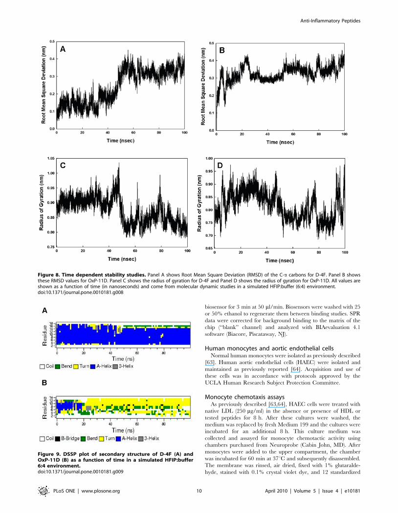
Secondary structure studies: molecular dynamicssimulations
Simulations of D-4F and OxP-11D in the ‘‘structure-promot-
ing’’ HFIP:buffer, solvent system were consistent with the
structures deduced from CD and FTIR measurements
(Figure 7). D-4F maintained a highly helical structure in the
simulated HFIP:buffer environment. After 40 nanoseconds, the
radius of gyration and RMSD of its C-a carbon backbone atoms
indicated a stable conformation with an uninterrupted helix
between residues 2 to 15. In contrast, within 20 nanoseconds,
OxP-11D changed its initially helical structure to a more flexible
Figure 1. Monocyte chemotaxis assay of OxP peptides. Results were standardized to LDL and are expressed as Inflammatory Index (II). Agentswith an II <1 are considered to be inactive. Agents with an II.1 are considered to be pro-inflammatory and those with an II,1 are considered to beanti-inflammatory. OxP-11, OxP-13 and D-4F showed very similar activity.doi:10.1371/journal.pone.0010181.g001
Figure 2. Dose response experiments. OxP-11 and OxP-13, the twomost active oxpholipins in the monocyte chemotaxis assay were re-tested at three concentrations: 1 mg/ml, 0.1 mg/ml and 0.01 mg/ml. A10-fold reduction, from 1.0 to 0.1 mg/ml caused a relatively smalldecrease in activity, but a 100-fold reduction to 10 ng/ml reducedactivity substantially.doi:10.1371/journal.pone.0010181.g002
Anti-Inflammatory Peptides
PLoS ONE | www.plosone.org 4 April 2010 | Volume 5 | Issue 4 | e10181
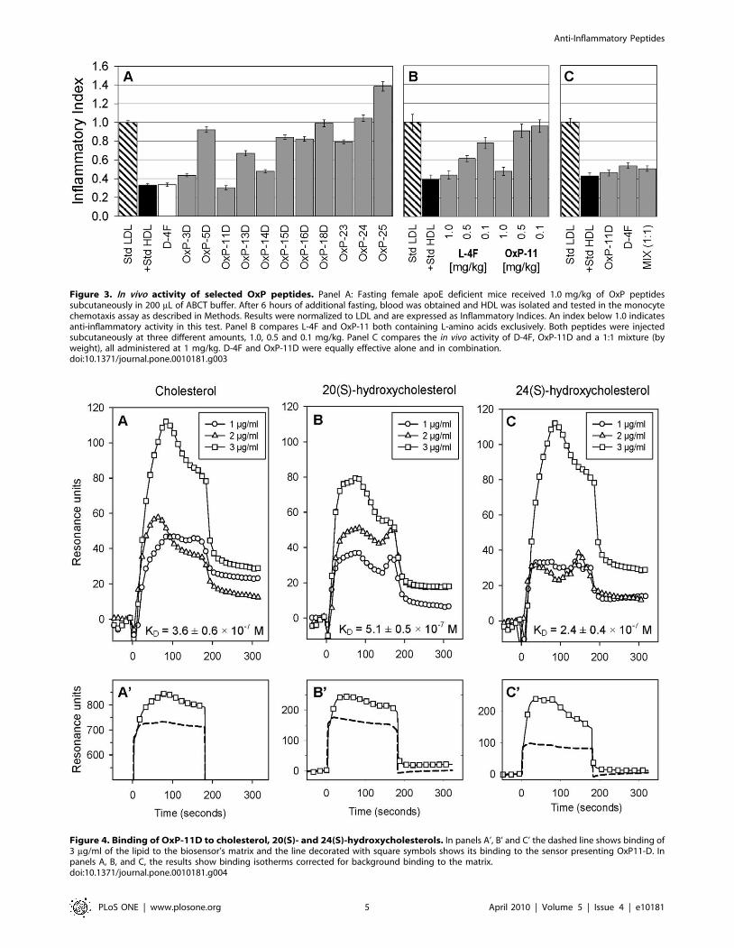
Figure 3. In vivo activity of selected OxP peptides. Panel A: Fasting female apoE deficient mice received 1.0 mg/kg of OxP peptidessubcutaneously in 200 mL of ABCT buffer. After 6 hours of additional fasting, blood was obtained and HDL was isolated and tested in the monocytechemotaxis assay as described in Methods. Results were normalized to LDL and are expressed as Inflammatory Indices. An index below 1.0 indicatesanti-inflammatory activity in this test. Panel B compares L-4F and OxP-11 both containing L-amino acids exclusively. Both peptides were injectedsubcutaneously at three different amounts, 1.0, 0.5 and 0.1 mg/kg. Panel C compares the in vivo activity of D-4F, OxP-11D and a 1:1 mixture (byweight), all administered at 1 mg/kg. D-4F and OxP-11D were equally effective alone and in combination.doi:10.1371/journal.pone.0010181.g003
Figure 4. Binding of OxP-11D to cholesterol, 20(S)- and 24(S)-hydroxycholesterols. In panels A’, B’ and C’ the dashed line shows binding of3 mg/ml of the lipid to the biosensor’s matrix and the line decorated with square symbols shows its binding to the sensor presenting OxP11-D. Inpanels A, B, and C, the results show binding isotherms corrected for background binding to the matrix.doi:10.1371/journal.pone.0010181.g004
Anti-Inflammatory Peptides
PLoS ONE | www.plosone.org 5 April 2010 | Volume 5 | Issue 4 | e10181
conformation with multiple turn-like features, evident from both
the C-a carbons and the radius of gyration (Figure 8). The DSSP
plot of OxP-11D after 20 nanoseconds revealed a stable turn
(residues 9 to 13), some helical propensity (residues 4–6), and more
disordered-coil conformations elsewhere (Figure 9).
Discussion
This study was undertaken to investigate the properties of
peptides derived from a highly conserved, cholesterol-binding
domain found in over 20 bacterial exotoxins (‘‘cholesterol
dependent cytolysins’’) secreted by Gram-positive bacteria.
Initially, we hoped only to design peptides or peptidomimetic
analogs that, by binding cholesterol, would protect human cells
from the lytic effects of these bacterial exotoxins, Indeed, several of
the peptides in Table 2 protected human erythrocytes from lysis
by anthrolysin O, listeriolysin O, and pneumolysin. These studies
will be described elsewhere.
Because peptides that bind cholesterol lipids have potential as
anti-inflammatory and/or anti-atherosclerosis agents we also
examined the binding of other lipids by the ‘‘Oxpholipin’’
peptides, as well as their activity in a monocyte chemotaxis assay
that guided the initial development of L-4F and related apoA-I-
mimetic peptides. The results of these studies caused us to focus on
OxP-11D, which is the principal subject of this report.
In vitro and in studies with apoE deficient mice, the effects of
OxP-11D on monocyte chemotaxis were dose-dependent and
similar in magnitude to those of D-4F. Surface plasmon resonance
studies revealed additional similarities to D-4F. Both peptides
bound normal and oxidized cholesterol, but OxP-11D did so with
higher affinity. Five of the 15 nonsteroid lipids in Table 3 were
apparently bound with higher affinity by OxP-11D and ten were
bound more effectively by D-4F. For example, the affinity of D-4F
for arachidonic and linoleic acid, POVPC, PEIPC and KODia-
PC appear to be considerably higher than that of OxP-11D.
Certain studies suggest that the factors responsible for the
different binding preferences of D-4F and OxP-11D reside in the
fine structure of the lipids as well as in those of the peptides
[52–57]. For example, OxP-11D bound 12(S)-HPETE (hydro-
peroxyeicosatetraenoic acid) with higher affinity than D-4F, and
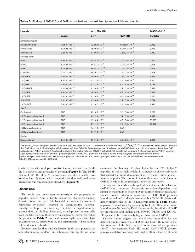
Table 3. Binding of OxP-11D and D-4F to oxidized and nonoxidized (phospho)lipids and sterols.
Ligands KD ± SEM (M) D-4F/OxP-11D
apoA-I D-4F OxP-11D KD Ratio
Nonoxidized lipids
Arachidonic acid 1.060.161028 1.260.161028 9.763.061027 0.012
Linoleic acid 6.562.561029 1.560.161028 6.061.361027 0.025
Palmitic acid 2.060.361026 6.760.761027 3.360.761027 2.030
Oxidized lipids
PGPC 4.264.261026 6.364.261029 3.260.661026 0.002
POVPC 3.162.861025 2.462.061029 3.060.461027 0.008
PEIPC 5.160.661025 6.065.0610211 1.260.461028 0.005
KOdiA-PC 2.161.161025 5.860.0610210 1.960.561027 0.003
5(S)-HPETE 1.660.361023 1.860.761028 1.760.561028 1.059
12(S)-HPETE 8.262.761024 1.761.261027 3.461.061028 5.000
15(S)-HPETE 1.060.261023 5.161.361028 4.561.861028 1.133
13(S)-HPODE 1.260.661023 2.160.561028 3.160.561028 0.677
12(S)-HETE 8.563.361024 1.860.561028 4.861.261028 0.375
15(S)-HETE 1.360.161023 2.260.861028 9.361.561028 0.236
9(S)-HODE 1.360.561023 1.560.461028 2.160.561028 0.714
13(S)-HODE 1.860.361023 1.160.661028 3.861.061029 2.895
Sterols
Cholesterol BND 9.260.361028 3.660.661027 0.255
20(S)-Hydroxycholesterol BND 3.962.161026 5.160.561027 7.647
22(S)-Hydroxycholesterol BND 1.560.461025 4.560.861027 33.333
24(S)-Hydroxycholesterol BND 4.361.661026 2.460.461027 17.917
25-Hydroxycholesterol BND 2.461.561026 BND _
4b-Hydroxycholesterol BND 3.161.761025 BND _
Control
Bovine albumin 2.160.461028 3.961.561026 6.961.061027 2.609
The mean KD values for apoA-I and D-4F are from [42], and those for OxP-11D are from this study. The ratio (KDD-4F)/(KD
OxP-11D) is also shown. Ratios below 1 indicatethat D-4F binds the lipid with higher affinity (lower KD) than OxP-11D. Ratios greater than 1 indicate that OxP-11D binds the lipid with higher affinity than D-4F.Abbreviations: PGPC-1-palmitoyl-2-glutaroyl-sn-glycero-3-phosphorylcholine; POVPC-1-palmitoyl-2-(5-oxovaleroyl)-sn-glycero-3-phosphorylcholine; PEIPC-1-palmitoyl-2-(5,6-epoxyisoprostane E2)-sn-glycero-3-phosphorylcholine; KOdiA-PC-1-palmitoyl-2-(5-keto-6-octene-dioyl)-sn-glycero-phosphatidylcholine; HPETE-hydroperoxyeicosatetraenoic acid; HPODE-hydroperoxyoctadecadienoic acid, HETE- hydroxyeicosatetraenoic acid; HODE- hydroxyoctadecadienoic acid.doi:10.1371/journal.pone.0010181.t003
Anti-Inflammatory Peptides
PLoS ONE | www.plosone.org 6 April 2010 | Volume 5 | Issue 4 | e10181

yet D-4F bound 12(S)-HETE (hydroxyeicosatetraenoic acid) with
much higher affinity than OxP-11D. Similarly, OxP-11D bound
13(S)-HODE (hydroxyoctadecadienoic acid) with higher affinity
than D-4F, yet D-4F had greater affinity for 9(S)-HODE.
Changes in the secondary structure of OxP-11D in lipid
environments were noted in our structural studies, and may
correlate with our functional measurements in vitro and in vivo.
CD, FTIR and molecular dynamics studies showed the peptide to
have considerable conformational freedom. OxP-11D had a less
defined conformation in more polar environments, however in
hydrophobic solvent systems and in lipid multilayer ensembles,
including these with cholesterol, the peptide showed highly
ordered helix and turn structures.
Recently, L-4F and D-4F were shown to bind oxidized lipids
with much higher affinity than human apoA-I [42] and it was
suggested that this property might contribute to its anti-
inflammatory activity by sequestering strongly pro-inflammatory
oxidized sterols and lipids [2,3,10,13,58,59]. How do the lipid
binding properties of D-4F and OxP-11D compare? If we can use
the KD values and ratios shown in Table 3 as a basis for
comparison, there are similarities and differences. Both peptides
bind cholesterol and its oxidized derivatives, with the advantage
going to OxP-11D. This is hardly surprising, given that 13 of the
14 residues (92.7%) in OxP-11D are identical to the library’s
cholesterol-binding design template.
Their respective binding of 5(S)-HPETE and 12(S)-HPETE is
also instructive. 5(S)-HPETE, which is derived from arachidonic
acid by the actions of 5-lipoxygenase, is a direct precursor of
leukotrienes A4, B4 and C4. The KD values (Table 3) show that
D-4F binds arachidonic acid, 5(S)-HPETE and 12(S)-HPETE with
similar affinities. In contrast, based on the KD ratios, the affinity of
OxP-11D for 5(S)-HPETE was approximately 57-fold higher than
its affinity for arachidonic acid, and its affinity for 12(S)-HPETE
was approximately 28.5-fold higher. Whether these differences will
prove advantageous or detrimental in animal trials of OxP-11D
cannot be predicted. Nonetheless, OxP-11D bound oxidized lipids
and sterols preferentially, and with much higher affinity than
apoA-I. Therefore it may function as oxidized-compounds’
sequestering agent influencing lipid metabolism on molecular
and cellular level. For these and other reasons, OxP-11D and
Figure 5. Binding of oxidized lipids to OxP-11D. Abbreviations: 13(S)-HODE: 13(S)-hydroxyoctadecadienoic acid, PEIPC: 1-palmitoyl-2-(5,6-deoxyisoprostane E2)-sn-glycero-3-phosphorylcholine, 12(S)-HPETE: 12(S)-hydroperoxyeicosatetraenoic acid, 5(S)-HPETE: 5(S)-hydroperoxyeicosate-traenoic acid. The biosensor presented 4686 RU of OxP-11D.doi:10.1371/journal.pone.0010181.g005
Anti-Inflammatory Peptides
PLoS ONE | www.plosone.org 7 April 2010 | Volume 5 | Issue 4 | e10181

structurally related peptides are interesting lead compounds whose
further study could result in the development of novel peptide and
peptidomimetic therapeutics.
Materials and Methods
Ethics StatementThe experiments were performed using protocols approved by
the Animal Research Committee at UCLA.
Peptide synthesis and characterizationSolid phase peptide synthesis was done with a SymphonyH
automated peptide synthesizer (Protein Technologies Inc., Tuc-
son, AZ) or a CEM Liberty automatic microwave peptide
synthesizer (CEM Corporation Inc., Matthews, NC), using 9-
fluorenylmethyloxycarbonyl (Fmoc) chemistry [60]. Amino acid
derivatives and reagents were from EMD Biosciences (San Diego,
CA) or Chem-Impex International (Wood Dale, IL). After cleaving
the peptides from the resin with modified reagent K (TFA 94% (v/
v); phenol, 2% (w/v); water, 2% (v/v); TIS, 2% (v/v); 2 hours)
they were precipitated with ice-cold diethyl ether and purified to
.95% homogeneity by preparative reverse-phase high perfor-
mance liquid chromatography (RP-HPLC).
Peptide purity was evaluated by matrix-assisted laser desorption
ionization spectrometry (MALDI-MS) and by analytical RP-
HPLC, using a ProStar 210 HPLC system with a ProStar 325
Figure 6. Circular dichroism (CD) and Fourier-Transform Infrared (FTIR) spectra. Panel A shows CD spectra of D-4F and Panel B shows thespectra of OxP-11D. Spectra were obtained in two solvent systems: 10 mM phosphate buffer, pH 7.4 (dashed line ----), HFIP:Buffer, (10 mM, pH 7.4)4:6, v:v (solid line —). The peptide concentration was 100 mM, the cuvette light path was 0.01 cm, the temperature was 25uC, and the spectra are theaverage of 8 scans. Panel C shows FTIR spectra of D-4F and Panel D shows FTIR spectra of OxP-11D. The spectra were obtained in D2O Buffer, TFE:deuterium Buffer 4:6, v:v, HFIP: deuterium Buffer, 4:6, v:v, deuterium vapor hydrated DMPC multilayers (peptide:lipid, 1:10, mole:mole), and deuteriumvapor hydrated DMPC:CHO (DMPC:CHO,1.2:1, mole:mole with a peptide to lipid ratio of 1:10, mole:mole).doi:10.1371/journal.pone.0010181.g006
Table 4. Proportions of different components of secondarystructure for D-4F and OxP-11D peptides based on CircularDichroic spectroscopic analysis.
Sample* Conformation (%)
a-helix turns b-sheet disordered
D-4F in Buffer 65.0 12.0 13.0 10.0
D-4F in HFIP:Buffer 74.0 9.0 11.0 6.0
OxP-11D in Buffer 15.0 19.0 30.0 36.0
OxP-11D in HFIP:Buffer 25.0 33.0 14.0 28.0
*peptides (100 mM) in 10 mM phosphate buffer pH 7.4 or HFIP:10 mMphosphate buffer pH 7.4 were analyzed for secondary conformation based asdescribed in the Methods section.
doi:10.1371/journal.pone.0010181.t004
Anti-Inflammatory Peptides
PLoS ONE | www.plosone.org 8 April 2010 | Volume 5 | Issue 4 | e10181

Dual Wavelength detector set at 220 nm and 280 nm (Varian
Inc., Palo Alto, CA). The mobile phases were: Solvent A, 0.1%
TFA in water; solvent B, 0.1% TFA in acetonitrile. Analytic
assessments used a reversed-phase, 4.6(250 mm C18 column
(Vydac 218TP54) and a linear 0 to 100% gradient of solvent B
applied over 100 min at 1 ml/min. Peptides OxP-3, OxP-3D and
OxP-21 were obtained by dimerizing the appropriate monomer
(10 mg/ml) in a 50% aqueous solution of DMSO at room
temperature over 48 h. OxP-22 was obtained by trimerizing OxP-
20 in 50% aqueous solution of DMF using Tris-[2-maleimi-
doethyl]amine and the manufacturer’s protocol (Pierce Biotech-
nology, Rockford, IL, Cat#33043). The progress of the reaction
was monitored by mass spectrometry. All peptides were lyophi-
lized for storage. Peptide stock solutions were made in HPLC
grade water containing 0.01% acetic acid, and peptide concen-
trations were determined by absorbance at 280 nm [61]. For
analytical details concerning synthesized peptides please see
Supporting Information (Table S1 and Figure S1).
LipidsPAPC (L-a-1-palmitoyl-2-arachidonoyl-sn-glycero-3-phospho-
rylcholine), PAPE (1-palmitoyl-2-arachidonoyl-sn-glycero-3-phos-
phatidylethanolamine), POVPC (1-palmitoyl-2-(5-oxovaleroyl)-sn-
glycero-3-phosphorylcholine, PGPC (1-palmitoyl-2-glutaroyl-sn-
glycero-3-phosphorylcholine), and POPC (1-Palmitoyl-2-oleoyl-
sn-glycero-3-phosphorylcholine) were from Avanti Polar Lipids
(Alabaster, AL). Cholesterol, $99% pure, 20(S)-hydroxycholes-
terol, 22(S)- and 25(S)-hydroxycholesterol, arachidonic acid,
linoleic acid, palmitic acid and bovine serum albumin were from
Sigma-Aldrich (St. Louis, MO). 13(S)-HPODE (hydroperoxyocta-
decadienoic acid), 5(S)-, 12(S)-, and 15(S)-HPETE (hydroperox-
yeicosatetraenoic acid), 12(S)- and 15(S)-HETE (hydroxyeicosate-
traenoic acid), were from BioMol, Plymouth Meeting, PA. 9(S)-
and 13(S)-HODE (hydroxyoctadecadienoic acid). KOdiA-PC was
from Cayman Chemical US (Ann Arbor, MI). 24(S)-hydroxycho-
lesterol was from Steraloids (Newport, RI). PEIPC (1-palmitoyl-2-
(5,6-deoxyisoprostane E2)-sn-glycero-3-phosphorylcholine) was
prepared as previously described [11,62].
Binding studiesBinding experiments were done by surface plasmon resonance
(SPR) on a BIAcore 3000 system (BiaCore AB, Piscataway, NJ).
Peptide ligands and apoA-I were immobilized on a BIAcore CM5
sensor chip activated per the manufacturer’s protocol with N-
hydroxysuccinimide and 1-ethyl-3-(3-dimethylaminoisopropyl)
carbodiimide. After achieving adequate immobilization, the
activated sensor surface was blocked with ethanolamine.
Lipid stock solutions were prepared in absolute ethanol and
then diluted into a standard BIAcore buffer (HBS-EP), containing
10 mM HEPES, pH 7.4, 150 mM NaCl, 3 mM EDTA and
0.005% (v/v) surfactant P20. Lipid concentrations used in the
binding studies were selected to give binding responses of 30-500
resonance units. Lipid stock solutions were prepared at 1 mg/ml
in ethanol. Since the highest analyte lipid concentrations did not
exceed 10 mg/ml, the highest ethanol concentration in any analyte
solutions was 1%, and for most lipids the ethanol concentration
was considerably lower. Ethanol-free HBS-EP buffer was used
during the dissociation phase. Lipid binding was measured by
observing the change in the SPR angle as 150 ml of lipid analyte
(various concentrations) in HBS-EP buffer flowed over the
Table 5. Secondary structural composition of D-4F and OxP-11D in different solvent systems, inferred from infrared (IR)spectroscopy.
Peptide/Solvent System Conformation (%)
a-helix turns b-sheet disordered
D-4F/Buffer 63.0 11.4 11.8 13.7
D-4F/TFE:Buffer 67.2 8.8 11.3 12.7
D-4F/HFIP:Buffer 71.5 8.0 11.9 8.6
D-4F/DMPC (1:10 mole:mole) 57.8 16.4 1.9 23.9
D-4F/DMPC:CHO (1:10 mole:mole) 58.1 15.2 4.1 22.6
OxP-11D/Buffer 20.1 24.8 33.7 21.4
OxP-11D/TFE:Buffer 32.9 31.0 22.9 13.2
OxP-11D/HFIP:Buffer 32.4 37.3 10.3 20.0
OxP-11D/DMPC (1:10 mole:mole) 26.5 24.4 24.9 24.2
OxP-11D/DMPC:CHO (1:10 mole:mole)
29.5 27.7 22.6 20.2
The peptides (1 mM) were studied in the following solvent systems: deuterated10 mM phosphate buffer pD 7.4 (‘‘DPB’’); trifluoroethanol (TFE):DPB; orhexafluoroisopropanol (HFIP): DPB. Measurements were done and spectra wereanalyzed as described in the Methods. The peak area error for these estimates is64%.doi:10.1371/journal.pone.0010181.t005
Figure 7. Molecular illustration of the structure of D-4F (A) and OxP-11D (B) after 83 nsec of molecular dynamics in HFIP:aqueousbuffer 6:4, v:v environment. a-helical segments are in red ribbon, disordered and turn segments are in green highlight The N-terminus is at thelower left and the adjacent C-terminus is in the upper right of the figure.doi:10.1371/journal.pone.0010181.g007
Anti-Inflammatory Peptides
PLoS ONE | www.plosone.org 9 April 2010 | Volume 5 | Issue 4 | e10181
biosensor for 3 min at 50 ml/min. Biosensors were washed with 25
or 50% ethanol to regenerate them between binding studies. SPR
data were corrected for background binding to the matrix of the
chip (‘‘blank’’ channel) and analyzed with BIAevaluation 4.1
software (Biacore, Piscataway, NJ).
Human monocytes and aortic endothelial cellsNormal human monocytes were isolated as previously described
[63]. Human aortic endothelial cells (HAEC) were isolated and
maintained as previously reported [64]. Acquisition and use of
these cells was in accordance with protocols approved by the
UCLA Human Research Subject Protection Committee.
Monocyte chemotaxis assaysAs previously described [63,64], HAEC cells were treated with
native LDL (250 mg/ml) in the absence or presence of HDL or
tested peptides for 8 h. After these cultures were washed, the
medium was replaced by fresh Medium 199 and the cultures were
incubated for an additional 8 h. This culture medium was
collected and assayed for monocyte chemotactic activity using
chambers purchased from Neuroprobe (Cabin John, MD). After
monocytes were added to the upper compartment, the chamber
was incubated for 60 min at 37uC and subsequently disassembled.
The membrane was rinsed, air dried, fixed with 1% glutaralde-
hyde, stained with 0.1% crystal violet dye, and 12 standardized
Figure 8. Time dependent stability studies. Panel A shows Root Mean Square Deviation (RMSD) of the C-a carbons for D-4F. Panel B showsthese RMSD values for OxP-11D. Panel C shows the radius of gyration for D-4F and Panel D shows the radius of gyration for OxP-11D. All values areshown as a function of time (in nanoseconds) and come from molecular dynamic studies in a simulated HFIP:buffer (6:4) environment.doi:10.1371/journal.pone.0010181.g008
Figure 9. DSSP plot of secondary structure of D-4F (A) andOxP-11D (B) as a function of time in a simulated HFIP:buffer6:4 environment.doi:10.1371/journal.pone.0010181.g009
Anti-Inflammatory Peptides
PLoS ONE | www.plosone.org 10 April 2010 | Volume 5 | Issue 4 | e10181

high power fields were examined microscopically. The number of
migrated monocytes was expressed as the mean 6SD of
monocytes counted. Values obtained in the absence of HDL were
normalized to a value of 1.0. Normalized values .1.0 after HDL
addition were considered to be pro-inflammatory, and values ,1.0
as being anti-inflammatory.
Mouse experimentsApoE null mice on a C57BL/6J background, originally from
Jackson Laboratories (Bar Harbor, ME), were maintained in a
breeding colony in the Department of Laboratory and Animal
Medicine at the David Geffen School of Medicine at UCLA. The
mice were maintained on a chow diet (Ralston Purina, St. Louis,
MO).
Groups of 6 fasting female apoE deficient mice, 4–6 months of
age, were injected subcutaneously with 200 mL of either ABCT
buffer (50 mM NH4HCO3, 0.1% Tween 20) or 1.0 mg/kg of OxP
peptides in ABCT buffer. Six hours later, and with continued
fasting, blood was removed from the retro-orbital sinus under mild
isoflurane anesthesia, and anticoagulated with heparin (2.5 U/ml).
Plasma was obtained and fractionated by FPLC. HDL-containing
fractions were tested in HAEC cultures for their ability to inhibit
LDL-induced monocyte chemotactic activity. All procedures
conformed to regulations of the UCLA Animal Research
Committee. An HDL inflammatory index was determined using
the standard monocyte chemotaxis assay. Indices .1 were
interpreted as pro-inflammatory, and indices ,1 as anti-
inflammatory.
Circular dichroism (CD) analyses of secondary structureCD spectra from 190–260 nm of D-4F and OxP-11D were
examined in different solution environments using a JASCO 715
spectropolarimeter (Jasco Inc., Easton, MD) that was calibrated for
wavelength and optical rotation with 10-camphorsulphonic acid
[65,66]. Peptides were scanned at 20 nm per minute in 0.01 cm
path-length cells at 25uC with a sample interval of 0.2 nm. Peptide
concentration was determined by UV absorbance at 280 nm.
After baseline correction, the spectra were expressed as the Mean
Residue Ellipticity [h]MRE. Quantitative estimates of the secondary
structural contributions were made with VARSLC [67] using the
spectral basis set for proteins implemented in the Olis Global
WorksTM software package (Olis Inc., Bogart, GA).
Fourier transform infrared (FTIR) spectroscopyInfrared spectra were recorded at 25uC using a Bruker Vector
22TM FTIR spectrometer with a deuterated triglycine sulfate
(DTGS) detector, and averaged over 256 scans at a gain of 4 with
a resolution of 2 cm21. Lipid and peptide samples were initially
freeze-dried several times from 10 mM HCl in D2O to remove
any interfering counter ions and residual H2O. Solution spectra of
peptides were made in deuterated 10 mM phosphate buffer, pD
7.4 (pD = pH+0.4) and in structure promoting mixed solvent-
buffer solutions (trifluoroethanol (TFE) or hexafluoroisopropanol
(HFIP) at a sample concentration of 1 mM.
Spectra were acquired using a temperature controlled, de-
mountable liquid cell with calcium fluoride windows fitted with a
50 mm thick spacer (Harrick Scientific, Pleasantville, NY). Lipid-
peptide films were prepared by air-drying mixtures of DMPC and
DMPC:cholesterol (1.2:1, mole:mole) in chloroform with D-4F or
OxP-11D in TFE onto a 5062062 mm, 45u ATR crystal (Pike
Technologies, Madison, WI) fitted to the Bruker spectrometer [68]
to form a multilayer film (lipid:peptide, 10:1, mole:mole). After
evaporation the solvent lipid:peptide film was hydrated by
passaging deuterium-saturated nitrogen gas through the sample
chamber for one hour prior to spectroscopy [69–71]. The relative
proportions of a-helix, b-turn, b-sheet, and disordered conforma-
tions of solution and multilayer IR spectra were determined by
Fourier self-deconvolution for band narrowing and area calcula-
tions of component peaks of the FTIR spectra using curve-fitting
software supplied by Galactic Software (GRAMS/AI, version 8.0;
Thermo Electron Corp., Waltham, MA). The frequency limits for
the different structures were: a-helix (1662-1645 cm21), b-sheet
(1637-1613 and 1710-1682 cm21), turns (1682-1662 cm21), and
disordered or random (1650-1637 cm21) [72].
Molecular dynamics modelingMonomeric starting structures for D-4F and OxP-11D were
obtained by using Hyperchem 7.5 (http://www.hyper.com) to
build the peptides in a helical conformation. These structures were
placed in a periodic 56 A3 box of TIP4P water or HFIP:TIP4P
water (4:6, v:v) and the ensemble was neutralized with counter-
ions to simulate the environment used for our CD measurements
(equilibrated HFIP solvent box and topology files courtesy of D.
Roccatano) [73]. The peptide in the solution box was conjugate-
gradient- minimized using the Polak-Ribiere approach imple-
mented in Hyperchem. Minimized monomeric D-4F or OxP-11D
ensembles were ported to the Gromacs program suite, version
4.0.4 (http://www.gromacs.org), and subjected to the steepest
descent method using the OPLS_AA option [74].
The system was subjected to 20 psec of pre-run molecular
dynamics at 300uK allowing the solvent to equilibrate while
restraining the peptide. After pre-run solvent equilibration the
peptides were subjected to 100 nsec of free MD simulations at
300uK without any experimental constraints, utilizing Berendsen
temperature and pressure coupling and the Particle Mesh Ewald
method for evaluating long-range electrostatic interactions. The
time-dependent evolution of the root mean square deviations
(RMSD) for the peptide C-a carbons, radius of gyration and
secondary structure (i.e., analyzed using the DSSP criteria [75] for
the peptide in the HFIP-water environment indicated when
equilibrium was reached. Molecular model illustrations were
rendered using PyMOL v0.99 (http://www.pymol.org).
Supporting Information
Table S1 Analytical data for OxP peptides. Peptide purity was
evaluated by matrix-assisted laser desorption ionization spectrom-
etry (MALDI-MS) and by analytical RP-HPLC, using a ProStar
210 HPLC system with a ProStar 325 Dual Wavelength detector
set at 220 nm and 280 nm (Varian Inc., Palo Alto, CA). The
mobile phases were: Solvent A, 0.1% TFA in water; solvent B,
0.1% TFA in acetonitrile. Analytic assessments used a reversed-
phase, 4.66250 mm C18 column (Vydac 218TP54) and a linear 0
to 100% gradient of solvent B applied over 100 min at 1 ml/min.
Found at: doi:10.1371/journal.pone.0010181.s001 (0.06 MB
DOC)
Figure S1 Two examples of analytical RP-HPLC profiles and
corresponding MS spectra obtained for OxP peptides. Panel A -
OxP-5; Panel B - OxP-11D.
Found at: doi:10.1371/journal.pone.0010181.s002 (0.13 MB
DOC)
Author Contributions
Conceived and designed the experiments: PPR RL. Performed the
experiments: PPR MN CLJ SHL EDM HL JR SS AJW. Analyzed the
data: PPR MN CLJ SS AJW AF RL. Contributed reagents/materials/
analysis tools: PPR AJW AF. Wrote the paper: PPR RL.
Anti-Inflammatory Peptides
PLoS ONE | www.plosone.org 11 April 2010 | Volume 5 | Issue 4 | e10181

References
1. Bjorkhem I (2002) Do oxysterols control cholesterol homeostasis? J Clin Invest110: 725–730.
2. Torocsik D, Szanto A, Nagy L (2009) Oxysterol signaling links cholesterol
metabolism and inflammation via the liver X receptor in macrophages. MolAspects Med 30: 134–152.
3. Olkkonen VM, Hynynen R (2009) Interactions of oxysterols with membranes
and proteins. Mol Aspects Med 30: 123–133.
4. Umetani M, Domoto H, Gormley AK, Yuhanna IS, Cummins CL, et al. (2007)27-Hydroxycholesterol is an endogenous SERM that inhibits the cardiovascular
effects of estrogen. Nat Med 13: 1185–1192.
5. Taylor FR, Saucier SE, Shown EP, Parish EJ, Kandutsch AA (1984) Correlationbetween oxysterol binding to a cytosolic binding protein and potency in the
repression of hydroxymethylglutaryl coenzyme A reductase. J Biol Chem 259:
12382–12387.
6. Suchanek M, Hynynen R, Wohlfahrt G, Lehto M, Johansson M, et al. (2007)The mammalian oxysterol-binding protein-related proteins (ORPs) bind 25-
hydroxycholesterol in an evolutionarily conserved pocket. Biochem J 405:473–480.
7. Goldstein JL, Bose-Boyd RA, Brown MS (2006) Protein sensors for membrane
sterols. Cell 124: 35–46.
8. Infante RE, bi-Mosleh L, Radhakrishnan A, Dale JD, Brown MS, et al. (2008)Purified NPC1 protein. I. Binding of cholesterol and oxysterols to a 1278-amino
acid membrane protein. J Biol Chem 283: 1052–1063.
9. Calder PC (2006) n-3 polyunsaturated fatty acids, inflammation, andinflammatory diseases. Am J Clin Nutr 83: 1505S–1519S.
10. Calder PC (2008) Polyunsaturated fatty acids, inflammatory processes and
inflammatory bowel diseases. Mol Nutr Food Res 52: 885–897.
11. Subbanagounder G, Wong JW, Lee H, Faull KF, Miller E, et al. (2002)Epoxyisoprostane and epoxycyclopentenone phospholipids regulate monocyte
chemotactic protein-1 and interleukin-8 synthesis. Formation of these oxidized
phospholipids in response to interleukin-1beta. J Biol Chem 277: 7271–7281.
12. Lewis RA, Austen KF, Soberman RJ (1990) Leukotrienes and other products of
the 5-lipoxygenase pathway. Biochemistry and relation to pathobiology in
human diseases. N Engl J Med 323: 645–655.
13. Tilley SL, Coffman TM, Koller BH (2001) Mixed messages: modulation ofinflammation and immune responses by prostaglandins and thromboxanes.
J Clin Invest 108: 15–23.
14. Boyce JA (2008) Eicosanoids in asthma, allergic inflammation, and host defense.Curr Mol Med 8: 335–349.
15. Capra V, Ambrosio M, Riccioni G, Rovati GE (2006) Cysteinyl-leukotriene
receptor antagonists: present situation and future opportunities. Curr MedChem 13: 3213–3226.
16. Peters-Golden M, Henderson WR, Jr. (2007) Leukotrienes. N Engl J Med 357:
1841–1854.
17. Riccioni G, Santilli F, D’Orazio N, Sensi S, Spoltore R, et al. (2002) The role ofantileukotrienes in the treatment of asthma. Int J Immunopathol Pharmacol 15:
171–182.
18. Samuelsson B, Dahlen SE, Lindgren JA, Rouzer CA, Serhan CN (1987)Leukotrienes and lipoxins: structures, biosynthesis, and biological effects. Science
237: 1171–1176.
19. Keane D, Newsholme P (2008) Saturated and unsaturated (including
arachidonic acid) non-esterified fatty acid modulation of insulin secretion frompancreatic beta-cells. Biochem Soc Trans 36: 955–958.
20. Claria J (2006) Regulation of cell proliferation and apoptosis by bioactive lipid
mediators. Recent Pat Anticancer Drug Discov 1: 369–382.
21. Krishnamoorthy S, Honn KV (2008) Eicosanoids in tumor progression andmetastasis. Subcell Biochem 49: 145–168.
22. Moreno JJ (2009) New aspects of the role of hydroxyeicosatetraenoic acids in cell
growth and cancer development. Biochem Pharmacol 77: 1–10.
23. Patel MI, Kurek C, Dong Q (2008) The arachidonic acid pathway and its role inprostate cancer development and progression. J Urol 179: 1668–1675.
24. Hirata I, Murano M, Nitta M, Sasaki S, Toshina K, et al. (2001) Estimation of
mucosal inflammatory mediators in rat DSS-induced colitis. Possible role ofPGE(2) in protection against mucosal damage. Digestion 63 Suppl 1: 73–80.
25. Nieto N, Torres MI, Rios A, Gil A (2002) Dietary polyunsaturated fatty acids
improve histological and biochemical alterations in rats with experimentalulcerative colitis. J Nutr 132: 11–19.
26. Ku IA, Imboden JB, Hsue PY, Ganz P (2009) Rheumatoid arthritis: model of
systemic inflammation driving atherosclerosis. Circ J 73: 977–985.
27. Montecucco F, Mach F (2009) Common inflammatory mediators orchestratepathophysiological processes in rheumatoid arthritis and atherosclerosis.
Rheumatology (Oxford) 48: 11–22.
28. Sharon P, Stenson WF (1984) Enhanced synthesis of leukotriene B4 by colonicmucosa in inflammatory bowel disease. Gastroenterology 86: 453–460.
29. Back M (2009) Leukotriene signaling in atherosclerosis and ischemia. Cardiovasc
Drugs Ther 23: 41–48.
30. Riccioni G, Capra V, D’Orazio N, Bucciarelli T, Bazzano LA (2008)Leukotriene modifiers in the treatment of cardiovascular diseases. J Leukoc
Biol 84: 1374–1378.
31. Hansson GK, Libby P (2006) The immune response in atherosclerosis: a double-edged sword. Nat Rev Immunol 6: 508–519.
32. Klingenberg R, Hansson GK (2009) Treating inflammation in atherosclerotic
cardiovascular disease: emerging therapies. Eur Heart J.
33. Montecucco F, Mach F (2009) Atherosclerosis is an inflammatory disease. Semin
Immunopathol 31: 1–3.
34. Montecucco F, Mach F (2009) Statins, ACE inhibitors and ARBs incardiovascular disease. Best Pract Res Clin Endocrinol Metab 23: 389–400.
35. Montecucco F, Mach F (2009) Update on statin-mediated anti-inflammatoryactivities in atherosclerosis. Semin Immunopathol 31: 127–142.
36. Moubayed SP, Heinonen TM, Tardif JC (2007) Anti-inflammatory drugs andatherosclerosis. Curr Opin Lipidol 18: 638–644.
37. Bloedon LT, Dunbar R, Duffy D, Pinell-Salles P, Norris R, et al. (2008) Safety,pharmacokinetics, and pharmacodynamics of oral apoA-I mimetic peptide D-4F
in high-risk cardiovascular patients. J Lipid Res 49: 1344–1352.
38. Navab M, Anantharamaiah GM, Reddy ST, Hama S, Hough G, et al. (2005)
Apolipoprotein A-I mimetic peptides. Arterioscler Thromb Vasc Biol 25:1325–1331.
39. Navab M, Anantharamaiah GM, Reddy ST, Fogelman AM (2006) Apolipo-protein A-I mimetic peptides and their role in atherosclerosis prevention. Nat
Clin Pract Cardiovasc Med 3: 540–547.
40. Navab M, Anantharamaiah GM, Reddy ST, Van Lenten BJ, Fogelman AM
(2008) Apo A-1 mimetic peptides as atheroprotective agents in murine models.Curr Drug Targets 9: 204–209.
41. Sethi AA, Amar M, Shamburek RD, Remaley AT (2007) Apolipoprotein AImimetic peptides: possible new agents for the treatment of atherosclerosis. Curr
Opin Investig Drugs 8: 201–212.
42. Van Lenten BJ, Wagner AC, Jung CL, Ruchala P, Waring AJ, et al. (2008) Anti-inflammatory apoA-I-mimetic peptides bind oxidized lipids with much higher
affinity than human apoA-I. J Lipid Res 49: 2302–2311.
43. Zhu X, Wu G, Zeng W, Xue H, Chen B (2005) Cysteine mutants of human
apolipoprotein A-I: a study of secondary structural and functional properties.J Lipid Res 46: 1303–1311.
44. Chiesa G, Sirtori CR (2003) Apolipoprotein A-I(Milano): current perspectives.Curr Opin Lipidol 14: 159–163.
45. Nissen SE, Tsunoda T, Tuzcu EM, Schoenhagen P, Cooper CJ, et al. (2003)Effect of recombinant ApoA-I Milano on coronary atherosclerosis in patients
with acute coronary syndromes: a randomized controlled trial. JAMA 290:2292–2300.
46. Navab M, Anantharamaiah GM, Reddy ST, Hama S, Hough G, et al. (2004)Oral D-4F causes formation of pre-beta high-density lipoprotein and improves
high-density lipoprotein-mediated cholesterol efflux and reverse cholesterol
transport from macrophages in apolipoprotein E-null mice. Circulation 109:3215–3220.
47. Navab M, Anantharamaiah GM, Reddy ST, Hama S, Hough G, et al. (2005)Oral small peptides render HDL antiinflammatory in mice and monkeys and
reduce atherosclerosis in ApoE null mice. Circ Res 97: 524–532.
48. Navab M, Ruchala P, Waring AJ, Lehrer RI, Hama S, et al. (2009) A novel
method for oral delivery of apolipoprotein mimetic peptides synthesized from allL-amino acids. J Lipid Res 50: 1538–1547.
49. Yamaguchi H, Kodama H, Osada S, Kato F, Jelokhani-Niaraki M, et al. (2003)Effect of alpha,alpha-dialkyl amino acids on the protease resistance of peptides.
Biosci Biotechnol Biochem 67: 2269–2272.
50. Navab M, Hama SY, Anantharamaiah GM, Hassan K, Hough GP, et al. (2000)
Normal high density lipoprotein inhibits three steps in the formation of mildly
oxidized low density lipoprotein: steps 2 and 3. J Lipid Res 41: 1495–1508.
51. Navab M, Hama-Levy S, Van Lenten BJ, Fonarow GC, Cardinez CJ, et al.
(1997) Mildly oxidized LDL induces an increased apolipoprotein J/paraoxonaseratio. J Clin Invest 99: 2005–2019.
52. Datta G, Chaddha M, Hama S, Navab M, Fogelman AM, et al. (2001) Effects ofincreasing hydrophobicity on the physical-chemical and biological properties of
a class A amphipathic helical peptide. J Lipid Res 42: 1096–1104.
53. Anantharamaiah G, Navab M, Reddy ST, Garber DW, Datta G, et al. (2006)
Synthetic peptides: managing lipid disorders. Curr Opin Lipidol 17: 233–237.
54. Datta G, Epand RF, Epand RM, Chaddha M, Kirksey MA, et al. (2004)
Aromatic residue position on the nonpolar face of class a amphipathic helicalpeptides determines biological activity. J Biol Chem 279: 26509–26517.
55. Epand RM, Epand RF, Sayer BG, Datta G, Chaddha M, et al. (2004) Twohomologous apolipoprotein AI mimetic peptides. Relationship between
membrane interactions and biological activity. J Biol Chem 279: 51404–51414.
56. Epand RM, Epand RF, Sayer BG, Melacini G, Palgulachari MN, et al. (2004)
An apolipoprotein AI mimetic peptide: membrane interactions and the role of
cholesterol. Biochemistry 43: 5073–5083.
57. Venkatachalapathi YV, Phillips MC, Epand RM, Epand RF, Tytler EM, et al.
(1993) Effect of end group blockage on the properties of a class A amphipathichelical peptide. Proteins 15: 349–359.
58. Vejux A, Lizard G (2009) Cytotoxic effects of oxysterols associated with humandiseases: Induction of cell death (apoptosis and/or oncosis), oxidative and
inflammatory activities, and phospholipidosis. Mol Aspects Med 30: 153–170.
59. Poli G, Sottero B, Gargiulo S, Leonarduzzi G (2009) Cholesterol oxidation
products in the vascular remodeling due to atherosclerosis. Mol Aspects Med 30:180–189.
60. Fields GB, Noble RL (1990) Solid phase peptide synthesis utilizing 9-fluorenylmethoxycarbonyl amino acids. Int J Pept Protein Res 35: 161–214.
Anti-Inflammatory Peptides
PLoS ONE | www.plosone.org 12 April 2010 | Volume 5 | Issue 4 | e10181

61. Pace CN, Vajdos F, Fee L, Grimsley G, Gray T (1995) How to measure and
predict the molar absorption coefficient of a protein. Protein Sci 4: 2411–2423.62. Gharavi NM, Gargalovic PS, Chang I, Araujo JA, Clark MJ, et al. (2007) High-
density lipoprotein modulates oxidized phospholipid signaling in human
endothelial cells from proinflammatory to anti-inflammatory. ArteriosclerThromb Vasc Biol 27: 1346–1353.
63. Fogelman AM, Elahi F, Sykes K, Van Lenten BJ, Territo MC, et al. (1988)Modification of the Recalde method for the isolation of human monocytes.
J Lipid Res 29: 1243–1247.
64. Navab M, Imes SS, Hama SY, Hough GP, Ross LA, et al. (1991) Monocytetransmigration induced by modification of low density lipoprotein in cocultures
of human aortic wall cells is due to induction of monocyte chemotactic protein 1synthesis and is abolished by high density lipoprotein. J Clin Invest 88:
2039–2046.65. Johnson WC, Jr. (1990) Protein secondary structure and circular dichroism: a
practical guide. Proteins 7: 205–214.
66. Miles AJ, Wien F, Lees JG, Janes RW, Wallace BA (2003) Calibration andstandardization of synchrotron radiation circular dichroism and conventional
circular dichroism spectrophotometers. Spectroscopy 17: 653–661.67. Johnson WC (1999) Analyzing protein circular dichroism spectra for accurate
secondary structures. Proteins 35: 307–312.
68. Gordon LM, Horvath S, Longo ML, Zasadzinski JA, Taeusch HW, et al. (1996)Conformation and molecular topography of the N-terminal segment of
surfactant protein B in structure-promoting environments. Protein Sci 5:
1662–1675.
69. Yamaguchi S, Huster D, Waring A, Lehrer RI, Kearney W, et al. (2001)
Orientation and dynamics of an antimicrobial peptide in the lipid bilayer by
solid-state NMR spectroscopy. Biophys J 81: 2203–2214.
70. Ulrich AS, Watts A (1994) Molecular response of the lipid headgroup to bilayer
hydration monitored by 2H-NMR. Biophys J 66: 1441–1449.
71. Bechinger B, Seelig J (1991) Conformational changes of the phosphatidylcholine
headgroup due to membrane dehydration. A 2H-NMR study. Chem Phys
Lipids 58: 1–5.
72. Byler DM, Susi H (1986) Examination of the secondary structure of proteins by
deconvolved FTIR spectra. Biopolymers 25: 469–487.
73. Roccatano D, Fioroni M, Zacharias M, Colombo G (2005) Effect of
hexafluoroisopropanol alcohol on the structure of melittin: a molecular
dynamics simulation study. Protein Sci 14: 2582–2589.
74. Hess B, Kutzner C, van der Spoel D (2008) GROMACS 4: Algorithms for
Highly Efficient, Load-Balanced, and Scalable Molecular Simulation. J Chem
Theory Comput, 4: 435–447.
75. Kabsch W, Sander C (1983) Dictionary of protein secondary structure: pattern
recognition of hydrogen-bonded and geometrical features. Biopolymers 22:
2577–2637.
Anti-Inflammatory Peptides
PLoS ONE | www.plosone.org 13 April 2010 | Volume 5 | Issue 4 | e10181