Role of Phospholipases and Oxidized Phospholipids in Inflammation
18
55 P.S. Tappia and N.S. Dhalla (eds.), Phospholipases in Health and Disease, Advances in Biochemistry in Health and Disease 10, DOI 10.1007/978-1-4939-0464-8_3, © Springer Science+Business Media New York 2014 Abstract Long thought of as a bystander in pathophysiological processes, lipid molecules have emerged as bioactive mediators of cellular activity. Oxidized phos- pholipids (OxPLs), generated during enzymatic and non-enzymatic processes, modulate cellular processes through receptor-mediated pathways that can effect a whole host of activities including apoptosis, monocyte adhesion, platelet aggrega- tion, and regulation of immune responses. Initially discovered as platelet activating factor analogs, there have been close to 50 distinct OxPL molecules that have been identified within biological tissues. With the advent of robust analytical systems, we are better able to identify and quantitate these molecules in an ever growing list of different biological tissues which has allowed for the generation of a comprehensive oxolipid profiles in both normal and disease states. Given the increased affinity of phospholipases towards OxPLs we are in the early stages of understanding of the complex interplay between the modification of OxPL through phospholipase activity and the cellular responses to the released hydrolyzed products. In this review we will summarize the role of OxPL in different pathological states and the specific phospholipases that have been shown to interact with OxPLs. Keywords Oxidized phospholipids • Phospholipases • Oxidative stress • Mass spectrometry • Lipidomics Chapter 3 Role of Phospholipases and Oxidized Phospholipids in Inflammation Devin Hasanally, Rakesh Chaudhary, and Amir Ravandi D. Hasanally • R. Chaudhary Institute of Cardiovascular Sciences, St. Boniface Hospital Research, Winnipeg, MB, Canada A. Ravandi (*) Institute of Cardiovascular Sciences, St. Boniface Hospital Research, Winnipeg, MB, Canada Institute of Cardiovascular Sciences, St. Boniface Hospital, 409 Tache Avenue, Winnipeg, MB, Canada R2H 2A6 e-mail: [email protected]
-
Upload
independent -
Category
Documents
-
view
1 -
download
0
Transcript of Role of Phospholipases and Oxidized Phospholipids in Inflammation

55P.S. Tappia and N.S. Dhalla (eds.), Phospholipases in Health and Disease, Advances in Biochemistry in Health and Disease 10, DOI 10.1007/978-1-4939-0464-8_3,© Springer Science+Business Media New York 2014
Abstract Long thought of as a bystander in pathophysiological processes, lipid molecules have emerged as bioactive mediators of cellular activity. Oxidized phos-pholipids (OxPLs), generated during enzymatic and non-enzymatic processes, modulate cellular processes through receptor-mediated pathways that can effect a whole host of activities including apoptosis, monocyte adhesion, platelet aggrega-tion, and regulation of immune responses. Initially discovered as platelet activating factor analogs, there have been close to 50 distinct OxPL molecules that have been identifi ed within biological tissues. With the advent of robust analytical systems, we are better able to identify and quantitate these molecules in an ever growing list of different biological tissues which has allowed for the generation of a comprehensive oxolipid profi les in both normal and disease states. Given the increased affi nity of phospholipases towards OxPLs we are in the early stages of understanding of the complex interplay between the modifi cation of OxPL through phospholipase activity and the cellular responses to the released hydrolyzed products. In this review we will summarize the role of OxPL in different pathological states and the specifi c phospholipases that have been shown to interact with OxPLs.
Keywords Oxidized phospholipids • Phospholipases • Oxidative stress • Mass spectrometry • Lipidomics
Chapter 3 Role of Phospholipases and Oxidized Phospholipids in Infl ammation
Devin Hasanally , Rakesh Chaudhary , and Amir Ravandi
D. Hasanally • R. Chaudhary Institute of Cardiovascular Sciences , St. Boniface Hospital Research , Winnipeg , MB , Canada
A. Ravandi (*) Institute of Cardiovascular Sciences , St. Boniface Hospital Research , Winnipeg , MB , Canada
Institute of Cardiovascular Sciences , St. Boniface Hospital , 409 Tache Avenue , Winnipeg , MB , Canada R2H 2A6 e-mail: [email protected]

56
3.1 Introduction
For many years phospholipids (PLs) were considered to be only cellular building blocks with very little biological activity. Due to their susceptibility to oxidation, they are modifi ed in the presence of reactive oxygen species (ROS). Apart from impairment of their structural function, oxidation makes oxidized phospholipids (OxPLs) acquire novel biological activities not characteristic of their unoxidized precursors (Fig. 3.1 ). The effects of OxPLs described in vitro and in vivo suggest their potential relevance in different pathologies including atherosclerosis, acute infl ammation, lung injury, and many other disease conditions [ 1 , 2 ]. The actions of OxPL can vary depending upon the specifi c species of phospholipid being oxidized. Recently, oxidized phosphatidylcholines (OxPC) have been recognized as not only products of oxidative damage but also mediators of its progression. These compounds exert their biological activity through multiple pathways. They have been shown to be potent stimulators of platelet-activating factor (PAF) receptor, prostaglandin receptors, and PPARγ receptors resulting in platelet aggregation, induction of the coagulation cascade, and apoptosis and cell death [ 3 , 4 ]. Recent advancements in softer methods of ionization, such as electrospray mass spectrometry, have allowed us to identify and quantitate OxPLs in biological tissues. With the better understanding of the OxPL structure, we are also identifying the specifi c role phospholipases play in modulating the effects of OxPL on cellular signaling. As we move forward in trying to better understand the role of OxPL in pathology, it necessitates a detailed understand-ing of the oxidized lipidome and the specifi c phospholipase that act as a defensive mechanism to protect the cell from their deleterious effects.
3.2 Generation of Oxidized Phospholipids
Phospholipids represent the major component of lipid bilayers due to their amphipa-thic structure. Polar head groups interact with the aqueous environment and cyto-plasm, and the fatty acid chains sequester to form the lipid core of the membrane acting as a semi-permeable barrier. During disease processes, not only does the struc-tural integrity of the phospholipid bilayer become compromised but also chemical modifi cation of the phospholipids through enzymatic and non- enzymatic pathways alters their function. One such disease process is the infl ammatory cascade that is a unifying mechanism in many pathological processes. A hallmark of infl ammation is the increased generation of ROS which can occur in multiple pathways [ 5 – 7 ] and results in the generation of superoxide radicals, OONO • and O • . This process is well described, for example, during ischemia- reperfusion injury within cardiomyocytes. The rapid correction of acidosis through the Na + /H + exchanger, the Na + /HCO 3− cotransporter [ 8 ], and the washout of lactate causes secondary activation of the Na + /Ca 2+ exchanger in the reverse direction aggravating the cytosolic Ca 2+ balance [ 9 ]. Abrupt re-exposure to oxygen of the ischemia-inhibited respiratory chain generates a mitochondrial membrane potential to drive ATP synthesis, which leads to a rapid cytosolic Ca 2+ overload and consequently a Ca 2+ accumulation in the matrix [ 10 ].
D. Hasanally et al.

57
Fig
. 3.1
Pe
roxi
datio
n of
uns
atur
ated
pho
spho
lipid
s ge
nera
tes
mul
tiple
est
erifi
ed o
xida
tion
prod
ucts
. Rea
ctiv
e ox
ygen
spe
cies
(R
OS)
gen
erat
ion
form
s pe
roxy
ra
dica
ls w
hich
may
cau
se th
e un
satu
rate
d fa
tty a
cid
to u
nder
go fu
rthe
r fra
gmen
tatio
n or
cyc
lizat
ion.
† D
enot
es m
ore
favo
red
oxid
atio
n lo
catio
n. O
xPL
(oxi
dize
d ph
osph
olip
ids)
, PU
FA (
poly
unsa
tura
ted
fatty
aci
d)
3 Oxidized Phospholipids in Infl ammation

58
Moreover, reactivation of the energy metabolism induces a large production of ROS. This localized oxidative burst and regional infl ammatory response results in non-enzymatic oxidation of cellular proteins, DNA, and lipids resulting in generation of molecules that have a powerful biological activity [ 11 , 12 ]. Dysfunctional and dying cells themselves can generate large amounts of mitochondrially derived ROS [ 13 ]. The targets of ROS include critical proteins and enzymes, lipids, nucleic acids, and nitric oxide (NO), among others. Certain cells, like active neutrophils, can release large amounts of enzymatically produced superoxide anions and hypochlorous acid from their nicotinamide adenine dinucleotide phosphate (NADPH) oxidase and myeloper-oxidase systems [ 14 , 15 ]. Many PLs contain poly-unsaturated fatty acid chains which make them susceptible to oxidative modifi cation. The location and number of double bonds in addition to the formation of stabilized intermediates by hydrogen transfer to neighboring carbon molecules determine the fi nal structure. The initial oxidation of a conjugated diene allows for the cleavage of carbon–carbon bonds after hydrogen removal that produces shorter chain, lower mass, fragmented species [ 16 ]. If the con-jugated diene becomes stabilized and remains intact, further oxidation yields longer chain, higher mass, non-fragmented oxidized species. The ROS-based oxidation of PL forms a heterogeneous pool of OxPL in which the oxidized fatty acid remains esterifi ed to the glycerol backbone (Fig. 3.1 ) [ 17 ]. The OxPLs can be broadly catego-rized into two groups: the fragmented OxPLs and the non-fragmented OxPLs. Fragmented OxPLs generally comprise of terminal aldehyde or carboxylic acid spe-cies. Non-fragmented species have hydroxide and/or peroxide additions and rear-rangement by cyclization generate other end-products like the eicosanoids.
OxPLs represent a heterogeneous group of oxidized lipids with multiple functional groups present at the sn -2 position. The generation of specifi c OxPLs and their physi-ological effects are tissue specifi c. For instance, in the setting of rat lung oxidative injury, the most abundant OxPC is an isoprostane containing PC [ 18 ] whereas in human atherosclerotic tissue, the fragmented OxPC molecule, POVPC (1-palmitoyl-2-5′-oxo-valeroyl- sn -glycero-3-phosphocholine) is the most abundant [ 19 ]. Not only is the structure of OxPC tissue specifi c but its biological roles are also cell and tissue specifi c. For example, POVPC acts as an anti-infl ammatory molecule by inhibiting LPS-induced intracellular signaling and the expression of adhesion molecules in human umbilical vein endothelial cells (HUVECs) [ 20 ], while in mouse lung macro-phages POVPC induce IL-6 production resulting in a pro-infl ammatory effect [ 21 ]. OxPL have been shown to play a role in multiple disease processes where oxidative stress and infl ammation are known mechanisms. These include atherosclerosis [ 17 ], diabetes [ 22 ], malignancy [ 23 ], chronic heart failure [ 24 ], cystic fi brosis, [ 25 ] and neurodegenerative diseases [ 26 ] like Parkinson’s disease.
3.3 Detection of Oxidized Phospholipids
Over the last 20 years there has been a revolution in the understanding of lipids and their biological activity [ 27 ]. This has been driven by the advent of new mass spec-trometric tools that allow us to identify and quantitate complex lipid mixtures [ 28 ].
D. Hasanally et al.

59
With the softer methods of ionization, we can identify phospholipid molecules as whole structures and this allows us to follow their chemical modifi cations through pathological processes.
Electrospray ionization (ESI) and matrix-assisted laser desorption ionization (MALDI) mass spectrometry allow us to ionize PL molecules without causing fragmentation permitting for identifi cation of whole molecules within heteroge-neous samples [ 27 ]. Mass spectrometry is being used to determine comprehensive lipid profi les in cells, tissues, and pathological samples. These lipidomic analyses usually follow the same workfl ow and employ extraction, separation, and detection methodology to establish the lipid profi le (Fig. 3.2 ). There has been great progress in applying this methodology to understand the oxidative changes that occur within the phospholipidome [ 29 , 30 ]; not only of OxPC which are the most abundant but also other OxPL species generated from phosphatidylserine (PS), phosphatidyl eth-anolamine (PE), cardiolipin (CL), and phosphatidylinositol (PI). With these novel techniques, both with a targeted approach or a wide spectrum approach, such as a gunshot lipidomic analysis, we can follow the changes that occur within a specifi c phospholipid class during disease processes [ 27 ]. Given that PC represents the largest phospholipid group in mammalian cells, the majority of our understanding of oxidative modifi cation comes from studies on OxPC molecules.
Through a joint research study conducted by the National Institute of Diabetes and Digestive and Kidney Diseases, the National Institute of Standards, and the LIPID MAPS Consortium, a comprehensive profi le of the human plasma lipidome encompassing all of the major lipid classes has been reported [ 31 ]. The study was able to identify over 500 individual lipid species from a pooled reference plasma sample. Recent follow-up studies correlated sex, smoking status, body mass index
Fig. 3.2 Phospholipid extraction workfl ow. Procedure from sample to data output established for phospholipid extraction, separation, and detection with a HPLC column linked to an electrospray ionization triple quadrupole tandem mass spectrometer [ 30 ]. HPLC (high performance liquid chromatography)
3 Oxidized Phospholipids in Infl ammation

60
(BMI), and age with changes in the lipid classes in plasma, with BMI and age showing signifi cant changes in PL amounts [ 32 ]. Even though phospholipid represented 43 % of the plasma lipidome by mass, the report did not address the identity of OxPL within plasma. Lipidomics has also made strides in identifying phospholipids from cell-specifi c samples such as macrophages, which have been shown to play a major role in the infl ammatory cascade. Macrophage activation by Toll-like receptor 4 (TLR-4) agonists led to changes within the lipid profi les identifi ed by mass spec-trometry at the cellular and subcellular levels [ 33 , 34 ]. Therefore, PLs are important for a rapid infl ammatory response before and after activation of macrophages.
There are fewer studies that have looked at the OxPL profi le within tissues since they represent only 1 % of the total phospholipid pool. The majority of the studies investigating the role of OxPL have been related to vascular pathology and athero-sclerosis in particular since there is a larger body of research correlating oxidized LDL (OxLDL) with initiation and progression of atherosclerotic plaques. Lipidomic profi le of atherosclerotic plaques at different stages of development has shown the presence of both fragmented and non-fragmented OxPCs within carotid endarterec-tomy plaque material [ 19 ]. The PCs represented the largest class of phospholipids within plaques with PC aldehydes, being the largest OxPC fraction. Both frag-mented and non-fragmented OxPCs were present through all stages of plaque progression which indicated continual generation and catabolism of these bioactive molecules within atherosclerotic plaques.
In other infl ammatory states, OxPCs have been shown to play a role in mediating pathological response. Recently in the setting of myocardial ischemia and reperfu-sion an oxolipidomics analysis of myocardial tissue demonstrated a signifi cant increase in OxPC species within the myocardial tissue during ex vivo model of ischemia and reperfusion [ 35 ]. In this experimental model, there was a correlation between ventricular function and OxPL levels in response to ischemia and reperfusion.
3.4 Biological Activity of Oxidized Phospholipids
Due to their fatty acid’s susceptibility to oxidation, phospholipids can be modifi ed in the presence of ROS. Once PL molecules are oxidized, they generate a multitude of different oxidation products that remain esterifi ed to the parent PL molecule. OxPLs gain bioactive properties that were not attributed to their precursors as a result of oxidation. OxPLs are able to induce cell-signaling pathways and cause an active cell response. Studies of human aortic endothelial cells (HAECs) indicate that just a brief exposure to a small number of OxPCs that are generated in vivo will affect the transcription of >1,000 genes involved in infl ammation, pro-coagulant activity, redox reaction, sterol metabolism, cell cycle, unfolded protein response, and angiogenesis [ 36 ]. Likewise phenotypic changes of cells are also observed and were demonstrated within macrophage populations within atherosclerotic plaques [ 37 ]. One of the fi rst defi ned OxPCs were the fragmented PAF-like lipids that through a
D. Hasanally et al.

61
G-protein mediated pathway resulted in cellular activation [ 38 ]. Since the initial discovery of OxPC molecules, there have been other classes of phospholipids that have been shown to undergo oxidative modifi cation in parallel with choline phos-pholipids, forming homologous products. Phosphatidylserine (PS) oxidation, in particular, has a distinct and key role in mitochondrial dysfunction, apoptosis, and recognition of apoptotic cells [ 39 ]. Ethanolamine phospholipids are oxidized during platelet activation and are the sites of prostanoid formation [ 40 ].
3.5 Oxidized Phospholipid Receptors
Due to increased polarity, OxPLs interact with membrane proteins resulting in bind-ing to a wide variety of infl ammatory receptors [ 41 – 43 ]. OxPLs were shown or hypothesized to stimulate several types of signal-transducing receptors located on the cell surface or in the nucleus, including G protein-coupled receptors, receptor tyrosine kinases, Toll-like receptors, receptors coupled to endocytosis, and nuclear ligand- activated transcription factors such as PPARs. The specifi city of OxPL receptor binding is likely a result of the chemical similarity of the OxPL to the receptor ligand. OxPCs containing esterifi ed isoprostaglandins (PEIPC) activate receptors recognizing prostaglandins E2 and D2 by EP2 and DP receptors, respec-tively [ 20 ]. The EP2 receptor is expressed in all cell types relevant to atherosclerosis including endothelial cells (ECs), monocytes, macrophages, and vascular smooth muscle cells (VSMCs). Activation of EP2 receptor on ECs results in activation of β1 integrin and increased binding of monocytes to ECs similar to that induced by OxPC, while EP2-receptor antagonists inhibit the action of OxPC.
Innate immune responses to OxPL are mediated by natural antibodies (N-Ab), C-reactive protein (CRP), and CD36 on macrophages [ 44 ]. PAF receptor and TLRs are well studied initiators of OxPL signaling and impact cascades like PI3K, Akt, JAK, ERK1/2, and MAPK signaling [ 44 , 45 ]. Multiple other receptors exist to mediate cellular activity of OxPL including EP2, VEGFR2, and SR-B1 [ 46 – 48 ]. The N-Ab against OxPL are encoded in germ line tissue and are produced by B-cells as IgM immunoglobulins [ 49 , 50 ]. They are able to bind antigens that rep-resent pathogens and stress-induced self-antigens as part of the humoral arc of innate immunity [ 51 , 52 ]. N-Ab have shown affi nity for OxPL in studies that used T15/E06 N-Ab to block the effects of OxPL on macrophage uptake of OxLDL [ 53 , 54 ]. Complement response to OxPL is mediated by interaction with the defense molecule CRP. High levels of CRP are used to identify an active infl ammatory response [ 45 ]. CRP has been shown to bind specifi cally OxPL within OxLDL [ 55 ]. The complex of CRP bound to OxLDL, by the cleaved product of OxPC, lysoPC, was shown to mediate the suppression of infl ammation in macrophages via reduced activation of the infl ammatory transcription factor NF-κB [ 56 ]. Macrophage acti-vation is central to infl ammation. OxPLs bind the macrophage by scavenger recep-tors specifi cally by CD36 which is the primary scavenger receptor capable of binding OxLDL and has been shown to bind OxPL [ 57 ]. The binding of OxLDL
3 Oxidized Phospholipids in Infl ammation

62
with CD36 is integral to the development of “foam cells” which are macrophages with large depositions of OxLDL including its lipid-rich core. These foam cells are believed to be the initial step in the generation of fatty streak resulting in athero-sclerotic plaque formation [ 58 ].
OxPLs have also been shown to play a role in the thrombosis and the clotting cascade through two particular receptors, tissue factor pathway inhibitor (TFPI) and PAF receptor. The accumulation of the PAF-like (alkyl-acyl) OxPLs and lysophos-pholipids (alkyl-hydroxyl) in plaques leads to platelet aggregation [ 4 , 59 ]. OxPL induces increased expression of P-selectin causing a change in the platelet shape which favors aggregation of platelets. In concert with ADP and other agonists of platelet accumulation, the diacyl-OxPLs appear to be active in inducing signifi cant platelet aggregation, but by themselves are only weak inducers of clotting factors [ 60 ]. Other OxPLs are able to increase transcription of the “master-switch” of coag-ulation, the tissue factor protein, and block the inhibitor TFPI, causing clotting sig-naling to be activated [ 44 ].
3.6 Cell Signaling Cascades Infl uenced by Oxidized Phospholipids
Transmission of signaling cascades initiated by OxPL has widespread effects. Infl ammation, cell cycle, and cell death pathways can be up-regulated or down- regulated when OxPLs bind to the cell [ 61 ]. There are multiple secondary messen-gers, like cAMP and Ca 2+ , that are increased by OxPL. Transcription factors, like NF-κB and STAT3, and modifying enzymes, like kinases and phosphatases, are also activated by OxPL. Together these infl uence diverse tissue and cell-specifi c responses [ 44 ]. OxPLs have been shown to infl uence PI3K/Akt signaling to mediate infl ammation by nitric oxide production by NADPH oxidases and endothelial nitric oxide synthase [ 62 ]. The study also demonstrated up-regulation of IL-8, a pro- infl ammatory cytokine, was generated in endothelial cells by this process. The Jun N-terminal kinase pathway can be up-regulated by OxPC while there is a simultane-ous down- regulation of phosphorylated-Akt signaling during oxidative stress within rat oligodendrocytes [ 63 ]. These pathways are infl uenced specifi cally by POVPC causing induction of neutral sphingomyelinases. The down-stream apoptotic signal-ing up-regulates caspase 3 and caspase 8 which are important for the completion of apoptosis. Infl ammatory genes and the unfolded protein response are pathways in which transcriptional activation occurs in response to OxPL. Activating transcrip-tion factor-6 (ATF-6) and X-box binding protein-1 (XBP-1) are transcription factors activated by OxPL that target infl ammation genes. ATF-6 induces XBP-1 mRNA and splicing is mediated by the ER membrane protein inositol requiring 1 (IRE1) allowing modulation in the nucleus [ 64 ]. Another mechanism described is the phos-phorylation of eIF2α catalyzed by double-stranded RNA-dependent protein kinase (PKR)-like ER kinase (PERK) leading to the presence of ATF-4 acting as a transcrip-tion factor [ 36 ]. XBP-1 and ATF-4 bind to promoter regions upstream of the target
D. Hasanally et al.

63
IL-6 and IL-8 infl ammatory signals causing them to be up-regulated. TLR signaling modulates the infl ammatory pathways relating to the innate immune system. OxPLs were able to initiate TLR-4 signaling through MAPK cascade to NF-κB and infl u-ence lipid metabolism and infl ammation [ 65 ]. When TLR-4 is activated, Bcl-2 fam-ily proteins in the mitochondria, Bid, Bad, Bax, and the nuclear transcription factor NF-κB shut down oxidative phosphorylation within the mitochondria and act together to increase the expression of pro-infl ammatory cytokines [ 66 , 67 ]. This process induces the pathways of infl ammation through a caspase 1-mediated mech-anism to increase active IL-1β and IL-18 in the extracellular spaces [ 66 ]. This pro- infl ammatory and pro-apoptotic environment catalyzed by OxPL catapults the cells into cell stress culminating in infl ammation or apoptosis if not reversed. Cells exposed to modifi ed and OxLDL demonstrate up-regulation of two adhesion mol-ecules, β1-integrin [ 68 ] and P-selectin [ 69 ], that specifi cally promotes monocyte adhesion to these cells. Infi ltration of macrophages past adjacent endothelial cells is also promoted during lung injury by disruption of adherens junctions. A short chain fragmented PC produced during oxidative stress, PGPC (1-palmitoyl-2- glutaroyl-sn - glycero-3-phosphocholine) was demonstrated to modulate the phosphorylation of VE-cadherin via activation of Src kinase that phosphorylated tyrosine residues important for adherens junctions stability [ 70 ].
Chemokines are important to modulate the infl ammatory response and OxPLs are able to target several chemokines that modulate the immune system. The che-mokines MCP-1, MCP-3, MCP-5, MIP-1α, MIP-1β, MIP-2β, IL-6, IL-8, and GROα [ 36 , 44 , 71 , 72 ] are infl uenced upon exposure to OxPL. MCP and MIP proteins are able to attract and activate macrophages causing sustained IL-8 production causing positive feedback to the infl ammatory response induced by OxPL. IL-6 is particu-larly important in the acute phase infl ammation as IL - 6 −/− knock-out mice demon-strate an impaired immune response [ 73 ]. These pro-infl ammatory signals cooperate to modulate other cell types in response to these stresses.
3.7 Apoptosis
Irreversible cell loss occurs through apoptosis signaling, a programmed sequence of cellular events that result in controlled cellular death [ 74 ]. What is central to the intrinsic cellular death pathway is the increase in the permeability of mitochondria, a result of apoptotic signals and caspase 3 activation [ 75 ]. Caspase 3 is a central apoptotic activator that allows for triggering the enzymatic cascade that leads to cell death [ 76 ]. Recently, it has been shown that PAzPC (1-palmitoyl-2-azelaoyl-sn - glycero -3-phosphocholine) has a receptor-independent cytotoxic effect on pro- myelocytic HL60 cells and HUVECs [ 77 ]. PAzPC-induced changes in cell morphology typical for apoptosis triggered phosphatidylserine exposure on the outer leafl et of the plasma membrane which then stimulated the release of mito-chondrial cytochrome C, apoptosis-inducing factor and activated caspase 3 [ 67 ]. In a caspase 3-mediated pathway, truncated OxPC molecules such as POVPC can
3 Oxidized Phospholipids in Infl ammation

64
produce VSMC apoptosis. In rat oligodendrocytes, POVPC was pro-apoptotic through activation of the caspase 3 pathway [ 63 ]. Macrophages, VMSCs, and dendritic cells are confi rmed to demonstrate increased apoptotic signaling in the presence of OxPL [ 36 , 63 , 78 , 79 ]. There are certain Bcl-2 family proteins like, Bax a mitochondrial pro-apoptotic protein, which are able to interact with OxPL potentially activating it at mitochondria exposed to oxidative stress [ 80 ]. This is clear activation of the intrinsic apoptotic pathway leading to mitochondrial dysfunction and caspase acti-vation [ 67 , 81 ].
3.8 Oxidized Phospholipids and Phospholipases
Phospholipases functionally impact the structures of OxPLs by hydrolyzing either the oxidized fatty acid or the functional head group of the phospholipids resulting in the generation of lysophospholipids, phosphatidic acids, and oxidized fatty acids (Fig. 3.3 ). There is growing evidence that phospholipases play a role in the media-tion of OxPL activity.
Oxidatively truncated phospholipids, but not their biosynthetic phospholipid precursors, are substrates for a class of phospholipases A2, the group VII class of PAF acetylhydrolases. These enzymes not only selectively recognize the sn -2 ace-tyl residue of PAF, but also specifi cally hydrolyze the fatty acyl fragment that remains esterifi ed in the sn -2 position of the phospholipid glycerol backbone after fragmentation of the oxidized fatty acyl residue [ 82 ]. These phospholipases are highly specifi c for OxPL recognition and cleavage. These enzymes are believed to be conserved over 100 Ma of evolution while maintaining their specialized func-tion, which demonstrates the continuing importance of specifi cally removing phos-pholipid oxidation products within aerobic organisms.
Fig. 3.3 Each phospholipase class cleaves at a particular site. Locations on phospholipid backbone associated with phospholipase cleavage by each class of phospholipase resulting in different products. PLA1 phospholipase A1, PLA2 phospholipase A2, PLC phospholipase C, PLD phospholipase D
D. Hasanally et al.

65
As an example, lipoprotein-associated phospholipase A2 (Lp-PLA2), associated with low-density lipoprotein (LDL) was found to bind OxPCs that are recognized by E06 antibodies [ 83 ]. The phospholipid pools within LDL are also infl uenced by the presence of Lp-PLA2 as noted when the oxidation of LDL in the presence of an irreversible Lp-PLA2 inhibitor, SB222657, resulted in the accumulation of short chain OxPCs but reduction of lysoPC species. This identifi ed short chain OxPC as the substrate for Lp-PLA2 and various saturated and mono-unsaturated lysoPC as the products [ 84 ]. Given that the oxidative modifi cation of phospholipids occurs at the sn -2 position we will limit out discussion to PLA2 enzymes and their activity towards OxPL molecules.
3.9 Phospholipase A2 Affi nity for OxPL
There are more than 20 different PLA2 enzymes. The three main groups are the calcium-dependent cytosolic, secretory PLA2, and calcium-independent PLA2. PLA2 enzymes bind phospholipids and their oxidized products to cleave at the sn -2 position releasing free oxidized fatty acids and lysoPL. LysoPLs are further broken down into lysophosphatidic acids which are themselves bioactive and exert their activity via the G-protein coupled receptors targeting adenylyl cyclase, ERK kinase, phospholipase C, phosphoinositol 3-kinase, and the Rho GTPase [ 85 ]. PLA2 has been proposed to serve as a secondary defense mechanism against the oxidative damage of phospholipids within membranes. However, extensive activation of this enzyme can also lead to membrane hydrolysis and loss of membrane integrity in the setting of membrane peroxidation. Increase in PLA2 activity following PL oxidation and disturbance in the lipid bilayer appears to support this hypothesis. This increase in activity has been seen for many PLA2 enzymes which appear to have substrate specifi city towards OxPL molecules with an oxidized fatty acid at the sn -2 position [ 86 ]. Contributing to the increased levels of PLA2 activity at site of PL oxidation are the increases in intracellular Ca 2+ levels that occur concurrently. Also the change in the physiochemical structure of the phospholipid bilayer results in exposure of the oxidized fatty acid to PLA2. This specifi city was originally shown in vesicles containing oxidized soy bean PC which results in increased PLA2 activity when compared to the vesicles containing non-oxidized PC molecules. This increased hydrolysis occurred at calcium concentrations of 10 μM and below, indicating that at physiological Ca 2+ concentrations there is an increased specifi city towards OxPC molecules by PLA2.
This increased PLA2 activity has also been shown within atherosclerotic tissue. Lipidomic analysis of atherosclerotic tissue has shown an increase in lysoPL as the plaque progresses from fatty streaks to necrotic cores in proportion to the OxPL levels [ 19 ]. PLA2 have also been shown to modulate the generation of OxPLs during LDL oxidation. In presence of PLA2 inhibitor, LDL oxidation progresses more rapidly with generation of larger amounts of OxPCs resulting in a more ath-erogenic particle [ 84 ]. The Lp-PLA2 is the main phospholipase present within LDL
3 Oxidized Phospholipids in Infl ammation

66
that has specifi c affi nity for fragmented OxPLs generated during LDL oxidation [ 84 ]. This specifi city has recently been shown to include the oxidized phosphatidylser-ines (OxPS) [ 87 ]. The interaction of Lp-PLA2 with different oxidized and non-oxidized PS species is mechanistically selective for hydrolysis based on the structure of the fragmented OxPS. His and Asp residues represent a catalytic dyad, and an essential Ser273 residue is present in Lp-PLA2 allowing for catalytic hydrolysis, the His/Asp dyad is also found in two important cytosolic PLA2s, GIVA, and GVIA [ 88 ]. In Lp-PLA2, Ser273 acts as a nucleophile that attacks the sn -2 ester bond of phospholipids within the active site which is composed of the catalytic triad involving Ser, His, and Asp [ 89 ]. This is the likely mechanism by which various PLA2 enzymes have different affi nities for different OxPLs depending on the presence of the dyad or the triad catalytic site. In particular the sn -2 ester bond’s proximity to the Ser273 residue in the active site determines the specifi city and effi ciency of Lp-PLA2 hydrolysis. It appears that in OxPS species which are hydrolyzed prefer-entially by Lp-PLA2, particularly 9-hydroxy or 9-hydroperoxy fatty acid chains, the sn -2 ester bond is closer than 3 Å to Ser273 compared to other species [ 87 ]. This suggests that Lp-PLA2 is more likely to hydrolyze a species that has an oxygen group closer to the sn -2 ester bond than one further away. PLA2 is activated by 1-palmitoyl-2-(9′-oxo-nonanoyl)- sn -glycero-3-phosphocholine (PONPC), a spe-cies with a terminal aldehyde at the C9 position [ 90 ]. Due to the activity of PONPC and the oxidized fatty acid, they may interact with lysine residues that are important for the interaction of PLA2 with membranes; this allosteric modifi cation as a Schiff base could cause cross-linking and permanent activation of PLA2 enzymes [ 91 ]. The anti- infl ammatory effects of secretory PLA2 group IIA (sPLA2(IIA)) are seen as increased levels of OxLDL within atherosclerotic tissue results in a marked increase in enzymatic activity. As oxidized lipoproteins contain signifi cant amounts of PC, OxPC, sphingomyelins (SM), and cholesterol, studies correlating these lipids to sPLA2(IIA) activity demonstrated no enzymatic effect from native PC and cho-lesterol, but opposing effects by OxPC and SM, stimulatory and inhibitory effects respectively [ 90 , 92 , 93 ]. OxPC and SM have similar PC head groups and it reason-ably follows that there is a competitive binding that could occur for the sPLA2(IIA) binding site. Experiments that incorporated various ratios of OxPC and SM into LDL proved that OxPC could out-compete SM, blocking the inhibitory effect on the activity of sPLA2(IIA), and SM could eliminate the stimulatory effect of OxPC in dose-dependent fashion [ 94 ]. However, OxPC exhibited a much more potent effect to stimulate sPLA2(IIA) activity as 1 nmol could overcome the inhibitory effect of 2 nmol of SM, while SM required 8 nmol to suppress 1 nmol of OxPC activation. This potent activation of sPLA2(IIA) shows how strong OxPCs are able to infl uence infl ammation. It is not simply due to the modifi cation that PLA2 are activated by the OxPL. Halogenated PL produced by myeloperoxidase, and hypohalous acids during infl ammation are actually inhibitory to the enzyme. High concentrations of chlori-nated and brominated PC molecules decreased sPLA2(IIA) activity twofold [ 95 ]. There appears to be a differential ability of stimulating versus inhibiting mole-cules on the regulation of sPLA2(IIA). The researchers concluded that the swift activation of the PLA2 enzyme is essential to eliminate OxPL during the initial stages
D. Hasanally et al.
67
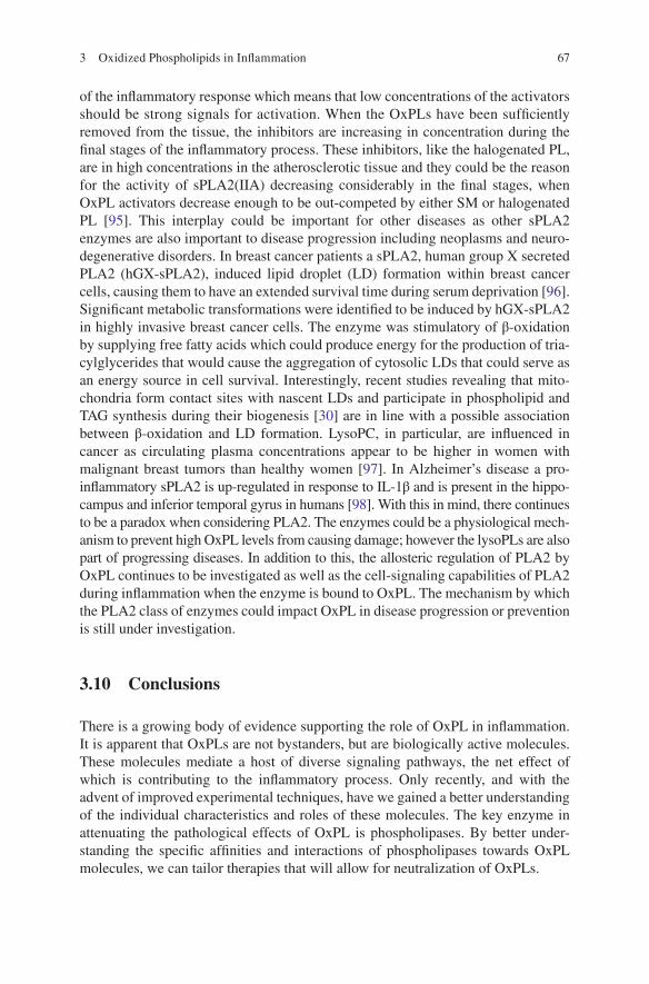
of the infl ammatory response which means that low concentrations of the activators should be strong signals for activation. When the OxPLs have been suffi ciently removed from the tissue, the inhibitors are increasing in concentration during the fi nal stages of the infl ammatory process. These inhibitors, like the halogenated PL, are in high concentrations in the atherosclerotic tissue and they could be the reason for the activity of sPLA2(IIA) decreasing considerably in the fi nal stages, when OxPL activators decrease enough to be out-competed by either SM or halogenated PL [ 95 ]. This interplay could be important for other diseases as other sPLA2 enzymes are also important to disease progression including neoplasms and neuro-degenerative disorders. In breast cancer patients a sPLA2, human group X secreted PLA2 (hGX-sPLA2), induced lipid droplet (LD) formation within breast cancer cells, causing them to have an extended survival time during serum deprivation [ 96 ]. Signifi cant metabolic transformations were identifi ed to be induced by hGX-sPLA2 in highly invasive breast cancer cells. The enzyme was stimulatory of β-oxidation by supplying free fatty acids which could produce energy for the production of tria-cylglycerides that would cause the aggregation of cytosolic LDs that could serve as an energy source in cell survival. Interestingly, recent studies revealing that mito-chondria form contact sites with nascent LDs and participate in phospholipid and TAG synthesis during their biogenesis [ 30 ] are in line with a possible association between β-oxidation and LD formation. LysoPC, in particular, are infl uenced in cancer as circulating plasma concentrations appear to be higher in women with malignant breast tumors than healthy women [ 97 ]. In Alzheimer’s disease a pro- infl ammatory sPLA2 is up-regulated in response to IL-1β and is present in the hippo-campus and inferior temporal gyrus in humans [ 98 ]. With this in mind, there continues to be a paradox when considering PLA2. The enzymes could be a physiological mech-anism to prevent high OxPL levels from causing damage; however the lysoPLs are also part of progressing diseases. In addition to this, the allosteric regulation of PLA2 by OxPL continues to be investigated as well as the cell-signaling capabilities of PLA2 during infl ammation when the enzyme is bound to OxPL. The mechanism by which the PLA2 class of enzymes could impact OxPL in disease progression or prevention is still under investigation.
3.10 Conclusions
There is a growing body of evidence supporting the role of OxPL in infl ammation. It is apparent that OxPLs are not bystanders, but are biologically active molecules. These molecules mediate a host of diverse signaling pathways, the net effect of which is contributing to the infl ammatory process. Only recently, and with the advent of improved experimental techniques, have we gained a better understanding of the individual characteristics and roles of these molecules. The key enzyme in attenuating the pathological effects of OxPL is phospholipases. By better under-standing the specifi c affi nities and interactions of phospholipases towards OxPL molecules, we can tailor therapies that will allow for neutralization of OxPLs.
3 Oxidized Phospholipids in Infl ammation

68
References
1. Chisolm G, Steinberg D (2000) The oxidative modifi cation hypothesis of atherogenesis: an overview. Free Radic Biol Med 28:1815–1826
2. Fessel J, Porter NA, Moore KP et al (2002) Discovery of lipid peroxidation products formed in vivo with a substituted tetrahydrofuran ring (isofurans) that are favored by increased oxygen tension. Proc Natl Acad Sci U S A 99:16713–16718
3. Weinstein E, Li H, Lawson JA et al (2000) Prothrombinase acceleration by oxidatively damaged phospholipids. J Biol Chem 275:22925–22930
4. Marathe G et al (2002) Activation of vascular cells by PAF-like lipids in oxidized LDL. Vasc Pharmacol 38(4):193–200
5. Apel K, Hirt H (2004) Reactive oxygen species: metabolism, oxidative stress, and signal trans-duction. Annu Rev Plant Biol 55:373–399
6. Murphy M (2009) How mitochondria produce reactive oxygen species. Biochem J 417:1–13 7. Lenaz G (2001) The mitochondrial production of reactive oxygen species: mechanisms and
implications in human pathology. IUBMB Life 52:159–164 8. Cour M, Gomez L, Mewton N et al (2011) Postconditioning: from the bench to bedside.
J Cardiovasc Pharmacol Ther 16:17–130 9. Piper H, Meuter K, Schäfer C (2003) Cellular mechanisms of ischemia-reperfusion injury.
Ann Thorac Surg 75:8 10. Crompton M (2000) Mitochondrial intermembrane junctional complexes and their role in cell
death. J Physiol 529:11–21 11. Oskolkova O, Afonyushkin T, Preinerstorfer B et al (2010) Oxidized phospholipids are more potent
antagonists of lipopolysaccharide than inducers of infl ammation. J Immunol 185:7706–7712 12. Lambeth J (2002) Nox/Duox family of nicotinamide adenine dinucleotide (phosphate)
oxidases. Curr Opin Hematol 9:11–17 13. Zweier J, Talukder M (2006) The role of oxidants and free radicals in reperfusion injury.
Cardiovasc Res 70:181–190 14. Vinten-Johansen J (2004) Involvement of neutrophils in the pathogenesis of lethal myocardial
reperfusion injury. Cardiovasc Res 61:481–497 15. Frangogiannis N, Smith C, Entman M (2002) The infl ammatory response in myocardial
infarction. Cardiovasc Res 53:31–47 16. Schneider C, Porter N, Brash A (2008) Routes to 4-hydroxynonenal: fundamental issues in the
mechanisms of lipid peroxidation. J Biol Chem 283:15539–15543 17. Allen D, Hasanally D, Ravandi A (2013) Role of oxidized phospholipids in cardiovascular
pathology. Clin Lipidol 8:205–215 18. Nonas S, Miller I, Kawkritinarong K et al (2006) Oxidized phospholipids reduce vascular leak and
infl ammation in rat model of acute lung injury. Am J Respir Crit Care Med 173:1130–1138 19. Ravandi A, Babaei S, Leung R et al (2004) Phospholipids and oxophospholipids in atheroscle-
rotic plaques at different stages of plaque development. Lipids 39:97–109 20. Li R, Mouillesseaux KP, Montoya D et al (2006) Identifi cation of prostaglandin E2 receptor
subtype 2 as a receptor activated by OxPAPC. Circ Res 98:642–650 21. Birukova A, Fu P, Chatchavalvanich S et al (2007) Polar head groups are important for barrier-
protective effects of oxidized phospholipids on pulmonary endothelium. Am J Physiol Lung Cell Mol Physiol 292:L924–L935
22. Furukawa M, Gohda T, Tanimoto M, Tomino Y (2013) Pathogenesis and novel treatment from the mouse model of type 2 diabetic nephropathy. Sci World J 2013:928197
23. Paschos A, Pandya R, Duivenvoorden WC, Pinthus JH (2013) Oxidative stress in prostate cancer: changing research concepts towards a novel paradigm for prevention and therapeutics. Prostate Cancer Prostatic Dis 16:217–225
24. Tsutsui H, Kinugawa S, Matsushima S (2011) Oxidative stress and heart failure. Am J Physiol Heart Circ Physiol 301:H2181–H2190
D. Hasanally et al.

69
25. Hammond V, Morgan AH, Lauder S et al (2012) Novel keto-phospholipids are generated by monocytes and macrophages, detected in cystic fi brosis, and activate peroxisome proliferator- activated receptor-γ. J Biol Chem 287:41651–41666
26. Hernandes M, Britto L (2012) NADPH oxidase and neurodegeneration. Curr Neuropharmacol 10:321–327
27. Wenk M (2010) Lipidomics: new tools and applications. Cell 143:888–895 28. Han X, Gross R (1994) Electrospray ionization mass spectroscopic analysis of human erythrocyte
plasma membrane phospholipids. Proc Natl Acad Sci U S A 91:10635–10639 29. Nakanishi H, Iida Y, Shimizu T, Taguchi R (2009) Analysis of oxidized phosphatidylcholines
as markers for oxidative stress, using multiple reaction monitoring with theoretically expanded data sets with reversed-phase liquid chromatography/tandem mass spectrometry. J Chromatogr B Analyt Technol Biomed Life Sci 877:1366–1374
30. Gruber F, Bicker W, Oskolkova OV et al (2012) A simplifi ed procedure for semi-targeted lipidomic analysis of oxidized phosphatidylcholines induced by UVA irradiation. J Lipid Res 53:1232–1242
31. Quehenberger O, Armando AM, Brown AH et al (2010) Lipidomics reveals a remarkable diversity of lipids in human plasma. J Lipid Res 51:3299–3305
32. Weir J, Wong G, Barlow CK et al (2013) Plasma lipid profi ling in a large population-based cohort. J Lipid Res 54:2898–2908
33. Andreyev A, Fahy E, Guan Z et al (2010) Subcellular organelle lipidomics in TLR-4-activated macrophages. J Lipid Res 51:2785–2797
34. Dennis E, Deems RA, Harkewicz R et al (2010) A mouse macrophage lipidome. J Biol Chem 285:39976–39985
35. White C, Ali A, Hasanally D et al (2013) A cardioprotective preservation strategy employing ex vivo heart perfusion facilitates successful transplant of donor hearts after cardiocirculatory death. J Heart Lung Transplant 32:734–743
36. Gargalovic P, Imura M, Zhang B et al (2006) Identifi cation of infl ammatory gene modules based on variations of human endothelial cell responses to oxidized lipids. Proc Natl Acad Sci U S A 103:12741–12746
37. Moore K, Sheedy F, Fisher E (2013) Macrophages in atherosclerosis: a dynamic balance. Nat Rev Immunol 13:709–721
38. Prescott SM, Zimmerman GA, Stafforini DM, McIntyre TM (2000) Platelet-activating factor and related lipid mediators. Annu Rev Biochem 69:419–445
39. Tyurina YY, Tyurin VA, Zhao Q et al (2004) Oxidation of phosphatidylserine: a mechanism for plasma membrane phospholipid scrambling during apoptosis? Biochem Biophys Res Commun 324:1059–1064
40. Thomas CP, Morgan LT, Maskrey BH et al (2010) Phospholipid-esterifi ed eicosanoids are generated in agonist-activated human platelets and enhance tissue factor-dependent thrombin generation. J Biol Chem 285:6891–6903
41. Podrez E, Byzova TV, Febbraio M (2007) Platelet CD36 links hyperlipidemia, oxidant stress and a prothrombotic phenotype. Nat Med 13:1086–1095
42. Androulakis N, Durand H, Ninio E, Tsoukatos DC (2005) Molecular and mechanistic charac-terization of platelet-activating factor-like bioactivity produced upon LDL oxidation. J Lipid Res 46:1923–1932
43. Singleton PA, Chatchavalvanich S, Fu P et al (2009) Akt-mediated transactivation of the S1P1 receptor in caveolin-enriched microdomains regulates endothelial barrier enhancement by oxidized phospholipids. Circ Res 104:978–986
44. Bochkov V, Oskolkova OV, Birukov KG et al (2010) Generation and biological activities of oxidized phospholipids. Antioxid Redox Signal 12:1009–1059
45. Weismann D, Binder C (2012) The innate immune response to products of phospholipid peroxidation. Biochim Biophys Acta 1818:2465–2475
46. Bochkov V (2007) Infl ammatory profi le of oxidized phospholipids. Thromb Haemost 97:348–354
3 Oxidized Phospholipids in Infl ammation

70
47. Zimman A, Mouillesseaux KP, Le T et al (2007) Vascular endothelial growth factor receptor 2 plays a role in the activation of aortic endothelial cells by oxidized phospholipids. Arterioscler Thromb Vasc Biol 27:332–338
48. Walton K, Hsieh X, Gharavi N et al (2003) Receptors involved in the oxidized 1-palmitoyl- 2-arachidonoyl-sn-glycero-3-phosphorylcholine-mediated synthesis of interleukin-8. A role for Toll-like receptor 4 and a glycosylphosphatidylinositol-anchored protein. J Biol Chem 278:29661–29666
49. Tsiantoulas D, Gruber S, Binder C (2012) B-1 cell immunoglobulin directed against oxidation- specifi c epitopes. Front Immunol 3:1–6
50. Perry H, Bender T, McNamara C (2012) B cell subsets in atherosclerosis. Front Immunol 3:1–11
51. Binder CJ, Chou MY, Fogelstrand L et al (2008) Natural antibodies in murine atherosclerosis. Curr Drug Targets 9:190–195
52. Chou MY, Hartvigsen K, Hansen LF et al (2008) Oxidation-specifi c epitopes are important targets of innate immunity. J Intern Med 263:479–488
53. Shaw P, Hörkkö S, Chang MK et al (2000) Natural antibodies with the T15 idiotype may act in atherosclerosis, apoptotic clearance, and protective immunity. J Clin Invest 105:1731–1740
54. Hörkkö S, Bird DA, Miller E et al (1999) Monoclonal autoantibodies specifi c for oxidized phospholipids or oxidized phospholipid-protein adducts inhibit macrophage uptake of oxi-dized low-density lipoproteins. J Clin Invest 103:117–128
55. Chang MK, Binder CJ, Torzewski M et al (2002) C-reactive protein binds to both oxidized LDL and apoptotic cells through recognition of a common ligand: phosphorylcholine of oxi-dized phospholipids. Proc Natl Acad Sci U S A 99:13043–13048
56. Chang MK, Hartvigsen K, Ryu J et al (2012) The pro-atherogenic effects of macrophages are reduced upon formation of a complex between C-reactive protein and lysophosphatidylcho-line. J Infl amm (London, England) 9:42
57. Boullier A, Friedman P, Harkewicz R et al (2005) Phosphocholine as a pattern recognition ligand for CD36. J Lipid Res 46:969–976
58. Febbraio M, Hajjar D, Silverstein R (2001) CD36: a class B scavenger receptor involved in angiogenesis, atherosclerosis, infl ammation, and lipid metabolism. J Clin Invest 108:785–791
59. Haserück N, Erl W, Pandey D et al (2004) The plaque lipid lysophosphatidic acid stimulates platelet activation and platelet-monocyte aggregate formation in whole blood: involvement of P2Y1 and P2Y12 receptors. Blood 103:2585–2592
60. Göpfert MS, Siedler F, Siess W, Sellmayer A (2005) Structural identifi cation of oxidized acyl- phosphatidylcholines that induce platelet activation. J Vasc Res 42:120–132
61. Berliner J, Leitinger N, Tsimikas S (2009) The role of oxidized phospholipids in atherosclero-sis. J Lipid Res 50:S207–S212
62. Gharavi NM, Baker NA, Mouillesseaux KP et al (2006) Role of endothelial nitric oxide syn-thase in the regulation of SREBP activation by oxidized phospholipids. Circ Res 98:768–776
63. Qin J, Testai FD, Dawson S et al (2009) Oxidized phosphatidylcholine formation and action in oligodendrocytes. J Neurochem 110:1388–1399
64. Yoshida H, Matsui T, Yamamoto A et al (2001) XBP1 mRNA is induced by ATF6 and spliced by IRE1 in response to ER stress to produce a highly active transcription factor. Cell 107:881–891
65. Dinasarapu RA, Gupta S, Ram Maurya M et al (2013) A combined omics study on activated macrophages—enhanced role of STATs in apoptosis, immunity and lipid metabolism. Bioinformatics (Oxford, England) 2013:1–9
66. Lartigue L, Faustin B (2013) Mitochondria: metabolic regulators of innate immune responses to pathogens and cell stress. Int J Biochem Cell Biol 45:2052–2056
67. Chen R, Feldstein A, McIntyre T (2009) Suppression of mitochondrial function by oxidatively truncated phospholipids is reversible, aided by bid, and suppressed by Bcl-XL. J Biol Chem 284:26297–26308
68. Shih PT, Elices MJ, Fang ZT et al (1999) Minimally modifi ed low-density lipoprotein induces monocyte adhesion to endothelial connecting segment-1 by activating beta1 integrin. J Clin Invest 103:613–625
D. Hasanally et al.

71
69. Vora DK, Fang ZT, Liva SM et al (1997) Induction of P-selectin by oxidized lipoproteins. Separate effects on synthesis and surface expression. Circ Res 80:810–818
70. Birukova AA, Starosta V, Tian X et al (2013) Fragmented oxidation products defi ne barrier disruptive endothelial cell response to OxPAPC. Transl Res 161:495–504
71. Kadl A, Galkina E, Leitinger N (2009) Induction of CCR2-dependent macrophage accumula-tion by oxidized phospholipids in the air-pouch model of infl ammation. Arthritis Rheum 60:1362–1371
72. Furnkranz A, Schober A, Bochkov VN et al (2005) Oxidized phospholipids trigger athero-genic infl ammation in murine arteries. Arterioscler Thromb Vasc Biol 25:633–638
73. Kopf M, Baumann H, Freer G et al (1994) Impaired immune and acute-phase responses in interleukin-6-defi cient mice. Nature 368(6469):339–342
74. Gottlieb RA, Burleson KO, Kloner RA et al (1994) Reperfusion injury induces apoptosis in rabbit cardiomyocytes. J Clin Invest 94:1621–1628
75. Gustafsson A, Gottlieb R (2003) Mechanisms of apoptosis in the heart. J Clin Immunol 23:447–459
76. Halestrap A, Kerr PM, Javadov S, Woodfi eld KY (1998) Elucidating the molecular mechanism of the permeability transition pore and its role in reperfusion injury of the heart. Biochim Biophys Acta 1366:79–94
77. Chen R, Yang L, McIntyre T (2007) Cytotoxic phospholipid oxidation products. Cell death from mitochondrial damage and the intrinsic caspase cascade. J Biol Chem 282:24842–24850
78. Fruhwirth G, Moumtzi A, Loidl A et al (2006) The oxidized phospholipids POVPC and PGPC inhibit growth and induce apoptosis in vascular smooth muscle cells. Biochim Biophys Acta 1761:1060–1069
79. Stemmer U, Dunai ZA, Koller D et al (2012) Toxicity of oxidized phospholipids in cultured macrophages. Lipids Health Dis 11:110
80. Wallgren M, Lidman M, Pham QD et al (2012) The oxidized phospholipid PazePC modulates interactions between Bax and mitochondrial membranes. Biochim Biophys Acta 1818:2718–2724
81. Mughal W, Kirshenbaum L (2011) Cell death signalling mechanisms in heart failure. Exp Clin Cardiol 16:102–108
82. Stremler KE, Stafforini DM, Prescott SM, McIntyre TM (1991) Human plasma platelet- activating factor acetylhydrolase. Oxidatively fragmented phospholipids as substrates. J Biol Chem 266:11095–11103
83. Bergmark C, Dewan A, Orsoni A et al (2008) A novel function of lipoprotein [a] as a prefer-ential carrier of oxidized phospholipids in human plasma. J Lipid Res 49:2230–2239
84. Davis B, Koster G, Douet LJ et al (2008) Electrospray ionization mass spectrometry identifi es substrates and products of lipoprotein-associated phospholipase A2 in oxidized human low density lipoprotein. J Biol Chem 283:6428–6437
85. Rivera R, Chun J (2006) Biological effects of lysophospholipids. Rev Physiol Biochem Pharmacol 160:25–46
86. Salgo MG, Corongiu FP, Sevanian A (1993) Enhanced interfacial catalysis and hydrolytic specifi city of phospholipase A2 toward peroxidized phosphatidylcholine vesicles. Arch Biochem Biophys 304:123–132
87. Tyurin VA, Yanamala N, Tyurina YY et al (2012) Specifi city of lipoprotein-associated phospholipase A(2) toward oxidized phosphatidylserines: liquid chromatography-electrospray ionization mass spectrometry characterization of products and computer modeling of interac-tions. Biochemistry 51:9736–9750
88. Kokotos G, Hsu YH, Burke JE et al (2010) Potent and selective fl uoroketone inhibitors of group VIA calcium-independent phospholipase A2. J Med Chem 53:3602–3610
89. Dennis E, Cao J, Hsu YH et al (2011) Phospholipase A2 enzymes: physical structure, biological function, disease implication, chemical inhibition, and therapeutic intervention. Chem Rev 111:6130–6185
90. Code C, Mahalka AK, Bry K, Kinnunen PK (2010) Activation of phospholipase A2 by 1-palmitoyl-2-(9′-oxo-nonanoyl)-sn-glycero-3-phosphocholine in vitro. Biochim Biophys Acta 1798:1593–1600
3 Oxidized Phospholipids in Infl ammation

72
91. Cordella-Miele E, Miele L, Mukherjee A (1990) A novel transglutaminase-mediated post- translational modifi cation of phospholipase A2 dramatically increases its catalytic activity. J Biol Chem 265:17180–17188
92. Samoilova EV, Pirkova AA, Prokazova NV, Korotaeva AA (2010) Effects of LDL lipids on activity of group IIA secretory phospholipase A2. Bull Exp Biol Med 150:39–41
93. Koumanov K, Wolf C, Béreziat G (1997) Modulation of human type II secretory phospholipase A2 by sphingomyelin and annexin VI. Biochem J 326:227–233
94. Korotaeva AA, Samoilova EV, Piksina GF, Prokazova NV (2010) Oxidized phosphatidylcho-line stimulates activity of secretory phospholipase A2 group IIA and abolishes sphingomyelin- induced inhibition of the enzyme. Prostaglandins Other Lipid Mediat 91:38–41
95. Korotaeva A, Samoilova E, Pavlunina T, Panasenko OM (2013) Halogenated phospholipids regulate secretory phospholipase A2 group IIA activity. Chem Phys Lipids 167–168:51–56
96. Pucer A, Brglez V, Payre C et al (2013) Group X secreted phospholipase A2 induces lipid droplet formation and prolongs breast cancer cell survival. Mol Cancer 12:111
97. Murph M, Tanaka T, Pang J et al (2007) Liquid chromatography mass spectrometry for quan-tifying plasma lysophospholipids: potential biomarkers for cancer diagnosis. Methods Enzymol 433:1–25
98. Moses G, Jensen MD, Lue LF et al (2006) Secretory PLA2-IIA: a new infl ammatory factor for Alzheimer’s disease. J Neuroinfl ammation 3:28
D. Hasanally et al.