Transbilayer phospholipids molecular imaging
14
REVIEW Open Access Transbilayer phospholipids molecular imaging Tarik Z Belhocine 1* and Frank S Prato 1,2 Abstract Nuclear medicine has become a key part of molecular imaging. In the present review article, we focus on the transbilayer phospholipids as exquisite targets for radiolabelled probes in molecular imaging. Asymmetry of phospholipid distribution is a characteristic of mammalian cell membranes. Phosphatidylcholine and sphyngomyelin cholinophospholipids are primarily located within the external leaflet of the cell membrane. Phosphatidylserine and phosphatidylethanolamine aminophospholipids, and also phosphatidylinositol are primarily located within the internal leaflet of the cell membrane. New radiolabelled tracers have been designed in preclinical and clinical research for PET-CT and SPECT-CT molecular imaging of transbilayer phospholipids. Keywords: phospholipids, molecular imaging, PET-CT, SPECT-CT Introduction In this beginning of the twenty-first century, personalized molecular medicine is the objective of molecular diagno- sis, molecular imaging, and molecular therapy [1,2]. Molecular imaging (MI) includes a number of morpholo- gical imaging techniques (i.e. ultrasound, computed tomography and magnetic resonance imaging), and opti- cal imaging techniques (i.e. bioluminescence and fluores- cence imaging) [3,4]. In addition, nuclear medicine with radiolabelled probes has become a key part of MI, espe- cially with 18 F-fluorodeoxyglucose ( 18 F-FDG) metabolic imaging [5]. New radiolabelled tracers have been designed for positron emission tomography-computed tomography (PET-CT) and single-photon emission com- puted tomography-computed tomography (SPECT-CT) molecular imaging [6]. In this review article, we focus on the transbilayer phospholipids as exquisite targets for radiolabelled probes in molecular imaging. Molecular imaging There is no universally accepted definition of molecular imaging [5,7]. In 2000, the Society of Molecular Imaging http://www.molecularimaging.org/ defined molecular imaging as: ‘ the characterization and measurement of biological processes in living animals at the cellular and molecular level’. In 2005, the European Society for Mole- cular Imaging http://www.e-smi.eu formulated a definition of molecular imaging as: ‘the characterisation of the dynamics of the molecular processes in the living organisms in vivo. In vivo molecular imaging is a science combining molecular biology, cellular biology and phy- siology with imaging in living subjects’. In 2006, the Fed- eration of Asian Societies for Molecular Imaging (FASMI: http://fasmi.org/) defined molecular imaging as: ‘ the characterization and measurement of biological pro- cesses in living animals at the cellular and molecular level by means of non-invasive (or minimally invasive) imaging’. In 2007, the Society of Nuclear Medicine Mole- cular Imaging Center of Excellence http://interactive. snm.org/ definitions task force approved this definition of molecular imaging as: ‘the visualization, characteriza- tion, and measurement of biological processes at the molecular and cellular levels in humans and other living systems’ [8]. A MI probe is a molecule used in molecular imaging to deliver a tracer to a specific organ or tissue. A probe typically consists of a ligand containing or linked to a signalling label. The label provides the signal (i.e. electromagnetic wave, light and radiation) that can be picked up by a detector, and the ligand carries the tracer to the site of interest [9]. A MI target used in molecular imaging is a molecule or structure in the body to which binds a probe delivered to a specific organ or tissue. The target may be a peptide, or a glucide, or a lipid; in many cases, the target is a protein [10,11]. Molecular imaging may be a single disease/gene or a general disease/biologic function control point for targeting [12]. Molecular imaging with transbilayer phospholipid tar- gets may be performed with radiolabelled probes such * Correspondence: [email protected] 1 Department of Medical Imaging, The University of Western Ontario, London, ON, Canada Full list of author information is available at the end of the article Belhocine and Prato EJNMMI Research 2011, 1:17 http://www.ejnmmires.com/content/1/1/17 © 2011 Belhocine and Prato; licensee Springer. This is an Open Access article distributed under the terms of the Creative Commons Attribution License (http://creativecommons.org/licenses/by/2.0), which permits unrestricted use, distribution, and reproduction in any medium, provided the original work is properly cited.
Transcript of Transbilayer phospholipids molecular imaging
REVIEW Open Access
Transbilayer phospholipids molecular imagingTarik Z Belhocine1* and Frank S Prato1,2
Abstract
Nuclear medicine has become a key part of molecular imaging. In the present review article, we focus on thetransbilayer phospholipids as exquisite targets for radiolabelled probes in molecular imaging. Asymmetry ofphospholipid distribution is a characteristic of mammalian cell membranes. Phosphatidylcholine andsphyngomyelin cholinophospholipids are primarily located within the external leaflet of the cell membrane.Phosphatidylserine and phosphatidylethanolamine aminophospholipids, and also phosphatidylinositol are primarilylocated within the internal leaflet of the cell membrane. New radiolabelled tracers have been designed inpreclinical and clinical research for PET-CT and SPECT-CT molecular imaging of transbilayer phospholipids.
Keywords: phospholipids, molecular imaging, PET-CT, SPECT-CT
IntroductionIn this beginning of the twenty-first century, personalizedmolecular medicine is the objective of molecular diagno-sis, molecular imaging, and molecular therapy [1,2].Molecular imaging (MI) includes a number of morpholo-gical imaging techniques (i.e. ultrasound, computedtomography and magnetic resonance imaging), and opti-cal imaging techniques (i.e. bioluminescence and fluores-cence imaging) [3,4]. In addition, nuclear medicine withradiolabelled probes has become a key part of MI, espe-cially with 18F-fluorodeoxyglucose (18F-FDG) metabolicimaging [5]. New radiolabelled tracers have beendesigned for positron emission tomography-computedtomography (PET-CT) and single-photon emission com-puted tomography-computed tomography (SPECT-CT)molecular imaging [6]. In this review article, we focus onthe transbilayer phospholipids as exquisite targets forradiolabelled probes in molecular imaging.
Molecular imagingThere is no universally accepted definition of molecularimaging [5,7]. In 2000, the Society of Molecular Imaginghttp://www.molecularimaging.org/ defined molecularimaging as: ‘the characterization and measurement ofbiological processes in living animals at the cellular andmolecular level’. In 2005, the European Society for Mole-cular Imaging http://www.e-smi.eu formulated a
definition of molecular imaging as: ‘the characterisationof the dynamics of the molecular processes in the livingorganisms in vivo. In vivo molecular imaging is a sciencecombining molecular biology, cellular biology and phy-siology with imaging in living subjects’. In 2006, the Fed-eration of Asian Societies for Molecular Imaging(FASMI: http://fasmi.org/) defined molecular imaging as:‘ the characterization and measurement of biological pro-cesses in living animals at the cellular and molecularlevel by means of non-invasive (or minimally invasive)imaging’. In 2007, the Society of Nuclear Medicine Mole-cular Imaging Center of Excellence http://interactive.snm.org/ definitions task force approved this definitionof molecular imaging as: ‘the visualization, characteriza-tion, and measurement of biological processes at themolecular and cellular levels in humans and other livingsystems’ [8]. A MI probe is a molecule used in molecularimaging to deliver a tracer to a specific organ or tissue. Aprobe typically consists of a ligand containing or linkedto a signalling label. The label provides the signal (i.e.electromagnetic wave, light and radiation) that can bepicked up by a detector, and the ligand carries the tracerto the site of interest [9]. A MI target used in molecularimaging is a molecule or structure in the body to whichbinds a probe delivered to a specific organ or tissue. Thetarget may be a peptide, or a glucide, or a lipid; in manycases, the target is a protein [10,11]. Molecular imagingmay be a single disease/gene or a general disease/biologicfunction control point for targeting [12].Molecular imaging with transbilayer phospholipid tar-
gets may be performed with radiolabelled probes such
* Correspondence: [email protected] of Medical Imaging, The University of Western Ontario,London, ON, CanadaFull list of author information is available at the end of the article
Belhocine and Prato EJNMMI Research 2011, 1:17http://www.ejnmmires.com/content/1/1/17
© 2011 Belhocine and Prato; licensee Springer. This is an Open Access article distributed under the terms of the Creative CommonsAttribution License (http://creativecommons.org/licenses/by/2.0), which permits unrestricted use, distribution, and reproduction inany medium, provided the original work is properly cited.
as radiolabelled annexin V or C2A synaptotagmindomain I or beta 2 glycoprotein I, radiolabelled duramy-cin, radiolabelled hypericin, radiolabelled lactadherin,radiolabelled choline or fluorocholine, radiolabelled dia-cylglycerols, radiolabelled sphyngomyelin for visualiza-tion, characterization and measurement of key biologicalfunctions (i.e. apoptosis, necrosis, thrombosis, vascula-ture endothelium, choline metabolism, myocardial andneuronal phosphoinositide turnover) or for assessingspecific diseases (i.e. cancers, immune diseases, inflam-matory diseases, infectious diseases, cardiac diseases andneurological diseases).
Membrane bilayerThe membrane bilayer is composed of 40% lipids andglycolipids, and 60% integral proteins and glycoproteins[13]. The lipids in the membrane bilayer are composedof phospholipids (75% to 88%), glycosphyngolipids (2%to 5%) and cholesterol (10% to 20%) [13]. The phospho-lipids include phosphatidylcholine (45% to 55%), phos-phatidylethanolamine (15% to 25%), phosphatidylinositol(10% to 15%), phosphatidylserine (2% to 10%), phospha-tidic acid (1% to 2%), sphyngomyelin (5% to 10%) andcardiolipin (2% to 5%). Liposomes are artificial lipid vesi-cles encapsulating drugs (e.g. chemotherapy drugs, anti-biotics, fungicides), enzymes, biological material (e.g.antigens, antibodies) and tracers (e.g. radiolabelled pro-ducts, contrast agents) [14]. Liposomes are nanoparticleswith a diameter < 100 nm characterised by the composi-tion of lipids, the number of membrane bilayers, and thesurface charges [7]. The material encapsulated is eitherdissolved in an aqueous phase or in a lipid phase. Radio-active phospholipid liposomes have been designed formolecular imaging [15].
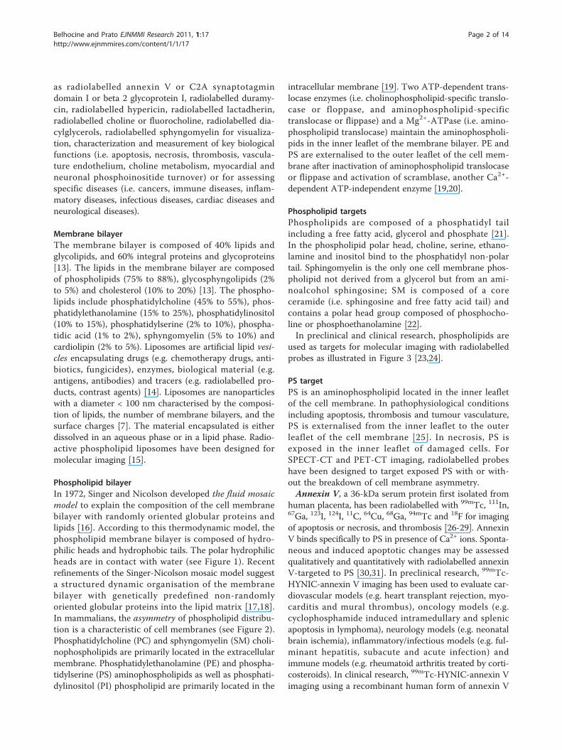
Phospholipid bilayerIn 1972, Singer and Nicolson developed the fluid mosaicmodel to explain the composition of the cell membranebilayer with randomly oriented globular proteins andlipids [16]. According to this thermodynamic model, thephospholipid membrane bilayer is composed of hydro-philic heads and hydrophobic tails. The polar hydrophilicheads are in contact with water (see Figure 1). Recentrefinements of the Singer-Nicolson mosaic model suggesta structured dynamic organisation of the membranebilayer with genetically predefined non-randomlyoriented globular proteins into the lipid matrix [17,18].In mammalians, the asymmetry of phospholipid distribu-tion is a characteristic of cell membranes (see Figure 2).Phosphatidylcholine (PC) and sphyngomyelin (SM) choli-nophospholipids are primarily located in the extracellularmembrane. Phosphatidylethanolamine (PE) and phospha-tidylserine (PS) aminophospholipids as well as phosphati-dylinositol (PI) phospholipid are primarily located in the
intracellular membrane [19]. Two ATP-dependent trans-locase enzymes (i.e. cholinophospholipid-specific translo-case or floppase, and aminophospholipid-specifictranslocase or flippase) and a Mg2+-ATPase (i.e. amino-phospholipid translocase) maintain the aminophospholi-pids in the inner leaflet of the membrane bilayer. PE andPS are externalised to the outer leaflet of the cell mem-brane after inactivation of aminophospholipid translocaseor flippase and activation of scramblase, another Ca2+-dependent ATP-independent enzyme [19,20].
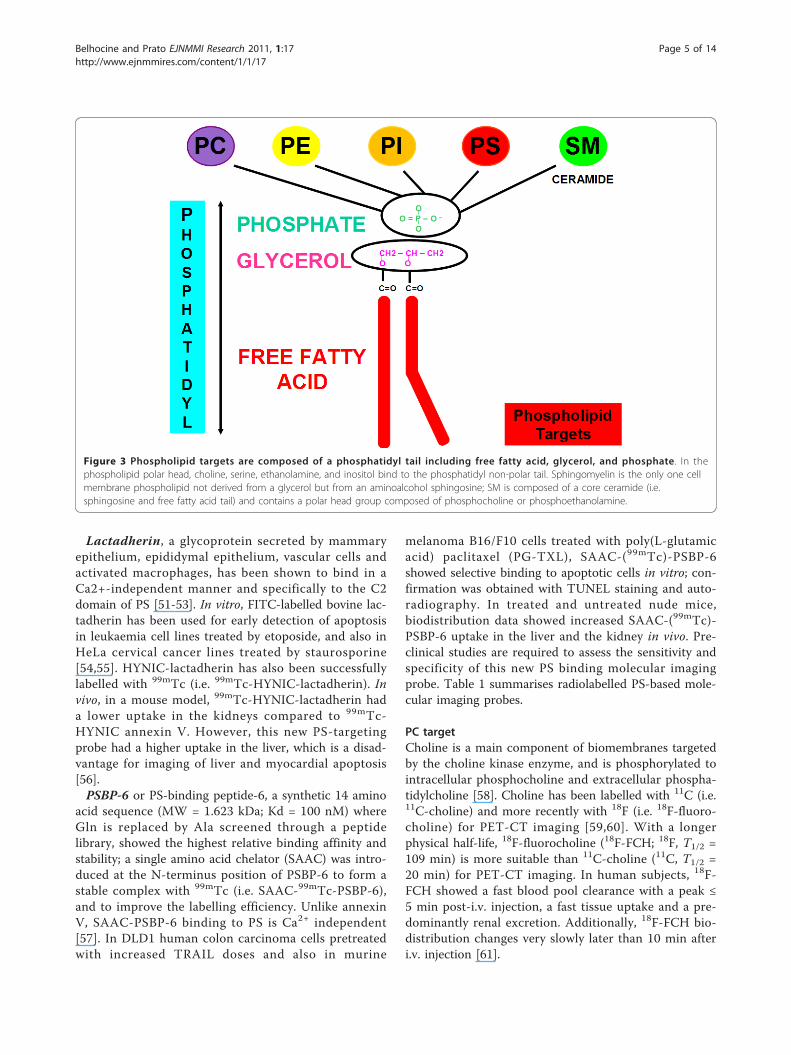
Phospholipid targetsPhospholipids are composed of a phosphatidyl tailincluding a free fatty acid, glycerol and phosphate [21].In the phospholipid polar head, choline, serine, ethano-lamine and inositol bind to the phosphatidyl non-polartail. Sphingomyelin is the only one cell membrane phos-pholipid not derived from a glycerol but from an ami-noalcohol sphingosine; SM is composed of a coreceramide (i.e. sphingosine and free fatty acid tail) andcontains a polar head group composed of phosphocho-line or phosphoethanolamine [22].In preclinical and clinical research, phospholipids are
used as targets for molecular imaging with radiolabelledprobes as illustrated in Figure 3 [23,24].
PS targetPS is an aminophospholipid located in the inner leafletof the cell membrane. In pathophysiological conditionsincluding apoptosis, thrombosis and tumour vasculature,PS is externalised from the inner leaflet to the outerleaflet of the cell membrane [25]. In necrosis, PS isexposed in the inner leaflet of damaged cells. ForSPECT-CT and PET-CT imaging, radiolabelled probeshave been designed to target exposed PS with or with-out the breakdown of cell membrane asymmetry.Annexin V, a 36-kDa serum protein first isolated from
human placenta, has been radiolabelled with 99mTc, 111In,67Ga, 123I, 124I, 11C, 64Cu, 68Ga, 94mTc and 18F for imagingof apoptosis or necrosis, and thrombosis [26-29]. AnnexinV binds specifically to PS in presence of Ca2+ ions. Sponta-neous and induced apoptotic changes may be assessedqualitatively and quantitatively with radiolabelled annexinV-targeted to PS [30,31]. In preclinical research, 99mTc-HYNIC-annexin V imaging has been used to evaluate car-diovascular models (e.g. heart transplant rejection, myo-carditis and mural thrombus), oncology models (e.g.cyclophosphamide induced intramedullary and splenicapoptosis in lymphoma), neurology models (e.g. neonatalbrain ischemia), inflammatory/infectious models (e.g. ful-minant hepatitis, subacute and acute infection) andimmune models (e.g. rheumatoid arthritis treated by corti-costeroids). In clinical research, 99mTc-HYNIC-annexin Vimaging using a recombinant human form of annexin V
Belhocine and Prato EJNMMI Research 2011, 1:17http://www.ejnmmires.com/content/1/1/17
Page 2 of 14
has been used to assess myocardial infarction, heart trans-plant rejection, atherosclerosis, ischemic pre-conditioningand chemotherapy/radiation therapy-induced apoptosis inhead and neck cancers, lymphomas, lung cancers, breastcancers, leukaemia and soft tissue sarcomas. 99mTc-HYNIC-annexin V imaging has also been used in patientswith dementia and stroke, and in patients with rheumatoidarthritis and Crohn’s disease for evaluation of early apop-totic response to Infliximab® therapy (i.e. anti-TNFamonoclonal antibody) [24,26]. Figures 4 and 5 illustrate PStargeting 99mTc-HYNIC-annexin V molecular imagingprobe in a patient with a non-small cell lung cancer(NSCLC). PS targeting 99mTc-annexin V-128 with an N-terminal site specific for endogenous binding of techne-tium-99m has been designed in order to improve the sen-sitivity of detection of apoptosis or necrosis (i.e. twofoldhigher than that of 99mTc-HYNIC-annexin V) [32].>C2A domain of synaptotagmin I (C2A) is a 12-kDa
protein predominantly located within the synaptic vesi-cles binding to PS. It has been fused with glutathione-s-transferase (GST) and radiolabelled with 99mTc for ima-ging of apoptosis or necrosis [33]. In a rat and pigmodel of acute myocardial infarction, increased 99mTc-
C2A-GST uptake was seen in the myocardial infarctarea at risk, and was associated with wall dysfunction[34-36]. In a mouse model of NSCLC, increased 99mTc-C2A-GST tumour uptake was noted after paclitaxel che-motherapy-induced apoptosis [37]. Also, C2A-GST hasbeen easily labelled with 18F for early imaging of apopto-sis after chemotherapy [38]. In a rabbit model of lungcancer, increased 18F-C2A-GST tumour uptake wasdetected 72 h after paclitaxel chemotherapy withincreased apoptotic index and caspase-3 activity.Recently, the C2A domain of synaptotagmin I has beenlabelled with a [99mTc(CO)3
+] core by using an efficientC-terminal site-specific radiolabelling method for theimaging of cell death [39].PS is not exposed in normal endothelium, but
increased exposure of PS is seen on the tumourendothelium vasculature [40,41]. Bavituximab, a chi-meric monoclonal antibody (MW = 145.3 kDa) bindingto the beta-2 glycoprotein I domain of PS, has beenradiolabelled with the b + emitter arsenicum-74 (74As,T1/2 = 17.8 days) for tumour vasculature PET imaging,and with the b-,g emitter 77As (T1/2 = 38.8 h) forSPECT imaging and potential tumour vasculature
Figure 1 Phospholipid bilayer composed of hydrophobic non-polar tails and hydrophilic polar heads. Liposomes are artificialphospholipid vesicles encapsulating materials either in an aqueous phase or in a lipid phase.
Belhocine and Prato EJNMMI Research 2011, 1:17http://www.ejnmmires.com/content/1/1/17
Page 3 of 14

endothelium therapy [42-44]. Increased 74As-bavituxi-mab uptake was seen in a rat model of prostate cancerwith the highest tumour-to-background activity ratio at72 h post-i.v injection (at 72 h p.i., tumour-to-liver ratio= 22, and tumour-to-muscle ratio = 470). Using autora-diography and immunohistochemical studies, 74As-bavi-tuximab was found to specifically bind to the tumourendothelium vasculature. Bavituximab radiolabelled with77As or 76As (b-, T1/2 = 26.3 h) may be used for dosime-try and immunotherapy.Hypericin, a non-porphyrin necrosis agent (MW = 504
Da) extracted from St John’s wort, has been labelled with64Cu (b+, T1/2 = 12.7 h) for PET imaging of necrosis. In afemale mouse model of BT474 breast xenograft tumour,64Cu-bis-DOTA-hypericin demonstrated increased uptakeat 24 h post-i.v. injection in necrotic injured tissues treatedwith near infrared photothermal ablation therapy. PETdistribution also showed higher uptake in the liver and thekidneys. Bis-DOTA-hypericin had a selective binding affi-nity for PS and PE phospholipids [45]. In necrosis, PS andPE are exposed to 64Cu-labelled hypericin probe in theinner leaflet of damaged cell membrane. Hypericin deriva-tives are efficient and yield reproducible results when
radiolabelled with 123I (i.e. mono-123I-iodohypericin andmono-123I-iodohypericin monocarboxylic acid). They havebeen also used in preclinical models of liver necrosis andmyocardial infarction as well as in clinical correlates ofthese pathophysiologic states [46,47]. In a preclinical ratmodel of liver rhabdomyosarcoma, 131I-labelled hypericinwas successfully used in theragnostics with a vascular dis-rupting agent (i.e. combretastatin A4 phosphate or CA4P);high radiolabelling efficiency was noted with minimumdeiodination. 131I-hypericin uptake colocalised tumournecrosis within 24 h post-i.v. injection on co-registeredplanar g scintigraphy with CT, MRI, histology and autora-diography [48]. Hypericin has also been labelled with99mTc (i.e. 99mTc-hypericin or 99mTc-mercaptoacetyldigly-cyl-1,5-diaminopentylene hypericin-carboxamide) forvisualisation of necrotic tissues in rats with reperfusedliver infarct [49]. 99mTc-hypericin, however, was found tobe not suitable for imaging of necrosis compared to 123I-hypericin derivatives [50]. Although the mechanism of tar-get uptake is unknown, it may be hypothesised that the PSand PE phospholipid targets are involved for 123I-, 131I-and 99mTc-labelled hypericin derivatives, but this is still tobe definitely proven.
Figure 2 Asymmetry of phospholipid distribution. Phosphatidylcholine (PC) and sphyngomyelin (SM) cholinophospholipids are primarilylocated in the external leaflet of the cell membrane (ECM). Phosphatidylserine (PS) and phosphatidylethanolamine (PE) aminophospholipids aswell as phosphatidylinositol (PI) phospholipid are primarily located in the internal leaflet of the cell membrane (ICM). In mammalians, theasymmetry of phospholipid distribution is maintained by the flip/flop movement of phospholipids. PS and PE are maintained to the inner leafletof the membrane bilayer by the Mg2+-ATPase (i.e. aminophospholipid translocase), and the ATP-dependent aminophospholipid-specific flippaseand cholinophospholipid-specific floppase (i.e. multidrug resistance proteins). In pathophysiological conditions, PS and PE are shuttled to theouter leaflet after inactivation of the aminophospholipid translocase or flippase, and activation of the Ca2+-dependent ATP-independentscramblase.
Belhocine and Prato EJNMMI Research 2011, 1:17http://www.ejnmmires.com/content/1/1/17
Page 4 of 14
Lactadherin, a glycoprotein secreted by mammaryepithelium, epididymal epithelium, vascular cells andactivated macrophages, has been shown to bind in aCa2+-independent manner and specifically to the C2domain of PS [51-53]. In vitro, FITC-labelled bovine lac-tadherin has been used for early detection of apoptosisin leukaemia cell lines treated by etoposide, and also inHeLa cervical cancer lines treated by staurosporine[54,55]. HYNIC-lactadherin has also been successfullylabelled with 99mTc (i.e. 99mTc-HYNIC-lactadherin). Invivo, in a mouse model, 99mTc-HYNIC-lactadherin hada lower uptake in the kidneys compared to 99mTc-HYNIC annexin V. However, this new PS-targetingprobe had a higher uptake in the liver, which is a disad-vantage for imaging of liver and myocardial apoptosis[56].PSBP-6 or PS-binding peptide-6, a synthetic 14 amino
acid sequence (MW = 1.623 kDa; Kd = 100 nM) whereGln is replaced by Ala screened through a peptidelibrary, showed the highest relative binding affinity andstability; a single amino acid chelator (SAAC) was intro-duced at the N-terminus position of PSBP-6 to form astable complex with 99mTc (i.e. SAAC-99mTc-PSBP-6),and to improve the labelling efficiency. Unlike annexinV, SAAC-PSBP-6 binding to PS is Ca2+ independent[57]. In DLD1 human colon carcinoma cells pretreatedwith increased TRAIL doses and also in murine
melanoma B16/F10 cells treated with poly(L-glutamicacid) paclitaxel (PG-TXL), SAAC-(99mTc)-PSBP-6showed selective binding to apoptotic cells in vitro; con-firmation was obtained with TUNEL staining and auto-radiography. In treated and untreated nude mice,biodistribution data showed increased SAAC-(99mTc)-PSBP-6 uptake in the liver and the kidney in vivo. Pre-clinical studies are required to assess the sensitivity andspecificity of this new PS binding molecular imagingprobe. Table 1 summarises radiolabelled PS-based mole-cular imaging probes.
PC targetCholine is a main component of biomembranes targetedby the choline kinase enzyme, and is phosphorylated tointracellular phosphocholine and extracellular phospha-tidylcholine [58]. Choline has been labelled with 11C (i.e.11C-choline) and more recently with 18F (i.e. 18F-fluoro-choline) for PET-CT imaging [59,60]. With a longerphysical half-life, 18F-fluorocholine (18F-FCH; 18F, T1/2 =109 min) is more suitable than 11C-choline (11C, T1/2 =20 min) for PET-CT imaging. In human subjects, 18F-FCH showed a fast blood pool clearance with a peak ≤5 min post-i.v. injection, a fast tissue uptake and a pre-dominantly renal excretion. Additionally, 18F-FCH bio-distribution changes very slowly later than 10 min afteri.v. injection [61].
Figure 3 Phospholipid targets are composed of a phosphatidyl tail including free fatty acid, glycerol, and phosphate . In thephospholipid polar head, choline, serine, ethanolamine, and inositol bind to the phosphatidyl non-polar tail. Sphingomyelin is the only one cellmembrane phospholipid not derived from a glycerol but from an aminoalcohol sphingosine; SM is composed of a core ceramide (i.e.sphingosine and free fatty acid tail) and contains a polar head group composed of phosphocholine or phosphoethanolamine.
Belhocine and Prato EJNMMI Research 2011, 1:17http://www.ejnmmires.com/content/1/1/17
Page 5 of 14
18F-fluorocholine (18F-FCH) and 11C-choline areincorporated into the membrane phospholipids, and arepredominantly used for prostate cancer imaging andbrain tumour imaging [62,63]. In prostate cancerpatients, 18F-FCH is a promising tracer for detection ofprimary prostate cancer, staging of lymph node and bonemetastases, and detection of recurrence after definitivetherapy [62-64]. In brain tumours, this agent may be use-ful to detect glioblastoma multiforme, to distinguishhigh-grade gliomas with a characteristic peri-tumouraluptake from metastases and benign lesions, to guide astereotactic biopsy, and also to differentiate post-radia-tion necrosis from recurrence [65-68]. Also, 11C-cholineand 18F-FCH have been used for hepatocellular carci-noma (HCC) imaging [69-71]. In a woodchuck model,11C-choline sensitivity for detection of well-differentiatedHCCs was higher than that of 18F-FDG, and in 12patients with moderately differentiated HCCs, the 11C-
choline detection rate was better than that of 18F-FDG.18F-FCH imaging has also successfully been performedfor detection of well-differentiated HCC in patients withliver nodules or cirrhosis or chronic liver disease, and fordetection of recurrences from HCC [72,73].Under normal glucose conditions, acetate free fatty
acid is metabolised into acetyl-CoA by acetyl-CoAsynthetase in both the cytosol and mitochondria forsynthesis of cholesterol and fatty acids [74]. In tumours,acetate is metabolised into fatty acids by fatty acidsynthetase, which is overexpressed in cancer cells [75].Acetate is mainly incorporated in the PC microdomainsthat play a major role for growth and metastasis [76].Acetoacetate ketone body is metabolised to acetoacetyl-CoA and to acetyl-CoA, and then incorporated into themetabolic pathway for lipid synthesis [77]. Like acetate,acetoacetate could be incorporated into the PC mem-brane. Acetate and acetoacetate have been labelled with
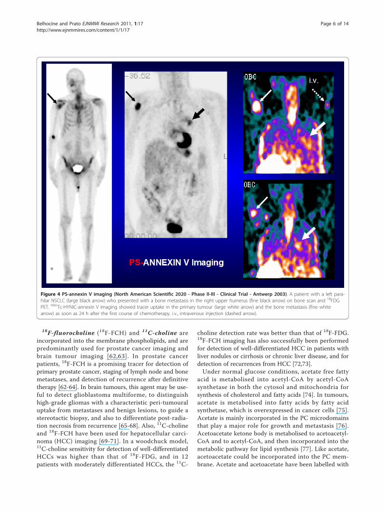
Figure 4 PS-annexin V imaging (North American Scientific 2020 - Phase II-III - Clinical Trial - Antwerp 2003). A patient with a left para-hilar NSCLC (large black arrow) who presented with a bone metastasis in the right upper humerus (fine black arrow) on bone scan and 18FDGPET. 99mTc-HYNIC-annexin V imaging showed tracer uptake in the primary tumour (large white arrow) and the bone metastasis (fine whitearrow) as soon as 24 h after the first course of chemotherapy. i.v., intravenous injection (dashed arrow).
Belhocine and Prato EJNMMI Research 2011, 1:17http://www.ejnmmires.com/content/1/1/17
Page 6 of 14
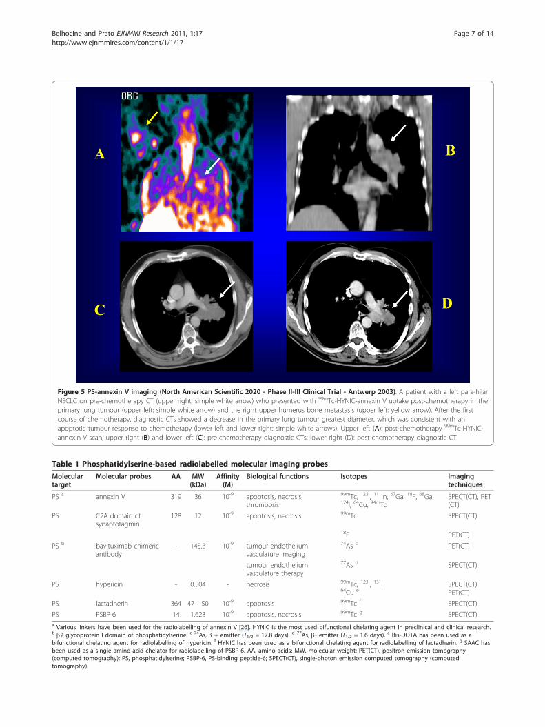
Figure 5 PS-annexin V imaging (North American Scientific 2020 - Phase II-III Clinical Trial - Antwerp 2003). A patient with a left para-hilarNSCLC on pre-chemotherapy CT (upper right: simple white arrow) who presented with 99mTc-HYNIC-annexin V uptake post-chemotherapy in theprimary lung tumour (upper left: simple white arrow) and the right upper humerus bone metastasis (upper left: yellow arrow). After the firstcourse of chemotherapy, diagnostic CTs showed a decrease in the primary lung tumour greatest diameter, which was consistent with anapoptotic tumour response to chemotherapy (lower left and lower right: simple white arrows). Upper left (A): post-chemotherapy 99mTc-HYNIC-annexin V scan; upper right (B) and lower left (C): pre-chemotherapy diagnostic CTs; lower right (D): post-chemotherapy diagnostic CT.
Table 1 Phosphatidylserine-based radiolabelled molecular imaging probes
Moleculartarget
Molecular probes AA MW(kDa)
Affinity(M)
Biological functions Isotopes Imagingtechniques
PS a annexin V 319 36 10-9 apoptosis, necrosis,thrombosis
99mTc, 123I, 111In, 67Ga, 18F, 68Ga,124I, 64Cu, 94mTc
SPECT(CT), PET(CT)
PS C2A domain ofsynaptotagmin I
128 12 10-9 apoptosis, necrosis 99mTc SPECT(CT)
18F PET(CT)
PS b bavituximab chimericantibody
- 145.3 10-9 tumour endotheliumvasculature imaging
74As c PET(CT)
tumour endotheliumvasculature therapy
77As d SPECT(CT)
PS hypericin - 0.504 - necrosis 99mTc, 123I, 131I64Cu e
SPECT(CT)PET(CT)
PS lactadherin 364 47 - 50 10-9 apoptosis 99mTc f SPECT(CT)
PS PSBP-6 14 1.623 10-9 apoptosis, necrosis 99mTc g SPECT(CT)a Various linkers have been used for the radiolabelling of annexin V [26]. HYNIC is the most used bifunctional chelating agent in preclinical and clinical research.b b2 glycoprotein I domain of phosphatidylserine. c 74As, b + emitter (T1/2 = 17.8 days). d 77As, b- emitter (T1/2 = 1.6 days). e Bis-DOTA has been used as abifunctional chelating agent for radiolabelling of hypericin. f HYNIC has been used as a bifunctional chelating agent for radiolabelling of lactadherin. g SAAC hasbeen used as a single amino acid chelator for radiolabelling of PSBP-6. AA, amino acids; MW, molecular weight; PET(CT), positron emission tomography(computed tomography); PS, phosphatidylserine; PSBP-6, PS-binding peptide-6; SPECT(CT), single-photon emission computed tomography (computedtomography).
Belhocine and Prato EJNMMI Research 2011, 1:17http://www.ejnmmires.com/content/1/1/17
Page 7 of 14
11C (i.e. 11C-acetate and 11C-acetoacetate) [74,77]. Acet-ate has also been labelled with 18F (i.e. 18F-fluoroacetate)[78]. 11C-acetate has a rapid blood clearance with a highaccumulation in liver and myocardium. In normal phy-siology, 18F-fluoroacetate is not a functional analogue of11C-acetate; 18F-fluoroacetate has a longer blood half-life,a rapid liver clearance and low myocardial uptake, exten-sive excretion into bile and urine, and suffers from skele-tal defluorination resulting in skeletal uptake of theformed 18F-fluoride [79]. In preclinical and clinicalresearch, 11C-acetate PET imaging has been successfullyused for tumour imaging in cancers of the prostate, kid-ney, pancreas, as well as gliomas, meningiomas and HCC[80,81]. In preclinical research, 18F-fluoroacetate hasbeen used for imaging of gliosis in glioblastoma, stroke,and ischemia-hypoxia associated with neuroinflammation[82]. In CWR22 tumour-bearing male nu/nu mice, 18F-fluoroacetate has been successfully used for prostate can-cer imaging with a higher tumour-to-prostate activityratio compared to 11C-acetate at 30 min and 2 h post-i.v.injection. In a patient with prostate cancer, 18F-fluoroace-tate was also able to detect several but not all bonemetastases [83,84]. In other preclinical research modelsof breast and prostate cancers, 11C-acetoacetate uptaketended to be higher than that of 11C-acetate [85]. Assess-ment of brain ketone metabolism in rats indicated a 7 to8 fold increase in 11C-acetoacetate under ketone dietarytreatment [86,87]. Table 2 summarises the radiolabelledPC-based molecular imaging probes.
PE targetPE is primarily located in the inner leaflet of the mem-brane bilayer [88]. Like PS, this major aminophospholi-pid is externalised from the inner leaflet to the outerleaflet of the cell membrane during apoptosis andtumour vascular endothelium [89,90]. In necrosis, PE isexposed in the inner leaflet of disintegrated cellmembrane.Duramycin, a 2-kDa small protein produced by Strep-
tovercillium cinnamoneus, has been designed in order totarget PE for imaging of apoptosis or necrosis [91].99mTc-HYNIC-duramycin has been successfully used in arat model of ischemia-reperfusion for imaging of myocar-dial infarction cell death [92]. 99mTc-duramycin is highly
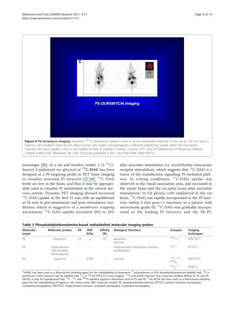
stable in vivo, and has favourable pharmacokinetics forearly imaging of acute cardiac cell death at 10 min post-i.v. injection with a fast blood clearance, a low liver uptakeand a high uptake in apoptotic and necrotic myocardium.Figure 6 illustrates the PE targeting 99mTc-duramycinmolecular imaging probe in a rat model of myocardialinfarction with correlation by autoradiography.In addition to this, radiolabelled ethanolamine (i.e.
ethanolamine labelled with 11C or 18F) is a potentialnew probe in oncology PET-CT imaging for assessmentof tumour proliferation [93]. 14C-ethanolamine hasbeen used in a variety of tumour cell types (i.e. mela-noma, prostate cancer, glioblastoma multiforme, diffuselarge B-cell lymphoma, colorectal adenocarcinoma). 14C-ethanolamine is incorporated into PE and has a two- tosevenfold significantly better uptake into tumour celltypes than 14C-choline. In an in vitro model of culturedprostate cancer cells, 14C-ethanolamine and 14C-N, N’-dimethyl-ethanolamine uptake was two- to fourfoldbetter in androgen-dependent and proliferating PC3cells compared to androgen-independent and growth-arrested LnCap cells.Hypericin, a PE- and PS-targeting probe, has been
radiolabelled with 64Cu (i.e. 64Cu-bis-DOTA-hypericin),123I (i.e. mono-123I-iodohypericin and mono-123I-iodohy-pericin monocarboxylic acid), 131I (i.e. 131I-hypericin)and 99mTc (i.e. 99mTc-hypericin) for imaging of necrosisin preclinical and clinical research as described above inthe PS target section [45-50]. Table 3 summarises thePE-based radiolabelled molecular probes.
PI targetPI phospholipid is located in the inner leaflet of themembrane bilayer [94]. 1,2 Diacylglycerol (DAG) ismetabolised to intermediate phosphoinositide metabo-lites (i.e. PA, PIP, PIP2, PIP3) including phosphatidylino-sitol (PI) [95]. DAG activates the intracellular proteinkinase C (PKC) transduction signalling pathway, whichis involved in higher cortical functions (e.g. memory,learning) and cardiac functions (e.g. hypertrophicgrowth, ventricular remodelling).Intact 11C-inositol (i.e. non-acetylated inositol) has
been suggested as a diagnostic agent in PET imaging forevaluation of PI brain metabolism and its role as second
Table 2 Phosphatidylcholine-based radiolabelled molecular imaging probes
Molecular target Molecular probes Biological functions Isotopes Imaging techniques
PC choline choline metabolism 11C PET(CT)
PC fluorocholine choline metabolism 18F PET(CT)
PC acetate acetate metabolism 11C PET(CT)
PC fluoroacetate acetate metabolism 18F PET(CT)
PC acetoacetate acetoacetate metabolism 11C PET(CT)
PC, phosphatidylcholine; PET(CT), positron emission tomography (computed tomography).
Belhocine and Prato EJNMMI Research 2011, 1:17http://www.ejnmmires.com/content/1/1/17
Page 8 of 14
messenger [96]. In a rat and monkey model, 1-[1-11C]-butyryl-2-palmitoyl-rac-glycerol or 11C-DAG has beendesigned as a PI-targeting probe in PET brain imagingto visualise neuronal PI turnover [97,98]. 11C-DAGlevels are low in the brain, and thus it may be appropri-ately used to visualise PI metabolism in the central ner-vous system. Dynamic PET imaging showed increased11C-DAG uptake in the first 15 min with an equilibriumat 16 min in pre-stimulation and post-stimulation con-ditions, which is suggestive of a membrane trappingmechanism. 11C-DAG uptake increased 20% to 30%
after arecoline stimulation (i.e. acetylcholine muscarinicreceptor stimulation), which suggests that 11C-DAG is atracer of the transduction signalling PI-mediated path-way. In resting conditions, 11C-DAG uptake wasobserved in the visual association area, and increased inthe whole brain and the occipital areas after arecolinestimulation. In C6 glioma cells implanted in the ratbrain, 11C-DAG was rapidly incorporated in the PI turn-over within 5 min post-i.v injection; in a patient withastrocytoma grade III, 11C-DAG was gradually incorpo-rated in the leading PI turnover and the PE-PC
Figure 6 PE-duramycin imaging. Increased 99mTc-Duramycin uptake is seen in acute myocardial infarction of the rat at 120 min post-i.v.injection with excellent infarct-to-non infarct activity ratio (right). Autoradiography confirmed radioactivity uptake within the myocardialinfarction. No tracer uptake is seen in the healthy rat (left). B, bladder; K, kidney. Courtesy of Pr. Zhao M (Department of Biophysics, MedicalCollege of Wisconsin, Milwaukee, WI, USA). Previously published in the J Nucl Med 2008, 49(8):1345-52.
Table 3 Phosphatidylethanolamine-based radiolabelled molecular imaging probes
Moleculartarget
Molecular probes AA MW(kDa)
Affinity(M)
Biological functions Isotopes Imagingtechniques
PE duramycin 19 2 10-9 apoptosisnecrosis
99mTc a SPECT(CT)
PE ethanolamineN’N-dimethylethanolamine
- - - ethanolamine metabolism (tumourproliferation)
14C b PET(CT)
PE c hypericin - 0.504 - necrosis 123I, 131I99mTc
SPECT(CT)
64Cu d PET(CT)a HYNIC has been used as a bifunctional chelating agent for the radiolabelling of duramycin. b ethanolamine or N,N’-dimethylethanolamine labelled with 14C inpreclinical in vitro research may be labelled with 11C or 18F for PET(CT) in vivo imaging. c 64Cu-bis-DOTA-hypericin has a selective binding affinity for PS and PE(ref.45); it may be hypothesised that 123I-, 131I- and 99mTc-labelled hypericin derivatives bind to PS and PE. d bis-DOTA has been used as a bifunctional chelatingagent for the radiolabelling of hypericin. AA, amino acids; MW, molecular weight; PE, phosphatidylethanolamine; PET(CT), positron emission tomography(computed tomography); SPECT(CT), single-photon emission computed tomography (computed tomography).
Belhocine and Prato EJNMMI Research 2011, 1:17http://www.ejnmmires.com/content/1/1/17
Page 9 of 14
secondary pool with an equilibrium period at 32-40 minpost-iv. injection [99]. In patients with Alzheimer’s dis-ease and ischemic stroke, 11C-DAG evaluated the PIcortical function and neural viability, respectively. Ineight patients with Alzheimer’s disease, 18F-FDG PETimaging showed bilateral hypometabolism in the par-ieto-temporal association areas, while 11C-DAG imagingshowed spotty uptake in the frontal lobes of the brainsuggestive of compensatory plastic process in non-damaged neural circuits to degenerative cognitiveimpairment [100]. In five patients with cerebral infarc-tion, dynamic PET brain imaging showed decreased11C-DAG uptake in comparison to normal cortex [101].11C-DAG pharmacokinetics demonstrated rapiddecrease in the plasma with a peak at 40 s post-i.v.injection, and gradual increase in the brain to reach aplateau at 15 to 20 min post-i.v. injection. Reflectingneural signal transduction activity, the incorporationconstant of 11C-DAG (k*DAG) was best correlated withthe cerebral metabolic rate of O2 (CMRO2). MaintainedPI metabolism suggested preservation of neural viabilityin the peri-infarct area of the ischemic stroke. Patientswith subacute local brain injury, either ischemic strokeor brain tumour, also exhibited 11C-DAG spots locatedin the associative areas distant from the lesion between2 weeks and 1 month after injury; one of the possiblefeatures of neural recovery in the intact brain related toPI metabolism in PET imaging [102].In a rat model, 11C-DAG has also been used for asses-
sing the myocardial PI turnover [103]. 11C-DAG incor-poration into intermediate PI metabolites was seen both inthe infarcted and the non-infarcted myocardium at 7 daysafter myocardial infarction. Furthermore, 11C-DAG hasbeen used to evaluate the PI-mediated angiotensin II sig-nalling pathway. In a rat model of infarcted myocardium,11C-DAG assessed the effect of angiotensin-convertingenzyme inhibitor (i.e. captropril) on activated PI metabo-lism in order to reduce left ventricular remodelling in theearly phase of myocardial infarction [104]. After 3 weeksof treatment with captropril, a significant decrease of11C-DAG uptake was seen in the infarcted myocardiumcompared to the non-infarcted myocardium, while 201Tlimaging showed decreased uptake in the infarcted myocar-dium with and without captopril treatment. In patientswith myocardial infarction, 11C-DAG has been used toevaluate the PI myocardial turnover and the left
ventricular remodelling [105]. In 13 patients with myocar-dial infarction, 11C-DAG was significantly correlated withthe myocardium-to-left atrium chamber ratio, the left ven-tricle ejection fraction, and the brain natriuretic peptideconcentration. Significant increase of 11C-DAG uptakewas seen in the remote viable region of myocardial infarc-tion patients compared to healthy subjects. DAG has alsobeen labelled with 18F to trace the PI metabolism into theventricular myocardium [106]. 18F has a longer half-life,and 18F-DAG may be more useful for clinical routine than11C-DAG. In the rat myocardium, 3, 5, 10, and 30 minafter i.v. injection of 1-[4-[18F]fluorobutyryl]-2-palmitoyl-glycerol (C4, C16), 18F-DAG reflected the myocardium PImetabolism in the early phase (3 to 5 min); in the latephase (≥ 30 min) after i.v. injection,18F-DAG may alsoreflect the PE metabolism. Quantification of PI metabo-lism requires further optimization with a 18F-radiolabelledDAG compound closer to the PI metabolism than 18F-DAG (C4, C16) [106]. Table 4 summarises the PI-basedradiolabelled molecular imaging probes.
SM targetSM is a cholinophospholipid located in the outer leafletof the membrane bilayer [19]. Liposomes composed ofSM have been experimentally used for tumour imaging.In a mouse tumour model, serum stable SM liposomes-encapsulating 67Ga prepared in a lipid phase with cho-lesterol (SM/cholesterol molar ratio, 2:1) enhancedblood circulation and increased 67Ga delivery to thetumour [107]. Tumour-to-blood activity ratio (T/B) andtumour index (TI = T/B × percentage dose per gram)were higher at 24, 48 and 72 h post-injection.Neutral SM liposomes with encapsulated 67Ga in an
aqueous phase offer better radiolabelling properties and67Ga accumulation for tumour imaging [108]. Table 5summarises the SM-based radiolabelled molecular ima-ging probes.
ConclusionIn nuclear medicine, the transbilayer phospholipid tar-gets are used in preclinical and clinical research forSPECT-CT and PET-CT molecular imaging with radi-olabelled probes to assess key biological functions:1. the PS target allows the visualisation, characterisa-
tion, and measurement of apoptosis or necrosis, andthrombosis with radiolabelled annexin V and the 99mTc
Table 4 Phosphatidylinositol-based radiolabelled molecular imaging probes
Molecular target Molecular probes Biological functions Isotopes Imaging techniques
PI inositol a PI brain metabolism 11C PET(CT)
PI DAG PI myocardial turnover 11C, 18F PET(CT)
PI DAG PI neuronal turnover 11C PET(CT)a intact inositol: no acetylated analogues. DAG, diacylglycerol; PET(CT), positron emission tomography (computed tomography); PI, phosphatidylinositol.
Belhocine and Prato EJNMMI Research 2011, 1:17http://www.ejnmmires.com/content/1/1/17
Page 10 of 14
or 18F-labelled C2A domain of synaptotagmin I and the99mTc-labelled HYNIC-lactadherin; 99mTc-labelled syn-thetic PSBP-6-SAAC probe has been designed for mole-cular imaging of cell death. PS targeting probes mayallow imaging of tumour endothelium vasculature with74As-bavituximab. 77/76As-bavituximab may also serve forb- radioimmunotherapy of tumours with PS exposedendothelium tumour vasculature.2. the PE target allows imaging of apoptosis or necro-
sis with 99mTc-HYNIC-duramycin. It may also allowtumour imaging with radiolabelled ethanolamine and N,N’ dimethylethanolamine.3. the PS and PE targets allow imaging of necrosis
with the radiolabelled 64Cu-bis-DOTA hypericin.4. the PC target allows assessment of choline metabo-
lism with 11C-choline or 18F-fluorocholine, and also theevaluation of the acetate and acetoacetate metabolismwith 11C-acetate or 18F-fluoroacetate and 11C-acetoacetate.5. the PI target allows evaluation of the neuronal and
myocardial PI turnover with 11C or 18F-labelled DAG,and also with 11C-inositol.6. the SM target may be used with liposomes encapsu-
lating 67Ga for imaging purposes.7. Translation of preclinical research to clinical
research will be necessary to optimally assess the phar-macokinetics of radiolabelled probes for molecular ima-ging of transbilayer phospholipids.
AbbreviationsAA: amino acids; ATP: adenosine triphosphate; CT: computed tomography;C2A-GST: C2A Synaptotagmin domain I glutathione-S-transferase; CA4P:combretastatin A4 phosphate; DAG: diacylglycerol; DOTA: 1,4,7,10-tetraazacyclododecane-N, N’, N″, N″’-tetraacetic acid; ECM: external leaflet ofthe cell membrane; FITC: fluorescein isothiocyanate; 18F-FCH: 18F-fluorocholine; 18F-FDG: 18F-fluorodeoxyglucose; kDa: kilodalton; HYNIC:hydrazinonicotinamide; i.e.: id est (in example with complete enumeration);e.g.: exampli gratia (for example with incomplete enumeration); ICM: internalleaflet of the cell membrane; M: molar; 10-9 M: nanomolar; MI: molecularimaging; MRI: magnetic resonance imaging; MW: molecular weight; NSCLC:non-small cell lung cancer; PA: phosphatidic acid; PC: phosphatidylcholine;PE: phosphatidylethanolamine; PET-CT: positron emission tomography-computed tomography; p.i: post-intra-venous injection; PI:phosphatidylinositol; PIP: phosphatidylinositol 4-phosphate; PIP 2:phosphatidylinositol 4,5-diphosphate; PIP 3: phosphatidylinositol 3,4,5-triphosphate; PS: phosphatidylserine; SAAC: single amino acid chelator; SM:sphyngomyelin; SPECT-CT: single-photon emission computed tomography-computed tomography; TRAIL: tumour necrosis factor-related apoptosis-inducing ligand; TUNEL: terminal deoxynucleotidyl transferase-mediateddUTP nick end labelling.
AcknowledgementsThe authors would like to acknowledge the contribution of Dr. Kimberley J.Blackwood (PhD), post-doctoral associate at The University of Western
Ontario and The Lawson Health Research Institute, for her valuableassistance in the manuscript editing.
Author details1Department of Medical Imaging, The University of Western Ontario,London, ON, Canada 2Departments of Medical BioPhysics and Physics,Lawson Health Research Institute, The University of Western Ontario,London, ON, Canada
Authors’ contributionsTB has made substantial contribution in drafting the manuscript. FP hasmade substantial contribution in reviewing the manuscript. All authors readand approved the final manuscript.
Authors’ informationTB is nuclear medicine physician (MD, PhD) and Adjunct Professor at TheUniversity of Western Ontario in the department of Medical Imaging(London, ON, Canada).FP is imaging program leader (PhD), Assistant Scientific Director at TheLawson Health Research Institute (LHRI), Professor at The University ofWestern Ontario in the departments of Medical Imaging, Medical BioPhysicsand Physics, and Chief Medical Physicist at St. Joseph’s Health Care andLondon Health Sciences Centre (London, ON, Canada).
Competing interestsThe authors declare that they have no competing interests.
Received: 20 May 2011 Accepted: 22 August 2011Published: 22 August 2011
References1. Jain KK: Personalized medicine. Curr Opin Mol Ther 2002, 4:548-558,
Review.2. Basu S: Personalized versus evidence-based medicine with PET-based
imaging. Nat Rev Clin Oncol 2010, 7:665-668.3. Hoffman JM, Gambhir SS: Molecular imaging: the vision and opportunity
for radiology in the future. Radiology 2007, 244:39-47, Review.4. Anderson CJ: A new vision for the MICoE. J Nucl Med 2010, 51, 19N, 24N.5. Hnatowich DJ: Observations on the role of nuclear medicine in
molecular imaging. J Cell Biochem Suppl 2002, 39:18-24, Review.6. Pappas V: Renewed energy for nuclear medicine and molecular imaging.
J Nucl Med 2011, 52:20N.7. Pomper MG, Gelovani JG: Molecular Imaging in Oncology Informa Healthcare
USA, Inc; 2008.8. Mankoff ADavid: A definition of molecular imaging. J Nucl Med 2007,
48:18N, 21N.9. Reubi JC, Maecke HR: Peptide-based probes for cancer imaging. J Nucl
Med 2008, 49:1735-1738, Review.10. Tait JF: Imaging of Apoptosis. J Nucl Med 2008, 49:1573-1576.11. Anderson CJ, Bulte JW, Chen K, Chen X, Khaw BA, Shokeen M, Wooley KL,
VanBrocklin HF: Design of targeted cardiovascular molecular imagingprobes. J Nucl Med 2010, 51(Suppl 1):3S-17S.
12. Eckelman WC, Reba RC, Kelloff GJ: Targeted imaging: an importantbiomarker for understanding disease progression in the era ofpersonalized medicine. Drug Discov Today 2008, 13:748-759.
13. Vance JE, Steenbergen R: Metabolism and functions ofphosphatidylserine. Prog Lipid Res 2005, 44:207-234, Review.
14. Ulrich AS: Biophysical aspects of using liposomes as delivery vehicles.Biosci Rep 2002, 22:129-150, Review.
15. Phillips WT, Goins BA, Bao A: Radioactive liposomes. Wiley Interdiscip RevNanomed Nanobiotechnol 2009, 1:69-83, Review.
16. Singer SJ, Nicolson GL: The fluid mosaic model of the structure of cellmembranes. Science 1972, 175:720-731.
Table 5 Sphyngomyelin-based radiolabelled molecular imaging probes
Molecular target Molecular probes Biological functions Isotopes Imaging techniques
SM liposomes a tumour imaging 67Ga SPECT(CT)a nanocarrier SM liposomes encapsulating 67Ga in lipid phase or aqueous phase. SM, sphyngomyelin; SPECT(CT), single-photon emission computed tomography(computed tomography).
Belhocine and Prato EJNMMI Research 2011, 1:17http://www.ejnmmires.com/content/1/1/17
Page 11 of 14

17. Bagatolli LA, Ipsen JH, Simonsen AC, Mouritsen OG: An outlook onorganization of lipids in membranes: searching for a realistic connectionwith the organization of biological membranes. Prog Lipid Res 2010,49:378-389.
18. Vereb G, Szöllosi J, Matkó J, Nagy P, Farkas T, Vigh L, Mátyus L,Waldmann TA, Damjanovich S: Dynamic, yet structured: the cellmembrane three decades after the Singer-Nicolson model. Proc NatlAcad Sci USA 2003, 100:8053-8058, Review.
19. Yamaji-Hasegawa A, Tsujimoto M: Asymmetric distribution ofphospholipids in biomembranes. Biol Pharm Bull 2006, 29:1547-1553,Review.
20. Balasubramanian K, Schroit AJ: Aminophospholipid asymmetry: a matterof life and death. Annu Rev Physiol 2003, 65:701-734, Review.
21. Garcia-Manyes S, Redondo-Morata L, Oncins G, Sanz F: Nanomechanics oflipid bilayers: heads or tails? J Am Chem Soc 2010, 132:12874-12886.
22. van Meer G, Voelker DR, Feigenson GW: Membrane lipids: where they areand how they behave. Nat Rev Mol Cell Biol 2008, 9:112-124, Review.
23. Groves AM, Win T, Haim SB, Ell PJ: Non-[18F]FDG PET in clinical oncology.Lancet Oncol 2007, 8:822-830, Review.
24. Belhocine TZ, Blankenberg FG: The imaging of apoptosis with theradiolabelled annexin A5: a new tool in translational research. Curr ClinPharmacol 2006, 1:129-137, Review.
25. Zwaal RF, Schroit AJ: Pathophysiologic implications of membranephospholipid asymmetry in blood cells. Blood 1997, 89:1121-1132, Review.
26. Lahorte CM, Vanderheyden JL, Steinmetz N, Van de Wiele C, Dierckx RA,Slegers G: Apoptosis-detecting radioligands: current state of the art andfuture perspectives. Eur J Nucl Med Mol Imaging 2004, 31:887-919, Review.
27. Belhocine T, Steinmetz N, Hustinx R, Bartsch P, Jerusalem G, Seidel L,Rigo P, Green A: Increased uptake of the apoptosis-imaging agent (99m)Tc recombinant human Annexin V in human tumors after one course ofchemotherapy as a predictor of tumor response and patient prognosis.Clin Cancer Res 2002, 8:2766-2774.
28. Boersma HH, Kietselaer BL, Stolk LM, Bennaghmouch A, Hofstra L, Narula J,Heidendal GA, Reutelingsperger CP: Past, present, and future of annexinA5: from discovery to clinical applications. J Nucl Med 2005, 46:2035-2050,Review.
29. Sarda-Mantel L, Coutard M, Rouzet F, Raguin O, Vrigneaud JM, Hervatin F,Martet G, Touat Z, Merlet P, Le Guludec D, Michel JB: 99mTc.annexin-Vfunctional imaging of luminal thrombus activity in abdominal aorticaneurysms. Arterioscler Thromb Vasc Biol 2006, 26:2153-2159.
30. Kartachova MS, Valdés Olmos RA, Haas RL, Hoebers FJ, van Herk M,Verheij M: 99mTc-HYNIC-rh-annexin-V scintigraphy: visual andquantitative evaluation of early treatment-induced apoptosis to predicttreatment outcome. Nucl Med Commun 2008, 29:39-44.
31. Vermeersch H, Ham H, Rottey S, Lahorte C, Corsetti F, Dierckx R,Steinmetz N, Van de Wiele C: Intraobserver, interobserver, and day-to-dayreproducibility of quantitative 99mTc-HYNIC annexin V imaging in headand neck carcinoma. Cancer Biother Radiopharm 2004, 19:205-210.
32. Tait JF, Smith C, Levashova Z, Patel B, Blankenberg FG, Vanderheyden JL:Improved detection of cell death in vivo with annexin V radiolabeled bysite-specific methods. J Nucl Med 2006, 47:1546-1553.
33. Zhao M, Zhu X, Ji S, Zhou J, Ozker KS, Fang W, Molthen RC, Hellman RS:99mTc-labelled C2A domain of synaptotagmin I as a target-specificmolecular probe for noninvasive imaging of acute myocardial infarction.J Nucl Med 2006, 47:1367-1374.
34. Zhu X, Li Z, Zhao M: Imaging acute cardiac cell death: temporal andspatial distribution of 99mTc-labelled C2A in the area at risk aftermyocardial ischemia and reperfusion. J Nucl Med 2007, 48:1031-1036.
35. Fang W, Wang F, Ji S, Meier HT, Hellman RS, Brindle KM, Davletov B,Zhao M: SPECT imaging of myocardial infarction using 99mTc-labelledC2A domain of synaptotagmin I a porcine ischemia-reperfusion model.Nucl Med Biol 2007, 34:917-923.
36. Zhu X, Migrino RQ, Hellman RS, Brahmbhatt T, Zhao M: Early uptake of99mTc-C2A: in the acute phase of myocardial infarction as a prognosticindicator for follow-up cardiac dysfunction. Nucl Med Commun 2008,29:764-769.
37. Wang F, Fang W, Zhao M, Wang Z, Ji S, Li Y, Zheng Y: Imaging paclitaxel(chemotherapy)-induced tumor apoptosis with 99mTc C2A, a domain ofsynaptotagmin I: a preliminary study. Nucl Med Biol 2008, 35:359-364.
38. Wang F, Fang W, Zhang MR, Zhao M, Liu B, Wang Z, Hua Z, Yang M,Kumata K, Hatori A, Yamasaki T, Yanamoto K, Suzuki K: Evaluation of
chemotherapy response in VX2 rabbit lung cancer with 18F-labelledC2A domain of synaptotagmin I. J Nucl Med 2011, 52:592-599.
39. Tavaré R, Torres Martin De Rosales R, Blower PJ, Mullen GE: Efficient site-specific radiolabeling of a modified C2A domain synaptotagmin I with[99mTc(CO)3]+: a new radiopharmaceutical for imaging cell death.Bioconjug Chem 2009, 20:2071-2081.
40. Ran S, Downes A, Thorpe PE: Increased exposure of anionicphospholipids on the surface of tumor blood vessels. Cancer Res 2002,62:6132-6140.
41. Ran S, Thorpe PE: Phosphatidylserine is a marker of tumor vasculatureand a potential target for cancer imaging and therapy. Int J Radiat OncolBiol Phys 2002, 54:1479-1484.
42. Ran S, He J, Huang X, Soares M, Scothorn D, Thorpe PE: Antitumor effectsof a monoclonal antibody that binds anionic phospholipids on thesurface of tumor blood vessels in mice. Clin Cancer Res 2005,11:1551-1562.
43. Jennewein M, Lewis MA, Zhao D, Tsyganov E, Slavine N, He J, Watkins L,Kodibagkar VD, O’Kelly S, Kulkarni P, Antich PP, Hermanne A, Rösch F,Mason RP, Thorpe PE: Vascular imaging of solid tumors in rats with aradioactive arsenic-labelled antibody that binds exposedphosphatidylserine. Clin Cancer Res 2008, 14:1377-1385.
44. Chopra A: [74]-Labeled monoclonal antibody against anionicphospholipids. Molecular Imaging and Contrast Agent Database (MICAD)[Internet] Bethesda (MD): National Center for Biotechnology Information(US); 2004-2010.
45. Song S, Xiong C, Zhou M, Lu W, Huang Q, Ku G, Zhao J, Flores LG Jr, Ni Y,Li C: Small-animal PET of tumor damage induced by photothermalablation with 64cu-bis-dota-hypericin. J Nucl Med 2011, 52:792-799.
46. Ni Y, Huyghe D, Verbeke K, de Witte PA, Nuyts J, Mortelmans L, Chen F,Marchal G, Verbruggen AM, Bormans GM: First preclinical evaluation ofmono-[123I]iodohypericin as a necrosis-avid tracer agent. Eur J Nucl MedMol Imaging 2006, 33:595-601.
47. Fonge H, Vunckx K, Wang H, Feng Y, Mortelmans L, Nuyts J, Bormans G,Verbruggen A, Ni Y: Non-invasive detection and quantification of acutemyocardial infarction in rabbits using mono-[123I]iodohypericinmicroSPECT. Eur Heart J 2008, 29:260-269.
48. Li J, Sun Z, Zhang J, Shao H, Miranda Cona M, Wang H, Marysael T, Chen F,Prinsen K, Zhou L, Huang D, Nuyts J, Yu J, Meng B, Bormans G, Fang Z, deWitte P, Li Y, Verbruggen A, Wang X, Mortelmans L, Xu K, Marchal G, Ni Y:A dual-targeting anticancer approach: soil and seed principle. Radiology2011, 260:799-807.
49. Chopra A: [99mTc]-Mercaptoacetyldiglycyl-1,5-diaminopentylenehypericincarboxamide. Molecular Imaging and Contrast Agent Database(MICAD) [Internet] Bethesda (MD): National Center for BiotechnologyInformation (US); 2004-2010.
50. Fonge H, Jin L, Wang H, Bormans G, Ni Y, Verbruggen A: Synthesis andpreliminary biological evaluation of a 99mTc-labelled hypericinderivative as a necrosis avid imaging agent. J Label Comp Radiopharm2008, 51:33-40.
51. Häggqvist B, Näslund J, Sletten K, Westermark GT, Mucchiano G,Tjernberg LO, Nordstedt C, Engström U, Westermark P: Medin: an integralfragment of aortic smooth muscle cell-produced lactadherin forms themost common human amyloid. Proc Natl Acad Sci USA 1999,96:8669-8674.
52. Hou J, Fu Y, Zhou J, Li W, Xie R, Cao F, Gilbert GE, Shi J: Lactadherinfunctions as a probe for phosphatidylserine exposure and as ananticoagulant in the study of stored platelets. Vox Sang 2011,100:187-195.
53. Schutters K, Reutelingsperger C: Phosphatidylserine targeting fordiagnosis and treatment of human diseases. Apoptosis 2010,15:1072-1082, Review.
54. Shi J, Shi Y, Waehrens LN, Rasmussen JT, Heegaard CW, Gilbert GE:Lactadherin detects early phosphatidylserine exposure on immortalizedleukemia cells undergoing programmed cell death. Cytometry A 2006,69:1193-1201.
55. Waehrens LN, Heegaard CW, Gilbert GE, Rasmussen JT: Bovine lactadherinas a calcium-independent imaging agent of phosphatidylserineexpressed on the surface of apoptotic HeLa cells. J Histochem Cytochem2009, 57:907-914.
56. Falborg L, Waehrens LN, Alsner J, Bluhme H, Frøkiaer J, Heegaard CW,Horsman MR, Rasmussen JT, Rehling M: Biodistribution of 99mTc-HYNIC-
Belhocine and Prato EJNMMI Research 2011, 1:17http://www.ejnmmires.com/content/1/1/17
Page 12 of 14

lactadherin in mice–a potential tracer for visualizing apoptosis in vivo.Scand J Clin Lab Invest 2010, 70:209-216.
57. Xiong C, Brewer K, Song S, Zhang R, Lu W, Wen X, Li C: Peptide-basedimaging agents targeting phosphatidylserine for the detection ofapoptosis. J Med Chem 2011, 54:1825-1835.
58. Price DT, Coleman RE, Liao RP, Robertson CN, Polascik TJ, DeGrado TR:Comparison of [18F]fluorocholine and [18F]fluorodeoxyglucose forpositron emission tomography of androgen dependent and androgenindependent prostate cancer. J Urol 2002, 168:273-280.
59. Leung K: [11C]Choline. Molecular Imaging and Contrast Agent Database(MICAD) [Internet] Bethesda (MD): National Center for BiotechnologyInformation (US); 2004-2011.
60. Leung K: [18F]Fluorocholine. Molecular Imaging and Contrast AgentDatabase (MICAD) [Internet] Bethesda (MD): National Center forBiotechnology Information (US); 2004-2011.
61. DeGrado TR, Reiman RE, Price DT, Wang S, Coleman RE: Pharmacokineticsand radiation dosimetry of 18F-fluorocholine. J Nucl Med 2002, 43:92-96.
62. Mertens K, Slaets D, Lambert B, Acou M, De Vos F, Goethals I: PET with (18)F-labelled choline-based tracers for tumour imaging: a review of theliterature. Eur J Nucl Med Mol Imaging 2010, 37:2188-2193.
63. Chiti A, Picchio M: The rising PET: the increasing use of choline PET/CT inprostate cancer. Eur J Nucl Med Mol Imaging 2011, 38:53-54.
64. Picchio M, Briganti A, Fanti S, Heidenreich A, Krause BJ, Messa C, Montorsi F,Reske SN, Thalmann GN: The role of choline positron emissiontomography/computed tomography in the management of patientswith prostate-specific antigen progression after radical treatment ofprostate cancer. Eur Urol 2011, 59:51-60.
65. Kwee SA, Coel MN, Lim J, Ko JP: Combined use of F-18 fluorocholinepositron emission tomography and magnetic resonance spectroscopyfor brain tumor evaluation. J Neuroimaging 2004, 14:285-289.
66. Hara T, Kondo T, Hara T, Kosaka N: Use of 18F-choline and 11C-choline ascontrast agents in positron emission tomography imaging-guidedstereotactic biopsy sampling of gliomas. J Neurosurg 2003, 99:474-479.
67. Kwee SA, Ko JP, Jiang CS, Watters MR, Coel MN: Solitary brain lesionsenhancing at MR imaging: evaluation with fluorine 18 fluorocholine PET.Radiology 2007, 244:557-565.
68. Lam WW, Ng DC, Wong WY, Ong SC, Yu SW, See SJ: Promising role of[18F] fluorocholine PET/CT vs [18F] fluorodeoxyglucose PET/CT inprimary brain tumors-Early experience. Clin Neurol Neurosurg 2011,113:156-161.
69. Kuang Y, Salem N, Tian H, Kolthammer JA, Corn DJ, Wu C, Wang F, Wang Y,Lee Z: Imaging lipid synthesis in hepatocellular carcinoma with [methyl-11c]choline: correlation with on vivo metabolic studies. J Nucl Med 2011,52:98-106.
70. Salem N, Kuang Y, Wang F, Maclennan GT, Lee Z: PET imaging ofhepatocellular carcinoma with 2-deoxy-2[18F]fluoro-d-deoxyglucose, 6-deoxy-6[18F] fluoro-d-glucose, [1-11C]-acetate and [N-methyl-11C]-choline. Q J Nucl Med Mol Imaging 2009, 53:144-156.
71. Yamamoto Y, Nishiyama Y, Kameyama R, Okano K, Kashiwagi H, Deguchi A,Kaji M, Ohkawa M: Detection of hepatocellular carcinoma using 11C-choline PET: comparison with 18F-FDG PET. J Nucl Med 2008,49:1245-1248.
72. Talbot JN, Gutman F, Fartoux L, Grange JD, Ganne N, Kerrou K, Grahek D,Montravers F, Poupon R, Rosmorduc O: PET/CT in patients withhepatocellular carcinoma using [(18)F]fluorocholine: preliminarycomparison with [(18)F]FDG PET/CT. Eur J Nucl Med Mol Imaging 2006,33:1285-1289.
73. Talbot JN, Fartoux L, Balogova S, Nataf V, Kerrou K, Gutman F, Huchet V,Ancel D, Grange JD, Rosmorduc O: Detection of hepatocellular carcinomawith PET/CT: a prospective comparison of 18F-fluorocholine and 18F-FDG in patients with cirrhosis or chronic liver disease. J Nucl Med 2010,51:1699-1706.
74. Leung K: [11C]Acetate. Molecular Imaging and Contrast Agent Database(MICAD) [Internet] Bethesda (MD): National Center for BiotechnologyInformation (US); 2004-2010.
75. Yoshimoto M, Waki A, Yonekura Y, Sadato N, Murata T, Omata N,Takahashi N, Welch MJ, Fujibayashi Y: Characterization of acetatemetabolism in tumor cells in relation to cell proliferation: acetatemetabolism in tumor cells. Nucl Med Biol 2001, 28:117-122.
76. Swinnen JV, Van Veldhoven PP, Timmermans L, De Schrijver E,Brusselmans K, Vanderhoydonc F, Van de Sande T, Heemers H, Heyns W,
Verhoeven G: Fatty acid synthase drives the synthesis of phospholipidspartitioning into detergent-resistant membrane microdomains. BiochemBiophys Res Commun 2003, 302:898-903.
77. Leung K: [11C]Acetoacetate. Molecular Imaging and Contrast AgentDatabase (MICAD) [Internet] Bethesda (MD): National Center forBiotechnology Information (US); 2004-2010.
78. Leung K: 2-[18F]Fluoroacetate. Molecular Imaging and Contrast AgentDatabase (MICAD) [Internet] Bethesda (MD): National Center forBiotechnology Information (US); 2004-2010, 2007 Mar 22 [updated 2008 Jun24].
79. Lindhe O, Sun A, Ulin J, Rahman O, Långström B, Sörensen J: [(18)F]Fluoroacetate is not a functional analogue of [(11)C]acetate in normalphysiology. Eur J Nucl Med Mol Imaging 2009, 36:1453-1459.
80. Jadvar H: Prostate cancer: PET with 18F-FDG, 18F. or 11C-Acetate, and18F. or 11C-choline. J Nucl Med 2011, 52:81-89.
81. Liu RS: Clinical applications of C-11-acetate in oncology [abstract]. ClinPositron Imaging 2000, 3:185.
82. Marik J, Ogasawara A, Martin-McNulty B, Ross J, Flores JE, Gill HS,Tinianow JN, Vanderbilt AN, Nishimura M, Peale F, Pastuskovas C, Greve JM,van Bruggen N, Williams SP: PET of glial metabolism using 2-18F-fluoroacetate. J Nucl Med 2009, 50:982-989.
83. Ponde DE, Dence CS, Oyama N, Kim J, Tai YC, Laforest R, Siegel BA,Welch MJ: 18F-fluoroacetate: a potential acetate analog for prostatetumor imaging–in vivo evaluation of 18F-fluoroacetate versus 11C-acetate. J Nucl Med 2007, 48:420-428.
84. Matthies A, Ezziddin S, Ulrich EM, Palmedo H, Biersack HJ, Bender H,Guhlke S: Imaging of prostate cancer metastases with 18F-fluoroacetateusing PET/CT. Eur J Nucl Med Mol Imaging 2004, 31:797.
85. Authier S, Tremblay S, Dumulon V, Dubuc C, Ouellet R, Lecomte R,Cunnane SC, Benard F: [(11)C] Acetoacetate utilization by breast andprostate tumors: a PET and biodistribution study in mice. Mol ImagingBiol 2008, 10:217-223.
86. Pifferi F, Tremblay S, Plourde M, Tremblay-Mercier J, Bentourkia M,Cunnane SC: Ketones and brain function: possible link topolyunsaturated fatty acids and availability of a new brain PET tracer,11C-acetoacetate. Epilepsia 2008, 49(Suppl 8):76-79, Review.
87. Bentourkia M, Tremblay S, Pifferi F, Rousseau J, Lecomte R, Cunnane S: PETstudy of 11C-acetoacetate kinetics in rat brain during dietary treatmentsaffecting ketosis. Am J Physiol Endocrinol Metab 2009, 296:E796-801.
88. Vance JE: Phosphatidylserine and phosphatidylethanolamine inmammalian cells: two metabolically related aminophospholipids. J LipidRes 2008, 49:1377-1387, Review.
89. Marconescu A, Thorpe PE: Coincident exposure ofphosphatidylethanolamine and anionic phospholipids on the surface ofirradiated cells. Biochim Biophys Acta 2008, 1778:2217-2224.
90. Stafford JH, Thorpe PE: Increased exposure of phosphatidylethanolamineon the surface of tumor vascular endothelium. Neoplasia 2011,13:299-308.
91. Leung K: 99mTc-(Hydrazinonicotinic acid-duramycin)(tricine)(TPPTS).Molecular Imaging and Contrast Agent Database (MICAD) [Internet] Bethesda(MD): National Center for Biotechnology Information (US); 2004-2010.
92. Zhao M, Li Z, Bugenhagen S: 99mTc-labelled duramycin as a novelphosphatidylethanolamine-binding molecular probe. J Nucl Med 2008,49:1345-1352.
93. Mintz A, Wang L, Ponde DE: Comparison of radiolabeled choline andethanolamine as probe for cancer detection. Cancer Biol Ther 2008,7:742-747.
94. Vance JE, Vance DE: Phospholipid biosynthesis in mammalians cells.Biochem Cell Biol 2004, 82:113-128, Review.
95. Imahori Y, Fujii R, Ueda S, Matsumoto K, Wakita K, Ido T, Nariai T,Nakahashi H: Membrane trapping of carbone-11-labelled 1,2-diacylglycerols as a basic concept for assessing phosphatidylinositolturnover in neurotransmission process. J Nucl Med 1992, 33:413-422.
96. Sasaki T, Ogihara-Umeda I, Kojima S, Nishigori H: Sequential enzymaticsynthesis and biodistribution of radiolabelled inositol and inositolanalogs. Int J Rad Appl Instrum A 1991, 42:97-101.
97. Fujii R, Ueda S, Kanatsuna T, Imahori Y, Ido T, Wakita K, Horii H,Yagyu T, Higashi N, Mabuchi H, Ohmori Y: [Carbon-11 labelleddiacylglycerol for signal transduction imaging by positron CT:evaluation of the quality and safety for clinical use]. Kaku Igaku 1995,32:191-198, Japanese.
Belhocine and Prato EJNMMI Research 2011, 1:17http://www.ejnmmires.com/content/1/1/17
Page 13 of 14
98. Imahori Y, Fujii R, Ueda S, Ohmori Y, Wakita K, Matsumoto K:Phosphoinositide turnover imaging linked to muscarinic cholinergicreceptor in the central nervous system by positron emissiontomography. J Nucl Med 1993, 34:1543-1551.
99. Imahori Y, Ohmori Y, Fujii R, Matsumoto K, Ueda S: Rapid incorporation ofcarbon-11-labeled diacylglycerol as a probe of signal transduction inglioma. Cancer Res 1995, 55:4225-4229.
100. Kondo M, Imahori Y, Mori S, Ueda Y, Fujii R, Nakajima K: Aberrant plasticityin Alzheimer’s disease. Neuroreport 1999, 10:1481-1484.
101. Matsumoto K, Imahori Y, Fujii R, Ohmori Y, Sekimoto T, Ueda S, Mineura K:Evaluation of phosphoinositides turnover on ischemic human brain with1-[1-11]-butyryl-2-palmitoyl-rac-glycerol using PET. J Nucl Med 1999,40:1590-1594.
102. Imahori Y, Fujii R, Kondo M, Ohmori Y, Nakajima K: Neural features ofrecovery from CNS injury revealed by PET in human brain. Neuroreport1999, 10:117-121.
103. Chida M, Kagaya Y, Imahori Y, Namiuchi S, Fujii R, Fukuchi M, Takahashi C,Tezuka F, Ido T, Shirato K: Visualization of myocardial phosphoinositideturn over with 1-[1-(11)C]-butyryl-2-palmitoyl-rac-glycerol in rats withmyocardial infarction. J Nucl Med 2000, 41:2063-2068.
104. Kagaya Y, Chida M, Imahori Y, Fujii R, Namiuchi S, Takeda M, Yamane Y,Otani H, Watanabe J, Fukuchi M, Tezuka F, Ido T, Shirato K: Effect ofangiotensin converting enzyme inhibition on myocardialphosphoinositides metabolism visualised with 1-[1-11C]-butyryl-2-palmitoyl-rac-glycerol in myocardial infarction in the rat. Eur J Nucl MedMol Imaging 2002, 29:1516-1522.
105. Otani H, Kagaya Y, Imahori Y, Yasuda S, Fujii R, Chida M, Namiuchi S,Takeda M, Sakuma M, Watanabe J, Ido T, Nonogi H, Shirato K: Myocardial11C-diacylglycerol accumulation and left ventricular remodelling inpatients after myocardial infarction. J Nucl Med 2005, 46:553-559.
106. Chida M, Kagaya Y, Nagata S, Mukoyoshi M, Namiuchi S, Yamane Y,Ishide N, Watanabe J, Takahashi T, Ido T, Shirato K: [18F] labelleddiacylglycerol analogue as a potential agent to trace myocardialphosphoinositide metabolism. Nucl Med Biol 2001, 28:815-819.
107. Ogihara-Umeda I, Kojima S: Increased delivery of gallium-67 to tumorsusing serum-stable liposomes. J Nucl Med 1988, 29:516-523.
108. Kostenikov NA, Fadeev NP, Serzhanina VA, Koresakova LN, Rozenberg OA:[An experimental study of the tumor affinity of gallium-67-labelledsphyngomyelin liposomes]. Vopr Onkol 1992, 38:1080-1084, Russian.
doi:10.1186/2191-219X-1-17Cite this article as: Belhocine and Prato: Transbilayer phospholipidsmolecular imaging. EJNMMI Research 2011 1:17.
Submit your manuscript to a journal and benefi t from:
7 Convenient online submission
7 Rigorous peer review
7 Immediate publication on acceptance
7 Open access: articles freely available online
7 High visibility within the fi eld
7 Retaining the copyright to your article
Submit your next manuscript at 7 springeropen.com
Belhocine and Prato EJNMMI Research 2011, 1:17http://www.ejnmmires.com/content/1/1/17
Page 14 of 14


![Molecular Imaging of Murine Intestinal Inflammation With 2-Deoxy-2-[18F]Fluoro-d-Glucose and Positron Emission Tomography](https://static.fdokumen.com/doc/165x107/6344fffc596bdb97a908b96f/molecular-imaging-of-murine-intestinal-inflammation-with-2-deoxy-2-18ffluoro-d-glucose.jpg)