Zwitterionic Phospholipids and Sterols Modulate Antimicrobial Peptide-Induced Membrane...
11
Zwitterionic Phospholipids and Sterols Modulate Antimicrobial Peptide-Induced Membrane Destabilization A. James Mason, Arnaud Marquette, and Burkhard Bechinger Faculte ´ de chimie, Universite ´ Louis Pasteur/Centre National de la Recherche Scientifique UMR 7177, Institut le Bel, Strasbourg, France ABSTRACT Cationic amphipathic a-helical peptides preferentially disrupt anionic lipids in mixed model membranes, potentially causing a catastrophic release of the cell contents or attenuation of the membrane potential. The effective role of such peptides requires considerable discrimination between target and host cells, which is likely to occur at the level of the cell membrane. Here, we explore the roles of a variety of common membrane constituents in mediating the interaction between the antimicrobial peptide pleurocidin and model membranes. We employ intrinsic tryptophan fluorescence and circular dichroism to observe the effect of increasing concentrations of sterol in the membrane on peptide binding, using 2 H solid-state NMR of chain deuterated lipids simultaneously to probe the effective chain disruption of the anionic phospholipid component of the membrane. We show that the degree of ordering of the lipid acyl chains in the membrane is dependent on the nature of the zwitterionic phospholipid headgroup in mixed anionic membranes. Furthermore, the presence of cholesterol and ergosterol increases acyl chain order in the liquid crystalline model membranes, but to differing degrees. Our results show how sterols can protect even negatively charged mem- branes from the disruptive effects of antimicrobial peptides, thereby providing a molecular view of the differences in sensitivity of various target membranes to linear cationic antibiotic peptides where bacteria (no sterols) are most susceptible, lower eukaryotes including fungi (containing ergosterol) exhibit an intermediate degree of sensitivity, and higher organisms (containing cholesterol) are largely resistant to antimicrobial peptides. INTRODUCTION Cationic a-helical peptides can have important antimicrobial (1,2) or gene transfer capabilities (3,4). For each of these roles, the peptide should have a high degree of selectivity to avoid host toxicity. Since it is thought that these peptides interact with the cell membrane and not a specific chiral cell membrane receptor, there is a significant risk that an effec- tive antimicrobial peptide may also prove toxic to its eukary- ote host or a vector peptide may damage a eukaryote cell membrane while delivering its nucleic acid cargo. Understanding why certain peptides are particularly effec- tive as antimicrobials, yet remain relatively well supported by eukaryote cells, requires detailed knowledge of a number of factors, including the volume, structure, and oligomeric state in solution of the peptide (5), synergistic effects (6), and the key peptide-lipid interactions that determine the activity of the peptide within its target membrane. When considering the peptide-lipid interactions, the selective activity of cat- ionic antimicrobial peptides has been primarily ascribed to the presence of negatively charged lipids in bacterial mem- branes (7), and indeed, a litany of previous studies have shown that cationic antimicrobial peptides associate far more readily with model membranes containing such lipids (e.g., (7–16)). The relationship between the activity and toxicity of antimicrobial peptides was therefore proposed to be related to the surface charge of the cell and then to the hydropho- bicity of the peptide (7). As a consequence, the role of other lipids, such as other phospholipids and sterols, in target membranes was considered to be secondary (7) and has been studied in much less detail, as it has been supposed that antimicrobial peptides would encounter neither negatively charged membranes containing cholesterol nor neutral mem- branes lacking cholesterol in vivo. However, in fact, such situations can be commonplace in vivo, such as when eu- karyotic membrane asymmetry is disrupted during tumori- genesis, where negatively charged lipids are presented at the cell surface (17); or in the membranes of parasites such as the eukaryote Plasmodium spp., whose neutral membranes contain much lower levels of cholesterol (18); or in the membranes of enveloped viruses, where the membrane is enriched in both negatively charged phosphatidylserine and cholesterol (19). Cationic peptides with a suitable level of hydrophobicity do interact with membranes with a neutral surface (e.g., (20,21)), and indeed, in silico studies show that they are capable of forming disordered toroidal pores in such membranes (22), they have been designed to attack tumor cells (23), and they also seem active against Plasmodium falciparum even within an erythrocyte cell host (24). Hence, revealing the subtlety of the peptide-lipid interactions in membranes mimicking these in vivo situations may yield a greater understanding of how antimicrobial peptides func- tion in nature and may aid our design of membrane inter- acting peptides, be they vector peptides or antimicrobial or antitumor therapeutics. Here, we illustrate our point by studying the interaction of pleurocidin with anionic membranes containing phos- phatidylglycerol (PG) as the anionic component and either doi: 10.1529/biophysj.107.116681 Submitted July 5, 2007, and accepted for publication August 17, 2007. Address reprint requests to A. James Mason, Dept. of Pharmacy, King’s College London, 150 Stamford St., London SE1 9NH, UK. Tel.: 33-3-90-24-51-52; Fax: 33-3-90-24-51-63; E-mail: [email protected]. Editor: Lukas K. Tamm. Ó 2007 by the Biophysical Society 0006-3495/07/12/4289/11 $2.00 Biophysical Journal Volume 93 December 2007 4289–4299 4289
-
Upload
independent -
Category
Documents
-
view
1 -
download
0
Transcript of Zwitterionic Phospholipids and Sterols Modulate Antimicrobial Peptide-Induced Membrane...
Zwitterionic Phospholipids and Sterols Modulate AntimicrobialPeptide-Induced Membrane Destabilization
A. James Mason, Arnaud Marquette, and Burkhard BechingerFaculte de chimie, Universite Louis Pasteur/Centre National de la Recherche Scientifique UMR 7177, Institut le Bel, Strasbourg, France
ABSTRACT Cationic amphipathic a-helical peptides preferentially disrupt anionic lipids in mixed model membranes, potentiallycausing a catastrophic release of the cell contents or attenuation of the membrane potential. The effective role of such peptidesrequires considerable discrimination between target and host cells, which is likely to occur at the level of the cell membrane. Here,we explore the roles of a variety of common membrane constituents in mediating the interaction between the antimicrobial peptidepleurocidin and model membranes. We employ intrinsic tryptophan fluorescence and circular dichroism to observe the effect ofincreasing concentrations of sterol in the membrane on peptide binding, using 2H solid-state NMR of chain deuterated lipidssimultaneously to probe the effective chain disruption of the anionic phospholipid component of the membrane. We show that thedegree of ordering of the lipid acyl chains in the membrane is dependent on the nature of the zwitterionic phospholipid headgroupin mixed anionic membranes. Furthermore, the presence of cholesterol and ergosterol increases acyl chain order in the liquidcrystalline model membranes, but to differing degrees. Our results show how sterols can protect even negatively charged mem-branes from the disruptive effects of antimicrobial peptides, thereby providing a molecular view of the differences in sensitivity ofvarious target membranes to linear cationic antibiotic peptides where bacteria (no sterols) are most susceptible, lower eukaryotesincluding fungi (containing ergosterol) exhibit an intermediate degree of sensitivity, and higher organisms (containing cholesterol)are largely resistant to antimicrobial peptides.
INTRODUCTION
Cationic a-helical peptides can have important antimicrobial
(1,2) or gene transfer capabilities (3,4). For each of these
roles, the peptide should have a high degree of selectivity to
avoid host toxicity. Since it is thought that these peptides
interact with the cell membrane and not a specific chiral cell
membrane receptor, there is a significant risk that an effec-
tive antimicrobial peptide may also prove toxic to its eukary-
ote host or a vector peptide may damage a eukaryote cell
membrane while delivering its nucleic acid cargo.
Understanding why certain peptides are particularly effec-
tive as antimicrobials, yet remain relatively well supported
by eukaryote cells, requires detailed knowledge of a number
of factors, including the volume, structure, and oligomeric
state in solution of the peptide (5), synergistic effects (6), and
the key peptide-lipid interactions that determine the activity
of the peptide within its target membrane. When considering
the peptide-lipid interactions, the selective activity of cat-
ionic antimicrobial peptides has been primarily ascribed to
the presence of negatively charged lipids in bacterial mem-
branes (7), and indeed, a litany of previous studies have
shown that cationic antimicrobial peptides associate far more
readily with model membranes containing such lipids (e.g.,
(7–16)). The relationship between the activity and toxicity of
antimicrobial peptides was therefore proposed to be related
to the surface charge of the cell and then to the hydropho-
bicity of the peptide (7). As a consequence, the role of other
lipids, such as other phospholipids and sterols, in target
membranes was considered to be secondary (7) and has been
studied in much less detail, as it has been supposed that
antimicrobial peptides would encounter neither negatively
charged membranes containing cholesterol nor neutral mem-
branes lacking cholesterol in vivo. However, in fact, such
situations can be commonplace in vivo, such as when eu-
karyotic membrane asymmetry is disrupted during tumori-
genesis, where negatively charged lipids are presented at the
cell surface (17); or in the membranes of parasites such as
the eukaryote Plasmodium spp., whose neutral membranes
contain much lower levels of cholesterol (18); or in the
membranes of enveloped viruses, where the membrane is
enriched in both negatively charged phosphatidylserine and
cholesterol (19). Cationic peptides with a suitable level of
hydrophobicity do interact with membranes with a neutral
surface (e.g., (20,21)), and indeed, in silico studies show that
they are capable of forming disordered toroidal pores in such
membranes (22), they have been designed to attack tumor
cells (23), and they also seem active against Plasmodiumfalciparum even within an erythrocyte cell host (24). Hence,
revealing the subtlety of the peptide-lipid interactions in
membranes mimicking these in vivo situations may yield a
greater understanding of how antimicrobial peptides func-
tion in nature and may aid our design of membrane inter-
acting peptides, be they vector peptides or antimicrobial or
antitumor therapeutics.
Here, we illustrate our point by studying the interaction
of pleurocidin with anionic membranes containing phos-
phatidylglycerol (PG) as the anionic component and either
doi: 10.1529/biophysj.107.116681
Submitted July 5, 2007, and accepted for publication August 17, 2007.
Address reprint requests to A. James Mason, Dept. of Pharmacy, King’s
College London, 150 Stamford St., London SE1 9NH, UK. Tel.:
33-3-90-24-51-52; Fax: 33-3-90-24-51-63; E-mail: [email protected].
Editor: Lukas K. Tamm.
� 2007 by the Biophysical Society
0006-3495/07/12/4289/11 $2.00
Biophysical Journal Volume 93 December 2007 4289–4299 4289
phosphatidylcholine (PC) or phosphatidylethanolamine (PE)
as zwitterionic lipids with and without either cholesterol or
ergosterol as sterol components. Pleurocidin is a cationic
amphipathic helical (25) peptide antibiotic found in the skin
(26) and intestine (27) of the winter flounder Pleuronectesamericanus. Pleurocidin inserts into membranes and adopts
a surface orientation (28), is capable of causing dye leakage
from liposomes (16) and translocating across model mem-
branes (16), and demonstrates pore-forming activity in pla-
nar lipid bilayers (29). These properties are greatly enhanced
when the membranes are partly composed of anionic lipids.
We use optical spectroscopy methods to show that the
presence of sterol reduces the insertion of pleurocidin into
anionic mixed membranes, whereas the peptide maintains a
mostly a-helical secondary structure. 2H NMR results show
that both sterols increase the lipid chain order of the labeled
anionic lipid model membranes, but in PE/PG membranes,
which most closely mimic the bacterial target membrane, an
apparent threshold is reached above which the membrane
order is increased by cholesterol but not by ergosterol.
Similar NMR measurements performed in the presence of
pleurocidin reveal how effective elevated levels of choles-
terol are at attenuating the membrane disruptive activity of
pleurocidin when compared with ergosterol, providing a
possible explanation for how certain natural membranes are
more resistant than others to the action of antimicrobial
peptides.
MATERIALS AND METHODS
Peptide and lipids
Pleurocidin amide (GWGSFFKKAAHVGKHVGKAALTHYL-NH2) was
synthesized using standard FMOC (9-fluorenylmethyloxycarbonyl) solid-
state chemistry on a Millipore 9050 synthesizer (Millipore, Billerica, MA).
In crude peptide preparations, a predominant peak was observed when
analyzed by high-performance liquid chromatography with acetonitrile/
water gradients. During high-performance liquid chromatography purifica-
tion, the main peak was collected and the identity of the product confirmed
by matrix-assisted laser desorption ionization mass spectrometry. The lipids
1-palmitoyl-2-oleoyl-sn-glycero-3-phosphatidylcholine (POPC), 1-palmitoyl-
2-oleoyl-sn-glycero-3-phosphatidylglycerol (POPG), 1-palmitoyl-2-oleoyl-
sn-glycero-3-phosphatidylethanolamine (POPE), 1-palmitoyld31-2-oleoyl-sn-
glycero-3-phosphatidylglycerol (POPG-d31), 1-palmitoyld31-2-oleoyl-sn-glycero-
3-phosphatidylethanolamine (POPE-d31), cholesterol, and Escherichia coli
total lipid extract were obtained from Avanti Polar Lipids (Alabaster, AL)
and used without further purification. Ergosterol was from Sigma (St. Louis,
MO). All other reagents were analytical grade or better.
Sample preparation for solid-state NMR
For solid-state NMR, samples with different lipid compositions were pre-
pared (molar ratios in parentheses): POPC/POPG-d31 (75:25) and POPE/
POPG-d31 (75:25). Further samples were prepared in which the amount of
phospholipid was kept constant as sterols were added in amounts relative to
the initial phospholipid composition (i.e., 10, 20, 30, and 40%), giving final
sterol molar concentrations of 9, 17, 23, and 29%, respectively. One further
sample was prepared wherein 20 mg E. coli total lipid extract was mixed
with 2 mg POPE-d31. For the binary and tertiary lipid mixtures, a total of
;5 mg of lipids per sample were dissolved and mixed in chloroform and
dried under rotor-evaporation at room temperature. To remove all organic
solvent, the lipid films were exposed to vacuum overnight. The films were
then rehydrated with 5 ml of 0.1-M Tris and 0.1 M KCl buffer at pH 7.5 at
room temperature. Samples were subjected to five rapid freeze-thaw cycles
for further sample homogenization and then centrifuged at 21,000 3 g for
20 min at room temperature. The pellets, containing lipid vesicles, were
transferred to Bruker 4-mm MAS rotors (Bruker, Karlsruhe, Germany) for
NMR measurements. The samples were then resuspended in 5 ml buffer and
pleurocidin was added at 2 mol %. The samples were briefly sonicated in
a bath sonicator to improve exposure of all lipids to the peptide and
then subjected to five more freeze-thaw cycles before being transferred to
MAS rotors, as above.
Tryptophan fluorescence spectroscopy
Emission spectra of the intrinsic fluorescence of Trp-2 were acquired using a
Fluorolog 3-22 spectrometer (HORIBA Jobin-Yvon, Longjumeau, France).
Vesicle suspensions were prepared as for solid-state NMR experiments
above in the absence of peptide, except that the freeze-thaw cycles were
omitted, yielding large multilamellar vesicles. Vesicles containing POPC/
POPG (75:25) and POPE/POPG (75:25) were prepared at a concentration of
5 mg/ml. From these suspensions, 150 ml was added to 0.85 ml PBS buffer,
and then peptide in solution (2 mg/ml in PBS) was added to produce a final
peptide concentration of ;0.01 mg/ml. A peptide/lipid molar ratio of 1:40
was maintained. Tryptophan emission spectra of the lipid/peptide suspension
were acquired by scanning from 310 to 450 nm using an excitation wavelength
of 295 nm and a spectral bandwidth of 5 nm for both excitation and emission.
A spectrum of the aqueous peptide was acquired at a peptide concentration of
0.1 mg/ml in the same buffer. For fluorescence quenching experiments, 30%
acrylamide solution was added stepwise to a final concentration of 0.18 M, and
at each step, equilibration of the sample was ensured. All spectra are an
average of three scans. The temperature was maintained at either 310 or 298 K
by connecting the cuvette holder to an external water bath.
Circular dichroism
Spectra were acquired on a Jasco J-810 spectrometer (Jasco, Tokyo, Japan)
with samples maintained at 310 K. Spectra were recorded from 250 to 190
nm using a spectral bandwidth of 1 nm and a scan rate of 100 nm/min.
Samples were prepared as for the fluorescence experiments above, but with
the lipid suspension undiluted. From the lipid suspension, 240 ml was added
to a 1-mm cuvette and then 12 ml peptide solution (2 mg/ml) was added and
thoroughly mixed. Spectra were treated using Jasco spectra analysis soft-
ware, where a spectrum of the peptide-free suspension was subtracted and
Means Movement smoothing with a convolution width of 5 points was
applied. Secondary-structure analysis was performed using CDPro (30).
Dye-release assay
Large unilamellar vesicles (LUV) loaded with calcein were prepared by
mechanical extrusion. Three lipid mixtures: POPE/POPG (75:25), POPE/
POPG (75:25) supplemented with 40% of cholesterol, and POPE/POPG
(75:25) supplemented with 40% of ergosterol, were dissolved separately in
chloroform/methanol. The solutions were dried and then hydrated in PBS
buffer (50 mM, pH 7.4) with 50 mM of calcein ions (Calcein disodium salt,
Fluka, Switzerland) before undergoing several freeze-thaw cycles and then
extrusion (11 times) through a 200-nm-pore membrane (Avestin, Ottawa,
Canada). The calcein-entrapped vesicles were separated from the dye solution
by gel filtration on a Sephadex G-50 column (2.5 3 3.5 mm) (Sigma) loaded
with PBS buffer (50 mM, pH 7.4) supplemented with 75 mM NaCl to
compensate for the change in osmolarity induced by the presence of calcein
molecules and Na1 counterions. The concentrations of the LUV suspensions
eluting from the column were determined by comparing 100% dye release
from suspensions before and after the gel filtration step.
4290 Mason et al.
Biophysical Journal 93(12) 4289–4299
Calcein efflux measurements were performed on a Fluorolog 3-22
spectrometer (Spex Jobin-Yvon). In a typical experiment, an aliquot of the
LUV solution was added to 1.5 ml PBS buffer (50 mM and 75 mM NaCl, pH
7.4) in a quartz cuvette and equilibrated for some minutes at 310 K inside the
spectrometer. We added 7 ml of pleurocidin solution (2 mg/ml) into the
cuvette with the sample excited at lexc ¼ 480 nm, and the intensity of
fluorescence (I) was recorded at lfluo ¼ 515 nm for ;10 min. A spectral
bandwidth of 1 nm was used for both excitation and emission. The per-
centage of calcein released from the vesicles (I%) was calculated according
to the formula I% ¼ 100 3 (I – I0) / (IMax – I0), where I0 represents the
intensity of fluorescence before adding the peptide to the solution and IMax
is the maximum intensity observed after dissolving the vesicle with 10 ml
of 10% Triton X-100. Care was taken to maintain constant IMax to allow
quantitative comparison between the multiple recordings.
Solid-state NMR
2H quadrupole experiments (31) for samples containing POPG-d31 as the
labeled lipid were performed at 46.10 MHz on a Bruker Avance 300 NMR
spectrometer using a 4-mm MAS probe with spectral width 200 kHz and
with recycle delay, echo delay, acquisition time, and 90� pulse lengths of
0.3 s, 42 ms, 2 ms, and 5 ms, respectively. The temperature was maintained at
310 K to keep the bilayers in their liquid-crystalline phase. During process-
ing, the first 40 points were removed to start Fourier-transformation at the
beginning of the echo. Spectra were zero-filled to 8192 points and 50-Hz
exponential line-broadening was applied. Smoothed and/or averaged deute-
rium order-parameter profiles were obtained from symmetrized and dePaked2H-NMR powder spectra of POPG-d31 using published procedures (32–34).
RESULTS
Tryptophan fluorescence spectroscopy
Intrinsic fluorescence of the Trp-2 residue in pleurocidin can
be used as a sensitive reporter of the environment experi-
enced by the peptide when interacting with vesicles of vary-
ing lipid compositions. We used two binary lipid mixtures as
a starting point, comprising POPG as the anionic component
and either POPC or POPE as the zwitterionic component,
and then increased the concentration of cholesterol in the
prepared model membranes. Ergosterol was found to con-
tribute a relatively strong background signal and was there-
fore unsuitable for inclusion in this aspect of the study. Three
parameters from the fluorescence experiments are used here
to characterize the peptide-lipid interactions and these in-
clude the emission maxima, emission intensity, and acces-
sibility to the aqueous quencher, acrylamide, where values
with shorter wavelengths, greater intensity, and a reduced
accessibility, respectively, represent a more hydrophobic
environment, as is found within the hydrophobic core of the
membrane. At 310 K, corresponding to physiological tem-
perature for a bacterium within a human host, it can be seen
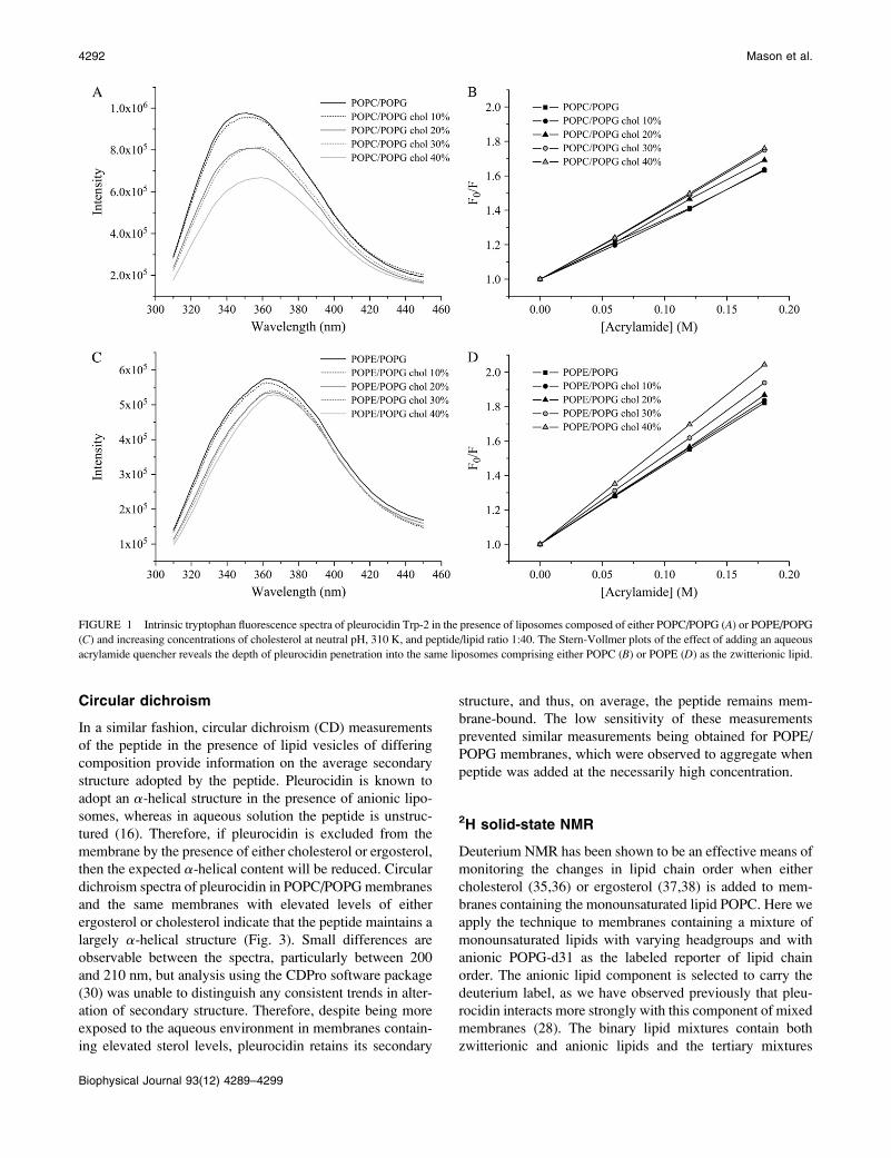
that the addition of increasing amounts of cholesterol to
POPC/POPG membranes leads to a reduction in intensity of
the fluorescence emission spectra, which accompanies a red
shift in the maxima (Fig. 1 A). The corresponding Stern-
Vollmer plots (Fig. 1 B) show a small but noticeable trend of
increasing accessibility to acrylamide. These results indicate
that, on average, the peptide experiences a less hydrophobic
environment for each stepwise addition of cholesterol.
Similar results are obtained when POPE is used as the
zwitterionic component (Fig. 1, C and D); however, the red
shifts and reductions in peak intensity (Fig. 1 C) are much
more reduced, whereas the slope on the Stern-Vollmer plots
(Fig. 1 D) is greater than for the corresponding membranes
containing POPC (Fig. 1, A and B). The emission maxima
obtained in the spectra for samples containing POPE are all
at relatively long wavelengths, between 364 and 367 nm,
indicating that at 310 K the peptide is quite accessible to the
external aqueous environment although still associated with
the hydrophobic membrane, as, for comparison, the emission
maximum for peptide in aqueous solution is at 374 nm
(Stern-Vollmer plot for pleurocidin in solution is available as
supplementary information). This is confirmed by the Stern-
Vollmer plots, which indicate a higher level of accessibility
of the peptide to the aqueous quencher (Fig. 1, C and D),
although again, the slope of the Stern-Vollmer plot for
pleurocidin in solution is over 1.5 times greater than those
for the peptide in the presence of lipid. Nevertheless, to
obtain a clearer picture of the effect of adding cholesterol, the
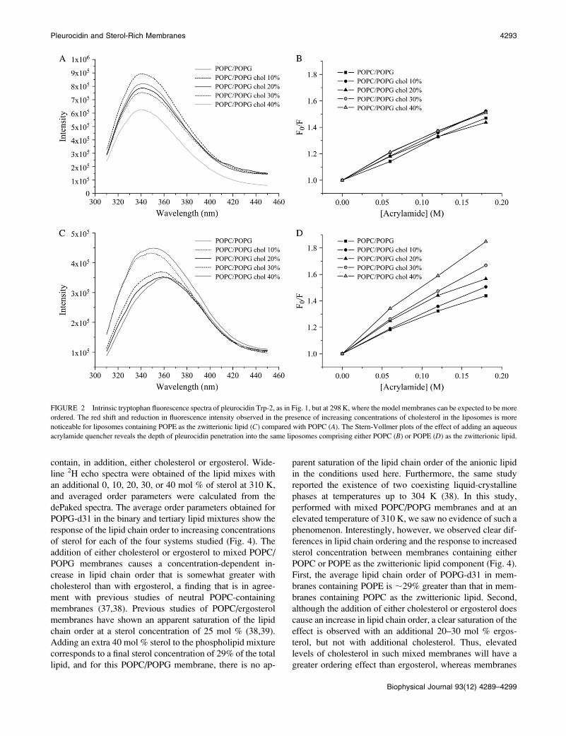
experiments were repeated at the lower temperature of 298
K. At this temperature, the membranes remain in the liquid-
crystalline phase, but the lipids become more ordered;
indeed, a comparison of averaged lipid-chain order param-
eters from this and our previous work (28) indicates that in
POPE/POPG (3:1) membranes, the order of the anionic lipid
chains increases by ;7% when the temperature decreases
from 310 to 298 K. At this lower temperature, the emission
maxima are all blue-shifted to lower wavelengths, indicating
that the peptide experiences a more hydrophobic environ-
ment at 298 K than at 310 K (Fig. 2, A and C). For POPC/
POPG liposomes, the reduction in emission intensity with
increasing cholesterol concentration is much more modest
at the lower temperature, whereas the red shifts observed at
310 K are not apparent (Fig. 2 A). The corresponding Stern-
Vollmer plots (Fig. 2 B) show a slight increase in accessi-
bility to acrylamide at higher cholesterol concentrations only
and confirm that under these conditions, the addition of
cholesterol does not have a large effect on pleurocidin
insertion into the membrane. In contrast, the effect on
pleurocidin insertion of stepwise addition of cholesterol to
POPE/POPG membranes is much clearer under these con-
ditions. When the cholesterol content is increased, large re-
ductions in emission maxima with concomitant red shifts
are observed (Fig. 2 C) which are reflected in clear increases
in the slope of the corresponding Stern-Vollmer plots (Fig.
2 D). Taken together, these data show that when the concen-
tration of cholesterol is increased in mixed anionic membranes,
the peptide experiences, on average, a less hydrophobic en-
vironment and is thus being increasingly excluded from the
hydrophobic core of the membrane. In addition, the fluores-
cence studies performed at both temperatures indicate that
the addition of cholesterol to mixed anionic membranes has a
greater effect on pleurocidin insertion when the zwitterionic
lipid is POPE rather than POPC.
Pleurocidin and Sterol-Rich Membranes 4291
Biophysical Journal 93(12) 4289–4299
Circular dichroism
In a similar fashion, circular dichroism (CD) measurements
of the peptide in the presence of lipid vesicles of differing
composition provide information on the average secondary
structure adopted by the peptide. Pleurocidin is known to
adopt an a-helical structure in the presence of anionic lipo-
somes, whereas in aqueous solution the peptide is unstruc-
tured (16). Therefore, if pleurocidin is excluded from the
membrane by the presence of either cholesterol or ergosterol,
then the expected a-helical content will be reduced. Circular
dichroism spectra of pleurocidin in POPC/POPG membranes
and the same membranes with elevated levels of either
ergosterol or cholesterol indicate that the peptide maintains a
largely a-helical structure (Fig. 3). Small differences are
observable between the spectra, particularly between 200
and 210 nm, but analysis using the CDPro software package
(30) was unable to distinguish any consistent trends in alter-
ation of secondary structure. Therefore, despite being more
exposed to the aqueous environment in membranes contain-
ing elevated sterol levels, pleurocidin retains its secondary
structure, and thus, on average, the peptide remains mem-
brane-bound. The low sensitivity of these measurements
prevented similar measurements being obtained for POPE/
POPG membranes, which were observed to aggregate when
peptide was added at the necessarily high concentration.
2H solid-state NMR
Deuterium NMR has been shown to be an effective means of
monitoring the changes in lipid chain order when either
cholesterol (35,36) or ergosterol (37,38) is added to mem-
branes containing the monounsaturated lipid POPC. Here we
apply the technique to membranes containing a mixture of
monounsaturated lipids with varying headgroups and with
anionic POPG-d31 as the labeled reporter of lipid chain
order. The anionic lipid component is selected to carry the
deuterium label, as we have observed previously that pleu-
rocidin interacts more strongly with this component of mixed
membranes (28). The binary lipid mixtures contain both
zwitterionic and anionic lipids and the tertiary mixtures
FIGURE 1 Intrinsic tryptophan fluorescence spectra of pleurocidin Trp-2 in the presence of liposomes composed of either POPC/POPG (A) or POPE/POPG
(C) and increasing concentrations of cholesterol at neutral pH, 310 K, and peptide/lipid ratio 1:40. The Stern-Vollmer plots of the effect of adding an aqueous
acrylamide quencher reveals the depth of pleurocidin penetration into the same liposomes comprising either POPC (B) or POPE (D) as the zwitterionic lipid.
4292 Mason et al.
Biophysical Journal 93(12) 4289–4299
contain, in addition, either cholesterol or ergosterol. Wide-
line 2H echo spectra were obtained of the lipid mixes with
an additional 0, 10, 20, 30, or 40 mol % of sterol at 310 K,
and averaged order parameters were calculated from the
dePaked spectra. The average order parameters obtained for
POPG-d31 in the binary and tertiary lipid mixtures show the
response of the lipid chain order to increasing concentrations
of sterol for each of the four systems studied (Fig. 4). The
addition of either cholesterol or ergosterol to mixed POPC/
POPG membranes causes a concentration-dependent in-
crease in lipid chain order that is somewhat greater with
cholesterol than with ergosterol, a finding that is in agree-
ment with previous studies of neutral POPC-containing
membranes (37,38). Previous studies of POPC/ergosterol
membranes have shown an apparent saturation of the lipid
chain order at a sterol concentration of 25 mol % (38,39).
Adding an extra 40 mol % sterol to the phospholipid mixture
corresponds to a final sterol concentration of 29% of the total
lipid, and for this POPC/POPG membrane, there is no ap-
parent saturation of the lipid chain order of the anionic lipid
in the conditions used here. Furthermore, the same study
reported the existence of two coexisting liquid-crystalline
phases at temperatures up to 304 K (38). In this study,
performed with mixed POPC/POPG membranes and at an
elevated temperature of 310 K, we saw no evidence of such a
phenomenon. Interestingly, however, we observed clear dif-
ferences in lipid chain ordering and the response to increased
sterol concentration between membranes containing either
POPC or POPE as the zwitterionic lipid component (Fig. 4).
First, the average lipid chain order of POPG-d31 in mem-
branes containing POPE is ;29% greater than that in mem-
branes containing POPC as the zwitterionic lipid. Second,
although the addition of either cholesterol or ergosterol does
cause an increase in lipid chain order, a clear saturation of the
effect is observed with an additional 20–30 mol % ergos-
terol, but not with additional cholesterol. Thus, elevated
levels of cholesterol in such mixed membranes will have a
greater ordering effect than ergosterol, whereas membranes
FIGURE 2 Intrinsic tryptophan fluorescence spectra of pleurocidin Trp-2, as in Fig. 1, but at 298 K, where the model membranes can be expected to be more
ordered. The red shift and reduction in fluorescence intensity observed in the presence of increasing concentrations of cholesterol in the liposomes is more
noticeable for liposomes containing POPE as the zwitterionic lipid (C) compared with POPC (A). The Stern-Vollmer plots of the effect of adding an aqueous
acrylamide quencher reveals the depth of pleurocidin penetration into the same liposomes comprising either POPC (B) or POPE (D) as the zwitterionic lipid.
Pleurocidin and Sterol-Rich Membranes 4293
Biophysical Journal 93(12) 4289–4299

that are largely composed of PE lipids, in preference to PC
lipids, will also have more ordered hydrophobic cores.
The large discrepancy between the average lipid chain
order observed for POPC/POPG and POPE/POPG mem-
branes calls into question which model is more appropriate
for representing natural membranes. To our knowledge,
order parameters are not known for natural membranes and a
detailed study of this is beyond the scope of this work.
However, we obtained an estimate of the lipid chain order
expected for the bacterium E. coli by adding POPE-d31 as a
labeled reporter to E. coli total lipid extract. Total E. colilipid extract contains 57.5% PE by weight (40), and
therefore, the effect of adding 2 mg POPE-d31 to 20 mg
total lipid should be minimized. In our previous study of the
interaction of pleurocidin with POPE/POPG membranes, we
showed that spectra of membranes carrying either POPE-d31
or POPG-d31 as reporter were very similar (28). The
spectrum of vesicles made from the E. coli lipid mix is itself
compared with that of POPE/POPG-d31 (Fig. 5) and can be
seen to be not too dissimilar, indicating that POPE/POPG
and not POPC/POPG model membranes are a much more
reliable mimic of the natural bacterial membrane in terms of
the lipid chain acyl order at least.
The membrane-destabilizing effect of pleurocidin can also
be measured by 2H NMR methods in the presence of ele-
vated levels of sterol. The wideline 2H echo spectra (Fig. 6)
and the corresponding order-parameter profiles calculated
relative to peptide-free membranes (Fig. 7) reveal how ef-
fective pleurocidin remains at destabilizing the anionic lipid
acyl chains in the absence or presence of either cholesterol or
ergosterol. Pleurocidin at 2 mol % has a modest chain-
disordering effect on POPG-d31 in mixed POPC/POPG
vesicles (Figs. 6 A and 7 A). However, since the lipid acyl
chains in these membranes are already rather disordered and
the relevance of this model membrane is uncertain, a com-
parison of the membranes with elevated sterol concentration
is more revealing. 2H NMR spectra of membranes containing
an additional 40 mol % of either cholesterol (Fig. 6 C) or
ergosterol (Fig. 6 E) show that although these sterols have
been shown, using optical methods, to inhibit the penetration
of pleurocidin into the hydrophobic core, the peptide is
nonetheless capable of reducing the lipid acyl chain order of
the anionic lipids. Furthermore, a quantitative comparison
of this chain destabilization (Fig. 7 A) reveals the reduction
FIGURE 5 Comparison of the 2H spectrum obtained for POPE/POPG-
d31 (3:1) and 2 mg of POPE-d31 added to 20 mg of total E. coli lipid extract.
The similarity of the spectra indicate that model membranes composed
of POPE/POPG are a good mimic for the natural E. coli membranes targeted
by antimicrobial peptides.
FIGURE 3 Circular dichroism spectra of pleurocidin in the presence of
liposomes of varying lipid composition at neutral pH, 310 K, and peptide/
lipid ratio 1:40. Spectra of peptide-liposomes containing POPC as the
zwitterionic lipid reveal that pleurocidin retains an a-helical secondary
structure even in the presence of sterol at an additional 40 mol %.
FIGURE 4 Average order parameters of binary and tertiary lipid mem-
branes obtained from dePaked 2H echo spectra of POPG-d31 containing
liposomes at pH 7.5 and 310 K. Original spectra, obtained on a Bruker Avance
300 spectrometer, and smoothed order-parameter profiles are available in the
Supplementary Material.
4294 Mason et al.
Biophysical Journal 93(12) 4289–4299
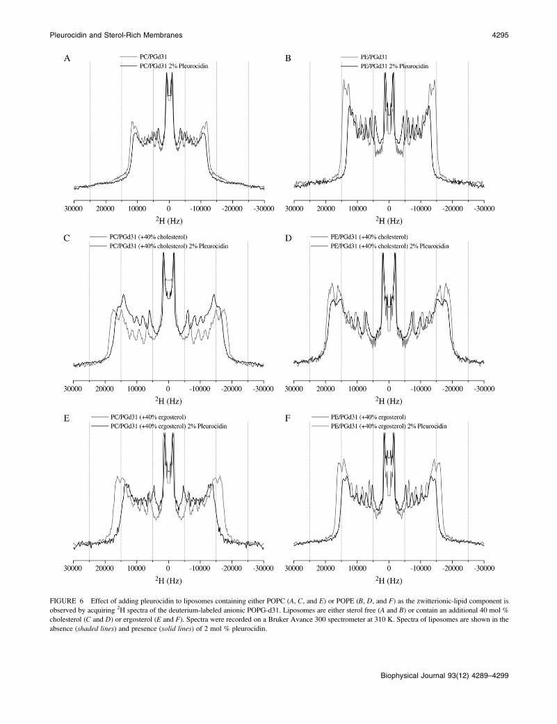
FIGURE 6 Effect of adding pleurocidin to liposomes containing either POPC (A, C, and E) or POPE (B, D, and F) as the zwitterionic-lipid component is
observed by acquiring 2H spectra of the deuterium-labeled anionic POPG-d31. Liposomes are either sterol free (A and B) or contain an additional 40 mol %
cholesterol (C and D) or ergosterol (E and F). Spectra were recorded on a Bruker Avance 300 spectrometer at 310 K. Spectra of liposomes are shown in the
absence (shaded lines) and presence (solid lines) of 2 mol % pleurocidin.
Pleurocidin and Sterol-Rich Membranes 4295
Biophysical Journal 93(12) 4289–4299
of lipid chain acyl order to be much greater in the presence of
ergosterol than of cholesterol. When POPE replaces POPC
as the zwitterionic lipid component a clear lipid-destabilizing
effect can be seen in the presence of 2 mol % pleurocidin
(Figs. 6 B and 7 B), indicating that despite the exposure of
the peptide to the aqueous environment at 310 K, as deter-
mined by fluorescence measurements, pleurocidin remains
associated with the membrane and capable of a strong inter-
action with the anionic lipid component. Again, a quantitative
comparison of the chain-disrupting effects of pleurocidin in
the presence of cholesterol (Figs. 6 D and 7 B) or ergosterol
(Figs. 6 F and 7 A) reveals that the activity of the peptide is
much reduced in the presence of cholesterol when compared
with ergosterol, and indeed, in this case the activity of pleu-
rocidin on the anionic lipid component is almost completely
attenuated. Notably, the chain-ordering effect of elevated
levels of cholesterol is greater in both cases than that of
ergosterol, whereas the greatest increases in sterol-induced
chain ordering correspond to the greatest reductions in
peptide-induced acyl-chain disruption.
Dye-release assay
Confirmation of the effects of incorporating elevated levels
of either ergosterol or cholesterol in the membranes was
obtained by studying pore formation in liposomes by means
of a dye-release assay. Liposomes comprised of POPE/
POPG and an additional 40 mol % of either cholesterol or
ergosterol were challenged by pleurocidin. A previous study
showed that pleurocidin causes release of ;30% of the dye
from negatively charged liposomes at the peptide/lipid ratios
used in this study (16). Here, we observe a similar release of
the calcein dye from POPE/POPG vesicles (Fig. 8). It is
interesting that the presence of elevated levels of ergosterol
reduces the amount of dye released when the liposomes are
challenged by pleurocidin. However, when ergosterol is re-
placed by cholesterol, the release of dye is almost completely
attenuated (Fig. 8).
DISCUSSION
The exact nature of how cationic amphipathic peptides kill
their microbial targets is a matter of some controversy, with a
debate ongoing as to the relative contributions of intracel-
lular targeting and membrane disruption to the overall killing
strategy for each different antimicrobial peptide (1). Anti-
microbial peptides are therefore diverse not only in structure
but also, potentially, in their mechanism of action. Never-
theless, the interaction between the bacterial cell membrane
and antimicrobial peptides is thought to be an important de-
terminant of the peptide activity. Although information re-
FIGURE 7 Smoothed order parameters shown for spectra in Fig. 5 with liposomes incubated with 2 mol % pleurocidin are calculated relative to profiles for
peptide-free liposomes.
FIGURE 8 Comparison of pore formation in anionic liposomes, assessed
by monitoring the release of fluorescent calcein from large unilamellar
vesicles when challenged with pleurocidin. The experiment was performed
at 310 K and pH 7.4 and the times of addition of pleurocidin and Triton
X-100 are indicated by solid and shaded arrows, respectively.
4296 Mason et al.
Biophysical Journal 93(12) 4289–4299
garding the membrane interaction of some classes of peptide,
such as defensins, is scarce, considerably more attention has
been focused on the membrane interaction of linear cationic
peptides such as magainin and pleurocidin. Despite a large
number of studies, there is, however, not yet a consensus on
how peptides from this class interact with cell membranes.
Solid-state NMR studies of aligned samples have shown that
at peptide/lipid ratios where channel conductance is observed,
a large number of amphipathic helical peptides including
magainin (41), pleurocidin (28), piscidins (42), LL-37 (43),
granulysin (44), MSI-843 (45)/ and LAH4 (21,46,47) have
either been shown or proposed to maintain a surface orien-
tation in their active conformation. Models for the action of
antimicrobial peptides on model membranes include the
Carpet mechanism (48) and pore-forming models (49). The
observation of a surface alignment of the peptides conflicts
with existing models that propose the formation of either
barrel-stave, or toroidal, pores, where a peptide orientation
parallel to the membrane normal is required (49,50),
although in the case of toroidal pores, pore formation may
be only transient. The NMR data, however, agrees very well
with a recent in silico study that predicts the formation of a
disordered toroidal pore, at a comparable peptide/lipid ratio,
with one peptide in the pore lumen and all peptides
maintaining an orientation roughly parallel to the membrane
surface (22). This model is useful for considering both a
pore-forming and an intracellular target bactericidal strategy
for linear cationic antibiotic peptides, since it shows that at
such peptide/lipid ratios, the peptide is capable of causing
sufficient membrane disruption that sizeable pores do form
and the peptide can migrate from one leaflet of the membrane
to another with the membrane remaining otherwise intact
(22). Recent studies of the mechanism of action of the
amphipathic cell penetrating peptide Tp-10 using fluores-
cence methods provide further experimental support for this
model (51). Interestingly, the molecular dynamics simula-
tions also revealed that the disorder in the lipid acyl chains of
those lipids in contact with the peptides increased, whereas
the remainder of the bilayer-forming lipids were unaffected
(22), a finding that agrees excellently with our (28,47,52) and
others’ (53,54) experimental observations in model mem-
brane systems studied by solid-state 2H NMR. We showed
that pleurocidin (28) and designed cationic helical peptides
(47,52) cause strong reductions in lipid acyl chain order in
the anionic but not the zwitterionic lipid component in mixed
membranes, indicating that the peptide has a strong effect on
the lipids associated with it but not on other lipids in the
membrane. Whether or not the local reduction of bilayer
order is a result of or a cause of pore formation and, hence,
cell viability, it is a useful probe of the effects of altering the
membrane composition and we have shown that the method
is sufficiently sensitive to detect small changes in chain
order, for example, as a result of altering peptide structure
(55). Hence, here we have assessed the role of the
zwitterionic lipid and the sterol component in modulating
the membrane-disrupting effect of pleurocidin, which we use
as a representative of the linear cationic amphipathic class of
antimicrobial peptides. Specifically, using fluorescence and
circular dichroism techniques, we have studied the binding
of pleurocidin to a variety of mixed membranes and have
then probed lipid acyl chain order in the anionic lipids, the
effect on this of insertion of pleurocidin, and how this is
related to pore formation and the bactericidal strategy of the
peptide.
The intrinsic tryptophan fluorescence measurements
presented here show that the presence of sterols in the
membranes does alter the location of pleurocidin in the
membrane, with increasing amounts of cholesterol increas-
ing the exposure of the peptide to the external aqueous
environment. The CD measurements indicate, however, that
despite this change in environment, the peptide retains its
secondary structure, whereas solid-state NMR measurements
show that it is capable of destabilizing the anionic lipid acyl
chains but to a more limited extent. Cholesterol has been
known for some time to inhibit the lytic activity of the
amphipathic a-helical peptide magainin 2 (56) and early 2H
solid-state NMR measurements indicated that cholesterol
also affected the membrane interaction of magainin 2 (57).
Here, though, we can present a detailed molecular under-
standing of how sterols can reduce the local membrane-
destabilizing effect of the antimicrobial peptide pleurocidin,
which is also closely related to the pore forming capability
of the peptide. Higher concentrations of sterol increase the
order of the acyl chains in the model membranes and by
quantifying both the increase in chain order due to the sterol
and the local chain disordering effect of the peptide we can
see that those membranes that become more ordered, in
particular those containing cholesterol in preference to
ergosterol or POPE in preference to POPC, are the most
resistant to the chain-disordering action of pleurocidin. At a
molecular level, the effect of inhibiting the local disruption
of chain order, which is linked to the formation of disordered
toroidal pores (22), can reduce or even prevent pore forma-
tion and the concomitant release of cell contents or atten-
uation of the membrane potential. Furthermore, since it has
been proposed that linear cationic antimicrobial peptides
translocate from one side of the membrane to another
through such pores (22,51), the inhibition of their formation
by elevated levels of cholesterol would also inhibit the entry
of antimicrobial peptides into a cell and help to protect from
any intracellular killing mechanism. Hence, the presence of
sterols, and in particular cholesterol, may protect against
many of the proposed (1) killing strategies of such antimi-
crobial peptides. Cholesterol and ergosterol are structurally
similar but differ in that ergosterol has two additional double
bonds (at positions C7-C8 and C22-C23) and a methyl group
at C24 of the side chain (38,39). By comparing the membrane
ordering effects of cholesterol and ergosterol with an
intermediate form, it was shown that the structure of both
the fused rings and the more flexible tail contribute to
Pleurocidin and Sterol-Rich Membranes 4297
Biophysical Journal 93(12) 4289–4299
determining lipid acyl chain order (38). There may be a
number of reasons for these structural differences and why
one sterol is chosen over any other in any given organism. In
yeast, for example, the evolution of the structure of ergos-
terol has led to enhanced membrane disorder without the
organism being reliant on the synthesis of unsaturated fatty
acids (58), but it seems likely that adopting ergosterol over
cholesterol can cost an organism in terms of resistance to the
action of antimicrobials. The clear differences in the capa-
bilities of cholesterol and ergosterol to order POPC/POPG,
and more noticeably POPE/POPG membranes, and the
interactions of these membranes with pleurocidin, provide a
molecular view of how ergosterol, which is the major sterol
of lower eukaryotes such as certain protozoa, yeast, and other
fungi (58) is often incapable of offering sufficient protection
from linear cationic antimicrobial peptides.
A variety of bacteria have been shown to be capable of
developing some level of resistance to antimicrobial peptides
(59,60), employing a variety of techniques, including adap-
tations to membrane lipids and membrane fluidity (61,62). In
light of this, the relative compositions of biological mem-
branes and their contributions to protection against the action
of antimicrobial peptides, which continue to be developed
as therapeutics (63), or toxicity of related vector or cell-
penetrating peptides should be an area of increased interest.
This study shows how alterations in lipid composition and
membrane order can affect the action of pleurocidin on its
target membrane. As discussed above, pleurocidin has been
shown to behave similarly to a number of other linear cationic
amphipathic antimicrobial peptides in terms of its topology
and effect on the membrane and, as such, is likely to be
representative of this class of peptide. Future studies will
determine how far this molecular view can be extended to
other classes of antimicrobial peptides, which may be struc-
turally and functionally distinct from those studied here, and
how the interaction of an antimicrobial peptide with a target
membrane fits in with the other known properties of anti-
microbial peptides, which are increasingly identified as hav-
ing a key modulatory role in the innate immune response (64,65).
SUPPLEMENTARY MATERIAL
To view all of the supplemental files associated with this
article, visit www.biophysj.org.
A.J.M. thanks Claire Gasnier for mass spectrometry analysis of peptides,
Thomas Ebbesen for access to the Fluorolog 3-22, and Alex Drake for
invaluable discussion of CD measurements.
This work was supported by Vaincre la Mucoviscidose (TG-0501).
REFERENCES
1. Brogden, K. A. 2005. Antimicrobial peptides: pore formers ormetabolic inhibitors in bacteria? Nat. Rev. Microbiol. 3:238–250.
2. Reddy, K. V. R., R. D. Yedery, and C. Aranha. 2004. Antimicrobialpeptides: premises and promises. Int. J. Antimicrob. Agents. 24:536–547.
3. Fernandez-Carneado, J., M. J. Kogan, S. Pujals, and E. Giralt. 2004.Amphipathic peptides and drug delivery. Biopolymers. 76:196–203.
4. Kichler, A., A. J. Mason, and B. Bechinger. 2006. Cationic amphi-pathic histidine-rich peptides for gene delivery. Biochim. Biophys.Acta. 1758:301–307.
5. Papo, N., and Y. Shai. 2003. Can we predict biological activity ofantimicrobial peptides form their interactions with model phospholipidmembranes? Peptides. 24:1693–1703.
6. Rosenfeld, Y., D. Barra, M. Simmaco, Y. Shai, and M. L. Mangoni.2006. A synergism between temporins toward Gram-negative bacteriaovercomes resistance imposed by the lipopolysaccharide protectivelayer. J. Biol. Chem. 281:28565–28574.
7. Glukhov, E., M. Stark, L. L. Burrows, and C. M. Deber. 2005. Basisfor selectivity of cationic antimicrobial peptides for bacterial versusmammalian membranes. J. Biol. Chem. 280:33960–33967.
8. Pouny, Y., D. Rapaport, A. Mor, P. Nicolas, and Y. Shai. 1992. Inter-action of antimicrobial dermaseptin and its fluorescently labeled ana-logues with phospholipid membranes. Biochemistry. 31:12416–12423.
9. Wimley, W. C., M. E. Selsted, and S. H. White. 1994. Interactionsbetween human defensins and lipid bilayers: evidence for formation ofmultimeric pores. Protein Sci. 3:1362–1373.
10. El Jastimi, R., K. Edwards, and M. Lafleur. 1999. Characterization ofpermeability and morphological perturbations induced by nisin onphosphatidylcholine membranes. Biophys. J. 77:842–852.
11. Zhang, L., A. Rozek, and R. E. W. Hancock. 2001. Interaction ofcationic antimicrobial peptides with model membranes. J. Biol. Chem.276:35714–35722.
12. Marcotte, I., K. L. Wegener, Y.-H. Lam, B. C. S. Cia, M. R. R. dePlanque, J. H. Bowie, M. Auger, and F. Separovic. 2003. Interaction ofantimicrobial peptides from Australian amphibians with lipid mem-branes. Chem. Phys. Lipids. 122:107–120.
13. Ambroggio, E. E., F. Separovic, J. Bowie, and G. D. Fidelio. 2004.Surface behaviour and peptide-lipid interactions of the antibiotic pep-tides, Maculatin and Citropin. Biochim. Biophys. Acta. 1664:31–37.
14. Mani, R., J. J. Buffy, A. J. Waring, R. I. Lehrer, and M. Hong. 2004.Solid-state NMR investigation of the selective disruption of lipidmembranes by Protegrin-1. Biochemistry. 43:13839–13848.
15. Buffy, J. J., M. J. McCormick, S. Wi, A. Waring, R. I. Lehrer, and M.Hong. 2004. Solid-state NMR investigation of the selective perturba-tion of lipid bilayers by the cyclic antimicrobial peptide RTD-1.Biochemistry. 43:9800–9812.
16. Yoshida, K., Y. Mukai, T. Niidome, C. Takashi, Y. Tokunaga, T.Hatakeyama, and H. Aoyagi. 2001. Interaction of pleurocidin and itsanalogs with phospholipid membrane and their antibacterial activity.J. Pept. Res. 57:119–126.
17. Utsugi, T., A. J. Schroit, J. Connor, C. D. Bucana, and I. J. Fidler.1991. Elevated expression of phosphatidylserine in the outer membraneleaflet of human tumor cells and recognition by activated human bloodmonocytes. Cancer Res. 51:3062–3066.
18. Sherman, I. W. 1979. Biochemistry of Plasmodium (malarial para-sites). Microbiol. Rev. 43:453–495.
19. Aloia, R. C., H. Tian, and F. C. Jensen. 1993. Lipid composition andfluidity of the human immunodeficiency virus envelope and host cellplasma membranes. Proc. Natl. Acad. Sci. USA. 90:5181–5185.
20. Hallock, K. J., D.-K. Lee, J. Omnaas, H. I. Mosberg, and A.Ramamoorthy. 2002. Membrane composition determines Pardaxin’smechanism of lipid bilayer disruption. Biophys. J. 83:1004–1013.
21. Vogt, T. C. B., and B. Bechinger. 1999. The interactions of histidine-containing amphipathic helical peptide antibiotics with lipid bilayers.J. Biol. Chem. 274:29115–29121.
22. Leontiadou, H., A. E. Mark, and S. J. Marrink. 2006. Antimicrobialpeptides in action. J. Am. Chem. Soc. 128:12156–12161.
23. Eliassen, L. T., B. E. Haug, G. Berge, and Ø. Rekdal. 2003. Enhancedantitumour activity of 15-residue bovine lactoferricin derivativescontaining bulky aromatic amino acids and lipophilic N-terminalmodifications. J. Pept. Sci. 9:510–517.
4298 Mason et al.
Biophysical Journal 93(12) 4289–4299
24. Nagaraj, G., M. V. Uma, M. S. Shivayogi, and H. Balaram. 2001.Antimalarial activities of peptide antibiotics isolated from fungi.Antimicrob. Agents Chemother. 45:145–149.
25. Syvitski, R. T., I. Burton, N. R. Mattatall, S. E. Douglas, andD. L. Jakeman. 2005. Structural characterization of the antimicrobialpeptide pleurocidin from winter flounder. Biochemistry. 44:7282–7293.
26. Cole, A. M., P. Weis, and G. Diamond. 1997. Isolation andcharacterisation of pleurocidin, an antimicrobial peptide in the skinsecretions of winter flounder. J. Biol. Chem. 272:12008–12013.
27. Cole, A. M., R. O. Darouiche, D. Legarda, N. Connell, andG. Diamond. 2000. Characterization of a fish antimicrobial peptide:gene expression, subcellular localization, and spectrum of activity.Antimicrob. Agents Chemother. 44:2039–2045.
28. Mason, A. J., I. N. H. Chotimah, P. Bertani, and B. Bechinger. 2006. Aspectroscopic study of the membrane interaction of the antimicrobialpeptide Pleurocidin. Mol. Membr. Biol. 23:185–194.
29. Saint, N., H. Cadiou, Y. Bessin, and G. Molle. 2002. Antibacterial pep-tide pleurocidin forms ion channels in planar lipid bilayers. Biochim.Biophys. Acta. 1564:359–364.
30. Sreerama, N., and R. W. Woody. 2000. Estimation of protein secon-dary structure from CD spectra: Comparison of CONTIN, SELCONand CDSSTR methods with an expanded reference set. Anal. Biochem.287:252–260.
31. Davis, J. H. 1983. The description of membrane lipid conformationorder and dynamics H-2-NMR. Biochim. Biophys. Acta. 737:117–171.
32. Schafer, H., B. Madler, and F. Volke. 1995. De-PAKE-ing of NMRpowder spectra by nonnegative least-squares analysis with Tikhonovregularization. J. Magn. Reson. 116:145–149.
33. Sternin, E., M. Bloom, and A. L. MacKay. 1983. De-PAKE-ing ofNMR spectra. J. Magn. Reson. 55:274–282.
34. Seelig, A., and J. Seelig. 1974. Dynamic structure of fatty acyl chainsin a phospholipid bilayer measured by deuterium magnetic resonance.Biochemistry. 13:4839–4845.
35. Thewalt, J. L., and M. Bloom. 1992. Phosphatidylcholine: cholesterolphase diagrams. Biophys. J. 63:1176–1181.
36. Henriksen, J., A. C. Rowat, E. Brief, Y.-W. Hseuh, J. L. Thewalt, M. J.Zuckermann, and J. H. Ipsen. 2006. Universal behaviour of membraneswith sterols. Biophys. J. 90:1639–1649.
37. Urbina, J. A., S. Pekerar, H. B. Le, J. Patterson, B. Montez, and O.Oldfield. 1995. Molecular order and dynamics of phosphatidylcholinebilayer membranes in the presence of cholesterol, ergosterol andlanosterol: a comparative study using 2H-, 13C- and 31P-NMRspectroscopy. Biochim. Biophys. Acta. 1238:163–176.
38. Hsueh, Y.-W., M.-T. Chen, P. J. Patty, C. Code, J. Cheng, B. J.Frisken, M. Zuckermann, and J. Thewalt. 2007. Ergosterol in POPCmembranes: physical properties and comparison with structurallysimilar sterols. Biophys. J. 92:1606–1615.
39. Arora, A., H. Raghuraman, and A. Chattopadhyay. 2004. Influence ofcholesterol and ergosterol on membrane dynamics: a fluorescenceapproach. Biochem. Biophys. Res. Commun. 318:920–926.
40. Morein, S., A.-S. Andersson, L. Rilfors, and G. Lindblom. 1996. Wild-type Escherichia coli cells regulate the membrane lipid composition ina ‘‘window’’ between gel and non-lamellar structures. J. Biol. Chem.271:6801–6809.
41. Bechinger, B., M. Zasloff, and S. J. Opella. 1993. Structure and orien-tation of the antibiotic peptide magainin in membranes by solid-statenuclear magnetic resonance spectroscopy. Protein Sci. 2:2077–2084.
42. Chekmenev, E. Y., B. S. Vollmar, K. T. Forseth, M. N. Manion,S. M. Jones, T. J. Wagner, R. M. Endicott, B. P. Kyriss, L. M. Homem, M.Pate, J. He, J. Raines, P. L. Gor’kov, W. W. Brey, D. J. Mitchell, A. J.Auman, M. J. Ellard-Ivey, J. Blazyk, and M. Cotton. 2006. Investigatingmolecular recognition and biological function at interfaces using piscidins,antimicrobial peptides from fish. Biochim. Biophys. Acta. 1758:1359–1372.
43. Durr, U. H. N., U. S. Sudheendra, and A. Ramamoorthy. 2006. LL-37,the only human member of the cathelicidin family of antimicrobialpeptides. Biochim. Biophys. Acta. 1758:1408–1425.
44. Ramamoorthy, A., S. Thennarasu, A. Tan, D.-K. Lee, C. Clayberger,and A. M. Krensky. 2006. Cell selectivity correlates with membrane-specific interactions: a case study on the antimicrobial peptide G15derived from granulysin. Biochim. Biophys. Acta. 1758:154–163.
45. Thennarasu, S., D.-K. Lee, A. Tan, U. P. Kari, and A. Ramamoorthy.2005. Antimicrobial activity and membrane selective interactions of asynthetic lipopeptide MSI-843. Biochim. Biophys. Acta. 1711:49–58.
46. Bechinger, B. 1996. Towards membrane protein design: pH-sensitivetopology of histidine-containing polypeptides. J. Mol. Biol. 263:768–775.
47. Mason, A. J., C. Gasnier, A. Kichler, G. Prevost, D. Aunis, M.-H.Metz-Boutigue, and B. Bechinger. 2006. Designed histidine-richpeptides show pH dependent antibiotic action against pathogenicbacteria peptides. Antimicrob. Agents Chemother. 50:3305–3311.
48. Oren, Z., and Y. Shai. 1998. Mode of action of linear amphipathica-helical antimicrobial peptides. Biopolymers. 47:451–463.
49. Bechinger, B. 2004. Membrane-lytic peptides. Crit. Rev. Plant Sci.23:271–292.
50. Bechinger, B. 1999. The structure, dynamics and orientation ofantimicrobial peptides in membranes by solid-state NMR spectroscopy.Biochim. Biophys. Acta. 1462:157–183.
51. Yandek, L. E., A. Pokorny, A. Floren, K. Knoelke, U. Langel, andP. F. F. Almeida. 2007. Mechanism of the cell-penetrating peptideTp10 permeation of lipid bilayers. Biophys. J. 92:2434–2444.
52. Mason, A. J., A. Martinez, C. Glaubitz, O. Danos, A. Kichler, and B.Bechinger. 2006. The antibiotic and DNA transfecting peptide LAH4selectively associates with, and disorders, anionic lipids in mixedmembranes. FASEB J. 20:320–322.
53. Ramamoorthy, A., S. Thennarasu, D.-K. Lee, A. Tan, and L. Maloy.2006. Solid-state NMR investigation of the membrane-disruptingmechanism of antimicrobial peptides MSI-78 and MSI-594 derivedfrom magainin 2 and melittin. Biophys. J. 91:206–216.
54. Henzler-Wildman, K. A., G. V. Martinez, M. F. Brown, and A.Ramamoorthy. 2004. Perturbation of the hydrophobic core of lipidbilayers by the human antimicrobial peptide LL-37. Biochemistry.43:8459–8469.
55. Mason, A. J., B. Bechinger, and A. Kichler. 2007. Rational designof vector and antibiotic peptides using solid-state NMR. Mini Rev.Med. Chem. 7:491–497.
56. Matsuzaki, K., K. Sugishita, N. Fujii, and K. Miyajima. 1995.Molecular basis for membrane selectivity of an antimicrobial peptide,magainin 2. Biochemistry. 43:3423–3429.
57. Bechinger, B., M. Zasloff, and S. J. Opella. 1992. Structure and inter-actions of magainin antibiotic peptides in lipid bilayers: a solid-statenuclear magnetic resonance investigation. Biophys. J. 62:12–14.
58. Bloch, K. E. 1983. Sterol structure and membrane function. CRC Crit.Rev. Biochem. 14:47–92.
59. Samuelsen, O., H. H. Haukland, H. Jenssen, M. Kramer, K. Sandvik,H. Ulvatne, and L. H. Vorland. 2005. Induced resistance to theantimicrobial peptide lactoferricin B in Staphylococcus aureus. FEBSLett. 579:3421–3426.
60. Perron, G. G., M. Zasloff, and G. Bell. 2006. Experimental evolution ofresistance to an antimicrobial peptide. Proc. Biol. Sci. 273:251–256.
61. Peschel, A. 2002. How do bacteria resist human antimicrobialpeptides? Trends Microbiol. 10:179–186.
62. Xiong, Y. Q., K. Mukhopadhyay, M. R. Yeaman, J. Adler-Moore,and A. S. Bayer. 2005. Functional interrelationships between cell mem-brane and cell wall in antimicrobial peptide-mediated killing ofStaphylococcus aureus. Antimicrob. Agents Chemother. 49:3114–3121.
63. Hancock, R. E. W., and H.-G. Sahl. 2006. Antimicrobial and host-defence peptides as new anti-infective therapeutic strategies. Nat.Biotechnol. 24:1551–1557.
64. Brown, K. L., and R. E. W. Hancock. 2006. Cationic host defense(antimicrobial) peptides. Curr. Opin. Immunol. 18:24–30.
65. Klotman, M. E., and T. L. Chang. 2006. Defensins in innate antiviralimmunity. Nat. Rev. Immunol. 6:447–456.
Pleurocidin and Sterol-Rich Membranes 4299
Biophysical Journal 93(12) 4289–4299