The Impact of Cyclopropane Configuration on the Biological Activity of Cyclopropyl-Epothilones
Upload
independentCategory
view
0download
0
Accepted Manuscript
C29-Sterols with a cyclopropane ring at C-25 and 26 from the Vietnamese marine
sponge Ianthella sp. and their anticancer properties
Nguyen Huu Tung, Chau Van Minh, Tran Thu Ha, Phan Van Kiem, Hoang
Thanh Huong, Nguyen Tien Dat, Nguyen Xuan Nhiem, Bui Huu Tai, Jae-Hee
Hyun, Hee-Kyoung Kang, Young Ho Kim
PII: S0960-894X(09)00944-5
DOI: 10.1016/j.bmcl.2009.06.097
Reference: BMCL 14565
To appear in: Bioorganic & Medicinal Chemistry Letters
Received Date: 27 May 2009
Revised Date: 24 June 2009
Accepted Date: 26 June 2009
Please cite this article as: Tung, N.H., Minh, C.V., Ha, T.T., Kiem, P.V., Huong, H.T., Dat, N.T., Nhiem, N.X., Tai,
B.H., Hyun, J-H., Kang, H-K., Kim, Y.H., C29-Sterols with a cyclopropane ring at C-25 and 26 from the Vietnamese
marine sponge Ianthella sp. and their anticancer properties, Bioorganic & Medicinal Chemistry Letters (2009), doi:
10.1016/j.bmcl.2009.06.097
This is a PDF file of an unedited manuscript that has been accepted for publication. As a service to our customers
we are providing this early version of the manuscript. The manuscript will undergo copyediting, typesetting, and
review of the resulting proof before it is published in its final form. Please note that during the production process
errors may be discovered which could affect the content, and all legal disclaimers that apply to the journal pertain.
ACCEPTED MANUSCRIPT
Graphical Abstract
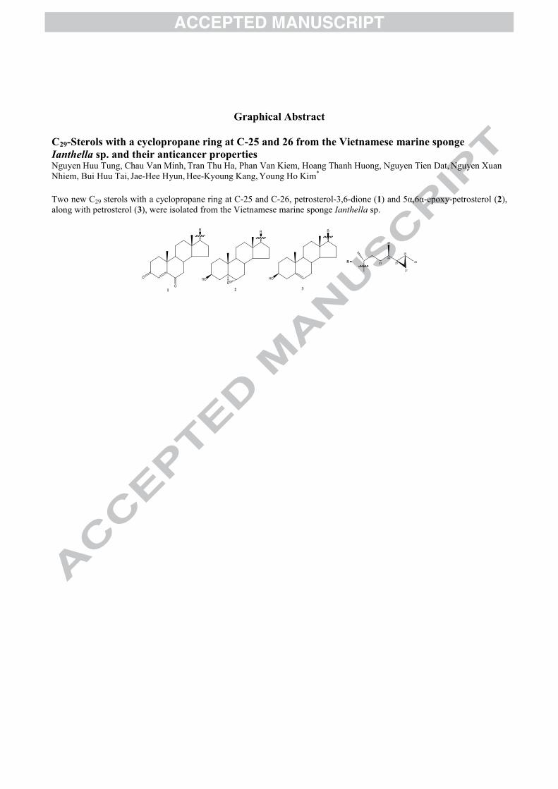
C29-Sterols with a cyclopropane ring at C-25 and 26 from the Vietnamese marine sponge Ianthella sp. and their anticancer propertiesNguyen Huu Tung, Chau Van Minh, Tran Thu Ha, Phan Van Kiem, Hoang Thanh Huong, Nguyen Tien Dat, Nguyen Xuan Nhiem, Bui Huu Tai, Jae-Hee Hyun, Hee-Kyoung Kang, Young Ho Kim*
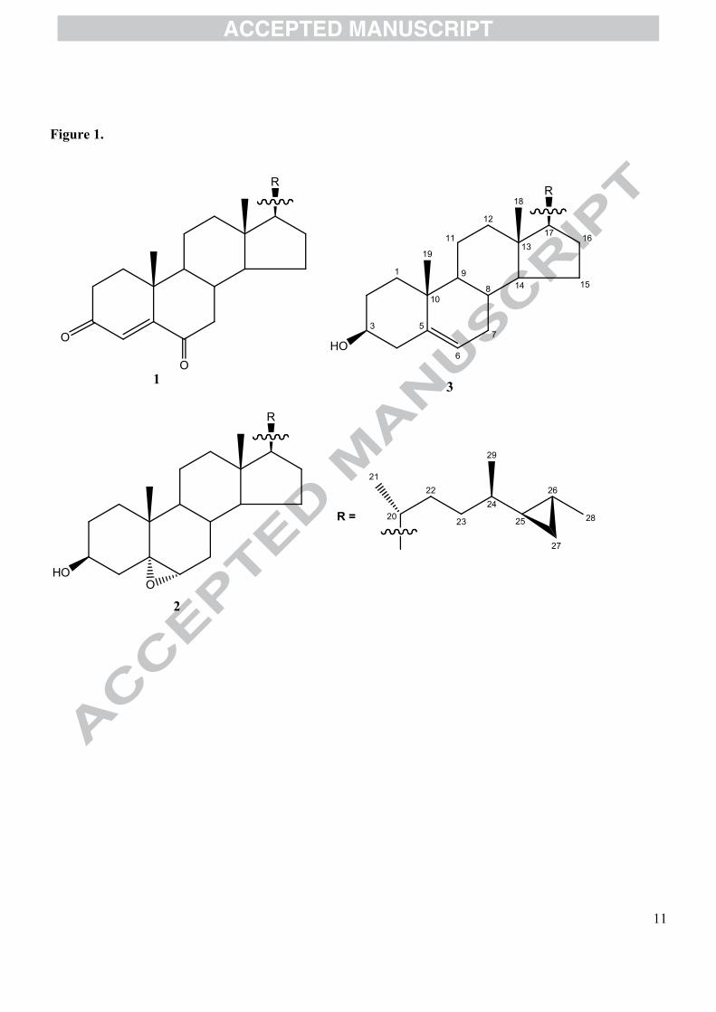
Two new C29 sterols with a cyclopropane ring at C-25 and C-26, petrosterol-3,6-dione (1) and 5α,6α-epoxy-petrosterol (2), along with petrosterol (3), were isolated from the Vietnamese marine sponge Ianthella sp.
R
O
O
R
HOO
1 2
23
24
29
25
26
27
28R =
R
HO
3
ACCEPTED MANUSCRIPT
1
C29-Sterols with a cyclopropane ring at C-25 and 26 from the
Vietnamese marine sponge Ianthella sp. and their anticancer properties
Nguyen Huu Tunga,b, Chau Van Minhb, Tran Thu Hab, Phan Van Kiemb, Hoang Thanh
Huongb, Nguyen Tien Datb, Nguyen Xuan Nhiema,b, Bui Huu Taia, Jae-Hee Hyunc, Hee-
Kyoung Kangc, Young Ho Kima,*
a College of Pharmacy, Chungnam National University, Daejeon 305-764, Korea b Institute of Natural Products Chemistry, Vietnam Academy of Science and Technology,
18 Hoang Quoc Viet, Nghiado, Caugiay, Hanoi, Vietnamc School of Medicine, Institute of Medical Sciences, Jeju National University, Jeju 690-
756, Korea
Correspondence to:
Young Ho Kim Ph.D.
College of Pharmacy
Chungnam National University
Daejeon 305-764, Korea
Tel: 82-42-821-5933
Fax: 82-42-823-6566
e-mail: [email protected]

ACCEPTED MANUSCRIPT
2
Two new C29 sterols with a cyclopropane ring at C-25 and C-26, petrosterol-3,6-dione (1) and 5α,6α-epoxy-petrosterol (2), along with petrosterol (3), were isolated from the Vietnamese marine sponge Ianthella sp. The structures of the new compounds were elucidated by comprehensive spectroscopic analyses. Compounds 1−3 showed cytotoxic activities on A549, HL-60, MCF-7, SK-OV-3, and U937 cancer cell lines with IC50 in the range of 8.4 to 22.6 μM, whereas compounds 1−3 exhibited only weak cytotoxic activities on HT-29 cell. After HL-60 cells were treated with the compounds, several apoptosis events like as chromatin condensation and the increase of the population of sub-G1 hypodiploid cells were observed. These data supported that the compounds might have potential for leukemia treatment.
Key words: Sponge, Ianthella sp., Sterol, Petrosterol, Cytotoxicity, Apoptosis
ACCEPTED MANUSCRIPT
3
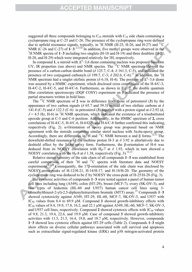
Marine sponges are sources of numerous novel sterols, particularly in terms of unique side chain structures such as those with high degree of alkylation and unusual function groups.1 In which, steroids with a cyclopropane ring at C-25 and C-26 are relatively rareand first reported in 1978 from sponges, the Petrosia ficiformis2 and Halichondria sp.3
Some of these analogs have been reported to show biological activities including anti-inflammation and cytotoxicity.4−6 The sponges of the genus Ianthella (order Verongida, family Ianthellidae) have been reported to contain several bromotyrosine-derived metabolites, such as the ianthesines, purealin, and others, which were mainly isolated from their polar extracts.7–9
In our research on secondary metabolites from Vietnamese marine organisms, chromatographic fractionation and isolation led to results of three sterols (1−3)10
including two new sterols, namely petrosterol-3,6-dione (1) and 5α,6α-epoxy-petrosterol (2), with a cyclopropane ring at C-25 and C-26 and a known one, petrosterol (3),4 from the MeOH extract of the sponge Ianthella sp.
The marine sponge Ianthella sp. (2.4 kg, wet weight) were collected at Namyet island, Khanh Hoa province in March, 2007, and was kept frozen (−20oC) until used; it was identified by Dr. Do Cong Thung, Institute of Marine Resources and Environment, Vietnam Academy of Science and Technology (VAST), Hanoi, Vietnam. The specimen vouchers of the Ianthella sp. (No 20070306) have been deposited at Institute of Natural Products Chemistry and Institute of Marine Resources and Environment, VAST, Vietnam.The frozen sample Ianthella sp. was ground and exhaustively extracted by the ultrasound-assisted manner with methanol (5 L × 3, each one day) at room temperature, and combined MeOH extract was concentrated in vacuo to give a black gum residue (70 g). The residue was suspended in H2O (0.5 L), and partitioned with EtOAc (0.5 L × 3). Next, the EtOAc-soluble portion (9.3 g) was subjected to silica gel column eluted with a gradient of MeOH in CHCl3 (30:1−1:1, v/v) to give four fractions (1a―d). The fraction 1a was repeatedly chromatogaphed using silica gel column with an eluent of CHCl3−MeOH (30:1, v/v) to afford 1 (20 mg). Thereafter, fraction 1b was further chromatographed using reversed-phase C-18 YMC column with an eluent of MeOH−acetone−H2O (5:1:3, v/v/v), followed by silica gel column chromatography (n-hexane−EtOAc, 3:1, v/v) to afford 2 (7.5 mg). Finally, repeated column chromatography of the fraction 1c (1.8 g) using silica gel (n-hexane−EtOAc, 5:1, v/v) and reversed-phase C-18 YMC (MeOH−H2O−acetone, 5:1:3, v/v/v) resulted in the isolation of 3 (12.5 mg).
The structure of compounds 1−3 (Fig. 1) were elucidated by using NMR, UV, IR and MS spectra. The IR (1685 cm−1) and UV (250 nm) spectra of 1 verified the presence of carbonyl groups and a conjugated double bond system, Δ4−3,6 dione. The quasimolecular peak was appeared at m/z 425 [M+H]+ in FABMS. The molecular formula of 1, C29H44O2, was deduced by HRFABMS (detected peak at m/z [M+H]+ 425.3397, calcd for C29H45O2,425.3375).
The IR spectrum of 2 displayed absorptions at 3443 cm−1 (OH group) and 1260 cm−1
(epoxy group). The molecular formula of 2, C29H48O2, was established based on HREIMS (detected peak at m/z [M]+ 428.3656, calcd for C29H48O2 428.3654) and FABMS (observed peak at m/z 429 [M+H]+).
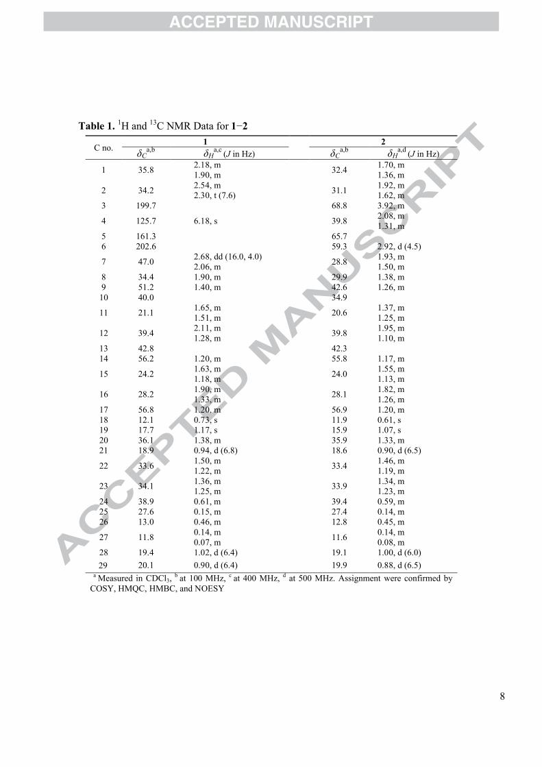
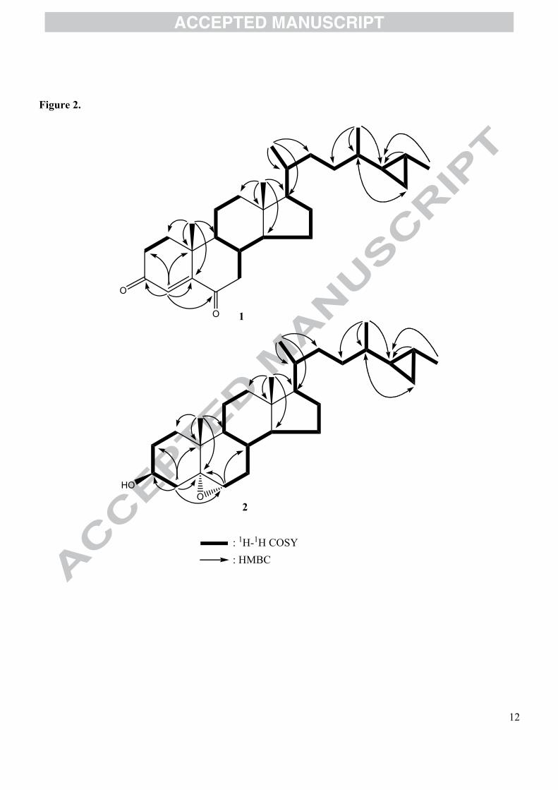
The 1H and 13C NMR data of 1 and 2 are summarized in Table 1. The assignments were confirmed by HMQC, HMBC, COSY, and NOESY spectra. The NMR data
ACCEPTED MANUSCRIPT
4
suggested all three compounds belonging to C29 steroids with C10 side chain containing a cyclopropane ring at C-25 and C-26. The presence of the cyclopropane ring were defined due to upfield resonance signals, typically, in 1H NMR (H-25, H-26, and H-27) and 13C NMR (C-26 and C-27) of 1−3.2,3,5,6 In addition, five methyl groups were observed in the1H NMR spectra of 1−3 including two singlets (H-18 and H-19) and three doublets (H-21,H-28, and H-29) which were integrated relatively for 3H, respectively.
In compound 1, a steroid with Δ4−3,6 dione containing nucleus was proposed based on UV, IR properties (see above) and NMR spectra. The 13C NMR spectrum showed the presence of a carbon-carbon double bond (δ 125.7, C-4; δ 161.3, C-5), and indicated the presence of two conjugated carbonyls (δ 199.7, C-3; δ 202.6, C-6).11 In addition, the 1H NMR spectrum had a singlet olefinic proton (δ 6.18, H-4). The position of Δ4−3,6 dione was assured by a HMBC experiment, which disclosed cross correlations of the H-4/C-3, H-4/C-2, H-4/C-5, and H-4/C-6. Furthermore, as shown in Fig. 2, the double quantum filter correlation spectroscopy (DQF COSY) experiment on 1 indicated the presence of partial structures written in bold lines.
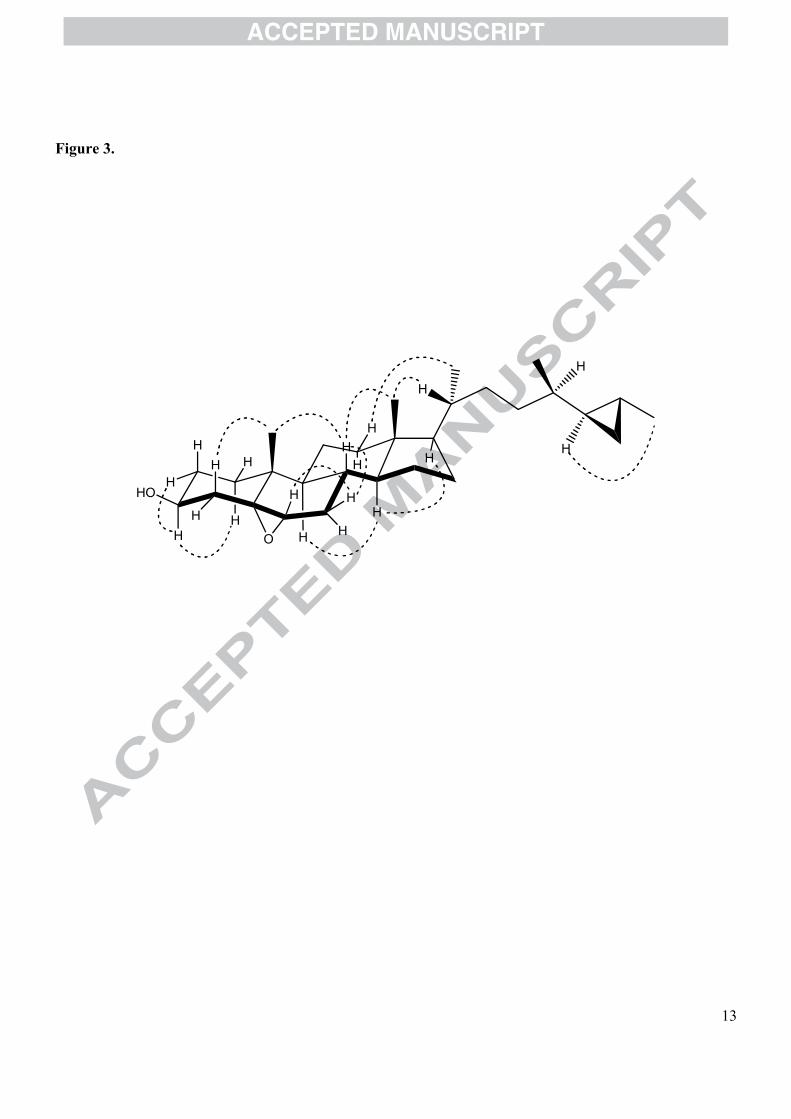
The 13C NMR spectrum of 2 was in difference from those of petrosterol (3) by theappearance of two carbon signals (δ 65.7 and 59.3) instead of two olefinic carbons at δ 141.8 (C-5) and δ 122.3 (C-6) in petrosterol (3) together with a proton signal at δ 2.92 (d,J = 4.5 Hz, H-6) in 1H NMR spectrum, which indicated the existence of a trisubstituted epoxide group at C-5 and C-6 position. Additionally, in the HMBC spectrum of 2, cross correlations of H-4/C-5, H-4/C-6, H-6/C-5, and H-6/C-8 further confirmed this indicating, respectively (Fig. 2). α-Orientation of epoxy group was proposed based on NMR agreement with the steroids containing similar sterol nucleus with 5α,6α-epoxy group. Accordingly, there are differences in 1H and 13C NMR between α and β forms.12,13 The downfield-shifted resonance of the methine proton H-3 at δ 3.92 is alterlatively due to deshield effect by the 5α,6α-epoxy form. Furthermore, the β-orientation of H-6 was deduced from its NOESY correlation with Hβ-7 at δ 1.93, which in turn showed a NOESY correlation with the Hβ-8 at δ 1.38, respectively (Fig. 3).12,13
Relative stereochemistry of the side chain of all compounds 1−3 was established from careful comparison of their 1H and 13C spectra with literature data and NOESYexperiments.4,5,14 Consequently, the 17β-orientation of the side chain was disclosed by NOESY cross-peaks of H-12/H-21, H-14/H-17, and H-18/H-20. The geometry of the cyclopropane ring was deduced to be E by NOESY the cross-peak of H-25/H-28 (Fig. 3).
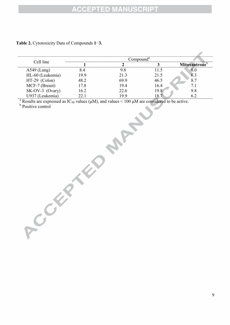
The cytotoxic activities of compounds 1−3 were tested against a panel of human tumor cell lines including lung (A549), colon (HT-29), breast (MCF-7), ovary (SK-OV-3), and two types of leukemia (HL-60 and U937) human cancer cell lines using 3-(dimethylthiazol-2-yl)-2,5-diphenyltetrazolium bromide (MTT) assay.15 Compounds 1−3showed cytotoxicity against A549, HT-29, HL-60, MCF-7, SK-OV-3, and U937 with IC50 values from 8.4 to 69.9 μM. Compound 1 showed growth-inhibitory effects with IC50 values of 8.4, 19.9, 17.8, 16.2, and 22.1 μM against A549, HL-60, MCF-7, SK-OV-3,and U937 cell lines, respectively. Compound 2 showed cytotoxic effects with IC50 values of 9.8, 21.3, 19.4, 22.6, and 19.9 μM. Case of compound 3 showed growth-inhibitoryactivities with 11.5, 21.5, 16.4, 19.8, and 18.7 μM, respectively. However, compounds1−3 showed less cytotoxic effects against HT-29 cells (Table 2). Compounds 1–3 might show effects on diverse cellular pathways associated with cell survival and apoptosis such as extracellular signal-regulated kinase (ERK) and p38 mitogen-activated protein
ACCEPTED MANUSCRIPT
5
kinase (MAPK) pathway as well as phosphoinositide 3 (PI3) kinase /Akt pathway.16–19 In order to explain the potency difference between cell lines, the cellular action mechanisms of the compounds should be studied in detail.
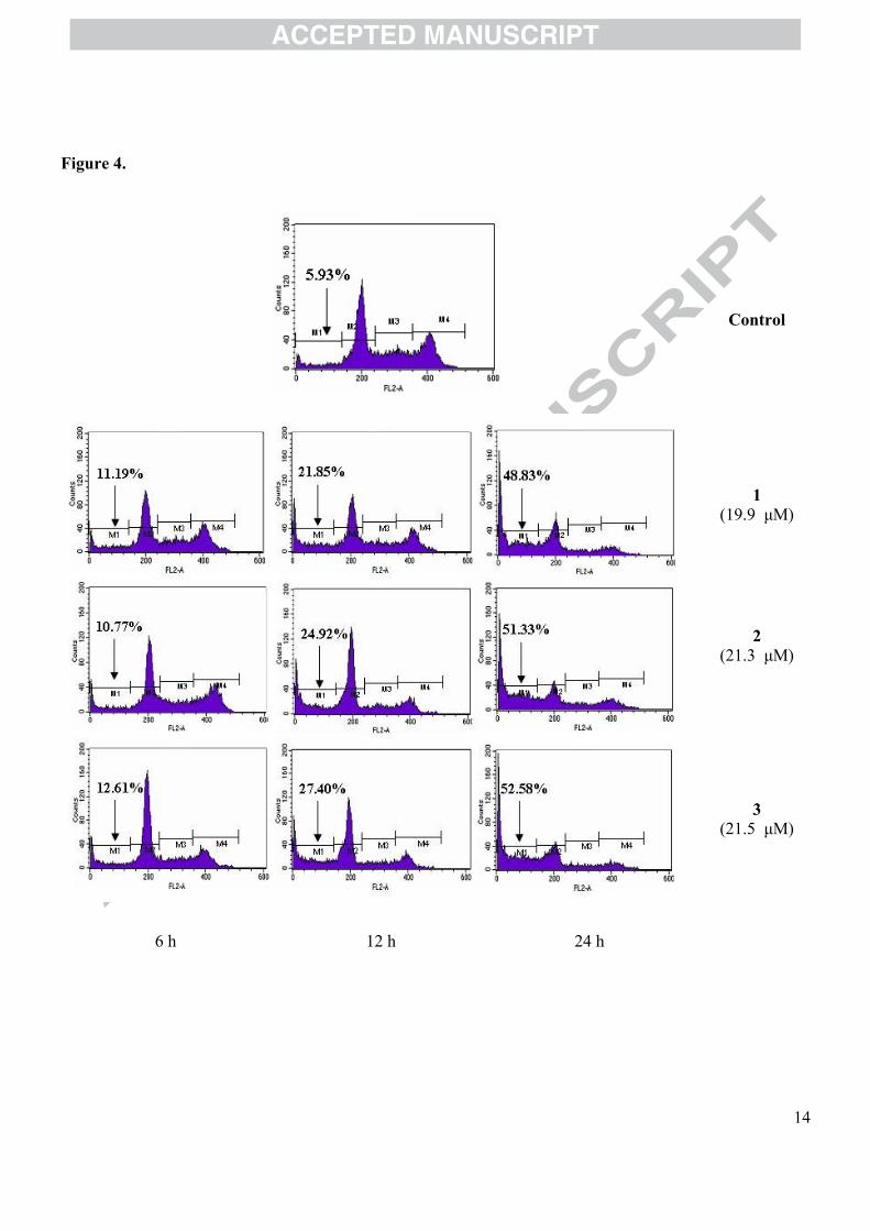
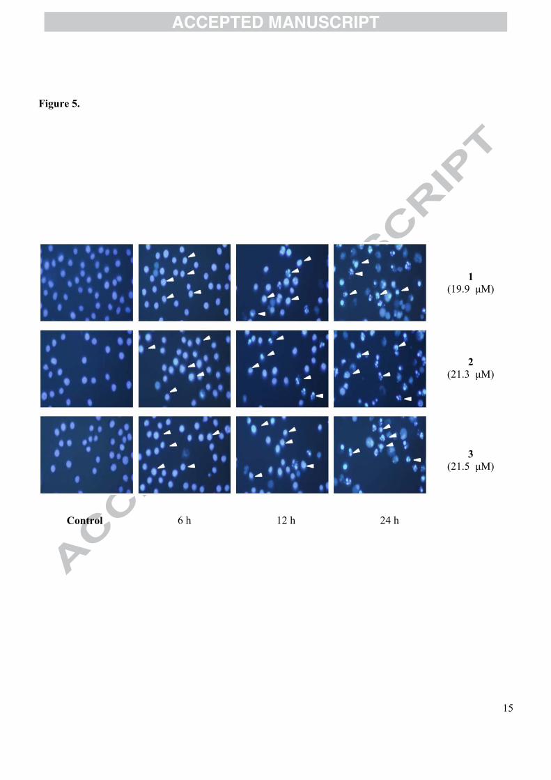
We investigated whether the inhibitory effects of compounds 1−3 on the growth of cancer cells might arise from the induction of apoptosis.20 The apoptotic characteristicswere examined after the HL-60 were treated with IC50 of compounds 1−3 for 6, 12, and 24 h. The percentage of sub-G1 hypodiploid cells by the treatment of compound 1 increased from 11.2% to 48.8% in a time-dependent manner. In the treatment of compounds 2 and 3, Sub-G1 hypodiploid cells increased from 11.8% to 51.3% and from12.6% to 52.6% in time-dependent manners (Fig. 4). These data show that compoundsinduce the apoptosis of HL-60 cells, and are supported by the increase in the number of apoptotic bodies which were easily found by H33342 staining in compounds-treated cells after over 6, 12, and 24 h-incubation (Fig. 5). This study observed that compounds 1–3markedly inhibited the growth of various cancer cells suggesting that the compounds induced necrosis or apoptosis. Epigallocatechin gallate (EGCG), one of green tea catechins, has been reported to induce the apoptosis of HL-60, acute promyelocytic leukemia cells, while EGCG was demonstrated to cause caspase-independent necrosis-like cell death in chronic myelogenous leukemia such as K562 and C2F8.21,22 As the compounds 1–3 induced the apoptosis of HL-60 leukemia cells, the compounds are expected to be able to induce the apoptosis in the other tested cancer cells. However, in further study, we need to evaluate the mode of cell death by compounds 1–3 based oncancer cell types.
In summary, this study demonstrated that compounds 1−3 inhibited the growth of the HL-60 cells by the induction of apoptosis.Acknowledgements
This work was partially supported by the Korea Foundation for International Cooperation of Science & Technology (KICOS) through a grant provided by the Korean Ministry of Science & Technology (MOST) in Korea (No. K2072100000208B010000210) and the Vietnam KC10.20/06-10 Project. The authors are grateful to Dr. Do Cong Thung, VAST, for his kind identification of the sponge, and Korean Basic Science Institute (KBSI) for NMR and MS measurements.
ACCEPTED MANUSCRIPT
6
References and Notes1. Faulkner, D. J. Nat. Prod. Rep. 1993, 10, 497 and previous papers in this series.2. Sica, D.; Zollo, F. Tetrahedron Lett. 1978, 9, 837.3. Ravi, B. N.; Kokke. W. C.; Delseth, C.; Djerassi, C. Tetrahedron Lett. 1978, 9,
4379.4. Mandeau, A.; Debitus, C.; Aries, M. F.; David, B. Steroids 2005, 70, 873.5. Kobayashi, M.; Chen, Y. J.; Higuchi, K.; Aoki, S.; Kitagawa, I. Chem. Pharm.
Bull. 1996, 44, 1840.6. Iguchi, K.; Shimura, H; Taira, S.; Yokoo, C.; Matsumoto, K.; Yamada, Y. J. Org.
Chem. 1994, 59, 7499.7. Okamoto, Y.; Ojika, M.; Kato, S.; Sakagami, Y. Tetrahedron 2000, 56, 5813.8. Nakamura, H.; Wu, H.; Kobayashi, J.; Nakamura, Y.; Ohizumi, Y.; Hirata, Y.
Tetrahedron Lett. 1985, 26, 4517.9. Ciminiello, P.; Fattorusso, E.; Forino, M.; Magno, S.; Pansini, M. Tetrahedron
1997, 53, 6565.10. Petrosterol-3,6-dione (1): yellow amorphous powder; 20][ D −2.8o (c 0.06,
CHCl3); UV: MeOHmax (nm) (log ε) 204.0 (1.04), 250.0 (1.49); IR (KBr) νmax 2945,
1685, 1242 cm−1; 1H NMR (CDCl3, 400 MHz) and 13C NMR (CDCl3, 100 MHz): see Table 1; positive FABMS m/z 425 [M+H]+; positive HRFABMS m/z [M+H]+
425.3397 (Calcd for C29H45O2 425.3375). 5α,6α-Epoxy-petrosterol (2): white solid; 20][ D −1.9o (c 0.07, CHCl3); UV
MeOHmax (nm) (log ε) 223.0 (2.29); IR (KBr) νmax 3443, 2951, 2924, 1260, 1041
cm−1; 1H NMR (CDCl3, 400 MHz) and 13C NMR (CDCl3, 100 MHz): see Table 1; FABMS m/z 429 [M+H]+; HREIMS m/z [M]+ 428.3656 (Calcd for C29H48O2 428.3654).Petrosterol (3): white powder; mp 121−122oC; 20][ D −16o (c 0.8, CHCl3);
1H NMR (CDCl3, 500 MHz): δ 5.30 (1H, br d, J = 5.5 Hz, H-6), 3.48 (1H, tt, J = 11.5, 5.5 Hz, H-3), 1.04 (3H, s, H-19), 1.03 (3H, d, J = 6.0 Hz, H-28), 0.92 (3H, d, J = 6.5, H-21), 0.88 (3H, d, J = 6.5 Hz, H-29), 0.68 (3H, s, H-18); 13C NMR (CDCl3, 125 MHz): δ 38.2 (C-1), 31.9 (C-2), 72.1 (C-3), 42.7 (C-4), 141.8 (C-5), 122.3 (C-6), 32.8 (C-7), 32.7 (C-8), 51.2 (C-9), 37.4 (C-10), 21.9 (C-11), 40.7 (C-12), 43.2 (C-13), 57.7 (C-14), 25.1 (C-15), 29.1 (C-16), 57.1 (C-17), 12.4 (C-18), 19.6 (C-19), 36.9 (C-20), 19.3 (C-21), 34.4 (C-22), 34.8 (C-23), 39.7 (C-24), 28.2 (C-25), 13.6 (C-26), 12.2 (C-27), 19.9 (C-28), 20.4 (C-29); FABMS m/z 413 [M+H]+.
11. Kontiza, I.; Abatis, D; Malakate, K.; Vagias, C.; Roussis, V. Steroids 2006, 71, 77.12. Krishna, V.; Chang, C.; Chou, C. H. Magn. Reson. Chem. 2006, 44, 817.13. Xu, S.; Liao, X.; Du, B.; Zhou, X.; Huang, Q.; Wu, C. Steroids 2008, 73, 568.14. Umeyama, A.; Ito, S.; Yoshigaki, A.; Arihara, S. J. Nat. Prod. 2000, 63, 1540.15. Carmichael, J.; DeGraff, W. G.; Gazdar, A. F.; Minna, J. D.; Mitchell, J. B.
Cancer. Res. 1987, 47, 943.16. Shi, Y.; Gaestel, M. Biol. Chem. 2002, 383, 1519.
17. Pearson, G.; Robinson, F.; Beers Gibson, T.; Xu, B.; Karandikar, M.; Berman, K.;
Cobb, M. H. Endocr. Rev. 2001, 22, 153.
18. Scheid, M. P.; Woodgctt, J. R. FEBS Lett. 2003, 546, 108.
ACCEPTED MANUSCRIPT
7
19. Nicholson, K. M.; Anderson, N. G. Cell. Signalling 2002, 14, 381.20. Flow cytometric analysis: HL-60 cells (3×10⁵ cells/mL) were treated with IC50 of
compounds for 6, 12, and 24 h. For the flow cytometric analysis to determine cell cycle phase distribution, the treated cells were washed twice with PBS and fixed in 70% ethanol for 30 min at 4°C. The cells were then rinsed with PBS and incubated in 50 μg/mL propidium iodide solution (PI; Sigma) and 50 μg/mLRNase A in the dark for 30 min at 37°C. Flow cytometry analysis was performed using an flow cytometer (Becton Dickinson FACS Caliber, BD Biosciences,USA). The DNA histograms obtained were analyzed to measure the proportion of sub-G1 hypodiploid cells.Morphology analysis: HL-60 cells (3×10⁵ cells/mL) were treated with IC50 of compounds for 6, 12, and 24 h. Cells were washed twice with PBS before being stained with 1 mg/mL Hoechst 33342 for 30 min at 37°C. Apoptotic bodies, with condensed and fragmented nuclei, were observed with a fluorescence microscope (×400, BX-50, Olympus, Japan).
21. Hibasami, H.; Achiwa, Y.; Fujikawa, T.; Komiya, T. Anticancer Res. 1996, 16, 1943.
22. Iwasaki, R.; Ito, K.; Ishida, T.; Hamanoue, M.; Adachi, S.; Watanabe, T.; Sato, Y.Cancer Sci. 2009, 100, 349.
ACCEPTED MANUSCRIPT
8
Table 1. 1H and 13C NMR Data for 1−2
C no.1 2
δCa,b δH
a,c (J in Hz) δC
a,b δHa,d
(J in Hz)
1 35.82.18, m1.90, m
32.41.70, m1.36, m
2 34.22.54, m2.30, t (7.6)
31.11.92, m1.62, m
3 199.7 68.8 3.92, m
4 125.7 6.18, s 39.82.08, m1.31, m
5 161.3 65.76 202.6 59.3 2.92, d (4.5)
7 47.02.68, dd (16.0, 4.0)2.06, m
28.81.93, m1.50, m
8 34.4 1.90, m 29.9 1.38, m9 51.2 1.40, m 42.6 1.26, m10 40.0 34.9
11 21.11.65, m1.51, m
20.61.37, m1.25, m
12 39.42.11, m1.28, m
39.81.95, m1.10, m
13 42.8 42.314 56.2 1.20, m 55.8 1.17, m
15 24.21.63, m1.18, m
24.01.55, m1.13, m
16 28.21.90, m1.33, m
28.11.82, m1.26, m
17 56.8 1.20, m 56.9 1.20, m18 12.1 0.73, s 11.9 0.61, s19 17.7 1.17, s 15.9 1.07, s20 36.1 1.38, m 35.9 1.33, m21 18.9 0.94, d (6.8) 18.6 0.90, d (6.5)
22 33.61.50, m1.22, m
33.41.46, m1.19, m
23 34.11.36, m1.25, m
33.91.34, m1.23, m
24 38.9 0.61, m 39.4 0.59, m25 27.6 0.15, m 27.4 0.14, m26 13.0 0.46, m 12.8 0.45, m
27 11.80.14, m0.07, m
11.60.14, m0.08, m
28 19.4 1.02, d (6.4) 19.1 1.00, d (6.0)
29 20.1 0.90, d (6.4) 19.9 0.88, d (6.5)a Measured in CDCl3,
b at 100 MHz, c at 400 MHz, d at 500 MHz. Assignment were confirmed by COSY, HMQC, HMBC, and NOESY
ACCEPTED MANUSCRIPT
9
Table 2. Cytotoxicity Data of Compounds 1−3.
Cell lineCompounda
1 2 3 Mitoxantroneb
A549 (Lung) 8.4 9.8 11.5 8.0HL-60 (Leukemia) 19.9 21.3 21.5 6.3HT-29 (Colon) 48.2 69.9 46.5 8.7MCF-7 (Breast) 17.8 19.4 16.4 7.1SK-OV-3 (Ovary) 16.2 22.6 19.8 9.8U937 (Leukemia) 22.1 19.9 18.7 6.2
a Results are expressed as IC50 values (μM), and values < 100 μM are considered to be active.b Positive control
ACCEPTED MANUSCRIPT
10
Legends
Figure 1: Structures of 1−3
Figure 2: 1H-1H COSY and selected HMBC correlations for 1 and 2
Figure 3: Key NOESY correlations of 2
Figure 4: The degree of apoptosis represented as the DNA content measured by flow cytometric analysis in
HL-60 cells.
Figure 5: The degree of apoptosis represented as the fluorescent image of nuclei in cells by fluorescent
microscope.
ACCEPTED MANUSCRIPT
11
Figure 1.
3 5
10
1
6
7
8
9
12
11
14
13
1716
15
R18
19
HO
R
O
O
R
HOO
20
22
23
24
21
29
25
26
27
28R =
31
2
ACCEPTED MANUSCRIPT
12
Figure 2.
O
O
HOO
: 1H-1H COSY
: HMBC
1
2
ACCEPTED MANUSCRIPT
13
Figure 3.
H
H
OH
HO
H
H
H
H H
H
H
H
H
H
H
H
H
HH
H
ACCEPTED MANUSCRIPT
14
Figure 4.
Control
1(19.9 μM)
2(21.3 μM)
3(21.5 μM)
6 h 12 h 24 h
ACCEPTED MANUSCRIPT
15
Figure 5.
1(19.9 μM)
2(21.3 μM)
3(21.5 μM)
Control 6 h 12 h 24 h
Copyright © 2022 FDOKUMEN