Active destabilization of base pairs by a DNA glycosylase wedge initiates damage recognition
10
272–281 Nucleic Acids Research, 2015, Vol. 43, No. 1 Published online 17 December 2014 doi: 10.1093/nar/gku1300 Active destabilization of base pairs by a DNA glycosylase wedge initiates damage recognition Nikita A. Kuznetsov 1,2,† , Christina Bergonzo 3,† , Arthur J. Campbell 3 , Haoquan Li 3 , Grigory V. Mechetin 1 , Carlos de los Santos 4 , Arthur P. Grollman 4 , Olga S. Fedorova 1,2,* , Dmitry O. Zharkov 1,2,* and Carlos Simmerling 3,5,* 1 SB RAS Institute of Chemical Biology and Fundamental Medicine, 8 Lavrentieva Ave., Novosibirsk 630090, Russia, 2 Department of Natural Sciences, Novosibirsk State University, 2 Pirogova St., Novosibirsk 630090, Russia, 3 Department of Chemistry, Stony Brook University, Stony Brook, NY 11794, USA, 4 Department of Pharmacological Sciences, Stony Brook University, Stony Brook, NY 11794, USA and 5 Laufer Center for Physical and Quantitative Biology, Stony Brook University, Stony Brook, NY 11794, USA Received November 09, 2014; Revised November 26, 2014; Accepted November 28, 2014 ABSTRACT Formamidopyrimidine-DNA glycosylase (Fpg) ex- cises 8-oxoguanine (oxoG) from DNA but ignores normal guanine. We combined molecular dynamics simulation and stopped-flow kinetics with fluores- cence detection to track the events in the recognition of oxoG by Fpg and its mutants with a key phenylala- nine residue, which intercalates next to the damaged base, changed to either alanine (F110A) or fluores- cent reporter tryptophan (F110W). Guanine was sam- pled by Fpg, as evident from the F110W stopped-flow traces, but less extensively than oxoG. The wedge- less F110A enzyme could bend DNA but failed to proceed further in oxoG recognition. Modeling of the base eversion with energy decomposition sug- gested that the wedge destabilizes the intrahelical base primarily through buckling both surrounding base pairs. Replacement of oxoG with abasic (AP) site rescued the activity, and calculations suggested that wedge insertion is not required for AP site desta- bilization and eversion. Our results suggest that Fpg, and possibly other DNA glycosylases, convert part of the binding energy into active destabilization of their substrates, using the energy differences be- tween normal and damaged bases for fast substrate discrimination. INTRODUCTION How enzymes recognize their cognate substrates is a long- standing question in molecular biology. As a case in point, DNA repair enzymes efficiently discriminate between nor- mal and damaged DNA, which often are quite similar in overall structure. For example, formamidopyrimidine- DNA glycosylase (Fpg) excises oxidized purines from DNA. Its substrates include 7,8-dihydro-8-oxoguanine (oxoG) and the formamidopyrimidine derivatives of gua- nine and adenine. The structural basis of DNA binding by Fpg from several bacterial species, including Escherichia coli (Eco-Fpg) and Geobacillus stearothermophilus (Bst- Fpg), has been revealed by X-ray crystallography (1–9) and molecular dynamics (MD) (6,7,10–16). As with other DNA glycosylases, complexity of substrate structure requires ex- tensive conformational changes in DNA after binding to the enzyme to achieve a catalytically competent conforma- tion. These changes include DNA kinking and eversion of the damaged dN from the helix into the lesion-binding pocket of the enzyme. Fpg also undergoes several confor- mational transitions, among them a possible closing move- ment of enzyme domains, insertion of several amino acid residues into the void vacated by the everted lesion and iso- merization of the active site. The kinetic scheme of oxoG excision by Fpg, studied by stopped-flow and quench-flow methods (17–22), reveals at least five reversible conformational transitions in the enzyme–substrate complex preceding one irreversible step of the reaction. As a result of this multistep conformational change in the enzyme and its substrate, Fpg discriminates very strongly in favor of oxoG and against undamaged G (17,18,21). Structural data suggest that most of the con- formational change in the Fpg–DNA complex seems to be promoted by insertion of three critical residues: Phe, Arg and Met. The roles ascribed to these residues are that Phe wedges between the pair containing the damaged base and * To whom correspondence should be addressed. Tel: +7 383 3635128; Fax: +7 383 3635153; Email: [email protected] Correspondence may also be addressed to Carlos Simmerling, Tel: +1 631 6321336; Fax: +1 631 6327960; Email: [email protected] Correspondence may also be addressed to Olga S. Fedorova. Tel: +7 383 3635175; Fax: +7 383 3635153; Email: [email protected] † These authors contributed equally to the paper as first authors. C The Author(s) 2014. Published by Oxford University Press on behalf of Nucleic Acids Research. This is an Open Access article distributed under the terms of the Creative Commons Attribution License (http://creativecommons.org/licenses/by-nc/4.0/), which permits non-commercial re-use, distribution, and reproduction in any medium, provided the original work is properly cited. For commercial re-use, please contact [email protected] at Russian Archive 2 on January 11, 2015 http://nar.oxfordjournals.org/ Downloaded from
Transcript of Active destabilization of base pairs by a DNA glycosylase wedge initiates damage recognition

272–281 Nucleic Acids Research, 2015, Vol. 43, No. 1 Published online 17 December 2014doi: 10.1093/nar/gku1300
Active destabilization of base pairs by a DNAglycosylase wedge initiates damage recognitionNikita A. Kuznetsov1,2,†, Christina Bergonzo3,†, Arthur J. Campbell3, Haoquan Li3, GrigoryV. Mechetin1, Carlos de los Santos4, Arthur P. Grollman4, Olga S. Fedorova1,2,*, DmitryO. Zharkov1,2,* and Carlos Simmerling3,5,*
1SB RAS Institute of Chemical Biology and Fundamental Medicine, 8 Lavrentieva Ave., Novosibirsk 630090, Russia,2Department of Natural Sciences, Novosibirsk State University, 2 Pirogova St., Novosibirsk 630090, Russia,3Department of Chemistry, Stony Brook University, Stony Brook, NY 11794, USA, 4Department of PharmacologicalSciences, Stony Brook University, Stony Brook, NY 11794, USA and 5Laufer Center for Physical and QuantitativeBiology, Stony Brook University, Stony Brook, NY 11794, USA
Received November 09, 2014; Revised November 26, 2014; Accepted November 28, 2014
ABSTRACT
Formamidopyrimidine-DNA glycosylase (Fpg) ex-cises 8-oxoguanine (oxoG) from DNA but ignoresnormal guanine. We combined molecular dynamicssimulation and stopped-flow kinetics with fluores-cence detection to track the events in the recognitionof oxoG by Fpg and its mutants with a key phenylala-nine residue, which intercalates next to the damagedbase, changed to either alanine (F110A) or fluores-cent reporter tryptophan (F110W). Guanine was sam-pled by Fpg, as evident from the F110W stopped-flowtraces, but less extensively than oxoG. The wedge-less F110A enzyme could bend DNA but failed toproceed further in oxoG recognition. Modeling ofthe base eversion with energy decomposition sug-gested that the wedge destabilizes the intrahelicalbase primarily through buckling both surroundingbase pairs. Replacement of oxoG with abasic (AP)site rescued the activity, and calculations suggestedthat wedge insertion is not required for AP site desta-bilization and eversion. Our results suggest that Fpg,and possibly other DNA glycosylases, convert partof the binding energy into active destabilization oftheir substrates, using the energy differences be-tween normal and damaged bases for fast substratediscrimination.
INTRODUCTION
How enzymes recognize their cognate substrates is a long-standing question in molecular biology. As a case in point,
DNA repair enzymes efficiently discriminate between nor-mal and damaged DNA, which often are quite similarin overall structure. For example, formamidopyrimidine-DNA glycosylase (Fpg) excises oxidized purines fromDNA. Its substrates include 7,8-dihydro-8-oxoguanine(oxoG) and the formamidopyrimidine derivatives of gua-nine and adenine. The structural basis of DNA bindingby Fpg from several bacterial species, including Escherichiacoli (Eco-Fpg) and Geobacillus stearothermophilus (Bst-Fpg), has been revealed by X-ray crystallography (1–9) andmolecular dynamics (MD) (6,7,10–16). As with other DNAglycosylases, complexity of substrate structure requires ex-tensive conformational changes in DNA after binding tothe enzyme to achieve a catalytically competent conforma-tion. These changes include DNA kinking and eversionof the damaged dN from the helix into the lesion-bindingpocket of the enzyme. Fpg also undergoes several confor-mational transitions, among them a possible closing move-ment of enzyme domains, insertion of several amino acidresidues into the void vacated by the everted lesion and iso-merization of the active site.
The kinetic scheme of oxoG excision by Fpg, studiedby stopped-flow and quench-flow methods (17–22), revealsat least five reversible conformational transitions in theenzyme–substrate complex preceding one irreversible stepof the reaction. As a result of this multistep conformationalchange in the enzyme and its substrate, Fpg discriminatesvery strongly in favor of oxoG and against undamaged G(17,18,21). Structural data suggest that most of the con-formational change in the Fpg–DNA complex seems to bepromoted by insertion of three critical residues: Phe, Argand Met. The roles ascribed to these residues are that Phewedges between the pair containing the damaged base and
*To whom correspondence should be addressed. Tel: +7 383 3635128; Fax: +7 383 3635153; Email: [email protected] may also be addressed to Carlos Simmerling, Tel: +1 631 6321336; Fax: +1 631 6327960; Email: [email protected] may also be addressed to Olga S. Fedorova. Tel: +7 383 3635175; Fax: +7 383 3635153; Email: [email protected]†These authors contributed equally to the paper as first authors.
C© The Author(s) 2014. Published by Oxford University Press on behalf of Nucleic Acids Research.This is an Open Access article distributed under the terms of the Creative Commons Attribution License (http://creativecommons.org/licenses/by-nc/4.0/), whichpermits non-commercial re-use, distribution, and reproduction in any medium, provided the original work is properly cited. For commercial re-use, please [email protected]
at Russian A
rchive 2 on January 11, 2015http://nar.oxfordjournals.org/
Dow
nloaded from

Nucleic Acids Research, 2015, Vol. 43, No. 1 273
the base pair 3′ to it, Arg stabilizes the initial bound con-formation of the complex and later competes for hydrogenbonding with the C opposite to the lesion and Met pre-vents the lesion, once everted, from falling back into thehelix stack (5,7,9). However, the order of events in the Fpg–DNA complex during the encounter and sampling of dam-aged and normal bases, and the degree to which these eventscontribute to the enzyme’s specificity, remain unclear.
High-quality atomistic simulation, based on availablestructures, is being used increasingly to analyze conforma-tional changes in enzymes during their catalytic cycle (23).On the other hand, transient enzyme kinetics, supplementedwith site-directed mutagenesis and substrate perturbationprovides valuable experimental information on protein dy-namics, but has low structural resolution (24). Combiningatomistic simulation with kinetic experiments is a power-ful tool that provides insights into the dynamic nature ofsubstrate recognition and processing by enzymes. In thiswork, we have used such a combined approach to addressthe mechanism of DNA substrate interrogation by Fpg.
MATERIALS AND METHODS
Oligonucleotides and enzymes
Eco-Fpg was purified and assayed as described (18,19)to >98% purity; the fraction of the active enzyme was∼90%. Site-directed mutants were constructed using theQuikChange kit (Stratagene), purified and analyzed in thesame way. Oligonucleotides were synthesized from com-mercially available phosphoramidites (Glen Research), pro-cessed if necessary and purified as described (18).
Stopped-flow measurements
All experiments were carried out at 25◦C in the buffer con-taining 50 mM Tris–HCl (pH 7.5), 50 mM KCl, 1 mMEDTA, 1 mM DTT and 9% glycerol (v/v). Stopped-flowmeasurements with fluorescence detection were carried outas described (18,19) using a model SX.18MV stopped-flowspectrometer (Applied Photophysics, UK). The dead timeof the instrument was 1.38 ms. Trp fluorescence experimentswere carried out with 1 �M Fpg and 0.5–4.0 �M ligand orsubstrate, while aPu and FRET experiments, with 1 �M lig-and or substrate and 0.25–4.0 �M Fpg. The following exci-tation and emission wavelengths were used: �ex = 293 nm,�em > 320 nm (Schott filter WG 320, Schott, Mainz, Ger-many) for Trp fluorescence only; �ex = 293 nm, �em = 335–345 nm (Corion filter P10–340, Newport, Franklin, MA,USA) for Trp fluorescence with a simultaneous aPu fluo-rescence detection; �ex = 310 nm, �em > 370 nm (Corionfilter LG-370) for aPu fluorescence; �ex = 550 nm, �em >645 nm (Schott filter RG 645) for Cy3/Cy5 FRET; �ex =491 nm, �em > 530 nm for fluorescein/DABCYL FRET.Each trace shown is an average of four or more individualexperiments. Kinetic schemes and constants were obtainedas described (17–19,22). Briefly, the data were corrected forbleaching using the equation:
F = (Fobs − Fbkgd) × ekbt + Fbkgd
where F is the corrected fluorescence, Fobs is the observedfluorescence, Fbkgd is the background fluorescence, t is the
time and kb is the bleaching rate constant. For Trp measure-ments, the fluorescence was then corrected for inner filtereffect due to absorption of the oligonucleotides at 293 nmusing the equation:
Fc = F × 100.5×A293
where Fc is the fluorescence corrected for both bleach-ing and the inner filter, F is the fluorescence corrected forbleaching and A293 is the absorption at 293 nm. The cor-rected fluorescence was then described as a sum of fluores-cences of individual species in the analyzed kinetic scheme,e.g. for Trp fluorescence and cleavable substrates (SchemesII and III):
Fc = f0[E]free +n∑
i=1
fi [ESi ] + fn+1[EP]
For other schemes or for DNA fluorophores, the equa-tion was modified to include the appropriate species. Fluxand mass ratio equations were included in the system(shown here for a productive reaction scheme with n re-versible steps, e.g. Schemes II and III):
d[ES1]dt
= k1[E]free[S] + k−2[ES2] − (k−1 + k2)[ES1]
d[ESi ]i �=1,n
dt= ki [ES(i − 1)] + k−(i+1) [ES(i + 1)] − (k−i + ki+1)[ESi ]
d[ESn]dt
= kn [ES(n − 1)] − (k−n − kn+1)[ESn ]
d[EP]dt
= kn+1[ESn] + k−p[E]free[P] − kp[EP]
[E]0 = [E]free +n∑
i=1
[ESi ] + [EP]
[S]0 = [S] +n∑
i=1
[ESi ] + [EP] + [P]
For the reactions of non-cleavable ligand binding (e.g.Schemes I, IV and V), the system was modified accordinglyto exclude the product formation step. The system was nu-merically integrated and fitted using DynaFit v4 software(25). For each type of experiments, several solutions withkinetic schemes containing different numbers of reversiblesteps preceding the irreversible step were obtained, and thegoodness of fit was inspected using the scree test (26) asdescribed earlier (19). The scheme was considered minimalwhen the number of reversible steps corresponded to the be-ginning of the shallow region of the SD versus number ofsteps graph.
Molecular dynamics
Simulations used the base eversion pathway described pre-viously (16). Protein and DNA force fields were ff99SB (27)
at Russian A
rchive 2 on January 11, 2015http://nar.oxfordjournals.org/
Dow
nloaded from

274 Nucleic Acids Research, 2015, Vol. 43, No. 1
Figure 1. (A) Comparison of structure at the wedge insertion site for simu-lation structures of Bst-Fpg–DNA complex with Phe113 (pink) or Trp113(colored by atom) wedge. (B) Experimental (jagged traces) and fit (smoothcurves) time courses of Trp fluorescence during the interaction of Eco-Fpg-F110W and Eco-Fpg-WT with G-ligand (G:C) and oxoG-substrate(oxoG:C). A.u., arbitrary units. (C) Kinetic simulation of accumulationand disappearance of various enzyme–substrate complexes during the in-teraction of Eco-Fpg-F110W with undamaged (G-ligand) and damaged(oxoG-substrate) DNA.
Figure 2. Comparison of free energy profiles for lesion eversion with Phe(A) and Ala (B) wedge. In each curve, the intrahelical position is on theleft. Subtracting the profiles (C) shows that the mutation stabilizes the in-trahelical state.
and parmbsc0 (28), respectively, with TIP3P explicit water(29) used in all simulations. The model system used for Bst-Fpg-WT/oxoG simulations was previously described else-where (16). The endpoint structures are based on coordi-nates from Bst-Fpg/DNA complexes with intrahelical G(2F5O; (4)) and fully everted oxoG (1R2Y; (3)). The se-quence and residue numbering of DNA in the model isshown in Supplementary Figure S3 and Table S2, respec-tively. The parameters used in this work for oxoG were re-ported previously (16). AMBER parameters for the abasicsite are included as Supplementary Files. The parameterswere generated in two parts: charge fitting and atom-typeassignment. The charges were generated from three alter-nate conformations and fit using the Restrained Electro-Static Potential (RESP) method (30). All other parametersand atom types were chosen through analogy.
Simulation details of abasic site
B-form DNA of sequence shown in Supplementary FigureS3B was built in AMBER nucgen. The central base waschanged into an abasic site by deleting the base and usingAMBER tleap to build in any missing atoms. Next the sys-tem was solvated with ∼8000 explicit solvent TIP3P watermolecules in a truncated octahedron periodic box with an 8A buffer (29). Two consecutive rounds of minimization wererun. The first round of minimization was run for 500 steps ofsteepest descent followed by 500 steps of conjugant gradientminimization while restraining all atoms in the DNA with arestraint force constant of 500.0 kcal/(mol·A2). In the sec-ond round of minimization, all restraints were removed and
the minimization was run for 1000 steps of steepest descentfollowed by 1500 steps of conjugant gradient. The mini-mized structure was then run through two rounds of equi-libration. In the first, the system was then heated to 330 Kover the course of 20 ps while positional restraints of allatoms on the DNA were employed with a force constantof 10.0 kcal/(mol·A2). All bonds involving hydrogen wereconstrained using SHAKE and a time step of 2 fs was used(31). In the second round of equilibration, all restraints wereremoved and the system was allowed to relax for 100 ps. Af-ter equilibration, the system was simulated for 5 ns at 330K and 1 atm. The last structure of this simulation is shownin Figure 3A.
Umbrella sampling
The goal of this work was to gauge the influence of thewedge in facilitating base eversion. Unlike our previouscomprehensive comparison of major and minor groovepathways for complete eversion that required separatelytracking base eversion and rotation around the glycosidictorsion (16), the process modeled here could be adequatelydescribed using a single reaction coordinate during um-brella sampling. We were able to simplify the calculation ofthe free energy by using the same structures mapping theNEB eversion pathway, but using only a distance measurefor base pair opening (Supplementary Figure S5). We chosea subset of structure snapshots from the 2D umbrella sam-pling to use for the 1D calculation. To obtain these struc-tures, and to confirm that the 1D distance metric adequatelydiscriminated progress along the 2D surface, we drew aline connecting the intrahelical and extrahelical free energyminima (Supplementary Figure S6A). Shown in black dotsoverlaid on the surface are structures from the 2D umbrellasampling runs that were close to the connecting line andwere therefore chosen for the 1D umbrella window initialcoordinates. In Supplementary Figure S11B, we show thatthese structures chosen along the 2D path are nearly linearin progress along the 1D base pair distance variable thatwe used for 1D umbrella sampling. Overall, this demon-strates that the 1D representation adequately represents theprogress in this region of our 2D free energy profile. Theprocedure described above provided initial structures forbase pair opening in the wild-type (WT) system. For thewedge mutant, in each of these initial structures all atomsof the Phe side chain except CB were deleted, and tleap wasused to add the H atoms on the Ala side chain. Each of theresulting F113A structures was equilibrated for 100 ps witha time step of 1 fs. During this equilibration, all heavy atomsof the DNA and Fpg were restrained with a restraint forceconstant of 100 kcal/(mol·A2), except the mutated residue113 which was allowed to relax along with the solvent. Dur-ing subsequent umbrella sampling, no restraints were usedother than the reaction coordinate of base pair opening dis-tance, which was the same as used for the WT system. The1D distance reaction coordinate (Supplementary FiguresS5 and S6) was calculated for each starting structure. Eachstructure was restrained to the closest 0.3 A interval, yield-ing 40 windows along the distance reaction coordinate in0.3 A intervals from 0.3 A to 12.0 A. Each window was re-strained to its respective grid point coordinates with a re-
at Russian A
rchive 2 on January 11, 2015http://nar.oxfordjournals.org/
Dow
nloaded from
Nucleic Acids Research, 2015, Vol. 43, No. 1 275
straint force constant of 10 kcal/(mol·A2). Distance valuescorresponding to the reaction coordinate were written eachtime step for post-processing of free energy. Each windowwas simulated for 500 ps with a time step of 2 fs. The tem-perature of each window was held constant at 330 K usinga Langevin thermostat with a Langevin collision frequencyof 2.0 ps−1, and the volume was held constant. An 8 A non-bonded cutoff was applied to all windows. All bonds to hy-drogen were constrained with SHAKE. After the umbrellasampling simulations were complete, the potential of meanforce was extracted using the weighted histogram analysismethod (32).
Simulation of the Bst-Fpg F113W mutant
To validate that Trp is a reasonable intercalating analog,we modeled the Bst-Fpg-F113W mutation in the Bst-Fpg-WT/oxoG system. After the last step of equilibration of theintrahelical oxoG bound to Bst-Fpg we converted Phe113to Trp keeping the backbone and part of the aromatic ringto maintain the plane of the side chain. After the mutationwe minimized the structure for 1000 steps of steepest de-scent followed by 20 ns of unrestrained dynamics under thesame conditions outlined above for Bst-Fpg-WT/oxoG.
Simulations of intrahelical Bst-Fpg-WT/oxoG and Bst-Fpg-F113A/oxoG
Starting from the minimized, solvated and equilibrated in-trahelical endpoints of Bst-Fpg-WT/oxoG and Bst-Fpg-F113A/oxoG, two sets of production dynamics were run foreach system, with each simulation totaling ∼15 ns. Berend-sen thermostat and barostat with 1 ps relaxation constantswere used to perform simulations in the NPT ensemble at1 atm and 330 K. SHAKE was used to constrain bonds tohydrogen, and a time step of 2 fs was employed. Differentinitial velocities were used to randomize trajectories. Thisprovided 30 ns of total structure information for each ofBst-Fpg-WT/oxoG and Bst-Fpg-F113A/oxoG.
Energy decomposition
Molecular mechanics generalized Born surface area (MM-GBSA) in the Sander module of AMBER 12 was used tomeasure the energy decomposition, where idecomp was setto 2 or 4. The GB-OBC model (33) was used with an icosa-hedral surface area term (GBSA = 2 in AMBER), with asurface tension value of 0.005 kcal/(mol·A2). Per residuedecomposition was performed on 50 frames each of the in-trahelical window and the extrahelical site window (1.2 Aand 10.5 A, respectively). The internal energy (idecomp =2) of every residue (Supplementary Figure S10) was calcu-lated and includes internal (bonds, angles, dihedrals), elec-trostatic, van der Waals and polar energies. The pairwiseper-residue interaction energy (idecomp = 4) was measuredfor every residue to every other residue (Supplementary Fig-ure S11). This included the internal (bonds, angles, dihe-drals), electrostatic, van der Waals and polar energies.
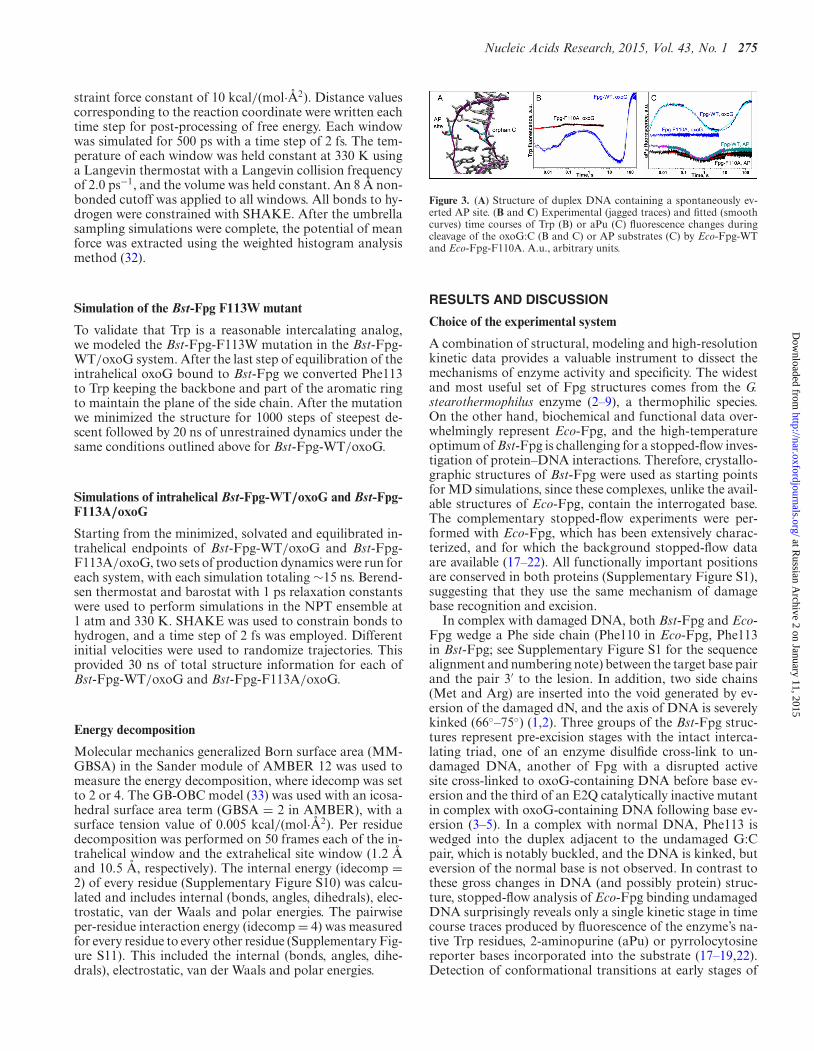
Figure 3. (A) Structure of duplex DNA containing a spontaneously ev-erted AP site. (B and C) Experimental (jagged traces) and fitted (smoothcurves) time courses of Trp (B) or aPu (C) fluorescence changes duringcleavage of the oxoG:C (B and C) or AP substrates (C) by Eco-Fpg-WTand Eco-Fpg-F110A. A.u., arbitrary units.
RESULTS AND DISCUSSION
Choice of the experimental system
A combination of structural, modeling and high-resolutionkinetic data provides a valuable instrument to dissect themechanisms of enzyme activity and specificity. The widestand most useful set of Fpg structures comes from the G.stearothermophilus enzyme (2–9), a thermophilic species.On the other hand, biochemical and functional data over-whelmingly represent Eco-Fpg, and the high-temperatureoptimum of Bst-Fpg is challenging for a stopped-flow inves-tigation of protein–DNA interactions. Therefore, crystallo-graphic structures of Bst-Fpg were used as starting pointsfor MD simulations, since these complexes, unlike the avail-able structures of Eco-Fpg, contain the interrogated base.The complementary stopped-flow experiments were per-formed with Eco-Fpg, which has been extensively charac-terized, and for which the background stopped-flow dataare available (17–22). All functionally important positionsare conserved in both proteins (Supplementary Figure S1),suggesting that they use the same mechanism of damagebase recognition and excision.
In complex with damaged DNA, both Bst-Fpg and Eco-Fpg wedge a Phe side chain (Phe110 in Eco-Fpg, Phe113in Bst-Fpg; see Supplementary Figure S1 for the sequencealignment and numbering note) between the target base pairand the pair 3′ to the lesion. In addition, two side chains(Met and Arg) are inserted into the void generated by ev-ersion of the damaged dN, and the axis of DNA is severelykinked (66◦–75◦) (1,2). Three groups of the Bst-Fpg struc-tures represent pre-excision stages with the intact interca-lating triad, one of an enzyme disulfide cross-link to un-damaged DNA, another of Fpg with a disrupted activesite cross-linked to oxoG-containing DNA before base ev-ersion and the third of an E2Q catalytically inactive mutantin complex with oxoG-containing DNA following base ev-ersion (3–5). In a complex with normal DNA, Phe113 iswedged into the duplex adjacent to the undamaged G:Cpair, which is notably buckled, and the DNA is kinked, buteversion of the normal base is not observed. In contrast tothese gross changes in DNA (and possibly protein) struc-ture, stopped-flow analysis of Eco-Fpg binding undamagedDNA surprisingly reveals only a single kinetic stage in timecourse traces produced by fluorescence of the enzyme’s na-tive Trp residues, 2-aminopurine (aPu) or pyrrolocytosinereporter bases incorporated into the substrate (17–19,22).Detection of conformational transitions at early stages of
at Russian A
rchive 2 on January 11, 2015http://nar.oxfordjournals.org/
Dow
nloaded from

276 Nucleic Acids Research, 2015, Vol. 43, No. 1
DNA binding could be hindered by a lack of fluorescentgroups appropriately placed at the enzyme–DNA interface.On the other hand, it is not clear whether the ‘insertion be-fore eversion’ structure (4) represents a true kinetic interme-diate or is artificially stabilized by covalent enzyme–DNAcross-linking.
Fluorescence tracking of wedge insertion
To address the sequence of events preceding and followingthe insertion, we decided to construct an F110W mutant ofEco-Fpg, in which the fluorescent Trp residue substitutesfor the Phe wedge, providing a suitable reporter at the Fpg–DNA interface. To analyze possible effects of the mutationon the structure of the Eco-Fpg–DNA recognition complex,we first built a model for intrahelical oxoG using the struc-ture of Bst-Fpg bound to undamaged DNA. We then intro-duced in silico the corresponding F113W wedge mutationin the Bst-Fpg–DNA complex and performed a 20-ns MDsimulation with both types of wedge. As shown in Figure1A, replacement of the WT Phe phenyl wedge with the Trpindole has little effect on the DNA structure at the inter-rogation site, with the Trp wedge inserted slightly deeperthan that of Phe. Relative to the crystal structure of Bst-Fpg cross-linked to undamaged DNA (PDB ID 2F5O), insimulations of both systems the root-mean-square deviation(RMSD) of the DNA fluctuated near 3 A while that of theprotein backbone remained near 1.5 A (Supplementary Fig-ure S2), suggesting that the mutation has little impact on theoverall structure of the complex.
Having determined that replacing Phe with Trp in theFpg–DNA complex was unlikely to affect its structure, weconstructed the Eco-Fpg F110W mutant and investigatedthe steady-state and stopped-flow kinetics of its reactionwith undamaged DNA (G-ligand), DNA containing anuncleavable abasic tetrahydrofuran moiety (THF), naturalabasic site (AP) or oxoG opposite to C (SupplementaryFigure S3). Steady-state KM and kcat of Eco-Fpg-F110Wcleaving the oxoG-substrate were similar to those of Eco-Fpg-WT (Supplementary Table S1), supporting the resultsof the modeling. When the rate constants associated withpre-catalytic conformational adjustment and catalysis weremeasured by stopped-flow kinetics using the THF-ligand,AP- or oxoG-substrate (Supplementary Figure S4), the ab-solute values for different steps varied but the minimal re-action schemes remained the same as for Eco-Fpg-WT (Ta-ble 1, Schemes I–III in the Supplementary Material). Noadditional stages in the reaction scheme were revealed byintroducing the Trp fluorophore, suggesting that, if a cog-nate lesion is present, conformational changes around thewedge coincide in time with those occurring in other Trp-containing parts of the Fpg molecule. Notably, the mostpronounced changes in rate constants occurred at the stagespresumed to involve substrate destabilization, wedge inser-tion and base eversion, processes most likely to be affectedkinetically by the wedge mutation; the steps involved in pri-mary binding (rate constants k1 and k–1) and late events thatmay correspond to product release and enzyme regenera-tion (k5, k–5, k6) (20) were influenced to a much lesser ex-tent.
The most drastic change was observed with the G-ligand,binding of which to Eco-Fpg-WT is described by a fastsingle-step equilibrium (characteristic time τ 1 ∼5 ms) (17–19,22). The association of Eco-Fpg-F110W with this lig-and could be separated into three fluorescently discerniblestages, two of which clearly followed the fast binding (τ 2∼50 ms, τ 3 ∼10 s) (Figure 1B and C, Table 1, Scheme IVin the Supplementary Material). These changes in fluores-cence most likely reflect conformational changes not ob-served in Eco-Fpg-WT due to lack of an appropriate flu-orescent reporter at the DNA-binding interface. A com-parison of the appearance and disappearance of differentspecies during binding of the G-ligand and oxoG-substrateby Eco-Fpg-F110W (Figure 1C) suggests that the ES1species in both Schemes III and IV are the same and char-acterize the primary encounter; Phe intercalation and basepair buckling likely occur at this stage as indicated by aPufluorescence. ES2 in Scheme IV apparently corresponds toES4 in Scheme III (22), the steps that reflect oxoG ever-sion and associated motions in the enzyme culminating inthe N-glycosidic bond breakage when the base is damaged.Obviously, even if G can be everted from the DNA duplex,further excision does not occur, thus, at this stage, the pro-cesses of sampling of the normal and the damaged basemust diverge. Finally, the ES3 species in Scheme IV mayreflect a non-productive attempt of formation of the cat-alytically competent complex, since species ES5 in the pro-ductive scheme likely corresponds to post-catalytic events(20–22).
In the structures of undamaged DNA bound to Bst-Fpg,the Phe wedge causes significant buckling at the interca-lation site, potentially probing the stability of the interro-gated base pair (4); this buckling may correspond to one ofthe early fluorescence changes detected in our experiments.In any case, sampling of the normal G base by Eco-Fpg-F110W seems to be extensive and involve several conforma-tional changes following insertion of the wedge. As similarreaction schemes are observed for other substrates and lig-ands processed by Eco-Fpg-WT and Eco-Fpg-F110W, onemay expect that Eco-Fpg-WT also samples deeply the Gbase. In the structure of a Bst-Fpg-N173C disulfide cross-link to undamaged DNA, in which the DNA backbone isdistorted artificially to stabilize the extrahelical G, the sam-pled base cannot enter the active site but, instead, is packedinto the DNA minor groove (6). Structural studies withthree other DNA glycosylases, human 8-oxoguanine-DNAglycosylase OGG1 (34,35), human uracil-DNA glycosylase(36) and E. coli alkylpurine-DNA glycosylase AlkA (37)suggest that extensive sampling of normal bases endingin a catalytically improper conformation of the enzyme–substrate complex may be a common theme in discrimina-tion of normal bases from damaged ones.
Computational analysis of the role of the wedge in lesion pro-cessing
As the wedge insertion by Fpg could represent a critical stepin initiating eversion from the helix of both undamaged anddamaged dN, abolishment of this residue should make theeversion unfavorable. The F113A mutation in Bst-Fpg de-creases the observed single-turnover kcat by ∼2 orders of
at Russian A
rchive 2 on January 11, 2015http://nar.oxfordjournals.org/
Dow
nloaded from

Nucleic Acids Research, 2015, Vol. 43, No. 1 277
Table 1. Pre-steady-state parameters of Eco-Fpg WT and mutants; Trp fluorescence
Eco-Fpg-WTa Eco-Fpg-F110WbEco-Fpg-F110Ab
G THFc APd oxoGe Gf THFc APd oxoGe oxoGg
k1, M−1s−1 (230 ± 40) ×106
(150 ± 20) ×106
(800 ± 30) ×106
(320 ± 15) ×106
(260 ± 10) ×106
(140 ± 6) × 106 (230 ± 8) × 106 (340 ± 10) ×106
(100 ± 14) ×106
k–1, s−1 2700 ± 200 270 ± 15 250 ± 20 890 ± 25 1100 ± 90 920 ± 30 300 ± 40 700 ± 25 990 ± 250k2, s−1 6.0 ± 0.2 36 ± 3 250 ± 18 50.6 ± 4.8 11.9 ± 1.3 13.1 ± 1.1 27.7 ± 1.4 54 ± 13k–2, s−1 0.02 ± 0.005 65 ± 8 2.4 ± 0.4 1.8 ± 0.4 31.6 ± 1.8 39.4 ± 5.2 63 ± 2 40 ± 3k3, s−1 10.0 ± 0.6 10.0 ± 0.5 6.7 ± 0.8 0.24 ± 0.05 7.1 ± 0.5 3.4 ± 0.1 22 ± 1k–3, s−1 0.6 ± 0.1 40 ± 4 46 ± 0.6 0.09 ± 0.01 1.71 ± 0.16 1.74 ± 0.08 28 ± 1k4, s−1 0.04 ± 0.007 11.0 ± 0.8 9.1 ± 0.6 0.26 ± 0.08 0.52 ± 0.01 4.5 ± 0.1k–4, s−1 0.01 ± 0.001 1.0 ± 0.1 2.4 ± 0.2 0.22 ± 0.04 0.03 ± 0.01 1.9 ± 0.1k5, s−1 0.20 ± 0.01 0.20 ± 0.01 0.30 ± 0.04 0.20 ± 0.01k–5, s−1 0.03 ± 0.007 0.02 ± 0.002k6, s−1 0.04 ± 0.006 0.021 ± 0.004KP, M (1.8±0.3)×10−6 (2.0±0.3)×10−6 (0.5±0.1)×10−6 (3.6±0.6)×10−6
Estimate ± standard error of the estimate.aFrom (18).bThis work.cScheme I (Supplementary Material).dScheme II (Supplementary Material).eScheme III (Supplementary Material).fScheme IV (Supplementary Material).gScheme V (Supplementary Material).
magnitude (9), and the same trend is observed for the E. colienzyme (38). To understand the effect of the wedge on lesionsampling, we performed free energy calculations for oxoGeversion for Bst-Fpg-WT and for its F113A mutant with thewedge absent. We utilized the eversion pathway map that wepreviously published for Fpg (see (16) for an in-depth dis-cussion of the major versus minor groove eversion pathway)to examine the impact of the wedge on the breaking of theinterrogated base pair. Unlike in our previous work com-paring major and minor groove eversion pathways, openingthrough the major groove could be well described by a sin-gle reaction coordinate, the distance between the center ofmass of the Watson Crick face of the everting base and thecenter of mass of the two flanking base pairs (Supplemen-tary Figures S5 and S6). The resulting free energy profilesfor base pair opening by Bst-Fpg-WT and Bst-Fpg-F113Aare shown in Figure 2. Essentially, the presence of Phe asthe intercalating residue destabilized the intrahelical con-formation but progressively less affected the further confor-mational intermediates. Intrahelical destabilization as thereason for the wedge effect was recently proposed based onthe crystal structure of Bst-Fpg-F113A (9).
Eversion of oxoG in the WT enzyme was a predomi-nantly low-barrier process in the simulations, encounter-ing barriers of 5–7 kcal/mol. An experimental analysis ofstepwise thermodynamics of oxoG recognition, binding andprocessing by Eco-Fpg-WT (22) also supports the low en-ergy barrier of oxoG eversion. However, the final adjust-ment of Fpg–DNA complex into the catalytically compe-tent conformation is highly unfavorable in enthalpy and isdriven by favorable entropy increase (22), possibly due toa release of water molecules from the protein–DNA inter-face (4). In contrast, the barrier to breaking the oxoG:Cbase pair by Bst-Fpg-F113A during initial eversion (ap-proaching 6 A) was nearly 15 kcal/mol, much higher thanfor Bst-Fpg-WT. Since single molecule fluorescence experi-ments estimate the energetic barrier to 1D diffusion of Bst-Fpg along DNA at only ∼2 kcal/mol (39), it seems unlikelythat the residence time of the enzyme at a particular inter-
rogation site would be sufficient to overcome an eversionbarrier that is more than 10 kcal/mol higher than that oftranslocation. Further supporting this conjecture, quantumdot-labeled single molecule fluorescence experiments showthat Eco-Fpg-F110A shifts to faster 1D diffusion rates onDNA, indicating the wedge plays a direct role in the intra-helical base interrogation attributed to slower diffusion ratecomponents (38).
Wedge deletion mutant lacks glycosylase function but retainsAP lyase activity
Based on results of our computational modeling, an Fpgwedge mutant should be less competent at the stage of le-sion eversion. We constructed the Eco-Fpg-F110A mutantand analyzed its activity on oxoG- and AP-substrates us-ing steady-state and stopped-flow kinetics. Under steady-state conditions, the mutant enzyme was unable to processoxoG-substrates but maintained residual activity on AP-substrates (Supplementary Table S1), in line with observa-tions that mutations of critical residues in Fpg and its ho-molog endonuclease VIII frequently abolish DNA glycosy-lase but not AP lyase activity (40–43).
When analyzed by the stopped-flow technique, interac-tion of Eco-Fpg-F110A with the oxoG-substrate could bedescribed by two stages with kinetic parameters similar tothe two initial steps observed for Eco-Fpg-WT (Table 1,Scheme V in the Supplementary Material); the reaction didnot proceed further (Figure 3B). When aPu was incorpo-rated 5′ or 3′ to the oxoG residue, Eco-Fpg-WT showedat least two conformational transitions preceding the irre-versible step, while Eco-Fpg-F110A produced no changes(Figure 3C). As the fluorescence of aPu reflects the hy-drophobicity of its environment (44), it may be inferred thatno appreciable conformational transitions occur in DNAin the vicinity of the damaged base when Eco-Fpg-F110Abinds its substrate. Consistent with the computational mod-eling, we conclude that Eco-Fpg-F110A forms a normal pri-mary encounter complex with the oxoG-substrate, but fail-
at Russian A
rchive 2 on January 11, 2015http://nar.oxfordjournals.org/
Dow
nloaded from

278 Nucleic Acids Research, 2015, Vol. 43, No. 1
Table 2. Pre-steady-state parameters of Eco-Fpg-WT and Eco-Fpg-F110A in binding and cleavage of the AP-substrate; aPu fluorescence
Eco-Fpg-WTa Eco-Fpg-F110Aa
k1, M−1×s−1 (9.7 ± 0.4) × 105 (3.6 ± 0.5) × 105
k–1, s−1 1.7 ± 0.1 2.6 ± 0.6k2, s−1 0.87 ± 0.07 0.50 ± 0.06KP, M (4.4 ± 0.6) × 10−6 (3.3 ± 0.6) × 10−6
Estimate ± standard error of the estimate is shown.aScheme VI (Supplementary Material).
ure to insert a bulky wedge next to the damaged base pre-cludes further reaction steps.
Reaction of Eco-Fpg-F110A with the AP-substrate alsowas visible by Trp fluorescence; however, rather small in-tensity changes rendered the signal-to-noise ratio low andquantification unreliable. Hence, we analyzed the AP sub-strate using aPu fluorescence, obtaining values for rate con-stants of individual conformational transitions that weresimilar for Eco-Fpg-WT and Eco-Fpg-F110A (Figure 3C,Table 2, Scheme VI in the Supplementary Material). In thiscase, changes in aPu fluorescence are likely due to interca-lation of other plugging residues (Met73 and Arg108). Thestopped-flow results agree with the steady-state activity ofEco-Fpg-F110A and with modeling that indicates relaxedrequirements for wedge insertion in AP site cleavage.
To understand the residual activity for the AP-substrate,we performed a simulation for duplex DNA with an AP sitereplacing the oxoG. We observed that the AP substrate pre-ferred the extrahelical position even in the absence of Fpg,due to rearrangement of the backbone that minimizes thegap resulting from lack of a base (Figure 3A). The ever-sion distance for the AP-substrate was stable near 12–15 A(Supplementary Figure S7); thus, the model indicates thatno wedge is required to facilitate eversion in this case. Com-bined, these evidence suggest that sampling barrier and Fpgresidence time at the AP site would be less affected by anPhe-to-Ala mutation compared with the oxoG lesion; in-deed, Eco-Fpg-F110A shows only a moderate change in theAP site cleavage processivity, a property that reflects boththe 1D diffusion rate and the catalytic efficiency of the en-zyme (45).
Finally, we followed the efficiency of Forster resonanceenergy transfer (FRET) in the stopped-flow mode to ex-plore effects of the wedge mutation on the ability of Fpgto bend substrate DNA. The oxoG-substrate was modi-fied at the opposite 5′-termini either with a pair of fluo-rescent dyes (Cy3 and Cy5), in which case the emissionfrom Cy5 after photon transfer from Cy3 was monitored,or with a dye–quencher pair (fluorescein and DABCYL),so as to observe quenching of fluorescein emission. Whenthe Cy3/Cy5 oxoG-substrate was cleaved by Eco-Fpg-WT,the Cy5 fluorescence initially increased and then startedto decay (Figure 4A and B); the fluorescein emission fromthe fluorescein/DABCYL pair almost exactly mirrored in-versely the Cy5 emission (Figure 4A). The data were op-timally fit by Scheme VII (Supplementary Material), withrate constants presented in Table 3. The enzyme essentiallyoverbends DNA during primary binding and initial confor-mational changes, then relaxes it. However, with the Eco-
Figure 4. FRET time courses during cleavage of oxoG-substrate by Fpg.(A) Comparison of Cy3/Cy5 (red) and fluorescein/DABCYL time coursesfor Eco-Fpg-WT. (B) FRET time courses for Eco-Fpg-WT and Eco-Fpg-F110A. Jagged traces represent experimental data, smooth curves show thefit to Scheme VII (Supplementary Material). A.u., arbitrary units.
Table 3. Pre-steady-state parameters of Eco-Fpg-WT and Eco-Fpg-F110A in binding and cleavage of the oxoG-substrate; Cy3/Cy5 FRET
Eco-Fpg-WTa Eco-Fpg-F110A
k1, M−1×s−1 (2.8 ± 0.3) × 105 (2.1 ± 0.1) × 105
k–1, s−1 0.88 ± 0.04 0.58 ± 0.05k2, s−1 0.35 ± 0.06 0.0023 ± 0.0001k–2, s−1 0.0020 ± 0.0004 0.00035 ± 0.00004k3, s−1 0.050 ± 0.010 n/cb
KP, M (2.1 ± 0.5) × 10−6 n/cb
Estimate ± standard error of the estimate is shown.aScheme VII (Supplementary Material).bn/c, not cleaved.
Fpg-F110A mutant, the Cy5 fluorescence decay was signifi-cantly prolonged (Figure 4B), suggesting that both enzymesquickly bend DNA but Eco-Fpg-F110A is much slower toproceed further, showing no appreciable cleavage of the sub-strate up to 400 s. Lack of appreciable aPu signal (see above)indicates that the mutant is able to bend DNA without sig-nificantly disrupting the stacking around oxoG.
How does the wedge destabilize the interrogated base pair?
The experimental data confirmed the role of the wedge, assuggested by the calculated free energy profiles for lesioneversion. Therefore, we performed further computationalanalyses to gain insight into the mechanism of lesion desta-bilization.
As noted above, the interrogated base pair and its 3′ GCbase pair are significantly buckled in the structure of Bst-Fpg encountering an intrahelical G. Importantly, the twobase pairs buckle in opposite directions, presumably cou-pled to wedge insertion. Our simulations reproduce thisbehavior for Bst-Fpg-WT interrogating oxoG, but in theBst-Fpg-F113A wedge deletion mutant the base pairs re-main buckled, but the values for two base pairs becomenearly equivalent (structure snapshot shown in Figure 5A,with histograms in Supplementary Figure S8) allowing thebases to adopt almost canonical stacking geometries. Thisfinding mirrors the observation in the structure of catalyt-ically inactive Bst-Fpg-E2Q+F113A thiol-crosslinked to aG-containing DNA (9). The buckle induced by the wedge inthe Bst-Fpg-WT structure implies weakening of stacking in-teractions between oxoG and its 3′ G neighbor. A shift in the
at Russian A
rchive 2 on January 11, 2015http://nar.oxfordjournals.org/
Dow
nloaded from

Nucleic Acids Research, 2015, Vol. 43, No. 1 279
Figure 5. (A) Structure overlap at the interrogation site of Bst-Fpg-F113A (colored by atom) and Bst-Fpg-WT (pink). (B and C) Color-coded differencein interaction energies due to the wedge, mapped onto the WT intrahelical (B) and extrahelical average structures (C). The figure shows the difference inper residue energy decomposition between WT and F113A mutant. Positive (red) values reflect more favorable interactions in the mutant.
rise helicoidal parameter from the regular 3.6 A to valuesabove 4.5 A (Supplementary Figure S9) further supportsthis hypothesis. Interestingly, the crystal structure of Bst-Fpg-E2Q+F113A thiol-crosslinked to an oxoG-substrate(9) shows an idosyncratic configuration of the damagedbase, never observed in other Fpg structures: it is intraheli-cal but rotated into the syn orientation and disengaged fromhydrogen bonding to the partner C. However, this struc-ture most likely represents a global energy minimum wherethe complex settles after the long crystallization procedurewith multiple cycles of DNA breathing and dissociation–reassociation events, whereas our F113A model is expectedto reflect the structural and energetic features of the primarylesion encounter complex, and in this sense is more relevantto the lesion recognition process.
Both per residue and pair-wise decomposition of the totalpotential energy for structures sampled in the energy land-scape minima, specifically at the intrahelical and extrahe-lical positions, were performed. We calculated the energycontribution of each residue to the system, mapping the dif-ference in resulting values (WT–F113A) onto the averageintrahelical and extrahelical WT structures (Figure 5B andC). The energy contribution of the G 3′ to the target oxoGbase favors the mutant in the intrahelical location, reflectingthe stability gained by removing the wedge, decreasing thebuckling and improving base pair stacking. Residue 113 fa-vors the mutant as well, illustrating the destabilizing role ofthis residue when it is Phe. Additionally, Met76 is allowed toshift away from the sugar–phosphate backbone in the mu-tant due to the gap created by loss of the wedge, improvingits van der Waals energy and reducing electrostatic clashes.The third member of the gap-filling triad, Arg111, did notshow a large energy effect on the stability of either extrahe-lical or intrahelical state.
We calculated the average interaction energy betweeneach pair of residues for the Bst-Fpg-WT and Bst-Fpg-F113A mutant in the intrahelical and extrahelical locations(Supplementary Figures S10 and S11, respectively). Exam-ination of the resulting data revealed that most interactionsin the intrahelical location differ only slightly between thesystems, and reflect small rearrangements and optimizationof geometries in non-bonded interactions. This aggregationof many small energy improvements contributes to an over-all stabilization of the mutant system intrahelical location.However, significant pairwise interactions which favor the
WT system in the intrahelical and extrahelical position arespecific to the eversion pathway. These pairs include an elec-trostatic interaction between oxoG and Arg263 in the intra-helical location, and stabilization of the 3′ base pair (282,298) and the Met loop and catalytic loop regions (77, 222,223) in the extrahelical position. Increased stabilization ofthe helix–two turn–helix DNA binding domain (156, 277) inthe mutant intrahelical location may contribute to the slowunbending of DNA mentioned above.
The key effects of the wedge arise directly from oxoGbuckling, and force the oxoG into a position in which fur-ther contacts along the base eversion pathway are stabi-lized, as shown in the energy decomposition. Conversely,the wedge mutant stabilization in the intrahelical locationmainly arises from improved stacking, assisted by othernon-specific pairwise interactions whose geometry is opti-mized from reduced spatial constraints. In the intrahelicalstate, Met76 is at the end of a strained loop region that un-dergoes a conformational change upon eversion, resultingin movement of Met76 into the minor groove and fillingof the gap created by eversion of the lesion. Replacementof Met76 with Ala in Bst-Fpg promotes the same struc-ture as do mutations in the loop binding the everted base,thus Met76 likely stabilizes the extrahelical state rather thandestabilizes the intrahelical one (9). In the absence of thebulky wedge in Bst-Fpg-F113A, the intrahelical state un-dergoes steric repacking that allows the loop to relax priorto eversion, forming a favorable interaction with its sur-rounding residues, including the catalytic loop, intercalatingArg111 and the G base 3′ to oxoG (Figure 5B). The addi-tional space also results in less effective gap filling by Met76in the F113A everted state, shown by the shift in pairwiseinteraction energies toward favoring the WT. The overalleffect is a significant stabilization of the intrahelical oxoG,contributing to the inability of the mutant Fpg to evert andprocess the lesion.
In conclusion, we have combined high-quality MD andstopped-flow kinetics with fluorescence detection to analyzea critical conformational change in the complex reactioncatalyzed by formamidopyrimidine-DNA glycosylase. Ourresults suggest that, during lesion search, Fpg likely samplesundamaged bases by wedge insertion and undergoes severalconformational changes in the enzyme–DNA complex. Asimilar scheme was proposed for uracil-DNA glycosylasebased on structural data (9). The strained conformation of
at Russian A
rchive 2 on January 11, 2015http://nar.oxfordjournals.org/
Dow
nloaded from

280 Nucleic Acids Research, 2015, Vol. 43, No. 1
kinked DNA after Fpg binding may provide a source of en-ergy for catalysis; however, the productive reaction must betriggered by wedge insertion, which therefore represents acritical step in the lesion recognition process. The wedge ac-tively destabilizes the lesion through a buckling process thatweakens the Watson–Crick hydrogen bonds and reduces 3′stacking. This mechanism may be common for DNA glyco-sylases that possess an aromatic intercalating wedge (41,46–48).
SUPPLEMENTARY DATA
Supplementary Data are available at NAR Online.
ACKNOWLEDGEMENT
We acknowledge assistance with data analysis from KunSong and Lin Fu (Stony Brook University).
FUNDING
National Cancer Institute [CA017395–33 to A.P.G.]; Na-tional Science Foundation [0549370 to C.B.]; Russian Foun-dation for Basic Research [13–04–00013 to O.S.F., 14–04–01879 to D.O.Z.]; Presidium of the Russian Academy ofSciences [MKB 6.11 to O.S.F., MKB 6.12 to D.O.Z.]. Partof the work with aPu and FRET experiments was specifi-cally funded by Russian Science Foundation [14–24–00093to D.O.Z.]. Part of the work with Trp stopped-flow experi-ments was specifically funded by Russian Science Founda-tion [14–14–00063 to N.A.K.]. NSF Teragrid [MCA02N028to C.S.]. Funding for open access charge: Novosibirsk StateUniversity.Conflict of interest statement. None declared.
REFERENCES1. Gilboa,R., Zharkov,D.O., Golan,G., Fernandes,A.S.,
Gerchman,S.E., Matz,E., Kycia,J.H., Grollman,A.P. and Shoham,G.(2002) Structure of formamidopyrimidine-DNA glycosylasecovalently complexed to DNA. J. Biol. Chem., 277, 19811–19816.
2. Fromme,J.C. and Verdine,G.L. (2002) Structural insights into lesionrecognition and repair by the bacterial 8-oxoguanine DNAglycosylase MutM. Nat. Struct. Biol., 9, 544–552.
3. Fromme,J.C. and Verdine,G.L. (2003) DNA lesion recognition by thebacterial repair enzyme MutM. J. Biol. Chem., 278, 51543–51548.
4. Banerjee,A., Santos,W.L. and Verdine,G.L. (2006) Structure of aDNA glycosylase searching for lesions. Science, 311, 1153–1157.
5. Qi,Y., Spong,M.C., Nam,K., Banerjee,A., Jiralerspong,S.,Karplus,M. and Verdine,G.L. (2009) Encounter and extrusion of anintrahelical lesion by a DNA repair enzyme. Nature, 462, 762–766.
6. Qi,Y., Spong,M.C., Nam,K., Karplus,M. and Verdine,G.L. (2010)Entrapment and structure of an extrahelical guanine attempting toenter the active site of a bacterial DNA glycosylase, MutM. J. Biol.Chem., 285, 1468–1478.
7. Qi,Y., Nam,K., Spong,M.C., Banerjee,A., Sung,R.-J., Zhang,M.,Karplus,M. and Verdine,G.L. (2012) Strandwise translocation of aDNA glycosylase on undamaged DNA. Proc. Natl Acad. Sci. U.S.A.,109, 1086–1091.
8. Sung,R.-J., Zhang,M., Qi,Y. and Verdine,G.L. (2012)Sequence-dependent structural variation in DNA undergoingintrahelical inspection by the DNA glycosylase MutM. J. Biol.Chem., 287, 18044–18054.
9. Sung,R.-J., Zhang,M., Qi,Y. and Verdine,G.L. (2013) Structural andbiochemical analysis of DNA helix invasion by the bacterial8-oxoguanine DNA glycosylase MutM. J. Biol. Chem., 288,10012–10023.
10. Amara,P., Serre,L., Castaing,B. and Thomas,A. (2004) Insights intothe DNA repair process by the formamidopyrimidine-DNAglycosylase investigated by molecular dynamics. Protein Sci., 13,2009–2021.
11. Perlow-Poehnelt,R.A., Zharkov,D.O., Grollman,A.P. and Broyde,S.(2004) Substrate discrimination by formamidopyrimidine-DNAglycosylase: distinguishing interactions within the active site.Biochemistry, 43, 16092–16105.
12. Amara,P. and Serre,L. (2006) Functional flexibility of Bacillusstearothermophilus formamidopyrimidine DNA-glycosylase. DNARepair, 5, 947–958.
13. Song,K., Hornak,V., de los Santos,C., Grollman,A.P. andSimmerling,C. (2006) Computational analysis of the mode of bindingof 8-oxoguanine to formamidopyrimidine-DNA glycosylase.Biochemistry, 45, 10886–10894.
14. Song,K., Kelso,C., de los Santos,C., Grollman,A.P. andSimmerling,C. (2007) Molecular simulations reveal a commonbinding mode for glycosylase binding of oxidatively damaged DNAlesions. J. Am. Chem. Soc., 129, 14536–14537.
15. Nam,K., Verdine,G.L. and Karplus,M. (2009) Analysis of ananomalous mutant of MutM DNA glycosylase leads to new insightsinto the catalytic mechanism. J. Am. Chem. Soc., 131, 18208–18209.
16. Bergonzo,C., Campbell,A.J., de los Santos,C., Grollman,A.P. andSimmerling,C. (2011) Energetic preference of 8-oxoG eversionpathways in a DNA glycosylase. J. Am. Chem. Soc., 133,14504–14506.
17. Fedorova,O.S., Nevinsky,G.A., Koval,V.V., Ishchenko,A.A.,Vasilenko,N.L. and Douglas,K.T. (2002) Stopped-flow kinetic studiesof the interaction between Escherichia coli Fpg protein and DNAsubstrates. Biochemistry, 41, 1520–1528.
18. Koval,V.V., Kuznetsov,N.A., Zharkov,D.O., Ishchenko,A.A.,Douglas,K.T., Nevinsky,G.A. and Fedorova,O.S. (2004)Pre-steady-state kinetics shows differences in processing of variousDNA lesions by Escherichia coli formamidopyrimidine-DNAglycosylase. Nucleic Acids Res., 32, 926–935.
19. Kuznetsov,N.A., Koval,V.V., Zharkov,D.O., Vorobjev,Y.N.,Nevinsky,G.A., Douglas,K.T. and Fedorova,O.S. (2007)Pre-steady-state kinetic study of substrate specificity of Escherichiacoli formamidopyrimidine-DNA glycosylase. Biochemistry, 46,424–435.
20. Kuznetsov,N.A., Zharkov,D.O., Koval,V.V., Buckle,M. andFedorova,O.S. (2009) Reversible chemical step and rate-limitingenzyme regeneration in the reaction catalyzed byformamidopyrimidine-DNA-glycosylase. Biochemistry, 48,11335–11343.
21. Koval,V.V., Kuznetsov,N.A., Ishchenko,A.A., Saparbaev,M.K. andFedorova,O.S. (2010) Real-time studies of conformational dynamicsof the repair enzyme E. coli formamidopyrimidine-DNA glycosylaseand its DNA complexes during catalytic cycle. Mutat. Res., 685, 3–10.
22. Kuznetsov,N.A., Vorobjev,Y.N., Krasnoperov,L.N. andFedorova,O.S. (2012) Thermodynamics of the multi-stage DNAlesion recognition and repair by formamidopyrimidine-DNAglycosylase using pyrrolocytosine fluorescence–stopped-flowpre-steady-state kinetics. Nucleic Acids Res., 40, 7384–7392.
23. Adcock,S.A. and McCammon,J.A. (2006) Molecular dynamics:survey of methods for simulating the activity of proteins. Chem. Rev.,106, 1589–1615.
24. Fisher,H.F. (2005) Transient-state kinetic approach to mechanisms ofenzymatic catalysis. Acc. Chem. Res., 38, 157–166.
25. Kuzmic,P. (1996) Program DYNAFIT for the analysis of enzymekinetic data: application to HIV proteinase. Anal. Biochem., 237,260–273.
26. Cattell,R.B. (1966) The scree test for the number of factors.Multivariate Behav. Res., 1, 245–276.
27. Hornak,V., Abel,R., Okur,A., Strockbine,B., Roitberg,A. andSimmerling,C. (2006) Comparison of multiple Amber force fields anddevelopment of improved protein backbone parameters. Proteins, 65,712–725.
28. Perez,A., Marchan,I., Svozil,D., Sponer,J., Cheatham,T.E. III,Laughton,C.A. and Orozco,M. (2007) Refinement of the AMBERforce field for nucleic acids: improving the description of �/�conformers. Biophys. J., 92, 3817–3829.
at Russian A
rchive 2 on January 11, 2015http://nar.oxfordjournals.org/
Dow
nloaded from

Nucleic Acids Research, 2015, Vol. 43, No. 1 281
29. Jorgensen,W.L., Chandrasekhar,J., Madura,J.D., Impey,R.W. andKlein,M.L. (1983) Comparison of simple potential functions forsimulating liquid water. J. Chem. Phys., 79, 926–935.
30. Cieplak,P., Cornell,W.D., Bayly,C. and Kollman,P.A. (1995)Application of the multimolecule and multiconformational RESPmethodology to biopolymers: charge derivation for DNA, RNA, andproteins. J. Comput. Chem., 16, 1357–1377.
31. Ryckaert,J.-P., Ciccotti,G. and Berendsen,H.J.C. (1977) Numericalintegration of the Cartesian equations of motion of a system withconstraints: molecular dynamics of n-alkanes. J. Comput. Phys., 23,327–341.
32. Kumar,S., Rosenberg,J.M., Bouzida,D., Swendsen,R.H. andKollman,P.A. (1995) Multidimensional free-energy calculations usingthe weighted histogram analysis method. J. Comput. Chem., 16,1339–1350.
33. Onufriev,A., Bashford,D. and Case,D.A. (2004) Exploring proteinnative states and large-scale conformational changes with a modifiedGeneralized Born model. Proteins, 55, 383–394.
34. Banerjee,A., Yang,W., Karplus,M. and Verdine,G.L. (2005) Structureof a repair enzyme interrogating undamaged DNA elucidatesrecognition of damaged DNA. Nature, 434, 612–618.
35. Radom,C.T., Banerjee,A. and Verdine,G.L. (2007) Structuralcharacterization of human 8-oxoguanine DNA glycosylase variantsbearing active site mutations. J. Biol. Chem., 282, 9182–9194.
36. Parker,J.B., Bianchet,M.A., Krosky,D.J., Friedman,J.I., Amzel,L.M.and Stivers,J.T. (2007) Enzymatic capture of an extrahelical thyminein the search for uracil in DNA. Nature, 449, 433–437.
37. Bowman,B.R., Lee,S., Wang,S. and Verdine,G.L. (2010) Structure ofEscherichia coli AlkA in complex with undamaged DNA. J. Biol.Chem., 285, 35783–35791.
38. Dunn,A.R., Kad,N.M., Nelson,S.R., Warshaw,D.M. andWallace,S.S. (2011) Single Qdot-labeled glycosylase molecules use awedge amino acid to probe for lesions while scanning along DNA.Nucleic Acids Res., 39, 7487–7498.
39. Blainey,P.C., van Oijen,A.M., Banerjee,A., Verdine,G.L. andXie,X.S. (2006) A base-excision DNA-repair protein finds intrahelical
lesion bases by fast sliding in contact with DNA. Proc. Natl Acad.Sci. U.S.A., 103, 5752–5757.
40. Lavrukhin,O.V. and Lloyd,R.S. (2000) Involvement ofphylogenetically conserved acidic amino acid residues in catalysis byan oxidative DNA damage enzyme formamidopyrimidineglycosylase. Biochemistry, 39, 15266–15271.
41. Zharkov,D.O., Golan,G., Gilboa,R., Fernandes,A.S.,Gerchman,S.E., Kycia,J.H., Rieger,R.A., Grollman,A.P. andShoham,G. (2002) Structural analysis of an Escherichia coliendonuclease VIII covalent reaction intermediate. EMBO J., 21,789–800.
42. Burgess,S., Jaruga,P., Dodson,M.L., Dizdaroglu,M. and Lloyd,R.S.(2002) Determination of active site residues in Escherichia coliendonuclease VIII. J. Biol. Chem., 277, 2938–2944.
43. Kropachev,K.Y., Zharkov,D.O. and Grollman,A.P. (2006) Catalyticmechanism of Escherichia coli endonuclease VIII: roles of theintercalation loop and the zinc finger. Biochemistry, 45, 12039–12049.
44. Rachofsky,E.L., Osman,R. and Ross,J.B.A. (2001) Probing structureand dynamics of DNA with 2-aminopurine: effects of localenvironment on fluorescence. Biochemistry, 40, 946–956.
45. Sidorenko,V.S. and Zharkov,D.O. (2008) Correlated cleavage ofdamaged DNA by bacterial and human 8-oxoguanine-DNAglycosylases. Biochemistry, 47, 8970–8976.
46. Lau,A.Y., Wyatt,M.D., Glassner,B.J., Samson,L.D. andEllenberger,T. (2000) Molecular basis for discriminating betweennormal and damaged bases by the human alkyladenine glycosylase,AAG. Proc. Natl Acad. Sci. U.S.A., 97, 13573–13578.
47. Bruner,S.D., Norman,D.P.G. and Verdine,G.L. (2000) Structuralbasis for recognition and repair of the endogenous mutagen8-oxoguanine in DNA. Nature, 403, 859–866.
48. Fromme,J.C., Banerjee,A., Huang,S.J. and Verdine,G.L. (2004)Structural basis for removal of adenine mispaired with 8-oxoguanineby MutY adenine DNA glycosylase. Nature, 427, 652–656.
at Russian A
rchive 2 on January 11, 2015http://nar.oxfordjournals.org/
Dow
nloaded from