Chronic valproate treatment blocks D2-like receptor-mediated brain signaling via arachidonic acid in...
9
Chronic valproate treatment blocks D 2 -like receptor-mediated brain signaling via arachidonic acid in rats Epolia Ramadan * , Mireille Basselin, Ameer Y. Taha, Yewon Cheon, Lisa Chang, Mei Chen, Stanley I. Rapoport Brain Physiology and Metabolism Section, National Institute on Aging, National Institutes of Health, Bethesda, MD 20892, USA article info Article history: Received 2 June 2011 Received in revised form 12 July 2011 Accepted 17 July 2011 Keywords: Arachidonic acid Phospholipase A 2 Valproate D 2 -like receptor Quinpirole Bipolar disorder abstract Background and objective: Hyperdopaminergic signaling and an upregulated brain arachidonic acid (AA) cascade may contribute to bipolar disorder (BD). Lithium and carbamazepine, FDA-approved for the treatment of BD, attenuate brain dopaminergic D 2 -like (D 2 ,D 3 , and D 4 ) receptor signaling involving AA when given chronically to awake rats. We hypothesized that valproate (VPA), with mood-stabilizing properties, would also reduce D 2 -like-mediated signaling via AA. Methods: An acute dose of quinpirole (1 mg/kg) or saline was administered to unanesthetized rats that had been treated for 30 days with a therapeutically relevant dose of VPA (200 mg/kg/day) or vehicle. Regional brain AA incorporation coefficients, k*, and incorporation rates, J in , markers of AA signaling and metabolism, were measured by quantitative autoradiography after intravenous [1- 14 C]AA infusion. Whole brain concentrations of prostaglandin (PG)E 2 and thromboxane (TX)B 2 also were measured. Results: Quinpirole compared to saline significantly increased k* in 40 of 83 brain regions, and increased brain concentrations of PGE 2 in chronic vehicle-treated rats. VPA treatment by itself reduced concen- trations of plasma unesterified AA and whole brain PGE 2 and TXB 2 , and blocked the quinpirole-induced increments in k* and PGE 2 . Conclusion: These results further provide evidence that mood stabilizers downregulate brain dopami- nergic D 2 -like receptor signaling involving AA. Published by Elsevier Ltd. 1. Introduction Valproate (2-propylpentanoate, VPA) has a wide clinical spectrum of use in both psychiatric and neurological disorders. It is one of the most frequently used antiepileptic drugs, has mood- stabilizing properties in the treatment of acute mania (Bowden, 2009), and might be effective for the reduction of depressive symptoms of acute bipolar depression (Smith et al., 2010; Wang et al., 2010). Despite more than 40 years of clinical use, the mechanism of action of VPA in bipolar disorder (BD) is still not fully understood. It is well known that VPA exerts multiple pharmacological effects, and has been found to affect glycogen synthase kinase-3 (GSK-3), the Wnt/b-catenin pathway, the extra- cellular signal-regulated kinase (ERK) pathway, g-aminobutyric acid (GABA)ergic neurotransmission, N-methyl-D-aspartate (NMDA) glutamatergic signaling, pre- and post-synaptic dopamine (DA) neurotransmission, voltage-gated sodium and T-type calcium channels, histone acetylation, and brain lipids and their metabo- lism (Basselin et al., 2008a; Montezinho et al., 2006; Phiel et al., 2001; Rapoport et al., 2009; Yatham et al., 2002). In addition, VPA is neuroprotective in several models of neurodegenerative disease (Monti et al., 2010, 2009). Hyperdopaminergic neurotransmission is suggested to be involved in the pathophysiology of mania in BD (Berk et al., 2007; Cousins et al., 2009; Diehl and Gershon, 1992; Goetz, 1997). Reports show that drugs that inhibit DAergic transmission (haloperidol, chlorpromazine) have antimanic actions whereas drugs that stimulate DA synthesis (levodopa), bind to D 2 receptors (bromocriptine) or reduce DA reuptake (amphetamine) often precipitate mania (Anand et al., 2000; Cipriani et al., 2006; Peet and Peters, 1995). In this context, postmortem BD brain shows higher D 2 receptor expression in the caudate and prefrontal cortex Abbreviations: AA, arachidonic acid; BD, bipolar disorder; BDNF, brain-derived neurotrophic factor; cPLA 2 , Ca 2þ -dependent cytosolic phospholipase A 2 ; COX, cyclooxygenase; DA, dopamine; DAT, dopamine reuptake transporter; GSK-3, glycogen synthase kinase-3; VPA, valproate; PGE 2 , prostaglandin E 2 ; TXB 2 , throm- boxane B 2 . * Corresponding author. Brain Physiology and Metabolism Section, National Institute on Aging, National Institutes of Health, Bldg. 9, Room 1S126, Bethesda, MD 20892, USA. Tel.: þ1 301 496 8994; fax: þ1 301 402 0074. E-mail address: [email protected] (E. Ramadan). Contents lists available at SciVerse ScienceDirect Neuropharmacology journal homepage: www.elsevier.com/locate/neuropharm 0028-3908/$ e see front matter Published by Elsevier Ltd. doi:10.1016/j.neuropharm.2011.07.025 Neuropharmacology 61 (2011) 1256e1264
-
Upload
independent -
Category
Documents
-
view
0 -
download
0
Transcript of Chronic valproate treatment blocks D2-like receptor-mediated brain signaling via arachidonic acid in...

at SciVerse ScienceDirect
Neuropharmacology 61 (2011) 1256e1264
Contents lists available
Neuropharmacology
journal homepage: www.elsevier .com/locate/neuropharm
Chronic valproate treatment blocks D2-like receptor-mediated brainsignaling via arachidonic acid in rats
Epolia Ramadan*, Mireille Basselin, Ameer Y. Taha, Yewon Cheon, Lisa Chang,Mei Chen, Stanley I. RapoportBrain Physiology and Metabolism Section, National Institute on Aging, National Institutes of Health, Bethesda, MD 20892, USA
a r t i c l e i n f o
Article history:Received 2 June 2011Received in revised form12 July 2011Accepted 17 July 2011
Keywords:Arachidonic acidPhospholipase A2
ValproateD2-like receptorQuinpiroleBipolar disorder
Abbreviations: AA, arachidonic acid; BD, bipolar dneurotrophic factor; cPLA2, Ca2þ-dependent cytosocyclooxygenase; DA, dopamine; DAT, dopamine rglycogen synthase kinase-3; VPA, valproate; PGE2, prboxane B2.* Corresponding author. Brain Physiology and M
Institute on Aging, National Institutes of Health, Bldg.20892, USA. Tel.: þ1 301 496 8994; fax: þ1 301 402
E-mail address: [email protected] (E. Rama
0028-3908/$ e see front matter Published by Elseviedoi:10.1016/j.neuropharm.2011.07.025
a b s t r a c t
Background and objective: Hyperdopaminergic signaling and an upregulated brain arachidonic acid (AA)cascade may contribute to bipolar disorder (BD). Lithium and carbamazepine, FDA-approved for thetreatment of BD, attenuate brain dopaminergic D2-like (D2, D3, and D4) receptor signaling involving AAwhen given chronically to awake rats. We hypothesized that valproate (VPA), with mood-stabilizingproperties, would also reduce D2-like-mediated signaling via AA.Methods: An acute dose of quinpirole (1 mg/kg) or saline was administered to unanesthetized rats thathad been treated for 30 days with a therapeutically relevant dose of VPA (200 mg/kg/day) or vehicle.Regional brain AA incorporation coefficients, k*, and incorporation rates, Jin, markers of AA signaling andmetabolism, were measured by quantitative autoradiography after intravenous [1-14C]AA infusion.Whole brain concentrations of prostaglandin (PG)E2 and thromboxane (TX)B2 also were measured.Results: Quinpirole compared to saline significantly increased k* in 40 of 83 brain regions, and increasedbrain concentrations of PGE2 in chronic vehicle-treated rats. VPA treatment by itself reduced concen-trations of plasma unesterified AA and whole brain PGE2 and TXB2, and blocked the quinpirole-inducedincrements in k* and PGE2.Conclusion: These results further provide evidence that mood stabilizers downregulate brain dopami-nergic D2-like receptor signaling involving AA.
Published by Elsevier Ltd.
1. Introduction
Valproate (2-propylpentanoate, VPA) has a wide clinicalspectrum of use in both psychiatric and neurological disorders.It is one of the most frequently used antiepileptic drugs, has mood-stabilizing properties in the treatment of acute mania (Bowden,2009), and might be effective for the reduction of depressivesymptoms of acute bipolar depression (Smith et al., 2010; Wanget al., 2010). Despite more than 40 years of clinical use, themechanism of action of VPA in bipolar disorder (BD) is still not fullyunderstood. It is well known that VPA exerts multiple
isorder; BDNF, brain-derivedlic phospholipase A2; COX,euptake transporter; GSK-3,ostaglandin E2; TXB2, throm-
etabolism Section, National9, Room 1S126, Bethesda, MD0074.dan).
r Ltd.
pharmacological effects, and has been found to affect glycogensynthase kinase-3 (GSK-3), the Wnt/b-catenin pathway, the extra-cellular signal-regulated kinase (ERK) pathway, g-aminobutyricacid (GABA)ergic neurotransmission, N-methyl-D-aspartate(NMDA) glutamatergic signaling, pre- and post-synaptic dopamine(DA) neurotransmission, voltage-gated sodium and T-type calciumchannels, histone acetylation, and brain lipids and their metabo-lism (Basselin et al., 2008a; Montezinho et al., 2006; Phiel et al.,2001; Rapoport et al., 2009; Yatham et al., 2002). In addition, VPAis neuroprotective in several models of neurodegenerative disease(Monti et al., 2010, 2009).
Hyperdopaminergic neurotransmission is suggested to beinvolved in the pathophysiology of mania in BD (Berk et al., 2007;Cousins et al., 2009; Diehl and Gershon, 1992; Goetz, 1997).Reports show that drugs that inhibit DAergic transmission(haloperidol, chlorpromazine) have antimanic actions whereasdrugs that stimulate DA synthesis (levodopa), bind to D2 receptors(bromocriptine) or reduce DA reuptake (amphetamine) oftenprecipitate mania (Anand et al., 2000; Cipriani et al., 2006; Peetand Peters, 1995). In this context, postmortem BD brain showshigher D2 receptor expression in the caudate and prefrontal cortex

E. Ramadan et al. / Neuropharmacology 61 (2011) 1256e1264 1257
(Feng, 2008; Pearlson et al., 1995) and genetic studies havelinked the DA reuptake transporter (DAT) and BD (Greenwoodet al., 2001, 2006), with a DAT mutation causing inhibitionof the transporter cell surface expression (Horschitz et al., 2005).Furthermore, analysis of postmortem cortex from BD patientsshows significantly elevated levels of the neuronal calcium sensor-1 (NCS-1), which inhibits D2 desensitization/internalization(Kabbani et al., 2002; Koh et al., 2003), changes in the levels of DAand cyclic adenosine 30:50-monophosphate-regulated phospho-protein of relative molecular mass 32,000 (DARPP-32) (Ishikawaet al., 2007; Zhan et al., 2011), and decreased protein and mRNAlevels of the DAT (Rao JS and Rapoport SI, unpublished data).
Dopaminergic D2-like (D2, D3, and D4) receptors in brain canbe coupled via a Gai/o-protein to Ca2þ-dependent cytosolicphospholipase (cPLA2, EC 3.1.1.4), which when activated releasesarachidonic acid (AA, 20:4n � 6) from the stereospecificallynumbered (sn)-2 position of synaptic membrane phospholipid(Clark et al., 1995; Nilsson et al., 1998; Ong et al., 1999; Vial andPiomelli, 1995). AA is an important second messenger in brainwith multiple effects, and is a precursor of bioactive eicosanoidssuch as prostaglandin E2 (PGE2) (Rapoport, 2008). Markers ofthe AA cascade have been reported to be abnormal in BD (Kim et al.,2011; Noponen et al., 1993).
Brain AA signaling can be measured in unanesthetized rodentsby infusing radiolabeled AA intravenously, quantifying integratedplasma radioactivity, using quantitative autoradiography todetermine regional brain radioactivity due to tracer AA incorpo-rated into membrane phospholipid, then applying a mathematicalmodel to calculate AA incorporation coefficients and rates, k* andJin, respectively (Rapoport et al., 2001; Robinson et al., 1992). Sincethe AA lost after release and metabolism cannot be synthesizedde novo from 2-carbon fragments, nor elongated significantly(<1%) from its shorter chain polyunsaturated precursor, linoleicacid (18:2n � 6) (Demar et al., 2006; Holman, 1986), k* and Jinfor AA represent net AA consumption following release fromphospholipid.
We showed using the intravenous infusion method that acuteadministration to unanesthetized rats of quinpirole (1 mg/kg,D2-like receptor agonist) (Seeman and Van Tol, 1994), amphet-amine or apomorphine (D1/D2 receptor agonist), but not the D1-like receptor agonist, SKF-38393, increased k* and Jin for AA inmany brain regions rich in D2-like receptors, and that the increasescould be blocked by pre-administration of a D2 receptor antagonist(e.g. butaclamol, raclopride) or of each of two FDA-approvedantimanic mood stabilizers, lithium and carbamazepine whengiven chronically (Basselin et al., 2005, 2008b; Bhattacharjee et al.,2005; Bhattacharjee et al., 2006, 2008; Hayakawa et al., 2001).Each mood stabilizer downregulated brain AA turnover and/orreduced levels and activities of essential enzymes and metabolitesof the brain AA cascade (Bazinet et al., 2006a,b; Bosetti et al.,2002; Chang et al., 1996; Ghelardoni et al., 2004; Rao et al.,2007a,b, 2005).
VPA also is approved as an antimanic mood stabilizer for BD, andwhen given chronically to rats reduces AA turnover within brainphospholipids and decreases activity of cyclooxygenase (COX) andits metabolites’ concentrations (Bosetti et al., 2003; Chang et al.,2001). We hypothesized that chronic administration of VPA toproduce therapeutically relevant plasma levels, also would blockthe quinpirole-initiated AA signal and other AA cascade markers inrat brain. We applied our established in vivo fatty acid method, andmeasured AA incorporation coefficients, k*, and rates, Jin, in each of83 brain regions after acutely giving saline or quinpirole (1 mg/kg)to unanesthetized rats that had received VPA (200 mg/kg/day, i.p.)or vehicle for 30 days. Whole brain concentrations of PGE2 andthromboxane (TX)B2 were also measured.
2. Material and methods
2.1. Animals and diets
Two-month-old male Fischer CDF 344 rats (Charles River Laboratories, Wil-mington, MA) were acclimated for 1 week in an animal facility with regulatedtemperature, humidity and light cycle, and had free access to food and water. Thediet (Rodent NIH-31 auto 18-4 diet, Zeigler Bros, Gardens, PA) contained (as % of totalfatty acid) 20.1% saturated, 22.5% monounsaturated, 47.9% linoleic, 5.1% a-linolenic,0.02% AA, 2.0% eicosapentaenoic, and 2.3% docosahexaenoic acid (Demar et al.,2006). Experiments were conducted following the “Guide for the Care and Use ofLaboratory Animals” (National Institutes of Health Publication No. 86-23) and wereapproved by the Animal Care and Use Committee of Eunice Kennedy ShriverNational Institute of Child Health and Human Development. Effort was made toreduce the number of animals used and to minimize animal suffering.
2.2. Drugs and tracers
Radiolabeled [1-14C]AA in ethanol (53 mCi/mmol, >98% pure, MoravekBiochemicals, Brea, CA) was evaporated and resuspended in HEPES buffer, pH 7.4,containing 50 mg/ml fatty acid-free bovine serum albumin (SigmaeAldrich, StLouis, MO). VPA (sodium salt, SigmaeAldrich)-treated rats received 200 mg/kgintraperitoneally (i.p.) once daily for 30 days. VPA was dissolved in saline (0.9%NaCl, Hospira, Lake Forest, IL) as described (Basselin et al., 2008a; Bazinet et al.,2005; Bosetti et al., 2003; Chang et al., 2001). A control group received the samevolume of saline (vehicle) under parallel conditions. An acute 1 mg/kg i.v. dose of(�)-quinpirole hydrochloride dissolved in saline (Sigma-Aldrich) was chosenbecause it produces widespread significant increments in k* for AA in the brain ofunanesthetized rats that can be blocked by D2-like receptor antagonists, butacla-mol or raclopride, without causing convulsions (Basselin et al., 2005; Bhattacharjeeet al., 2005).
2.3. Surgical procedures and tracer infusion
On the morning of day 30, a rat was injected with the last VPA or vehicle dose,and then anesthetized with halothane (2e3% v/v in O2). Polyethylene (PE 50)catheters were inserted into the right femoral artery and vein as described previ-ously (Basselin et al., 2005). The wound was closed with surgical clips and the ratwas wrapped loosely, with its upper body remaining free, in a fast-setting plastercast taped to a wooden block. Surgery lasted 20e25 min. The rat was allowed torecover from anesthesia for 3e4 h in an environment maintained at 25 �C. Rectaltemperature was maintained at 36.4e37.1 �C using a feedback-heating device andrectal thermometer. Arterial blood pressure and heart rate were measured witha blood pressure recorder (CyQ 103/302; Cybersense, Nicholasville, KY). One minuteafter an i.v. injection of quinpirole or saline, [1-14C]AA (170 mCi/kg, 2 ml) was infusedinto the femoral vein for 5 min at a rate of 400 ml/min, using an infusion pump(Harvard Apparatus Model 22, Natick, MA). Twenty minutes after beginning tracerinfusion, the rat was euthanized with an overdose of Nembutal� (90 mg/kg, i.v.) anddecapitated. The brain was removed (<30 s), frozen in 2-methylbutane maintainedat �40 �C in dry ice, and stored at �80 �C until sectioned.
2.4. Chemical analysis
Blood samples, collected before, during and after [1-14C]AA infusion, werecentrifuged immediately at 18,000 g for 30 s. Total lipids were extracted from plasma(30 ml) using a modified Folch procedure (Folch et al., 1957). One hundred ml of thelower organic phase was used to determine the radiolabeled unesterified plasma AAconcentration by liquid scintillation counting. As reported (DeGeorge et al., 1989),greater than 95e98% of total plasma and brain radioactivity at 5 min following[1-14C]AA infusion is radiolabeled AA. Concentrations of unlabeled, unesterifiedfatty acids were determined from 150 microLiter frozen/thawed arterial plasma.Total lipids were extracted and separated by thin layer chromatography on silica gel60 plates (Whatman, Clifton, NJ) using the solvent system heptane:diethylether:-glacial acetic acid (60:40:3, v/v/v). Unesterified fatty acids (identified under UVlight) were scraped from the plate and methylated with 1% H2SO4 (by vol) inanhydrous methanol (3 h at 70 �C), then separated and quantified by gas-liquidchromatography using heptadecanoic acid (17:0) as an internal standard.
2.5. Quantitative autoradiography
Quantitative autoradiography was performed as described (Basselin et al.,2006a). A total of 83 brain regions from autoradiographs of coronal brainsections were identified from a stereotaxic rat brain atlas (Paxinos and Watson,1987), and were sampled in both hemispheres. The average of bilateralmeasurements for each region from three consecutive brain sections was used tocalculate regional radioactivity (nCi/g wet brain) by digital quantitative densi-tometry, using the public domain 1.62 Analysis NIH Image program. Regionalbrain incorporation coefficients k* (ml plasma/s/g wet brain) of AA were calcu-lated as (Robinson et al., 1992),

E. Ramadan et al. / Neuropharmacology 61 (2011) 1256e12641258
k� ¼ c�brainð20minÞZ20
0
c�plasmadt
(1)
c�brain (nCi/g wet brainwt) is brain radioactivity 20 min after beginning infusion,c�plasma (nCi/ml plasma) is arterial labeled unesterified AA, and t (min) is time afterbeginning [1-14C]AA infusion. Integrated plasma radioactivity (input function) wasdetermined by trapezoidal integration and used to calculate k* for each experiment.
The regional rate of incorporation of unesterified AA from plasma into brainphospholipids, Jin (pmol/s/g), was calculated as follows:
Jin ¼ k�cplasma (2)
where cplasma is the plasma concentration (nmol/ml) of unlabeled unesterified AA.
2.6. Brain PGE2 and TXB2 concentrations
In a separate experiment and after the last of 30 daily administrations of VPA orvehicle, a rat was injected with quinpirole (1 mg/kg, i.p.) or saline. Twenty-oneminutes later (Basselin et al., 2008b), it was anesthetized with Nembutal� (45 mg/kg, i.p.), and immediately subjected to head-focused microwave irradiation (5.5 kW,3.8 s; Cober Electronics, Stamford, CT) to stop postmortem brain metabolism (Fariaset al., 2008; Poddubiuk et al., 1982). A half-brainwas weighed, homogenized with 18volumes of hexane:isopropanol (3:2, by volume) using a glass Tenbroeck homoge-nizer and the homogenates were centrifuged (800 g, 5 min). Tissue residues wererinsed with 3 � 2 volumes of the same solvent. The resultant lipid extracts wereconcentrated to dryness under N2 and resuspended in the enzyme immunoassaybuffer provided by the polyclonal PGE2 and TXB2 immunoassay kits (OxfordBiochemical Research, Oxford, MI).
2.7. Statistical analyses
A paired t test using GraphPad Prism version 4.0b (GraphPad Software, SanDiego, CA) was applied to compare mean physiological parameters in the sameanimal before and after drug injection. A standard two-way analysis of variance(ANOVA) was performed to compare chronic VPA and vehicle treatment with acutequinpirole vs saline administration with regard to: integrated arterial plasmaradioactivity input functions, plasma unesterified fatty acid concentrations, brainPGE2 and TXB2 concentrations, and regional values of k* and Jin for AA. If interac-tions between VPA and quinpirole were statistically insignificant, probabilities ofeffects of VPA and quinpirole were reported. If interactions were statisticallysignificant, probabilities of main effects of VPA and quinpirole were not reported(Tabachnick and Fidell, 2001), and a one-way ANOVA with Bonferroni’s post-testwas used to compare quinpirole and saline responses between chronic VPA- andvehicle-treated rats, as well as saline responses in VPA-treated compared withvehicle-treated rats. Data are reported as the mean � SD, with statistical signifi-cance taken as P � 0.05.
3. Results
3.1. Physiology, behavior and arterial plasma radioactivity
After 30 days of treatment, themean bodyweight of VPA-treatedrats was significantly lower than that of vehicle-treated rats(294.1 þ 25.9 g [n ¼ 14] vs 263.4 þ 21.0 g [n ¼ 14], P ¼ 0.002),as reported (Basselin et al., 2008a; Daoud et al., 2004; Hassel et al.,
Table 1Physiological parameters following drug administration in unanesthetized rats.
Chronic vehicle
Saline Quinpirole
Before After Before A
Rectal temperature (�C) 36.8 � 0.5 36.7 � 0.4 36.8 � 0.3 3Heart rate (beats/min) 414 � 26 402 � 48 409 � 42Arterial blood pressure (mmHg)Systolic 178 � 13 166 � 7 174 � 10Diastolic 109 � 8 101 � 7 109 � 7
Orofacial activity duration (s)Orofacial activityCalm period
Values are means � SD (n ¼ 7) measured before drug injection (quinpirole 1 mg/kg, i.v.Paired t-tests were used to compare means in the same animal before and after drug in
2001). There was no significant difference between rats chronicallyinjected with VPA or saline with regard to rectal temperature, heartrate or arterial blood pressure (Table 1). Acute quinpirole provokedrepeated cycles of an “active” period of repetitive head and mouthmovements and sniffing, followed by a “calm” period (Horvitz et al.,2001). No significant difference in mean cycling periods wasobserved in VPA-treated compared to vehicle-treated rats (Table 1).
Neither chronic VPA nor acute quinpirole modified the timecourse of arterial plasma radioactivity (Eq. (1)) following intravenous[14C]AA infusion. The mean integral of radioactivity in the plasmaorganic fraction (nCi � s)/ml (n ¼ 7), did not differ significantlyamong groups: chronic vehicle þ saline, 149,317 � 30,502; chronicvehicle þ quinpirole, 152,433 � 32,473; chronic VPA þ saline,121,565 � 8959; chronic VPA þ quinpirole, 144,614 � 24,116.
3.2. Plasma concentrations of unlabeled unesterified fatty acids
A two-way ANOVA showed a significant VPA and quinpiroleinteraction for plasma concentrations of unesterified stearic andAA but not of unesterified palmitic, oleic, linoleic, a-linolenic,or docosahexaenoic acids (Table 2). A one-way ANOVA withBonferroni’s post-test showed that chronic VPA comparedto vehicle significantly reduced plasma concentrations of stearicacid and AA by 39% and 66%, respectively. Compared withvehicle, chronic VPA had a significant main negative effect(�57% to �70%) on each of the remaining six unesterified fattyacid concentrations, while acute quinpirole had no main effecton any concentration.
3.3. Regional brain AA incorporation coefficients, k*
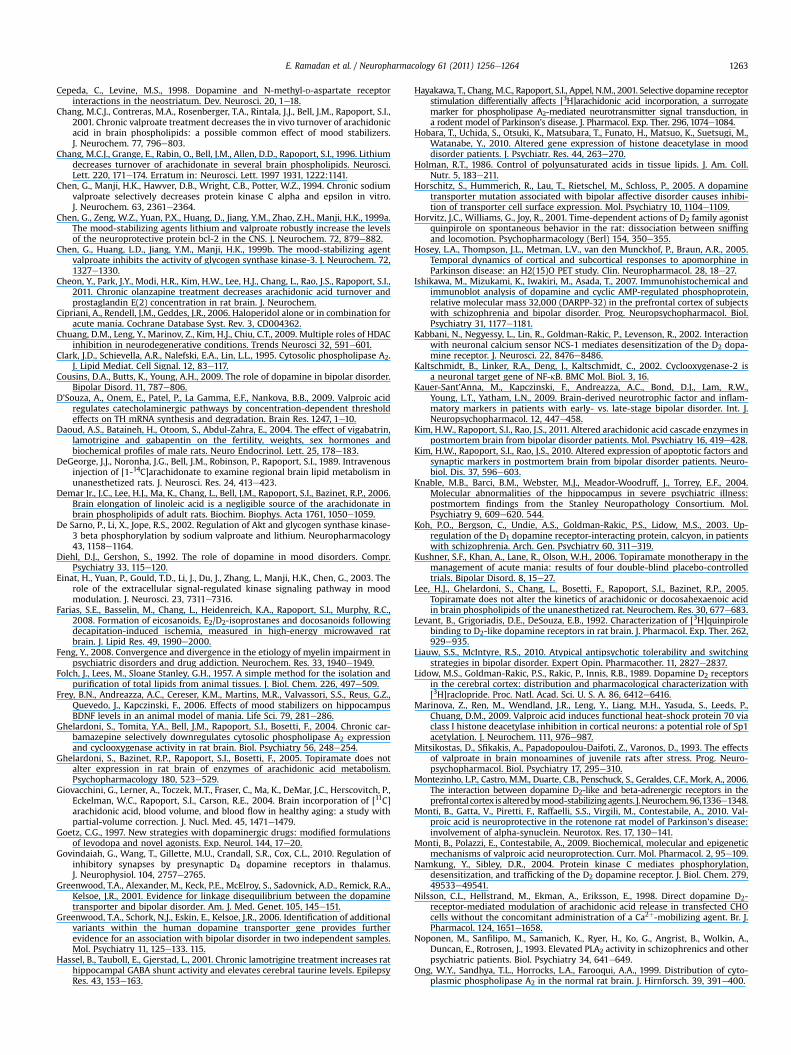
Fig. 1 presents coronal autoradiographs of brains from ratstreated chronically (30 days) with vehicle (control) or VPA, thenacutely injected with saline or quinpirole. k* for AA, calculated byEq. (1), is color-coded. The figure shows no difference in regionalvalues of k* in response to saline between VPA- and vehicle-treatedrats. Acute quinpirole increased k* in multiple brain regions of thechronic vehicle-treated but not VPA-treated rats. Data obtainedfrom such autoradiographs are summarized in Table 3.
Values of mean AA incorporation coefficients, k*, determined ineach of 83 brain regions were subjected to a two-way ANOVA.Statistically significant interactions between VPA and quinpirolewere found in 40 regions belonging primarily to the nigrostriataland mesocorticolimbic systems, which comprise DAergic circuits ofthe basal ganglia (Baldessarini and Tarazi, 1996) (Table 3, Fig. 1). Inall 40 regions, a one-way ANOVA with Bonferroni’s post-testshowed that chronic VPA did not significantly change mean base-line (after saline) k* in any region (Table 3). The same one-way
Chronic valproate
Saline Quinpirole
fter Before After Before After
6.9 � 0.2 36.9 � 0.4 37.0 � 0.3 36.8 � 0.4 37.0 � 0.2430 � 30 425 � 37 434 � 27 441 � 31 433 � 53
181 � 13 175 � 10 166 � 8 162 � 14 162 � 11103 � 7 109 � 6 96 � 11 102 � 7 100 � 6
10 � 2 11 � 317 � 4 15 � 3
) and 10 min after [1-14C]AA infusion.jection.

Table 2Effects of valproate and quinpirole on plasma unesterified fatty acid concentrations.
Fatty acid Chronic vehicle Chronic VPA VPA � Quininteraction
VPA effect Quin effect
Saline(nmol/ml plasma)
Quin(nmol/ml plasma)
Saline(nmol/ml plasma)
Quin(nmol/ml plasma)
P-value P-value P-value
Palmitic (16:0) 199.9 � 82.8 164.3 � 43.1 75.7 � 20.4 70.6 � 15.9 0.4133 <0.0001 0.2768Stearic (18:0) 49.2 � 10.1 36.6 � 14.3 30.0 � 4.8** 33.7 � 5.9 0.0334Oleic (18:1n � 9) 124.0 � 50.8 89.9 � 42.4 53.2 � 15.8 45.2 � 11.1 0.3264 0.0002 0.1192Linoleic (18:2n � 6) 181.9 � 73.3 146.5 � 68.1 70.2 � 19.4 72.0 � 16.9 0.3505 <0.0001 0.3977a-Linolenic (18:3n � 3) 11.9 � 5.3 10.3 � 4.4 4.1 � 1.5 3.8 � 1.5 0.6485 <0.0001 0.4819Arachidonic (20:4n � 6) 16.0 � 6.1 12.6 � 2.9 5.5 � 1.0*** 7.9 � 1.9 0.0411Docosahexaenoic (22:6n � 3) 19.5 � 10.2 15.7 � 4.8 5.8 � 1.6 6.4 � 1.3 0.3199 <0.0001 0.4665
Values are means � SD (n ¼ 7) measured from arterial plasma collected at 19 min after the beginning of [1-14C]AA infusion.**P < 0.01, ***P < 0.001; vehicle plus quinpirole vs vehicle plus saline, VPA plus saline vs vehicle plus saline, and VPA plus quinpirole vs VPA plus saline (one-way ANOVABonferroni post-tests). VPA, valproate; Quin, quinpirole.
E. Ramadan et al. / Neuropharmacology 61 (2011) 1256e1264 1259
ANOVA showed that acute quinpirole compared with salineincreased k* by 22e58% in chronic vehicle-treated rats. Affectedregions included caudate putamen (36e43%), globus pallidus (45%),subthalamic nucleus (33%), substantia nigra (41%), prefrontal cortex(39e50%), primary olfactory cortex (35%), frontal cortex (29e38%),pyriform and anterior cingulated cortex (22%), motor (31e44%),somatosensory, auditory (29e33%), visual (41e48%) cortical areas(26e39%), bed nucleus of the stria terminalis (53%), amygdala(58%), nucleus accumbens (42%), ventral tegmental area (44%),arcuate nucleus of the hypothalamus (28%), ventroposteriorthalamic nuclei (40e45%) and zona incerta (29%). Quinpirolecompared to saline did not significantly increase k* in any of the 40regions in chronic VPA-treated rats.
In the 43 regions where the VPA and quinpirole interaction wasstatistically insignificant, neither VPA nor quinpirole had anysignificant main effect on k* for AA (data not shown). Thus, chronic
Fig. 1. Coronal autoradiographs of brain showing effects of quinpirole and valproate on rebrain � 10�4) are on a color scale from 4 (blue) to 10 (orange). Acb, nucleus accumbens; CP, cSN, substantia nigra; VTA, ventral tegmental area. VPA, valproate; Quin, quinpirole. (For inteweb version of this article.)
VPA blocked each of the 40 quinpirole-induced k* increments thatwere observed in the chronic vehicle-treated rat.
3.4. Regional rates of incorporation of unlabeledunesterified AA into brain
Rates of incorporation of unlabeled unesterified AA from plasmainto brain, Jin, (data not shown) were calculated by Eq. (2) fromregional k* (Table 3) and cplasma for AA (Table 2). A two-way ANOVAshowed no statistically significant interaction between VPA andquinpirole in any of the 83 brain regions examined. Chronic VPAcompared with vehicle had a significant main negative effect ineach of the 83 brain regions while acute quinpirole had no maineffect on any. In vehicle-treated rats Jin ranged from 5.9 pmol/s/g inthe internal capsule to 28 pmol/s/g in the choroid plexus, whereas
gional arachidonic acid incorporation coefficients k* in rats. Values of k* (ml/s/g wetaudate putamen; Fr8, frontal cortex (8); Fr 10, frontal cortex (10); PFr, prefrontal cortex;rpretation of the references to color in this figure legend, the reader is referred to the
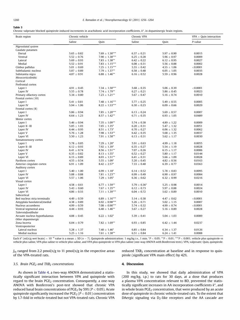
Table 3Chronic valproate blocked quinpirole-induced increments in arachidonic acid incorporation coefficients, k*, in dopaminergic brain regions.
Brain region Chronic vehicle Chronic VPA VPA � Quin interaction
Saline Quin Saline Quin P-value
Nigrostiatal systemCaudate putamenDorsal 5.65 � 0.82 7.69 � 1.30*** 6.37 � 0.21 5.97 � 0.90 0.0015Ventral 5.52 � 0.76 7.90 � 1.38*** 6.25 � 0.28 5.96 � 0.97 0.0009Lateral 5.69 � 0.93 7.83 � 1.38** 6.42 � 0.22 6.12 � 0.95 0.0027Medial 5.52 � 0.91 7.83 � 1.15*** 6.08 � 0.31 5.56 � 0.88 0.0002
Globus pallidus 5.01 � 0.69 7.25 � 1.15*** 5.55 � 0.42 4.55 � 1.06 <0.0001Subthalamic nucleus 5.87 � 0.89 7.79 � 1.37** 6.58 � 0.48 6.05 � 1.05 0.0035Substantia nigra 4.87 � 0.91 6.88 � 1.46** 6.16 � 0.52 5.59 � 0.96 0.0028MesocorticolimbicCorticalPrefrontal cortexLayer I 4.91 � 0.45 7.34 � 1.50*** 5.68 � 0.35 5.06 � 0.39 <0.0001Layer IV 5.55 � 0.78 7.74 � 1.79** 6.27 � 0.21 5.86 � 0.45 0.0023
Primary olfactory cortex 5.34 � 0.80 7.23 � 1.21** 5.67 � 0.47 5.16 � 0.74 0.0010Frontal cortex (10)Layer I 5.41 � 0.81 7.48 � 1.16*** 5.77 � 0.25 5.49 � 0.55 0.0005Layer IV 5.94 � 1.06 8.22 � 1.53*** 6.30 � 0.23 6.09 � 0.66 0.0029
Frontal cortex (8)Layer I 5.86 � 0.94 7.85 � 1.20*** 6.13 � 0.24 5.69 � 0.57 0.0007Layer IV 6.64 � 1.23 8.57 � 1.42** 6.71 � 0.35 6.93 � 1.05 0.0489
Motor cortexLayer I 5.46 � 0.94 7.35 � 1.09** 5.74 � 0.38 4.89 � 1.22 0.0009Layer IIeIII 5.85 � 1.03 7.65 � 1.35* 6.20 � 0.31 5.47 � 1.32 0.0051Layer IV 6.44 � 0.95 8.51 � 1.73* 6.70 � 0.27 6.06 � 1.32 0.0062Layer V 5.76 � 1.28 7.98 � 1.53** 6.42 � 0.35 5.68 � 1.35 0.0037Layer VI 5.50 � 1.23 7.91 � 1.58** 6.13 � 0.31 5.62 � 1.17 0.0030
Somatosensory cortexLayer I 5.78 � 0.85 7.29 � 1.20* 5.91 � 0.63 4.99 � 1.18 0.0055Layer IIeIII 6.12 � 0.93 7.92 � 1.39* 6.35 � 0.27 5.54 � 1.19 0.0028Layer IV 6.41 � 0.74 8.56 � 1.51** 7.07 � 0.26 6.15 � 1.17 0.0006Layer V 6.35 � 0.82 8.13 � 1.55* 6.52 � 0.27 5.80 � 1.17 0.0144Layer VI 6.15 � 0.89 8.03 � 1.51** 6.41 � 0.31 5.66 � 1.09 0.0028
Pyriform cortex 4.55 � 0.54 5.55 � 1.09* 5.20 � 0.45 4.82 � 0.56 0.0163Anterior cingulate cortex 6.91 � 1.09 8.42 � 2.57* 7.33 � 0.40 6.39 � 0.77 0.0363Auditory cortexLayer I 5.40 � 1.00 6.99 � 1.18* 6.14 � 0.52 5.78 � 0.83 0.0095Layer IV 5.88 � 0.88 7.81 � 1.23** 6.99 � 0.49 6.90 � 0.97 0.0084Layer VI 5.57 � 1.06 7.29 � 1.05* 6.36 � 0.62 6.32 � 0.99 0.0216
Visual cortexLayer I 4.58 � 0.61 6.77 � 1.59** 5.79 � 0.36* 5.25 � 0.98 0.0014Layer IV 5.43 � 0.60 7.67 � 1.35** 6.12 � 0.73 5.97 � 0.88 0.0024Layer VI 4.86 � 0.55 7.11 � 1.39*** 6.04 � 0.72 5.62 � 0.89 0.0010
SubcorticalBed nucleus stria terminalis 4.49 � 0.59 6.89 � 1.16*** 5.14 � 0.38 4.47 � 0.62 <0.0001Amygdala basolateral/medial 4.38 � 0.89 6.92 � 0.98*** 5.26 � 0.71 5.02 � 1.15 0.0007Nucleus accumbens 4.99 � 0.59 7.08 � 0.90*** 5.74 � 0.32 4.99 � 0.74 <0.0001Ventral tegmental area 4.44 � 0.93 6.40 � 1.46** 5.67 � 0.27 5.16 � 0.89 0.0028TuberoinfundibularArcuate nucleus hypothalamus 4.88 � 0.45 6.22 � 1.02* 5.39 � 0.41 5.04 � 1.03 0.0089Other dopaminergicZona Incerta 6.04 � 0.79 7.82 � 1.69** 6.93 � 0.85 6.42 � 1.44 0.0237VentroposteriorLateral nucleus 5.28 � 1.37 7.40 � 1.46* 6.85 � 0.84 6.34 � 1.37 0.0126Medial nucleus 5.25 � 1.16 7.61 � 1.38** 6.51 � 0.84 6.24 � 1.41 0.0088
Each k* (ml/s/g wet brain) � 10�4 value is a mean � SD (n ¼ 7). Quinpirole administration: 1 mg/kg i.v., 1 min. *P < 0.05; **P < 0.01; ***P < 0.001; vehicle plus quinpirole vsvehicle plus saline, VPA plus saline vs vehicle plus saline, and VPA plus quinpirole vs VPA plus saline (one-way ANOVAwith Bonferroni tests). VPA, valproate; Quin, quinpirole.
E. Ramadan et al. / Neuropharmacology 61 (2011) 1256e12641260
Jin ranged from 2.2 pmol/s/g to 11 pmol/s/g in the respective areasof the VPA-treated rats.
3.5. Brain PGE2 and TXB2 concentrations
As shown in Table 4, a two-way ANOVA demonstrated a statis-tically significant interaction between VPA and quinpirole withregard to the brain PGE2 concentration. Consequently, a one-wayANOVA with Bonferroni’s post-test showed that chronic VPAreduced basal brain concentrations of PGE2 by 59% (P< 0.05). Acutequinpirole significantly increased the PGE2 (P< 0.01) concentrationby 1.7-fold in vehicle-treated but not VPA-treated rats. Chronic VPA
reduced TXB2 concentration at baseline and in response to quin-pirole (significant VPA main effect) by 42%.
4. Discussion
In this study, we showed that daily administration of VPA(200 mg/kg, i.p.) to rats for 30 days, at a dose that producesa plasma VPA concentration relevant to BD, prevented the statis-tically significant increases in AA incorporation coefficients k*, andinwhole brain PGE2 concentration, that were produced by an acutedose of quinpirole in chronic vehicle-treated rats. To the extent thatDAergic signaling via D2-like receptors and the AA cascade are

Table 4Effects of quinpirole and valproate on brain PGE2 and TXB2 concentrations in rats.
Chronic vehicle Chronic VPA VPA � Quin interaction VPA effect Quin effect
Saline Quin Saline Quin P-value P-value P-value
PGE2 (ng/g brain) 10.3 � 3.1 17.7 � 6.8** 4.2 � 1.1* 5.3 � 0.8 0.0348TXB2 (pg/g brain) 41.3 � 11.9 31.4 � 9.1 22.2 � 6.8 20.2 � 9.0 0.2710 0.0002 0.1034
Each value is a mean� SD (n ¼ 7). Bonferroni’s multiple comparison tests were performed. *P< 0.05; **P< 0.01; vehicle plus quinpirole vs vehicle plus saline, VPA plus salinevs vehicle plus saline, and VPA plus quinpirole vs VPA plus saline. VPA, valproate; Quin, quinpirole.
E. Ramadan et al. / Neuropharmacology 61 (2011) 1256e1264 1261
pathologically upregulated in BD patients, for which evidenceexists (see “Introduction”) (Berk et al., 2007; Cousins et al., 2009;Diehl and Gershon, 1992; Goetz, 1997; Kim et al., 2011), theseresults suggest that the efficacy of VPA in the disease treatment isdue in part to its ability to dampen upregulated D2-like signalinginvolving AA and its downstream metabolites. In agreement,chronic administration to rats of a therapeutically relevant plasmaconcentration of lithium or carbamazepine also dampensD2-induced elevations in k* for AA and in brain eicosanoids(Basselin et al., 2005, 2008b; Bosetti et al., 2002; Bosetti et al.,2003). Taken together, reduced D2-like signal involving AA and itsmetabolites may be common to the therapeutic action of moodstabilizers effective in BD. In contrast, topiramate, which appearedeffective in Phase II trials in BD, but later failed Phase III placebo-controlled trials (Kushner et al., 2006), did not change markers ofthe rat brain AA cascade (Ghelardoni et al., 2005; Lee et al., 2005).Topiramate has not been tested with regard to the D2-like signal.
Similar to lithium and carbamazepine, chronic VPA significantlydecreased baseline PGE2 and TXB2 concentrations as reported(Basselin et al., 2008a; Bosetti et al., 2002, 2003; Ghelardoni et al.,2004). We ascribe this to VPA selectively decreasing the bindingactivity of the transcription factor NF-kB that regulates neuronalCOX-2 gene expression, as well as reducing COX-1 and COX-2protein levels and whole brain COX activity (Bosetti et al., 2003;Kaltschmidt et al., 2002; Rao et al., 2007a,b).
Acute quinpirole significantly increased k* for AA in 40 brainregions, most of which are rich in D2-like receptors (Levant et al.,1992; Lidow et al., 1989) and are related to the topographicaldistribution of DAergic innervation in the brain (mesocorticolimbic,nigrostriatal, and tuberoinfundibular pathways). The zona incerta,located in the ventral thalamus, and cerebral cortical areas (layersI to VI) including auditory and visual cortex also contain DAneurons (Berger et al., 1985; Bjorklund and Lindvall, 1975; Lidowet al., 1989; Rivera and Chun, 2008). The globus pallidus, sub-thalamic nucleus, and ventrobasal thalamus also receive DAergicinnervation and express DA receptors (Baldessarini and Tarazi,1996; Govindaiah et al., 2010).
Mechanisms underlying VPA’s ability to block the D2-likereceptor-induced increases in k* for AA and to reduce PGE2 andTXB2 concentrations in rat brain are not clear. VPA could have actedby reducing COX activity, and COX-1 and COX-2 protein levels(Bosetti et al., 2003). When COX enzymes are pharmacologicallyinhibited or abscent in rodent brain, k* responses to drugs acting atcPLA2-coupled neuroreceptors are reduced or lost, as are increasesin brain PGE2 and/or TXB2 concentrations (Basselin et al., 2006b,2007b). VPA also may have interfered with the DAergic systemand D2-like receptors. Consistent with altered gene expression ofhistone deacetylases and increased histone H3 and H4 acetylationin BD patients (Hobara et al., 2010; Sharma et al., 2006), VPA,a direct histone deacetylase inhibitor (Phiel et al., 2001), maymodify the transcription of the rate-limiting enzyme in DAbiosynthesis, tyrosine hydroxylase (D’Souza et al., 2009), or of Sp1(Marinova et al., 2009), a transcription factor of the D2 receptor(Yajima et al., 1998), and/or DAT gene acetylation (Wang andBannon, 2005). Consistent with our findings, VPA has been
shown to decrease D2 receptor protein in the rat prefrontal cortex(Montezinho et al., 2006), lower presynaptic DA function in thestriatum of patients with mania (Yatham et al., 2002), and increaseDAT gene expression in rat midbrain DA neurons (Wang et al.,2007), thus decreasing extracellular DA concentration at thesynaptic cleft. Although VPA has been reported to inhibit GSK-3(Chen et al., 1999a,b), which can be regulated by DA via the Aktsignaling pathway (Beaulieu et al., 2004; Beaulieu, 2011), this effectis indirect and has been attributed to inhibition of activation of Aktand inactivation of GSK-3 following inhibition of histone deacety-lase (Phiel et al., 2001; De Sarno et al., 2002).
Therapeutically relevant concentration of VPA has been shown todecrease the activity andprotein level of protein kinaseC (Chen et al.,1994), which mediates phosphorylation, desensitization and traf-ficking of the D2 receptor (Namkung and Sibley, 2004). Alternatively,chronic VPA may have indirectly attenuated the D2-mediated AAsignaling by (i) enhancing GABAergic transmission, which partici-pates in regulating DA release and of inhibiting DAergic activity(Agmo et al., 1996) and/or by (ii) reducing excitatory neurotrans-mission and blocking the AA signaling mediated by glutamatergic-NMDA receptors (Basselin et al., 2008a). D2-like and NMDA recep-tors are often functionally coupled and colocalized on the sameneurons in the brain (Cepeda and Levine, 1998; Wang et al., 2003).Together with our previous reports (Basselin et al., 2005, 2008b,2007a, 2006a), the results suggest that antimanic mood stabilizerseffective in BD suppress AA signaling coupled to both NMDA andD2-like receptors. Combined, these data are consistent with VPA pro-tecting DA neurons in lipopolysaccharide-induced neurotoxicity(Peng et al., 2005). In agreement, VPA was neuroprotective inexperimental models of cerebral ischemia, Parkinson’s disease andglutamate-induced excitotoxicity via histone deacetylase inhibition(Monti et al., 2010, 2009; Ren et al., 2004;Wang et al., 2010; Chuanget al., 2009).
VPA increased brain-derived neurotrophic factor (BDNF) (Einatet al., 2003; Yasuda et al., 2009), hippocampal neurotrophin-3(Walz et al., 2008), anti-apoptotic factor B-cell lymphoma-2 (Bcl-2)(Chen et al., 1999a,b), and restored amphetamine-induced down-regulation of BDNF and of neurotrophin-3 in rat brain (Frey et al.,2006; Walz et al., 2008). Given that the brain and serum in BDpatients have reduced BDNF and other neurotrophic factors (Kauer-Sant’Anna et al., 2009; Kim et al., 2010; Knable et al., 2004;Tramontina et al., 2009), these actions may contribute to VPA’sneuroprotective effect in BD, a disease characterized by progressivesynaptic loss and apoptosis (Benes et al., 2006; Kim et al., 2010;Rapoport et al., 2009).
Consistent with our previous studies, chronic VPA significantlydecreased the plasma concentration of unlabeled unesterified fattyacids including AA (Bazinet et al., 2005; Chang et al., 2001), indi-cating a widespread effect on whole body fatty acid metabolism. Asimilar reduction in plasma unesterified fatty acids has been foundwith othermood stabilizers andantipsychotics used to treat BD, suchas lamotrigine, olanzapine and clozapine (Ramadan et al., 2011;Cheon et al., 2011) suggesting a common peripheral effect of thesedrugs. The decrease in plasma unesterified fatty acids may be due to(i) reduced liver secretion of lipoprotein-bound esterified fatty acids,
E. Ramadan et al. / Neuropharmacology 61 (2011) 1256e12641262
the main source of unesterified fatty acids in plasma, or (ii) reducedrelease of unesterified fatty acids from adipose tissue by lipases.Although the effects of VPA on free fatty acid release have not beeninvestigated, evidence of impaired secretion of esterified fatty acidshas been demonstrated with a marked reduction in triglyceridesecretion following VPA treatment (Bellringer et al., 1988).
The baseline values of k* and Jin in this study agreewith previousreports (Basselin et al., 2005, 2008a; Bhattacharjee et al., 2005;Bhattacharjee et al., 2006, 2008). Values of baseline k* were notaltered by chronic VPA, which is consistent with our previous data(Basselin et al., 2008a), and supports the finding that chronic VPAdoes not affect basal cPLA2-IV expression (Bosetti et al., 2003).Chronic VPA compared to vehicle had a significant main negativeeffect on all Jin values at baseline, indicating that metabolic AA lossfrom brainwas lower in the VPA-treated animals. We ascribe this toVPA’s significant reduction of the plasma concentration of unla-beled unesterified AA and of brain PGE2 and TXB2 concentrations,and to its selective inhibition of acyl-CoA synthetase 4-mediatedconversion of AA to AAeCoA (Bazinet et al., 2006a; Shimshoniet al., 2011).
Chronic VPA, like chronic carbamazepine but unlike chroniclithium, did not prevent quinpirole-induced hyperactivity orstereotypy (Basselin et al., 2005, 2008b; Beaulieu et al., 2004;Shaldubina et al., 2002). As each of the three mood stabilizersdownregulates the brain AA cascade, their different effects onquinpirole-induced behaviors suggest that these behaviors do notinvolve AA signaling, and that the quinpirole-induced activitycycles are not modeling BD. In contrast, VPA attenuated hyperac-tivity and locomotor behavior in DAT knockdown mice (Ralph-Williams et al., 2003).
We investigated the effects only of chronic (30 days) VPA in thisstudy, as mood stabilization in BD patients only appears after 10days of treatment with VPA. An acute injection of VPA(200e300 mg/kg) in rats caused no/very transient change in thebrain DA level (Ahmad et al., 2005; Mitsikostas et al., 1993).
In conclusion, chronic VPA prevented the statistically significantincreases in k* for AA and in PGE2 concentrations that wereobserved in response to quinpirole in chronic vehicle-treated rats.These and observations in rats administered chronic lithium orcarbamazepine support the hypothesis that mood stabilizerscommonly downregulate brain AA signaling via D2-like receptors,and are consistent with evidence that some BD symptoms arisefrom excessive DAergic neurotransmission (Goetz, 1997). It wouldbe worthwhile to see if atypical antipsychotics (e.g. clozapine,olanzapine), which are D2-like receptor antagonists, do so as well,which would suggest a more general receptor action of theseagents on cPLA2-mediated AA signaling (Liauw and McIntyre,2010). Additionally positron emission tomography using [1-11C]AA might be employed in BD patients with or without chronic VPAtreatment, before and after drug induced D2-like receptor activa-tion, to see if VPA has a similar transient effect on AA signaling BD(Giovacchini et al., 2004; Goetz, 1997; Hosey et al., 2005).
Acknowledgments
This work was supported entirely by the Intramural ResearchProgram of the National Institute on Aging, NIH. We thank theFellows Editorial Board for editorial assistance. None of the authorshas a financial or other conflict of interest related to this work.
References
Agmo, A., Belzung, C., Giordano, M., 1996. Interactions between dopamine andGABA in the control of ambulatory activity. J. Neural Transm. 103, 925e934.
Ahmad, S., Fowler, L.J., Whitton, P.S., 2005. Effects of combined lamotrigine andvalproate on basal and stimulated extracellular amino acids and monoamines inthe hippocampus of freely moving rats. Naunyn Schmiedebergs Arch. Phar-macol. 371, 1e8.
Anand, A., Verhoeff, P., Seneca, N., Zoghbi, S.S., Seibyl, J.P., Charney, D.S., Innis, R.B.,2000. Brain SPECT imaging of amphetamine-induced dopamine release ineuthymic bipolar disorder patients. Am. J. Psychiatry 157, 1108e1114.
Baldessarini, R.J., Tarazi, F.I., 1996. Brain dopamine receptors: a primer on theircurrent status, basic and clinical. Harv. Rev. Psychiatry 3, 301e325.
Basselin, M., Chang, L., Bell, J.M., Rapoport, S.I., 2005. Chronic lithium chlorideadministration to unanesthetized rats attenuates brain dopamine D2-likereceptor-initiated signaling via arachidonic acid. Neuropsychopharmacology30, 1064e1075.
Basselin, M., Chang, L., Bell, J.M., Rapoport, S.I., 2006a. Chronic lithium chlorideadministration attenuates brain NMDA receptor-initiated signaling viaarachidonic acid in unanesthetized rats. Neuropsychopharmacology 31,1659e1674.
Basselin, M., Villacreses, N.E., Langenbach, R., Ma, K., Bell, J.M., Rapoport, S.I., 2006b.Resting and arecoline-stimulated brain metabolism and signaling involvingarachidonic acid are altered in the cyclooxygenase-2 knockout mice.J. Neurochem. 96, 669e679.
Basselin, M., Villacreses, N.E., Chen, M., Bell, J.M., Rapoport, S.I., 2007a. Chroniccarbamazepine administration reduces NMDA receptor-initiated signaling viaarachidonic acid in rat brain. Biol. Psychiatry 62, 934e943.
Basselin, M., Villacreses, N.E., Lee, H.-J., Bell, J.M., Rapoport, S.I., 2007b. Flurbiprofen,a cyclooxygenase inhibitor, reduces the brain arachidonic acid signal inresponse to the cholinergic muscarinic, arecoline, in awake rats. Neurochem.Res. 32, 1857e1867.
Basselin, M., Chang, L., Chen, M., Bell, J.M., Rapoport, S.I., 2008a. Chronic adminis-tration of valproic acid reduces brain NMDA signaling via arachidonic acid inunanesthetized rats. Neurochem. Res. 33, 2229e2240.
Basselin, M., Chang, L., Chen, M., Bell, J.M., Rapoport, S.I., 2008b. Chronic carba-mazepine administration attenuates dopamine D2-like receptor-initiatedsignaling via arachidonic acid in rat brain. Neurochem. Res. 33, 1373e1383.
Bazinet, R.P., Rao, J.S., Chang, L., Rapoport, S.I., Lee, H.J., 2005. Chronic valproate doesnot alter the kinetics of docosahexaenoic acid within brain phospholipids of theunanesthetized rat. Psychopharmacology (Berl) 1, 180e185.
Bazinet, R.P., Weis, M.T., Rapoport, S.I., Rosenberger, T.A., 2006a. Valproic acidselectively inhibits conversion of arachidonic acid to arachionoyl-CoA by brainmicrosomal long-chain fatty acyl-CoA synthetases: relevance to bipolardisorder. Psychopharmacology (Berl) 184, 122e129.
Bazinet, R.P., Rao, J.S., Chang, L., Rapoport, S.I., Lee, H.J., 2006b. Chronic carbama-zepine decreases the incorporation rate and turnover of arachidonic acid butnot docosahexaenoic acid in brain phospholipids of the unanesthetized rat:relevance to bipolar disorder. Biol. Psychiatry 59, 401e407.
Beaulieu, J.M., Sotnikova, T.D., Yao, W.D., Kockeritz, L., Woodgett, J.R.,Gainetdinov, R.R., Caron, M.G., 2004. Lithium antagonizes dopamine-dependentbehaviors mediated by an AKT/glycogen synthase kinase 3 signaling cascade.Proc. Natl. Acad. Sci. U. S. A. 101, 5099e5104.
Beaulieu, J.M., 2011. A role of Akt and glycogen synthase kinase-3 as integrators ofdopamine and serotonin neurotransmission in mental health. J. PsychiatryNeurosci. 36, 1110011.
Bellringer, M.E., Rahman, K., Coleman, R., 1988. Sodium valproate inhibits themovement of secretory vesicles in rat hepatocytes. Biochem. J. 249, 513e519.
Benes, F.M., Matzilevich, D., Burke, R.E., Walsh, J., 2006. The expression of proa-poptosis genes is increased in bipolar disorder, but not in schizophrenia. Mol.Psychiatry 11, 241e251.
Berger, B., Verney, C., Alvarez, C., Vigny, A., Helle, K.B., 1985. New dopaminergicterminal fields in the motor, visual (area 18b) and retrosplenial cortex in theyoung and adult rat. Immunocytochemical and catecholamine histochemicalanalyses. Neuroscience 15, 983e998.
Berk, M., Dodd, S., Kauer-Sant’Anna, M., Malhi, G.S., Bourin, M., Kapczinski, F.,Norman, T., 2007. Dopamine dysregulation syndrome: implications for a dopa-mine hypothesis of bipolar disorder. Acta Psychiatr. Scand. Suppl., 41e49.
Bhattacharjee, A.K., Chang, L., Lee, H.J., Bazinet, R.P., Seemann, R., Rapoport, S.I.,2005. D2 but not D1 dopamine receptor stimulation augments brain signalinginvolving arachidonic acid in unanesthetized rats. Psychopharmacology (Berl)180, 735e742.
Bhattacharjee, A.K., Chang, L., White, L., Bazinet, R.P., Rapoport, S.I., 2006.D-Amphetamine stimulates D2 dopamine receptor-mediated brain signalinginvolving arachidonic acid in unanesthetized rats. J. Cereb. Blood Flow Metab.26, 1378e1388.
Bhattacharjee, A.K., Chang, L., White, L., Bazinet, R.P., Rapoport, S.I., 2008. Imagingapomorphine stimulation of brain arachidonic acid signaling via D2-likereceptors in unanesthetized rats. Psychopharmacology (Berl) 197, 557e566.
Bjorklund, A., Lindvall, O., 1975. Dopamine in dendrites of substantia nigra neurons:suggestions for a role in dendritic terminals. Brain Res. 83, 531e537.
Bosetti, F., Rintala, J., Seemann, R., Rosenberger, T.A., Contreras, M.A., Rapoport, S.I.,Chang, M.C., 2002. Chronic lithium downregulates cyclooxygenase-2 activityand prostaglandin E2 concentration in rat brain. Mol. Psychiatry 7, 845e850.
Bosetti, F., Weerasinghe, G.R., Rosenberger, T.A., Rapoport, S.I., 2003. Valproic aciddown-regulates the conversion of arachidonic acid to eicosanoids viacyclooxygenase-1 and -2 in rat brain. J. Neurochem. 85, 690e696.
Bowden, C.L., 2009. Anticonvulsants in bipolar disorders: current research andpractice and future directions. Bipolar Disord. 11 (Suppl. 2), 20e33.
E. Ramadan et al. / Neuropharmacology 61 (2011) 1256e1264 1263
Cepeda, C., Levine, M.S., 1998. Dopamine and N-methyl-D-aspartate receptorinteractions in the neostriatum. Dev. Neurosci. 20, 1e18.
Chang, M.C.J., Contreras, M.A., Rosenberger, T.A., Rintala, J.J., Bell, J.M., Rapoport, S.I.,2001. Chronic valproate treatment decreases the in vivo turnover of arachidonicacid in brain phospholipids: a possible common effect of mood stabilizers.J. Neurochem. 77, 796e803.
Chang, M.C.J., Grange, E., Rabin, O., Bell, J.M., Allen, D.D., Rapoport, S.I., 1996. Lithiumdecreases turnover of arachidonate in several brain phospholipids. Neurosci.Lett. 220, 171e174. Erratum in: Neurosci. Lett. 1997 1931, 1222:1141.
Chen, G., Manji, H.K., Hawver, D.B., Wright, C.B., Potter, W.Z., 1994. Chronic sodiumvalproate selectively decreases protein kinase C alpha and epsilon in vitro.J. Neurochem. 63, 2361e2364.
Chen, G., Zeng, W.Z., Yuan, P.X., Huang, D., Jiang, Y.M., Zhao, Z.H., Manji, H.K., 1999a.The mood-stabilizing agents lithium and valproate robustly increase the levelsof the neuroprotective protein bcl-2 in the CNS. J. Neurochem. 72, 879e882.
Chen, G., Huang, L.D., Jiang, Y.M., Manji, H.K., 1999b. The mood-stabilizing agentvalproate inhibits the activity of glycogen synthase kinase-3. J. Neurochem. 72,1327e1330.
Cheon, Y., Park, J.Y., Modi, H.R., Kim, H.W., Lee, H.J., Chang, L., Rao, J.S., Rapoport, S.I.,2011. Chronic olanzapine treatment decreases arachidonic acid turnover andprostaglandin E(2) concentration in rat brain. J. Neurochem.
Cipriani, A., Rendell, J.M., Geddes, J.R., 2006. Haloperidol alone or in combination foracute mania. Cochrane Database Syst. Rev. 3, CD004362.
Chuang, D.M., Leng, Y., Marinov, Z., Kim, H.J., Chiu, C.T., 2009. Multiple roles of HDACinhibition in neurodegenerative conditions. Trends Neurosci 32, 591e601.
Clark, J.D., Schievella, A.R., Nalefski, E.A., Lin, L.L., 1995. Cytosolic phospholipase A2.J. Lipid Mediat. Cell Signal. 12, 83e117.
Cousins, D.A., Butts, K., Young, A.H., 2009. The role of dopamine in bipolar disorder.Bipolar Disord. 11, 787e806.
D’Souza, A., Onem, E., Patel, P., La Gamma, E.F., Nankova, B.B., 2009. Valproic acidregulates catecholaminergic pathways by concentration-dependent thresholdeffects on TH mRNA synthesis and degradation. Brain Res. 1247, 1e10.
Daoud, A.S., Bataineh, H., Otoom, S., Abdul-Zahra, E., 2004. The effect of vigabatrin,lamotrigine and gabapentin on the fertility, weights, sex hormones andbiochemical profiles of male rats. Neuro Endocrinol. Lett. 25, 178e183.
DeGeorge, J.J., Noronha, J.G., Bell, J.M., Robinson, P., Rapoport, S.I., 1989. Intravenousinjection of [1-14C]arachidonate to examine regional brain lipid metabolism inunanesthetized rats. J. Neurosci. Res. 24, 413e423.
Demar Jr., J.C., Lee, H.J., Ma, K., Chang, L., Bell, J.M., Rapoport, S.I., Bazinet, R.P., 2006.Brain elongation of linoleic acid is a negligible source of the arachidonate inbrain phospholipids of adult rats. Biochim. Biophys. Acta 1761, 1050e1059.
De Sarno, P., Li, X., Jope, R.S., 2002. Regulation of Akt and glycogen synthase kinase-3 beta phosphorylation by sodium valproate and lithium. Neuropharmacology43, 1158e1164.
Diehl, D.J., Gershon, S., 1992. The role of dopamine in mood disorders. Compr.Psychiatry 33, 115e120.
Einat, H., Yuan, P., Gould, T.D., Li, J., Du, J., Zhang, L., Manji, H.K., Chen, G., 2003. Therole of the extracellular signal-regulated kinase signaling pathway in moodmodulation. J. Neurosci. 23, 7311e7316.
Farias, S.E., Basselin, M., Chang, L., Heidenreich, K.A., Rapoport, S.I., Murphy, R.C.,2008. Formation of eicosanoids, E2/D2-isoprostanes and docosanoids followingdecapitation-induced ischemia, measured in high-energy microwaved ratbrain. J. Lipid Res. 49, 1990e2000.
Feng, Y., 2008. Convergence and divergence in the etiology of myelin impairment inpsychiatric disorders and drug addiction. Neurochem. Res. 33, 1940e1949.
Folch, J., Lees, M., Sloane Stanley, G.H., 1957. A simple method for the isolation andpurification of total lipids from animal tissues. J. Biol. Chem. 226, 497e509.
Frey, B.N., Andreazza, A.C., Cereser, K.M., Martins, M.R., Valvassori, S.S., Reus, G.Z.,Quevedo, J., Kapczinski, F., 2006. Effects of mood stabilizers on hippocampusBDNF levels in an animal model of mania. Life Sci. 79, 281e286.
Ghelardoni, S., Tomita, Y.A., Bell, J.M., Rapoport, S.I., Bosetti, F., 2004. Chronic car-bamazepine selectively downregulates cytosolic phospholipase A2 expressionand cyclooxygenase activity in rat brain. Biol. Psychiatry 56, 248e254.
Ghelardoni, S., Bazinet, R.P., Rapoport, S.I., Bosetti, F., 2005. Topiramate does notalter expression in rat brain of enzymes of arachidonic acid metabolism.Psychopharmacology 180, 523e529.
Giovacchini, G., Lerner, A., Toczek, M.T., Fraser, C., Ma, K., DeMar, J.C., Herscovitch, P.,Eckelman, W.C., Rapoport, S.I., Carson, R.E., 2004. Brain incorporation of [11C]arachidonic acid, blood volume, and blood flow in healthy aging: a study withpartial-volume correction. J. Nucl. Med. 45, 1471e1479.
Goetz, C.G., 1997. New strategies with dopaminergic drugs: modified formulationsof levodopa and novel agonists. Exp. Neurol. 144, 17e20.
Govindaiah, G., Wang, T., Gillette, M.U., Crandall, S.R., Cox, C.L., 2010. Regulation ofinhibitory synapses by presynaptic D4 dopamine receptors in thalamus.J. Neurophysiol. 104, 2757e2765.
Greenwood, T.A., Alexander, M., Keck, P.E., McElroy, S., Sadovnick, A.D., Remick, R.A.,Kelsoe, J.R., 2001. Evidence for linkage disequilibrium between the dopaminetransporter and bipolar disorder. Am. J. Med. Genet. 105, 145e151.
Greenwood, T.A., Schork, N.J., Eskin, E., Kelsoe, J.R., 2006. Identification of additionalvariants within the human dopamine transporter gene provides furtherevidence for an association with bipolar disorder in two independent samples.Mol. Psychiatry 11, 125e133. 115.
Hassel, B., Tauboll, E., Gjerstad, L., 2001. Chronic lamotrigine treatment increases rathippocampal GABA shunt activity and elevates cerebral taurine levels. EpilepsyRes. 43, 153e163.
Hayakawa, T., Chang, M.C., Rapoport, S.I., Appel, N.M., 2001. Selective dopamine receptorstimulation differentially affects [3H]arachidonic acid incorporation, a surrogatemarker for phospholipase A2-mediated neurotransmitter signal transduction, ina rodent model of Parkinson’s disease. J. Pharmacol. Exp. Ther. 296, 1074e1084.
Hobara, T., Uchida, S., Otsuki, K., Matsubara, T., Funato, H., Matsuo, K., Suetsugi, M.,Watanabe, Y., 2010. Altered gene expression of histone deacetylase in mooddisorder patients. J. Psychiatr. Res. 44, 263e270.
Holman, R.T., 1986. Control of polyunsaturated acids in tissue lipids. J. Am. Coll.Nutr. 5, 183e211.
Horschitz, S., Hummerich, R., Lau, T., Rietschel, M., Schloss, P., 2005. A dopaminetransporter mutation associated with bipolar affective disorder causes inhibi-tion of transporter cell surface expression. Mol. Psychiatry 10, 1104e1109.
Horvitz, J.C., Williams, G., Joy, R., 2001. Time-dependent actions of D2 family agonistquinpirole on spontaneous behavior in the rat: dissociation between sniffingand locomotion. Psychopharmacology (Berl) 154, 350e355.
Hosey, L.A., Thompson, J.L., Metman, L.V., van den Munckhof, P., Braun, A.R., 2005.Temporal dynamics of cortical and subcortical responses to apomorphine inParkinson disease: an H2(15)O PET study. Clin. Neuropharmacol. 28, 18e27.
Ishikawa, M., Mizukami, K., Iwakiri, M., Asada, T., 2007. Immunohistochemical andimmunoblot analysis of dopamine and cyclic AMP-regulated phosphoprotein,relative molecular mass 32,000 (DARPP-32) in the prefrontal cortex of subjectswith schizophrenia and bipolar disorder. Prog. Neuropsychopharmacol. Biol.Psychiatry 31, 1177e1181.
Kabbani, N., Negyessy, L., Lin, R., Goldman-Rakic, P., Levenson, R., 2002. Interactionwith neuronal calcium sensor NCS-1 mediates desensitization of the D2 dopa-mine receptor. J. Neurosci. 22, 8476e8486.
Kaltschmidt, B., Linker, R.A., Deng, J., Kaltschmidt, C., 2002. Cyclooxygenase-2 isa neuronal target gene of NF-kB. BMC Mol. Biol. 3, 16.
Kauer-Sant’Anna, M., Kapczinski, F., Andreazza, A.C., Bond, D.J., Lam, R.W.,Young, L.T., Yatham, L.N., 2009. Brain-derived neurotrophic factor and inflam-matory markers in patients with early- vs. late-stage bipolar disorder. Int. J.Neuropsychopharmacol. 12, 447e458.
Kim, H.W., Rapoport, S.I., Rao, J.S., 2011. Altered arachidonic acid cascade enzymes inpostmortem brain from bipolar disorder patients. Mol. Psychiatry 16, 419e428.
Kim, H.W., Rapoport, S.I., Rao, J.S., 2010. Altered expression of apoptotic factors andsynaptic markers in postmortem brain from bipolar disorder patients. Neuro-biol. Dis. 37, 596e603.
Knable, M.B., Barci, B.M., Webster, M.J., Meador-Woodruff, J., Torrey, E.F., 2004.Molecular abnormalities of the hippocampus in severe psychiatric illness:postmortem findings from the Stanley Neuropathology Consortium. Mol.Psychiatry 9, 609e620. 544.
Koh, P.O., Bergson, C., Undie, A.S., Goldman-Rakic, P.S., Lidow, M.S., 2003. Up-regulation of the D1 dopamine receptor-interacting protein, calcyon, in patientswith schizophrenia. Arch. Gen. Psychiatry 60, 311e319.
Kushner, S.F., Khan, A., Lane, R., Olson, W.H., 2006. Topiramate monotherapy in themanagement of acute mania: results of four double-blind placebo-controlledtrials. Bipolar Disord. 8, 15e27.
Lee, H.J., Ghelardoni, S., Chang, L., Bosetti, F., Rapoport, S.I., Bazinet, R.P., 2005.Topiramate does not alter the kinetics of arachidonic or docosahexaenoic acidin brain phospholipids of the unanesthetized rat. Neurochem. Res. 30, 677e683.
Levant, B., Grigoriadis, D.E., DeSouza, E.B., 1992. Characterization of [3H]quinpirolebinding to D2-like dopamine receptors in rat brain. J. Pharmacol. Exp. Ther. 262,929e935.
Liauw, S.S., McIntyre, R.S., 2010. Atypical antipsychotic tolerability and switchingstrategies in bipolar disorder. Expert Opin. Pharmacother. 11, 2827e2837.
Lidow, M.S., Goldman-Rakic, P.S., Rakic, P., Innis, R.B., 1989. Dopamine D2 receptorsin the cerebral cortex: distribution and pharmacological characterization with[3H]raclopride. Proc. Natl. Acad. Sci. U. S. A. 86, 6412e6416.
Marinova, Z., Ren, M., Wendland, J.R., Leng, Y., Liang, M.H., Yasuda, S., Leeds, P.,Chuang, D.M., 2009. Valproic acid induces functional heat-shock protein 70 viaclass I histone deacetylase inhibition in cortical neurons: a potential role of Sp1acetylation. J. Neurochem. 111, 976e987.
Mitsikostas, D., Sfikakis, A., Papadopoulou-Daifoti, Z., Varonos, D., 1993. The effectsof valproate in brain monoamines of juvenile rats after stress. Prog. Neuro-psychopharmacol. Biol. Psychiatry 17, 295e310.
Montezinho, L.P., Castro, M.M., Duarte, C.B., Penschuck, S., Geraldes, C.F., Mork, A., 2006.The interaction between dopamine D2-like and beta-adrenergic receptors in theprefrontal cortex isalteredbymood-stabilizingagents. J.Neurochem.96,1336e1348.
Monti, B., Gatta, V., Piretti, F., Raffaelli, S.S., Virgili, M., Contestabile, A., 2010. Val-proic acid is neuroprotective in the rotenone rat model of Parkinson’s disease:involvement of alpha-synuclein. Neurotox. Res. 17, 130e141.
Monti, B., Polazzi, E., Contestabile, A., 2009. Biochemical, molecular and epigeneticmechanisms of valproic acid neuroprotection. Curr. Mol. Pharmacol. 2, 95e109.
Namkung, Y., Sibley, D.R., 2004. Protein kinase C mediates phosphorylation,desensitization, and trafficking of the D2 dopamine receptor. J. Biol. Chem. 279,49533e49541.
Nilsson, C.L., Hellstrand, M., Ekman, A., Eriksson, E., 1998. Direct dopamine D2-receptor-mediated modulation of arachidonic acid release in transfected CHOcells without the concomitant administration of a Ca2þ-mobilizing agent. Br. J.Pharmacol. 124, 1651e1658.
Noponen, M., Sanfilipo, M., Samanich, K., Ryer, H., Ko, G., Angrist, B., Wolkin, A.,Duncan, E., Rotrosen, J., 1993. Elevated PLA2 activity in schizophrenics and otherpsychiatric patients. Biol. Psychiatry 34, 641e649.
Ong, W.Y., Sandhya, T.L., Horrocks, L.A., Farooqui, A.A., 1999. Distribution of cyto-plasmic phospholipase A2 in the normal rat brain. J. Hirnforsch. 39, 391e400.

E. Ramadan et al. / Neuropharmacology 61 (2011) 1256e12641264
Paxinos, G., Watson, C., 1987. The Rat Brain in Stereotaxic Coordinates, third ed.Academic Press, New York.
Pearlson, G.D., Wong, D.F., Tune, L.E., Ross, C.A., Chase, G.A., Links, J.M., Dannals, R.F.,Wilson, A.A., Ravert, H.T., Wagner Jr., H.N., et al., 1995. In vivo D2 dopaminereceptor density in psychotic and nonpsychotic patients with bipolar disorder.Arch. Gen. Psychiatry 52, 471e477.
Peet, M., Peters, S., 1995. Drug-induced mania. Drug Saf. 12, 146e153.Peng, G.S., Li, G., Tzeng, N.S., Chen, P.S., Chuang, D.M., Hsu, Y.D., Yang, S., Hong, J.S.,
2005. Valproate pretreatment protects dopaminergic neurons from LPS-induced neurotoxicity in rat primary midbrain cultures: role of microglia.Brain Res. Mol. Brain Res. 134, 162e169.
Phiel, C.J., Zhang, F., Huang, E.Y., Guenther, M.G., Lazar, M.A., Klein, P.S., 2001.Histone deacetylase is a direct target of valproic acid, a potent anticonvulsant,mood stabilizer, and teratogen. J. Biol. Chem. 276, 36734e36741.
Poddubiuk, Z.M., Blumberg, J.B., Kopin, I.J., 1982. Brain prostaglandin content in ratssacrificed by decapitation vs focused microwave irradiation. Experientia 38,987e988.
Ralph-Williams, R.J., Paulus, M.P., Zhuang, X., Hen, R., Geyer, M.A., 2003. Valproateattenuates hyperactive and perseverative behaviors in mutant mice witha dysregulated dopamine system. Biol. Psychiatry 53, 352e359.
Ramadan, E., Basselin, M., Rao, J.S., Chang, L., Chen, M., Ma, K., Rapoport, S.I., 2011.Lamotrigine blocks NMDA receptor-initiated arachidonic acid signalling inrat brain: implications for its efficacy in bipolar disorder. Int. J. Neuro-psychopharmacol. 28, 1e13.
Rao, J.S., Bazinet, R.P., Rapoport, S.I., Lee, H.J., 2007a. Chronic treatment of rats withsodium valproate downregulates frontal cortex NF-kB DNA binding activity andCOX-2 mRNA. Bipolar Disord. 9, 513e520.
Rao, J.S., Bazinet, R.P., Rapoport, S.I., Lee, H.J., 2007b. Chronic administration ofcarbamazepine downregulates AP-2 DNA binding activity and AP-2a proteinexpression in rat frontal cortex. Biol. Psychiatry 61, 154e161.
Rao, J.S., Rapoport, S.I., Bosetti, F., 2005. Decrease in the AP-2 DNA-binding activityand in the protein expression of AP-2 alpha and AP-2 beta in frontal cortex of ratstreated with lithium for 6 weeks. Neuropsychopharmacology 30, 2006e2013.
Rapoport, S.I., 2008. Arachidonic acid and the brain. J. Nutr. 138, 2515e2520.Rapoport, S.I., Basselin, M., Kim, H.-W., Rao, J.S., 2009. Bipolar disorder and mech-
anism of action of mood stabilizers. Brain Res. Rev. 61, 185e209.Rapoport, S.I., Chang, M.C., Spector, A.A., 2001. Delivery and turnover of plasma-
derived essential PUFAs in mammalian brain. J. Lipid Res. 42, 678e685.Ren, M., Leng, Y., Jeong, M., Leeds, P.R., Chuang, D.M., 2004. Valproic acid reduces
brain damage induced by transient focal cerebral ischemia in rats: potentialroles of histone deacetylase inhibition and heat shock protein induction.J. Neurochem. 89, 1358e1367.
Rivera, R., Chun, J., 2008. Biological effects of lysophospholipids. Rev. Physiol. Bio-chem. Pharmacol. 160, 25e46.
Robinson, P.J., Noronha, J., DeGeorge, J.J., Freed, L.M., Nariai, T., Rapoport, S.I., 1992.A quantitative method for measuring regional in vivo fatty-acid incorporationinto and turnover within brain phospholipids: review and critical analysis.Brain Res. Brain Res. Rev. 17, 187e214.
Seeman, P., Van Tol, H.H., 1994. Dopamine receptor pharmacology. Trends Phar-macol. Sci. 15, 264e270.
Shaldubina, A., Einat, H., Szechtman, H., Shimon, H., Belmaker, R.H., 2002. Prelim-inary evaluation of oral anticonvulsant treatment in the quinpirole model ofbipolar disorder. J. Neural Transm. 109, 433e440.
Sharma, R.P., Rosen, C., Kartan, S., Guidotti, A., Costa, E., Grayson, D.R., Chase, K.,2006. Valproic acid and chromatin remodeling in schizophrenia and bipolardisorder: preliminary results from clinical population. Schizophr. Res. 88,227e231.
Shimshoni, J.A., Basselin, M., Li, L.O., Coleman, R.A., Rapoport, S.I., Modi, H.R., 2011.Valproate uncompetitively inhibits arachidonic acid acylation by rat acyl-CoAsynthetase 4: relevance to valproates’ efficacy against bipolar disorder. Bio-chim. Biophys. Acta 1811, 163e169.
Smith, L.A., Cornelius, V.R., Azorin, J.M., Perugi, G., Vieta, E., Young, A.H.,Bowden, C.L., 2010. Valproate for the treatment of acute bipolar depression:systematic review and meta-analysis. J. Affect. Disord. 122, 1e9.
Tabachnick, B.G., Fidell, L.S., 2001. Computer-assisted Research Design and Analysis.Allyn and Bacon Ed., Boston.
Tramontina, J.F., Andreazza, A.C., Kauer-Sant’Anna, M., Stertz, L., Goi, J., Chiarani, F.,Kapczinski, F., 2009. Brain-derived neurotrophic factor serum levels before andafter treatment for acute mania. Neurosci. Lett. 452, 111e113.
Vial, D., Piomelli, D., 1995. Dopamine D2 receptors potentiate arachidonate releasevia activation of cytosolic, arachidonic-specific phospholipase A2. J. Neurochem.64, 2765e2772.
Walz, J.C., Frey, B.N., Andreazza, A.C., Cereser, K.M., Cacilhas, A.A., Valvassori, S.S.,Quevedo, J., Kapczinski, F., 2008. Effects of lithium and valproate on serum andhippocampal neurotrophin-3 levels in an animal model of mania. J. Psychiatr.Res. 42, 416e421.
Wang, J., Bannon, M.J., 2005. Sp1 and Sp3 activate transcription of the humandopamine transporter gene. J. Neurochem. 93, 474e482.
Wang, J., Michelhaugh, S.K., Bannon, M.J., 2007. Valproate robustly increases Sptranscription factor-mediated expression of the dopamine transporter genewithin dopamine cells. Eur. J. Neurosci. 25, 1982e1986.
Wang, P.W., Nowakowska, C., Chandler, R.A., Hill, S.J., Nam, J.Y., Culver, J.L.,Keller, K.L., Ketter, T.A., 2010. Divalproex extended-release in acute bipolar IIdepression. J. Affect. Disord. 124, 170e173.
Wang, X., Zhong, P., Gu, Z., Yan, Z., 2003. Regulation of NMDA receptors by dopa-mine D4 signaling in prefrontal cortex. J. Neurosci. 23, 9852e9861.
Yajima, S., Lee, S.H., Minowa, T., Mouradian, M.M., 1998. Sp family transcriptionfactors regulate expression of rat D2 dopamine receptor gene. DNA Cell Biol. 17,471e479.
Yasuda, S., Liang, M.H., Marinova, Z., Yahyavi, A., Chuang, D.M., 2009. The moodstabilizers lithium and valproate selectively activate the promoter IV of brain-derived neurotrophic factor in neurons. Mol. Psychiatry 14, 51e59.
Yatham, L.N., Liddle, P.F., Lam, R.W., Shiah, I.S., Lane, C., Stoessl, A.J., Sossi, V.,Ruth, T.J., 2002. PET study of the effects of valproate on dopamine D2 receptorsin neuroleptic- and mood-stabilizer-naive patients with nonpsychotic mania.Am. J. Psychiatry 159, 1718e1723.
Zhan, L., Kerr, J.R., Lafuente, M.J., Maclean, A., Chibalina, M.V., Liu, B., Burke, B.,Bevan, S., Nasir, J., 2011. Altered expression and coregulation of dopamine sig-nalling genes in schizophrenia and bipolar disorder. Neuropathol. Appl. Neu-robiol. 37, 206e219.