Cloning and Characterization of a Functional Human Homolog of Escherichia coli Endonuclease III
Upload
independentCategory
view
2download
0
Published online 27 May 2009 Nucleic Acids Research, 2009, Vol. 37, No. 13 4453–4463doi:10.1093/nar/gkp380
Physical and functional interactions betweenEscherichia coli MutL and the Vsr repairendonucleaseRoger J. Heinze1, Luis Giron-Monzon1, Alexandra Solovyova2, Sarah L. Elliot2,
Sven Geisler1, Claire G. Cupples3, Bernard A. Connolly2 and Peter Friedhoff1,*
1Institut fur Biochemie, Justus-Liebig-Universitat, D-35392 Giessen, Germany, 2Institute for Cell and MolecularBiosciences (ICaMB), University of Newcastle, Newcastle upon Tyne NE2 4HH, UK and 3Department ofBiochemistry and Microbiology, University of Victoria, Victoria, BC, V8W 3P6, Canada
Received February 9, 2009; Revised April 16, 2009; Accepted April 27, 2009
ABSTRACT
DNA mismatch repair (MMR) and very-short patch(VSP) repair are two pathways involved in therepair of T:G mismatches. To learn about competi-tion and cooperation between these two repairpathways, we analyzed the physical and functionalinteraction between MutL and Vsr using biophysicaland biochemical methods. Analytical ultracentrifu-gation reveals a nucleotide-dependent interactionbetween Vsr and the N-terminal domain of MutL.Using chemical crosslinking, we mapped the inter-action site of MutL for Vsr to a region betweenthe N-terminal domains similar to that describedbefore for the interaction between MutL and thestrand discrimination endonuclease MutH of theMMR system. Competition between MutH and Vsrfor binding to MutL resulted in inhibition of the mis-match-provoked MutS- and MutL-dependent activa-tion of MutH, which explains the mutagenic effectof Vsr overexpression. Cooperation between MMRand VSP repair was demonstrated by the stimulationof the Vsr endonuclease in a MutS-, MutL- and ATP-hydrolysis-dependent manner, in agreement withthe enhancement of VSP repair by MutS and MutLin vivo. These data suggest a mobile MutS–MutLcomplex in MMR signalling, that leaves the DNAmismatch prior to, or at the time of, activation ofdownstream effector molecules such as Vsr orMutH.
INTRODUCTION
Genome stability requires continual repair of DNAdamage and mismatches prior to replication. Most
mismatches that result from DNA polymerase errorsduring replication are the target of the post-replicativeDNA mismatch repair (MMR) system. Base damage canalso generate mismatches, e.g. oxidation of G results in8-oxoG:A mismatches after replication, which are recog-nized by either MMR or MutY/OGG2 (1,2), or methyla-tion of G which results in O6-methylguanine:Cmismatches that are the target of MMR or 6-O-methyl-guanine-DNA methyltransferases (MGMT) (3). In addi-tion to replication errors, T:G mismatches can arise viadeamination of 5-methylcytosine which is used by manyorganisms as a chemical tag that allows the distinguishingof foreign DNA from organism or viruses lacking thismodification (4). T:G mismatches can also be repairedby specialized enzymes such as the ubiquitous T:G DNAglycosylases or the very-short patch (VSP) repair systemfound in many bacteria (5,6). In E. coli post-replicativeMMR is initiated by MutS recognizing the mismatch.MutS recruits MutL to form a ternary complex that coor-dinates subsequent repair steps. This complex activates thelatent endonuclease MutH, which nicks the erroneousDNA daughter strand at hemimethylated GATC-sitesthat can up to 1000 bp from the mismatch (7). Thenicked strand is removed in a MutSL-dependent mannerby action of UvrD helicase, single-strand-binding protein(SSB) and one of several exonucleases. Repair is com-pleted by the DNA polymerase III holoenzyme andDNA ligase I (7). T:G mismatches are targeted by theVSP repair system when they occur within 50-CTWGG/CCWGG (W is A or T) sequences, which arise from dea-mination of 50-C5meCWGG/CCWGG, methyl group addi-tion resulting from the action of the E. coli DNA cytosinemethyltransferase (Dcm) (3,8). Thus while the principalbiological function of VSP repair is to prevent 5meC to Tmutations, its overall effect on the bacterial genomes is tomaintain Dcm sites.Several experimental observations have led to the
conclusion that VSP repair has evolved a well-nuanced
*To whom correspondence should be addressed. Tel: +49 641 9935407; Fax: +49 641 9935409; Email: [email protected]
� 2009 The Author(s)This is an Open Access article distributed under the terms of the Creative Commons Attribution Non-Commercial License (http://creativecommons.org/licenses/by-nc/2.0/uk/) which permits unrestricted non-commercial use, distribution, and reproduction in any medium, provided the original work is properly cited.
by guest on January 14, 2015http://nar.oxfordjournals.org/
Dow
nloaded from
relationship with the general MMR proteins. Strains with-out a vsr gene are completely deficient in VSP repair, andhence, have a high frequency of C!T mutations at5-methylcytosines (9,10). VSP repair is reduced (11), butnot eliminated, in cells which are unable to produce MutSor MutL (12–14). Overexpression of Vsr leads to a muta-tor phenotype in E. coli which can be attenuated by simul-taneous overexpression of MutL or MutH but not MutS(15,16). The two repair pathways seem to be separated intime, MMR being active mainly in the exponential phasewhereas VSP repair operates in the stationary phase (17).Levels of MutS and MutH are down regulated as E. colicells enter stationary phase whereas the amount of MutLdoes not alter (18,19). Finally, a physical and functionalinteraction and between MutL and Vsr has been demon-strated using bacterial- and yeast-two hybrid analysis (20),and the observed stimulation of Vsr DNA binding andDNA cleavage by MutL (21,22). A mutant Vsr proteinlacking the N-terminal 14 amino acids (Vsr-�14) hasdiminished endonuclease and VSP repair activity, butinteracts with MutL as strongly as the wild type in a bac-terial two-hybrid assay (20). However, the endonucleaseactivation of Vsr-�14 by MutL was eliminated (21).Recently, based on in vivo data, it has been suggestedthat MMR MutS and MutL collaborate with Vsr endo-nuclease in the repair of O6-methylguanine by methyl-transferases, i.e. Ada and Ogt (23). However, littlebiochemical data is available that directly demonstratescompetition or synergism between Vsr and the MMR-protein MutS in vitro.Here we present a detailed biochemical and biophysical
analysis of the physical and functional interaction betweenMutL and Vsr. For the first time we have investigated thefunctional interaction between MutS, MutL and Vsr in anin vitro assay using a long circular DNA substrate contain-ing a T:G mismatch in the sequence context of the Vsrrecognition sequence (see above). Our data suggest thatthe activation of MutH and Vsr by MutL follow a similarmechanism involving a mobile MutS–MutL complex.
MATERIALS AND METHODS
Strains, plasmids, enzymes and reagents
E. coli K12 strains CC106 (P90C [ara�[lac-proXIII][F’lacIZ proAB+]) (23), TX2929 (CC106 mutS201::Tn5;Kmr) and the pET-15b (Novagen) derived plasmidspTX412 and pTX418 containing the mutS and mutLgenes, respectively, under control of the T7 promoterwere kindly provided by Dr M. Winkler (18). Expressionplasmid for MutL-E29A was a kind gift of Dr. W. Yang(24). Single-cysteine variants of MutL have been describedbefore and are named according to the position of theunique cysteine residue, e.g. MutL-314 is a variant withcysteine at position 314 introduced into the cysteine-freebackground (25). Plasmid pMQ402 (His6-MutH), apBAD18 derivative, was a kind gift of Dr M. Marinus(26). Plasmids coding for Vsr (pRSETB-Vsr), His-Vsr(pDV111) or His-Vsr�14 (pDV114) have been describedbefore (21,27,28). Assays for complementation ofthe mutator phenotype were carried out as reported
elsewhere (29). MutS, MutL and Vsr proteins wereexpressed using E. coli strain HMS174 (�DE3) (Novagen);for MutH E. coli strain XL1 blue MRF’ (Stratagene) wasused. Protein purification and analytical size-exclusionchromatography was performed as described previously(29). Untagged Vsr protein was expressed usingpRSETB-Vsr and purified as described before (30).
Analytical ultracentrifugation sample preparation
Wild-type Vsr was stored in 25mM HEPES (pH 8.0)100mM NaCl, 0.5mM DTT (buffer A), whereas histidinetagged MutL–NTD was stored in 10mM HEPES (pH 8.0)500mM KCl, 1mM EDTA, 1mM DTT. Since sedimen-tation velocity experiments can be affected by the presenceof high concentrations of KCl, 500mM KCl was replacedwith 300mM KCl, via extensive dialysis. The concentra-tions of purified proteins were determined by absorbancespectroscopy at 280 nm using calculated extinctioncoefficients of 32 400 and 26 030M–1 cm–1, respectively.Dimerisation of MutL–NTD and formation of theMutL–NTD–Vsr complex were promoted by addition ofMgCl2/AMPPNP to a final concentration of 5mM to thesample prior to analytical ultracentrifugation (AUC)experiment.
AUC and the data treatment
Sedimentation velocity experiments were carried out in aBeckman Coulter (Palo Alto, CA, USA) ProteomeLabXL-I analytical ultracentrifuge using both absorbance at280 nm and interference optics. All runs were carried outat the rotation speed of 48 000 r.p.m. and experimentaltemperature of 48C. The sample volume was 400 ml,and the sample concentrations ranged between 0.2 and1.9mg/ml. The weight average sedimentation coefficientwas calculated by integrating the differential sedimenta-tion coefficient distribution (31) and value of molecularmass from sedimentation velocity data was calculated asdescribed before (32). Details for the quantitative analysisof the sedimentation velocity data are given in Supplemen-tary Data.
Circular heteroduplex DNA substrates
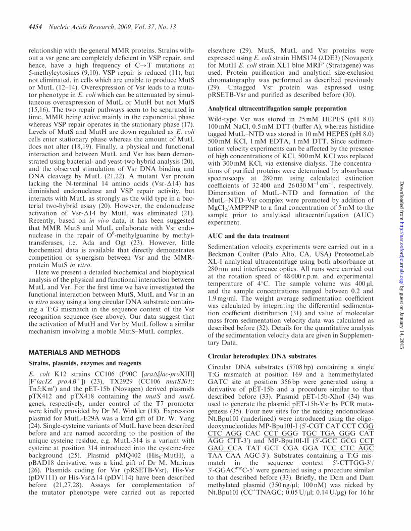
Circular DNA substrates (5708 bp) containing a singleT:G mismatch at position 169 and a hemimethylatedGATC site at position 356 bp were generated using aderivative of pET-15b and a procedure similar to thatdescribed before (33). Plasmid pET-15b-XhoI (34) wasused to generate the plasmid pET-15b-Vsr by PCR muta-genesis (35). Four new sites for the nicking endonucleaseNt.Bpu10I (underlined) were introduced using the oligo-deoxynucleotides MP-Bpu10I-I (50-CGT CAT CCT CGGCTC AGG CAC CCT GGG TGC TGA GGG CATAGG CTT-30) and MP-Bpu10I-II (50-GCC GCG CCTGAG CCA TAT GCT CGA GGA TCC CTC AGCTAA CAA AGC-30). Substrates containing a T:G mis-match in the sequence context 50-CTTGG-30/30-GGACmeC-50 were generated using a procedure similarto that described before (33). Briefly, the Dcm and Dammethylated plasmid (350 ng/ml; 100 nM) was nicked byNt.Bpu10I (CC^TNAGC; 0.05U/ml; 0.14U/mg) for 16 hr
4454 Nucleic Acids Research, 2009, Vol. 37, No. 13
by guest on January 14, 2015http://nar.oxfordjournals.org/
Dow
nloaded from

at 378C, followed by denaturation and re-annealing in thepresence of the 50-phosphorylated oligodeoxynucleotidesNt-G:T (50-TCA GCA CCT AGG GTG CC-30) and Nt-GATC (50-TGA GCC ATA TGC TCG AGG ATC CC-30)in 50-fold molar excess. After ligation with T4 DNA ligase(0.1U/ml) for 8 h at 258C, the reaction mixture was treatedwith (0.04U/ml) exonuclease I (Fermentas) and (0.05U/ml)exonuclease III (New England Biolabs) at 378C for 16 h toremove any nicked and linear DNA fragments.Optionally, the DNA was methylated at the hemimethy-lated GATC-site by dam methylatransferase (NewEngland Biolabs). Finally, the DNA was precipitatedwith one volume of isopropanol and 1/10 volume of 3Msodium acetate. The resulting covalently-closed circularDNA, containing a single hemimethylated Dcm site witha T:G mismatch and an additional hemimethylatedGATC site, was resuspended in 50 ml 10mM Tris–HCl,pH 7.9.
Thrombin-cleavage of MutL
Full-length MutL was cleaved with 20 units of thrombin(Sigma) per mg of MutL for 30min at 378C giving twodefined protein fragments, MutL–NTD (comprisingresidues 1–375) and MutL–CTD (comprising residues376–615). Once cleavage of MutL was complete, asjudged by SDS–PAGE, MutL–NTD was separated fromMutL–CTD by gel filtration using a L-6200A Merck-Hitachi HPLC system with a Superdex 75TM column equi-librated with 10mM HEPES/KOH pH7.9, 200mM KCl,1mM EDTA and 1mM DTT. MutL–NTD and MutL–CTD aliquots were snap-frozen in liquid nitrogen andstored at –708C.
Site-directed mutagenesis of Vsr
The gene encoding the C139S Vsr variant was generatedusing the oligodeoxynucleotide 50-CGA TCT GCG CGCTGG CCC CTT CGC CGC TGA TCC-30 in the mutagen-esis protocol described before (25) with pDV111 as thetemplate (27).The whole vsr gene was sequenced.
Mismatch-provoked activation of MutH
Ten nanomolars of heteroduplex DNA substrate (484 bp)containing a T:G mismatch at position 385 and a singleunmethylated GATC site at position 210 (34) of DNA wasincubated with 200 nM MutH, 1 mM MutL, 400 nM(monomer equivalents) MutS and the indicated concentra-tion of Vsr in 10mM Tris–HCl (pH 7.9), 5mM MgCl2,1mM ATP, 50 mg/ml BSA and 125mM KCl at 378C.MutH endonuclease activity was scored by the appearanceof cleaved products (analyzed by 6% polyacrylamide gelelectrophoresis). Alternatively, a circular heteroduplexDNA containing a single hemimethylated GATC sitesand a single T:G mismatch in the sequence contextCC5meAGG/CTTGG was used.
Photocrosslinking
Single-cysteine MutL variants (10 mM) were pre-incubatedin the presence or absence of 1mM ATP, ADP orAMPPNP for 30min on ice in 10mM Tris–HCl pH 7.5,
10mM MgCl2 and 125mM KCl (crosslinking buffer).Either single-cysteine MutL variants or Vsr were modifiedwith 100M excess of benzophenone-4-maleimide (BPM)for 30min at room temperature. The modification reac-tion was stopped with 5mM DTT. Modified proteinswere mixed with the respective interaction partners in a1:1 molar ratio (e.g. 2.5 mMVsr/5 mMMutL monomers) in25 ml and irradiated for 30min on ice at 366 nm with ahandheld UV lamp (Bachofer, Reutlinger) at a distanceof 5 cm. After addition of 6.25ml of 160mM Tris–HCl pH6.8, 5% (v/v), 2% (w/v) SDS, 40% (v/v) glycerol (SDS-loading buffer) the reaction mixture was subjected toSDS–PAGE.
Thiol–thiol crosslinking
Vsr and single-cysteine MutL variants were pre-incubatedon ice at 2.5mM each for 30min in the presence orabsence of ATP or AMPPNP in crosslinking buffer.Methanethiosulfonate MTS (methanethiosulfonate) cross-linkers of varying spacer arm length were purchasedfrom Toronto Research Chemicals. Stock solutions ofthe crosslinkers were made in DMSO and stored at–208C. Crosslinkers were diluted in DMSO and addedto the proteins resulting in an indicated molar excess ofcrosslinker:protein and a final concentration of 10%DMSO. After 5min, reactions were quenched with1mM N-ethylmaleimide prior to addition of SDS-loadingbuffer. Samples were analyzed as described above for thephotocrosslinking procedure.A scheme of all the crosslinking experiments is shown in
Supplementary Data.
RESULTS
AUC of the MutL-NTD and Vsr
The molecular matchmaker MutL coordinates MMR andcrosstalk with other repair processes by interacting with avariety of proteins. For the interaction between MutL andVsr, the N-terminal domain (NTD) of MutL had beendemonstrated to be sufficient in vivo (20). However, it isnot clear whether this interaction requires nucleotide bind-ing by MutL, as observed for the interaction betweenMutL and MutH (24). To answer this question the inter-actions between MutL and Vsr were assessed in an AUCsedimentation velocity experiment. Vsr distributed in theAUC cell mainly as a species with sedimentation coeffi-cients of 1.9 S (Figure 1A) which is in good agreementwith the sedimentation coefficient of 1.93 S calculatedfrom the crystal structure for a Vsr monomer withHYDROPRO (36); a second minor species of 2.9 S wasobserved, however, the identity of this species could notbe assigned (see Supplementary Data for additional infor-mation). The addition of AMPPNP, a non-hydrolysableanalogue of ATP to Vsr had minimal effect on the distri-bution of the species: Vsr remained basically a monomer(Figure 1B) although the distribution became broader.MutL–NTD was a single species with the peak centredaround 3.1 S (Figure 1C) indicative of a monomericform. The addition of 5mM AMPPNP resulted in theformation of second species with sedimentation coefficient
Nucleic Acids Research, 2009, Vol. 37, No. 13 4455
by guest on January 14, 2015http://nar.oxfordjournals.org/
Dow
nloaded from

of 5.2 S indicative of the formation of the MutL–NTDdimer (Figure 1D). In the absence of nucleotide, Vsr andMutL–NTD did not form any significant amount of com-plex (Figure 1E). However, in the presence of AMPPNPan additional species with sedimentation coefficient ofabout 6.1 S was observed concomitant with the disappear-ance of the MutL–NTD dimer. This species could be inter-preted as complex of a MutL-dimer with one or two Vsrmolecules (Figure 1F). In summary, our sedimentationvelocity analysis demonstrated that the interactionbetween MutL–NTD and Vsr is nucleotide dependent,as described previously for the MutL–NTD MutH inter-action (24,25).
Photocrosslinking MutL to Vsr
In order to gain insights into the structural arrangementof the MutL–Vsr complex, we employed a strategy usedpreviously for analyzing the MutL–MutH complex (25).To this end, we modified a series of single-cysteine MutLvariants (Figure 2) with benzophenone-4-maleimide
(BPM), (Figure 3A) and subsequently tested the benzo-phenone-modified proteins for photocrosslinking to Vsr(Figure 3B). Only MutL-314 was able to form a photo-crosslink with Vsr in an ATP-dependent manner(Figure 3B); this was verified by in-gel tryptic digestionand mass spectrometry analysis (data not shown).Interestingly, MutL-314 had been shown before to forma photocrosslink to MutH suggesting that the bindingsites of MutL for MutH and Vsr are overlapping or inclose proximity (25). Next we tested, whether MutL–NTD is also sufficient to allow the formation of the photo-crosslink with Vsr. To this end, MutL-314 was cleavedwith thrombin which removes the N-terminal His-tagand cleaves the protein between Pro-364 and Arg-375resulting in an N-terminal (MutL–NTD-314) and aC-terminal (MutL–CTD) fragment (37). After gel filtra-tion purification MutL–NTD-314 was modified withBPM followed by photocrosslinking to Vsr in the absenceor presence of ATP or AMPPNP essentially as describedfor full-length MutL (Figure 3C). In the absence of
Figure 1. Nucleotide-dependent interaction between Vsr and the N-terminal domain of MutL. General size distributions (expressed in s20,w) of Vsr,MutL–NTD and complexes of Vsr and MutL–NTD (2:1 molar ratio) as revealed by AUC. (A) Vsr, (B) Vsr with AMPPNP, (C) MutL-NTD, (D),MutL-NTD with AMPPNP, (E) Vsr and MutL-NTD, and (F) Vsr and MutL-NTD with AMPPNP. See ‘Materials and Methods’ section for details.
4456 Nucleic Acids Research, 2009, Vol. 37, No. 13
by guest on January 14, 2015http://nar.oxfordjournals.org/
Dow
nloaded from

nucleotide, no photocrosslink formation was observed(Figure 3C, lane 1). However, MutL–NTD-314 incubatedwith either ATP or AMPPNP was able to form a photo-crosslink product with Vsr (Figure 3C, lanes 2 and 3).In the absence of Vsr, no photocrosslink product wasobserved regardless whether MutL–NTD-314 was incu-bated in the absence or presence nucleotide (data notshown). These results are in agreement with the AUCdata (Figure 1), indicating that the C-terminal domainof MutL is not required for the physical interactionwith Vsr.
Photocrosslinking Vsr to MutL
Vsr contains five cysteine residues, of which three areinvolved in zinc binding (Cys-66, 73 and 117), one isburied (Cys-59) and only one is surface exposed (Cys-139) (Figure 4A). Chemical modification experimentsusing PEG-5000 maleimide indicated that one to twocysteine residues can be modified (Figure 4B). Notably,the variant Vsr-C139S, in which the surface exposedcysteine residue was exchanged to serine, displayed a sig-nificantly reduced but still observable labelling. This vari-ant retained endonuclease activity and its ability to inhibitthe MutL mediated mismatch-provoked activation ofMutH, similar to wild type Vsr (data not shown), indicat-ing that the mutation has not impaired the interactionwith either DNA or MutL. In order to test whether anycysteine residue of Vsr is in close proximity to MutL,we attempted to crosslink benzophenone-modified Vsrand Vsr-C139S to full-length MutL and MutL–NTD.In contrast to the photocrosslinking, the benzophenone-modified MutL–NTD to Vsr, benzophenone-modifiedVsr/Vsr-C139S did not form a photocrosslink withMutL–NTD (Figure 4C, lane 2). Crosslinked complexesto full-length MutL were observed with Vsr but not withVsr-C139S (Figure 4C, lanes 4 and 8) suggesting that Cys-139 is involved in the photocrosslinking reaction to MutL.
Figure 3. Photocrosslinking MutL-314 to Vsr. (A) Structure of thebenzophenone-4-maleimide (BPM). (B) Single-cysteine MutL variants(2.5 mM) modified with benzophenone at cysteine 314 or 327, respec-tively, were photocrosslinked in the presence of ATP to Vsr orVsr�14 (2.5mM) by irradiation at 366 nm for 30min. Reactions mix-tures were analyzed by SDS–PAGE. The identity of the crosslinkedspecies (L-Vsr) was verified by in-gel tryptic digest and massspectrometry analysis (data not shown). Photocrosslinks wereobserved only with MutL-314. MutL-327 is shown as an examplefor a MutL-variant, not forming a photocrosslink with Vsr. (C)Nucleotide dependence of photocrosslinking MutL–NTD-314 to Vsr.MutL–NTD-314 was incubated in the absence or presence of theindicated nucleotide for 30min prior photocrosslinking to Vsr essen-tially as described above.
Figure 2. Location of cysteine residues in MutL. Location of cysteineresidues in single-cysteine MutL variants mapped to the dimeric struc-tures of the MutL–NTD (25) (pdb code 1b63). All molecular graphicswere produced with PyMOL (www.pymol.org).
Nucleic Acids Research, 2009, Vol. 37, No. 13 4457
by guest on January 14, 2015http://nar.oxfordjournals.org/
Dow
nloaded from

Following photocrosslinking of benzophenone-modifiedVsr to a full-length MutL variant, labelled with a fluoro-phore at residue 297 in the NTD, limited proteolysisdemonstrated that the photocrosslink is formed to resi-dues of NTD (Supplementary Data). The absence of thephotocrosslink with MutL–NTD may be due to subtlestructural changes in the orientation of Vsr bound toMutL in the absence of the MutL–CTD thereby prevent-ing the photocrosslink formation. Taken together ourdata support the idea that in complex with MutL the sur-face exposed Cys-139 of Vsr is in proximity to residues ofthe NTD of MutL.
Chemical crosslinking MutL and Vsr
In order to get additional information about the orienta-tion of Vsr and MutL, we applied the cysteine–cysteinecrosslinking approach. We tested Vsr for crosslinking toselected single-cysteine variants of MutL in the presenceof AMPPNP using the long-range homobifunctionalcrosslinker MTS-11-MTS (Figure 5A). Variant MutL-327 formed a crosslink with both Vsr and the truncatedform (Vsr-�N), indicating that the loss of N-terminalamino acids did not influence the interaction betweenMutL and Vsr (Figure 5A, lane 3). A quantitative analysisof the crosslinking yields revealed that MutL-327 formedsignificantly more crosslinked products to Vsr than anyother MutL variant tested (Figure 5B). Similar to thephysical interaction analysis and the photocrosslinkingexperiments, chemical crosslinking was dependent on thepresence of nucleotide (ATP, AMPPNP or ADP) and wasnot observed in the absence of nucleotide (data notshown). These results suggest that one or more cysteineresidues clustering in two regions of Vsr, are in close prox-imity to residue 327 of MutL. Since Cys-139 was impor-tant for the formation of a photocrosslinked product frombenzophenone-modified Vsr to MutL, we asked whetherthis residue is also important for chemical crosslinking.In contrast to the photocrosslinking reaction, chemicalcrosslinking was still possible between MutL-327 andVsr-C139S indicating that Cys-139 is not essential for for-mation of the chemical crosslink (Figure 5A, lane 5). Weconcluded that one of the exposed residues from the zinc-binding site is involved in the chemical crosslink reaction.This assumption is supported by the fact that similarresults were obtained with the double mutant Vsr-C59S/C139S and that chemical crosslinking between MutL-327and either Vsr or Vsr-C139S was strongly impaired uponaddition of 100mM ZnCl2 (data not shown).
Vsr inhibits mismatch-provoked activation of MutH byMutS and MutL
Overexpression of plasmid-borne Vsr in E. coli has beenshown to be mutagenic (38), an effect attenuated byco-overexpression of MutL or MutH but not MutS (15).We asked whether the physical interaction between Vsrand MutL is sufficient to explain the inhibitory effect onMMR in vivo. Here, we tested the influence of Vsr on theinitial steps in DNA MMR, i.e. the mismatch-provokedactivation of the MutH endonuclease by MutS and MutL.To this end, we tested the cleavage of a linear DNA
Figure 4. Photocrosslinking of Vsr to MutL. (A) Structure of Vsr incomplex with DNA-product (pdb code 1cw0) (52) showing the locationof cysteine residues and the structural Zn-ion. The N-terminal residuesmissing in Vsr-�14 are highlighted in red. (B) Vsr and Vsr C139Swere modified with a 25-molar excess of PEG-MAL and analyzed bySDS–PAGE (see ‘Materials and Methods’ section). (C) Vsr and VsrC139S were modified with benzophenone-4-maleimide (BPM) and ana-lyzed for photocrosslinking to MutL–NTD or full-length MutL in theAMPPNP-bound form essentially as described in Figure 3. Note theintermolecular crosslink within Vsr (labelled with asterisks) andthe crosslink between Vsr and MutL (triangles).
4458 Nucleic Acids Research, 2009, Vol. 37, No. 13
by guest on January 14, 2015http://nar.oxfordjournals.org/
Dow
nloaded from

substrate containing a single unmethylated GATC site anda T:G mismatch in a sequence context not related to theVsr-recognition sequence (Figure 6A). Upon incubationwith MutH (200 nM), MutL (1mM) and MutS (400 nM),this DNA substrate is nicked twice in both unmethylatedDNA strands resulting in two linear products (Figure 6B).Under the conditions used, the DNA cleavage rate byMutH was >20-fold lower in the absence of MutS,MutL, or the T:G mismatch (data not shown). Additionof about 200 nM levels of Vsr inhibited the reaction by50% and Vsr in excess over MutL almost completely abol-ished DNA cleavage (Figure 6B). Similar results were
obtained using a circular heteroduplex DNA containinga single T:G mismatch and a hemimethylated GATC-site(data not shown). Next we tested the accessibility of theGATC site in the presence of MutS, MutL and Vsr bymonitoring the DNA cleavage at the GGATCC site byBamHI. No inhibitory effect on BamHI cleavage by Vsrwas observed, ruling out blocking of the GATC-site bynon-specific DNA binding of Vsr (data not shown).Using a different circular heteroduplex DNA substratewith a BamHI-site only 4 bp away from the T:G mismatchbinding of MutS is blocking the action of BamHI whereasVsr is not, indicating that Vsr is not strongly binding tothis mismatch. However, Vsr is inhibiting the mismatch-provoked activation of MutH on this substrate similar asfor the linear heteroduplex DNA shown in Figure 6(Supplementary Data). Finally, we tested the inhibitoryaction of Vsr on the MutS and mismatch-independentactivation of MutH by MutL that is observed only atlow ionic strength (25,37,39). Under the conditions used,the rate of DNA cleavage by MutH (500 nM) is0.24min�1. Upon addition of 2 mM Vsr this rate decreased
Figure 5. Thiol–thiol crosslinking: (A) The homobifunctional crosslin-ker MTS-11-MTS was used to crosslink MutL-327 in its AMPPNPform to Vsr or Vsr-C139S, respectively. (B) Quantitative analysis ofcrosslinking the indicated single-cysteine MutL variants and Vsr withMTS-11-MTS in the presence of AMPPNP. Mean crosslink yields �SEare based on three independent experiments.
Figure 6. Inhibition of mismatch-provoked MutS/MutL-dependentactivation of MutH by Vsr: (A) Scheme for double nicking of a T:Gmismatch DNA containing a single unmethylated GATC-site (484 bp)by MutH resulting in two cleavage products (209 and 275 bp). (B) T:Gmismatch DNA (484 bp, 10 nM) containing a single unmethylatedGATC-site was incubated with 200 nM MutH, 400 nM MutS and1mM MutL in the presence of the indicated concentration of Vsr.Apparent first-order rate constants (kst) for the appearance of thecleavage products are plotted against concentration of Vsr.
Nucleic Acids Research, 2009, Vol. 37, No. 13 4459
by guest on January 14, 2015http://nar.oxfordjournals.org/
Dow
nloaded from

almost 10-fold to 0.03min–1 (data not shown). In conclu-sion, a competition between Vsr and MutS for mismatchbinding is not necessary for the interference by Vsr ofMutH mediated DNA cleavage, which is a consequenceof competition between Vsr and MutH for bindingto MutL.
Vsr endonuclease is stimulated by MutS and MutL
The efficiency of Vsr repair in vivo is influenced by thepresence of both MutL and MutS (40). The present andprevious studies have provided evidence for a physical andfunctional interaction between Vsr and MutL, but littlein vitro data is available for the role of MutS. Therefore,we analyzed the role of MutS and ATP hydrolysis on theactivity of Vsr using a covalently closed circular DNAsubstrate containing a single T:G mismatch in the recog-nition sequence of Vsr (Figure 7A). Our analysis demon-strates that the endonuclease activity of Vsr is greatlystimulated in the presence of MutS, MutL and ATP(Figure 7B). Under the experimental conditions contain-ing near physiological salt concentrations (125mM KCl)neither MutL nor MutS alone were able to stimulate theVsr endonuclease (Figure 7B, lanes 3 and 4). Moreover,the stimulation was dependent on the presence of ATP
which cannot be substituted by ADP (compare lanes 6and 8). In the absence of Vsr, no cleavage was observedwith MutS, MutL and ATP (lane 10). Furthermore, DNAsubstrates without a mismatch or a mismatch in a differentsequence context were not cleaved by Vsr indicating theobserved DNA nicking was specific for Vsr (data notshown). AUC and crosslinking revealed that the interac-tion of Vsr and MutL required nucleotide binding but nothydrolysis. Therefore, we asked whether the MutS-depen-dent stimulation of Vsr requires the ATPase activity ofMutL. To this end the ATP-binding proficient butATPase impaired variant MutL-E29A was tested for itsability to stimulate the Vsr endonuclease (Figure 8) (24).In contrast to the results obtained with wild-type MutLlittle or no stimulation of DNA nicking by Vsr wasobserved with this variant suggesting that ATP-bindingand ATP-hydrolysis are required for a functional interac-tion between MutL and Vsr under physiological saltconcentrations.
DISCUSSION
Many DNA repair systems have overlapping substratespecificities, giving rise to the need to co-ordinate theiractivities. The repair of T:G mismatches by the MMRand the VSP systems falls into this category. MMR isinhibited by excess Vsr, reflecting a physical and func-tional interaction between MutL and Vsr, as shown byseveral studies (20–22,40). Our in vitro data, using sedi-mentation velocity analysis (Figure 1), clearly showthe formation of a protein–protein complex betweenMutL and Vsr, supporting the in vivo observations.Indeed, using photochemical and chemical crosslinking
Figure 7. Stimulation of the Vsr endonuclease activity requires MutS,MutL and ATP-hydrolysis: (A) A covalently closed circular (ccc) DNAcontaining a T:G mismatch (shown in bold) was generated as describedin Materials and Methods. The recognition sequence of Vsr is boxed.(B) DNA nicking was monitored after incubation 15 nM DNA for5min at 378C in the absence or presence of the indicated compounds:Vsr (75 nM), MutS (400 nM), MutL (400 nM) and ATP or ADP(1mM). ccc, covalently closed circle; oc, open circle; li, linear.
Figure 8. Stimulation of the Vsr endonuclease activity requires ATPhydrolysis by MutL: Nicking of 15 nM covalently closed circularDNA containing a T:G mismatch within the Vsr recognitionsequence (see Figure 7) was monitored after incubation for 5min at378C in the presence of increasing concentrations of Vsr (0, 1, 3, 9, 27,81, 243, 729, 2187 nM), 1mM ATP in the (A) absence or presenceof MutS (100 nM) and (B) MutL (200 nM) or (C) MutL-E29A(200 nM).
4460 Nucleic Acids Research, 2009, Vol. 37, No. 13
by guest on January 14, 2015http://nar.oxfordjournals.org/
Dow
nloaded from

(Figures 3–5) we could map the interaction site of MutLfor Vsr to a site overlapping with the binding site forMutH (25,41). This finding supports a model of competi-tion for a common binding site on MutL between theMMR MMR proteins MutH and the VSP protein Vsr,explaining the inhibitory effect of Vsr overexpression onMMR in vivo, which is reversed by overproduction ofeither MutL or MutH (37). Indeed, we could demonstratethat Vsr does not influence initial steps in DNAMMR, i.e.ternary complex formation between DNA, MutS andMutL (data not shown). However, Vsr efficiently inhibitsthe mismatch-provoked MutS and MutL-dependent acti-vation of MutH (Figure 6 and Supplementary Data), sug-gesting that the mutagenic effect of Vsr arises as a directconsequence of competition between MutH and Vsr forbinding to MutL, postulated before (40). As expected,MutH inhibited the activation of Vsr by MutS andMutL (Supplementary Data).
The stimulatory effect of both MutS and MutL on VSPrepair has been puzzling, especially since structural analy-sis of the MutS-DNA and Vsr-DNA complexes clearlyrevealed that MutS and Vsr cannot be bound simulta-neously to the same DNA mismatch (40). In the presentstudy we demonstrated that the physical interactionbetween MutL and Vsr is important but not sufficientfor an enhancement of Vsr activity under physiologicalionic strength. Our analysis revealed that both MutLand MutS are necessary and sufficient for the stimulationof Vsr endonuclease and that this functional interactionrequires ATP hydrolysis (Figure 7), similar to the mis-match-provoked activation of MutH (42). Whereas thephysical interaction between MutL and Vsr could beobserved in the absence of ATP-hydrolysis and with theN-terminal domain of MutL (Figure 1), the functionalinteraction, resulting in activation of Vsr, requires ATP-hydrolysis, as the ATPase deficient MutL-E29A hasalmost completely lost its stimulatory ability (Figure 8).Similarly, MutL-E29A is not able to support the mis-match-provoked activation of MutH at physiologicalionic strength (125mM KCl). Our conclusions are atodds with experiments conducted at low ionic strength(20mM NaCl), suggesting a role for ATP-hydrolysis byMutL only for steps after DNA incision by MutH andloading of DNA helicase II (43). However, the conclusionsare fully consistent with data reporting the inability ofMutS to further increase the MutH activation by MutL-E29A at higher ionic strength (44). Notably, at lower saltconcentration (e.g. 50mM KCl) MutL-E29A is able toactivate MutH in a MutS independent manner as similarlydescribed for MutL wild type (data not shown) (44).
Several conclusions can be drawn from the presentstudy. MutL shares a common or overlapping interactionsite for two of its effector proteins, MutH and Vsr. Theinteraction site for MutH is located between the N- andC-terminal domains reminiscent to the position of theclient binding site of Hsp90 which shares structuralhomology to MutL in the ATPase domain (45,46).Although binding of the effector proteins (Vsr or MutH)to the MutL-NTD is dependent on nucleotide binding butnot hydrolysis, this is not true for the functional interac-tion. At physiological ionic strength, the activation of Vsr
and also MutH (data not shown) in a mismatch andMutS-dependent manner requires ATP-hydrolysis byMutL (Figure 8). Finally, since mismatch bindingby MutS and Vsr is mutually exclusive, only models invol-ving a mobile rather a stationary MutS that doesnot remain bound at the mismatch are consistent withthe observed MutS–MutL-dependent activation of Vsr(7,47). Similarly, MMR has been reported to be efficienteven if the mismatch and the GATC-site are separated byonly 4 bp, a distance which is too short to allow simulta-neous binding of MutS at the mismatch and MutH at theGATC-site (48).We envision at least two possible ways that MutS and
MutL could allow access of Vsr to the T/G mismatch, orMutH to adjacent GATC sites. One possibility is thatMutS and MutL leave the site of the mismatch beforethe arrival of the effector proteins, as has been proposedpreviously (40). The only way to explain the stimulatoryeffect of the MutS–MutL complex is to postulate that itchanges the conformation of the DNA to make it easier tobind. Our data show that Vsr and MutL interact, whichmakes this scenario unlikely. The other possibility is thatMutS and MutL act as damage sensors, recruiting any oneof a number of effectors to the site. The resulting ATP-dependent interactions between sensor(s) and effector(s)both displace the sensors and activate the effectors. Sucha model is supported by recent in vivo studies showing thatMutL recruits the Vsr and MutH endonucleases inresponse to DNA damage (49). If the recruited proteinis able to mediate repair, the MutS–MutL complexwould dissociate. If the recruited protein is unable tomediate repair, the MutS–MutL complex would remain,allowing subsequent recruitment of another effector. Sucha model would account for the fact that MutS and MutLenhance Vsr activity, but are not required for VSP repair.The inhibitory effect of Vsr on MMR explains why reg-
ulation of Vsr expression is required for proper MMRfunction (and vice versa for MutH in stationary phasealthough). Indeed, MutH is inhibiting the activation ofVsr by MutS and MutL (Supplementary Data).However, beside this, it is possible that Vsr is kept lowin growing cells not because it interferes with MMR butbecause it can stimulate CTAGG-to-CCAGG mutations,as shown before (38).The question of when and how Vsr or MutH enter and
leave the repair pathway needs to be addressed before themechanistic details of competition and cooperationbetween various proteins interacting with MutL can beunderstood. The observed similarities between the stimu-lation of Vsr and MutH by MutS and MutL offers newroutes for future in-depth analysis of the activation ofeffector proteins in the MMR pathways in a comparativemanner. Only one Vsr homolog from another organismhas been analyzed but not in the context of MMR (50).Since homologs of a Vsr endonuclease are present in 198bacterial species [REFSEQ 01-23-09 (51)] from across thebacterial kingdom in contrast to MutH homologs that arealmost exclusively found in g-proteobacteria (currently130 in REFSEQ), it will be important to understandwhether the observed physical and functional interactionbetween Vsr and MutL in E. coli is a general DNA repair
Nucleic Acids Research, 2009, Vol. 37, No. 13 4461
by guest on January 14, 2015http://nar.oxfordjournals.org/
Dow
nloaded from

pathway used also in other bacteria. The functional assayinvolving the activation of Vsr by MutS and MutLdescribed in the present study might be exploited for themechanistic analysis of DNA MMR pathways in bacterialsystems.
SUPPLEMENTARY DATA
Supplementary Data are available at NAR Online.
ACKNOWLEDGEMENTS
The technical assistance of Ina Dern is gratefully acknowl-edged. We thank Ines Winkler for critical reading themanuscript.
FUNDING
German Science Foundation [FR1495/3-2 and GRK1384to PF], UK BBSRC [13/E19804 to BAC, PhD studentshipto SLE] and the Canadian Institutes for Health Research[to C.C.]. Funding for open access charge: GermanScience Foundation GRK1384.
Conflict of interest statement. None declared.
REFERENCES
1. Slupphaug,G., Kavli,B. and Krokan,H.E. (2003) The interactingpathways for prevention and repair of oxidative DNA damage.Mutat. Res., 531, 231–251.
2. Bai,H. and Lu,A.L. (2007) Physical and functional interactionsbetween Escherichia coli MutY glycosylase and mismatch repairprotein MutS. J. Bacteriol., 189, 902–910.
3. York,S.J. and Modrich,P. (2006) Mismatch repair-dependentiterative excision at irreparable O6-methylguanine lesions in humannuclear extracts. J. Biol. Chem., 281, 22674–22683.
4. Bhagwat,A.S. and Connolly,B. (2006) In Siede,W., Kow,Y.W. andDoetsch,P.W. (eds), DNA Damage Recognition. Marcel Dekker Inc,New York, pp. 476–492.
5. Yang,W. (2006) Poor base stacking at DNA lesions may initiaterecognition by many repair proteins. DNA Repair, 5, 654–666.
6. Hennecke,F., Kolmar,H., Brundl,K. and Fritz,H.J. (1991) Thevsr gene product of E. coli K-12 is a strand- and sequence-specificDNA mismatch endonuclease. Nature, 353, 776–778.
7. Iyer,R.R., Pluciennik,A., Burdett,V. and Modrich,P.L. (2006)DNA mismatch repair: functions and mechanisms. Chem. Rev., 106,302–323.
8. Lieb,M. and Bhagwat,A.S. (1996) Very short patch repair: reducingthe cost of cytosine methylation. Mol. Microbiol., 20, 467–473.
9. Lieb,M. (1991) Spontaneous mutation at a 5-methylcytosinehotspot is prevented by very short patch (VSP) mismatch repair.Genetics, 128, 23–27.
10. Petropoulos,L., Vidmar,J.J., Passi,E. and Cupples,C.G. (1994)A simple assay for monitoring the mutagenic effects of5-methylcytosine deamination in Escherichia coli. Mut. Res., 304,181–185.
11. Bell,D.C. and Cupples,C.G. (2001) Very-short-patch repair inEscherichia coli requires the dam adenine methylase. J. Bacteriol.,183, 3631–3635.
12. Jones,M., Wagner,R. and Radman,M. (1987) Mismatch repair andrecombination in E. coli. Cell, 50, 621–626.
13. Lieb,M. (1987) Bacterial genes mutL, mutS, and dcm participate inrepair of mismatches at 5-methylcytosine sites. J. Bacteriol., 169,5241–5246.
14. Zell,R. and Fritz,H.J. (1987) DNA mismatch-repair in Escherichiacoli counteracting the hydrolytic deamination of 5-methyl-cytosineresidues. EMBO J., 6, 1809–1815.
15. Macintyre,G., Doiron,K.M. and Cupples,C.G. (1997) The Vsrendonuclease of Escherichia coli: an efficient DNA repair enzymeand a potent mutagen. J. Bacteriol., 179, 6048–6052.
16. Lieb,M., Rehmat,S. and Bhagwat,A.S. (2001) Interaction of MutSand Vsr: some dominant-negative mutS mutations that disablemethyladenine-directed mismatch repair are active in very-short- patch repair. J. Bacteriol., 183, 6487–6490.
17. Macintyre,G., Pitsikas,P. and Cupples,C.G. (1999) Growthphase-dependent regulation of Vsr endonuclease may contributeto 5-methylcytosine mutational hot spots in Escherichia coli.J. Bacteriol., 181, 4435–4436.
18. Feng,G., Tsui,H.C. and Winkler,M.E. (1996) Depletion of thecellular amounts of the MutS and MutH methyl-directed mismatchrepair proteins in stationary-phase Escherichia coli K-12 cells.J. Bacteriol., 178, 2388–2396.
19. Tsui,H.C., Feng,G. and Winkler,M.E. (1997) Negative regulationof mutS and mutH repair gene expression by the Hfq and RpoSglobal regulators of Escherichia coli K-12. J. Bacteriol., 179,7476–7487.
20. Mansour,C.A., Doiron,K.M. and Cupples,C.G. (2001)Characterization of functional interactions among the Escherichiacoli mismatch repair proteins using a bacterial two-hybrid assay.Mutat. Res., 485, 331–338.
21. Monastiriakos,S.K., Doiron,K.M., Siponen,M.I. and Cupples,C.G.(2004) Functional interactions between the MutL and Vsr proteinsof Escherichia coli are dependent on the N-terminus of Vsr. DNARepair, 3, 639–647.
22. Drotschmann,K., Aronshtam,A., Fritz,H.J. and Marinus,M.G.(1998) The Escherichia coli MutL protein stimulates binding ofVsr and MutS to heteroduplex DNA. Nucleic Acids Res., 26,948–953.
23. Rye,P.T., Delaney,J.C., Netirojjanakul,C., Sun,D.X., Liu,J.Z. andEssigmann,J.M. (2008) Mismatch repair proteins collaborate withmethyltransferases in the repair of O(6)-methylguanine. DNARepair, 7, 170–176.
24. Ban,C., Junop,M. and Yang,W. (1999) Transformation of MutLby ATP binding and hydrolysis: a switch in DNA mismatch repair.Cell, 97, 85–97.
25. Giron-Monzon,L., Manelyte,L., Ahrends,R., Kirsch,D., Spengler,B.and Friedhoff,P. (2004) Mapping protein-protein interactionsbetween MutL and MutH by cross-linking. J. Biol. Chem., 279,49338–49345.
26. Loh,T., Murphy,K.C. and Marinus,M.G. (2001) Mutationalanalysis of the MutH protein from Escherichia coli. J. Biol. Chem.,276, 12113–12119.
27. Cupples,C.G. and Macintyre,G. (2000) The E. coli Vsr endonu-clease. Assaying activity in vitro and in vivo. Methods Mol. Biol.,152, 63–73.
28. Turner,D.P. and Connolly,B.A. (2000) Interaction of the E. coliDNA G:T-mismatch endonuclease (vsr protein) with oligonucleo-tides containing its target sequence. J. Mol. Biol., 304, 765–778.
29. Manelyte,L., Urbanke,C., Giron-Monzon,L. and Friedhoff,P.(2006) Structural and functional analysis of the MutS C-terminaltetramerization domain. Nucleic Acids Res., 34, 5270–5279.
30. Elliott,S.L., Brazier,J., Cosstick,R. and Connolly,B.A. (2005)Mechanism of the Escherichia coli DNA T:G-mismatchendonuclease (Vsr protein) probed with thiophosphate-containingoligodeoxynucleotides. J. Mol. Biol., 353, 692–703.
31. Schuck,P. (2003) On the analysis of protein self-association bysedimentation velocity analytical ultracentrifugation. Anal.Biochem., 320, 104–124.
32. Brown,P.H. and Schuck,P. (2006) Macromolecular size-and-shapedistributions by sedimentation velocity analytical ultracentrifuga-tion. Biophys. J., 90, 4651–4661.
33. Wang,H. and Hays,J.B. (2001) Simple and rapid preparation ofgapped plasmid DNA for incorporation of oligomers containingspecific DNA lesions. Mol. Biotechnol., 19, 133–140.
34. Thomas,E., Pingoud,A. and Friedhoff,P. (2002) An efficient methodfor the preparation of long heteroduplex DNA as substrate formismatch repair by the Escherichia coli MutHLS system. Biol.Chem., 383, 1459–1462.
4462 Nucleic Acids Research, 2009, Vol. 37, No. 13
by guest on January 14, 2015http://nar.oxfordjournals.org/
Dow
nloaded from

35. Kirsch,R.D. and Joly,E. (1998) An improved PCR-mutagenesisstrategy for two-site mutagenesis or sequence swapping betweenrelated genes. Nucleic Acids Res., 26, 1848–1850.
36. Garcia De La Torre,J., Huertas,M.L. and Carrasco,B. (2000)Calculation of hydrodynamic properties of globular proteins fromtheir atomic-level structure. Biophys. J., 78, 719–730.
37. Ban,C. and Yang,W. (1998) Crystal structure and ATPase activityof MutL: implications for DNA repair and mutagenesis. Cell, 95,541–552.
38. Doiron,K.M., Viau,S., Koutroumanis,M. and Cupples,C.G. (1996)Overexpression of vsr in Escherichia coli is mutagenic. J. Bacteriol.,178, 4294–4296.
39. Hall,M.C., Jordan,J.R. and Matson,S.W. (1998) Evidence for aphysical interaction between the Escherichia coli methyl- directedmismatch repair proteins MutL and UvrD. EMBO J., 17,1535–1541.
40. Bhagwat,A.S. and Lieb,M. (2002) Cooperation and competition inmismatch repair: very short-patch repair and methyl-directedmismatch repair in Escherichia coli. Mol. Microbiol., 44, 1421–1428.
41. Kosinski,J., Steindorf,I., Bujnicki,J.M., Giron-Monzon,L. andFriedhoff,P. (2005) Analysis of the quaternary structure of theMutL C-terminal domain. J. Mol. Biol., 351, 895–909.
42. Lahue,R.S., Su,S.S. and Modrich,P. (1987) Requirement ford(GATC) sequences in Escherichia coli mutHLS mismatchcorrection. Proc. Natl Acad. Sci. USA, 84, 1482–1486.
43. Robertson,A.B., Pattishall,S.R., Gibbons,E.A. and Matson,S.W.(2006) MutL-catalyzed ATP hydrolysis is required at a post-UvrDloading step in methyl-directed mismatch repair. J. Biol. Chem.,281, 19949–19959.
44. Junop,M.S., Yang,W., Funchain,P., Clendenin,W. and Miller,J.H.(2003) In vitro and in vivo studies of MutS, MutL and MutHmutants: correlation of mismatch repair and DNA recombination.DNA Repair, 2, 387–405.
45. Pearl,L.H. and Prodromou,C. (2006) Structure and mechanism ofthe Hsp90 molecular chaperone machinery. Annu. Rev. Biochem.,75, 271–294.
46. Ahrends,R., Kosinski,J., Kirsch,D., Manelyte,L., Giron-Monzon,L., Hummerich,L., Schulz,O., Spengler,B. and Friedhoff,P.(2006) Identifying an interaction site between MutH and theC-terminal domain of MutL by crosslinking, affinity purification,chemical coding and mass spectrometry. Nucleic Acids Res., 34,3169–3180.
47. Li,G.M. (2008) Mechanisms and functions of DNA mismatchrepair. Cell Res., 18, 85–98.
48. Bruni,R., Martin,D. and Jiricny,J. (1988) d(GATC) sequencesinfluence Escherichia coli mismatch repair in a distance-dependentmanner from positions both upstream and downstream of themismatch. Nucleic Acids Res., 16, 4875–4890.
49. Polosina,Y.Y., Mui,J., Pitsikas,P. and Cupples,C.G. (2009) TheEscherichia coli mismatch repair protein MutL recruits theVsr and MutH endonucleases in response to DNA damage.J. Bacteriol. doi:101128/JB.00066-09.
50. Laging,M., Lindner,E., Fritz,H.J. and Kramer,W. (2003) Repair ofhydrolytic DNA deamination damage in thermophilic bacteria:cloning and characterization of a Vsr endonuclease homologfrom Bacillus stearothermophilus. Nucleic Acids Res., 31,1913–1920.
51. Pruitt,K.D., Tatusova,T. and Maglott,D.R. (2007) NCBIreference sequences (RefSeq): a curated non-redundant sequencedatabase of genomes, transcripts and proteins. Nucleic Acids Res.,35, D61–D65.
52. Tsutakawa,S.E., Jingami,H. and Morikawa,K. (1999)Recognition of a TG mismatch: the crystal structure of veryshort patch repair endonuclease in complex with a DNA duplex.Cell, 99, 615–623.
Nucleic Acids Research, 2009, Vol. 37, No. 13 4463
by guest on January 14, 2015http://nar.oxfordjournals.org/
Dow
nloaded from
Copyright © 2022 FDOKUMEN