Characterization of Escherichia coli harboring colibactin ...
Upload
independentCategory
view
0download
0
Research in Microbiology 162 (2011) 108e116www.elsevier.com/locate/resmic
Environmental factors affecting indole production in Escherichia coli
Thi Hiep Han a, Jin-Hyung Lee a, Moo Hwan Cho a, Thomas K. Wood b, Jintae Lee a,*
a School of Chemical Engineering, Yeungnam University, Gyeongsan-si, Gyeonsangbuk-do 712-749, South KoreabDepartment of Chemical Engineering, 220 Jack E. Brown Building, Texas A&M University, College Station, TX 77843-3122, USA
Received 26 June 2010; accepted 17 September 2010
Available online 8 December 2010
Abstract
A variety of both Gram-positive and Gram-negative bacteria produce large quantities of indole as an intercellular signal in microbialcommunities. Biosynthesis of indole is well-studied, and while carbon sources and amino acids are important environmental cues for indoleproduction in Escherichia coli, other environmental factors affecting indole production for this strain are less clear. This study demonstrates thatthe environmental cue pH is an important factor for indole production that further controls biofilm formation of E. coli. Moreover, E. coliproduced a higher level of extracellular indole in the presence of the antibiotics ampicillin and kanamycin, and the increased indole enhancedcell survival during antibiotic stress. Additionally, we found here that temperature is another important factor for indole production; E. coliproduces and accumulates a large amount of indole at 50 �C, even at low cell densities. Overall, our results suggest that indole is a stablebiological compound, and E. coli may utilize indole to protect itself against other microorganisms.� 2010 Institut Pasteur. Published by Elsevier Masson SAS. All rights reserved.
Keywords: Indole; Signal molecule; Escherichia coli; Environmental factors; Antibiotics
1. Introduction
In nature, bacteria are most commonly found in complexcommunities. Many bacteria have developed their ability tosense the local environment, such as the nutritional limitation,their population, the presence of toxic chemicals from otherbacteria and host signals. Therefore, it is important to coor-dinate their behavior in order to adapt and survive in envi-ronmental niches. For example, bacteria have developedsignaling systems, such as intercellular signaling systems(Waters and Bassler, 2005) and diverse two-component regu-latory systems (Mitrophanov and Groisman, 2008) tosynchronize cellular behavior.
Among intercellular signal molecules, indole is oftenproduced by a variety of both Gram-positive and Gram-negative bacteria (to date, more than 85 species) (Lee and Lee,
* Corresponding author. Tel.: þ82 53 810 2533; fax: þ82 53 810 4631.
E-mail address: [email protected] (J. Lee).
0923-2508/$ - see front matter � 2010 Institut Pasteur. Published by Elsevier Ma
doi:10.1016/j.resmic.2010.11.005
2010). Importantly, recent studies have demonstrated manybiological functions of indole, such as drug resistance inEscherichia coli (Hirakawa et al., 2005; Lee et al., 2010),plasmid stability in E. coli (Chant and Summers, 2007),virulence control in pathogenic E. coli (Anyanful et al., 2005;Hirakawa et al., 2009), and biofilm formation in E. coli (DiMartino et al., 2003; Lee et al., 2007b) as well as in Vibriocholerae (Mueller et al., 2009). Interestingly, indole controlsphenotypes of other bacteria that cannot produce indole. Forexample, indole decreases the growth of fungus Aspergillusniger (Kamath and Vaidyanathan, 1990), increases drugresistance in Salmonella enterica (Nikaido et al., 2008), andattenuates virulence in Pseudomonas aeruginosa (Lee et al.,2009). Moreover, indole significantly affects gene expressionfor human epithelial cells leading to tighter cell-junctions andincreases in beneficial cytokines (Bansal et al., 2010).
An early report showed that Bacillus coli (E. coli) andAsiatic cholera (Vibrio cholerae) produced indole duringa stationary cell growth phase in 1897 (Smith, 1897). Indolebiosynthesis in E. coli has been investigated over many
sson SAS. All rights reserved.
109T.H. Han et al. / Research in Microbiology 162 (2011) 108e116
decades (Newton and Snell, 1965; Botsford and DeMoss,1971). In E. coli, indole is produced by tryptophanase(TnaA; EC 4.1.99.1) that can reversibly convert tryptophaninto indole, pyruvate, and ammonia (Newton and Snell, 1965)in the tryptophan pathway in E. coli (Lee et al., 2007b). E. coliuses several mechanisms (repression, transcription attenuation,and feedback inhibition) to regulate the expression of thetryptophan (trpABCDE ) and tna operons (tnaCAB) in trypto-phan metabolism (Gong and Yanofsky, 2002; Yanofsky et al.,1991; Lee et al., 2007b). Environmental conditions andcomposition of media critically influence the level of extra-cellular indole. For example, the extracellular indole concen-tration is cell population density-dependent in E. coli (Wanget al., 2001; Kobayashi et al., 2006). Extracellular indolereached 0.5 mM in a rich medium in the stationary phase sothat it has been called a stationary phase signal molecule(Wang et al., 2001; Kobayashi et al., 2006). In addition,glucose repressed indole biosynthesis (John and Wyeth, 1919)due to catabolic repression of tnaA (Botsford and DeMoss,1971). Different carbon sources also influence the accumula-tion of extracellular indole (John and Wyeth, 1919; Botsfordand DeMoss, 1971). Additionally, temperature and pH affectindole biosynthesis in E. coli. A low pH inhibited indoleproduction in E. coli (John and Wyeth, 1919), and TnaA wasone of the most induced proteins at pH 9.0 (Blankenhorn et al.,1999). Gene expression of tnaAB was induced in a tempera-ture sensitive E. coli rnpA mutant by temperature shifting from30 �C to 43 �C (Li et al., 2003), and E. coli lost the ability ofindole biosynthesis at 44.5 �C (Bueschkens and Stiles, 1984).
Although the importance of indole signaling in microbialcommunities (Lee and Lee, 2010) as well as in the humanimmune system (Bansal et al., 2010; Wikoff et al., 2009) hasbeen recently demonstrated, environmental factors thatcontribute to and regulate indole production remain poorlydefined. Recently, several transcriptomic studies provide
Table 1
Transcriptomic data for gene expression of tnaA in wild-type E. coli with various en
in the same reference. Studies with mutant strains are not included here.
Factors Conditions
Culture time 15 h Suspension cells vs. 4 h suspension cells in LB
Glucose No glucose vs. 0.4% glucose in LB
Carbon sources Glycerol vs. glucose in a minimal medium
Succinate vs. glucose in a minimal medium
Acetate vs. glucose in a minimal medium
Amino acids Alanine vs. glucose in a minimal medium
Proline vs. glucose in a minimal medium
50 mg/mL L-tryptophan for 60 min in a minimal
medium with 0.2% glucose
Acid pH 5.0 vs. pH 7.0 in LB
Base pH 8.7 vs. pH 5.0 in LB
10 mM Trimethylamine N-oxide vs. none in LB
Pressure 0.1 Mpa vs. 30 MPa in LB
Oxidative stress 35 ppm Paraquat vs. none for 60 min in LB
1 mM Hydrogen peroxide vs. none in LB
Heavy metals 1.2 mM Cadmium (II) vs. none in LB
4 mM Nickel (II) vs. none in LB
Aeration Aerobic vs. anaerobic for 60 min in a minimal
medium with 30 mM glucose
informative data on the expression of tnaA in response tovarious environmental cues in E. coli (Table 1). Hence, theoverall objective of this study is to identify the importantenvironmental factors that control indole production as well ascell growth to better understand indole signaling in microbialcommunities.
2. Materials and methods
2.1. Bacterial strains, materials and growth ratemeasurements
E. coli K-12 BW25113 (lacIq rrnBT14 DlacZWJ16 hsdR514DaraBA-DAH33 DrhaBADLD78) and its isogenic tnaA mutant(Baba et al., 2006) that does not produce indole (Lee et al., 2008)were used. LuriaeBertani (LB) was used as a basic medium forgrowth unless indicated. Indole, ampicillin, kanamycin, chlor-amphenicol, N-hexanoyl-DL-homoserine lactone, N-(3-oxote-tradecanoyl)-L-homoserine lactone, N-(3-oxododecanoyl)-L-homoserine, N-(3-oxooctanoyl)-L-homoserine lactone, paraquat,cadmium chloride, sodium dodecyl sulfate (SDS), and 4-(dime-thylamino)-benzaldehyde were purchased from SigmaeAldrichCo. (Missouri, USA). Hydrogen peroxide (H2O2), HCl, NaOH,NaCl, FeCl3, ethanol, methanol, and dimethyl sulfoxide (DMSO)were purchased fromDuksan Pure Chemical Co. (Ansan, Korea).Bacterial strains were initially streaked from �80 �C glycerolstocks on LB plates, and a fresh single colonywas inoculated intoLB medium (25 mL) in 250-mL flasks and routinely cultured at250 rpm at 37 �C unless indicated. Overnight cultures werediluted 1:100 using LB medium. For cell growth measurements,the optical density was measured at 600 nm (OD600) witha spectrophotometer (UV-160, Shimadzu, Japan).When thevalueof OD600 was above 0.7, culture sample was diluted to fit ina linear range of 0.2 and 0.7. The specific growth rates weredetermined by measuring OD600 and calculated by using the
vironmental factors. Each comparison was made with its own control condition
Fold change of tnaA Reference
�588 (Domka et al., 2007)
100 (Gosset et al., 2004)
5 (Liu et al., 2005)
5 (Liu et al., 2005)
13 (Liu et al., 2005)
6 (Liu et al., 2005)
12 (Liu et al., 2005)
18 (Khodursky et al., 2000)
�14 (Maurer et al., 2005)
16 (Maurer et al., 2005)
15 (Bordi et al., 2003)
�25 (Ishii et al., 2005)
20 (Kim et al., 2005)
18 (Zheng et al., 2001)
�15 (Brocklehurst and Morby, 2000)
�15 (Brocklehurst and Morby, 2000)
7 (Patridge and Ferry, 2006)
110 T.H. Han et al. / Research in Microbiology 162 (2011) 108e116
linear portion of the natural logarithm of OD600 versus time. Inorder to measure cell viability and cell number, diluted cells wereenumerated with LB agar plates.
2.2. Indole assays
Extracellular and intracellular indole concentrations weremeasured as indicated previously using Kovac’s reagent(Kawamura-Sato et al., 1999). Briefly, Kovac’s reagent(0.4 mL) was mixed with supernatants (1 mL) of bacterialcultures. The reaction mixture was diluted 1:10 in HCl-amylalcohol solution (30 mL of HCl and 90 mL of amyl alcohol),and the absorbance of the mixture was measured at 540 nmwith a spectrophotometer (Shimadzu UV-160). For the intra-cellular indole concentrations, cells were completely lysedwith 1% SDS and 0.2% NaOH by mixing well. In addition, thespectrophotometric indole assay was corroborated withreverse-phase HPLC (Lee et al., 2007b) using a 100 � 4.6 mmChromolith Performance RP-18e column (Merck KGaA,Darmstadt, Germany) and elution with H2O-0.1% (v/v) tri-fluoroacetic acid and acetonitrile as the mobile phases ata flow rate of 0.5 mL/min (50:50). Under these conditions, theretention time and the absorbance maximum was 5.1 min/271 nm for indole.
2.3. Crystal-violet biofilm assay
A static biofilm formation assay was performed in 96-wellpolystyrene plates (Fisher Scientific, Pittsburg, USA) aspreviously reported (Pratt and Kolter, 1998). Briefly, cellswere inoculated at an initial turbidity at 600 nm of 0.05 for24 h without shaking at 30 �C and 37 �C. Cell density(turbidity at 620 nm) and total biofilm (absorbance at 540 nm)were measured using crystal violet staining. Total biofilm wasnormalized by bacterial growth for each condition. Each datapoint was averaged from at least twelve replicate wells (sixwells from each of two independent cultures).
2.4. Scanning electron microscopy (SEM)
In order to investigate the impact on cell morphology uponaddition of ampicillin and indole, SEM was used by modifyingthe protocol (Hossain et al., 1996). Briefly, E. coli cells werecultured in LB medium at 37 �C for 6.5 h, and cells weredirectly fixed by adding glutaraldehyde (2.5% final) andformaldehyde (2% final) and incubated at 4 �C overnight.Fixed cells were collected by filtering with a 0.45 mm nylonfilter (Nalgene, New York, USA) with vacuum. The filtercontained cells was cut into 0.5 � 0.5 mm squares and washedwith 0.2 M sodium phosphate buffer before fixating for 90 minwith osmium solution (containing 1.5 mL of sodium phos-phate buffer 0.2 M, 3 mL of 2% OsO4 and 3 mL deionizedwater). Then, samples were washed and dehydrated bysuccessive 10 min incubations in 50% ethanol, 70% ethanol,80% ethanol, 90% ethanol, and 95% ethanol followed by twosuccessive incubations for 20 min in 100% ethanol. Afterdehydrating, the filters with cells were incubated in isoamyl
acetate for 20 min and dried with critical-point dryer (HCP-2,Hitachi, Japan). The nylon filters were affixed to SEM stubsand coated with white gold for 200 s using ion sputtering (E-1030, Hitachi). Specimens were examined with the SEMS-4100 (Hitachi). The voltage was set at 15 kV and viewed ata magnification from x2000 to x15,000.
3. Results
The main purpose of this research was to determine keyenvironmental factors affecting the production of extracellularindole in E. coli. Environmental conditions such as pH, antibi-otics, temperature, heavymetals, oxidative reagents, osmolarity,solvents, and quorum-sensing molecules (acyl homoserinelactones) from other bacteria were tested. Since indole signalingshows different responses at different temperatures (Lee et al.,2008), both a human body temperature (37 �C) and a lowtemperature (25 �C or 30 �C) were investigated with variousconditions.
3.1. pH controls indole production and biofilm formation
Low pH (pH 4) inhibits indole production (John and Wyeth,1919) while high pH (pH 9) induces the expression of tnaA-significantly in E. coli (Blankenhorn et al., 1999; Yohanneset al., 2004). Hence, different pHs (4, 5, 6, 7, 8, 9, and 10adjusted with 37% HCl and 5 N NaOH which caused the totalvolume change less than 0.2%) were tested to measure theproduction of extracellular indole at the same cell turbidity of1.5 at 30 �C and 37 �C, respectively. Growth rates ofE. coliweresimilar between pH 5 and pH 9 as 0.83 � 0.01 h�1 at pH 5,1.01� 0.01 h�1 at pH6, 1.04� 0.02 h�1 at pH7, 0.87� 0.01 h�1
at pH 8, and 0.70 � 0.08 h�1 at pH 9, respectively at 30 �C and1.37� 0.02 h�1 at pH5, 1.51� 0.04 h�1 at pH6, 1.50� 0.02 h�1
at pH 7, 1.35 � 0.01 at pH 8, and 1.21 � 0.04 h�1 at pH 9,respectively at 37 �C,while cell growth ofE. coliwasmuch slowat pH 4 and 10 so that data at pH 4 and 10 were excluded in thecomparison. The initial pH (between pH 5 and pH 9) was notchanged until the cell turbidity of 1.5 at which the production ofindole was measured.
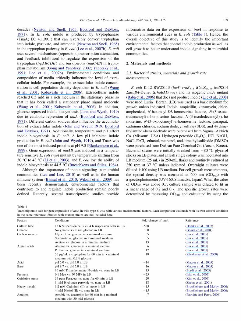
Fig. 1A clearly shows that low pH inhibits indole produc-tion while high pH increases indole production both at 30 �Cand at 37 �C. The results match well with the previous tran-scriptomic data (Table 1) in which tnaA was repressed underacidic conditions while tnaA was induced with a basiccondition. It was also observed that indole production washigher at 37 �C than at 30 �C for all tested pHs.
Since indole inhibited the biofilm formation of E. coliBW25113 strain (Lee et al., 2007b) and pH significantlychanges indole production (Fig. 1A), the effect of pH wasinvestigated for biofilm formation in E. coli BW25113(Fig. 1B). Biofilm formation was clearly decreased at alkaliconditions (pH 8 and 9) due to the high level of indoleaccumulation. To confirm the biofilm reduction by indole,when indole (0.5 mM) was exogenously added in the mediumat pH 5, the biofilm formation/cell was significantly decreasedas 0.29 � 0.06 at 30 �C and 0.27 � 0.09 at 37 �C. This result
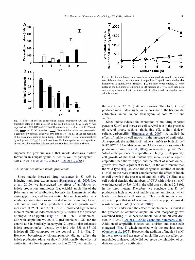
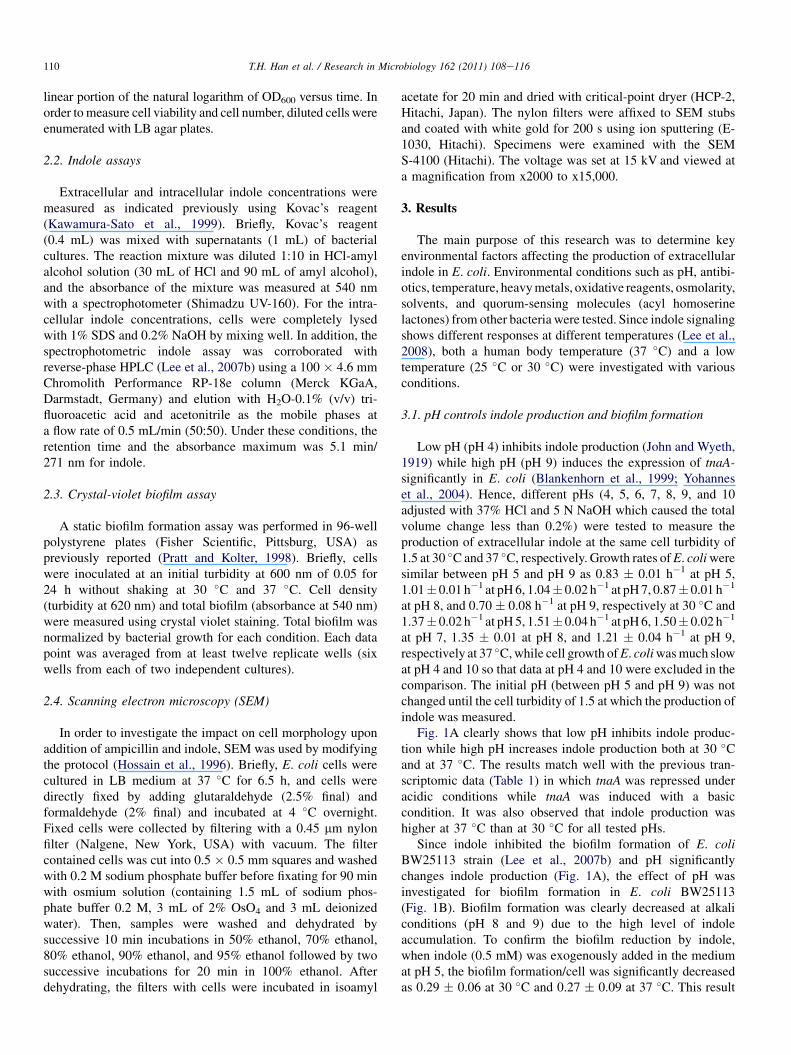
Fig. 2. Effect of antibiotics on extracellular indole production/cell growth in E.
coli. Sub-inhibitory concentrations of ampicillin (2 mg/mL, solid circles C),
kanamycin (2 mg/mL, solid triangles, ;), and none (open circles, B) were
added at the beginning of culturing in LB medium at 37 �C. Each data point
was averaged from at least four independent cultures and one standard devi-
ation is shown.
Fig. 1. Effect of pH on extracellular indole production (A) and biofilm
formation after 24 h (B) in E. coli in LB medium. pH (5, 6, 7, 8, and 9) was
adjusted with 37% HCl and 5 N NaOH and cells were cultured at 30 �C (solid
bars, ) and 37 �C (open bars, ). Extracellular indole was measured at
a cell turbidity (optical density at 600 nm) of 1.5. The pH at the cell turbidity
of 1.5 was almost same as the initial pH. Total biofilm (OD540) was normalized
by cell growth (OD620) for each condition. Each data point was averaged from
at least two independent cultures and one standard deviation is shown.
111T.H. Han et al. / Research in Microbiology 162 (2011) 108e116
supports the previous result that indole decreases biofilmformation in nonpathogenic E. coli as well as pathogenic E.coli O157:H7 (Lee et al., 2007a,b; Lee et al., 2008).
3.2. Antibiotics induce indole production
Since indole increased drug resistance in E. coli byinducing multidrug export genes (Hirakawa et al., 2005; Leeet al., 2010), we investigated the effect of antibiotics onindole production. Antibiotics (bactericidal ampicillin of theb-lactam class of antibiotics, bactericidal kanamycin of theaminoglycosides, and bacteriostatic chloramphenicol) at sub-inhibitory concentrations were added in the beginning of eachcell culture and indole production and cell growth weremeasured at 25 �C and 37 �C. E. coli produced significantlymore extracellular indole/cell density (22-fold) in the presenceof ampicillin (2 mg/mL) (Fig. 2); 1900 � 260 mM indole/cellOD with ampicillin vs. 88 � 3 mM indole/cell OD for thecontrol at 8 h. Similarly, kanamycin (2 mg/mL) also enhancedindole production/cell density by 4-fold with 338 � 87 mMindole/cell OD compared to the control at 8 h (Fig. 2).However, bacteriostatic chloramphenicol did not affect theindole production (data not shown). Additionally, the effect ofantibiotics at a low temperature, such as 25 �C, was similar to
the results at 37 �C (data not shown). Therefore, E. coliproduced more indole signal in the presence of the bactericidalantibiotics, ampicillin and kanamycin, at both 25 �C and37 �C.
Since indole induced the expression of multidrug exportergenes in E. coli and increased cell survival rate in the presenceof several drugs, such as rhodamine 6G, sodium dodecylsulfate, carbenicillin (Hirakawa et al., 2005), we studied theeffect of indole on cell growth in the presence of antibiotics.As expected, the addition of indole (1 mM) to both E. coliK-12 BW25113 wild-type and tnaA knock mutant (non-indoleproducing strain (Lee et al., 2008)) increased cell growth 2- to5-fold in the presence of ampicillin at 8 h (Fig. 3). Apparently,cell growth of the tnaA mutant was more sensitive againstampicillin than the wild-type, and the effect of indole on cellgrowth was more significant (5-fold) in the tnaA mutant thanthe wild-type (Fig. 3). Also the exogenous addition of indole(1 mM) to the tnaA mutant complemented the effect of indoleon cell growth in the presence of ampicillin (Fig. 3). Similar tocell optical density, the numbers of CFU with indole (1 mM)were increased by 3.6- fold in the wild-type strain and 2.8-foldin the tnaA mutant. Therefore, we conclude that E. coliproduces a high amount of indole against antibiotics, whichleads to enhanced cell survival. This result supports wella recent report that indole eventually leads to population-wideresistance in E. coli (Lee et al., 2010).
To better understand the effect of indole on cell survival inthe presence of antibiotic ampicillin, cell morphology wasexamined using SEM because indole could inhibit cell divi-sion in E. coli (Lee et al., 2008; Chant and Summers, 2007).Addition of ampicillin blocked cell division and made cellselongated (Fig. 4) which matched with the previous result(Comber et al., 1975). However, the addition of indole (1 mM)in the presence and absence of ampicillin does not affect cellmorphology. Hence, indole did not rescue the inhibition of celldivision caused by antibiotics.
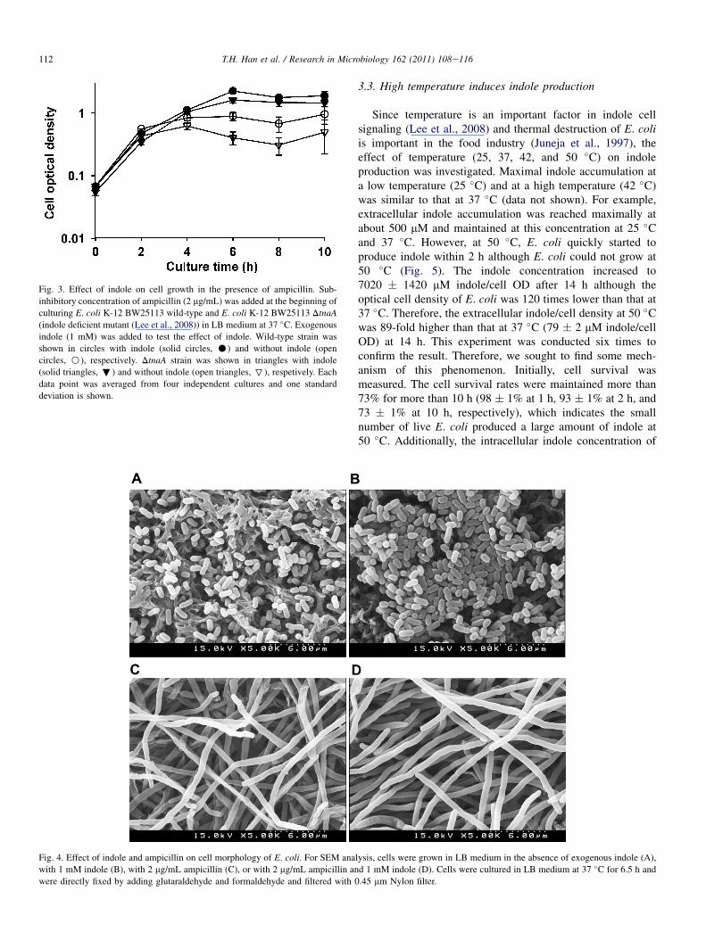
Fig. 3. Effect of indole on cell growth in the presence of ampicillin. Sub-
inhibitory concentration of ampicillin (2 mg/mL) was added at the beginning of
culturing E. coli K-12 BW25113 wild-type and E. coli K-12 BW25113 DtnaA
(indole deficient mutant (Lee et al., 2008)) in LB medium at 37 �C. Exogenousindole (1 mM) was added to test the effect of indole. Wild-type strain was
shown in circles with indole (solid circles, C) and without indole (open
circles, B), respectively. DtnaA strain was shown in triangles with indole
(solid triangles,;) and without indole (open triangles, 7), respetively. Each
data point was averaged from four independent cultures and one standard
deviation is shown.
Fig. 4. Effect of indole and ampicillin on cell morphology of E. coli. For SEM ana
with 1 mM indole (B), with 2 mg/mL ampicillin (C), or with 2 mg/mL ampicillin an
were directly fixed by adding glutaraldehyde and formaldehyde and filtered with 0
112 T.H. Han et al. / Research in Microbiology 162 (2011) 108e116
3.3. High temperature induces indole production
Since temperature is an important factor in indole cellsignaling (Lee et al., 2008) and thermal destruction of E. coliis important in the food industry (Juneja et al., 1997), theeffect of temperature (25, 37, 42, and 50 �C) on indoleproduction was investigated. Maximal indole accumulation ata low temperature (25 �C) and at a high temperature (42 �C)was similar to that at 37 �C (data not shown). For example,extracellular indole accumulation was reached maximally atabout 500 mM and maintained at this concentration at 25 �Cand 37 �C. However, at 50 �C, E. coli quickly started toproduce indole within 2 h although E. coli could not grow at50 �C (Fig. 5). The indole concentration increased to7020 � 1420 mM indole/cell OD after 14 h although theoptical cell density of E. coli was 120 times lower than that at37 �C. Therefore, the extracellular indole/cell density at 50 �Cwas 89-fold higher than that at 37 �C (79 � 2 mM indole/cellOD) at 14 h. This experiment was conducted six times toconfirm the result. Therefore, we sought to find some mech-anism of this phenomenon. Initially, cell survival wasmeasured. The cell survival rates were maintained more than73% for more than 10 h (98 � 1% at 1 h, 93 � 1% at 2 h, and73 � 1% at 10 h, respectively), which indicates the smallnumber of live E. coli produced a large amount of indole at50 �C. Additionally, the intracellular indole concentration of
lysis, cells were grown in LB medium in the absence of exogenous indole (A),
d 1 mM indole (D). Cells were cultured in LB medium at 37 �C for 6.5 h and
.45 mm Nylon filter.
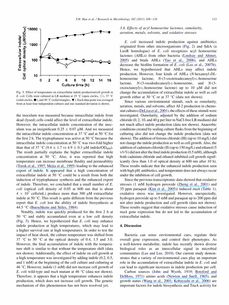
Fig. 5. Effect of temperature on extracellular indole production/cell growth in
E. coli. Cells were cultured in LB medium at 25 �C (open circles, B), 37 �C(solid circles,C), and 50 �C (solid triangles,;). Each data point was averaged
from at least four independent cultures and one standard deviation is shown.
113T.H. Han et al. / Research in Microbiology 162 (2011) 108e116
the inoculum was measured because intracellular indole fromdead (lysed) cells could affect the level of extracellular indole.However, the intracellular indole concentration of the inoc-ulum was an insignificant 0.25 � 0.07 mM. And we measuredthe intracellular indole concentration at 37 �C and at 50 �C forthe first 2 h. The tryptophanase was active at 50 �C because theintracellular indole concentration at 50 �C was two-fold higherthan that of 37 �C (9.4 � 1.7 vs 4.9 � 0.3 mM indole/OD600).The result partially explains the higher extracellular indoleconcentration at 50 �C. Also, it was reported that hightemperature can increase membrane fluidity and permeability(Torok et al., 1997; Mejia et al., 1995) leading to the enhancedexport of indole. It appeared that a high concentration ofextracellular indole at 50 �C could be a result from both theinduction of tryptophanase synthesis and the enhanced exportof indole. Therefore, we concluded that a small number of E.coli (optical cell density of 0.05 at 600 nm that is about4 � 107 cells/mL) produce more than 300 mM extracellularindole at 50 �C. This result is quite different from the previousreport that E. coli lost the ability of indole biosynthesis at44.5 �C (Bueschkens and Stiles, 1984).
Notably, indole was quickly produced for the first 2 h at50 �C and stably accumulated even at a low cell density(Fig. 5). Hence, we hypothesized that E. coli may enhanceindole production at high temperatures, which may lead toa higher survival rate at high temperatures. In order to test theimpact of heat shock, the culture temperature was shifted from37 �C to 50 �C at the optical density of 0.4, 1.5 and 3.0.However, the final accumulation of indole with the tempera-ture shift is similar to that without the temperature shift (datanot shown). Additionally, the effect of indole on cell growth ata high temperature was investigated by adding indole (0.2, 0.5,and 1 mM) at the beginning of the cell culture and culturing at46 �C. However, indole (1 mM) did not increase cell growth ofE. coli wild-type and tnaA mutant at 46 �C (data not shown).Therefore, it appears that a high temperature enhances indoleproduction, which does not increase cell growth. The geneticmechanism of this phenomenon has not been resolved yet.
3.4. Effects of acyl homoserine lactones, osmolarity,aeration, metals, solvents, and oxidative stresses
E. coli increased indole production against antibioticsoriginated from other microorganisms (Fig. 2) and SdiA (aLuxR homologue) of E. coli recognizes acyl homoserinelactones (AHLs) from other bacteria (Lindsay and Ahmer,2005) and binds AHLs (Yao et al., 2006), and AHLsdecrease the biofilm formation of E. coli (Lee et al., 2007b).Hence, we hypothesized that AHLs may affect indoleproduction. However, four kinds of AHLs (N-hexanoyl-DL-homoserine lactone, N-(3-oxotetradecanoyl)-L-homoserinelactone, N-(3-oxododecanoyl)-L-homoserine, and N-(3-oxooctanoyl)-L-homoserine lactone) up to 10 mM did notchange the accumulation of extracellular indole as well as cellgrowth either at 30 �C or at 37 �C (data not shown).
Since various environmental stimuli, such as osmolarity,aeration, metals, and solvents, affect AI-2 production in chemo-stat cultures (DeLisa et al., 2001), the effects of these stimuliwereinvestigated. Osmolarity, adjusted by the addition of sodiumchloride (0, 2, 10, and 40 g per liter in NaCl-free LBmedium) didnot much affect indole production (data not shown). Anaerobicconditions created by sealing culture flasks from the beginning ofculturing also did not change the indole production (data notshown). The addition of ferrous chloride (III) (up to 10mg/L) didnot change the indole production as well as cell growth. Also, theaddition of cadmiumchloride (II) (up to 150mg/L) and ethanol (5v/v%) did not alter the final indole production (0.5mM), althoughboth cadmium chloride and ethanol inhibited cell growth signif-icantly (less than 1.0 of optical density at 600 nm after 10 h).These results indicate that the enhanced indole production seenwith high pH, antibiotics, and temperature does not always occurunder the inhibition of cell growth.
Since the previous transcriptomic data showed that oxidativestresses (1 mM hydrogen peroxide (Zheng et al., 2001) and35 ppm paraquat (Kim et al., 2005)) induced tnaA (Table 1),oxidative stress was investigated. However, unexpectedly,hydrogen peroxide up to 5 mM and paraquat up to 200 ppm didnot alter indole production and cell growth (data not shown).These results suggest that oxidative stresses cause induction oftnaA gene expression but do not led to the accumulation ofextracellular indole.
4. Discussion
Bacteria can sense environmental cues, regulate theiroverall gene expression, and control their phenotypes. Asa well-known metabolite, indole has recently shown diversebiological roles as an intercellular signal in microbialcommunities (Lee and Lee, 2010). Our current study demon-strates that a variety of environmental cues play an importantrole in the accumulation of extracellular indole in E. coli andcan lead to significant increases in indole production per cell.
Carbon sources (John and Wyeth, 1919; Botsford andDeMoss, 1971) amino acids (Newton and Snell, 1965), andgrowth status (Wang et al., 2001; Kobayashi et al., 2006) areimportant factors for indole biosynthesis and TnaA activity for
114 T.H. Han et al. / Research in Microbiology 162 (2011) 108e116
E. coli. Several reports also indicated that pH is importantfactor for indole production (John and Wyeth, 1919). It waseven proposed that TnaA counteracted alkaline stress, such astrimethylamine N-oxide because an indole-mutant (DtnaLAB)showed low survival in trimethylamine N-oxide (Bordi et al.,2003). We clearly demonstrated that E. coli produces moreindole at high pH, which further impacts biofilm formation(Fig. 1). These results support the hypothesis that E. coli mayturn off tnaA in the acidic stomach and turn it on in the basicgut (Lee et al., 2007b).
This study demonstrated for the first time that E. coli has theability to enhance the accumulation of indole against antibi-otics (Fig. 2) and the enhanced indole production increases cellsurvival (Fig. 3). Although highly speculative, these resultssuggest that E. coli may utilize indole to compete against othermicroorganisms that could produce antibiotics. For example,the b-lactam antibiotic penicillin may be derived from Peni-cillium fungi (Fleming, 1929) and bactericidal kanamycin maybe isolated from Streptomyces kanamyceticus (Garrod, 1981).In natural environments, E. coli may encounter these antibi-otic-producing microorganisms outside animal host. It waspreviously reported that indole increases drug resistance byinducing intrinsic xenobiotic exporter genes (mdtEF and acrD)in E. coli, where indole acts via two-component signal trans-duction systems (BaeSR and CpxAR) (Hirakawa et al., 2005).Similar to E. coli, non-indole producing bacteria, such as S.enterica (Nikaido et al., 2008) and P. aeruginosa (Lee et al.,2009) also increased drug resistance in the presence ofindole. The results indicate that the presence of environmentalsignal indole could seriously affect the drug resistance ofpathogenic bacteria.
The degradation of indole is possible because various oxy-genases from non-indole producing bacteria, plants, and animalsmay transform indole (Lee and Lee, 2010). Hence, non-indoleproducing bacteria may have acquired some defense againstindole. For example, P. aeruginosa, could rapidly decrease thelevel of extracellular indole (Lee et al., 2009). Furthermore,previous results showed that indole from E. coli diminishedPseudomonas virulence by repressing quorum-sensing regulatedgenes encoding the mexGHI-opmD multidrug efflux pump, phzoperon, pqs operon, pch operon, and pvd operon while AHLsinduce these genes (Lee et al., 2009). Therefore, it appears thatsignaling interference for indole and defense against indolecould be widespread in microbial communities.
Temperature is another important cue for indole signaling(Lee et al., 2008) as well as for indole production (Fig. 5). tnaABwas induced in a temperature sensitive E. coli rnpA mutant bytemperature shifting from 30 �C to 43 �C (Li et al., 2003) andmutants that lack heat shock proteins IbpA and IbpB elevatedextracellular concentrations of indole (Kuczy�nska-Wi�snik et al.,2009). The current study confirms that the indole production issignificantly increased at high temperatures (Figs. 1 and 5).Moreover, E. coli produced indole even at 50 �C, which wasdifferent from a previous result that E. coli lost the ability ofindole production at 44.5 �Cafter 6 days (Bueschkens and Stiles,1984). The difference is probably because of different culturetimes (16 h at 50 �C vs. 6 days at 44.5 �C) and different cell lines
(BW25113 vs. ATCC 11775). It was also previously reportedthat indole cell signaling for biofilm formation, antibioticresistance, and cell division in E. coli occurs primarily at a lowtemperatures (Lee et al., 2008), while indole could controlplasmid stability in E. coli (Chant and Summers, 2007) andattenuate the virulence in P. aeruginosa at 37 �C (Lee et al.,2009). Hence, it appears that E. coli may utilize indolesignaling in different ways depending on the environmentaltemperature.
Humans maintain a symbiotic relationship with their intes-tinal microbial flora over a long time, and some bacteria arecrucial for nutrient assimilation and are beneficial for theimmune system (Hooper and Gordon, 2001). A variety ofintestinal bacteria produce a large quantity of indole in theanimal gut (a weak basic condition) including the humanintestine (Lee and Lee, 2010; DeMoss and Moser, 1969). Arecent study suggested that indole could be important in theintestinal epithelial cells response to gastrointestinal tractpathogens because indole beneficially affected gene expressionof human epithelial cells (Bansal et al., 2010). Moreover, a massspectrometry-based metabolomics study demonstrated that theproduction of a powerful antioxidant compound indole-3-pro-pionic acid in animal blood completely depended on indole-producing enteric bacteria (Wikoff et al., 2009). The resultsuggested that animals possibly utilize indole derivatives orig-inated from gut microflora for their immune systems (Wikoffet al., 2009). The current study further suggests that the intes-tinal microbial flora may protect itself using indole againstpathogenic bacteria.
Recently, the role of indole has been also studied in Vibriocholerae in which indole decreases its biofilm formation whileincreasing its grazing resistance to the phagocytic eukaryoteDictyostelium discoideum, probably by inducing expression ofvirulence-associated secretion proteins (Mueller et al., 2009).Furthermore, more than 85 species of both Gram-positive andGram-negative bacteria produce large quantities of extracel-lular indole in microbial communities (Lee and Lee, 2010).Because the habitats of these bacteria are diverse, the mech-anisms of indole production are probably different amongbacterial species. Hence, it would be interesting to investigateindole production in other indole-producing bacteria. Furtherstudies are essentially required to understand why manybacterial species produce indole, how bacteria regulate indolesignaling, and how other species react to environmentalindole.
Acknowledgements
This research was supported by the Yeungnam Universityresearch grant, and the Human Resources DevelopmentProgram (R&D Workforce Cultivation Track for Solar CellMaterials and Processes) of Korea Institute of Energy Tech-nology Evaluation and Planning (KETEP) grant funded by theKorean Ministry of Knowledge Economy. T. Wood is theT. Michael O’Connor endowed chair and is also supported bythe NIH (R01 GM089999).

115T.H. Han et al. / Research in Microbiology 162 (2011) 108e116
References
Anyanful, A., Dolan-Livengood, J.M., Lewis, T., Sheth, S., Dezalia, M.N.,
Sherman, M.A., Kalman, L.V., Benian, G.M., Kalman, D., 2005. Paralysis
and killing of Caenorhabditis elegans by enteropathogenic Escherichia coli
requires the bacterial tryptophanase gene. Mol. Microbiol. 57, 988e1007.
Baba, T., Ara, T., Hasegawa, M., Takai, Y., Okumura, Y., Baba, M.,
Datsenko, K.A., Tomita, M., Wanner, B.L., Mori, H., 2006. Construction
of Escherichia coli K-12 in-frame, single-gene knockout mutants: the Keio
collection. Mol. Syst. Biol. 2 2006.0008.
Bansal, T., Alaniz, R.C., Wood, T.K., Jayaraman, A., 2010. The bacterial signal
indole increases epithelial-cell tight-junction resistance and attenuates
indicators of inflammation. Proc. Natl. Acad. Sci. U S A 107, 228e233.
Blankenhorn, D., Phillips, J., Slonczewski, J.L., 1999. Acid- and base-induced
proteins during aerobic and anaerobic growth of Escherichia coli revealed
by two-dimensional gel electrophoresis. J. Bacteriol. 181, 2209e2216.Bordi, C., Theraulaz, L., Mejean, V., Jourlin-Castelli, C., 2003. Anticipating
an alkaline stress through the Tor phosphorelay system in Escherichia coli.
Mol. Microbiol. 48, 211e223.
Botsford, J.L., DeMoss, R.D., 1971. Catabolite repression of tryptophanase in
Escherichia coli. J. Bacteriol. 105, 303e312.
Brocklehurst, K.R., Morby, A.P., 2000. Metal-ion tolerance in Escherichia
coli: analysis of transcriptional profiles by gene-array technology. Micro-
biology 146, 2277e2282.
Bueschkens, D.H., Stiles, M.E., 1984. Escherichia coli variants for gas and
indole production at elevated incubation temperatures. Appl. Environ.
Microbiol. 48, 601e605.Chant, E.L., Summers, D.K., 2007. Indole signalling contributes to the stable
maintenance of Escherichia coli multicopy plasmids. Mol. Microbiol. 63,
35e43.
Comber, K.R., Osborne, C.D., Sutherland, R., 1975. Comparative effects of
amoxycillin and ampicillin in the treatment of experimental mouse
infections. Antimicrob. Agents Chemother. 7, 179e185.
DeLisa, M.P., Valdes, J.J., Bentley, W.E., 2001. Mapping stress-induced
changes in autoinducer AI-2 production in chemostat-cultivated Escher-
ichia coli K-12. J. Bacteriol. 183, 2918e2928.
DeMoss, R.D., Moser, K., 1969. Tryptophanase in diverse bacterial species. J.
Bacteriol. 98, 167e171.Di Martino, P., Fursy, R., Bret, L., Sundararaju, B., Phillips, R.S., 2003. Indole
can act as an extracellular signal to regulate biofilm formation of
Escherichia coli and other indole-producing bacteria. Can. J. Microbiol.
49, 443e449.Domka, J., Lee, J., Bansal, T., Wood, T.K., 2007. Temporal gene-expression in
Escherichia coli K-12 biofilms. Environ. Microbiol. 9, 332e346.
Fleming, A., 1929. On the antibacterial action of cultures of a penicillium,
with special reference to their use in the isolation of B. influenzæ. Br. J.
Exp. Pathol. 10, 226e236.
Garrod, L.P., 1981. In: Antibiotic and Chemotherapy. Churchill Livingstone,
p. 131.
Gong, F., Yanofsky, C., 2002. Analysis of tryptophanase operon expression in
vitro: accumulation of TnaC-peptidyl-tRNA in a release factor 2-depleted
S-30 extract prevents Rho factor action, simulating induction. J. Biol.
Chem. 277, 17095e17100.Gosset, G., Zhang, Z., Nayyar, S., Cuevas, W.A., Saier Jr., M.H., 2004.
Transcriptome analysis of Crp-dependent catabolite control of gene
expression in Escherichia coli. J. Bacteriol. 186, 3516e3524.
Hirakawa, H., Inazumi, Y., Masaki, T., Hirata, T., Yamaguchi, A., 2005. Indole
induces the expression of multidrug exporter genes in Escherichia coli.
Mol. Microbiol. 55, 1113e1126.
Hirakawa, H., Kodama, T., Takumi-Kobayashi, A., Honda, T., Yamaguchi, A.,
2009. Secreted indole serves as a signal for expression of type III secretion
system translocators in enterohaemorrhagic Escherichia coli O157:H7.
Microbiology 155, 541e550.
Hooper, L.V., Gordon, J.I., 2001. Commensal host-bacterial relationships in
the gut. Science 292, 1115e1118.
Hossain,M.M., Nakayama, H., Goto, N., 1996. In vitro induction of apoptosis of
developing brain cells by 5-azacytidine. Int. J. Dev. Neurosci. 14, 11e17.
Ishii, A., Oshima, T., Sato, T., Nakasone, K., Mori, H., Kato, C., 2005.
Analysis of hydrostatic pressure effects on transcription in Escherichia coli
by DNA microarray procedure. Extremophiles 9, 65e73.
John, F., Wyeth, S., 1919. The effects of acids, alkalies, and sugars on the
growth and indole formation of Bacillus Coli. Biochem. J. 13, 10e24.Juneja, V.K., Snyder Jr., O.P., Marmer, B.S., 1997. Thermal destruction of
Escherichia coli O157:H7 in beef and chicken: determination of D- and z-
values. Int. J. Food Microbiol. 35, 231e237.Kamath, A.V., Vaidyanathan, C.S., 1990. New pathway for the biodegradation
of indole in Aspergillus niger. Appl. Environ. Microbiol. 56, 275e280.
Kawamura-Sato, K., Shibayama, K., Horii, T., Iimuma, Y., Arakawa, Y.,
Ohta, M., 1999. Role of multiple efflux pumps in Escherichia coli in indole
expulsion. FEMS Microbiol. Lett. 179, 345e352.
Khodursky, A.B., Peter, B.J., Cozzarelli, N.R., Botstein, D., Brown, P.O.,
Yanofsky, C., 2000.DNAmicroarray analysis of gene expression in response
to physiological and genetic changes that affect tryptophan metabolism in
Escherichia coli. Proc. Natl. Acad. Sci. U S A 97, 12170e12175.
Kim, B.C., Youn, C.H., Ahn, J.M., Gu, M.B., 2005. Screening of target-
specific stress-responsive genes for the development of cell-based
biosensors using a DNA microarray. Anal. Chem. 77, 8020e8026.
Kobayashi, A., Hirakawa, H., Hirata, T., Nishino, K., Yamaguchi, A., 2006.
Growth phase-dependent expression of drug exporters in Escherichia coli
and its contribution to drug tolerance. J. Bacteriol. 188, 5693e5703.Kuczy�nska-Wi�snik, D., Matuszewska, E., Laskowska, E., 2009. Escherichia
coli heat-shock proteins IbpA and IbpB affect biofilm formation by
influencing the level of extracellular indole. Microbiology 156, 148e157.
Lee, H.H., Molla, M.N., Cantor, C.R., Collins, J.J., 2010. Bacterial charity
work leads to population-wide resistance. Nature 467, 82e85.
Lee, J.-H., Lee, J., 2010. Indole as an intercellular signal in microbial
community. FEMS Microbiol. Rev 34, 426e444.Lee, J., Attila, C., Cirillo, S.L.G., Cirillo, J.D., Wood, T.K., 2009. Indole and
7-hydroxyindole diminish Pseudomonas aeruginosa virulence. Microb.
Biotech. 2, 75e90.
Lee, J., Bansal, T., Jayaraman, A., Bentley, W.E., Wood, T.K., 2007a. Enter-
ohemorrhagic Escherichia coli biofilms are inhibited by 7-hydroxyindole
and stimulated by isatin. Appl. Environ. Microbiol. 73, 4100e4109.
Lee, J., Jayaraman, A., Wood, T.K., 2007b. Indole is an inter-species biofilm
signal mediated by SdiA. BMC Microbiol. 7, 42.
Lee, J., Zhang, X.S., Hegde, M., Bentley, W.E., Jayaraman, A., Wood, T.K.,
2008. Indole cell signaling occurs primarily at low temperatures in
Escherichia coli. ISME J. 2, 1007e1023.
Li, Y., Cole, K., Altman, S., 2003. The effect of a single, temperature-sensitive
mutation on global gene expression in Escherichia coli. RNA 9, 518e532.
Lindsay, A., Ahmer, B.M.M., 2005. Effect of sdiA on biosensors of N-acyl-
homoserine lactones. J. Bacteriol. 187, 5054e5058.Liu, M., Durfee, T., Cabrera, J.E., Zhao, K., Jin, D.J., Blattner, F.R., 2005.
Global transcriptional programs reveal a carbon source foraging strategy
by Escherichia coli. J. Biol. Chem. 280, 15921e15927.
Maurer, L.M., Yohannes, E., Bondurant, S.S., Radmacher, M., Slonczewski, J.
L., 2005. pH regulates genes for flagellar motility, catabolism, and
oxidative stress in Escherichia coli K-12. J. Bacteriol. 187, 304e319.
Mejia, R., Gomez-Eichelmann, M.C., Fernandez, M.S., 1995. Membrane
fluidity of Escherichia coli during heat-shock. Biochim. Biophys. Acta
1239, 195e200.
Mitrophanov, A.Y., Groisman, E.A., 2008. Signal integration in bacterial two-
component regulatory systems. Genes Dev. 22, 2601e2611.
Mueller, R.S., Beyhan, S., Saini, S.G., Yildiz, F.H., Bartlett, D.H., 2009. Indole
acts as an extracellular cue regulating gene expression in Vibrio cholerae.
J. Bacteriol. 191, 3504e3516.
Newton, W.A., Snell, E.E., 1965. Formation and interrelationships of trypto-
phanase and tryptophan synthetases in Escherichia coli. J. Bacteriol. 89,
355e364.
Nikaido, E., Yamaguchi, A., Nishino, K., 2008. AcrAB multidrug efflux pump
regulation in Salmonella enterica serovar Typhimurium by RamA in
response to environmental signals. J. Biol. Chem. 283, 24245e24253.
Patridge, E.V., Ferry, J.G., 2006. WrbA from Escherichia coli and Archae-
oglobus fulgidus is an NAD(P)H:quinone oxidoreductase. J. Bacteriol.
188, 3498e3506.
116 T.H. Han et al. / Research in Microbiology 162 (2011) 108e116
Pratt, L.A., Kolter, R., 1998. Genetic analysis of Escherichia coli biofilm
formation: roles of flagella, motility, chemotaxis and type I pili. Mol.
Microbiol. 30, 285e293.
Smith, T., 1897. A modification of the method for determining the production
of indol by bacteria. J. Exp. Med. 2, 543e547.Torok, Z., Horvath, I., Goloubinoff, P., Kovacs, E., Glatz, A., Balogh, G.,
Vigh, L., 1997. Evidence for a lipochaperonin: association of active
protein-folding GroESL oligomers with lipids can stabilize membranes
under heat shock conditions. Proc. Natl. Acad. Sci. U S A 94, 2192e2197.
Wang, D., Ding, X., Rather, P.N., 2001. Indole can act as an extracellular
signal in Escherichia coli. J. Bacteriol. 183, 4210e4216.
Waters, C.M., Bassler, B.L., 2005. Quorum sensing: cell-to-cell communica-
tion in bacteria. Annu. Rev. Cell Dev. Biol. 21, 319e346.
Wikoff, W.R., Anfora, A.T., Liu, J., Schultz, P.G., Lesley, S.A., Peters, E.C.,
Siuzdak, G., 2009. Metabolomics analysis reveals large effects of gut
microflora on mammalian blood metabolites. Proc. Natl. Acad. Sci. U S A
106, 3698e3703.
Yanofsky, C., Horn, V., Gollnick, P., 1991. Physiological studies of tryptophan
transport and tryptophanase operon induction in Escherichia coli. J. Bac-
teriol. 173, 6009e6017.Yao, Y., Martinez-Yamout, M.A., Dickerson, T.J., Brogan, A.P., Wright, P.E.,
Dyson, H.J., 2006. Structure of the Escherichia coli quorum sensing
protein SdiA: activation of the folding switch by acyl homoserine lactones.
J. Mol. Biol. 355, 262e273.
Yohannes, E., Barnhart, D.M., Slonczewski, J.L., 2004. pH-dependent cata-
bolic protein expression during anaerobic growth of Escherichia coli K-12.
J. Bacteriol. 186, 192e199.Zheng, M., Wang, X., Templeton, L.J., Smulski, D.R., LaRossa, R.A., Storz, G.,
2001. DNA microarray-mediated transcriptional profiling of the Escher-
ichia coli response to hydrogen peroxide. J. Bacteriol. 183, 4562e4570.
Copyright © 2022 FDOKUMEN