Nitric Oxide Potentiates Hydrogen Peroxide-induced Killing of Escherichia coli
Upload
khangminh22Category
view
0download
0
1
Vol.:(0123456789)
Scientific Reports | (2022) 12:5305 | https://doi.org/10.1038/s41598-022-09274-x
www.nature.com/scientificreports
Characterization of Escherichia coli harboring colibactin genes (clb) isolated from beef production and processing systemsManita Guragain, John W. Schmidt, Norasak Kalchayanand, Aaron M. Dickey & Joseph M. Bosilevac*
Certain strains of Escherichia coli possess and express the toxin colibactin (Clb) which induces host mutations identical to the signature mutations of colorectal cancer (CRC) that lead to tumorigenic lesions. Since cattle are a known reservoir of several Enterobacteriaceae including E. coli, this study screened for clb amongst E. coli isolated from colons of cattle-at-harvest (entering beef processing facility; n = 1430), across the beef processing continuum (feedlot to finished subprimal beef; n = 232), and in ground beef (n = 1074). Results demonstrated that clb+ E. coli were present in cattle and beef. Prevalence of clb+ E. coli from colonic contents of cattle and ground beef was 18.3% and 5.5%, respectively. clb+ E. coli were found susceptible to commonly used meat processing interventions. Whole genome sequencing of 54 bovine and beef clb+ isolates showed clb occurred in diverse genetic backgrounds, most frequently in phylogroup B1 (70.4%), MLST 1079 (42.6%), and serogroup O49 (40.7%).
Colorectal cancer (CRC), the fourth most common cancer in the United States1, is a multifactorial disease asso-ciated with risk factors such as obesity, genetics, and lifestyle including smoking, alcohol, and diet (as reviewed in2). The International Agency for Research on Cancer reports a positive association between high consumption of red meat and CRC, thereby, classifying red meat as “probably carcinogenic to humans”. Association between CRC and red meat consumption is classically attributed to the formation of carcinogenic compounds generated during high temperature cooking3. However, as reviewed in4, intrinsic properties of red meat itself do not provide adequate mechanistic links between increased CRC risk and red meat consumption. This warrants investigation into extrinsic factors such as meat-borne tumorigenic bacteria as a missing link between red meat and CRC.
The abundance of E. coli carrying DNA damaging genes like colibactin (clb) and cytotoxic necrotizing factor (cnf) is increased in colon biopsies from CRC patients compared to non-CRC patients and healthy individuals5. Clb is an important virulence factor of E. coli and other Enterobactericeae6,7. This genotoxic, nonribosomal, hybrid peptide-polyketide is encoded within the 54 kb polyketide synthase (pks) genomic island8. Clb expressed by clb+ E. coli alkylates DNA and induces double strand DNA breaks and interstrand cross linking of mam-malian DNA8–10. These cross-links result in chromosomal aberrations, cell cycle arrest, and cell senescence8,11. Mouse model studies show Clb induces the formation of invasive colonic tumors associated with increased DNA damage12. Unique single strand breaks and indels induced by Clb are found to be identical with mutational signatures from a subset of human cancer genomes, particularly, colorectal cancer (CRC) and though rare, in tumors derived from head, neck, urinary tract13, and oral squamous carcinoma14.
The gastrointestinal tract of cattle is a natural reservoir of both commensal and pathogenic E. coli. These E. coli are categorized using various schemes which allow quick assignment of an isolate to specific host, pathogenesis, and phylogenetic lineages. E. coli are defined based on the variation in O-antigen of outer membrane lipopoly-saccharide to any of 185 O-serogroups15. DNA sequence variations in internal regions of multiple housekeeping genes define clonal diversity and categorize E. coli into different multilocus sequence type (MLST) profiles16. Lastly, there are 8 phylogenetic groups of E. coli defined by sets of five genes, each set specific to individual phylogroup17. During beef processing, E. coli of these various sorts from hides and feces may directly contami-nate the carcasses that are intended to be further processed into final beef products 18,19. Despite the advances
OPEN
Meat Safety and Quality Research Unit, US Department of Agriculture, Agricultural Research Service, U.S. Meat Animal Research Center, State Spur 18D, P.O. Box 166, Clay Center, Nebraska 68933, USA. *email: [email protected]
2
Vol:.(1234567890)
Scientific Reports | (2022) 12:5305 | https://doi.org/10.1038/s41598-022-09274-x
www.nature.com/scientificreports/
in pathogen control in beef production and processing, ground beef still continues to be an important source of E. coli infections that occasionally lead to recalls and outbreaks20 (as reviewed in21). Therefore, the beef chain (cattle to finished beef) was examined for the presence of clb+ E. coli to test the hypothesis that these E. coli may be contributing to the classification of beef as a potential carcinogen.
The results presented herein show the potential of cattle as a reservoir and beef as a vehicle of clb+ E. coli. Arbitrarily isolated E. coli from cattle and ground beef were found to carry clb in their genomes. Further, currently used processing interventions are shown to be effective control measures that can reduce clb+ E. coli contamina-tion of beef. Whole genome sequencing showed that the organization of the pks island is highly conserved in clb+ E. coli isolated from cattle and beef, and revealed that the majority of clb+ isolates from cattle and beef belong to a limited number of phylogroups, serogroups, and MLST types.
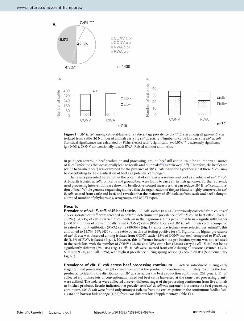
ResultsPrevalence of clb+ E. coli in US beef cattle. E. coli isolates (n = 1430) previously collected from colons of 709 eviscerated cattle 22 were screened in order to determine the prevalence of clb+ E. coli in beef cattle. Overall, 18.7% (134/715) of cattle carried E. coli with clb in their genomes. On a per animal basis a significantly higher (P < 0.05) number of conventionally raised (CONV) cattle (85/351) carried clb+ E. coli in their colons compared to raised without antibiotics (RWA) cattle (49/364) (Fig. 1). Since two isolates were selected per animal22, this amounted to 11.7% (167/1430) of the cattle borne E. coli testing positive for clb. Significantly higher prevalence of clb+ E. coli was observed among isolates from CONV cattle (15% of CONV isolates) compared to RWA cat-tle (8.5% of RWA isolates) (Fig. 1). However, this difference between the production system was not reflected in the cattle lots, with the number of CONV (28/36) and RWA cattle lots (22/36) carrying clb+ E. coli not being significantly different (P > 0.05) (Fig. 1). clb+ E. coli were isolated from cattle during all seasons (Winter, 11.7%, Summer, 9.2%, and Fall, 8.2%), with highest prevalence during spring season (17.5%, p < 0.05) (Supplementary Fig. S1).
Prevalence of clb+ E. coli across beef processing continuum. Bacteria introduced during early stages of meat processing may get carried over across the production continuum, ultimately reaching the final products. To identify the distribution of clb+ E. coli across the beef production continuum, 232 generic E. coli collected from three lots of conventionally raised fed beef cattle harvested at the same beef processing plant23 were utilized. The isolates were collected at seven different stages of the processing continuum from live animals to finished products. Results indicated that prevalence of clb+ E. coli was extremely low across the beef processing continuum. clb+ E. coli were found only amongst isolates from the earliest points in the continuum: feedlot fecal (1/36) and harvest hide sponge (1/36) from two different lots (Supplementary Table T1).
7.4% ***
42.3%
4.3%***
46.0% CONV clb+CONV clb-RWA clb+RWA clb-
n=1430
a.
0
10
20
30
40
CONV RWA
Num
ber o
f cal
le lo
ts
clb -clb+
n=72
c.
080
160240320400
CONV RWA
Num
ber o
f cat
tle
clb -clb+
***
n=715
b.
Figure 1. clb+ E. coli among cattle-at-harvest. (a) Percentage prevalence of clb+ E. coli among all generic E. coli isolated from cattle (b) Number of animals carrying clb+ E. coli. (c) Number of cattle lots carrying clb+ E. coli. Statistical significance was calculated by Fisher’s exact test. *, significant (p < 0.05); ***, extremely significant (p < 0.001). CONV, conventionally raised; RWA, Raised without antibiotics.

3
Vol.:(0123456789)
Scientific Reports | (2022) 12:5305 | https://doi.org/10.1038/s41598-022-09274-x
www.nature.com/scientificreports/
Prevalence of clb+ E. coli in retail ground beef. To estimate the prevalence of clb+ E. coli in ground beef, 1,074 generic E. coli previously isolated from 507 retail ground beef samples collected from 6 US cities24 were screened. Overall, prevalence of clb+ E. coli in ground beef samples was found to be 5.5% (28/507) (Fig. 2). With two isolates selected per sample24, 4.1% (44/1074) of the generic E. coli from ground beef carried clb in their genome. clb+ E. coli appeared significantly lower (P < 0.05) among isolates recovered from ground beef with RWA label claims (2.4%) compared to isolates from CONV ground beef (5.5%). However, there was no significant difference on a per sample basis where clb+ E. coli were isolated from 9 of 240 RWA and 19 of 267 CONV samples. Interestingly, among the samples from the southwestern region, clb+ E. coli were entirely absent from RWA samples (Fig. 2). Prevalence of clb appears to be lowest among isolates from ground beef purchased in the Pacific West region (1.9%) compared to Plains (5.3%), Southeast (4.6%), and North East (5.5%) regions (Supplementary Fig. S2).
Susceptibility of clb+ E. coli towards meat processing interventions. Forty-four selected clb+ E. coli were screened for susceptibility towards current beef processing antimicrobial compounds used as interven-tions by using a benchtop killing assay. clb+ E. coli were treated with 2% lactic acid (LA), 200 ppm Peroxyacetic acid (PAA), 300 ppm Bromine (BR), and hot water (HW, 80 °C) in a 96-well setting. Fold change in cell density (OD600) of treated samples compared to untreated samples was calculated and compared to that of E. coli O157:H7 FSIS-4. It was observed that clb+ E. coli were susceptible to the antimicrobials with fold reductions for hot water, 2.5 ± 0.4 to 4.2 ± 2.2; LA, 2.5 ± 0.7 to 3.9 ± 1.0; PAA, 2.6 ± 0.4 to 4.7 ± 2.3; and BR, 2.0 ± 1.2 to 5.4 ± 2.8. All strains were as sensitive or more sensitive to the treatments as the reference E. coli O157:H7 FSIS-4 isolate25 (Supplementary Fig. S3).
Next, to assess the efficacy of antimicrobial interventions on controlling clb+ E. coli on beef carcass surfaces, an inoculum pool of 4 clb+ isolates and the reference E. coli O157:H7 FSIS-4 isolate was inoculated on beef flank (cutaneous trunci muscle) surfaces and treated in the USMARC pilot processing plant. The inoculated flanks were exposed to antimicrobial interventions and log reductions in Colony forming unit (CFU) per cm2 were calculated. clb+ isolates inoculated on the beef flank surfaces were found to be equally susceptible to LA, PAA, and BR with Log reductions of 0.7–1.3, 0.6–1.0, and 0.4 ± 1.0 CFU/cm2 respectively. Dry steam was found to be most effective at reducing clb+ E. coli on beef flank surfaces (1.6–2.8 2 Log CFU/cm2reduction) (Fig. 3). There were no statistically significant differences between clb+ E. coli and the reference E. coli O157:H7 FSIS-4 (log reduction: steam, 1.7–3.1; LA, 0.8–1.4 1; PAA, 0.6–1.0; BR, 0.5–0.9) (P > 0.05, one-way ANOVA).
Genome characteristics of clb+ E. coli. An in-house genome analysis pipeline was used to assemble the genomes, screen them against Center for Genomic Epidemiology (CGE) databases and obtain the assembly sta-tistics. Assembly statistics and characteristics of fifty-four clb+ E. coli genomes are summarized in Supplementary Data File SD1.
Phylogroups of clb+ E. coli were determined in-silico with ClermonTyping 21.0326. Overall, 70.4% (38/54) of the clb+ E. coli in this study belong to phylogroup B1, followed by B2 (27.8%, 15/54), and A (1.9%, 1/54) (Fig. 4). Among clb+ E. coli isolates from the colons of cattle, prevalence of phylogroups B1 and B2 were 65.9% and 34.1% respectively. Among ground beef clb+ E. coli, 90.9% belonged to phylogroup B1, while 9.1% to B2. One clb + isolate from beef processing continuum belonged to phylogroup A, and the other to phylogroup B1. Owing to difference in number of genomes, no attempt was made to compare phylogroups between the sample sources.
Based on in-silico O-serotyping using SerotypeFinder version 2018_09_2427, the most pre-dominant O-sero-group among sequenced clb+ isolates was O49 (40.7%, n = 22), followed by O2 (27.8%, n = 15), and O42 (11.1%, n = 6), and other O-groups (7.4%, n = 4). O-serogroup could not be determined for 7 isolates (Fig. 4). In-silico multilocus sequence typing using MLST version 2019_05_0828 showed three-quarters of the sequenced clb+ E. coli in our study belonged to three MLST types; 23 to MLST 1079, 10 to MLST 95, and 8 to MLST 278 (Fig. 4).
0.02.04.06.08.0
10.012.0
% clb
+ sa
mpl
es
CONVRWA
n=507
b.3% *
50.7%
1.1%*
45.2
CONV clb+CONV clb-RWA clb+RWA clb-
n=1074
a.
Figure 2. clb+ E. coli occurring in ground beef. (a) Percentage prevalence of clb+ E. coli in ground beef among all generic E. coli isolated; (b) Percentage of ground beef samples carrying clb+ E. coli. Statistical significance was calculated by Fisher’s exact test. *, significant (p < 0.05). CONV: ground beef produced from conventionally raised cattle; RWA: ground beef produced with label claim of “from beef raised without antibiotics”.

4
Vol:.(1234567890)
Scientific Reports | (2022) 12:5305 | https://doi.org/10.1038/s41598-022-09274-x
www.nature.com/scientificreports/
Antibiotic resistance genes were identified by ResFinder version 2020_02_0629–31. Prevalence of genes associ-ated with tetracycline resistance (tetA, tetB, tetC, tetM), aminoglycoside resistance (aadA1), fosfomycin resist-ance (fosA7), and beta-lactamase resistance (bla TEM-1A) were 51.8, 31.5, 7.4, and 1.9% respectively (Fig. 5) (Supplementary Data File SD1).
VirulenceFinder 2.032,33 identified 50 virulence genes belonging to several functional categories like toxin, stress resistance, adherence, siderophores, immune evasion, serum survival, colicin, and secretion system, in a decreasing order of abundance among clb+ isolates. Tellurium resistance gene terC, with implicated role in gen-eral stress tolerance in the host environment, was present in all clb+ isolates identified in this study. Other highly distributed genes were long polar fimbriae lpfA (67%), glutamate decarboxylase gad (63%), outer membrane protease ompT (57%), and outer membrane protein complement resistance traT (57%). Cytolethal distending toxin cdtB, another genotoxin which promotes colorectal cancer34, was present in 15 genomes (14 group B2 and 1 group B1). Overall, group B2 clb+ E. coli isolates harbored more virulence factors compared to group B1 isolates (Fig. 6) (Supplementary Data File SD1).
Phylogenetic analyses. Core genomes of the 54 sequenced clb+ isolates were aligned with PARSNP ver-sion 1.235, maximum likelihood tree was constructed with iQ-tree 1.6.1036 and was visualized with iTol V637. The genomes distinctly grouped according to phylogroups, serogroups, and MLST profiles. Isolates from different sources as wells as different production systems (i.e., RWA and CONV) grouped together in the same clades (Fig. 4).
High sensitivity mapping using Geneious mapper in Geneious prime 2020.1.2 (Biomatters) against the refer-ence pks sequence regions spanning clbA-clbS identified pks islands in all fifty-four newly sequenced genomes. Among these newly identified pks islands, thirty eight pks sequences present in full length in individual contigs were then aligned (Supplementary Fig. S4) and a maximum likelihood tree was constructed. Compared to the chromosome, G + C contents of all pks islands were higher (53.5% VS ~ 50.1%). Nucleotide sequence compari-son showed that pks islands were highly conserved (> 99.64%) in comparison to each other and the reference sequences and maintained genetic synteny (Supplementary Fig. S4). When differences in nucleotide sequences were found, they mostly resulted from substitutions within genes.
Phylogenetic analysis showed that pks islands grouped into two major clades, largely by phylogroups, O-sero-groups, and MLST profiles of the isolate in which they were found (Fig. 4). Nucleotide sequence identity between the clades ranged from 99.63 to 99.82%. Clades were not specific to sample sources or production system. Clade 1 exclusively consisted of twelve pks sequences belonging to group B1 isolates, all of which belonged to MLST 1079 and serogroup O49. Only one sequence from a MLST 1079 isolate grouped with clade 2 sequences. Clade 1 sequences were 100.0% identical to each other, while 99.95–99.98% identical with clusters of reference sequences from human clinical isolates. Interestingly, pks sequences from cattle B2 strains are phylogenetically closer to B1 strains than to the B2 human clinical reference strains. pks sequences in clade 2 further distinctly grouped in two subclades. Clade 2A pks sequences originated from twelve isolates, exclusively of phylogroup B1 and includes pks sequence from human commensal strain JML285. Isolates in this clade appear to be the most diverse of all three clades in context of their serogroups and MLST profile yet 100.0% identical regarding pks sequences, except for reference sequence SI-NP020 from a bovine isolate, which shared 99.97% identity with rest of the clade. Clade 2B consisted of fourteen sequences, of which twelve belong to isolates of phylogroup B2 and serogroup O2, sharing > 99.97% nucleotide sequence identity. Ten of these sequences belonged to MLST 95 and the remaining two MLST 1619. Two reference pks sequences from bovine fecal isolates KS-P003 and KS-P027 (phylogroup B2, MLST95) also clustered together with these ten sequences. The remaining clade 2B sequences belonged to isolates of group A and group B1 each and shared 99.70% identity with rest of the clade 2B.
Since clonal complex 95 (CC95) strains are known as significant extraintestinal pathogens in humans17, we further analyzed these strains to understand the distribution of pks islands. Core genomes of all pks+ CC95 strains, including two previously reported bovine strains KS-P003 and KS-P027 closely clustered together. All
0.0
1.0
2.0
3.0
4.0
LA PAA Brom Steam ni noitcuder golcf
u/cm
2
Figure 3. Beef flank inoculation assay. LA, 2% lactic acid (1 min). PAA, 200 ppm peroxyacetic acid (1 min). Brom, 300 ppm BoviBromine (1 min). Steam, flank surface temperature achieved 82 °C with dry steam (15 s). Statistical significance between clb+ E. coli and reference E. coli O157:H7 was calculated by Fisher’s exact test (p ≥ 0.05, not significant). Data presented represents average of 2 biological replicates, 12 technical replicates each on individual beef flanks. clb+ E. coli, E. coli O157:H7.

5
Vol.:(0123456789)
Scientific Reports | (2022) 12:5305 | https://doi.org/10.1038/s41598-022-09274-x
www.nature.com/scientificreports/
Figure 4. Maximum likelihood phylogenetic tree of (a) 54 core genomes of clb+ E. coli isolated from beef production and processing continuum, and (b) 39 pks island sequences extracted from the contigs carrying full length pks island (clbA-clbS). Phylogenetic trees were constructed using IQ-tree and visualized with iTOL v6. Numbers on the outermost column represent multilocus sequence types (MLST).
Figure 5. Distribution of acquired antibiotic resistance genes in clb+ E. coli. Heat map was created and visualized using ggplot2 package of R. Column represents E. coli isolates and row represents acquired antibiotic resistance genes. *, Phylogroup B2; # Phylogroup A.

6
Vol:.(1234567890)
Scientific Reports | (2022) 12:5305 | https://doi.org/10.1038/s41598-022-09274-x
www.nature.com/scientificreports/
CC95 strains identified in this study belonged to the same serogroup (O2) and phylogroup (B2), but different serogroup than KS-P003 and KS-P027 (Fig. 4A). pks islands in these strains grouped together in same clade (2B), but away from pks islands in CC95 human isolates in clade 1 (Fig. 4B). An in-silico search for genes and primer sequences specific to unique subgroups38 among the CC95 pks+ strains from this study was unsuccessful therefore fimH typing was performed. In combination with phylogroups, fimH typing has been described as a useful approach in classifying strains of E. coli39. The fimH allele in these strains were identified using fimtype 1.0 available from center for genomic epidemiology. All CC95 strains including KS-P003 and KS-P027 were found to carry the same fimH allele (fimH15).
DiscussionBeef-borne pathogenic E. coli outbreaks are often associated with diarrheagenic pathotypes, that cause acute intestinal diseases, but other beef-borne chronic infections due to non-classical pathotypic E. coli are often over-looked. The association of red meat consumption with increased risk of CRC 3, which in turn is associated with increased tumorigenic bacterial populations5,40, undoubtedly demands the investigation of beef as a vehicle for such bacteria including clb+ E. coli. The clb+ E. coli further poses a multigenerational threat as vertical transmis-sion has been reported in humans41 and experimental animals42, where they produce long-lasting effects. This study demonstrates that the clb gene cluster is present in commensal E. coli isolated from cattle and beef. In the U.S., cattle presented for harvest are found to be colonized by pathogenic E. coli (E. coli O157:H7 and non-O157 Shiga toxin producing E. coli; STEC) and Salmonella at rates of 1.9–34.3%19 and 1.7–9.2%22,43, respectively, using culture methods that enrich and select for the pathogen. In contrast, 18.5% of cattle were determined to carry arbitrary clb+ E. coli that were isolated and identified without enrichment or selective pressure. Although antimi-crobial interventions focus on controlling E. coli O157:H7 and reduce their prevalence in finished beef products
Figure 6. Distribution of acquired virulence genes in clb+ E. coli. Heat map was created and visualized using ggplot2 package of R. Column represents E. coli isolates and row represents acquired virulence genes. *, Phylogroup B2; # Phylogroup A.

7
Vol.:(0123456789)
Scientific Reports | (2022) 12:5305 | https://doi.org/10.1038/s41598-022-09274-x
www.nature.com/scientificreports/
to less than a fraction of one percent44, arbitrary clb+ E. coli was found at a rate of 4.1% in finished ground beef. This is despite the fact that clb+ E. coli reacted equally or were more sensitive to the processing interventions tested as E. coli O157:H7. Thus, finished beef is a potential vehicle for clb+ E. coli transmission.
E. coli contamination of beef primarily occurs during steps of sanitary dressing when the hide is removed from the carcass, with minor inputs from the steps of evisceration45.Further investigation of alternate foodborne sources of clb+ E. coli are needed before proper risk assessments can be performed to determine the actual impact of beef-borne clb+ E. coli on public health. Our examination of the E. coli isolates recovered from the beef pro-cessing continuum were too limited in number to reveal an alternate route of clb+ E. coli contamination of beef. Pathogenic E. coli, however, have been reported to be present in beef lymph nodes, which upon grinding may release the bacteria into the ground beef46. Alternately there may be environmental niches where clb+ E. coli may persist in the processing plant environment protected within a biofilm47.
The overall prevalence of clb+ E. coli was higher among conventionally raised cattle and their beef products, however, the number of cattle lots and ground beef samples with clb+ E. coli did not differ between CONV and RWA production systems. Therefore, this study does not indicate that use of antibiotics in beef production selects for clb+ E. coli. In fact, the types of antibiotic resistance genes identified within clb+ E. coli are among those fre-quently detected regardless of antimicrobial use48,49.
The whole genome sequencing of the clb+ E. coli in this study revealed several novel results regarding their phylogeny. Unlike human clinical samples50,51, frequency of clb+ is highest among phylogroup B1 E. coli isolated from beef. This may be a source-specific difference as animal E. coli strains often belong to group B1, whereas group B2 includes many human pathogenic strains52–54. There appears to be a preference for particular genetic backgrounds among the clades of pks. All CC95 pks sequences in this study as well as two reference bovine CC95 isolates KS-P003 and KS-P027, but none of the human clinical CC95 strains in clade 1, carried fimH15 suggesting host specificity among CC95 strains. Further, the potential mechanism of pks transfer appears to be specific to genetic background as well as pks sequence clades. Occasionally, however, the same mechanisms appear to allow pks transfer to different genetic backgrounds, as well as transfer of different pks sequence variants. Colibactin production may be independent of phylogeny of clb genes55, nevertheless, future studies are needed to assess the production and activity of Clb among cattle and meat-borne isolates.
In this study, 27% of sequenced isolates belonged to phylogroup B2, a phylogroup that possesses more viru-lence factors and increasingly has become dominant in industrialized nations56,57, where CRC is also on the rise58. Further, high association of colicin genes, cdtB, and clb among these isolates will likely contribute towards their success in gut colonization, leading to microbial dysbiosis and ultimate progression to CRC. However, the CC95 pks islands identified in this study cluster distantly from reference human CC95 strains, similar to a recently reported observation50. Therefore, for proper assessment of risk posed by cattle and meat-borne CC95 pks+ strains in human CRC, future studies aimed at establishing human gut colonization status and lesion formation in colonic tissues by these isolates are necessary.
It is well established how pathogenic E. coli such as E. coli O157:H7 contaminates beef during harvest and processing59, and herds of cattle with twenty percent fecal prevalence have greater than eighty percent prevalence on their hides60. Thus, clb+ E. coli may be present on hides and contaminated beef in a similar fashion as E. coli O157:H7. Over the last twenty years, hide and carcass directed interventions have been implemented to reduce E. coli O157:H7 contamination18,61. These interventions may have influenced clb+ E. coli on beef during this same time span. Historic data or samples are unfortunately lacking to address this. However, modern beef processing interventions focused on pathogen control may be what is confounding a more recent epidemiological report that cannot establish strong correlations between red meat consumption and CRC 62. For instance, recent studies have shown an association of high red meat intake with an alkylating mutational signature in the colorectum, suggesting this to be a mechanistic link between CRC and red meat consumption63. Interestingly, clb+ E. coli also alkylates host DNA and lead to tumorigenic lesions10. One could speculate that the cohorts of individuals in the recent studies have been exposed to less clb+ E. coli (and other toxigenic E. coli) through the red meat they consume than those in the past.
Colorectal cancer is the third most common malignancy worldwide1 and is arguably associated with red meat consumption. Understanding the distribution and characteristics of clb+ E. coli in beef production and processing systems is the first step towards identifying microbial factors that may contribute to red meat associated CRC. Red meat, but not poultry and fish, is associated with increased risk of CRC. Therefore, expanding this study to the latter two, as well as other red meat sources is essential to draw conclusions regarding role of clb+ E. coli in red meat associated CRC.
MethodsEthics statement. All E. coli isolates used in this work were archived strains from previous work22–24 where sets of arbitrarily selected E. coli were examined. Samples from cattle were collected at a commercial feedlot22 and immediately after commercial harvest processes at USDA–FSIS federally inspected establishments where humane animal handling was practiced22–24. All experimental protocols involving cattle were approved and car-ried out in accordance with relevant guidelines from USD-ARS-US Meat Animal Research Center, Institutional Biosafety Committee (IBC #2.1 and #3.0) and Institutional Animal Care & Use Committee (IACUC #81.0). This study is reported in accordance to ARRIVE guidelines.
Study design. Generic E. coli isolates previously recovered and archived at the United States Meat Animal Research Center (USMARC) were utilized for the current study. First, generic E. coli (n = 1430) isolated from the colonic contents of cattle-at-harvest were utilized22. These samples were collected from 72 cattle lots (36 raised without antibiotics (RWA) and 36 raised conventionally (CONV) on a monthly basis (3 lots of each per

8
Vol:.(1234567890)
Scientific Reports | (2022) 12:5305 | https://doi.org/10.1038/s41598-022-09274-x
www.nature.com/scientificreports/
month) over a period of one year. Colon contents were collected from colons immediately after carcass eviscera-tion. Eviscera was tracked to coincide with targeted lots of cattle. Next, to examine clb+ E. coli across the beef production continuum, 232 generic E. coli isolated from three lots of fed beef cattle along different stages of beef processing continuum (feces and hides at feedlot, feces and hides at abattoir, pre-evisceration carcasses, final car-casses, and packaged strip loins) were utilized23. Finally, for finished beef products, 1074 generic E. coli collected from 507 retail ground beef samples (267 with RWA label claims and 240 without, considered CONV) obtained from 6 U.S. cities were examined24. The prevalence of clb+ E. coli was unknown, therefore the full collections of strains from each study were examined in lieu of a power analysis.
clb screening assay. To screen E. coli isolates for the pks island, a multiplex PCR targeting clb genes at the 5’ and 3’ regions of the 54 kb pks island from E. coil Nissel 1917 (GenBank accession No: CP022686) was designed and utilized. An aliquot of the overnight culture growing at 37 °C in Luria Bertani (LB) broth (Fisher Scientific, Pittsburg, PA) was combined with BAX lysis buffer (Hygiena, Camarillo, CA) following manufacturer’s recom-mendations to generate template DNA. Primer sequences are provided in Supplementary Table T2. Amplicons were resolved on 1.2% agarose gels and stained with ethidium bromide for visualization.
Antimicrobial susceptibility screening assay. Forty-four clb+ E. coli (35 colon isolates + 9 ground beef isolates) were evaluated for susceptibility towards commonly used meat processing interventions. These iso-lates were arbitrarily chosen to represent diverse cattle production systems, months, and regions. Isolate E. coli O157:H7 FSIS-4 from the USMARC collection, which was previously characterized for its susceptibility towards the antimicrobial treatments25, was utilized as a reference. The following antimicrobial solutions were prepared in tap water at room temperature: 2% lactic acid (LA, pH = 2.3) (Sigma-Aldrich, St. Louis, MO), 200 ppm peroxy-acetic acid (PAA, pH = 3.4) (BLITZ organic, FMC Corp., Philadelphia, PA), and 300 ppm 1,3-dibromo-5,5-di-methyl hydrantoin (BR, pH = 7.0) (Albemarle, Baton Rouge, LA). For non-treated control, an equal volume of sterile, deionized water was used. PAA and BR concentrations were determined by following manufacturer’s rec-ommendation using a PAA test kit (Peroxychem, Philadelphia, PA) and bromine photometer II (Hanna Instru-ment, Smithfield, RI) respectively.
Individual isolates were grown overnight in LB broth at 37 °C. Optical density at 600 nm (OD600) of each cul-ture was adjusted to an equivalent cell density of ~ 1.0X10⁸ CFU/mL. Each individual culture (200 μL) was mixed with 800 μL of each antimicrobial solution in deep-well 96 well blocks (USAScientific, Ocala, FL). Lastly, a non-chemical treatment using hot water (80 °C) was examined, where 200 μL culture was mixed with pre-warmed 800 μL sterile deionized water in thin-walled polypropylene tubes (USAScientific, Ocala, FL) and incubated in 80 °C water-bath (PolyScience, Niles, IL). Treatment durations were 5 min for LA or hot water, and 30 s for PAA or BR. At the end of each exposure time, 100 μL of treated cells were transferred to 900 μL Difco D/E neutraliza-tion broth (BD), held at room temperature for 1 h, then 100 μL was transferred to 900 μL LB broth and incubated at 37 °C for 6 h. At the end of the incubation period, fold reductions in OD600 of treated samples compared to untreated samples were calculated from three independent experiments and compared to E. coli O157:H7 FSIS-4.
Fresh beef inoculation study. Beef inoculation studies were performed as described previously with slight modifications44. Four clb+ E. coli (Col222, Col317, Col1011, and Gb644) were selected based on their phe-notype on Sorbitol MacConkey agar containing 5-bromo-4-chloro-3-indolyl-β-D-glucuronide (BCIG-SMAC; Oxoid Basingstoke UK), which allowed their differential enumeration from E. coli O157:H7 (E. coli O157:H7, straw yellow color and clb+ E. coli purple to violet color). Each clb+ E. coli isolate and E. coli O157:H7 FSIS-4 were individually grown for 16 h at 37 °C in LB broth and OD600 was adjusted to a cell density of ~ 108 CFUmL. An inoculum was prepared by mixing equal volumes of each culture with final cell density adjusted to ~ 107 CFU/mL. For each intervention, two beef flanks were used, and surface pH measured before treatment (mean pH 5.6 to 5.8). The exterior of each flank was marked to 16 sections of 25 cm2 using branding ink and inocu-lated with ~ 105 CFU/25 cm2 as described previously44, then freshly prepared antimicrobial compounds (2% LA, 200 ppm PAA, or 300 ppm BR) were applied individually using a handheld electrostatic spray gun (ES sprayer Model VP200ESK, Victory Innovations Co., St. Louis Park, MN) without the electrostatic mode from a distance of 15 cm, at 110 lb/in2 for 1 min with a flow rate of 0.32 L/min. After each treatment, excess liquid was allowed to drip for 30 s. In the case of heat treatment, vacuum packaged flanks were pre-warmed to 31 °C in a water bath to avoid heat loss during the treatment. Dry steam was generated using Vapamore MR-100 steamer (Vapamore, Scottsdale, Az) and applied to each 25 cm2 section for 15 s. During heat treatment, meat surface temperature was continuously monitored to reach approximately 80–82 °C using Digi-Sense thermometer with type J ther-mocouple (Cole-Parmer, Vernon Hills, IL). After each treatment, the 25 cm2 sections were randomly cut and individually put into sterile filtered bags, resuspended in 75 mL of Dey-Engle broth (BD) supplemented with 200 mM K2HPO4, homogenized, and tenfold serially diluted as previously described44 Appropriate dilutions were spiral plated onto BCIG-SMAC agar, then incubated at 37 °C for 24 h for enumeration, and CFUs were counted. Bacterial counts were transformed to log CFU/cm2 and reduction in Log CFU/ cm2 of treated samples compared to untreated samples was calculated.
Whole genome sequencing of pks+ E. coli isolates. For whole genome analysis, 69 clb+ E. coli isolates (55 colon, 12 ground beef, and 2 continuum isolates) were selected based on cattle types, months, and regions to have the greatest variety of possible sources represented. Genomic DNA (gDNA) was extracted from overnight culture (LB broth, 37 °C) and purified using Qiamp DNA mini kit (Qiagen, Valencia, CA). gDNA was sheared to an average size of 350 bp using Covaris microtubes (Covaris Inc, Woburn MA). Illumina sequencing librar-
9
Vol.:(0123456789)
Scientific Reports | (2022) 12:5305 | https://doi.org/10.1038/s41598-022-09274-x
www.nature.com/scientificreports/
ies were prepared using a TruSeq DNA PCR free LP kit (Illumina, San Diego, CA). Paired read sequencing was performed on MiSeq platform using kit v2 (300 cycles) (Illumina, San Diego, CA).
Genome assembly, analysis, and annotation. An in-house genome analysis pipeline was used to assemble the genomes, screen them against Center for Genomic Epidemiology (CGE) databases and obtain the assembly statistics. Detail of the in-house genome assembly pipeline is provided in Supplementary Data File SD2. Genome assembly quality was further assessed with QUAST64. Fifteen genomes were discarded due to low coverage and/or poor sequence quality. Metadata for the remaining fifty-four clb+ E. coli isolates (41 colon, 11 ground beef, and 2 continuum isolates) are provided in Supplementary Data File SD1. Phylogroups were predicted with ClermonTyping 21.03. The pks island was identified through high sensitivity mapping of new genomes against the reference pks sequence from E. coli strain IHE3034 (GenBank Accession Number: AM229678.1) using Geneious mapper in Geneious prime 2020.1.2. Draft shovill genomes were annotated with NCBI prokaryotic genome annotation pipeline65.
Phylogenetic analysis. For phylogenetic analysis of newly identified pks islands, ~ 51 kb region spanning the first amino acid of first gene clbA to the last amino acid of last gene clbS was extracted from 38 individ-ual contigs, each harboring the full length pks island. Regions upstream of clbR containing variable number of tandem repeat (5’-ACA GAT AC-3’) were removed before analysis55. These pks islands were mapped against each-other and 10 reference pks sequences from GenBank (Supplementary Table T3) using MAFFT aligner66 in Geneious prime V2020.1.2 (Biomatters). The best fit model for maximum parsimony phylogenetic analysis was determined to be GTR using Akaike’s information criteria (AIC) by Modeltest-NG v0.1.767,68.
For phylogenetic analysis of core genomes of clb+ isolates, E. coli LY180 (Genbank Accession number: CP006584.1) was selected as the closest matching reference genome using Patric 3.6.1069. Prophage sequences within the reference genome were identified with PHASTER70,71 and masked using Bedtools72. Core genomes of 54 clb+ isolates and the reference genome were aligned using ParSNP version 1.2. The best fit model for maximum parsimony phylogenetic analysis was determined to be GTR + I + G4 using AIC by Modeltest-NG v0.1.767,68.
Maximum likelihood phylogenetic tree was constructed using iQ-tree1.6.10, and visualized with iTol v6.
Statistical analyses. Statistical analysis for clb prevalence was performed using Fisher’s Chi-squared test (GraphPad Prism). For flank inoculation assay, log reductions in CFU/cm2 of clb isolates and E. coli O157:H7 reference isolate after treatment were compared using one-way analysis of variance (ANOVA).
Data availabilityAll genome sequences have been uploaded to NCBI under Bioproject no. PRJNA761561. GenBank accession numbers for individual PGAP annotated genomes are provided in Supplementary Data File SD1.
Received: 30 September 2021; Accepted: 15 March 2022
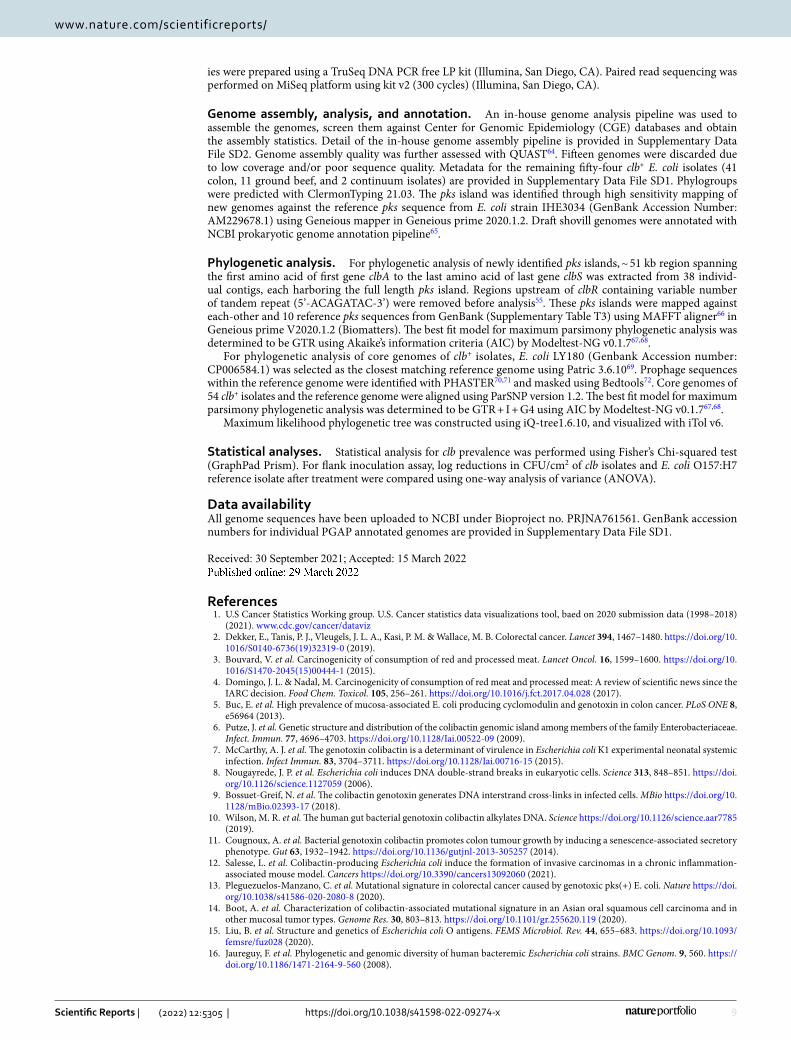
References 1. U.S Cancer Statistics Working group. U.S. Cancer statistics data visualizations tool, baed on 2020 submission data (1998–2018)
(2021). www. cdc. gov/ cancer/ datav iz 2. Dekker, E., Tanis, P. J., Vleugels, J. L. A., Kasi, P. M. & Wallace, M. B. Colorectal cancer. Lancet 394, 1467–1480. https:// doi. org/ 10.
1016/ S0140- 6736(19) 32319-0 (2019). 3. Bouvard, V. et al. Carcinogenicity of consumption of red and processed meat. Lancet Oncol. 16, 1599–1600. https:// doi. org/ 10.
1016/ S1470- 2045(15) 00444-1 (2015). 4. Domingo, J. L. & Nadal, M. Carcinogenicity of consumption of red meat and processed meat: A review of scientific news since the
IARC decision. Food Chem. Toxicol. 105, 256–261. https:// doi. org/ 10. 1016/j. fct. 2017. 04. 028 (2017). 5. Buc, E. et al. High prevalence of mucosa-associated E. coli producing cyclomodulin and genotoxin in colon cancer. PLoS ONE 8,
e56964 (2013). 6. Putze, J. et al. Genetic structure and distribution of the colibactin genomic island among members of the family Enterobacteriaceae.
Infect. Immun. 77, 4696–4703. https:// doi. org/ 10. 1128/ Iai. 00522- 09 (2009). 7. McCarthy, A. J. et al. The genotoxin colibactin is a determinant of virulence in Escherichia coli K1 experimental neonatal systemic
infection. Infect Immun. 83, 3704–3711. https:// doi. org/ 10. 1128/ Iai. 00716- 15 (2015). 8. Nougayrede, J. P. et al. Escherichia coli induces DNA double-strand breaks in eukaryotic cells. Science 313, 848–851. https:// doi.
org/ 10. 1126/ scien ce. 11270 59 (2006). 9. Bossuet-Greif, N. et al. The colibactin genotoxin generates DNA interstrand cross-links in infected cells. MBio https:// doi. org/ 10.
1128/ mBio. 02393- 17 (2018). 10. Wilson, M. R. et al. The human gut bacterial genotoxin colibactin alkylates DNA. Science https:// doi. org/ 10. 1126/ scien ce. aar77 85
(2019). 11. Cougnoux, A. et al. Bacterial genotoxin colibactin promotes colon tumour growth by inducing a senescence-associated secretory
phenotype. Gut 63, 1932–1942. https:// doi. org/ 10. 1136/ gutjnl- 2013- 305257 (2014). 12. Salesse, L. et al. Colibactin-producing Escherichia coli induce the formation of invasive carcinomas in a chronic inflammation-
associated mouse model. Cancers https:// doi. org/ 10. 3390/ cance rs130 92060 (2021). 13. Pleguezuelos-Manzano, C. et al. Mutational signature in colorectal cancer caused by genotoxic pks(+) E. coli. Nature https:// doi.
org/ 10. 1038/ s41586- 020- 2080-8 (2020). 14. Boot, A. et al. Characterization of colibactin-associated mutational signature in an Asian oral squamous cell carcinoma and in
other mucosal tumor types. Genome Res. 30, 803–813. https:// doi. org/ 10. 1101/ gr. 255620. 119 (2020). 15. Liu, B. et al. Structure and genetics of Escherichia coli O antigens. FEMS Microbiol. Rev. 44, 655–683. https:// doi. org/ 10. 1093/
femsre/ fuz028 (2020). 16. Jaureguy, F. et al. Phylogenetic and genomic diversity of human bacteremic Escherichia coli strains. BMC Genom. 9, 560. https://
doi. org/ 10. 1186/ 1471- 2164-9- 560 (2008).

10
Vol:.(1234567890)
Scientific Reports | (2022) 12:5305 | https://doi.org/10.1038/s41598-022-09274-x
www.nature.com/scientificreports/
17. Clermont, O. et al. Characterization and rapid identification of phylogroup G in Escherichia coli, a lineage with high virulence and antibiotic resistance potential. Environ. Microbiol. 21, 3107–3117. https:// doi. org/ 10. 1111/ 1462- 2920. 14713 (2019).
18. Nou, X. et al. Effect of chemical dehairing on the prevalence of Escherichia coli O157:H7 and the levels of aerobic bacteria and enterobacteriaceae on carcasses in a commercial beef processing plant. J Food Prot 66, 2005–2009. https:// doi. org/ 10. 4315/ 0362- 028x- 66. 11. 2005 (2003).
19. Barkocy-Gallagher, G. A. et al. Seasonal prevalence of Shiga toxin-producing Escherichia coli, including O157: H7 and non-O157 serotypes, and Salmonella in commercial beef processing plants. J. Food Protect. 66, 1978–1986. https:// doi. org/ 10. 4315/ 0362- 028x- 66. 11. 1978 (2003).
20. Marshall, K. E., Nguyen, T.-A., Ablan, M., Nichols, M. C., Robyn, M. P., Sundararaman, P., Whitlock, L., Wise, M. E., Jhung, M. A. Investigations of possible multistate outbreaks of Salmonella, Shiga toxin–producing Escherichia coli, and Listeria monocytogenes Infections 1–14 (Center for Disease Control, United States, 2016).
21. Heiman, K. E., Mody, R. K., Johnson, S. D., Griffin, P. M. & Gould, L. H. Escherichia coli O157 outbreaks in the United States, 2003–2012. Emerg. Infect. Dis. 21, 1293–1301. https:// doi. org/ 10. 3201/ eid21 08. 141364 (2015).
22. Vikram, A. et al. Impact of “Raised without Antibiotics” beef cattle production practices on occurrences of antimicrobial resistance. Appl. Environ. Microbiol. https:// doi. org/ 10. 1128/ AEM. 01682- 17 (2017).
23. Schmidt, J. W. et al. Occurrence of antimicrobial-resistant Escherichia coli and Salmonella enterica in the beef cattle production and processing continuum. Appl. Environ. Microbiol. 81, 713–725. https:// doi. org/ 10. 1128/ AEM. 03079- 14 (2015).
24. Schmidt, J. W. et al. Antimicrobial resistance in US retail ground beef with and without label claims regarding antibiotic use. J. Food Protect. 84, 827–842. https:// doi. org/ 10. 4315/ Jfp- 20- 376 (2021).
25. Kalchayanand, N., Koohmaraie, M. & Wheeler, T. L. Effect of exposure time and organic matter on efficacy of antimicrobial compounds against shiga toxin-producing Escherichia coli and Salmonella. J. Food Prot. 79, 561–568. https:// doi. org/ 10. 4315/ 0362- 028X. JFP- 15- 204 (2016).
26. Beghain, J., Bridier-Nahmias, A., Le Nagard, H., Denamur, E. & Clermont, O. ClermonTyping: An easy-to-use and accurate in silico method for Escherichia genus strain phylotyping. Microb. Genom. https:// doi. org/ 10. 1099/ mgen.0. 000192 (2018).
27. Joensen, K. G., Tetzschner, A. M., Iguchi, A., Aarestrup, F. M. & Scheutz, F. Rapid and easy in silico serotyping of Escherichia coli isolates by use of whole-genome sequencing data. J. Clin. Microbiol. 53, 2410–2426. https:// doi. org/ 10. 1128/ JCM. 00008- 15 (2015).
28. Larsen, M. V. et al. Multilocus sequence typing of total-genome-sequenced bacteria. J. Clin. Microbiol. 50, 1355–1361. https:// doi. org/ 10. 1128/ JCM. 06094- 11 (2012).
29. Zankari, E. et al. PointFinder: a novel web tool for WGS-based detection of antimicrobial resistance associated with chromosomal point mutations in bacterial pathogens. J. Antimicrob. Chemother. 72, 2764–2768. https:// doi. org/ 10. 1093/ jac/ dkx217 (2017).
30. Bortolaia, V. et al. ResFinder 4.0 for predictions of phenotypes from genotypes. J. Antimicro.b Chemother. 75, 3491–3500. https:// doi. org/ 10. 1093/ jac/ dkaa3 45 (2020).
31. Camacho, C. et al. BLAST+: Architecture and applications. BMC Bioinform. 10, 421. https:// doi. org/ 10. 1186/ 1471- 2105- 10- 421 (2009).
32. Joensen, K. G. et al. Real-time whole-genome sequencing for routine typing, surveillance, and outbreak detection of verotoxigenic Escherichia coli. J. Clin. Microbiol. 52, 1501–1510. https:// doi. org/ 10. 1128/ JCM. 03617- 13 (2014).
33. Malberg Tetzschner, A. M., Johnson, J. R., Johnston, B. D., Lund, O. & Scheutz, F. In silico genotyping of Escherichia coli isolates for extraintestinal virulence genes by use of whole-genome sequencing data. J. Clin. Microbiol. https:// doi. org/ 10. 1128/ JCM. 01269- 20 (2020).
34. He, Z. et al. Campylobacter jejuni promotes colorectal tumorigenesis through the action of cytolethal distending toxin. Gut 68, 289–300. https:// doi. org/ 10. 1136/ gutjnl- 2018- 317200 (2019).
35. Treangen, T. J., Ondov, B. D., Koren, S. & Phillippy, A. M. The Harvest suite for rapid core-genome alignment and visualization of thousands of intraspecific microbial genomes. Genome Biol. 15, 524. https:// doi. org/ 10. 1186/ s13059- 014- 0524-x (2014).
36. Nguyen, L.-T., Schmidt, H. A., von Haeseler, A. & Minh, B. Q. IQ-TREE: A fast and effective stochastic algorithm for estimating maximum-likelihood phylogenies. Mol. Biol. Evol. 32, 268–274. https:// doi. org/ 10. 1093/ molbev/ msu300 (2014).
37. Letunic, I. & Bork, P. Interactive Tree Of Life (iTOL) v5: An online tool for phylogenetic tree display and annotation. Nucl. Acids Res. 49, W293–W296. https:// doi. org/ 10. 1093/ nar/ gkab3 01 (2021).
38. Gordon, D. M. et al. Fine-scale structure analysis shows epidemic patterns of clonal complex 95, a cosmopolitan Escherichia coli lineage responsible for extraintestinal infection. mSphere https:// doi. org/ 10. 1128/ mSphe re. 00168- 17 (2017).
39. Dias, R. C. S., Moreira, B. M. & Riley, L. W. Use of fimH single-nucleotide polymorphisms for strain typing of clinical isolates of Escherichia coli for epidemiologic investigation. J. Clin. Microbiol. 48, 483–488. https:// doi. org/ 10. 1128/ Jcm. 01858- 09 (2010).
40. Dejea, C. M. et al. Patients with familial adenomatous polyposis harbor colonic biofilms containing tumorigenic bacteria. Science https:// doi. org/ 10. 1126/ scien ce. aah36 48 (2018).
41. Tsunematsu, Y. et al. Mother-to-infant transmission of the carcinogenic colibactin-producing bacteria. BMC Microbiol. 21, 235. https:// doi. org/ 10. 1186/ s12866- 021- 02292-1 (2021).
42. Tronnet, S. et al. The genotoxin colibactin shapes gut microbiota in mice. mSphere https:// doi. org/ 10. 1128/ mSphe re. 00589- 20 (2020).
43. Schmidt, J. W. et al. Antimicrobial resistance at Two U.S. cull cow processing establishments. J. Food Protect. 83, 2216–2228 (2020). 44. Kalchayanand, N. et al. Efficacy of antimicrobial compounds on surface decontamination of seven Shiga toxin-producing Escheri-
chia coli and Salmonella inoculated onto fresh beef. J. Food Prot. 78, 503–510. https:// doi. org/ 10. 4315/ 0362- 028X. JFP- 14- 268 (2015). 45. Wheeler, T. L., Kalchayanand, N. & Bosilevac, J. M. Pre- and post-harvest interventions to reduce pathogen contamination in the
U.S. beef industry. Meat Sci. 98, 372–382 (2014). 46. Grispoldi, L. et al. Bovine lymph nodes as a source of Escherichia coli contamination of the meat. Int. J. Food Microbiol. https://
doi. org/ 10. 1016/j. ijfoo dmicro. 2020. 108715 (2020). 47. Chitlapilly Dass, S. et al. Impact of mixed biofilm formation with environmental microorganisms on E. coli O157:H7 survival
against sanitization. npj Sci. Food https:// doi. org/ 10. 1038/ s41538- 020- 00076-x (2020). 48. Doster, E. et al. Metagenomic characterization of the microbiome and resistome of retail ground beef products. Front Microbiol.
https:// doi. org/ 10. 3389/ fmicb. 2020. 541972 (2020). 49. Miller, E., Vikram, A., Agga, G. E., Arthur, T. M. & Schmidt, J. W. Effects of in-feed chlortetracycline prophylaxis in beef cattle on
antimicrobial resistance genes. Foodborne Pathog. Dis. 15, 689–697. https:// doi. org/ 10. 1089/ fpd. 2018. 2475 (2018). 50. Auvray, F. et al. Insights into the acquisition of the pks island and production of colibactin in the Escherichia coli population. Microb.
Genom. https:// doi. org/ 10. 1099/ mgen.0. 000579 (2021). 51. Suresh, A. et al. Evolutionary dynamics based on comparative genomics of pathogenic Escherichia coli lineages harboring polyketide
synthase (pks) Island. MBio https:// doi. org/ 10. 1128/ mBio. 03634- 20 (2021). 52. Carlos, C. et al. Escherichia coli phylogenetic group determination and its application in the identification of the major animal
source of fecal contamination. BMC Microbiol. 10, 161. https:// doi. org/ 10. 1186/ 1471- 2180- 10- 161 (2010). 53. Higgins, J. et al. Genotyping of Escherichia coli from environmental and animal samples. J. Microbiol. Methods 70, 227–235. https://
doi. org/ 10. 1016/j. mimet. 2007. 04. 009 (2007). 54. Tenaillon, O., Skurnik, D., Picard, B. & Denamur, E. The population genetics of commensal Escherichia coli. Nat. Rev. Microbiol.
8, 207–217. https:// doi. org/ 10. 1038/ nrmic ro2298 (2010).

11
Vol.:(0123456789)
Scientific Reports | (2022) 12:5305 | https://doi.org/10.1038/s41598-022-09274-x
www.nature.com/scientificreports/
55. Wami, H. et al. Insights into evolution and coexistence of the colibactin- and yersiniabactin secondary metabolite determinants in enterobacterial populations. Microb. Genom. https:// doi. org/ 10. 1099/ mgen.0. 000577 (2021).
56. Stoppe, N. C. et al. Worldwide phylogenetic group patterns of Escherichia coli from commensal human and wastewater treatment plant isolates. Front Microbiol. 8, 2512. https:// doi. org/ 10. 3389/ fmicb. 2017. 02512 (2017).
57. Martin, P., Tronnet, S., Garcie, C. & Oswald, E. Interplay between siderophores and colibactin genotoxin in Escherichia coli. IUBMB Life 69, 435–441. https:// doi. org/ 10. 1002/ iub. 1612 (2017).
58. Sung, H. et al. Global cancer statistics 2020: GLOBOCAN estimates of incidence and mortality worldwide for 36 cancers in 185 Countries. CA Cancer J. Clin. 71, 209–249. https:// doi. org/ 10. 3322/ caac. 21660 (2021).
59. Stromberg, Z. R., Redweik, G. A. J. & Mellata, M. Detection, prevalence, and pathogenicity of non-O157 shiga toxin-producing Escherichia coli from cattle hides and carcasses. Foodborne Pathog. Dis. 15, 119–131. https:// doi. org/ 10. 1089/ fpd. 2017. 2401 (2018).
60. Arthur, T. M. et al. Longitudinal study of Escherichia coli O157:H7 in a beef cattle feedlot and role of high-level shedders in hide contamination. Appl. Environ. Microbiol. 75, 6515–6523. https:// doi. org/ 10. 1128/ aem. 00081- 09 (2009).
61. Bosilevac, J. M., Nou, X., Osborn, M. S., Allen, D. M. & Koohmaraie, M. Development and evaluation of an on-line hide decontami-nation procedure for use in a commercial beef processing plantt. J. Food Prot. 68, 265–272. https:// doi. org/ 10. 4315/ 0362- 028x- 68.2. 265 (2005).
62. Johnston, B. C. et al. Unprocessed red meat and processed meat consumption: Dietary guideline recommendations from the nutritional recommendations (NutriRECS) consortium. Ann. Intern. Med. 171, 756–764. https:// doi. org/ 10. 7326/ m19- 1621 (2019).
63. Gurjao, C. et al. Discovery and features of an alkylating signature in colorectal cancer. Cancer Discov. https:// doi. org/ 10. 1158/ 2159- 8290. Cd- 20- 1656 (2021).
64. Gurevich, A., Saveliev, V., Vyahhi, N. & Tesler, G. QUAST: Quality assessment tool for genome assemblies. Bioinformatics 29, 1072–1075. https:// doi. org/ 10. 1093/ bioin forma tics/ btt086 (2013).
65. Li, W. et al. RefSeq: expanding the prokaryotic genome annotation pipeline reach with protein family model curation. Nucl. Acids Res 49, D1020–D1028. https:// doi. org/ 10. 1093/ nar/ gkaa1 105 (2021).
66. Katoh, K. & Standley, D. M. MAFFT multiple sequence alignment software version 7: Improvements in performance and usability. Mol. Biol. Evol 30, 772–780. https:// doi. org/ 10. 1093/ molbev/ mst010 (2013).
67. Darriba, D. et al. ModelTest-NG: A new and scalable tool for the selection of DNA and protein evolutionary models. Mol. Biol. Evol. 37, 291–294. https:// doi. org/ 10. 1093/ molbev/ msz189 (2020).
68. Flouri, T. et al. The phylogenetic likelihood library. Syst. Biol. 64, 356–362. https:// doi. org/ 10. 1093/ sysbio/ syu084 (2015). 69. Davis, J. J. et al. The PATRIC bioinformatics resource center: Expanding data and analysis capabilities. Nucl. Acids Res. 48, D606–
D612. https:// doi. org/ 10. 1093/ nar/ gkz943 (2020). 70. Zhou, Y., Liang, Y., Lynch, K. H., Dennis, J. J. & Wishart, D. S. PHAST: A fast phage search tool. Nucl. Acids Res. 39, W347-352.
https:// doi. org/ 10. 1093/ nar/ gkr485 (2011). 71. Arndt, D. et al. PHASTER: A better, faster version of the PHAST phage search tool. Nucl. Acids Res. 44, W16-21. https:// doi. org/
10. 1093/ nar/ gkw387 (2016). 72. Quinlan, A. R. & Hall, I. M. BEDTools: A flexible suite of utilities for comparing genomic features. Bioinformatics 26, 841–842.
https:// doi. org/ 10. 1093/ bioin forma tics/ btq033 (2010).
AcknowledgementsThis work was funded by the Nebraska Beef Council (ARS #58-3040-0-015). Thanks to Jody Gallagher for sec-retarial assistance. We acknowledge Greg Smith, Bruce Jasch, and USMARC core sequencing facility for techni-cal assistance. Thanks to Dr. Frederick Auvray (IRSD, INSERM, Universite de Toulouse, INRAE, ENVT, UPS, Toulouse, France) for providing the full length pks sequences for E. coli strains KS-P003 and KS-P027.USDA is an equal opportunity provider and employer. Names are necessary to report factually on available data; however, the USDA neither guarantees nor warrants the standard of the product, and the use of the name by USDA implies no approval of the product to the exclusion of others that may also be suitable.
Author contributionsM.G. conceived the project, designed, and executed the study, collected and analyzed the data, and prepared original draft of the manuscript. J.W.S. provided consultation on isolate selection, provided the isolates. N.K. provided the consultation and collected data on beef flank inoculation. A.M.D. built and provided the in-house bioinformatic pipeline for genome analysis. J.M.B. conceptualized and supervised the study and revised the manuscript. M.G. and J.M.B. acquired the funding. All authors reviewed the manuscript.
Competing interests The authors declare no competing interests.
Additional informationSupplementary Information The online version contains supplementary material available at https:// doi. org/ 10. 1038/ s41598- 022- 09274-x.
Correspondence and requests for materials should be addressed to J.M.B.
Reprints and permissions information is available at www.nature.com/reprints.
Publisher’s note Springer Nature remains neutral with regard to jurisdictional claims in published maps and institutional affiliations.

12
Vol:.(1234567890)
Scientific Reports | (2022) 12:5305 | https://doi.org/10.1038/s41598-022-09274-x
www.nature.com/scientificreports/
Open Access This article is licensed under a Creative Commons Attribution 4.0 International License, which permits use, sharing, adaptation, distribution and reproduction in any medium or
format, as long as you give appropriate credit to the original author(s) and the source, provide a link to the Creative Commons licence, and indicate if changes were made. The images or other third party material in this article are included in the article’s Creative Commons licence, unless indicated otherwise in a credit line to the material. If material is not included in the article’s Creative Commons licence and your intended use is not permitted by statutory regulation or exceeds the permitted use, you will need to obtain permission directly from the copyright holder. To view a copy of this licence, visit http:// creat iveco mmons. org/ licen ses/ by/4. 0/.
© The Author(s) 2022
Copyright © 2022 FDOKUMEN