An Influenza AH1N1 2009 Hemagglutinin Vaccine Produced in Escherichia coli
14
An Influenza A/H1N1/2009 Hemagglutinin Vaccine Produced in Escherichia coli Jose ´ M. Aguilar-Ya ´n ˜ ez 1 , Roberto Portillo-Lara 1 , Gonzalo I. Mendoza-Ochoa 1 , Sergio A. Garcı´a-Echauri 1 , Felipe Lo ´ pez-Pacheco 1 , David Bulnes-Abundis 1 , Johari Salgado-Gallegos 1 , Itzel M. Lara-Mayorga 1 , Yenny Webb-Vargas 1 , Felipe O. Leo ´ n-Angel 1 , Ramo ´ n E. Rivero-Aranda 1 , Yuriana Oropeza-Almaza ´n 1 , Guillermo M. Ruiz-Palacios 1 , Manuel I. Zertuche-Guerra 1 , Rebecca M. DuBois 2 , Stephen W. White 2 , Stacey Schultz-Cherry 3 , Charles J. Russell 3 , Mario M. Alvarez 1 * 1 Centro de Biotecnologı ´a-FEMSA, Tecnolo ´ gico de Monterrey at Monterrey, Monterrey, Me ´xico, 2 Department of Structural Biology, St. Jude Children’s Research Hospital, Memphis, Tennessee, United States of America, 3 Department of Infectious Diseases, St. Jude Children’s Research Hospital, Memphis, Tennessee, United States of America Abstract Background: The A/H1N1/2009 influenza pandemic made evident the need for faster and higher-yield methods for the production of influenza vaccines. Platforms based on virus culture in mammalian or insect cells are currently under investigation. Alternatively, expression of fragments of the hemagglutinin (HA) protein in prokaryotic systems can potentially be the most efficacious strategy for the manufacture of large quantities of influenza vaccine in a short period of time. Despite experimental evidence on the immunogenic potential of HA protein constructs expressed in bacteria, it is still generally accepted that glycosylation should be a requirement for vaccine efficacy. Methodology/Principal Findings: We expressed the globular HA receptor binding domain, referred to here as HA 63–286 - RBD, of the influenza A/H1N1/2009 virus in Escherichia coli using a simple, robust and scalable process. The recombinant protein was refolded and purified from the insoluble fraction of the cellular lysate as a single species. Recombinant HA 63–286 - RBD appears to be properly folded, as shown by analytical ultracentrifugation and bio-recognition assays. It binds specifically to serum antibodies from influenza A/H1N1/2009 patients and was found to be immunogenic, to be capable of triggering the production of neutralizing antibodies, and to have protective activity in the ferret model. Conclusions/Significance: Projections based on our production/purification data indicate that this strategy could yield up to half a billion doses of vaccine per month in a medium-scale pharmaceutical production facility equipped for bacterial culture. Also, our findings demonstrate that glycosylation is not a mandatory requirement for influenza vaccine efficacy. Citation: Aguilar-Ya ´n ˜ ez JM, Portillo-Lara R, Mendoza-Ochoa GI, Garcı ´a-Echauri SA, Lo ´ pez-Pacheco F, et al. (2010) An Influenza A/H1N1/2009 Hemagglutinin Vaccine Produced in Escherichia coli. PLoS ONE 5(7): e11694. doi:10.1371/journal.pone.0011694 Editor: Neeraj Vij, Johns Hopkins School of Medicine, United States of America Received March 1, 2010; Accepted June 9, 2010; Published July 22, 2010 Copyright: ß 2010 Aguilar-Ya ´n ˜ ez et al. This is an open-access article distributed under the terms of the Creative Commons Attribution License, which permits unrestricted use, distribution, and reproduction in any medium, provided the original author and source are credited. Funding: This work was funded by the Zambrano-Hellion family, FEMSA, and Tecnologico de Monterrey (seed fund CAT-122). This work was also funded by the National Institutes of Health, National Institute of Allergy and Infectious Diseases, under contract number HHSN266200700005C, Cancer Center core grant CA21765, the American Lebanese Syrian Associated Charities (ALSAC), and the Childrens Infection Defense Center (CIDC) at St. Jude Children’s Research Hospital. The funders had no role in study design, data collection and analysis, decision to publish, or preparation of the manuscript. Competing Interests: The authors declare that they submitted a patent application related to the ELISA protocol referred to in the manuscript. (Alvarez M.M. et al; patent application MX/a/2009/014098; December 18, 2009). The authors confirm that this does not alter their adherence to all the PLoS ONE policies on sharing data and materials, as detailed online in the guide for authors. * E-mail: [email protected] Introduction The emergence of pandemic H1N1 subtype influenza in April 2009 emphasizes the need for rapid methods to manufacture large quantities of influenza vaccine. To curtail a second wave of influenza A/H1N1/2009 in the U.S.A, it was estimated that up to 70% of citizens would need to be vaccinated by the Fall of 2009 [1]. More than 20% vaccination coverage has been proposed based on other reports [2]. While 20% vaccine coverage was at least partially achieved in some First World European countries, in nations such as Me ´xico (the epidemiological epicenter of the current pandemic), sufficient vaccine dosages were not available even by March 2010. All commercial influenza vaccines are produced by propagating the virus in embryonated chicken eggs. Further processing is then needed to separate and inactivate viral particles and to purify the hemagglutinin (HA) protein, the primary vaccine antigen. This technology is slow and requires one embryonated egg per vaccine dose [3]. To vaccinate one third of the population in the United States and Me ´xico, 150 million eggs would be required, and an additional 150 million doses would be needed for the rest of Latin America. Several alternative strategies have been proposed to produce pandemic and seasonal influenza vaccines [4,5]. These include viral culture in mammalian cells [5–7] and the use of recombinant proteins [3,8–12]. The concept of producing subunit influenza vaccines was first proposed three decades ago [13]. The expression and purification of a single antigenic protein in bacterial culture [3,10,11] may be the simplest and fastest strategy for generating PLoS ONE | www.plosone.org 1 July 2010 | Volume 5 | Issue 7 | e11694
Transcript of An Influenza AH1N1 2009 Hemagglutinin Vaccine Produced in Escherichia coli

An Influenza A/H1N1/2009 Hemagglutinin VaccineProduced in Escherichia coliJose M. Aguilar-Yanez1, Roberto Portillo-Lara1, Gonzalo I. Mendoza-Ochoa1, Sergio A. Garcıa-Echauri1,
Felipe Lopez-Pacheco1, David Bulnes-Abundis1, Johari Salgado-Gallegos1, Itzel M. Lara-Mayorga1,
Yenny Webb-Vargas1, Felipe O. Leon-Angel1, Ramon E. Rivero-Aranda1, Yuriana Oropeza-Almazan1,
Guillermo M. Ruiz-Palacios1, Manuel I. Zertuche-Guerra1, Rebecca M. DuBois2, Stephen W. White2,
Stacey Schultz-Cherry3, Charles J. Russell3, Mario M. Alvarez1*
1 Centro de Biotecnologıa-FEMSA, Tecnologico de Monterrey at Monterrey, Monterrey, Mexico, 2 Department of Structural Biology, St. Jude Children’s Research Hospital,
Memphis, Tennessee, United States of America, 3 Department of Infectious Diseases, St. Jude Children’s Research Hospital, Memphis, Tennessee, United States of America
Abstract
Background: The A/H1N1/2009 influenza pandemic made evident the need for faster and higher-yield methods for theproduction of influenza vaccines. Platforms based on virus culture in mammalian or insect cells are currently underinvestigation. Alternatively, expression of fragments of the hemagglutinin (HA) protein in prokaryotic systems canpotentially be the most efficacious strategy for the manufacture of large quantities of influenza vaccine in a short period oftime. Despite experimental evidence on the immunogenic potential of HA protein constructs expressed in bacteria, it is stillgenerally accepted that glycosylation should be a requirement for vaccine efficacy.
Methodology/Principal Findings: We expressed the globular HA receptor binding domain, referred to here as HA63–286-RBD, of the influenza A/H1N1/2009 virus in Escherichia coli using a simple, robust and scalable process. The recombinantprotein was refolded and purified from the insoluble fraction of the cellular lysate as a single species. Recombinant HA63–286-RBD appears to be properly folded, as shown by analytical ultracentrifugation and bio-recognition assays. It bindsspecifically to serum antibodies from influenza A/H1N1/2009 patients and was found to be immunogenic, to be capable oftriggering the production of neutralizing antibodies, and to have protective activity in the ferret model.
Conclusions/Significance: Projections based on our production/purification data indicate that this strategy could yield upto half a billion doses of vaccine per month in a medium-scale pharmaceutical production facility equipped for bacterialculture. Also, our findings demonstrate that glycosylation is not a mandatory requirement for influenza vaccine efficacy.
Citation: Aguilar-Yanez JM, Portillo-Lara R, Mendoza-Ochoa GI, Garcıa-Echauri SA, Lopez-Pacheco F, et al. (2010) An Influenza A/H1N1/2009 HemagglutininVaccine Produced in Escherichia coli. PLoS ONE 5(7): e11694. doi:10.1371/journal.pone.0011694
Editor: Neeraj Vij, Johns Hopkins School of Medicine, United States of America
Received March 1, 2010; Accepted June 9, 2010; Published July 22, 2010
Copyright: � 2010 Aguilar-Yanez et al. This is an open-access article distributed under the terms of the Creative Commons Attribution License, which permitsunrestricted use, distribution, and reproduction in any medium, provided the original author and source are credited.
Funding: This work was funded by the Zambrano-Hellion family, FEMSA, and Tecnologico de Monterrey (seed fund CAT-122). This work was also funded by theNational Institutes of Health, National Institute of Allergy and Infectious Diseases, under contract number HHSN266200700005C, Cancer Center core grantCA21765, the American Lebanese Syrian Associated Charities (ALSAC), and the Childrens Infection Defense Center (CIDC) at St. Jude Children’s Research Hospital.The funders had no role in study design, data collection and analysis, decision to publish, or preparation of the manuscript.
Competing Interests: The authors declare that they submitted a patent application related to the ELISA protocol referred to in the manuscript. (Alvarez M.M. etal; patent application MX/a/2009/014098; December 18, 2009). The authors confirm that this does not alter their adherence to all the PLoS ONE policies on sharingdata and materials, as detailed online in the guide for authors.
* E-mail: [email protected]
Introduction
The emergence of pandemic H1N1 subtype influenza in April
2009 emphasizes the need for rapid methods to manufacture large
quantities of influenza vaccine. To curtail a second wave of
influenza A/H1N1/2009 in the U.S.A, it was estimated that up to
70% of citizens would need to be vaccinated by the Fall of 2009 [1].
More than 20% vaccination coverage has been proposed based on
other reports [2]. While 20% vaccine coverage was at least partially
achieved in some First World European countries, in nations such as
Mexico (the epidemiological epicenter of the current pandemic),
sufficient vaccine dosages were not available even by March 2010.
All commercial influenza vaccines are produced by propagating
the virus in embryonated chicken eggs. Further processing is then
needed to separate and inactivate viral particles and to purify the
hemagglutinin (HA) protein, the primary vaccine antigen. This
technology is slow and requires one embryonated egg per vaccine
dose [3]. To vaccinate one third of the population in the United
States and Mexico, 150 million eggs would be required, and an
additional 150 million doses would be needed for the rest of Latin
America.
Several alternative strategies have been proposed to produce
pandemic and seasonal influenza vaccines [4,5]. These include
viral culture in mammalian cells [5–7] and the use of recombinant
proteins [3,8–12]. The concept of producing subunit influenza
vaccines was first proposed three decades ago [13]. The expression
and purification of a single antigenic protein in bacterial culture
[3,10,11] may be the simplest and fastest strategy for generating
PLoS ONE | www.plosone.org 1 July 2010 | Volume 5 | Issue 7 | e11694

large quantities of new influenza vaccines. In fact, the develop-
ment of a bacterial clone capable of producing an antigen against
a new influenza strain would require less than one week, and
scaling up production using bioreactors would allow the
generation of hundreds of thousands of doses in less than a day.
Moreover, recombinant vaccines produced in bacteria, free of
other viral and cellular components, are expected to reduce
complications associated with whole virus vaccines such as
pyogenic reaction and Guillain-Barre syndrome [13].
One concern is that complete viral particles may be orders of
magnitude more immunogenic than recombinant peptides [11]
because the former are polyantigenic and undergo post-transcrip-
tional modifications such as glycosylation. Commercial vaccines
based on recombinant technology are presented as ‘‘virus like
particles’’ and/or are expressed in eukaryotic systems capable of
glycosylation. For example, GARDASILH (Merck) against Human
Papilloma virus, and RecombivaxH (Merck) against Hepatitis B
virus are expressed in Saccharomyces cerevisiae. Nonetheless, multiple
and single antigen experimental vaccines produced in bacteria
have proved to be protective in animal models [14,15]. In the case
of influenza viruses, there is experimental evidence to suggest that
HA glycosylation might be important for proper folding [16] and
virus-host receptor recognition [5,6,17–19], but not for immuno-
genicity to any significant degree [3,10,11,19–21].
Results
Design and expression of the HA receptor-bindingdomain
In this paper, we document the production of a recombinant
HA receptor-binding domain (HA RBD) in E. coli that specifically
binds serum antibodies from positive influenza A H1N1/2009
patients. When intramuscularly administered, the protein triggers
a specific immune response, produces neutralizing antibodies, and
provides protection against influenza A/H1N1/2009 challenge in
ferrets. This 25 kDa protein comprises a highly conserved region
of the HA1 domain of the hemagglutinin of the A/H1N1/2009
virus spanning amino acids 63 to 286 of the native sequence, and
is therefore designated HA63–286-RBD (residues 55–271 in H3
numbering, v.gr. Accession No. ACQ99608) (Figure 1a, 1b). In
addition, it contains all of the predicted antigenic sites for the HA
protein of the A/H1N1/2009 strain [22]. A sequence encoding a
six-histidine purification tag was added at the N-terminus of the
protein, and an enterokinase cleavage site (EKCS) was added to
facilitate tag removal (Figure 1c, 1d, 1e).
The selection of this precise HA subdomain purposely excludes all
residues of the metastable HA2 stalk domain including the
hydrophobic fusion peptide and transmembrane domain. Computer
simulations predict that the isolated HA1 receptor-binding domain
has much less surface hydrophobicity than the entire HA protein
ectodomain (compare Figure 2a and 2b) while still preserving its
antigenic structure (Figure 2c). For these simulations, the full length
HA of the Influenza A H1N1/1918 virus [23] was taken as a
template for the estimation of the most probable structure of protein
HA63–286-RBD. Given the close similitude in primary and tertiary
structure between the HA H1N1/2009 and the HA H1N1/1918
protein [22], the accuracy of the predicted structure of the HA63–286-
RBD is expected to be high. Hydrophobicity minimization is
generally conducive to higher expression levels in E. coli which
typically recognize prominent hydrophobic regions as being
misfolded and subsequently degrade them [24]. Indeed, in our
experiments, high production levels of the complete H1N1/2009
Figure 1. Construction of HA63–286-RBD. (A) Crystal structure of HA protein (PDB entry 1RUY) showing HA1 (light grey), HA2 (dark grey), and theglobular domain of HA63–286-RBD (red) used for these studies. (B) Schematic for the construction of HA63–286-RBD. The cDNA sequence encodingresidues 63–286 of influenza A H1N1 virus (without transmembrane regions) was cloned for expression in Escherichia coli. (C) Schematicrepresentation of the HA63–286-RBD containing an N-terminal 66Histidine tag and an enterokinase cleavage sequence (EkCS). (D) Same as (C) exceptthat this construct contains a periplasmic signal sequence. (E) Amino acid sequence of the N-terminus in both (C) and (D). * indicates the enterokinasecleavage site.doi:10.1371/journal.pone.0011694.g001
An H1N1 Vaccine in E. coli
PLoS ONE | www.plosone.org 2 July 2010 | Volume 5 | Issue 7 | e11694
HA1 subunit were not achievable using either conventional E. coli
strains or E. coli strains BL21 (DE3) pLysS variants C41 and C43
from LucigenH Corporation (Middleton, WI) which are known
to successfully express transmembrane proteins [25,26]. Protein
HA63–286-RBD was expressed in both E. coli Rosetta-gami and C41,
but not in C43. Producer strains were deposited at ATCCH under
Patent Deposit Designation PTA-10320. In terms of global yield and
growth rate, clones derived from the E. coli C41 strain were more
suitable for large-scale production (Figure 3a). Specific growth rates
of 1.69 h21 and average yields of 3.4 g/L were observed at 5L scale
settings after 12 h cultivation.
Protein recovery, purification and refoldingIn these culture conditions, practically all of the recombinant
protein was produced as insoluble inclusion bodies (Figure 3b).
Although this facilitated the primary recovery of protein, the
Figure 2. Surface hydrophobicity and antigenic structure of protein HA63–286-RBD. (A) Hydrophobic (red) and hydrophilic regions (blue) atthe surface of protein HA63–286-RBD calculated by simulations; (B) The hydrophobicity map of the HA1 subunit expressed by Chiu et al (10) ispresented for comparison. (C) Simulation results show that protein HA63–286-RBD preserves the conformational antigenic sites Sa, Sb, Ca1, Ca2, Cbcomputationally predicted by Igarashi et al. [22] for the HA of the influenza A H1N1/CA2009 virus. Three dimensional structures were obtained usingSwiss-model. The full length HA of the Influenza A H1N1/1918 virus [23] was taken as a template for the estimation of the most probable structure ofprotein HA63–286-RBD. Visualization and highlighting of immunogenic sites was done using UCSF-Chimera. The structure of the antigenic epitopes ofthe HA of the influenza A H1N1/CA2009 virus was taken from Igarashi et al. [22]. They are also consistent with structural data published recently by Xuet al [41].doi:10.1371/journal.pone.0011694.g002
An H1N1 Vaccine in E. coli
PLoS ONE | www.plosone.org 3 July 2010 | Volume 5 | Issue 7 | e11694

proper folding and resulting bioactivity of the recombinant protein
recovered from inclusion bodies requires an effective method to
solubilize, refold and purify the protein [10,24]. By optimizing a
recovery and refolding procedure, we eventually obtained a
bioactive protein that recognizes antibodies from serum of H1N1/
2009 positive patients and provides protection against virus
infection in the ferret model. Briefly, HA63–286-RBD was
recovered using standard chemical lysis procedures, dissolved in
8M urea, and refolded and purified by Immobilized Metal Affinity
Chromatography (IMAC) using 400 mM arginine in PBS at pH 8
(Figure 3c). The refolded protein was eluted using 150 mM
imidazole at pH 7. This simple purification scheme produced
HA63–286-RBD solutions in the range of 400 to 650 mg/L with
purities exceeding 99.5%, as estimated by microelectrophoresis
using an ExperionH platform from Bio-rad (Hercules, CA). At the
present time, we observe an overall process yield of <0.02 g/L of
bioreactor volume. After process optimization, average overall
yields of 0.5–1.0 g/L (refolded protein per Liter of bacterial
culture) could be expected.
We further characterized the folded state of protein HA63–286-
RBD in solution by analytical ultracentrifugation, specifically
using sedimentation velocity and equilibrium analysis assays. Both
experiments showed that HA-RBD exists mainly as a monomer in
solution (Figure 4), and there are no dimmers observed in the c(s)
distribution profile (Figure 4a) at the concentration used. The
analytical results are presented in Table 1. The frictional ratio
value (f/f0 – value) of 1.30 reflects a slightly elongated globular
protein, consistent with the predicted three-dimensional structure.
The standard s-value, s020,w (water as solvent at 20uC and zero
concentration), and frictional ratio calculated with the standard
s-value (in parenthesis) are also listed in Table 1. The
sedimentation equilibrium data do not fit quite as well to a
discrete single monomer species model which predicts a mass
value of 28,585 Da, slightly larger than the monomeric molecular
weight. The dissociation equilibrium constant of the monomer-
dimmer self-association model determined from the equilibrium
data is KD = 288 mM, and this suggests a very weak dimerization
interaction (root mean square deviation of the model was 0.0037
absorbance units at 280 nm; Figure 4b).
HA63–286-RBD specifically recognizes antibodies fromH1N1-infected subjects
The resulting HA63–286-RBD protein is specifically recognized
by antibodies in serum samples from patients positive for the 2009
H1N1 virus (Figure 5). In comparative experiments, serum from
positive patients diagnosed by the RT-PCR protocols established
by the CDC and recommended by the WHO [27], or serum from
subjects negative for influenza A H1N1, were measured in an
HA63–286-RBD-specific ELISA as described by Alvarez et al. [28].
At 1:50 dilution, the absorbance signal observed in samples from
positive patients was between 2 to 4-fold higher when compared to
signal from samples from negative subjects (Figure 5a).
To establish if HA63–286-RBD obtained from inclusion bodies is
properly folded, a soluble form of HA63–286-RBD was produced by
expression in E. coli BL21 (DE3) pLysS variant C41 using a genetic
construction that included a signal peptide for periplasmic
expression [29] and a 6His tag sequence (Figure 1d). Extraction
from the periplasmic space was performed using a saline gradient,
and practically all of the protein was found in solution, as
confirmed by a western blot assay using anti-histidine antibodies.
The protein was recovered and purified by affinity chromatogra-
phy using its histidine tag. Yields of this soluble version of HA63–
286-RBD are two orders of magnitude lower than its refolded
analog, which would make its large scale production unfeasible,
but soluble HA63–286-RBD is useful as a reference for proper
folding.
Figure 3. Expression of HA63–286-RBD in E. coli. (A) Protein profile of cell lysates from culture experiments of E. coli C41, BL21 (DE3) pLysS orRosetta-gami transformed with genes to produce (1) GFP+histidine tag (clone C41 1); (2) GFP+histidine tag (clone C41 2); (3) GFP+histidine tag (cloneC41 3); (4) negative control, C41(5) HA63–286-RBD (clone C41 1); (6) HA63–286-RBD (clone C41 2); (7) HA63–286-RBD (clone Rosetta-gami clone 1); (8)HA63–286-RBD (clone Rosetta-gami clone 2). (9) Precision Plus Kaleidoscope molecular mass ruler showing 25 kD (pink) and 20 kD (blue) bands. Theblue arrow indicates the 26 kD band corresponding to HA63–286-RBD. (B) SDS-PAGE showing (1) the soluble and (2) insoluble fraction of the C41 strainlysate after 8 hours induction with 1mM IPTG. (C) SDS-PAGE showing the protein profiles at different stages of recovery, purification and on-columnrefolding. (1) Crude lysate of the 8M urea solubilized inclusion bodies, (2) Unbound fraction, (3) 1st wash step, (4) 2nd wash step, (5,6) refolding steps,(7–12) Elution fraction using imidazole 300 mM, (13) chromatographic resin. (M) Precision Plus Kaleidoscope molecular mass ruler.doi:10.1371/journal.pone.0011694.g003
An H1N1 Vaccine in E. coli
PLoS ONE | www.plosone.org 4 July 2010 | Volume 5 | Issue 7 | e11694

Selective biorecognition of native soluble and refolded HA63–286-
RBD by antibodies from the sera of positive influenza A H1N1/
2009 subjects was then compared. Refolded HA63–286-RBD
exhibited more than 90% selective biorecognition with respect to
native soluble HA63–286-RBD. This was a consistent observation
among different batches of product (Figure 5b).
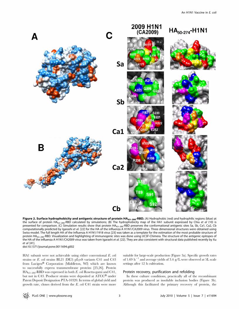
HA63–286-RBD is immunogenic in ferretsThe immunogenic and protective potential of HA63–286-RBD
was evaluated in experiments with ferrets, the preferred animal
model for influenza studies [30–33]. Sixteen ferrets were
intramuscularly administered with different doses of HA63–286-
RBD with or without adjuvant as described in Table 2. Five
additional animals were used as non-vaccinated controls. Serum
was collected at different times post-vaccination for 34 days and
analyzed for HA-specific antibodies by ELISA. Our results suggest
that the immune response to the HA63–286-RBD prime alone is
variable. Most animals (56.25%) exhibited a moderate primary
response that was clearly observable by day 6 or 7 post-vaccination
(as in ferret 4C; Figure 5b). Others (43.75%) did not display any
significant primary response (as in ferret 4A; Figure 6a). However,
all ferrets subjected to a boost at day fifteen exhibited a significant
Figure 4. Analytical ultracentrifugation of HA63–286-RBD. (A) The sedimentation velocity profiles (fringe displacement) were fitted to acontinuous sedimentation coefficient distribution model c(s). The experiment was conducted at a loading protein concentration of 0.45 mg/mL in10 mM Tris pH 8.0, 100 mM NaCl at 20uC and at a rotor speed of 60,000 rpm. The s-values of the proteins are listed in Table 1. (B) Absorbance scansat 280 nm at equilibrium are plotted versus the distance from the axis of rotation. The protein was centrifuged in the above buffer at 4uC for at least24 h at each rotor speed of 20, 30 and 38 k rpm. The solid lines represent the global nonlinear least squares best-fit of all the data sets to a monomer-dimer self-association model with a very weak KD (288 mM). For clarity, only the loading protein concentration of 5 mM is shown. The r.m.s. deviationfor this fit was 0.0037 absorbance units.doi:10.1371/journal.pone.0011694.g004
An H1N1 Vaccine in E. coli
PLoS ONE | www.plosone.org 5 July 2010 | Volume 5 | Issue 7 | e11694

sera IgG response between four and six days after the boost
injection. No statistically significant difference was observed
between the 125 mg treatments with or without adjuvant
(Figure 6a). However, our results suggest that the antibody
response is dose dependent. Figures 6c and 6d show the specific
antibody levels observed in animals vaccinated with 125 mg and
200 mg dosages. The antibody response was similar at both dosage
levels after the prime, but after the boost the group primed with
200 mg of protein and boosted at day 23 had ,1.5 fold higher HA-
specific IgG levels as compared to the 125 mg primed and boosted
group. As discussed later, this difference in immunological
response does not conclusively imply a higher protective efficacy
of the 200 mg dose. A 275 mg dosage, the highest dosage tested,
also did not increase the primary immunogenic response
compared to those observed for 125 and 200 mg dosages. Overall,
the results demonstrate that HA63–286-RBD is immunogenic in the
ferret model and is capable of triggering a specific IgG immune
response.
Table 3 presents the results of a neutralization assay conducted
on selected serum samples from immunized ferrets (ferret 1A, 2C,
2D, 2E, 4A, 4C, 4D, 4E) and a non-vaccinated control (ferret C3).
The study was conducted such that all dosage groups were
represented. All samples were tested at a dilution of 1:40. This
dilution was similar to the one used for the ELISA. Convention-
ally, it is accepted that a serum sample that neutralizes virus
infection at a dilution of 1:40 or higher will be at least partially
protective [30]. All samples tested from vaccinated ferrets resulted
in neutralization in at least 50% of the conducted replicates (see
fourth column in Table 3). No obvious correlation can be
established between neutralization potential and vaccine dosage,
and even a single dosage of 125 mg was found to be capable of
stimulating the production of neutralizing antibodies. Pre-immune
samples and dilutions of 1:160 of the vaccinated ferrets resulted
non-neutralizing. Similarly, samples from a non-vaccinated
control that became severely symptomatic after viral challenge
(ferret C3) did not neutralized virus infection.
HA63–286-RBD protects ferrets against challenge by H1N1influenza virus
Twenty-one ferrets (sixteen vaccinated and five non-vaccinated
controls) were then challenged with influenza A/H1N1/2009
virus. The virus was cultured in MDCK cells in our laboratory
using techniques reported elsewhere [6]. The identity of the virus
was confirmed by a WHO-recommended real time RT-PCR
protocol [27]. Briefly, ferrets were lightly anesthetized and a
200 mL aliquot of virus suspension from a cell culture supernatant
with a virus titer of 105.83 TCID50 mL21 was administered
intranasally at day 0 of the challenge experiment, 45 days after first
vaccination. Body temperature (as measured by a microchip
implanted in the animals four days before the challenge), weight,
sneezing and the presence and appearance of mucus were
monitored for one week after the challenge in all animals. The
efficacy of infection was determined by verifying the presence of
influenza A/H1N1/2009 virus through real time RT-PCR in
pharyngeal samples taken at days 2 and 4 after challenge. All
animals tested positive for H1N1 infection, with CT values in the
range of 20 to 25 cycles at both days 2 and 4 post infection. These
values are considered high in human samples, implying that
infective concentrations of the virus were successfully delivered to
each experimental subject.
Table 2 shows a summary of symptoms displayed by each ferret
based on four commonly accepted indicators of disease [31,32].
Each index was constructed such that its value range falls between
0 and 5. An overall sickness index (OSI) resulted from the
weighted addition of all four individual symptom indexes,
according to the expression:
OSIInfluenza~0:30 IndexDweigth
� �z0:30 IndexDTð Þ
z0:20 Indexsneezing
� �z0:20 Indexmuccusð Þ
ð1Þ
Weights were empirically assigned based on the relative
significance of the symptom; increases in temperature and weight
loss were assumed more relevant and objective than mucus quality
and sneezing. Visual inspection of data revealed that lower intensity
influenza symptoms were observed in vaccinated ferrets. The
overall sickness index (OSI) provides a better estimate of the severity
of symptoms in vaccinated and non-vaccinated animals. The
average OSI for all ferrets was 1.6 units, and ferrets with OSI values
lower than this value were rated as slightly symptomatic. Subjects
with OSI values between 1.8 and 2.0 were rated as moderately
symptomatic, and animals with OSI values above 2.3 units were
rated as severely symptomatic. From the vaccinated population,
only 6.25% (1/16) of the animals were identified as severely
symptomatic and 18.75% (3/16) exhibited moderate influenza
symptoms. In contrast, 100% of non-vaccinated ferrets (5 out of 5
subjects) exhibited severe influenza symptoms. Ferret 2E, the only
vaccinated ferret that displayed severe influenza symptoms, received
a single but relatively high dose of protein HA63–286-RBD that
resulted in high titers of specific antibodies (as measured by ELISA
[28]). In addition, 1:40 dilutions of its serum samples were capable
of neutralizing virus infection in MCDK cultures (Table 3).
OSI values did not exhibit a normal distribution, and a Mann-
Whitney test based on median comparisons was therefore conducted.
OSI was significantly lower in the vaccinated than in the non-
vaccinated group (Pvalue,0.001). Comparatively, all individual sickness
Table 1. Summary of results of velocity experiment of HA-RBD in 10 mM Tris pH 8, 100 mM NaCl at 20uC.
Samplea s20 (Svedberg)b s020,w (Svedberg)c M (Da)d f/f0
e
Monomeric HA-RBD (0.45) 2.45 (92%) 2.51 26,700 (26,378) 1.30(1.29)
others (0.03) 0.88 (8%) N/D N/D N/D
aConcentration of peak in mg/mL in parenthesis.bSedimentation coefficient taken from the ordinate maximum of each peak in the best-fit c(s) distribution at 20uC with percentage protein amount in parenthesis.Sedimentation coefficient (s-value) is a measure of the size and shape of a protein in a solution with a specific density and viscosity at a specific temperature.cStandard sedimentation coefficient (s0
20,w -value) at zero concentration, in water at 20uC.dMolar mass values taken from the c(s) distribution that was transformed to the c(M) distribution. The theoretical mass of the monomer is given in parenthesis.eBest-fit weight-average frictional ratio values (f/f0)w taken from the c(s) distribution. The frictional ratios calculated with s0
20,w -values via the v-bar method (SEDNTERP)is in parenthesis.doi:10.1371/journal.pone.0011694.t001
An H1N1 Vaccine in E. coli
PLoS ONE | www.plosone.org 6 July 2010 | Volume 5 | Issue 7 | e11694

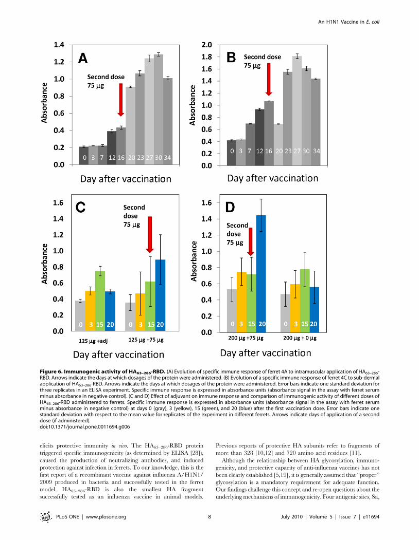
indicators were significantly higher in non-vaccinated than in
vaccinated ferrets (Figure 7a,b). Average IndexDT was significantly
higher in non-vaccinated ferrets according to a T test (Pvalue,0.001). In
non-vaccinated ferrets the average maximum weight loss was
significantly more severe (T test; Pvalue = 0.056) and the average
Indexmucus was also significantly higher (T test; Pvalue,0.023). Values
for Indexsneezing did not exhibit a normal distribution and were
analyzed using a sign test based on a comparison of medians.
Indexsneezing was significantly higher in non-vaccinated ferrets
(Pvalue = 0.001). Significant differences in temperature excursion
profiles and weight loss profiles were observed in vaccinated and
non-vaccinated ferrets (Figure 7c,d). At the moment of sacrifice (three
weeks after challenge), all non-vaccinated animals tested positive to the
H1N1/2009 virus by PCR, while only 40% of immunized animals did.
Discussion
Is glycosylation crucial for efficacy?Our results suggest that the non-glycosylated, refolded HA1
receptor-binding domain produced in E. coli is immunogenic and
Figure 5. Serum from patients infected with Influenza A H1N1/2009 specifically recognize HA63–286-RBD. (A) Bars 1–8, in gray tones,correspond to absorbance signals from non-exposed subjects (samples taken from March to May 2008). Bar 9, in black, shows the averageabsorbance value of samples 1 to 8. Bars 10 to 14, shown in colour, correspond to absorbance signals from Influenza A/H1N1 negative subjects. Bars15–23, shown in colour, correspond to absorbance signals from samples of Influenza A H1N1 positive subjects (diagnosed two or three weekspreviously by RT-PCR). Error bars represent one standard deviation (B) Proper refolding (biorecognition of antibodies from a positive patient), wasevaluated for 4 different production batches of HA63–286-RBD. Batch 5 is a reference batch where HA63–286-RBD was expressed in its soluble formusing a signal peptide for periplasmic expression.doi:10.1371/journal.pone.0011694.g005
An H1N1 Vaccine in E. coli
PLoS ONE | www.plosone.org 7 July 2010 | Volume 5 | Issue 7 | e11694
elicits protective immunity in vivo. The HA63–286-RBD protein
triggered specific immunogenicity (as determined by ELISA [28]),
caused the production of neutralizing antibodies, and induced
protection against infection in ferrets. To our knowledge, this is the
first report of a recombinant vaccine against influenza A/H1N1/
2009 produced in bacteria and successfully tested in the ferret
model. HA63–286-RBD is also the smallest HA fragment
successfully tested as an influenza vaccine in animal models.
Previous reports of protective HA subunits refer to fragments of
more than 328 [10,12] and 720 amino acid residues [11].
Although the relationship between HA glycosylation, immuno-
genicity, and protective capacity of anti-influenza vaccines has not
been clearly established [5,19], it is generally assumed that ‘‘proper’’
glycosylation is a mandatory requirement for adequate function.
Our findings challenge this concept and re-open questions about the
underlying mechanisms of immunogenicity. Four antigenic sites, Sa,
Figure 6. Immunogenic activity of HA63–286-RBD. (A) Evolution of specific immune response of ferret 4A to intramuscular application of HA63–286-RBD. Arrows indicate the days at which dosages of the protein were administered. (B) Evolution of a specific immune response of ferret 4C to sub-dermalapplication of HA63–286-RBD. Arrows indicate the days at which dosages of the protein were administered. Error bars indicate one standard deviation forthree replicates in an ELISA experiment. Specific immune response is expressed in absorbance units (absorbance signal in the assay with ferret serumminus absorbance in negative control). (C and D) Effect of adjuvant on immune response and comparison of immunogenic activity of different doses ofHA63–286-RBD administered to ferrets. Specific immune response is expressed in absorbance units (absorbance signal in the assay with ferret serumminus absorbance in negative control) at days 0 (gray), 3 (yellow), 15 (green), and 20 (blue) after the first vaccination dose. Error bars indicate onestandard deviation with respect to the mean value for replicates of the experiment in different ferrets. Arrows indicate days of application of a seconddose (if administered).doi:10.1371/journal.pone.0011694.g006
An H1N1 Vaccine in E. coli
PLoS ONE | www.plosone.org 8 July 2010 | Volume 5 | Issue 7 | e11694

Sb, Ca (composed of the Ca1 and Ca2 subunits) and Cb, have been
conclusively identified in the globular region of hemaglutinnin from
influenza A H1N1 viruses [22,33–39]. Although some of these are
in the vicinity of glycosylation sites, none are glycosylated
[17,22,36]. Moreover, some native HA proteins have fewer
glycosylation sites than others. For example, the HA protein from
H1N1/2009 or H5N1 viruses have fewer glycosylation sites than
the HA proteins of currently circulating human H3N2 viruses. In
particular, a recently published structural analysis of the HA protein
of the influenza A/H1N1/2009 virus [40] shows that its globular
region is only glycosylated at one site (only one Asp at the protein
surface). This glycosylation site does not interfere with any of the
antigenic sites anticipated by simulations. The same computational
study [22] predicts that only one of the five antigenic sites of the HA
protein of the H1N1/2009 could become glycosylated by a single
nucleotide mutation. In addition, all typical antigenic sites in other
HA molecules have been reported to be accessible to specific
antibodies [34–38]. Early work by Skehel et al., suggested that
glycosylation could even interfere with proper antibody recognition
[41]. It was recently demonstrated that HA molecules with poor or
truncated glycan structures have similar secondary structures, and
exhibit higher binding affinity for cellular receptors and anti-HA
antibodies than do the fully glycosylated forms [19]. Significantly,
superior protective performance was observed with HA with
truncated glycans [19]. Antibodies from sera of animals adminis-
tered with a monoglycosylated HA molecule exhibited higher
binding affinities for native HAs and stronger neutralization of the
virus. Notably, in lethal challenge experiments in mice using an
H5N1 virus, an HA version with truncated glycosylation exhibited
higher protective efficacy.
Practical relevanceThe efficacy of the recombinant protein presented here has yet
to be compared to currently available Influenza A/H1N1 vaccines
Table 2. Results from an Influenza A H1N1 challenge experiment in ferrets.
Code Dose I (mg) Dose II (mg) Symptom Indexes Overall Sickness Index
D T index (C) Mucus Sneezing index D weigth index (C)
VACCINATED
1A 125a 4.34({) 1.50 1({) 21.16 1.07
1B 125a 21.87 2.50 0 23.98({) 1.18
2A 200 75 3.12({) 3.50 ({) 0 21.29 0.93
2B 200 75 2.19 1.50 0 21.69 0.82
2C 200 1.59 1.50 0 21.32 0.65
2D 200 75 2.75 7.00({) 0 23.42({) 1.78({)
2E 200 5.44({) 3.50({) 0 25.31({) 2.37({)
2F 200 2.00 1.50 1({) 21.43 0.92
3A 275 20.15 4.00({) 0 21.77 0.79
3B 275 20.63 3.50({) 2({) 21.57 1.03
3C 275 21.51 0.00 1({) 21.87 0.60
4A 125 75 2.15 2.00 2({) 23.39({) 1.75({)
4C 125 75 5.69({) 1.00 2({) 22.73({) 1.83({)
4D 125 1.92 2.00 0 20.52 0.48
4E 125 0.16 2.50 0 24.08({) 1.41
4F 125 1.13 3.00({) 0 22.41({) 1.04
sum 28.34 40.50 9 237.92 18.65
average 1.77 2.53 0.56 22.37 1.17
Std Dev. 2.13 1.58 0.81 1.28 0.52
CONTROLS (NON-VACCINATED)
C1 0 0 8.04({) 6.00({) 3({) 22.43({) 2.50({)
C2 0 0 6.41({) 4.00({) 2({) 23.45({) 2.32({)
C3 0 0 6.47({) 5.00({) 3({) 22.64({) 2.34({)
C4 0 0 6.31({) 3.00({) 2({) 24.60({) 2.59({)
C5 0 0 8.48({) 3.00({) 3({) 26.22({) 3.56({)
sum 35.70 21.00 13.00 219.34 13.31
average 7.14 4.20 2.60 23.87 2.66
Std Dev. 0.93 1.17 0.49 1.40 0.46
Values for five symptom indicators and an overall sickness index are presented for each of 21 animals, distributed in 5 experimental groups, coded as 1,2,3,4,and C.Groups 1 to 4 were administered with different doses of HA63–286-RBD, in one or two immunizations (as indicated in column Dose I and Dose II). Group C was thenegative control (non-vaccinated). Symbols were assigned based on magnitude deviation with respect to the average value for that particular indicator: no symbol forvalues lower than average; ({) for values around mean value; and ({ and value in bold) for values significantly above the average.doi:10.1371/journal.pone.0011694.t002
An H1N1 Vaccine in E. coli
PLoS ONE | www.plosone.org 9 July 2010 | Volume 5 | Issue 7 | e11694

produced in embryonated chicken eggs. Thus far, we have only used
antigen concentrations of 125–275 mg/dose in our experiments
with ferrets. These dosages are one order of magnitude higher than
those previously used in similar experiments with adjuvanted
influenza virus vaccines [42,43] (1.5 to 15 mg/dose). Our results
indicate that a single dose of 125 mg of HA63–286-RBD is safe in
ferrets and could provide adequate protection (above 90%) with no
need of adjuvant. This has practical importance because adjuvant
use has not been approved for human use in many countries. A
recent report documents that rabbit serum antibodies raised against
a full-length, glycosylated HA-H5N1 produced in a baculovirus-
insect cell system (insect cells produce ‘‘trimmed’’ glycosylation
compared to egg/mammalian cell systems) exhibited four times
higher viral neutralizing titer than serum antibodies against an
HA1-H5N1 produced in E. coli (12). As the E. coli construct was not
shown to fold into its proper antigenic structure, this difference in
immunogenicity cannot be solely attributed to differences in
glycosylation. However, even accepting that the potency of the
non-glycosylated recombinant HA63–286-RBD described here could
be an order of magnitude lower than that of current influenza virus
vaccines, a bacterial system will produce at least 1000 more doses
per unit of time per unit of volume than current egg-based systems,
and at least 100 more doses than other technologies currently being
explored. Typical productivity of egg-based technologies can be
calculated at <30–40 mg L21 h21. According to average final
concentrations and yields reported in literature, the expected yields
for emerging technologies such as viral culture in insect or
mammalian cells are <1000 mg L21 h21 [3,11,44,45]. In contrast,
the expected productivities for recombinant expression of a single
antigenic protein in bacterial cultures are on the order of
<210,000 mg L21 h1. Considering levels of expression and recov-
ery yields known for production of complex recombinant proteins in
E. coli cultures, i.e. (HA)final = 3.0 g/L after 24 hours of process [45]
and recovery yields = 0.4 [46,47], the technology presented here
would allow the production of 2.2 million doses of influenza vaccine
in a conventional 1,000 L bioreactor (pilot plant scale) per day, even
if we considered a dose of 450 mg/person which represents one
order of magnitude higher than conventional viral HA equivalent.
Influenza vaccines based on HA fragments produced in E. coli
could be the next generation of commercial influenza vaccines.
Recently, other groups have also reported experimental evidence
for the potential of E. coli platforms for influenza vaccine
production [3,10–12]. As an illustration, the activity of the
complete HA1 domain of the hemagglutinin of the influenza
H5N1 virus produced in E. coli cultures, exclusively measured in
terms of specific recognition from infected rat serum antibodies,
was found to be strongly dependent on the refolding method [10].
Song et al. recently proposed a bacterial expression vaccine
platform as a cost and time effective solution for pandemic and
seasonal influenza outbreaks [11]. The authors demonstrated that
recombinant fusion proteins consisting of HA1 subunits linked to
the Toll-like receptor 5 (TLR5) ligand (flagellin) produced in E. coli
displayed strong immunogenic and protective activity in rodents
[11], although the protective activity of the HA1 subunits
themselves was not studied. Biesova et al. reported [12] that an
adjuvanted formulation based on an HA-H5N1 recombinant
fragment of 60 kDa produced in E. coli showed antigenic and
protective activity in mice. The design and production strategies
that we have developed represent a major advance in this
approach and could be a general low cost and high volume
platform for the rapid and copious production of pandemic and
seasonal influenza vaccines.
Materials and Methods
Genetic constructionWe expressed a 25 kDa fragment of the globular region of the
hemagglutinin of the influenza A/H1N1/2009 virus, from
residues 63 to 286 (v.gr. GenBank accession No. ACQ99608), in
three strains of E. coli: Rosetta-gamiTM (DE3) pLysS from
NovagenH (EMD4 Biosciences, NJ), and BL21 (DE3) pLysS
variants C41 and C43 obtained from LucigenH Corporation
(Middleton, WI). The protein is referred to here as HA63–286-
RBD. A sequence coding for a series of six histidines was added at
the N-terminus of the protein followed by an enterokinase
recognition site to facilitate removal of the 6His tag (Figure 1).
The corresponding DNA sequence was obtained by back
translation of the open reading frame and was optimized for E.
coli expression. This gene was synthesized at DNA2.0 (Menlo Park,
CA), and cloned in a pJexpress404 vector.
Table 3. Evaluation of neutralization of H1N1/2009 infection in MDCK cultures.
HA-RBD dose Ferret identifierPositivecontrol1
Pre-immuneserum2
Post-immuneserum3,4 Observations
Single dose/125mg+adj 1A (+) (2) 100% (+); 2/2 Slightly symptomatic
Single dose/125 mg 4D (+) (2) 75% (+); 3/4 Slightly symptomatic
Single dose/125 mg 4E (+) (2) 66% (+); 2/3 Slightly symptomatic
Double dose/125 mg+75 mg 4A (+) (2) 66% (+); 2/3 Moderately symptomatic
Double dose/125 mg+75 mg 4C (+) (2) 100% (+); 2/2 Moderately symptomatic
Single dose/200 mg 2C (+) (2) 100% (+); 2/2 Slightly symptomatic
Single dose/200 mg 2E (+) (2) 100% (+); 4/4 Severely symptomatic
Double dose/200 mg+75 mg 2D (+) (2) 50% (+); 1/2 Moderately symptomatic
Non-vaccinated C3 (+) (2) 0% (+); 0/2 Severely symptomatic
Pre-immune serum samples from selected ferrets did not exhibit neutralization against H1N1/2009 infection. Thirty days post-immune diluted serum samples (1:40)taken from vaccinated ferrets neutralized virus infection in MDCK cultures.1Human serum from a 30 days H1N1/2009 convalescent subject.2Serum sample taken at the moment of vaccination.3Serum sample taken 30 days after vaccination.4Percentage of assays that rendered neutralization at 1:40 dilution.doi:10.1371/journal.pone.0011694.t003
An H1N1 Vaccine in E. coli
PLoS ONE | www.plosone.org 10 July 2010 | Volume 5 | Issue 7 | e11694
Production in bioreactorsE. coli culture experiments in LB medium were conducted in
instrumented bioreactors at scales of 250 mL and 5 L, under
culture conditions of 37uC, pH 7.0, and 20% dissolved oxygen.
Protein production was induced during early exponential growth
phase, once optical density (as measured at 580 nm) reached
values between 0.6 and 0.8 absorbance units, by the addition of
0.4 to 1 mM IPTG (isopropyl-tiogalactoside or 1-metil-etil 1-tio-
b-D-galactopiranoside). After induction, culture was extended for
eight to ten hours at 30uC.
Recovery, purification and refoldingAfter cultivation, biomass was centrifuged at 3000g for
10 minutes. 20 mL of TALONH xTtractor Buffer (Clontech
Laboratories, Inc.) were added per gram of wet cellular pellet to
disrupt cell membranes and extract the inclusion bodies. A
concentrated solution of type I DNAses and Lysozyme 1X, was
added to further degrade cell membranes and degrade DNA,
consequently decreasing the viscosity of the solution and
facilitating further processing. The resulting solution was centri-
fuged at 12,000g for 30 minutes at 4uC. A series of consecutive
washing steps using PBS buffer rendered a precipitate containing
the protein of interest, in its insoluble form, with purity higher than
90%. The recombinant protein was dissolved in an 8 M urea, and
the protein solution was loaded onto chromatography columns
containing 2 mL of TALONH Metal Affinity Resin (Clontech
Laboratories, Mountain View, CA) containing Co2+ ions and
equilibrated at pH 8. While still attached to the resin via its 6His
Figure 7. Protective effect of HA63–286-RBD in ferrets challenged with influenza A/H1N1/2009 virus. (A) Averages from four differentsymptom indexes are compared for vaccinated (green bars) and non-vaccinated ferrets (red bars). Error bars represent one standard deviation. (B)Averages of the overall sickness index (as defined in text) are compared for vaccinated (green bars) and non-vaccinated ferrets (red bars). Error barsrepresent one standard deviation. (C) Evolution of body weight loss in ferrets challenged intra-nasally with infective dosages of influenza A/H1N1/2009 virus (at day 0). Averages of daily Dweight (Weightdayx–Weightbasal) for five non-vaccinated controls (&), five ferrets administered with a singledose of 125mg of HA63–286-RBD (#); three ferrets administered with a single dose of 200 mg of HA63–286-RBD (N); three ferrets administered with asingle dose of 275 mg of HA63–286-RBD (¤); and all sixteen vaccinated ferrets (%) are compared. Error bars represent one standard deviation. (D)Evolution of body temperature in ferrets challenged intra-nasally with infective dosages of Influenza A/H1N1/2009 virus (at day 0). Averages of dailyDT (Tdayx–Tbasal) for five non-vaccinated controls (&), five ferrets administered with a single dose of 275 mg of HA63–286-RBD in a double dose (m), and275 mg of HA63–286-RBD in a single dose are compared (¤). Error bars represent one standard deviation.doi:10.1371/journal.pone.0011694.g007
An H1N1 Vaccine in E. coli
PLoS ONE | www.plosone.org 11 July 2010 | Volume 5 | Issue 7 | e11694

tag, HA63–286-RBD was treated with successive PBS or 400 mM
arginine washes at pH 7 or 8 respectively to promote refolding.
Protein was eluted using 150 mM imidazole at pH 7. The
resulting protein solution was dialyzed to remove imidazole and
quantified by microelectrophoresis using an ExperionH platform
from Bio-rad (Hercules, CA). For these studies, the 6His tag and
EK-sequence was not removed.
3D structure modelingThree dimensional structures were predicted using Swiss-model.
This program uses an algorithm that finds the most thermody-
namically stable 3D structure by minimization of the free Gibbs
energy of a preliminary inferred structure. To calculate this
preliminary structure, the algorithm requires as inputs (a) the
amino acid sequence of protein HA63–286-RBD, and (b) and the
amino acid sequence and crystal structure reported for a
structurally similar protein. Here, the full length HA of the
Influenza A H1N1/1918 virus [23] was taken as template for the
estimation of the most probable structure of protein HA63–286-
RBD. Visualization and highlighting of immunogenic sites was
done using UCSF-Chimera. The structure of the antigenic
epitopes of the HA of the influenza A H1N1/CA2009 virus was
taken from Igarashi et al. [22].
Analytical UltracentrifugationExperiments were carried out in a ProteomeLab XL-I analytical
ultracentrifuge with a four-hole rotor (Beckman An-60Ti) and cells
containing sapphire or quartz windows and charcoal-filled Epon
double-sector centre pieces (Beckman Coulter, Fullerton, CA).
The density and viscosity of the ultracentrifugation buffer, 10 mM
Tris pH 8.0, 100 mM NaCl at 4 and 20uC were calculated from
its composition and the partial specific volume at 4 and 20uC and
the molecular weight of the protein was calculated based on its
amino acid composition using the software SEDNTERP [48].
Buffer from the size-exclusion column was used as the ultracen-
trifugation buffer and optical reference. For the sedimentation
velocity experiment the loading volume of 400 ml was identical for
the reference and sample chambers of the double-sector centre-
piece. Fringe displacement data at time intervals of 1.0 min were
collected with the Rayleigh interference system for 10 hours at a
rotor speed of 60,000 rpm and analysed with SEDFIT software
(www.analyticalultracentrifugation.com) using the model for
continuous sedimentation coefficient distribution c(s) with decon-
volution of diffusional effects [49,50]. The sedimentation coeffi-
cient distribution c(s) was calculated with maximum entropy
regularization at a confidence level of p = 0.7 and at a resolution of
sedimentation coefficients of n = 100. The positions of the
meniscus and bottom, as well as time-invariant and radial noises,
were fitted. Sedimentation equilibrium was attained at 24 h at a
rotor temperature of 4uC at increasing speeds of 20, 30 &
38 k rpm [51]. Protein at concentrations of between 5 and 17 mM
(120 mL) were loaded into double-sector centrepieces and
absorbance distributions recorded at 280 and 250 nm in
0.001 cm radial intervals with 20 replicates for each point. Global
least squares modelling were performed at multiple rotor speeds
with the software SEDPHAT using a reversible monomer-dimer
self-association model as well as the single species model [51].
ELISA protocolSpecific binding to antibodies from serum samples of Influenza
A/H1N1/2009 convalescent patients was determined by a specific
ELISA protocol using protein HA63–286-RBD as antigen ([28];
Figure S1). All patients provided written informed consent for the
collection of samples and subsequent analysis at the moment that
the blood sample was taken. This study was conducted according
to the principles expressed in the Declaration of Helsinki. The
study was approved by the Institutional Review Board of the
School of Biotechnology and Health at Tecnologico de Monterrey
at Monterrey, Mexico. Anti-histidine antibodies were fixed to the
surface of 96 well immunoassay microplates. A commercial
solution was used to block surface spaces within the plate.
HA63–286-RBD was added to the micro-wells, and non-attached
excess was removed by successive washes with PBS. Serum
samples to be assayed (1:50 dilution in PBS) were added to each
well to test for specific bio-recognition.
Serum samples were incubated at room temperature for one
hour. After incubation, wells were washed repeatedly. To reveal
the amount of antibody specifically bound in each well, a volume
of 100 mL/well of an anti-human IgG antibody solution (1:30000
dilution in PBS-Tween 0.05%) marked with horse radish
peroxidase (PierceH, USA) was used. After incubating for one
hour at room temperature and washing repeatedly, a 100 mL
volume of substrate solution (1-Step Ultra TMB-ELISA; Lot.
34028, PierceH) was added to each well. After incubation for
1565 min at room temperature in darkness, the enzymatic
reaction was stopped by addition of 50 mL/well of a 1M
H2SO4. Color produced by the enzymatic reaction (from colorless
to yellow) was evaluated by absorbance at 450nm in a BiotekHmicroplate reader, USA [28].
Immunology studies in ferretsExperiments with animal models were conducted in accordance
with international, national (NOM-062-ZOO-1999) and institu-
tional guidelines. In particular, the immunization and challenge
experiments documented in this study were submitted to and
approved by the Institutional Committee for humanitarian animal
use and care of the School of Biotechnology and Health of the
Tecnologico de Monterrey (Protocol #11 H1N1 CB, approved on
11/09/2009; and Protocol #16 H1N1 CB, approved on 11/11/
2009). All recommendations of the committee were followed to
minimize animal suffering or unnecessary manipulation.
The immunogenic potential of HA63–286-RBD was evaluated in
experiments with 16 ferrets (Mustela putorius furo: age approxi-
mately 8 months, bodyweight 0.8–1.5 kg). Sixteen ferrets were
administered intramuscularly with different concentrations of
HA63–286-RBD: 275, 200, and 125 mg of protein without adjuvant
and 125 mg of protein with adjuvant MF59C.1, NovartisH(containing 9.3 mg of esqualene). Selected animals were admin-
istered with a second dose of 75 mg of protein (without adjuvant)
fifteen days after the first dose was applied (Table 2). Five
additional animals were used as non-vaccinated controls (C1–C5).
Blood samples were taken from each vaccinated animal at day 3,
7, 12, 16, 20, 23, 27, 30 and 34. Serum was isolated and analyzed
for specific antibodies using the ELISA technique previously
described, with the only exception of using a peroxidated anti-
ferret antibody instead of an anti-human one.
Neutralization experimentsNeutralization assays were conducted with serum samples from
selected vaccinated ferrets (ferret 1A, 2C, 4B, 4C, 4D, 4E, 4F and
a non-vaccinated control (ferret C3). MCDK cells were cultured to
confluency in 96 well micro-plates at 30uC in DMEM culture
medium. To infect cells, a stock of H1N1/2009 viral suspension
with a 102 TCID50/mL titer was used. Cytopathic effect was
observed when 100 mL of a 1:1 dilution of this viral suspension in
PBS was administered to confluent cell cultures (for a virus final
titer of 52 TCID50/mL). Similarly, to test neutralization of each
serum sample, 50 mL of the undiluted viral stock suspension and
An H1N1 Vaccine in E. coli
PLoS ONE | www.plosone.org 12 July 2010 | Volume 5 | Issue 7 | e11694

50 mL of a 1:20 dilution of the serum sample in PBS were
administered to confluent cell cultures (for a final virus titer of 52
TCID50/mL and a final serum dilution of 1:40) in each well.
Serum samples were diluted 1:160 in K-PBS to serve the purpose
of negative controls. After an incubation period of 1 hour at 30uC,
the viral suspension and serum solution was washed twice from
each well with culture media and 200 mL of medium supplement-
ed with 0.20% of BSA and 2 mg/mL of TPCK trypsin. At least
two replicates for each serum sample were conducted.
Protective studies in ferretsThe protective potential of HA63–286-RBD was evaluated in
experiments with 21 ferrets, 16 vaccinated animals and 5 controls.
Vaccinated ferrets were challenged intra-nasally 45 days after first
vaccination with 200 mL of Influenza A/H1N1/2009 virus
suspension from a MDCK cell culture supernatant with a virus
titer of 105.83 TCID50 mL21. The influenza A/H1N1/2009 virus
strain used was isolated from an infected subject and was kindly
donated by INDRE (Instituto Nacional de Referencia Epidemio-
logica, Mexico).
Four symptoms of influenza infection were monitored in
experimental animals during seven days after intra-nasal viral
challenge with 200 mL of virus suspension from a MDCK cell
culture supernatant with a virus titer of 105.83 TCID50 mL21. For
each symptom, an indicator was defined. The temperature
excursion index (IndexDT) equals the sum of DT values (Tday x–
Tday 0) from day 0 to day 7 of the experiment. Tday x was
determined as the average of three scanning lectures of the micro-
ship implanted in each animal, always taken at the same time of
the day and at least 15 minutes after the animals were fully
awaken. The sneezing index (Indexsneezing) was built as the sum of
days at which the animal exhibited frequent events of consecutive
sneezing. Sneezing was exclusively monitored at days 3, 5, and 6.
Mucus quality was graded in a scale of 0 to 3 (0- no mucus is
evident; 1- transparent mucus is present; 2- colored mucus present;
3- abundant and colored mucus is present). The mucus index
(Indexmucus) was calculated as the sum of daily values, from 0 to 3,
registered for mucus quality at days 3, 5, and 6. Weight loss index
(IndexDweight) was calculated as the maximum loss in weight
(grams/25 grams) with respect to body weight registered at the
beginning of the challenge. In all cases, maximum weight loss was
observed at day 6 or 7 of the challenge.
Supporting Information
Figure S1 Schematic representation of an immunoassay used to
validate the preferential biological affinity of the recombinant
protein for antibodies present in serum of patients infected with
influenza A H1N1/2009. (A) Anti-histidine antibodies were fixed
to the surface of 96 well immunoassay microplates. (B) After
blocking with a commercial agent, a solution of protein HA63–286-
RBD was added to each well. (C) In comparative experiments,
serum samples (1:50 dilution) from positive and negative
volunteers were added; left panel illustrates a scenario with a
higher concentration of specific influenza antibodies. (D) Addition
of a peroxidated anti-IgG human antibody to specifically bind the
retained serum antibodies. The addition of peroxidase substrate
enables the enzymatic peroxidation with an associated propor-
tional development of colour.
Found at: doi:10.1371/journal.pone.0011694.s001 (1.82 MB TIF)
Acknowledgments
We thankfully acknowledge to Dr. Amanda Nourse, at the Hartwell Center
for Bioinformatics and Biotechnology at St. Jude Children’s Research
Hospital, who kindly conducted all analytical ultracentrifugation experi-
ments. We thank Dr. Jose Angel Cordoba Villalobos, Minister of Health in
Mexico, for his valuable advice.
Author Contributions
Conceived and designed the experiments: JMAY GIMO SAGE FLP
IMLM GMRP RMD SWW CJR MMA. Performed the experiments:
JMAY RPL GIMO SAGE FLP DBA JSG IMLM YWV FOLA RERA
YOA MMA. Analyzed the data: JMAY RPL SAGE FLP YWV RERA
YOA RMD SWW SSC CJR MMA. Contributed reagents/materials/
analysis tools: GMRP MIZG SWW SSC CJR MMA. Wrote the paper:
MMA.
References
1. Yang Y, Sugimoto JD, Halloran ME, Basta NE, Chao DL, et al. (2009) The
transmissibility and control of pandemic influenza a (H1N1) virus. Science 326:
729–733.
2. Chowell G, Viboud C, Wang X, Bertozzi SM, Miller MA (2009) Adaptive
Vaccination Strategies to Mitigate Pandemic Influenza: Mexico as a Case Study.
Plos ONE 4: e8164 1–9.
3. Biesova Z, Miller MA, Schneerson R, Shiloach J, Green KY (2009) Preparation,
characterization, and immunogenicity in mice of a recombinant influenza H5
hemagglutinin vaccine against the avian H5N1 A/Vietnam/1203/2004
influenza virus. Vaccine 27: 6234–6238.
4. Fedson DS (2008) New technologies for meeting the global demand for
pandemic influenza vaccines. Biologicals 36: 346–349.
5. Schwarzer J, Rapp E, Hennig R, Genzel Y, Jordan I, et al. (2009) Glycan
analysis in cell culture-based influenza vaccine production: Influence of host cell
line and virus strain on the glycosylation pattern of viral hemagglutinin. Vaccine
27: 4325–4336.
6. Liu J, Shi X, Schwartz R, Kemble G (2009) Use of MDCK cells for production
of live attenuated influenza vaccine. Vaccine 27: 6460–6463.
7. Szymczakiewicz-Multanowska A, Groth N, Bugarini R, Lattanzi M, Casula D,
et al. (2009) Safety and immunogenicity of a novel influenza subunit vaccine
produced in mammalian cell culture. Journal of Infectious Diseases 200:
841–848.
8. Wang K, Holtz KM, Anderson K, Chubet R, Mahmoud W, et al. (2006)
Expression and Purification of an Influenza Haemagglutinin-one step closer to a
recombinant protein-based influenza vaccine. Vaccine 24: 2176–2185.
9. King JC, Jr., Cox MM, Reisinger K, Hedrick J, Graham I, et al. (2006)
Evaluation of the safety, reactogenicity and immunogenicity of FluBlokHtrivalent recombinant baculovirus-expressed hemagglutinin influenza vaccine
administered intramuscularly to healthy children aged 6–59 months. Vaccine
27: 6589–6594.
10. Chiu FF, Venkatesan N, Wu CR, Chou AH, Chen HW, et al. (2009)Immunological studies of HA1 domain of hemagglutinin of influenza H5N1
virus. Biochemical and Biophysical Research Communications 383: 27–31.
11. Song L, Nakaar V, Kavita U, Price A, Huleatt J, et al. (2008) EfficaciousRecombinant Influenza Vaccines Produced by High Yield Bacterial Expression:
A Solution to Global Pandemic and Seasonal Needs. PLoS ONE 3: e2257.doi:10.1371/journal.pone.0002257.
12. Shen S, Mahadevappa G, Oh HL, Wee BY, Choi YW, et al. (2008) Comparingthe antibody responses against recombinant hemagglutinin proteins of avian
influenza A (H5N1) virus expressed in insect cells and bacteria. Journal of
Medical Virology 80: 1972–1983.
13. Davis AR, Nayak DP, Ueda M, Hiti AL, Dowbenko D, et al. (1981) Expression
of antigenic determinants of the hemagglutinin gene of a human influenza virusin E. coli. Proc Natl Acad Sci USA 78: 5376–5380.
14. Berhanu A, Wilson RL, Kirkwood-Watts DL, King DS, Warren TK, et al.
(2008) Vaccination of BALB/c mice with Escherichia coli-expressed vacciniavirus proteins A27L, B5R, and D8L protects mice from lethal vaccinia virus
challenge, J. Virology 82: 3517–3529.
15. Fang M, Cheng H, Dai Z, Bu Z, Sigal LJ (2006) Immunization with a single
extracellular enveloped virus protein produced in bacteria provides partial
protection from a lethal orthopoxvirus infection in a natural host. Virology 345:231–43.
16. Hebert DN, Zhang JX, Chen W, Foellmer B, Helenius A (1997) The numberand location of glycans on influenza hemagglutinin determine folding and
association with calnexin and calreticulin. J Cell Biol 139: 613–623.
17. Skehel JJ, Wiley DC (2000) Receptor binding and membrane fusion in virusentry: The Influenza Hemagglutinin. Annual Review of Biochemistry 69:
531–569.
18. Wiley DC, Skehel JJ (1987) The structure and function of the hemagglutinin
membrane glycoprotein of influenza virus. Annual Review of Biochemistry 56:
365–394.
An H1N1 Vaccine in E. coli
PLoS ONE | www.plosone.org 13 July 2010 | Volume 5 | Issue 7 | e11694

19. Wang CC, Chen JR, Tseng YC, Hsu CH, Hung YF (2009) Glycans on influenza
hemagglutinin affect receptor binding and immune response. Proc Natl Acad Sci106: 18137–18142.
20. Brigth RA, Ross TM, Subbarao K, Robinson HL, Katz JM (2003) Impact of
glycosylation on the immunogenicity of a DNA-based Influenza H5 HA vaccine.Virology 308: 270–278.
21. Shih AC, Hsiao TC, Ho MS, Li WH (2007) Simultaneous amino acidsubstitutions at antigenic sites drive Influenza A hemagglutinin evolution. Proc
Natl Acad Sci 104: 6283–6288.
22. Igarashi M, Ito K, Yoshida R, Tomabechi D, Kida H, et al. (2010) Predictingthe Antigenic Structure of the Pandemic (H1N1) 2009 Influenza Virus
Hemagglutinin. PLoS ONE 5: e8553. doi:10.1371/journal.pone.0008553.23. Stevens J, Corper AL, Basler CF, Taubenberger JK, Palese P, et al. (2004)
Structure of the Uncleaved Human H1 Hemagglutinin from the Extinct 1918Influenza Virus. Science 303: 1866–1870. DOI: 10.1126/science.1093373.
24. Baneyx F, Mujacic M (2004) Recombinant protein folding and misfolding in
Escherichia coli. Nature Biotechnol 22: 1399–1408.25. Miroux B, Walker JE (1996) Over-production of Proteins in Escherichia coli:
Mutant Hosts that Allow Synthesis of some Membrane Proteins and GlobularProteins at high levels. J Mol Biol 260: 289–298.
26. Dumon-Seignovert L, Cariot G, Vuillard L (2004) The toxicity of recombinant
proteins in Escherichia coli: a comparison of overexpression in BL21(DE3),C41(DE3), and C43(DE3). Protein expression and purification 37: 203–206.
27. World Health Organization (WHO) website (2009) CDC protocol of realtimeRTPCR for influenza A (H1N1). Available: http://www.who.int/csr/resources/
publications/swineflu/realtimeptpcr/en/index.htmL. Accessed 2009 April 30.28. Alvarez MM, Lopez-Pacheco F, Aguilar-Yanez JM, Portillo-Lara R, Mendoza-
Ochoa GI, et al. (2010) Specific Recognition of Influenza A/H1N1/2009
Antibodies in Human Serum: A Simple Virus-Free ELISA Method. PLoS ONE5: e10176. doi:10.1371/journal.pone.0010176.
29. Paal M, Heel T, Schneider R, Auer B (2009) A novel Ecotin-Ubiquitin-Tag(ECUT) for efficient, soluble peptide production in the periplasm of Escherichia
coli. Microbial Cell Factories 8: 7–16.
30. Lu X, Tumpey TM, Morken T, Zaki SR, Cox NJ, et al. (1999) A Mouse Modelfor the Evaluation of Pathogenesis and Immunity to Influenza A (H5N1) Viruses
Isolated from Humans. Journal of Virology 73: 5903–5911.31. Maines TR, Jayaraman A, Belser JA, Wadford DA, Pappas C, et al. (2009)
Transmission and Pathogenesis of Swine-Origin 2009 A(H1N1) InfluenzaViruses in Ferrets and Mice. Science 325: 484–487.
32. Munster VJ, de Wit E, van den Brand JMA, Herfst S, Schrauwen EJA, et al.
(2009) Pathogenesis and Transmission of Swine-Origin 2009 A(H1N1) InfluenzaVirus in Ferrets. Science 325: 481–483.
33. Caton AJ, Brownlee GG, Yewdell JW, Gerhard W (1982) The antigenicstructure of the influenza virus A/PR/8/34 hemagglutinin (H1 subtype). Cell
31: 417–427.
34. Wiley DC, Wilson IA, Skehel JJ (1981) Structural identification of the antibodybinding sites of Hong Kong influenza haemagglutinin and their involvement in
antigenic variation. Nature 289: 373–378.35. Gerhard W, Yewdell J, Frankel ME, Webster R (1981) Antigenic structure of
influenza virus haemagglutinin defined by hybridoma antibodies. Nature 290:713–717.
36. Shen J, Ma J, Wang Q (2009) Evolutionary Trends of A(H1N1) Influenza Virus
Hemagglutinin Since 1918. PLoS ONE 4: e7789. doi:10.1371/journal.pone.0007789.
37. Yoshida R, Igarashi M, Ozaki H, Kishida N, Tomabechi D, et al. (2009) Cross-
Protective Potential of a Novel Monoclonal Antibody Directed against AntigenicSite B of the Hemagglutinin of Influenza A Viruses. PLoS Pathog 5: e1000350.
doi:10.1371/journal.ppat.1000350.38. Schwahn AB, Downard KM (2009) Antigenicity of a type a influenza virus
through comparison of hemagglutination inhibition and mass spectrometry
immunoassays. Journal of Immunoassay and Immunochemistry 30: 245–261.39. Hidayatullah TA (2009) Cloning and expression of antigenic sites of
hemagglutinin of Influenza A virus. International Journal of Integrative Biology6: 137–142.
40. Xu R, Ekiert DC, Krause JC, Hai R, Crowe Jr JE, et al. (2010) Structural basisof preexisting immunity to the 2009 H1N1 pandemic influenza virus. Science
328: 357–360.
41. Skehel JJ, Stevens DJ, Daniels RS, Douglas AR, Knossow M, et al. (1984) Acarbohydrate side chain on hemagglutinins of Hong Kong influenza viruses
inhibits recognition by a monoclonal antibody. Proc Natl Acad Sci USA 81:1779–1783.
42. Pascua PNQ, Song MS, Lee JH, Park KJ, Kwon H, et al. (2009) Evaluation of
the Efficacy and Cross-Protectivity of Recent Human and Swine Vaccinesagainst the Pandemic (H1N1) 2009 Virus Infection. PLoS ONE 4: e8431.
doi:10.1371/journal.pone.0008431.43. Baras B, Stittelaar KJ, Simon JH, Thoolen RJMM, Mossman SP, et al. (2008)
Cross-Protection against Lethal H5N1 Challenge in Ferrets with an AdjuvantedPandemic Influenza Vaccine. PLoS ONE 3: e1401. doi:10.1371/journal.
pone.0001401.
44. Chu C, Lugovtsev V, Golding H, Betenbaugh M, Shiloach J (2009) Conversionof MDCK cell line to suspension culture by transfecting with human siat7e gene
and its application for influenza virus production. Proc Natl Acad Sci USA 106:14802–14807.
45. Liu J, Schwartz R, Thompson M, Camilo-Maranga LJ, Sheng-Tsiung Hsu S,
et al. (2008) MDCK cell lines supporting viral growth to high titers andbioreactor process using the same. Pub. No: US 2008/0286850 A1; WO/2008/
105931.46. Panda AK (2003) Bioprocessing of therapeutic proteins from the inclusion bodies
of Escherichia coli. Adv Biochem Eng Biotechnol 85: 43–93.47. Singh SM, Panda AK (2005) Solubilization and refolding of bacterial inclusion
body proteins. J Biosci Bioeng 99: 303–310.
48. Laue TM, Shah BD, Ridgeway TM, Pelletier SL (1992) In: Harding SE,Rowe AJ, Horton JC, eds. Analytical Ultracentrifugation in Biochemistry and
Polymer Science, The Royal Society of Chemistry, Cambridge. pp 90–125.49. Schuck P (2000) Size-distribution analysis of macromolecules by sedimentation
velocity ultracentrifugation and Lamm equation modelling. Biophys J 78:
1606–1619.50. Schuck P, Perugini MA, Gonzales NR, Howlett GJ, Schubert D (2002) Size-
distribution analysis of proteins by analytical ultracentrifugation: strategies andapplication to model systems. Biophys J 82: 1096–111.
51. Balbo A, Brown PH, Braswell EH, Schuck P (2007) Measuring protein-proteininteractions by equilibrium sedimentation. Curr Protoc Immunol Unit 18: 1–28.
An H1N1 Vaccine in E. coli
PLoS ONE | www.plosone.org 14 July 2010 | Volume 5 | Issue 7 | e11694