Patogénesis molecular, epidemiología y diagnóstico de Escherichia coli enteropatógena
Microreview
The cell death response to enteropathogenicEscherichia coli infection
Tania Wong Fok Lung, Jaclyn S. Pearson,Ralf Schuelein and Elizabeth L. Hartland*Department of Microbiology and Immunology, Universityof Melbourne at the Peter Doherty Institute for Infectionand Immunity, Melbourne 3000, Australia.
Summary
Given the critical roles of inflammation and pro-grammed cell death in fighting infection, it is notsurprising that many bacterial pathogens haveevolved strategies to inactivate these defences.The causative agent of infant diarrhoea, enter-opathogenic Escherichia coli (EPEC), is anextracellular, intestinal pathogen that blocks bothinflammation and programmed cell death. EPECattaches to enterocytes, remains in the gut lumenand utilizes a type III secretion system (T3SS) toinject multiple virulence effector proteins directlyinto the infected cell, many of which subvert hostantimicrobial processes through the disruption ofsignalling pathways. Recently, T3SS effector pro-teins from EPEC have been identified that inhibitdeath receptor-induced apoptosis. Here we reviewthe mechanisms used by EPEC T3SS effectors tomanipulate apoptosis and promote host cell sur-vival and discuss the role of these activities duringinfection.
Introduction
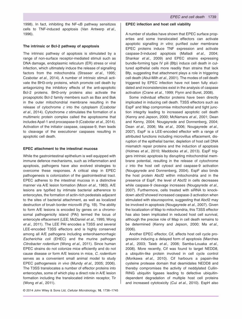
The gastrointestinal epithelium utilizes a plethora ofinnate immune defence mechanisms to fight entericpathogens. The host intestinal epithelium is com-posed of enterocytes, mucus-secreting goblet cells,enteroendocrine cells and Paneth cells and lies above thelamina propria where lymphocytes and other immunecells reside (Fig. 1A). All these cell types play a vital rolein sensing bacterial pathogens through recognition ofpathogen-associated molecular patterns (PAMPs) by a
range of surface-located and intracellular pattern recog-nition receptors including Toll-like receptors (TLRs), NOD-like receptors, RIG-I like helicases and C-type lectinreceptors (Creagh and O’Neill, 2006). Receptor stimula-tion leads to a cascade of signalling events that ultimatelyresults in the production of inflammatory cytokines, infil-tration of inflammatory immune cells and/or death of theinfected cell.
Although EPEC is an extracellular pathogen thatremains attached to the apical surface of enterocytes(Fig. 1B), EPEC can interfere with intracellular signallingpathways by injecting type III secretion system (T3SS)effectors into the enterocyte, many of which subvert hostcell signalling pathways (Giogha et al., 2014). Forexample, EPEC utilizes NleE, NleC and NleD to inhibitinflammation by blocking distinct steps in the nuclearfactor-κB (NF-κB) and mitogen-activated protein kinase(MAPK) signalling pathways that lead to the production ofpro-inflammatory cytokines (Marchès et al., 2005; Nadleret al., 2010; Newton et al., 2010; Yen et al., 2010; Baruchet al., 2011; Muhlen et al., 2011; Pearson et al., 2011;Zhang et al., 2012). EPEC not only blocks inflammationbut recent work has shown that EPEC T3SS effectors arealso potent inhibitors of cell death. Hence, host cell sur-vival and the prevention of apoptosis have emerged ascritical virulence mechanisms of EPEC and other attach-ing and effacing (A/E) pathogens.
Death receptor-mediated apoptosis
Apoptosis can be stimulated by either intracellularligands, known as the intrinsic or mitochondrial pathway,or by extracellular ligands, known as the extrinsic or deathreceptor pathway. Both pathways converge at the point ofactivation or cleavage of the executioner caspase,caspase-3, resulting in the late morphological changesof apoptosis (Igney and Krammer, 2002). The extrinsicpathway of apoptosis involves a family of transmembranereceptors termed death receptors that are members ofthe tumour necrosis factor receptor (TNFR) superfamily.Members of this family have conserved cysteine-richregions in the extracellular ligand-binding domain anda cytoplasmic domain of about 80 amino acids calledthe death domain (DD; Ashkenazi and Dixit, 1998). A
Received 19 August, 2014; revised 24 September, 2014; accepted26 September, 2014. *For correspondence. E-mail [email protected]; Tel.: +613 8344 8041; Fax +613 9347 1540.
Cellular Microbiology (2014) 16(12), 1736–1745 doi:10.1111/cmi.12371First published online 30 October 2014
© 2014 John Wiley & Sons Ltd
cellular microbiology

functional member of the TNFR superfamily is typicallya trimeric complex that is stabilized by intracysteinedisulfide bonds formed between the cysteine-rich regionsof individual subunit members (Banner et al., 1993).However, TNFRs can also exist in a soluble form (Grussand Dower, 1995). The DD of death receptors is essentialfor relaying the death signal from the cell surface to the
intracellular signalling pathway via a range of adaptormolecules that ultimately results in activation of theinitiator caspase, caspase-8. The best-characterizeddeath receptors include Fas/CD95/APO-1, TNFR1/p55/CD120a, TNF-related apoptosis-inducing ligand receptor1 (TRAIL-R1)/DR4 and TRAIL-R2/DR5/Apo2/KILLER andtheir respective ligands are the FasL, TNF and TRAIL/
DED
DED
DD
DD DD
Fas
Pro-caspase-8
FADD
FADD
GlcNAc
NleB1
Fas
FasL FasL
Lamina propria
Apoptosis
Nucleus
Actin
Activatedcaspase-8
NleB1
Mucus
Gobletcell Enteroendocrine
cell
Panethcell
Enterocyte
Commensal bacteria
sIgA
Lamina propria
p18 p43/41
caspase-3
B
41
? ?
EPECDnleB
T3SS
Enterocyte
Neutrophil
Gut lumenA C
Fig. 1. EPEC attachment and inhibition of Fas signalling in enterocytes during EPEC infection.A. Diagram of the cellular architecture of the intestinal epithelium. Enterocytes constitute the majority of the intestinal epithelium, which alsocontains goblet cells, enteroendocrine cells and Paneth cells. The epithelium lies above the lamina propria and is covered with a layer ofmucus in the gut lumen. Red dashed circle indicates that A/E pathogens target the tips of intestinal villi for attachment.B. Transmission electron micrograph of C. rodentium (asterisk) attachment during infection showing A/E lesion formation.C. Schematic diagram of NleB1-mediated inhibition of FasL-induced cell death during EPEC infection. Wild-type EPEC inhibits caspase-8activation and apoptotic cell death using NleB1,which GlcNAcylates FADD (blue squares) and prevents formation of the DISC. EPEC lackingNleB1 (ΔnleB1) cannot inhibit FasL-driven apoptotic cell death. The specific source of FasL during EPEC infection has not yet been identified.
EPEC and cell death 1737
© 2014 John Wiley & Sons Ltd, Cellular Microbiology, 16, 1736–1745

Apo2L (for TRAIL-R1 and TRAIL-R2; Chicheporticheet al., 1997; Ashkenazi and Dixit, 1998; Peter andKrammer, 1998; Suliman et al., 2001; Rubio-Moscardoet al., 2005). These trimeric ligands belong to the TNFfamily of cytokines and are mainly expressed astransmembrane proteins although they can also bereleased as soluble cytokines by proteolytic cleavage.Recent studies have demonstrated that only themembrane-bound form of FasL possesses apoptosis-inducing capacity (O’Reilly et al., 2009).
FasL and Fas signalling
Fas is expressed ubiquitously and although it is particu-larly abundant in lymphoid tissues and on activatedmature T lymphocytes, functional Fas is also expressedon the basolateral membrane of intestinal epithelial cells(Moller et al., 1994; Strasser et al., 2009). In contrast tothe ubiquitous expression of Fas, FasL is expressed on aselective cellular pool including activated cytotoxic T-cells,natural killer cells (NK) and NK T-cells (NKT). Upon stimu-lation of Fas by FasL, there is an aggregation of pre-assembled Fas trimers at the plasma membrane, whichleads to internalization of Fas (Lee et al., 2006). Fas thenrecruits the adaptor protein, Fas-associated protein withDD (FADD), via homotypic interactions between DDsfound in the cytoplasmic region of Fas and the C-terminusof FADD. FADD recruits procaspase-8 via a secondhomotypic interaction, this time involving the death effec-tor domain (DED) of FADD and procaspase-8. The inter-actions between Fas, FADD and procaspase-8 form thedeath-inducing signalling complex (DISC) that inducesproximity-induced activation (cleavage) of caspase-8(Kischkel et al., 1995). Activated caspase-8 then cleavesand activates the executioner caspase-3 leading toapoptotic cell death. In certain cell types, including epithe-lial cells, FasL stimulation induces apoptotic cell deaththrough caspase-8 mediated cleavage of a pro-apoptoticBcl-2 homology 3 (BH3)-only protein called Bid (Li et al.,1998; Jost et al., 2009). Bid is a member of the Bcl-2family of proteins, which is involved in the mitochondrialcell death pathway and so its activation by caspase-8 isan example of a crosstalk between the intrinsic and extrin-sic pathways of apoptosis.
The TRAIL and TNF pathways
The death receptors, TRAIL-R1 and TRAIL-R2, haveextensive tissue distribution, whereas the ligand, TRAIL,is expressed mainly on NK cells, NKT cells andmacrophages. Tumour necrosis factor-related apoptosis-inducing ligand (TRAIL) induces apoptosis by binding toeither TRAIL-R1 or TRAIL-R2, although TRAIL-R2 is moresignificant in inducing apoptosis. Receptor binding of
TRAIL leads to recruitment of FADD via homotypic DDinteractions, which in turn recruits procaspase-8 to theDISC via DED interactions leading to activation of acaspase cascade in a similar manner to FasL-induced celldeath (Kuang et al., 2000).
Similar to TRAIL, two different TNF receptors exist,TNFR1 and TNFR2. Although both receptors sharesequence similarity in their extracellular ligand-bindingregion, their cytoplasmic region sequences are different,thereby accounting for their different signalling activities(Tartaglia et al., 1991; Vandenabeele et al., 1995). BothTNFR1 and TNFR2 can bind TNF. However, only TNFR1is involved in death receptor signalling as TNFR2 lacks anintracellular DD. TNFR1 is expressed ubiquitously and isrequired for TNF-induced apoptosis of pathogen-infectedcells (Pfeffer et al., 1993; Zhao et al., 2000). Upon stimu-lation of TNFR1 by TNF, TNFR1 translocates to lipid rafts(Legler et al., 2003), whereupon receptor-interactingprotein kinase 1 (RIPK1), TNFR type1-associated DDprotein (TRADD), TNF receptor-associated factor 2(TRAF2) and cellular inhibitor of apoptosis protein 1 and 2(c-IAP1/2) are recruited to form the membrane-boundreceptor complex I (Micheau and Tschopp, 2003). Thereceptor complex is then internalized by endocytosisduring which RIPK1, TRAF2 and TRADD dissociate fromTNFR1 enabling the DD of TRADD to bind to the DD ofFADD. This in turn recruits procaspase-8 via homotypicDED interactions to the newly formed cytosolic complex II(Micheau and Tschopp, 2003; Schneider-Brachert et al.,2004). Activated caspase-8 then cleaves the executionercaspases leading to cell death by apoptosis. If caspase-8 is depleted or inhibited, complex II cannot induceapoptotic cell death and instead TNFR1 stimulation leadsto a recently described form of cell death known as pro-grammed necrosis or necroptosis, which relies on RIPK3and mixed lineage kinase domain-like (MLKL) (Murphyet al., 2013; Newton et al., 2014). Most recently, RIPK1was shown to play a dual regulatory role in gut homo-eostasis by inhibiting both apoptosis and necroptosis inintestinal epithelial cells through its scaffolding function(Dannappel et al., 2014; Takahashi et al., 2014).
Even though TNFR1 stimulation can lead to cell death,the primary function of TNFR1 is in inflammation. In thispathway, RIPK1 in membrane-bound receptor complex Iis polyubiquitinated at lysine-377 by the E3 ubiquitinligases, cIAP-1/2 (Ea et al., 2006; Li et al., 2006).Polyubiquitinated RIPK1 recruits TAK1 and the associ-ated TAK1-binding proteins 1/2/3 (TAB1/2/3; Ea et al.,2006). TAK1 then activates the IKK complex leading tophosphorylation of the NF-κB inhibitory protein, IκB,which is then ubiquitinated and degraded enablingnuclear translocation of NF-κB and transcriptionalactivation of NF-κB-dependent genes including pro-inflammatory and anti-apoptotic genes (Wang et al.,
1738 T. Wong Fok Lung et al.
© 2014 John Wiley & Sons Ltd, Cellular Microbiology, 16, 1736–1745
1998). In fact, inhibiting the NF-κB pathway sensitizescells to TNF-induced apoptosis (Van Antwerp et al.,1996).
The intrinsic or Bcl-2 pathway of apoptosis
The intrinsic pathway of apoptosis is stimulated by arange of non-surface receptor-mediated stimuli such asDNA damage, endoplasmic reticulum (ER) stress or viralinfection, which ultimately induce the release of signallingfactors from the mitochondria (Strasser et al., 1995;Czabotar et al., 2014). A number of intrinsic stimuli acti-vate the BH3-only proteins, which promote cell death byantagonizing the inhibitory effects of the anti-apoptoticBcl-2 proteins. BH3-only proteins also activate theproapoptotic Bcl-2 family members such as Bax and Bakin the outer mitochondrial membrane resulting in therelease of cytochrome c into the cytoplasm (Czabotaret al., 2014). Cytochrome c stimulates the formation of amultimeric protein complex called the apoptosome thatincludes Apaf-1 and procaspase-9 (Czabotar et al., 2014).Activation of the initiator caspase, caspase-9, then leadsto cleavage of the executioner caspases resulting inapoptotic cell death.
EPEC attachment to the intestinal mucosa
While the gastrointestinal epithelium is well equipped withimmune defence mechanisms, such as inflammation andapoptosis, pathogens have also evolved strategies toovercome these responses. A critical step in EPECpathogenesis is colonization of the gastrointestinal tract.EPEC adheres to the intestinal mucosa in a distinctivemanner via A/E lesion formation (Moon et al., 1983). A/Elesions are typified by intimate bacterial adherence toenterocytes, the formation of actin-rich pedestals adjacentto the sites of bacterial attachment, as well as localizeddestruction of brush border microvilli (Fig. 1B). The abilityto form A/E lesions is encoded by genes on a chromo-somal pathogenicity island (PAI) termed the locus ofenterocyte effacement (LEE; McDaniel et al., 1995; Wonget al., 2011). The LEE PAI encodes a T3SS and severalLEE-encoded T3SS effectors and is highly conservedamong all A/E pathogens including enterohaemorrhagicEscherichia coli (EHEC) and the murine pathogenCitrobacter rodentium (Wong et al., 2011). Since humanEPEC strains do not colonize mice efficiently and do notcause disease or form A/E lesions in mice, C. rodentiumserves as a convenient small animal model to studyEPEC pathogenesis in vivo (Mundy et al., 2005; 2006).The T3SS translocates a number of effector proteins intoenterocytes, some of which play a direct role in A/E lesionformation including the translocated intimin receptor, Tir(Wong et al., 2011).
EPEC infection and host cell viability
A number of studies have shown that EPEC surface prop-erties and some translocated effectors can activateapoptotic signalling in vitro; purified outer membraneEPEC proteins induce TNF expression and activatecaspase-3-induced apoptosis (Malladi et al., 2004;Shankar et al., 2009) and EPEC strains expressingbundle-forming type IV pili (Bfp) induce cell death in cul-tured epithelial cells more readily than strains that lackBfp, suggesting that attachment plays a role in triggeringcell death (Abul-Milh et al., 2001). The modes of cell deathtriggered by EPEC infection have not been fully eluci-dated and inconsistencies exist in the analysis of caspaseactivation (Crane et al., 1999; Flynn and Buret, 2008).
Some individual effector proteins have been directlyimplicated in inducing cell death. T3SS effectors such asEspF and Map compromise mitochondrial and tight junc-tion integrity leading to increased apoptotic cell death(Kenny and Jepson, 2000; McNamara et al., 2001; Deanand Kenny, 2004; Nougayrede and Donnenberg, 2004;Dean et al., 2006; Ma et al., 2006; Nougayrede et al.,2007). EspF is a LEE-encoded effector with a range ofattributed functions including microvillus effacement, dis-ruption of the epithelial barrier, depletion of host cell DNAmismatch repair proteins and the induction of apoptosis(Holmes et al., 2010; Maddocks et al., 2013). EspF trig-gers intrinsic apoptosis by disrupting mitochondrial mem-brane potential, resulting in the release of cytochromec into the host cell cytosol and caspase-9 activation(Nougayrede and Donnenberg, 2004). EspF also bindsthe host protein Abcf2 within mitochondria and in thepresence of EspF, the level of Abcf2 in cells decreaseswhile caspase-9 cleavage increases (Nougayrede et al.,2007). Furthermore, cells treated with siRNA to knock-down abcf2 showed increased caspase-3 activation whenstimulated with staurosporine, suggesting that Abcf2 maybe involved in apoptosis (Nougayrede et al., 2007). Giventhe localization of Map to mitochondria, this T3SS effectorhas also been implicated in reduced host cell survival,although the precise role of Map in cell death remains tobe determined (Kenny and Jepson, 2000; Ma et al.,2006).
Another EPEC effector, Cif, affects host cell cycle pro-gression inducing a delayed form of apoptosis (Marcheset al., 2003; Taieb et al., 2006; Samba-Louaka et al.,2008). More recently, Cif was found to target NEDD8,a ubiquitin-like protein involved in cell cycle control(Morikawa et al., 2010). Cif harbours a papain-likecysteine protease domain that deamidates NEDD8 andthereby compromises the activity of neddylated Cullin-RING ubiquitin ligases leading to defective ubiquitin-dependent degradation of multiple host cell proteinsand increased cytotoxicity (Cui et al., 2010). EspH also
EPEC and cell death 1739
© 2014 John Wiley & Sons Ltd, Cellular Microbiology, 16, 1736–1745
induces a cytotoxic effect and induces caspase-3 cleav-age, which is related to disassembly of focal adhesions(Dong et al., 2010; Wong et al., 2012). EspH binds theDH-PH domain in multiple RhoGEFs, which prevents acti-vation of Rho and therefore Rho-dependent events suchas phagocytosis as well as disruption of the host actincytoskeleton structure (Dong et al., 2010).
Anti-apoptotic effectors of EPEC
Despite the fact that the effectors described above havebeen associated with cytotoxic effects and/or activation ofexecutioner caspases, in general, cells infected withEPEC do not display characteristic late apoptotic featuressuch as membrane blebbing, nuclear condensation andcell shrinkage. In addition, in vivo studies report thatapoptosis is not observed in tissues from humans infectedwith EPEC (Rothbaum et al., 1982; 1983; Fagundes-Netoet al., 1996) or in tissues from C57BL/6 mice infected withC. rodentium (Vallance et al., 2003). Hence, apoptosis isnot a characteristic feature of infection with A/E patho-gens. In fact, compared with Salmonella, Shigella andYersinia, three genera of invasive enteric bacteria, whichare known to induce host cell death, EPEC is experimen-tally much weaker in its ability to cause cell death(Monack et al., 1996; 1997; Crane et al., 1999).
The first EPEC T3SS effectors associated with the inhi-bition of cell death were NleH1 and NleH2. nleH1/H2double mutants of EPEC are highly cytotoxic for cells,which can be complemented by the reintroduction ofeither nleH1 or nleH2 (Hemrajani et al., 2010). Furtherinvestigation of NleH1 showed that the effector inhibitscaspase-3 activation/cleavage even when cells aretreated with a wide range of proapoptotic compoundsincluding staurosporine, tunicamycin and brefeldin A.Caspase-3 inhibition was independent of predicted NleHkinase activity (Hemrajani et al., 2010). The exact mecha-nism by which NleH1 blocks apoptosis has not been fullyelucidated although NleH1 interacts with an anti-apoptoticprotein called Bax inhibitor-1 (BI-1; Hemrajani et al.,2010). BI-1 is an ER protein that when overexpressed inmammalian cells suppresses apoptosis induced by avariety of stimuli including deprivation of growth factor,treatment with the anti-cancer agent, etoposide as well asstaurosporine and the proapototic protein Bax (Robinsonet al., 2011). NleH1 also blocks cell death inducedby the TcdB toxin from Clostridium difficile. TcdB is aglucosyltransferase that inactivates Rho family proteinscausing disruption of the cell cytoskeleton and cell death(Matarrese et al., 2007). The modification of Rho by TcdBis detected in cells by a newly described pyrin-containinginflammasome (Xu et al., 2014) that activates caspase-1and induces cell death. More work is needed to elucidatethe mechanism by which NleH blocks cell death, espe-
cially given the ability of NleH1 to inhibit cell death fromsuch a diverse range of intrinsic stimuli. Interestingly, bothNleH1 and NleH2 are also associated with inhibition of IκBphosphorylation and NF-κB activation in response to TNFstimulation (Royan et al., 2010). The mechanism by whichNleH inhibits NF-κB activation is also not fully understoodas one study implicated the kinase activity of NleH (Royanet al., 2010), while a kinase-independent mechanisminvolving the ribosomal protein S3 was proposed byothers (Wan et al., 2007; Gao et al., 2009).
The NleD effector is a zinc metalloprotease that cleavesthe c-Jun N-terminal kinase (JNK) and MAPK, p38(Baruch et al., 2011). In response to stimulation of TLRs,the interleukin 1 receptor or TNFR, several mitogen-activated protein 3 kinases are activated that can activateJNK, which in turn phosphorylates c-Jun, a component ofthe AP-1 transcription factor that regulates cell prolifera-tion, apoptosis and inflammation (Shaulian and Karin,2001; Eferl and Wagner, 2003). JNK activation involvesphosphorylation on threonine-183 and tyrosine-185located in the activation loop (Kallunki et al., 1994). NleDcleaves JNK in the activation loop, which blocks JNK-driven apoptosis (Baruch et al., 2011). NleD also cleavesp38 in the conserved activation loop and the combinedinactivation of JNK and p38 by NleD is linked to suppres-sion of the inflammatory response via inhibition of IL8expression (Baruch et al., 2011).
Inhibition of the extrinsic cell death response
EPEC and EHEC express two homologues of the T3SSeffector NleB, NleB1 and NleB2. Initial studies in EPECidentified a role for NleB1 in the inhibition of (NF-κBactivation upon TNF but not IL-1β stimulation Newtonet al., 2010). Despite this, however, NleB1 did not inhibitIL-8 production during EPEC infection (Pearson et al.,2013). Instead, recent studies have demonstratedthat NleB1 inhibits the extrinsic apoptotic pathway bymodifying DD-containing proteins including FADD,RIPK1 and TRADD (Li et al., 2013; Pearson et al.,2013). The fact that RIPK1 and TRADD form part of theTNFR1 receptor complex is most likely the reason thatNleB1 inhibits NF-κB activation under certain experi-mental conditions, namely, ectopic expression or over-expression in the absence of nleE (Newton et al., 2010;Li et al., 2013).
NleB1 is a glycosyltransferase (Gao et al., 2013) thatbinds to and modifies the DD of FADD by adding a singleGlcNAc moiety at arginine 117 (Arg117), thereby inhibitingTNF or FasL-induced DISC formation and subsequentcaspase-8 activation and apoptotic cell death (Li et al.,2013; Pearson et al., 2013). Arg117 in FADD is essentialfor Fas-FADD as well as TRADD–FADD DD interactions(Imtiyaz et al., 2005; Wang et al., 2010) and the critical
1740 T. Wong Fok Lung et al.
© 2014 John Wiley & Sons Ltd, Cellular Microbiology, 16, 1736–1745
role of this conserved residue in apoptosis is evident inthe embryonic lethality of mice carrying an R117Q muta-tion (Imtiyaz et al., 2009). NleB1 also GlcNAcylatesTRADD at the equivalent arginine residue (Arg235), pre-venting recruitment of TRADD, TRAF2 and ubiquitinatedRIPK1 to TNFR1 (Li et al., 2013). Furthermore, NleB1inhibits TRAIL-induced DISC formation and apopto-sis (Li et al., 2013). NleB2 is also a predictedglycosyltransferase but does not bind FADD and has noimpact on the inhibition of cell death during EPEC infec-tion (Li et al., 2013; Pearson et al., 2013).
The role of NleB1 in vivo has been investigated usingnleB deletion mutants of C. rodentium, which carriesonly one copy of nleB. nleB mutants are attenuated forcolonization (Kelly et al., 2006) and mice infected withC. rodentium lacking nleB show increased numbers ofcells positive for cleaved caspase-8 in the colon (Pearsonet al., 2013). The importance of the FasL-Fas signallingpathway during C. rodentium infection was demonstratedfurther by infecting Fas-deficient (Faslpr/lpr) and FasL-deficient (Fasgld/gld) mice with C. rodentium (Pearson et al.,2013). Both strains developed severe, watery diarrhoeaand showed increased colitis in response to C. rodentiumcompared with wild-type mice. Although the source ofFasL during infection is unclear, this model suggests thatFasL-positive immune cells recognize and delete infectedcells by inducing apoptosis, thereby controlling pathogenload. In the presence of NleB, this cell death response isthwarted and the bacteria maintain their attachment to aviable enterocyte (Fig. 1C).
While NleB1 is specific for the extrinsic apoptoticpathway, NleF has been implicated in the inhibition ofapoptosis stimulated by both the intrinsic and extrinsicpathways (Blasche et al., 2013). Because of the ability ofNleF to bind and inhibit caspase-9 activation, the mainfocus of work on NleF has been the inhibition of intrinsicapoptosis. However, NleF also prevents TRAIL-inducedapoptosis in HeLa cells by binding to and inhibitingcaspase-8 (Blasche et al., 2013). NleF-mediated inhibi-tion results from insertion of the carboxy terminal end ofNleF into the active site of the caspase (Blasche et al.,2013). However, the role of NleF in apoptosis inhibitionduring infection is still unclear and may be secondary toeffectors such as NleB1, as no significant differenceswere observed in the activation of downstream execu-tioner caspases-3 and 7 in HeLa cells infected withwild-type EPEC or a ΔnleF EPEC derivative (Blascheet al., 2013). NleF also binds to caspase-4, which wasrecently described as an innate sensor of intracellularlipopolysaccharide (LPS; Shi et al., 2014). The CARDdomain of caspase-4 binds directly to LPS leading tostimulation of pyroptosis. Hence, during infection, the roleof NleF may be to counteract inflammasome formation ininfected enterocytes.
Protecting epithelial integrity
Two further T3SS effector proteins influence cell survivalthrough maintaining cellular junctions. EspZ (SepZ) is aLEE-encoded effector protein that promotes maintenanceof the intestinal epithelium by stabilizing focal adhesionsduring infection (Deng et al., 2004; Shames et al., 2010;Berger et al., 2012). Epithelial cells infected with an EPECΔespZ mutant exhibit increased cytotoxicity compared tocells infected with wild-type EPEC. EspZ-mediated pro-tection from cytotoxicity has been attributed to its ability tobind CD98 (Shames et al., 2010). CD98 enhances signal-ling from the β1-integrin receptor, including pro-survivalsignals from the Akt kinase and focal adhesion kinase(FAK; Feral et al., 2005). Interestingly, the T3SS effector,OspE, from the invasive enteric pathogen Shigellaflexneri, also stabilizes focal adhesion sites of host intes-tinal cells, thereby reinforcing enterocyte adherence to thebasement membrane and preventing the exfoliation ofinfected cells into the gut lumen (Kim et al., 2009). Themechanism of action of OspE is via binding to integrin-linked kinase (ILK), which contributes to the inhibition offocal adhesion disassembly rather than affecting focaladhesion assembly (Kim et al., 2009). Indeed, thephosphorylation of both FAK and paxillin, indicating dis-assembly, was significantly reduced in cells expressingOspE and ILK (Kim et al., 2009). The residue impor-tant for OspE binding to ILK was identified astryptophan-68 (Trp68). Trp68 is highly conserved amongOspE homologues from EHEC, EPEC and C. rodentium(termed EspO), which also bind to ILK (Kim et al., 2009).Hence EspO effectors in A/E pathogens may act inconcert with EspZ and effectors that regulate the actincytoskeleton to block sloughing of infected cells into thegut lumen (Morita-Ishihara et al., 2013).
Conclusions
The exposure of intestinal cells to EPEC-derived PAMPs,intimate attachment of the bacteria as well as the activityof effector proteins such as Map, EspF, Cif and EspH allstimulate intrinsic apoptosis pathways and stressresponses in the host cell that are countered by effectorssuch as NleH, NleD, NleF, EspZ and EspO. The action ofthese cell death-suppressing effectors promotes survivalof the infected cell thereby prolonging EPEC adherence tothe intestinal mucosa. In addition, EPEC has evolvedspecific mechanisms to inhibit death receptor-mediatedinflammation and apoptosis in response to immunesystem surveillance and host death ligand-mediatedelimination of infected cells. A diagrammatic summary ofthese effectors and comparison with other pathogens ispresented in Giogha et al. (2014). Importantly, the activityof NleB has revealed a previously unappreciated role for
EPEC and cell death 1741
© 2014 John Wiley & Sons Ltd, Cellular Microbiology, 16, 1736–1745

Fas signalling in fighting gut infection and exemplifies howEPEC has adapted to avoid the intestinal innate immuneresponse.
There is still much to understand about the activities ofEPEC T3SS effector proteins, in particular where hostbinding targets and biological effects have not been con-firmed in the context of infection. In addition, the discoveryof new enzymatic activities for several EPEC effectorproteins warrants re-examination of other effectors thatmay also be novel enzymes. Lastly, especially in the fieldof cell signalling, studies relying on in vitro models andectopic expression of effectors should be interpretedwith caution if not confirmed by infection, ideally usingC. rodentium as an in vivo model for EPEC infection.
Acknowledgements
We gratefully acknowledge Ms Vicki Bennett-Wood for the elec-tron micrograph of C. rodentium. This work was supported byfunding awarded to ELH from the Australian National Health andMedical Research Council (APP1044061). TWFL was supportedby a University of Melbourne International Research Scholarship(MIRS).
References
Abul-Milh, M., Wu, Y., Lau, B., Lingwood, C.A., andBarnett Foster, D. (2001) Induction of epithelial cell deathincluding apoptosis by enteropathogenic Escherichia coliexpressing bundle-forming pili. Infect Immun 69: 7356–7364.
Ashkenazi, A., and Dixit, V.M. (1998) Death receptors:signaling and modulation. Science 281: 1305–1308.
Banner, D.W., D’Arcy, A., Janes, W., Gentz, R., Schoenfeld,H.J., Broger, C., et al. (1993) Crystal structure of thesoluble human 55 kd TNF receptor-human TNF betacomplex: implications for TNF receptor activation. Cell 73:431–445.
Baruch, K., Gur-Arie, L., Nadler, C., Koby, S., Yerushalmi, G.,Ben-Neriah, Y., et al. (2011) Metalloprotease type III effec-tors that specifically cleave JNK and NF-kB. EMBO J 30:221–231.
Berger, C.N., Crepin, V.F., Baruch, K., Mousnier, A.,Rosenshine, I., and Frankel, G. (2012) EspZ of enter-opathogenic and enterohemorrhagic Escherichia coli regu-lates type III secretion system protein translocation. MBio3: e00317-12.
Blasche, S., Mortl, M., Steuber, H., Siszler, G., Nisa, S.,Schwarz, F., et al. (2013) The E. coli effector protein NleFis a caspase inhibitor. PLoS ONE 8: e58937.
Chicheportiche, Y., Bourdon, P.R., Xu, H., Hsu, Y.M., Scott,H., Hession, C., et al. (1997) TWEAK, a new secretedligand in the tumor necrosis factor family that weaklyinduces apoptosis. J Biol Chem 272: 32401–32410.
Crane, J.K., Majumdar, S., and Pickhardt, D.F., 3rd (1999)Host cell death due to enteropathogenic Escherichiacoli has features of apoptosis. Infect Immun 67: 2575–2584.
Creagh, E.M., and O’Neill, L.A. (2006) TLRs, NLRs andRLRs: a trinity of pathogen sensors that co-operate ininnate immunity. Trends Immunol 27: 352–357.
Cui, J., Yao, Q., Li, S., Ding, X., Lu, Q., Mao, H., et al. (2010)Glutamine deamidation and dysfunction of ubiquitin/NEDD8 induced by a bacterial effector family. Science329: 1215–1218.
Czabotar, P.E., Lessene, G., Strasser, A., and Adams, J.M.(2014) Control of apoptosis by the BCL-2 protein family:implications for physiology and therapy. Nat Rev Mol CellBiol 15: 49–63.
Dannappel, M., Vlantis, K., Kumari, S., Polykratis, A., Kim, C.,Wachsmuth, L., et al. (2014) RIPK1 maintains epithelialhomeostasis by inhibiting apoptosis and necroptosis.Nature 513: 90–94.
Dean, P., and Kenny, B. (2004) Intestinal barrier dysfunctionby enteropathogenic Escherichia coli is mediated by twoeffector molecules and a bacterial surface protein. MolMicrobiol 54: 665–675.
Dean, P., Maresca, M., Schuller, S., Phillips, A.D., and Kenny,B. (2006) Potent diarrheagenic mechanism mediatedby the cooperative action of three enteropathogenicEscherichia coli-injected effector proteins. Proc Natl AcadSci USA 103: 1876–1881.
Deng, W., Puente, J.L., Gruenheid, S., Li, Y., Vallance, B.A.,Vazquez, A., et al. (2004) Dissecting virulence: systematicand functional analyses of a pathogenicity island. Proc NatlAcad Sci USA 101: 3597–3602.
Dong, N., Liu, L., and Shao, F. (2010) A bacterial effectortargets host DH-PH domain RhoGEFs and antagonizesmacrophage phagocytosis. EMBO J 29: 1363–1376.
Ea, C.K., Deng, L., Xia, Z.P., Pineda, G., and Chen, Z.J.(2006) Activation of IKK by TNFalpha requires site-specificubiquitination of RIP1 and polyubiquitin binding by NEMO.Mol Cell 22: 245–257.
Eferl, R., and Wagner, E.F. (2003) AP-1: a double-edgedsword in tumorigenesis. Nat Rev Cancer 3: 859–868.
Fagundes-Neto, U., Freymuller, E., Gandolfi Schimitz, L., andScaletsky, I. (1996) Nutritional impact and ultrastructuralintestinal alterations in severe infections due to enter-opathogenic Escherichia coli strains in infants. J Am CollNutr 15: 180–185.
Feral, C.C., Nishiya, N., Fenczik, C.A., Stuhlmann, H.,Slepak, M., and Ginsberg, M.H. (2005) CD98hc (SLC3A2)mediates integrin signaling. Proc Natl Acad Sci USA 102:355–360.
Flynn, A.N., and Buret, A.G. (2008) Caspases-3, -8, and -9are required for induction of epithelial cell apoptosis byenteropathogenic E. coli but are dispensable for increasedparacellular permeability. Microb Pathog 44: 311–319.
Gao, X., Wan, F., Mateo, K., Callegari, E., Wang, D., Deng,W., et al. (2009) Bacterial effector binding to ribosomalprotein s3 subverts NF-kB function. PLoS Pathog 5:e1000708.
Gao, X., Wang, X., Pham, T.H., Feuerbacher, L.A., Lubos,M.L., Huang, M., et al. (2013) NleB, a bacterial effectorwith glycosyltransferase activity, targets GAPDH func-tion to inhibit NF-kB activation. Cell Host Microbe 13:87–99.
Giogha, C., Wong Fok Lung, T., Pearson, J.S., and Hartland,E.L. (2014) Inhibition of death receptor signaling by
1742 T. Wong Fok Lung et al.
© 2014 John Wiley & Sons Ltd, Cellular Microbiology, 16, 1736–1745
bacterial gut pathogens. Cytokine Growth Factor Rev 25:235–243.
Gruss, H.J., and Dower, S.K. (1995) Tumor necrosis factorligand superfamily: involvement in the pathology of malig-nant lymphomas. Blood 85: 3378–3404.
Hemrajani, C., Berger, C.N., Robinson, K.S., Marches, O.,Mousnier, A., and Frankel, G. (2010) NleH effectors inter-act with Bax inhibitor-1 to block apoptosis during enter-opathogenic Escherichia coli infection. Proc Natl Acad SciUSA 107: 3129–3134.
Holmes, A., Muhlen, S., Roe, A.J., and Dean, P. (2010) TheEspF effector, a bacterial pathogen’s Swiss army knife.Infect Immun 78: 4445–4453.
Igney, F.H., and Krammer, P.H. (2002) Death and anti-death:tumour resistance to apoptosis. Nat Rev Cancer 2: 277–288.
Imtiyaz, H.Z., Zhang, Y., and Zhang, J. (2005) Structuralrequirements for signal-induced target binding of FADDdetermined by functional reconstitution of FADD defi-ciency. J Biol Chem 280: 31360–31367.
Imtiyaz, H.Z., Zhou, X., Zhang, H., Chen, D., Hu, T., andZhang, J. (2009) The death domain of FADD is essentialfor embryogenesis, lymphocyte development, and prolif-eration. J Biol Chem 284: 9917–9926.
Jost, P.J., Grabow, S., Gray, D., McKenzie, M.D., Nachbur,U., Huang, D.C., et al. (2009) XIAP discriminates betweentype I and type II FAS-induced apoptosis. Nature 460:1035–1039.
Kallunki, T., Su, B., Tsigelny, I., Sluss, H.K., Derijard, B.,Moore, G., et al. (1994) JNK2 contains a specificity-determining region responsible for efficient c-Jun bindingand phosphorylation. Genes Dev 8: 2996–3007.
Kelly, M., Hart, E., Mundy, R., Marches, O., Wiles, S.,Badea, L., et al. (2006) Essential role of the type IIIsecretion system effector NleB in colonization ofmice by Citrobacter rodentium. Infect Immun 74: 2328–2337.
Kenny, B., and Jepson, M. (2000) Targeting of an enter-opathogenic Escherichia coli (EPEC) effector protein tohost mitochondria. Cell Microbiol 2: 579–590.
Kim, M., Ogawa, M., Fujita, Y., Yoshikawa, Y., Nagai, T.,Koyama, T., et al. (2009) Bacteria hijack integrin-linkedkinase to stabilize focal adhesions and block cell detach-ment. Nature 459: 578–582.
Kischkel, F.C., Hellbardt, S., Behrmann, I., Germer, M.,Pawlita, M., Krammer, P.H., and Peter, M.E. (1995)Cytotoxicity-dependent APO-1 (Fas/CD95)-associatedproteins form a death-inducing signaling complex (DISC)with the receptor. EMBO J 14: 5579–5588.
Kuang, A.A., Diehl, G.E., Zhang, J., and Winoto, A. (2000)FADD is required for DR4- and DR5-mediated apopto-sis: lack of trail-induced apoptosis in FADD-deficientmouse embryonic fibroblasts. J Biol Chem 275: 25065–25068.
Lee, K.H., Feig, C., Tchikov, V., Schickel, R., Hallas, C.,Schutze, S., et al. (2006) The role of receptor internaliza-tion in CD95 signaling. EMBO J 25: 1009–1023.
Legler, D.F., Micheau, O., Doucey, M.A., Tschopp, J., andBron, C. (2003) Recruitment of TNF receptor 1 to lipid raftsis essential for TNFalpha-mediated NF-kappaB activation.Immunity 18: 655–664.
Li, H., Zhu, H., Xu, C.J., and Yuan, J. (1998) Cleavage of BIDby caspase 8 mediates the mitochondrial damage in theFas pathway of apoptosis. Cell 94: 491–501.
Li, H., Kobayashi, M., Blonska, M., You, Y., and Lin, X. (2006)Ubiquitination of RIP is required for tumor necrosis factoralpha-induced NF-kappaB activation. J Biol Chem 281:13636–13643.
Li, S., Zhang, L., Yao, Q., Li, L., Dong, N., Rong, J., et al.(2013) Pathogen blocks host death receptor signalling byarginine GlcNAcylation of death domains. Nature 501:242–246.
Ma, C., Wickham, M.E., Guttman, J.A., Deng, W., Walker, J.,Madsen, K.L., et al. (2006) Citrobacter rodentium infectioncauses both mitochondrial dysfunction and intestinal epi-thelial barrier disruption in vivo: role of mitochondrial asso-ciated protein (Map). Cell Microbiol 8: 1669–1686.
McDaniel, T.K., Jarvis, K.G., Donnenberg, M.S., and Kaper,J.B. (1995) A genetic locus of enterocyte effacement con-served among diverse enterobacterial pathogens. ProcNatl Acad Sci USA 92: 1664–1668.
McNamara, B.P., Koutsouris, A., O’Connell, C.B.,Nougayrede, J.P., Donnenberg, M.S., and Hecht, G. (2001)Translocated EspF protein from enteropathogenicEscherichia coli disrupts host intestinal barrier function. JClin Invest 107: 621–629.
Maddocks, O.D., Scanlon, K.M., and Donnenberg, M.S.(2013) An Escherichia coli effector protein promotes hostmutation via depletion of DNA mismatch repair proteins.MBio 4: e152–e113.
Malladi, V., Shankar, B., Williams, P.H., and Balakrishnan,A. (2004) Enteropathogenic Escherichia coli outer mem-brane proteins induce changes in cadherin junctions ofCaco-2 cells through activation of PKCa. Microbes Infect 6:38–50.
Marchès, O., Wiles, S., Dziva, F., La Ragione, R.M., Schuller,S., Best, A., et al. (2005) Characterization of two non-locusof enterocyte effacement-encoded type III-translocatedeffectors, NleC and NleD, in attaching and effacing patho-gens. Infect Immun 73: 8411–8417.
Marches, O., Ledger, T.N., Boury, M., Ohara, M., Tu, X.,Goffaux, F., et al. (2003) Enteropathogenic andenterohaemorrhagic Escherichia coli deliver a novel effec-tor called Cif, which blocks cell cycle G2/M transition. MolMicrobiol 50: 1553–1567.
Matarrese, P., Falzano, L., Fabbri, A., Gambardella, L.,Frank, C., Geny, B., et al. (2007) Clostridium difficile toxinB causes apoptosis in epithelial cells by thrilling mitochon-dria. Involvement of ATP-sensitive mitochondrial potassiumchannels. J Biol Chem 282: 9029–9041.
Micheau, O., and Tschopp, J. (2003) Induction of TNF recep-tor I-mediated apoptosis via two sequential signaling com-plexes. Cell 114: 181–190.
Moller, P., Koretz, K., Leithauser, F., Bruderlein, S., Henne,C., Quentmeier, A., and Krammer, P.H. (1994) Expressionof APO-1 (CD95), a member of the NGF/TNF receptorsuperfamily, in normal and neoplastic colon epithelium. IntJ Cancer 57: 371–377.
Monack, D.M., Raupach, B., Hromockyj, A.E., and Falkow, S.(1996) Salmonella typhimurium invasion induces apoptosisin infected macrophages. Proc Natl Acad Sci USA 93:9833–9838.
EPEC and cell death 1743
© 2014 John Wiley & Sons Ltd, Cellular Microbiology, 16, 1736–1745

Monack, D.M., Mecsas, J., Ghori, N., and Falkow, S. (1997)Yersinia signals macrophages to undergo apoptosis andYopJ is necessary for this cell death. Proc Natl Acad SciUSA 94: 10385–10390.
Moon, H.W., Whipp, S.C., Argenzio, R.A., Levine, M.M., andGiannella, R.A. (1983) Attaching and effacing activities ofrabbit and human enteropathogenic Escherichia coli in pigand rabbit intestines. Infect Immun 41: 1340–1351.
Morikawa, H., Kim, M., Mimuro, H., Punginelli, C., Koyama,T., Nagai, S., et al. (2010) The bacterial effector Cif inter-feres with SCF ubiquitin ligase function by inhibitingdeneddylation of Cullin1. Biochem Biophys Res Commun401: 268–274.
Morita-Ishihara, T., Miura, M., Iyoda, S., Izumiya, H.,Watanabe, H., Ohnishi, M., and Terajima, J. (2013)EspO1-2 regulates EspM2-mediated RhoA activity to sta-bilize formation of focal adhesions in enterohemorrhagicEscherichia coli-infected host cells. PLoS ONE 8: e55960.
Muhlen, S., Ruchaud-Sparagano, M.H., and Kenny, B. (2011)Proteasome-independent degradation of canonical NFkBcomplex components by the NleC protein of pathogenicEscherichia coli. J Biol Chem 286: 5100–5107.
Mundy, R., MacDonald, T.T., Dougan, G., Frankel, G., andWiles, S. (2005) Citrobacter rodentium of mice and man.Cell Microbiol 7: 1697–1706.
Mundy, R., Girard, F., FitzGerald, A.J., and Frankel, G. (2006)Comparison of colonization dynamics and pathology ofmice infected with enteropathogenic Escherichia coli,enterohaemorrhagic E. coli and Citrobacter rodentium.FEMS Microbiol Lett 265: 126–132.
Murphy, J.M., Czabotar, P.E., Hildebrand, J.M., Lucet, I.S.,Zhang, J.G., Alvarez-Diaz, S., et al. (2013) Thepseudokinase MLKL mediates necroptosis via a molecularswitch mechanism. Immunity 39: 443–453.
Nadler, C., Baruch, K., Kobi, S., Mills, E., Haviv, G., Farago,M., et al. (2010) The type III secretion effector NleE inhibitsNF-kB activation. PLoS Pathog 6: e1000743.
Newton, H.J., Pearson, J.S., Badea, L., Kelly, M., Lucas, M.,Holloway, G., et al. (2010) The type III effectors NleE andNleB from enteropathogenic E. coli and OspZ from Shigellablock nuclear translocation of NF-kappaB p65. PLoSPathog 6: e1000898.
Newton, K., Dugger, D.L., Wickliffe, K.E., Kapoor, N., deAlmagro, M.C., Vucic, D., et al. (2014) Activity of proteinkinase RIPK3 determines whether cells die by necroptosisor apoptosis. Science 343: 1357–1360.
Nougayrede, J.P., and Donnenberg, M.S. (2004) Enter-opathogenic Escherichia coli EspF is targeted to mitochon-dria and is required to initiate the mitochondrial deathpathway. Cell Microbiol 6: 1097–1111.
Nougayrede, J.P., Foster, G.H., and Donnenberg, M.S.(2007) Enteropathogenic Escherichia coli effector EspFinteracts with host protein Abcf2. Cell Microbiol 9: 680–693.
O’Reilly, L.A., Tai, L., Lee, L., Kruse, E.A., Grabow, S., Fairlie,W.D., et al. (2009) Membrane-bound Fas ligand only isessential for Fas-induced apoptosis. Nature 461: 659–663.
Pearson, J.S., Riedmaier, P., Marches, O., Frankel, G., andHartland, E.L. (2011) A type III effector protease NleC fromenteropathogenic Escherichia coli targets NF-kB for deg-radation. Mol Microbiol 80: 219–230.
Pearson, J.S., Giogha, C., Ong, S.Y., Kennedy, C.L., Kelly,M., Robinson, K.S., et al. (2013) A type III effector antago-nizes death receptor signalling during bacterial gut infec-tion. Nature 501: 247–251.
Peter, M.E., and Krammer, P.H. (1998) Mechanisms of CD95(APO-1/Fas)-mediated apoptosis. Curr Opin Immunol 10:545–551.
Pfeffer, K., Matsuyama, T., Kundig, T.M., Wakeham, A.,Kishihara, K., Shahinian, A., et al. (1993) Mice deficient forthe 55 kd tumor necrosis factor receptor are resistant toendotoxic shock, yet succumb to L. monocytogenes infec-tion. Cell 73: 457–467.
Robinson, K.S., Clements, A., Williams, A.C., Berger, C.N.,and Frankel, G. (2011) Bax inhibitor 1 in apoptosis anddisease. Oncogene 30: 2391–2400.
Rothbaum, R., McAdams, A.J., Giannella, R., and Partin, J.C.(1982) A clinicopathologic study of enterocyte-adherentEscherichia coli: a cause of protracted diarrhea in infants.Gastroenterology 83: 441–454.
Rothbaum, R.J., Partin, J.C., Saalfield, K., and McAdams,A.J. (1983) An ultrastructural study of enteropathogenicEscherichia coli infection in human infants. UltrastructPathol 4: 291–304.
Royan, S.V., Jones, R.M., Koutsouris, A., Roxas, J.L.,Falzari, K., Weflen, A.W., et al. (2010) Enteropathogenic E.coli non-LEE encoded effectors NleH1 and NleH2 attenu-ate NF-kB activation. Mol Microbiol 78: 1232–1245.
Rubio-Moscardo, F., Blesa, D., Mestre, C., Siebert, R.,Balasas, T., Benito, A., et al. (2005) Characterizationof 8p21.3 chromosomal deletions in B-cell lymphoma:TRAIL-R1 and TRAIL-R2 as candidate dosage-dependenttumor suppressor genes. Blood 106: 3214–3222.
Samba-Louaka, A., Nougayrede, J.P., Watrin, C., Jubelin, G.,Oswald, E., and Taieb, F. (2008) Bacterial cyclomodulin Cifblocks the host cell cycle by stabilizing the cyclin-dependent kinase inhibitors p21 and p27. Cell Microbiol10: 2496–2508.
Schneider-Brachert, W., Tchikov, V., Neumeyer, J., Jakob,M., Winoto-Morbach, S., Held-Feindt, J., et al. (2004)Compartmentalization of TNF receptor 1 signaling: inter-nalized TNF receptosomes as death signaling vesicles.Immunity 21: 415–428.
Shames, S.R., Deng, W., Guttman, J.A., de Hoog, C.L., Li, Y.,Hardwidge, P.R., et al. (2010) The pathogenic E. coli typeIII effector EspZ interacts with host CD98 and facilitateshost cell prosurvival signalling. Cell Microbiol 12: 1322–1339.
Shankar, B., Krishnan, S., Malladi, V., Balakrishnan, A., andWilliams, P.H. (2009) Outer membrane proteins of wild-type and intimin-deficient enteropathogenic Escherichiacoli induce Hep-2 cell death through intrinsic and extrinsicpathways of apoptosis. Int J Med Microbiol 299: 121–132.
Shaulian, E., and Karin, M. (2001) AP-1 in cell proliferationand survival. Oncogene 20: 2390–2400.
Shi, J., Zhao, Y., Wang, Y., Gao, W., Ding, J., Li, P., et al.(2014) Inflammatory caspases are innate immune recep-tors for intracellular LPS. Nature 514: 187–192.
Strasser, A., Harris, A.W., Huang, D.C., Krammer, P.H., andCory, S. (1995) Bcl-2 and Fas/APO-1 regulate distinctpathways to lymphocyte apoptosis. EMBO J 14: 6136–6147.
1744 T. Wong Fok Lung et al.
© 2014 John Wiley & Sons Ltd, Cellular Microbiology, 16, 1736–1745

Strasser, A., Jost, P.J., and Nagata, S. (2009) The many rolesof FAS receptor signaling in the immune system. Immunity30: 180–192.
Suliman, A., Lam, A., Datta, R., and Srivastava, R.K. (2001)Intracellular mechanisms of TRAIL: apoptosis throughmitochondrial-dependent and -independent pathways.Oncogene 20: 2122–2133.
Taieb, F., Nougayrede, J.P., Watrin, C., Samba-Louaka, A.,and Oswald, E. (2006) Escherichia coli cyclomodulin Cifinduces G2 arrest of the host cell cycle without activationof the DNA-damage checkpoint-signalling pathway. CellMicrobiol 8: 1910–1921.
Takahashi, N., Vereecke, L., Bertrand, M.J., Duprez, L.,Berger, S.B., Divert, T., et al. (2014) RIPK1 ensures intes-tinal homeostasis by protecting the epithelium againstapoptosis. Nature 513: 95–99.
Tartaglia, L.A., Weber, R.F., Figari, I.S., Reynolds, C.,Palladino, M.A., Jr, and Goeddel, D.V. (1991) The twodifferent receptors for tumor necrosis factor mediatedistinct cellular responses. Proc Natl Acad Sci USA 88:9292–9296.
Vallance, B.A., Deng, W., Jacobson, K., and Finlay, B.B.(2003) Host susceptibility to the attaching and effacingbacterial pathogen Citrobacter rodentium. Infect Immun71: 3443–3453.
Van Antwerp, D.J., Martin, S.J., Kafri, T., Green, D.R., andVerma, I.M. (1996) Suppression of TNF-alpha-inducedapoptosis by NF-kappaB. Science 274: 787–789.
Vandenabeele, P., Declercq, W., Beyaert, R., and Fiers, W.(1995) Two tumour necrosis factor receptors: structure andfunction. Trends Cell Biol 5: 392–399.
Wan, F., Anderson, D.E., Barnitz, R.A., Snow, A., Bidere, N.,Zheng, L., et al. (2007) Ribosomal protein S3: a KH domainsubunit in NF-kB complexes that mediates selective generegulation. Cell 131: 927–939.
Wang, C.Y., Mayo, M.W., Korneluk, R.G., Goeddel, D.V., andBaldwin, A.S., Jr (1998) NF-kB antiapoptosis: induction ofTRAF1 and TRAF2 and c-IAP1 and c-IAP2 to suppresscaspase-8 activation. Science 281: 1680–1683.
Wang, L., Yang, J.K., Kabaleeswaran, V., Rice, A.J., Cruz,A.C., Park, A.Y., et al. (2010) The Fas-FADD death domaincomplex structure reveals the basis of DISC assemblyand disease mutations. Nat Struct Mol Biol 17: 1324–1329.
Wong, A.R., Pearson, J.S., Bright, M.D., Munera, D.,Robinson, K.S., Lee, S.F., et al. (2011) Enteropatho-genic and enterohaemorrhagic Escherichia coli: evenmore subversive elements. Mol Microbiol 80: 1420–1438.
Wong, A.R., Clements, A., Raymond, B., Crepin, V.F., andFrankel, G. (2012) The interplay between the Escherichiacoli Rho guanine nucleotide exchange factor effectorsand the mammalian RhoGEF inhibitor EspH. MBio 3:e00250-11.
Xu, H., Yang, J., Gao, W., Li, L., Li, P., Zhang, L., et al. (2014)Innate immune sensing of bacterial modifications of RhoGTPases by the Pyrin inflammasome. Nature 513: 237–241.
Yen, H., Ooka, T., Iguchi, A., Hayashi, T., Sugimoto, N., andTobe, T. (2010) NleC, a type III secretion protease, com-promises NF-kB activation by targeting p65/RelA. PLoSPathog 6: e1001231.
Zhang, L., Ding, X., Cui, J., Xu, H., Chen, J., Gong, Y.N., et al.(2012) Cysteine methylation disrupts ubiquitin-chainsensing in NF-kB activation. Nature 481: 204–208.
Zhao, Y.X., Lajoie, G., Zhang, H., Chiu, B., Payne, U., andInman, R.D. (2000) Tumor necrosis factor receptor p55-deficient mice respond to acute Yersinia enterocoliticainfection with less apoptosis and more effective host resist-ance. Infect Immun 68: 1243–1251.
EPEC and cell death 1745
© 2014 John Wiley & Sons Ltd, Cellular Microbiology, 16, 1736–1745
Copyright © 2022 FDOKUMEN