Pathogenic Escherichia coli - ScienceOpen
16
Use of Zebrafish to Probe the Divergent Virulence Potentials and Toxin Requirements of Extraintestinal Pathogenic Escherichia coli Travis J. Wiles 1 , Jean M. Bower 1 , Michael J. Redd 2 , Matthew A. Mulvey 1 * 1 Division of Cell Biology and Immunology, Department of Pathology, University of Utah, Salt Lake City, Utah, United States of America, 2 Huntsman Cancer Institute, University of Utah, Salt Lake City, Utah, United States of America Abstract Extraintestinal pathogenic E. coli (ExPEC) cause an array of diseases, including sepsis, neonatal meningitis, and urinary tract infections. Many putative virulence factors that might modulate ExPEC pathogenesis have been identified through sequencing efforts, epidemiology, and gene expression profiling, but few of these genes have been assigned clearly defined functional roles during infection. Using zebrafish embryos as surrogate hosts, we have developed a model system with the ability to resolve diverse virulence phenotypes and niche-specific restrictions among closely related ExPEC isolates during either localized or systemic infections. In side-by-side comparisons of prototypic ExPEC isolates, we observed an unexpectedly high degree of phenotypic diversity that is not readily apparent using more traditional animal hosts. In particular, the capacity of different ExPEC isolates to persist and multiply within the zebrafish host and cause disease was shown to be variably dependent upon two secreted toxins, a-hemolysin and cytotoxic necrotizing factor. Both of these toxins appear to function primarily in the neutralization of phagocytes, which are recruited in high numbers to sites of infection where they act as an essential host defense against ExPEC as well as less virulent E. coli strains. These results establish zebrafish as a valuable tool for the elucidation and functional analysis of both ExPEC virulence factors and host defense mechanisms. Citation: Wiles TJ, Bower JM, Redd MJ, Mulvey MA (2009) Use of Zebrafish to Probe the Divergent Virulence Potentials and Toxin Requirements of Extraintestinal Pathogenic Escherichia coli. PLoS Pathog 5(12): e1000697. doi:10.1371/journal.ppat.1000697 Editor: Lalita Ramakrishnan, University of Washington, United States of America Received June 8, 2009; Accepted November 17, 2009; Published December 18, 2009 Copyright: ß 2009 Wiles et al. This is an open-access article distributed under the terms of the Creative Commons Attribution License, which permits unrestricted use, distribution, and reproduction in any medium, provided the original author and source are credited. Funding: This work was supported by National Institutes of Health (NIH) Microbial Pathogenesis Training Grant T32 AI055434 (to T.J.W.) and by NIH Grant DK068585. The funders had no role in study design, data collection and analysis, decision to publish, or preparation of the manuscript. Competing Interests: The authors have declared that no competing interests exist. * E-mail: [email protected] Introduction Escherichia coli is a laboratory workhorse that has helped expand our fundamental understanding of many biological processes. Outside the laboratory, E. coli are a remarkably diverse species both genetically and with respect to their ability to exist as either harmless commensals or as pathogens in a variety of animal hosts. Substantial morbidity and annual medical costs in the billions of dollars are attributed to a group of E. coli strains referred to as extraintestinal pathogenic E. coli (ExPEC) [1,2,3,4]. These pathogens have the capacity to persist within the human gut among the normal microbiota without any overt consequences. However, once outside the intestinal tract, ExPEC pathotypes can cause an array of diseases including urinary tract infections (UTIs), sepsis, and meningitis. The frequency of ExPEC-induced infec- tions in human populations may be aggravated by the broad host range of these pathogens. For example, production birds raised for human consumption are susceptible to colibacillosis, a lethal infection caused by ExPEC strains known as avian pathogenic E. coli (APEC). These pathogens are highly similar to ExPEC strains like uropathogenic E. coli (UPEC), which are the primary cause of UTIs in humans [5,6]. Such observations highlight the zoonotic potential of APEC and related bacteria, suggesting that the widespread dissemination of ExPEC-associated virulence traits among human isolates may occur through consumption of contaminated poultry or other food products [6,7,8]. In recent years, an enormous amount of information has been accrued by sequencing the genomes of several prototypic ExPEC isolates and other E. coli strains. These data, together with epidemiological analyses, confirm that distinct ExPEC pathotypes share many known and putative virulence factors. These include a number of secreted toxins, iron acquisition systems (siderophores), adhesins, and capsular antigens [9]. Secreted toxins, which include a-hemolysin (HlyA), cytotoxic necrotizing factor-1 (CNF1), and the secreted autotransporter SAT, can alter host signaling cascades, disrupt inflammatory responses, and induce host cell death, while siderophores like aerobactin, bacteriocin, and enterobactin allow ExPEC to sequester essential iron away from the host [10,11,12]. Adhesive organelles such as type 1, F1C, P, and S pili (or fimbriae) can mediate ExPEC interactions with, and entry into, host cells and tissues, while capsule expression may enable ExPEC to better avoid professional phagocytes [9,13,14]. These and other virulence factors are often encoded within genomic regions, known as pathogenicity islands (PAIs), which are acquired by horizontal gene transfer [15,16,17,18]. The modular exchange of PAIs and other genetic elements likely provides ExPEC with a high degree of versatility over time, enabling these bacteria to adapt to and colonize assorted host and PLoS Pathogens | www.plospathogens.org 1 December 2009 | Volume 5 | Issue 12 | e1000697
-
Upload
khangminh22 -
Category
Documents
-
view
0 -
download
0
Transcript of Pathogenic Escherichia coli - ScienceOpen
Use of Zebrafish to Probe the Divergent VirulencePotentials and Toxin Requirements of ExtraintestinalPathogenic Escherichia coliTravis J. Wiles1, Jean M. Bower1, Michael J. Redd2, Matthew A. Mulvey1*
1 Division of Cell Biology and Immunology, Department of Pathology, University of Utah, Salt Lake City, Utah, United States of America, 2 Huntsman Cancer Institute,
University of Utah, Salt Lake City, Utah, United States of America
Abstract
Extraintestinal pathogenic E. coli (ExPEC) cause an array of diseases, including sepsis, neonatal meningitis, and urinary tractinfections. Many putative virulence factors that might modulate ExPEC pathogenesis have been identified throughsequencing efforts, epidemiology, and gene expression profiling, but few of these genes have been assigned clearly definedfunctional roles during infection. Using zebrafish embryos as surrogate hosts, we have developed a model system with theability to resolve diverse virulence phenotypes and niche-specific restrictions among closely related ExPEC isolates duringeither localized or systemic infections. In side-by-side comparisons of prototypic ExPEC isolates, we observed anunexpectedly high degree of phenotypic diversity that is not readily apparent using more traditional animal hosts. Inparticular, the capacity of different ExPEC isolates to persist and multiply within the zebrafish host and cause disease wasshown to be variably dependent upon two secreted toxins, a-hemolysin and cytotoxic necrotizing factor. Both of thesetoxins appear to function primarily in the neutralization of phagocytes, which are recruited in high numbers to sites ofinfection where they act as an essential host defense against ExPEC as well as less virulent E. coli strains. These resultsestablish zebrafish as a valuable tool for the elucidation and functional analysis of both ExPEC virulence factors and hostdefense mechanisms.
Citation: Wiles TJ, Bower JM, Redd MJ, Mulvey MA (2009) Use of Zebrafish to Probe the Divergent Virulence Potentials and Toxin Requirements of ExtraintestinalPathogenic Escherichia coli. PLoS Pathog 5(12): e1000697. doi:10.1371/journal.ppat.1000697
Editor: Lalita Ramakrishnan, University of Washington, United States of America
Received June 8, 2009; Accepted November 17, 2009; Published December 18, 2009
Copyright: � 2009 Wiles et al. This is an open-access article distributed under the terms of the Creative Commons Attribution License, which permitsunrestricted use, distribution, and reproduction in any medium, provided the original author and source are credited.
Funding: This work was supported by National Institutes of Health (NIH) Microbial Pathogenesis Training Grant T32 AI055434 (to T.J.W.) and by NIH GrantDK068585. The funders had no role in study design, data collection and analysis, decision to publish, or preparation of the manuscript.
Competing Interests: The authors have declared that no competing interests exist.
* E-mail: [email protected]
Introduction
Escherichia coli is a laboratory workhorse that has helped expand
our fundamental understanding of many biological processes.
Outside the laboratory, E. coli are a remarkably diverse species
both genetically and with respect to their ability to exist as either
harmless commensals or as pathogens in a variety of animal hosts.
Substantial morbidity and annual medical costs in the billions of
dollars are attributed to a group of E. coli strains referred to as
extraintestinal pathogenic E. coli (ExPEC) [1,2,3,4]. These
pathogens have the capacity to persist within the human gut
among the normal microbiota without any overt consequences.
However, once outside the intestinal tract, ExPEC pathotypes can
cause an array of diseases including urinary tract infections (UTIs),
sepsis, and meningitis. The frequency of ExPEC-induced infec-
tions in human populations may be aggravated by the broad host
range of these pathogens. For example, production birds raised for
human consumption are susceptible to colibacillosis, a lethal
infection caused by ExPEC strains known as avian pathogenic E.
coli (APEC). These pathogens are highly similar to ExPEC strains
like uropathogenic E. coli (UPEC), which are the primary cause of
UTIs in humans [5,6]. Such observations highlight the zoonotic
potential of APEC and related bacteria, suggesting that the
widespread dissemination of ExPEC-associated virulence traits
among human isolates may occur through consumption of
contaminated poultry or other food products [6,7,8].
In recent years, an enormous amount of information has been
accrued by sequencing the genomes of several prototypic ExPEC
isolates and other E. coli strains. These data, together with
epidemiological analyses, confirm that distinct ExPEC pathotypes
share many known and putative virulence factors. These include a
number of secreted toxins, iron acquisition systems (siderophores),
adhesins, and capsular antigens [9]. Secreted toxins, which include
a-hemolysin (HlyA), cytotoxic necrotizing factor-1 (CNF1), and
the secreted autotransporter SAT, can alter host signaling
cascades, disrupt inflammatory responses, and induce host cell
death, while siderophores like aerobactin, bacteriocin, and
enterobactin allow ExPEC to sequester essential iron away from
the host [10,11,12]. Adhesive organelles such as type 1, F1C, P,
and S pili (or fimbriae) can mediate ExPEC interactions with, and
entry into, host cells and tissues, while capsule expression may
enable ExPEC to better avoid professional phagocytes [9,13,14].
These and other virulence factors are often encoded within
genomic regions, known as pathogenicity islands (PAIs), which are
acquired by horizontal gene transfer [15,16,17,18].
The modular exchange of PAIs and other genetic elements
likely provides ExPEC with a high degree of versatility over time,
enabling these bacteria to adapt to and colonize assorted host and
PLoS Pathogens | www.plospathogens.org 1 December 2009 | Volume 5 | Issue 12 | e1000697

environmental niches. While an individual ExPEC isolate will
typically encode about 5,000 genes, the total number of genes that
may be swapped among all ExPEC isolates and related strains in
nature is currently estimated to total more than 17,800 [19,20].
However, most of these genes remain uncharacterized and factors
that dictate the host and niche specificities of distinct ExPEC
isolates remain enigmatic. Complicating matters further are results
showing that even relatively well-studied virulence factors such as
HlyA and CNF1, which are closely linked with UPEC and other
ExPEC pathotypes, have effects on bacterial colonization and
persistence that can be difficult to discern using established mouse
infection models (unpublished observations and [21,22,23,24]).
Such observations, coupled with the sheer number of genes that
ExPEC isolates can potentially acquire, indicate that these
pathogens have likely evolved multiple and probably redundant
mechanisms to overcome the many challenges encountered within
diverse host environments.
To formally address this possibility, and to establish a more
experimentally amenable high-throughput approach to phenotyp-
ically assess ExPEC virulence potentials during both systemic and
localized infections, we developed a novel model system employing
zebrafish (Danio rerio) embryos. Results obtained using this
surrogate host model demonstrate that even very closely related
ExPEC isolates can differ greatly in their virulence capacity in a
niche- and dose-specific fashion. In addition, we found that the in
vivo survival and virulence of different ExPEC isolates can be
variably dependent upon the secreted toxins HlyA and CNF1,
which appear to act primarily in the neutralization of host
phagocytes. In total, this work highlights the phenotypic diversity
of closely related ExPEC isolates and establishes zebrafish as a
valuable tool for the discovery and analysis of both ExPEC
virulence factors and key host defense mechanisms.
Results
Localized and systemic ExPEC infections in zebrafishThe pathogenic potential of ExPEC isolates has generally been
assessed in vivo using rodent models in which bacteria are
inoculated via either trans-urethral catheterization or other
injection routes. These model systems mimic many important
aspects of both localized UTIs and the often more serious systemic
infections caused by ExPEC. However, these models do not lend
themselves readily to rapid high-throughput analyses or real-time
observations of the infection process. Over the past several years,
zebrafish have emerged as a powerful vertebrate model system for
deciphering virulence mechanisms employed by both fish-specific
and mammalian pathogens [25,26,27,28,29,30,31]. At 48 h post
fertilization (hpf) the zebrafish immune system is composed solely
of innate defenses, including antimicrobial peptides, complement,
toll-like receptors, and phagocytes [32,33,34,35]. This develop-
mental state makes zebrafish embryos a convenient model
organism to study ExPEC pathogenesis since immunity to these
pathogens in both humans and mice is also primarily dependent
upon innate defenses [36]. Furthermore, the transparency and
external development of zebrafish embryos facilitate the direct
observation of pathogenic events in live animals [37]. To test the
utility of zebrafish as an alternative host for ExPEC, we inoculated
48 hpf embryos with bacteria at two sites: a fluid-filled sac
surrounding the heart referred to as the pericardial cavity and the
blood, via the circulation valley (Figure S1). The pericardial
cavity is an infection site that restricts bacterial dissemination and
serves as a model for a localized infection [38]. Inoculation into
the circulation valley results in the rapid dispersion of bacteria
throughout the embryo, mimicking a systemic infection. Each site
likely presents ExPEC with different challenges in terms of
nutrient availability, phagocyte numbers, and the types and levels
of antimicrobial agents.
To address the phenotypic diversity of ExPEC in vivo, we
examined seven fully sequenced ExPEC isolates: two cystitis
strains (UTI89 and F11), two pyelonephritis isolates (CFT073 and
536), one neonatal meningitis E. coli (NMEC) strain (RS218), an
APEC isolate (APEC01), and one strain (83972) responsible for
asymptomatic bacteruria (ABU). ABU isolates like 83972 can exist
in a commensal-like relationship with the host, and in some cases
appear to protect the urinary tract from colonization by more
pathogenic bacterial strains [39]. We also utilized a recently
sequenced human gut isolate (HS) and two laboratory K12
reference strains (MG1655 and W3110) as controls. Bacteria were
grown in M9 minimal medium and washed in phosphate buffered
saline (PBS) prior to injection into either the pericardial cavity or
blood of 48 hpf zebrafish embryos (Videos S1 and S2). Low
(2,000 to 3,500 CFU), medium (4,000 to 6,500 CFU), and high
(6,600 to 11,500 CFU) inoculation doses were used. Infected
embryos were examined every 6 h over a 24 h period, and death
was scored as the absence of a heartbeat and lack of blood flow.
Figure 1A shows the dose- and niche-dependent killing of
zebrafish embryos by the UPEC strains UTI89 and CFT073, the
ABU isolate 83972, and the gut isolate HS, presented as Kaplan-
Meier survival curves. Points on the curves only appear when at
least one event (death) is recorded. Both high and medium
inoculation doses of UTI89 and CFT073 were lethal after
inoculation into the pericardial cavity, but only CFT073 was
effective at killing the host when inoculated into the blood. In
contrast, the ABU isolate 83972 was only lethal at high inoculation
doses when delivered into the pericardial cavity, and had little
effect when injected into the blood. HS was similarly avirulent
when injected into the blood, but displayed moderate levels of
lethality within the pericardial cavity. Interestingly, HS possesses
many virulence-associated genes and has previously been
suggested to act as a ‘‘precursor’’ pathogen [19].
Figure 1B summarizes results from all ten tested E. coli strains
24 h after inoculation of either the pericardial cavity or blood with
,5,000 CFU. The laboratory K12 strains MG1655 and W3110
Author Summary
Escherichia coli can exist among the normal intestinalmicrobiota without causing any overt problems for thehuman host. However, humans as well as other animalscan often acquire less-mild mannered variants of E. colistrains known as extraintestinal pathogenic E. coli (ExPEC)that can colonize sites outside of the intestinal tract andcause a range of serious illnesses, including sepsis,meningitis, and urinary tract infections. Despite manyadvances over the years using cell culture and rodentinfection models, the spectrum of genes that control theability of different ExPEC strains to colonize and growwithin specific host niches and cause disease remain, forthe most part, elusive. Here, we report the development ofa new model system that uses zebrafish as surrogate hostsfor ExPEC and related isolates. Using zebrafish to modelboth localized and systemic infections, we found thatclosely related ExPEC isolates display an unexpected arrayof virulence characteristics and toxin requirements that arenot readily apparent from genomic information alone. Thismodel system is amenable to high-throughput geneticand pharmacological screens and should prove useful inthe development of more efficacious therapeutics.
Deciphering ExPEC Virulence in Zebrafish
PLoS Pathogens | www.plospathogens.org 2 December 2009 | Volume 5 | Issue 12 | e1000697
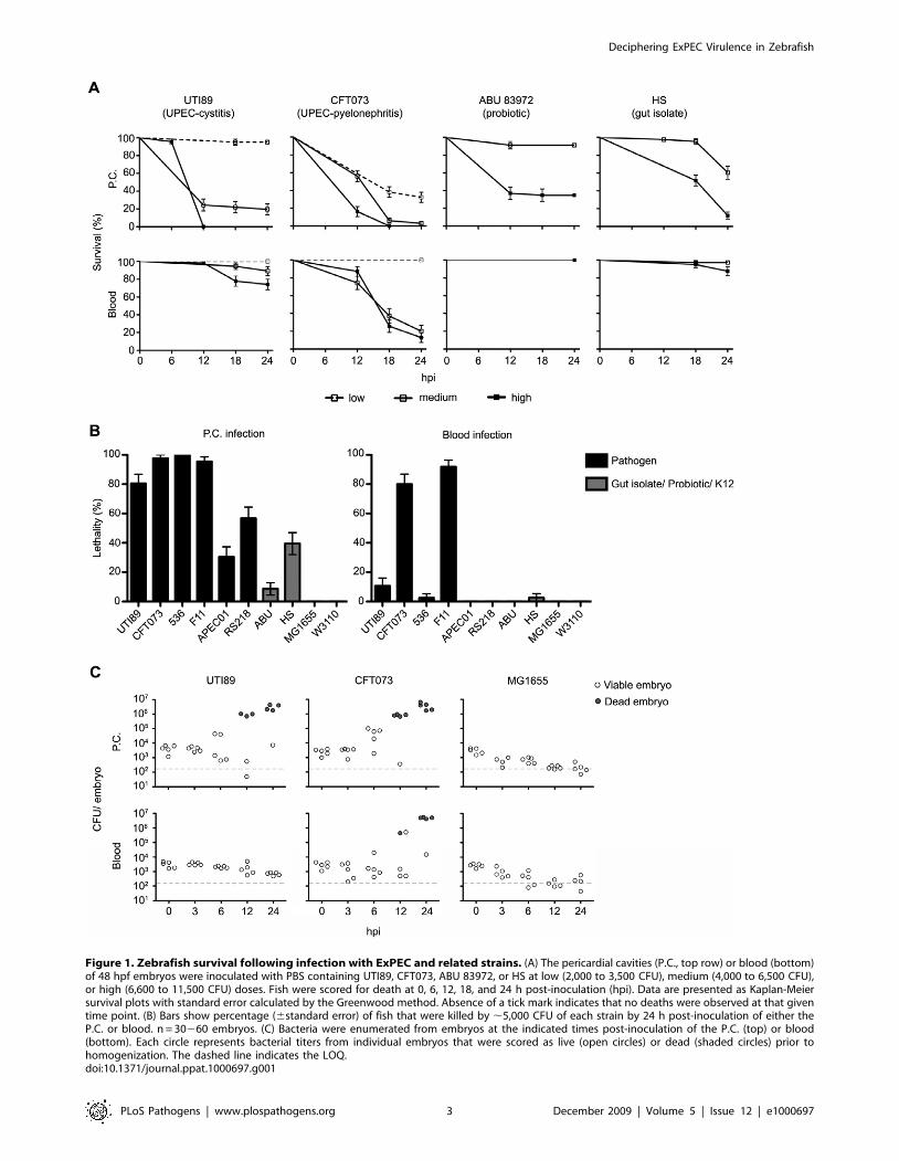
Figure 1. Zebrafish survival following infection with ExPEC and related strains. (A) The pericardial cavities (P.C., top row) or blood (bottom)of 48 hpf embryos were inoculated with PBS containing UTI89, CFT073, ABU 83972, or HS at low (2,000 to 3,500 CFU), medium (4,000 to 6,500 CFU),or high (6,600 to 11,500 CFU) doses. Fish were scored for death at 0, 6, 12, 18, and 24 h post-inoculation (hpi). Data are presented as Kaplan-Meiersurvival plots with standard error calculated by the Greenwood method. Absence of a tick mark indicates that no deaths were observed at that giventime point. (B) Bars show percentage (6standard error) of fish that were killed by ,5,000 CFU of each strain by 24 h post-inoculation of either theP.C. or blood. n = 30260 embryos. (C) Bacteria were enumerated from embryos at the indicated times post-inoculation of the P.C. (top) or blood(bottom). Each circle represents bacterial titers from individual embryos that were scored as live (open circles) or dead (shaded circles) prior tohomogenization. The dashed line indicates the LOQ.doi:10.1371/journal.ppat.1000697.g001
Deciphering ExPEC Virulence in Zebrafish
PLoS Pathogens | www.plospathogens.org 3 December 2009 | Volume 5 | Issue 12 | e1000697
were the least virulent of the tested strains, causing no death unless
exceptionally high inoculation doses of .30,000 CFU were used.
Among the ExPEC strains, only the UTI isolates CFT073 and F11
were lethal in both the pericardial cavity and blood, while 536 and
UTI89 caused significant host death only when inoculated into the
pericardial cavity. In comparison to these UPEC strains, APEC01
and the NMEC isolate RS218 showed moderate levels of killing
only after injection into the pericardial cavity, similar to gut isolate
HS. Of note, all bacterial strains used in this study grew at similar
rates in M9 medium at 28.5uC, the temperature at which zebrafish
are maintained. Consequently, differences in virulence levels
observed among these strains are likely not directly attributable to
temperature effects on the rate of bacterial replication.
By microscopy, we observed that massive bacterial growth
within either the pericardial cavity or blood was associated with
eventual death of the zebrafish host. In all cases, the relatively clear
pericardial cavity seen in uninfected or mock-infected embryos
(Video S3) became increasingly cloudy following inoculation with
lethal isolates such as CFT073 (Video S4). For these and later
imaging studies, the E. coli strains used were transformed with the
low-copy, high-retention plasmid pGEN-GFP(LVA), which en-
codes a constitutively expressed destabilized variant of GFP having
a half-life of ,40 min. The pGEN plasmid backbone contains the
hok sok post-segregation killing system and two par loci that allow
for high levels of plasmid retention over multiple generations in the
absence of antibiotic selection [40,41]. Use of the pGEN-
GFP(LVA) construct facilitated the unambiguous detection of
bacteria that were recently viable and translationally active. We
observed that CFT073 and other lethal isolates eventually filled
the entire pericardial cavity, but rarely spread into other niches
until after the host had died (Video S4). In contrast, inoculation of
either CFT073 or F11 into the blood resulted in the rapid spread
and disseminated growth of these pathogens throughout the host
circulatory system and, eventually, other tissues (Video S5).
During the course of non-lethal infections, bacteria inoculated into
either the pericardial cavity or blood failed to replicate appreciably
and instead survived in only limited numbers within the host.
These trends were validated by determining bacterial titers in
zebrafish homogenates at 0, 3, 6, 12, and 24 h post-inoculation of
UTI89, CFT073, or MG1655 into the pericardial cavity or blood
(Figure 1C; see below). In these assays, lethal infections were
associated with increases in bacterial burden by up to 4 orders of
magnitude, while during non-lethal infections bacterial numbers
declined to near or below the limit of quantitation (LOQ).
Interestingly, embryos that were challenged via the pericardial
cavity with F11, UTI89, APEC01, RS218, 83972, and, less
frequently, MG1655 and survived appeared at times to be notably
sick, displaying restricted blood flow, heart deformation, edema,
and/or curled tails, despite the absence of large numbers of
bacteria (Video S6). These pathologies likely arise as a
consequence of both host inflammatory responses and bacterial
toxicity. In total, our data demonstrate that zebrafish embryos are
relatively resistant to non-pathogenic laboratory E. coli strains, but
are susceptible to ExPEC infection in a strain-, dose-, and niche-
specific fashion.
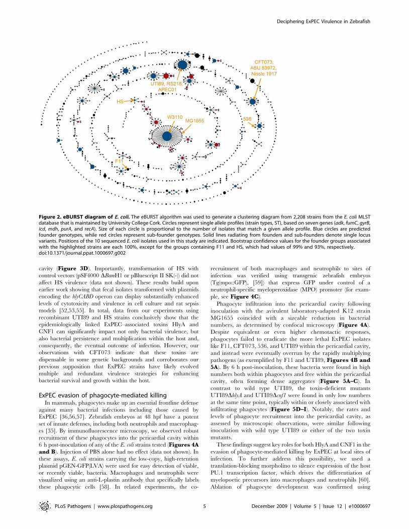
ExPEC genetic diversity and virulence in zebrafishThe genetic relatedness of the strains used in this study to one
another and to the broader worldwide E. coli community was
addressed using multilocus sequence typing (MLST) and the
eBURST algorithm [42,43], by which the allelic profiles of 2,208
E. coli isolates were compared and organized based on similarities
among seven housekeeping genes (adk, fumC, gyrB, icd, mdh, purA,
and recA) (Figure 2). For this analysis, isolates with identical allelic
profiles were grouped together as strain types (circles) that were
further organized into clonal clusters. These are defined as groups
of genotypes that have six out of seven alleles in common with a
founder genotype located in the center of each cluster (blue circles
in Figure 2). This form of allelic-based cluster analysis is useful for
identifying patterns of descent within bacterial populations in
which horizontal gene transfer is common [44]. A similar
approach was employed recently to infer relationships among
bacteremic ExPEC and other E. coli isolates [45], but here we
wished to assess if the genetic relatedness of the specific bacterial
strains used in our assays could be correlated with the varying
levels of virulence observed in the zebrafish host.
As shown in Figure 2, many of the E. coli strains used in this
study possess the founder genotype of their respective clonal
clusters and several share identical strain type profiles. Of note, the
avian pathogen APEC01 and the human cystitis isolate UTI89,
which were previously shown to have highly homologous genomes
[5,20], were grouped together as the same strain type in our
analysis. Despite this similarity, APEC01 differed markedly from
UTI89 in its virulence capacity within the pericardial cavity
(p,0.001), and APEC01 instead appeared to be phenotypically
akin to the more distantly related gut isolate HS (p.0.38;
Figure 1B). Likewise, the pyelonephritis isolate CFT073 and ABU
83972 belong to the same clonal cluster, and yet display distinct
levels of virulence in both the pericardial cavity and blood.
Interestingly, current evidence indicates that some ABU isolates
like 83972 were once pathogens, rendered less virulent by the loss
or mutation of key virulence factors by genomic decay [46]. In
total, our data confirm the idea that even pathogens with highly
similar epidemiological and genetic origins can vary greatly in
their virulence potential and niche-adaptability, probably due in
part to acquisition of specific virulence factors by horizontal gene
transfer.
ExPEC toxins and bacterial virulence potential withinzebrafish
We next asked if ExPEC virulence within zebrafish could be
linked to known ExPEC-associated virulence factors, such as HlyA
and CNF1. The pore-forming toxin HlyA is encoded by ,50% of
all ExPEC isolates, including strains UTI89, F11, CFT073, 536,
and RS218 used here [47,48]. Three of these isolates, UTI89, F11,
and RS218, also encode CNF1, which is genetically linked with
the hly operon in some ExPEC-associated PAIs [11,49,50]. In
rodent infection models, both HlyA and CNF1 have been shown
to influence ExPEC virulence, but the effects of either of these
toxins on pathogen growth and persistence within the host have
been more difficult to assess (data not shown and
[21,22,23,24,51,52,53,54]). We found that deletion of either hlyA
or cnf1 in UTI89 clearly attenuated both the virulence and growth
capacity of this strain within the pericardial cavity of 48 hpf
zebrafish embryos using medium inoculation doses of 4,000 to
6,500 CFU (Figure 3A and B, see below). Virulence was
partially restored when either mutant strain was complemented
with a plasmid carrying the respective deleted toxin. Surprisingly,
disruption of hlyA in CFT073, which naturally lacks cnf1, did not
diminish CFT073 virulence within the pericardial cavity and had
only a slight attenuating effect in the blood (Figure 3C). This is
despite the fact that the HlyA toxin encoded by CFT073 is nearly
identical to the one expressed by UTI89.
The contribution of both HlyA and CNF1 to bacterial virulence
in the zebrafish host was further addressed by transforming the gut
isolate HS with plasmid pSF4000 or pHLK102, encoding the
hlyCABD operon and cnf1, respectively. Expression of either toxin
significantly enhanced the virulence of HS within the pericardial
Deciphering ExPEC Virulence in Zebrafish
PLoS Pathogens | www.plospathogens.org 4 December 2009 | Volume 5 | Issue 12 | e1000697
cavity (Figure 3D). Importantly, transformation of HS with
control vectors (pSF4000 DBamH1 or pBluescript II SK(-)) did not
affect HS virulence (data not shown). These results build upon
earlier work showing that fecal isolates transformed with plasmids
encoding the hlyCABD operon can display substantially enhanced
levels of cytotoxicity and virulence in cell culture and rat sepsis
models [52,53,55]. In total, data from our experiments using
recombinant UTI89 and HS strains conclusively show that the
epidemiologically linked ExPEC–associated toxins HlyA and
CNF1 can significantly impact not only bacterial virulence, but
also bacterial persistence and multiplication within the host and,
consequently, the eventual outcome of infection. However, our
observations with CFT073 indicate that these toxins are
dispensable in some genetic backgrounds and corroborates our
previous supposition that ExPEC strains have likely evolved
multiple and redundant virulence strategies for enhancing
bacterial survival and growth within the host.
ExPEC evasion of phagocyte-mediated killingIn mammals, phagocytes make up an essential frontline defense
against many bacterial infections including those caused by
ExPEC [36,56,57]. Zebrafish embryos at 48 hpf have a potent
set of innate defenses, including both neutrophils and macrophag-
es [35]. By immunofluorescence microscopy, we observed robust
recruitment of these phagocytes into the pericardial cavity within
6 h post-inoculation of any of the E. coli strains tested (Figures 4Aand B). Injection of PBS alone had no effect (data not shown). In
these assays, E. coli strains carrying the low-copy, high-retention
plasmid pGEN-GFP(LVA) were used for easy detection of viable,
or recently viable, bacteria. Macrophages and neutrophils were
visualized using an anti-L-plastin antibody that specifically labels
these phagocytic cells [58]. In related experiments, the co-
recruitment of both macrophages and neutrophils to sites of
infection was verified using transgenic zebrafish embryos
(Tg(mpo::GFP), [59]) that express GFP under control of a
neutrophil-specific myeloperoxidase (MPO) promoter (for exam-
ple, see Figure 4C).
Phagocyte infiltration into the pericardial cavity following
inoculation with the avirulent laboratory-adapted K12 strain
MG1655 coincided with a sizeable reduction in bacterial
numbers, as determined by confocal microscopy (Figure 4A).
Despite equivalent or even higher chemotactic responses,
phagocytes failed to eradicate the more lethal ExPEC isolates
like F11, CFT073, 536, and UTI89 within the pericardial cavity,
and instead were eventually overrun by the rapidly multiplying
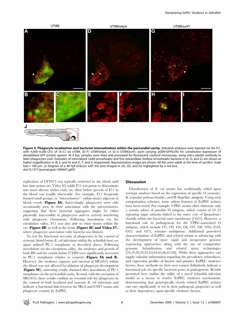
pathogens (as exemplified by F11 and UTI89, Figures 4B and5A). By 6 h post-inoculation, these bacteria were found in high
numbers both within phagocytes and free within the pericardial
cavity, often forming dense aggregates (Figure 5A–C). In
contrast to wild type UTI89, the toxin-deficient mutants
UTI89DhlyA and UTI89Dcnf1 were found in only low numbers
at the same time point, typically within or closely associated with
infiltrating phagocytes (Figure 5D–I). Notably, the rates and
levels of phagocyte recruitment into the pericardial cavity, as
assessed by microscopic observations, were similar following
inoculation with wild type UTI89 or either of the two toxin
mutants.
These findings suggest key roles for both HlyA and CNF1 in the
evasion of phagocyte-mediated killing by ExPEC at local sites of
infection. To further address this possibility, we used a
translation-blocking morpholino to silence expression of the host
PU.1 transcription factor, which drives the differentiation of
myelopoetic precursors into macrophages and neutrophils [60].
Ablation of phagocyte development was confirmed using
Figure 2. eBURST diagram of E. coli. The eBURST algorithm was used to generate a clustering diagram from 2,208 strains from the E. coli MLSTdatabase that is maintained by University College Cork. Circles represent single allele profiles (strain types, ST), based on seven genes (adk, fumC, gyrB,icd, mdh, purA, and recA). Size of each circle is proportional to the number of isolates that match a given allele profile. Blue circles are predictedfounder genotypes, while red circles represent sub-founder genotypes. Solid lines radiating from founders and sub-founders denote single locusvariants. Positions of the 10 sequenced E. coli isolates used in this study are indicated. Bootstrap confidence values for the founder groups associatedwith the highlighted strains are each 100%, except for the groups containing F11 and HS, which had values of 99% and 93%, respectively.doi:10.1371/journal.ppat.1000697.g002
Deciphering ExPEC Virulence in Zebrafish
PLoS Pathogens | www.plospathogens.org 5 December 2009 | Volume 5 | Issue 12 | e1000697

immunofluorescence microscopy of 48 to 72 hpf PU.1 morphants,
by which substantial reduction in L-plastin staining was evident
when compared with control embryos that were injected with
PBS alone (data not shown). Following inoculation of the
pericardial cavity, both UTI89DhlyA and UTI89Dcnf1 were
significantly more lethal and grew to much higher titers in the
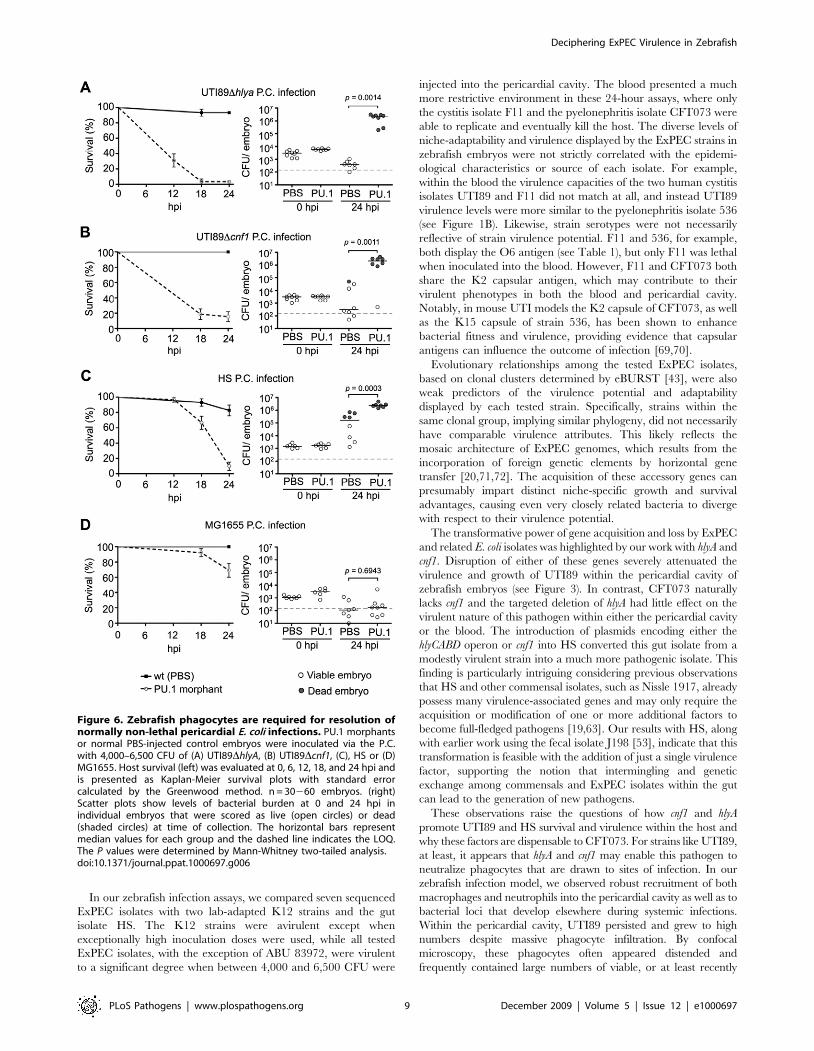
PU.1 morphants relative to the PBS-injected control embryos
(Figure 6A and B). Likewise, the gut isolate HS also displayed
substantially increased lethality and growth within PU.1 mor-
phants (Figure 6C). In contrast, the K12 strain MG1655
exhibited only slightly elevated virulence and no enhanced
replication within the PU.1 morphants (Figure 6D). Presumably,
other innate defenses and/or nutrient restrictions within the
zebrafish host are sufficient to limit MG1655 survival in the
absence, or near absence, of phagocytes.
Not unexpectedly, phagocytes also appeared to be major
facilitators of bacterial clearance within the blood and other
niches outside of the pericardial cavity. By 12 h post-inoculation
via the circulation valley, bacterial strains like MG1655 and
UTI89 (which are for the most part non-lethal within the blood
during the course of these assays, see Figure 1) were observed only
in small numbers, primarily localized within phagocytes
(Figure 7A–D). In contrast, and despite the presence of large
numbers of circulating phagocytes, CFT073 and F11 multiplied to
high levels within the blood and infiltrated multiple tissues before
eventually killing the host (Figure 7E–H). In these assays,
Figure 3. Differential effects of ExPEC-encoded toxins on zebrafish survival. (A) and (B), left panels - Zebrafish viability was determined at 0,6, 12, 18, and 24 hpi of the P.C. with medium doses (4,000–6,500 CFU) of wild type (wt) UTI89, UTI89DhlyA, or UTI89DhlyA/pSF4000 (+Hly),UTI89Dcnf1, or UTI89Dcnf1/pHLK102 (+CNF1). Right panels - Bacterial burden was determined for UTI89DhlyA and UTI89Dcnf1 at the indicated hpi.Embryos were scored as live (open circles) or dead (shaded circles) prior to homogenization. The dashed line denotes the LOQ. (C) Zebrafish survivalfollowing inoculation of the P.C. (left) or blood (right) with wt CFT073 or CFT073DhlyA. Inoculum sizes used were 2,000–3,500 CFU for P.C. injectionsand 4,000–6,500 CFU for blood injections. (D) Zebrafish survival after P.C. inoculation with 4,000–6,500 CFU of wt HS (Hly- and CNF1-negative), HS/pSF4000 (+Hly), or HS/pHLK102 (+CNF1). Survival plots (A–D) are presented as Kaplan-Meier survival plots with standard error calculated by theGreenwood method. n = 30260 embryos for all results.doi:10.1371/journal.ppat.1000697.g003
Deciphering ExPEC Virulence in Zebrafish
PLoS Pathogens | www.plospathogens.org 6 December 2009 | Volume 5 | Issue 12 | e1000697
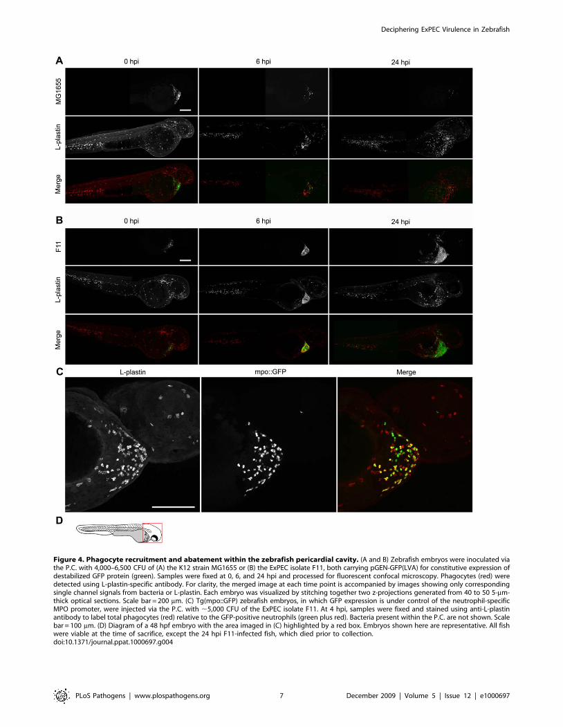
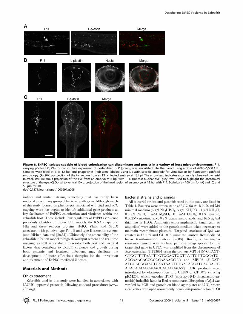
Figure 4. Phagocyte recruitment and abatement within the zebrafish pericardial cavity. (A and B) Zebrafish embryos were inoculated viathe P.C. with 4,000–6,500 CFU of (A) the K12 strain MG1655 or (B) the ExPEC isolate F11, both carrying pGEN-GFP(LVA) for constitutive expression ofdestabilized GFP protein (green). Samples were fixed at 0, 6, and 24 hpi and processed for fluorescent confocal microscopy. Phagocytes (red) weredetected using L-plastin-specific antibody. For clarity, the merged image at each time point is accompanied by images showing only correspondingsingle channel signals from bacteria or L-plastin. Each embryo was visualized by stitching together two z-projections generated from 40 to 50 5-mm-thick optical sections. Scale bar = 200 mm. (C) Tg(mpo::GFP) zebrafish embryos, in which GFP expression is under control of the neutrophil-specificMPO promoter, were injected via the P.C. with ,5,000 CFU of the ExPEC isolate F11. At 4 hpi, samples were fixed and stained using anti-L-plastinantibody to label total phagocytes (red) relative to the GFP-positive neutrophils (green plus red). Bacteria present within the P.C. are not shown. Scalebar = 100 mm. (D) Diagram of a 48 hpf embryo with the area imaged in (C) highlighted by a red box. Embryos shown here are representative. All fishwere viable at the time of sacrifice, except the 24 hpi F11-infected fish, which died prior to collection.doi:10.1371/journal.ppat.1000697.g004
Deciphering ExPEC Virulence in Zebrafish
PLoS Pathogens | www.plospathogens.org 7 December 2009 | Volume 5 | Issue 12 | e1000697
replication of CFT073 was typically restricted to the blood until
late time points (see Video S5) while F11 was prone to disseminate
into more diverse niches early on, often before growth of F11 in
the blood was readily observable. For example, F11 frequently
formed small groups, or ‘‘microclusters’’, within tissues adjacent to
blood vessels (Figure 8A). Interestingly, phagocytes were only
occasionally seen in close association with the microsclusters,
suggesting that these bacterial aggregates might be either
physically inaccessible to phagocytes and/or actively interfering
with phagocyte chemotaxis. Following inoculation via the
circulation valley, F11 was also able to enter tissues within the
eye (Figure 8B) as well as the brain (Figure 8C and Video S7),
where phagocyte association with bacteria was limited.
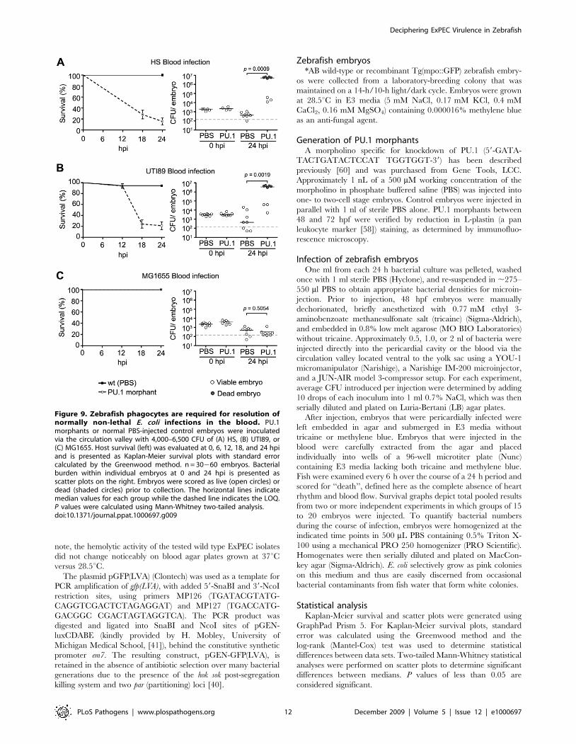
To test the functional necessity of phagocytes in the control of
systemic blood-borne E. coli infections within the zebrafish host, we
again utilized PU.1 morphants as described above. Following
inoculation via the circulation valley, the virulence and growth of
both HS and the cystitis isolate UTI89 were significantly increased
in PU.1 morphants relative to controls (Figure 9A and B).
However, the virulence capacity and survival of MG1655 within
the blood was not affected by ablation of phagocyte development
(Figure 9C), mirroring results obtained after inoculation of PU.1
morphants via the pericardial cavity. In total, with the exception of
MG1655, these results confirm an essential role for phagocytes in
the control of both localized and systemic E. coli infections and
indicate a functional link between the HlyA and CNF1 toxins and
phagocyte evasion by ExPEC.
Discussion
Classification of E. coli strains has traditionally relied upon
serotype analyses based on the expression of specific O (somatic),
K (capsular polysaccharide), and H (flagellar) antigens. Using such
categorization schemes, some salient features of ExPEC isolates
have been noted. For example, UPEC strains often elaborate only
a certain subset of possible O antigens, which consist of 10–25
repeating sugar subunits linked to the outer core of lipopolysac-
charide within the bacterial outer membrane [14,61]. However, a
functional role in pathogenesis for the UPEC-associated O
antigens, which include O1, O2, O4, O6, O7, O8, O16, O18,
O25, and O75, remains ambiguous. Additional gene-level
characterization of ExPEC and related strains is advancing with
the development of more rapid and inexpensive genome
sequencing approaches, along with the use of comparative
genomic hybridization and related array technologies
[16,19,20,45,62,63,64,65,66,67,68]. While these approaches can
supply valuable information regarding the prevalence, relatedness,
and expression profiles of known and putative ExPEC virulence
factors, these methods on their own cannot definitively indicate a
functional role for specific bacterial genes in pathogenesis. Results
presented here outline the utility of a novel zebrafish infection
model as a means to resolve ExPEC virulence mechanisms,
demonstrating that genotypically closely related ExPEC isolates
can vary significantly in vivo in their pathogenic properties as well
as their dependency upon specific virulence factors.
Figure 5. Phagocyte localization and bacterial internalization within the pericardial cavity. Zebrafish embryos were injected via the P.C.with 4,000–6,500 CFU of (A–C) wt UTI89, (D–F) UTI89DhlyA, or (G–I) UTI89Dcnf1, each carrying pGEN-GFP(LVA) for constitutive expression ofdestabilized GFP protein (green). At 6 hpi, samples were fixed and processed for fluorescent confocal microscopy, using anti-L-plastin antibody tolabel phagocytes (red). Examples of internalized (solid arrowheads) and free extracellular (hollow arrowheads) bacteria in (A, D, and G) are shown athigher magnification in (B, E, and H) and (C, F, and I), respectively. Representative images are shown. All fish were viable at the time of sacrifice. Scalebars = 100 mm. (J) Diagram of a 48 hpf embryo with the area imaged in (A), (D), and (G) highlighted by a red box.doi:10.1371/journal.ppat.1000697.g005
Deciphering ExPEC Virulence in Zebrafish
PLoS Pathogens | www.plospathogens.org 8 December 2009 | Volume 5 | Issue 12 | e1000697
In our zebrafish infection assays, we compared seven sequenced
ExPEC isolates with two lab-adapted K12 strains and the gut
isolate HS. The K12 strains were avirulent except when
exceptionally high inoculation doses were used, while all tested
ExPEC isolates, with the exception of ABU 83972, were virulent
to a significant degree when between 4,000 and 6,500 CFU were
injected into the pericardial cavity. The blood presented a much
more restrictive environment in these 24-hour assays, where only
the cystitis isolate F11 and the pyelonephritis isolate CFT073 were
able to replicate and eventually kill the host. The diverse levels of
niche-adaptability and virulence displayed by the ExPEC strains in
zebrafish embryos were not strictly correlated with the epidemi-
ological characteristics or source of each isolate. For example,
within the blood the virulence capacities of the two human cystitis
isolates UTI89 and F11 did not match at all, and instead UTI89
virulence levels were more similar to the pyelonephritis isolate 536
(see Figure 1B). Likewise, strain serotypes were not necessarily
reflective of strain virulence potential. F11 and 536, for example,
both display the O6 antigen (see Table 1), but only F11 was lethal
when inoculated into the blood. However, F11 and CFT073 both
share the K2 capsular antigen, which may contribute to their
virulent phenotypes in both the blood and pericardial cavity.
Notably, in mouse UTI models the K2 capsule of CFT073, as well
as the K15 capsule of strain 536, has been shown to enhance
bacterial fitness and virulence, providing evidence that capsular
antigens can influence the outcome of infection [69,70].
Evolutionary relationships among the tested ExPEC isolates,
based on clonal clusters determined by eBURST [43], were also
weak predictors of the virulence potential and adaptability
displayed by each tested strain. Specifically, strains within the
same clonal group, implying similar phylogeny, did not necessarily
have comparable virulence attributes. This likely reflects the
mosaic architecture of ExPEC genomes, which results from the
incorporation of foreign genetic elements by horizontal gene
transfer [20,71,72]. The acquisition of these accessory genes can
presumably impart distinct niche-specific growth and survival
advantages, causing even very closely related bacteria to diverge
with respect to their virulence potential.
The transformative power of gene acquisition and loss by ExPEC
and related E. coli isolates was highlighted by our work with hlyA and
cnf1. Disruption of either of these genes severely attenuated the
virulence and growth of UTI89 within the pericardial cavity of
zebrafish embryos (see Figure 3). In contrast, CFT073 naturally
lacks cnf1 and the targeted deletion of hlyA had little effect on the
virulent nature of this pathogen within either the pericardial cavity
or the blood. The introduction of plasmids encoding either the
hlyCABD operon or cnf1 into HS converted this gut isolate from a
modestly virulent strain into a much more pathogenic isolate. This
finding is particularly intriguing considering previous observations
that HS and other commensal isolates, such as Nissle 1917, already
possess many virulence-associated genes and may only require the
acquisition or modification of one or more additional factors to
become full-fledged pathogens [19,63]. Our results with HS, along
with earlier work using the fecal isolate J198 [53], indicate that this
transformation is feasible with the addition of just a single virulence
factor, supporting the notion that intermingling and genetic
exchange among commensals and ExPEC isolates within the gut
can lead to the generation of new pathogens.
These observations raise the questions of how cnf1 and hlyA
promote UTI89 and HS survival and virulence within the host and
why these factors are dispensable to CFT073. For strains like UTI89,
at least, it appears that hlyA and cnf1 may enable this pathogen to
neutralize phagocytes that are drawn to sites of infection. In our
zebrafish infection model, we observed robust recruitment of both
macrophages and neutrophils into the pericardial cavity as well as to
bacterial loci that develop elsewhere during systemic infections.
Within the pericardial cavity, UTI89 persisted and grew to high
numbers despite massive phagocyte infiltration. By confocal
microscopy, these phagocytes often appeared distended and
frequently contained large numbers of viable, or at least recently
Figure 6. Zebrafish phagocytes are required for resolution ofnormally non-lethal pericardial E. coli infections. PU.1 morphantsor normal PBS-injected control embryos were inoculated via the P.C.with 4,000–6,500 CFU of (A) UTI89DhlyA, (B) UTI89Dcnf1, (C), HS or (D)MG1655. Host survival (left) was evaluated at 0, 6, 12, 18, and 24 hpi andis presented as Kaplan-Meier survival plots with standard errorcalculated by the Greenwood method. n = 30260 embryos. (right)Scatter plots show levels of bacterial burden at 0 and 24 hpi inindividual embryos that were scored as live (open circles) or dead(shaded circles) at time of collection. The horizontal bars representmedian values for each group and the dashed line indicates the LOQ.The P values were determined by Mann-Whitney two-tailed analysis.doi:10.1371/journal.ppat.1000697.g006
Deciphering ExPEC Virulence in Zebrafish
PLoS Pathogens | www.plospathogens.org 9 December 2009 | Volume 5 | Issue 12 | e1000697

viable, bacteria (see Figure 5). The presence of so many wild type
GFP-positive bacteria within phagocytes may be a consequence of
highly active phagocytosis during the course of our infection assays,
but may also reflect an innate ability of some ExPEC isolates to
persist and perhaps even multiply with these effector immune cells.
This possibility requires further investigation. In sharp contrast to
wild type UTI89, the hlyA and cnf1 UTI89 mutants failed to grow
within the pericardial cavity and instead were typically found only in
small clusters within the infiltrating phagocytes.
Work in mouse and cell culture model systems has demonstrated
that high levels of HlyA can cause osmotic lysis of host cells, while
sublytic concentrations of this pore-forming toxin can modulate
host survival pathways and thereby interfere with phagocyte
chemotaxis and bactericidal activities [12,73,74,75,76]. By causing
the aberrant activation of host Rho-family GTPases, CNF1 can
similarly inhibit phagocyte functions [11,77,78]. Both HlyA and
CNF1 may also stimulate the breakdown of tissue barriers and the
release of host nutrients [24,51,79], but in our assays phagocytes
appear to be the primary targets of these toxins. This conclusion is
strongly supported by our results showing that UTI89DhlyA,
UTI89Dcnf1, and wt HS (without either hlyA or cnf1) are lethal in
PU.1 morphants that lack both neutrophils and macrophages (see
Figures 6 and 9). Depletion of these host immune cells also
substantially enhanced the virulence of wild type UTI89 when
injected into the blood, suggesting that phagocytes are the major
obstacles to the growth of ExPEC strains like UTI89 during
systemic infections.
The differential requirements for hlyA and cnf1 observed in
UTI89 versus CFT073 indicate a degree of functional redundancy
in the spectrum of virulence factors that ExPEC strains can
acquire. Specifically, our data indicate that CFT073 must have
alternate mechanisms to neutralize or overwhelm host phagocytes,
independent of HlyA or CNF1. It is likely that the disparate
degrees of virulence observed among the tested ExPEC isolates are
also influenced by differences in expression levels and/or allelic
variation of toxins like HlyA, as has been documented previously
[52,53]. However, this cannot completely account for the
differential requirements that strains like CFT073, F11, and
UTI89 have for HlyA and CNF1. Specifically, CFT073 remains
highly virulent even in the absence of hlyA and ongoing work in
our lab suggests the same is true for F11. In addition, in vitro levels
of hemolytic activity do not necessarily correlate with the virulence
capacity of individual strains. For example, we recently found that
the pyelonephritis isolate CP9 exhibits high levels of hemolytic
activity on blood agar plates at both 28.5 and 37uC, similar to
CFT073 and F11, and yet CP9 is substantially less virulent in the
zebrafish host (unpublished observations).
In conclusion, development of a zebrafish infection model has
allowed us to perform side-by-side phenotypic comparisons of the
virulence potentials and niche restrictions of multiple ExPEC
Figure 7. Differential growth and phagocytosis of E. coli isolates within the blood. Zebrafish embryos were infected via the blood with4,000–6,500 CFU of (A and B) MG1655, (C and D) UTI89, (E and F) CFT073, or (G and H) F11. All bacterial strains carry pGEN-GFP(LVA) for constitutiveexpression of destabilized GFP (green). At 12 hpi, samples were fixed and phagocytes (red) were labeled using L-plastin-specific antibody forvisualization by fluorescent confocal microscopy. Regions highlighted by arrowheads in (A), (C), (E), and (G) are shown further magnified in panels (B),(D), (F), and (H), respectively. All images shown are representative of the pool of embryos imaged. MG1655- and UTI89-infected embryos were viableand healthy in appearance prior to sacrifice for microscopy, whereas fish inoculated with CFT073 or F11 were notably sick and near death at time ofcollection. Scale bars = 100 mm. (I) Diagram of a 48 hpf embryo with the region imaged in (A), (C), (E), and (G) denoted by a red box.doi:10.1371/journal.ppat.1000697.g007
Deciphering ExPEC Virulence in Zebrafish
PLoS Pathogens | www.plospathogens.org 10 December 2009 | Volume 5 | Issue 12 | e1000697
isolates and mutant strains, something that has rarely been
undertaken with any group of bacterial pathogens. Although much
of this study focused on phenotypes associated with hlyA and cnf1,
ongoing work has begun to identify additional gene products as
key facilitators of ExPEC colonization and virulence within the
zebrafish host. These include four regulators of ExPEC virulence
previously identified in mouse UTI models: the RNA chaperone
Hfq and three secretin proteins (HofQ, YheF, and GspD)
associated with putative type IV pili and type II secretion systems
(unpublished data and [80,81]). Ultimately, the amenability of the
zebrafish infection model to high-throughput screens and real-time
imaging, as well as its ability to resolve both host and bacterial
factors that contribute to ExPEC virulence and growth during
both systemic and localized infections, may facilitate the
development of more efficacious therapies for the prevention
and treatment of ExPEC-mediated illnesses.
Materials and Methods
Ethics statementZebrafish used in this study were handled in accordance with
IACUC-approved protocols following standard procedures (www.
zfin.org).
Bacterial strains and plasmidsAll bacterial strains and plasmids used in this study are listed in
Table 1. Bacteria were grown static at 37uC for 24 h in 20 ml M9
minimal medium (6 g/l Na2HPO4, 3 g/l KH2PO4, 1 g/l NH4Cl,
0.5 g/l NaCl, 1 mM MgSO4, 0.1 mM CaCl2, 0.1% glucose,
0.0025% nicotinic acid, 0.2% casein amino acids, and 16.5 mg/ml
thiamine in H2O). Antibiotics (chloramphenicol, kanamycin, or
ampicillin) were added to the growth medium when necessary to
maintain recombinant plasmids. Targeted knockout of hlyA was
created in UTI89 and CFT073 using the lambda Red-mediated
linear transformation system [82,83]. Briefly, a kanamycin
resistance cassette with 40 base pair overhangs specific for the
target hlyA gene in UPEC was amplified from the chromosome of
Salmonella strain TT23691 using the primers MP104 (59-GTAGT-
GTGCTTTTAATTTGTGCAGTGGTTATTGTTGGCATC-
ACCAAACACCCCCCAAAACC-39) and MP105 (59-CAT-
ATGGACGGAACTCAATAACTTTGACAGCATCAGCA T-
ACACACAACCACACCACACCAC-39). PCR products were
introduced by electroporation into UTI89 or CFT073 carrying
pKM208, which encodes IPTG (isopropyl-b-D-thiogalactopyra-
noside)-inducible lambda Red recombinase. Disruption of hlyA was
verified by PCR and growth on blood agar plates at 37uC, where
clear zones developed around only hemolysin-positive colonies. Of
Figure 8. ExPEC isolates capable of blood colonization can disseminate and persist in a variety of host microenvironments. F11,carrying pGEN-GFP(LVA) for constitutive expression of destabilized GFP (green), was inoculated into the blood using a dose of 4,000–6,500 CFU.Samples were fixed at 6 or 12 hpi and phagocytes (red) were labeled using L-plastin-specific antibody for visualization by fluorescent confocalmicroscopy. (A) 20X z-projection of the tail region from an F11-infected embryo at 12 hpi. The arrowhead indicates a commonly observed bacterialmicrocluster. (B) 40X z-projection of the eye from an embryo at 6 hpi with F11. Hoechst nuclear dye (grey) was used to highlight the anatomicalstructure of the eye. (C) Dorsal to ventral 10X z-projection of the head region of an embryo at 12 hpi with F11. Scale bars = 100 mm for (A) and (C) and50 mm for (B).doi:10.1371/journal.ppat.1000697.g008
Deciphering ExPEC Virulence in Zebrafish
PLoS Pathogens | www.plospathogens.org 11 December 2009 | Volume 5 | Issue 12 | e1000697
note, the hemolytic activity of the tested wild type ExPEC isolates
did not change noticeably on blood agar plates grown at 37uCversus 28.5uC.
The plasmid pGFP(LVA) (Clontech) was used as a template for
PCR amplification of gfp(LVA), with added 59-SnaBI and 39-NcoI
restriction sites, using primers MP126 (TGATACGTATG-
CAGGTCGACTCTAGAGGAT) and MP127 (TGACCATG-
GACGGC CGACTAGTAGGTCA). The PCR product was
digested and ligated into SnaBI and NcoI sites of pGEN-
luxCDABE (kindly provided by H. Mobley, University of
Michigan Medical School, [41]), behind the constitutive synthetic
promoter em7. The resulting construct, pGEN-GFP(LVA), is
retained in the absence of antibiotic selection over many bacterial
generations due to the presence of the hok sok post-segregation
killing system and two par (partitioning) loci [40].
Zebrafish embryos*AB wild-type or recombinant Tg(mpo::GFP) zebrafish embry-
os were collected from a laboratory-breeding colony that was
maintained on a 14-h/10-h light/dark cycle. Embryos were grown
at 28.5uC in E3 media (5 mM NaCl, 0.17 mM KCl, 0.4 mM
CaCl2, 0.16 mM MgSO4) containing 0.000016% methylene blue
as an anti-fungal agent.
Generation of PU.1 morphantsA morpholino specific for knockdown of PU.1 (59-GATA-
TACTGATACTCCAT TGGTGGT-39) has been described
previously [60] and was purchased from Gene Tools, LCC.
Approximately 1 nL of a 500 mM working concentration of the
morpholino in phosphate buffered saline (PBS) was injected into
one- to two-cell stage embryos. Control embryos were injected in
parallel with 1 nl of sterile PBS alone. PU.1 morphants between
48 and 72 hpf were verified by reduction in L-plastin (a pan
leukocyte marker [58]) staining, as determined by immunofluo-
rescence microscopy.
Infection of zebrafish embryosOne ml from each 24 h bacterial culture was pelleted, washed
once with 1 ml sterile PBS (Hyclone), and re-suspended in ,275–
550 ml PBS to obtain appropriate bacterial densities for microin-
jection. Prior to injection, 48 hpf embryos were manually
dechorionated, briefly anesthetized with 0.77 mM ethyl 3-
aminobenzoate methanesulfonate salt (tricaine) (Sigma-Aldrich),
and embedded in 0.8% low melt agarose (MO BIO Laboratories)
without tricaine. Approximately 0.5, 1.0, or 2 nl of bacteria were
injected directly into the pericardial cavity or the blood via the
circulation valley located ventral to the yolk sac using a YOU-1
micromanipulator (Narishige), a Narishige IM-200 microinjector,
and a JUN-AIR model 3-compressor setup. For each experiment,
average CFU introduced per injection were determined by adding
10 drops of each inoculum into 1 ml 0.7% NaCl, which was then
serially diluted and plated on Luria-Bertani (LB) agar plates.
After injection, embryos that were pericardially infected were
left embedded in agar and submerged in E3 media without
tricaine or methylene blue. Embryos that were injected in the
blood were carefully extracted from the agar and placed
individually into wells of a 96-well microtiter plate (Nunc)
containing E3 media lacking both tricaine and methylene blue.
Fish were examined every 6 h over the course of a 24 h period and
scored for ‘‘death’’, defined here as the complete absence of heart
rhythm and blood flow. Survival graphs depict total pooled results
from two or more independent experiments in which groups of 15
to 20 embryos were injected. To quantify bacterial numbers
during the course of infection, embryos were homogenized at the
indicated time points in 500 mL PBS containing 0.5% Triton X-
100 using a mechanical PRO 250 homogenizer (PRO Scientific).
Homogenates were then serially diluted and plated on MacCon-
key agar (Sigma-Aldrich). E. coli selectively grow as pink colonies
on this medium and thus are easily discerned from occasional
bacterial contaminants from fish water that form white colonies.
Statistical analysisKaplan-Meier survival and scatter plots were generated using
GraphPad Prism 5. For Kaplan-Meier survival plots, standard
error was calculated using the Greenwood method and the
log-rank (Mantel-Cox) test was used to determine statistical
differences between data sets. Two-tailed Mann-Whitney statistical
analyses were performed on scatter plots to determine significant
differences between medians. P values of less than 0.05 are
considered significant.
Figure 9. Zebrafish phagocytes are required for resolution ofnormally non-lethal E. coli infections in the blood. PU.1morphants or normal PBS-injected control embryos were inoculatedvia the circulation valley with 4,000–6,500 CFU of (A) HS, (B) UTI89, or(C) MG1655. Host survival (left) was evaluated at 0, 6, 12, 18, and 24 hpiand is presented as Kaplan-Meier survival plots with standard errorcalculated by the Greenwood method. n = 30260 embryos. Bacterialburden within individual embryos at 0 and 24 hpi is presented asscatter plots on the right. Embryos were scored as live (open circles) ordead (shaded circles) prior to collection. The horizontal lines indicatemedian values for each group while the dashed line indicates the LOQ.P values were calculated using Mann-Whitney two-tailed analysis.doi:10.1371/journal.ppat.1000697.g009
Deciphering ExPEC Virulence in Zebrafish
PLoS Pathogens | www.plospathogens.org 12 December 2009 | Volume 5 | Issue 12 | e1000697
MicroscopyZebrafish embryos were injected with either PBS or
,5,000 CFU of the indicated bacterial strain, each carrying
pGEN-GFP(LVA). At 0, 6, 12, and 24 h post-inoculation,
embryos were fixed by rotating overnight at 4uC in PBS
containing 4% paraformaldehyde and 0.4% Triton X-100.
Embryos were then washed 365 min at room temperature in
wash buffer (PBS containing 0.8% Triton X-100), placed in
blocking buffer (PBS plus 0.25% casein, 0.1% Tween, and 1%
dimethyl sulfoxide) for 2 h, and then labeled with rabbit anti-L-
plastin antibody (1:5000, [58]) overnight at 4uC with rotation. The
embryos were again washed 4620 min at room temperature prior
to addition of donkey anti-rabbit Alexa555 fluor-conjugated
secondary antibody (1:1000; Invitrogen) in blocking buffer. After
rocking overnight at 4uC and subsequent washes, the embryos
were rinsed 3X in 30%, 50%, and 80% glycerol/PBS solutions.
Samples were then stored in 80% glycerol at 220uC or directly
mounted in 80% glycerol and imaged using an Olympus IX81
FV1000 confocal microscope equipped with 10X, 20X, and 40X
objectives. Confocal stacks were assembled using ImageJ (National
Institutes of Health) and subsequently stitched together using
Photoshop CS3 (Adobe Systems Inc.). Live injection and infection
videos were obtained using a Canon PowerShot A640 10
megapixel camera mounted on an SZX10 stereomicroscope
(Olympus) using a Camadapter kit (camadapter.com).
Cluster analysis of E. coliAllelic profiles from 2,208 E. coli isolates were downloaded from
the MLST database at the Environmental Research Institute
(ERI, University College Cork, http://mlst.ucc.ie). The eBURST
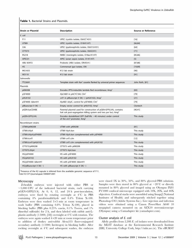
Table 1. Bacterial Strains and Plasmids.
Strain or Plasmid Description Source or Reference
E. coli
F11 UPEC (cystitis isolate, O6:K2*:H31) [16]
UTI89 UPEC (cystitis isolate, O18:K1:H7) [66,84]
536 UPEC (pyelonephritis isolate, O6:K15:H31) [64]
CFT073 UPEC (pyelonephritis isolate, O6:K2:H1) [71]
RS218 NMEC (meningitis isolate, O18ac:K1:H7) [85,86]
APEC01 APEC (avian sepsis isolate, O1:K1:H7) [5]
ABU 83972 Probiotic (ABU isolate, OR:K5:H-) [87,88]
HS Commensal (gut isolate, O9) [19,89]
MG1655 K12 lab strain [90]
W3110 K12 lab strain [91]
Salmonella
TT23691 Template strain with Kanr cassette flanked by universal primer sequences John Roth, [81]
Plasmids
pKM208 Encodes IPTG-inducible lambda Red recombinase; Ampr [83]
pSF4000 hlyCABD in pACYC184; Clmr [79]
pHLK102 cnf1 in pBluescript II SK(2) (pHLK102); Ampr [21]
pSF4000 DBamH1 hlyABD DhylC, control for pSF4000; Clmr [79]
pBluescript II SK(2) Empty vector control for pHLK102; Ampr Clontech
pGEN-luxCDABE Parental plasmid used for construction of pGEN-GFP(LVA), containshok sok post-segregation killing system and two par loci; Ampr
[40,41]
pGEN-GFP(LVA) Encodes destabilized GFP (half-life ,40 minutes) under controlof the em7 promoter; Ampr
This study
Recombinant strains
UTI89/pKM208 UTI89 with pKM208 [81]
UTI89DhlyA UTI89 hlyA::kan This study
UTI89DhlyA/pSF4000 UTI89 hlyA::kan complemented with pSF4000 This study
UTI89Dcnf1 UTI89 cnf1::clm [12]
UTI89Dcnf1/pHLK102 UTI89 cnf1::clm complemented with pHLK102 This study
CFT073/pKM208 CFT073 with pKM208 This study
CFT073DhlyA CFT073 hlyA::kan This study
HS/pSF4000 HS with pSF4000 This study
HS/pHLK102 HS with pHLK102 This study
HS/pSF4000 DBamH1 HS with pSF4000 DBamH1 This study
HS/pBluescript II SK(2) HS with pBluescript II SK(2) This study
*Presence of the K2 capsule is inferred from the available genomic sequence of F11.doi:10.1371/journal.ppat.1000697.t001
Deciphering ExPEC Virulence in Zebrafish
PLoS Pathogens | www.plospathogens.org 13 December 2009 | Volume 5 | Issue 12 | e1000697
algorithm (http://eburst.mlst.net/) was used with 1,000 bootstrap
iterations to organize strains into clonal clusters and to generate an
eBURST diagram [43].
Supporting Information
Figure S1 Schematic of 48 hpf zebrafish anatomical features.
Microinjection sites used in this study are indicated by shaded
arrowheads. Definitions: P.C., pericardial cavity; circ. val.,
circulation valley; C.V., caudal vein; Nc, notochord; D.A., dorsal
aorta.
Found at: doi:10.1371/journal.ppat.1000697.s001 (0.19 MB TIF)
Video S1 The movie depicts the inoculation of the pericardial
cavity of a 48 hpf zebrafish embryo with approximately
10,000 CFU of MG1655/pGEN-GFP(LVA).
Found at: doi:10.1371/journal.ppat.1000697.s002 (3.48 MB
MOV)
Video S2 The movie shows about 10,000 CFU of MG1655/
pGEN-GFP(LVA) being injected into the blood of a 48 hpf
zebrafish embryo via the circulation valley.
Found at: doi:10.1371/journal.ppat.1000697.s003 (2.93 MB
MOV)
Video S3 Movie depicts healthy uninfected 48 hpf embryos
using dark field and oblique light, as well as fluorescent (green) as a
control. Note the clarity of the pericardial cavity, heart structure,
heartbeat, and blood flow in comparison to Movies S4, S5 and S6
taken using ExPEC-infected embryos.
Found at: doi:10.1371/journal.ppat.1000697.s004 (4.20 MB
MOV)
Video S4 Zebrafish embryo infected with CFT073/pGEN-
GFP(LVA) via the pericardial. Clips show state of the pericardial
cavity at 6, 12, and 24 hpi.
Found at: doi:10.1371/journal.ppat.1000697.s005 (5.79 MB
MOV)
Video S5 Zebrafish embryo infected with CFT073/pGEN-
GFP(LVA) via the circulation valley. Clips show bacterial
dissemination throughout the circulation at 12 hpi. By 18-24 hpi
most CFT073-infected fish will have died and bacteria become
dispersed throughout most tissues.
Found at: doi:10.1371/journal.ppat.1000697.s006 (10.64 MB
MOV)
Video S6 Embryos that survive challenge with F11, UTI89,
APEC01, RS218, 83972, and, less often, MG1655 may still appear
notably sick, despite the absence of large numbers of bacteria.
Clips show examples of restricted blood flow, heart deformation,
edema, and curled tails in embryos at various time points post
inoculation of the pericardial cavity with UTI89 or APEC01.
Found at: doi:10.1371/journal.ppat.1000697.s007 (7.43 MB
MOV)
Video S7 Movie shows sequential merged 10X z-stacks of the
F11/pGEN-GFP(LVA)-infected embryo depicted in Figure 8C.
Phagocytes labeled with anti-L-plastin antibodies are shown in red.
Found at: doi:10.1371/journal.ppat.1000697.s008 (1.80 MB
MOV)
Acknowledgments
We thank Drs. P. Jensen, B. Bass, and J. Habig (University of Utah) for
providing equipment for the establishment of the zebrafish infection model
system in our lab, as well as S. Johnson and Dr. G. King at the Centralized
Zebrafish Animal Resource Facility (CZAR, University of Utah) for help in
setting up and maintaining the zebrafish tanks. We are also grateful to Drs.
J. Kaper (University of Maryland), K. S. Kim (Johns Hopkins University),
H. Lockman (Battelle Memorial Institute), H. Mobley (University of
Michigan Medical School), L. Nolan and Y. Wannemuehler (Iowa State
University), A. O’Brien and S. B. Rasmussen (Uniformed Services
University of the Health Sciences), C. Svanborg (Lund University), and
R. Welch (University of Wisconsin, Madison) for bacterial strains and
plasmid constructs. Thanks are also due to all those who submit strain
information to the MLST Database at the ERI (University College Cork),
which is publicly available at http://mlst.ucc.ie and supported by a grant
from the Science Foundation of Ireland (05/FE1/B882). Finally, we are
thankful to Dr. C. Rodesch and K. Carney for their expert help with the
microscopy and Dr. S. Renshaw (MRC Centre for Developmental and
Biomedical Genetics, University of Sheffield) for providing the
Tg(mpo::GFP) zebrafish.
Author Contributions
Conceived and designed the experiments: TJW MAM. Performed the
experiments: TJW. Analyzed the data: TJW MAM. Contributed reagents/
materials/analysis tools: TJW JMB MJR. Wrote the paper: TJW MAM.
References
1. Kaper JB, Nataro JP, Mobley HL (2004) Pathogenic Escherichia coli. Nat Rev
Microbiol 2: 123–140.
2. Foxman B (2003) Epidemiology of urinary tract infections: incidence, morbidity,
and economic costs. Dis Mon 49: 53–70.
3. Russo TA, Johnson JR (2000) Proposal for a new inclusive designation for
extraintestinal pathogenic isolates of Escherichia coli: ExPEC. J Infect Dis 181:
1753–1754.
4. Smith JL, Fratamico PM, Gunther NW (2007) Extraintestinal pathogenic
Escherichia coli. Foodborne Pathog Dis 4: 134–163.
5. Johnson TJ, Kariyawasam S, Wannemuehler Y, Mangiamele P, Johnson SJ,
et al. (2007) The genome sequence of avian pathogenic Escherichia coli strain
O1:K1:H7 shares strong similarities with human extraintestinal pathogenic E.
coli genomes. J Bacteriol 189: 3228–3236.
6. Ewers C, Li G, Wilking H, Kiessling S, Alt K, et al. (2007) Avian pathogenic,
uropathogenic, and newborn meningitis-causing Escherichia coli: how closely
related are they? Int J Med Microbiol 297: 163–176.
7. Ron EZ (2006) Host specificity of septicemic Escherichia coli: human and avian
pathogens. Curr Opin Microbiol 9: 28–32.
8. Rodriguez-Siek KE, Giddings CW, Doetkott C, Johnson TJ, Fakhr MK,
et al. (2005) Comparison of Escherichia coli isolates implicated in human urinary
tract infection and avian colibacillosis. Microbiology 151: 2097–2110.
9. Wiles TJ, Kulesus RR, Mulvey MA (2008) Origins and virulence mechanisms of
uropathogenic Escherichia coli. Exp Mol Pathol 85: 11–19.
10. Guyer DM, Radulovic S, Jones FE, Mobley HL (2002) Sat, the secreted
autotransporter toxin of uropathogenic Escherichia coli, is a vacuolating
cytotoxin for bladder and kidney epithelial cells. Infect Immun 70: 4539–4546.
11. Lemonnier M, Landraud L, Lemichez E (2007) Rho GTPase-activating
bacterial toxins: from bacterial virulence regulation to eukaryotic cell biology.
FEMS Microbiol Rev 31: 515–534.
12. Wiles TJ, Dhakal BK, Eto DS, Mulvey MA (2008) Inactivation of host Akt/
protein kinase B signaling by bacterial pore-forming toxins. Mol Biol Cell 19:
1427–1438.
13. Dhakal BK, Kulesus RR, Mulvey MA (2008) Mechanisms and consequences of
bladder cell invasion by uropathogenic Escherichia coli. Eur J Clin Invest 38
Suppl 2: 2–11.
14. Bidet P, Mahjoub-Messai F, Blanco J, Dehem M, Aujard Y, et al. (2007)
Combined multilocus sequence typing and O serogrouping distinguishes
Escherichia coli subtypes associated with infant urosepsis and/or meningitis.
J Infect Dis 196: 297–303.
15. Hacker J, Blum-Oehler G, Muhldorfer I, Tschape H (1997) Pathogenicity
islands of virulent bacteria: structure, function and impact on microbial
evolution. Mol Microbiol 23: 1089–1097.
16. Lloyd AL, Rasko DA, Mobley HL (2007) Defining genomic islands and
uropathogen-specific genes in uropathogenic Escherichia coli. J Bacteriol 189:
3532–3546.
17. Hacker J, Kaper JB (2000) Pathogenicity islands and the evolution of microbes.
Annu Rev Microbiol 54: 641–679.
18. Gal-Mor O, Finlay BB (2006) Pathogenicity islands: a molecular toolbox for
bacterial virulence. Cell Microbiol 8: 1707–1719.
19. Rasko DA, Rosovitz MJ, Myers GS, Mongodin EF, Fricke WF, et al. (2008) The
pangenome structure of Escherichia coli: comparative genomic analysis of E. coli
commensal and pathogenic isolates. J Bacteriol 190: 6881–6893.
Deciphering ExPEC Virulence in Zebrafish
PLoS Pathogens | www.plospathogens.org 14 December 2009 | Volume 5 | Issue 12 | e1000697
20. Touchon M, Hoede C, Tenaillon O, Barbe V, Baeriswyl S, et al. (2009)
Organised genome dynamics in the Escherichia coli species results in highly
diverse adaptive paths. PLoS Genet 5: e1000344. doi:10.1371/journal.p-
gen.1000344.
21. Rippere-Lampe KE, O’Brien AD, Conran R, Lockman HA (2001) Mutation of
the gene encoding cytotoxic necrotizing factor type 1 (cnf(1)) attenuates the
virulence of uropathogenic Escherichia coli. Infect Immun 69: 3954–3964.
22. Khan NA, Wang Y, Kim KJ, Chung JW, Wass CA, et al. (2002) Cytotoxic
necrotizing factor-1 contributes to Escherichia coli K1 invasion of the central
nervous system. J Biol Chem 277: 15607–15612.
23. Johnson DE, Drachenberg C, Lockatell CV, Island MD, Warren JW, et al.
(2000) The role of cytotoxic necrotizing factor-1 in colonization and tissue injury
in a murine model of urinary tract infection. FEMS Immunol Med Microbiol
28: 37–41.
24. Smith YC, Rasmussen SB, Grande KK, Conran RM, O’Brien AD (2008)
Hemolysin of uropathogenic Escherichia coli evokes extensive shedding of the
uroepithelium and hemorrhage in bladder tissue within the first 24 hours after
intraurethral inoculation of mice. Infect Immun 76: 2978–2990.
25. Brannon MK, Davis JM, Mathias JR, Hall CJ, Emerson JC, et al. (2009)
Pseudomonas aeruginosa Type III secretion system interacts with phagocytes to
modulate systemic infection of zebrafish embryos. Cell Microbiol.
26. Davis JM, Clay H, Lewis JL, Ghori N, Herbomel P, et al. (2002) Real-time
visualization of mycobacterium-macrophage interactions leading to initiation of
granuloma formation in zebrafish embryos. Immunity 17: 693–702.
27. Neely MN, Pfeifer JD, Caparon M (2002) Streptococcus-zebrafish model of
bacterial pathogenesis. Infect Immun 70: 3904–3914.
28. van der Sar AM, Musters RJ, van Eeden FJ, Appelmelk BJ, Vandenbroucke-
Grauls CM, et al. (2003) Zebrafish embryos as a model host for the real time
analysis of Salmonella typhimurium infections. Cell Microbiol 5: 601–611.
29. Prajsnar TK, Cunliffe VT, Foster SJ, Renshaw SA (2008) A novel vertebrate
model of Staphylococcus aureus infection reveals phagocyte-dependent
resistance of zebrafish to non-host specialized pathogens. Cell Microbiol 10:
2312–2325.
30. Kizy AE, Neely MN (2009) First Streptococcus pyogenes signature-tagged
mutagenesis screen identifies novel virulence determinants. Infect Immun 77:
1854–1865.
31. Clatworthy AE, Lee JS, Leibman M, Kostun Z, Davidson AJ, et al. (2009)
Pseudomonas aeruginosa infection of zebrafish involves both host and pathogen
determinants. Infect Immun 77: 1293–1303.
32. Trede NS, Langenau DM, Traver D, Look AT, Zon LI (2004) The use of
zebrafish to understand immunity. Immunity 20: 367–379.
33. Li X, Wang S, Qi J, Echtenkamp SF, Chatterjee R, et al. (2007) Zebrafish
peptidoglycan recognition proteins are bactericidal amidases essential for defense
against bacterial infections. Immunity 27: 518–529.
34. Jault C, Pichon L, Chluba J (2004) Toll-like receptor gene family and TIR-
domain adapters in Danio rerio. Mol Immunol 40: 759–771.
35. Lieschke GJ, Oates AC, Crowhurst MO, Ward AC, Layton JE (2001)
Morphologic and functional characterization of granulocytes and macrophages
in embryonic and adult zebrafish. Blood 98: 3087–3096.
36. Mulvey MA, Schilling JD, Martinez JJ, Hultgren SJ (2000) Bad bugs and
beleaguered bladders: interplay between uropathogenic Escherichia coli and
innate host defenses. Proc Natl Acad Sci U S A 97: 8829–8835.
37. Pradel E, Ewbank JJ (2004) Genetic models in pathogenesis. Annu Rev Genet
38: 347–363.
38. Herbomel P, Thisse B, Thisse C (1999) Ontogeny and behaviour of early
macrophages in the zebrafish embryo. Development 126: 3735–3745.
39. Roos V, Ulett GC, Schembri MA, Klemm P (2006) The asymptomatic
bacteriuria Escherichia coli strain 83972 outcompetes uropathogenic E. coli
strains in human urine. Infect Immun 74: 615–624.
40. Galen JE, Nair J, Wang JY, Wasserman SS, Tanner MK, et al. (1999)
Optimization of plasmid maintenance in the attenuated live vector vaccine strain
Salmonella typhi CVD 908-htrA. Infect Immun 67: 6424–6433.
41. Lane MC, Alteri CJ, Smith SN, Mobley HL (2007) Expression of flagella is
coincident with uropathogenic Escherichia coli ascension to the upper urinary
tract. Proc Natl Acad Sci U S A 104: 16669–16674.
42. Maiden MC, Bygraves JA, Feil E, Morelli G, Russell JE, et al. (1998) Multilocus
sequence typing: a portable approach to the identification of clones within
populations of pathogenic microorganisms. Proc Natl Acad Sci U S A 95:
3140–3145.
43. Feil EJ, Li BC, Aanensen DM, Hanage WP, Spratt BG (2004) eBURST:
inferring patterns of evolutionary descent among clusters of related bacterial
genotypes from multilocus sequence typing data. J Bacteriol 186: 1518–1530.
44. Feil EJ (2004) Small change: keeping pace with microevolution. Nat Rev
Microbiol 2: 483–495.
45. Jaureguy F, Landraud L, Passet V, Diancourt L, Frapy E, et al. (2008)
Phylogenetic and genomic diversity of human bacteremic Escherichia coli
strains. BMC Genomics 9: 560.
46. Klemm P, Hancock V, Schembri MA (2007) Mellowing out: adaptation to
commensalism by Escherichia coli asymptomatic bacteriuria strain 83972. Infect
Immun 75: 3688–3695.
47. Marrs CF, Zhang L, Foxman B (2005) Escherichia coli mediated urinary tract
infections: are there distinct uropathogenic E. coli (UPEC) pathotypes? FEMS
Microbiol Lett 252: 183–190.
48. Johnson JR (1991) Virulence factors in Escherichia coli urinary tract infection.
Clin Microbiol Rev 4: 80–128.
49. Landraud L, Gibert M, Popoff MR, Boquet P, Gauthier M (2003) Expression of
cnf1 by Escherichia coli J96 involves a large upstream DNA region including the
hlyCABD operon, and is regulated by the RfaH protein. Mol Microbiol 47:
1653–1667.
50. Landraud L, Gauthier M, Fosse T, Boquet P (2000) Frequency of Escherichia
coli strains producing the cytotoxic necrotizing factor (CNF1) in nosocomial
urinary tract infections. Lett Appl Microbiol 30: 213–216.
51. Linggood MA, Ingram PL (1982) The role of alpha haemolysin in the virulence
of Escherichia coli for mice. J Med Microbiol 15: 23–30.
52. Welch RA, Falkow S (1984) Characterization of Escherichia coli hemolysins
conferring quantitative differences in virulence. Infect Immun 43: 156–160.
53. Welch RA, Dellinger EP, Minshew B, Falkow S (1981) Haemolysin contributes
to virulence of extra-intestinal E. coli infections. Nature 294: 665–667.
54. Haugen BJ, Pellett S, Redford P, Hamilton HL, Roesch PL, et al. (2007) In vivo
gene expression analysis identifies genes required for enhanced colonization of
the mouse urinary tract by uropathogenic Escherichia coli strain CFT073 dsdA.
Infect Immun 75: 278–289.
55. Mobley HL, Green DM, Trifillis AL, Johnson DE, Chippendale GR, et al.
(1990) Pyelonephritogenic Escherichia coli and killing of cultured human renal
proximal tubular epithelial cells: role of hemolysin in some strains. Infect Immun
58: 1281–1289.
56. Weichhart T, Haidinger M, Horl WH, Saemann MD (2008) Current concepts
of molecular defence mechanisms operative during urinary tract infection.
Eur J Clin Invest 38 Suppl 2: 29–38.
57. Ragnarsdottir B, Fischer H, Godaly G, Gronberg-Hernandez J, Gustafsson M,
et al. (2008) TLR- and CXCR1-dependent innate immunity: insights into the
genetics of urinary tract infections. Eur J Clin Invest 38 Suppl 2: 12–20.
58. Redd MJ, Kelly G, Dunn G, Way M, Martin P (2006) Imaging macrophage
chemotaxis in vivo: studies of microtubule function in zebrafish wound
inflammation. Cell Motil Cytoskeleton 63: 415–422.
59. Renshaw SA, Loynes CA, Trushell DM, Elworthy S, Ingham PW, et al. (2006) A
transgenic zebrafish model of neutrophilic inflammation. Blood 108: 3976–3978.
60. Rhodes J, Hagen A, Hsu K, Deng M, Liu TX, et al. (2005) Interplay of pu.1 and
gata1 determines myelo-erythroid progenitor cell fate in zebrafish. Dev Cell 8:
97–108.
61. Stenutz R, Weintraub A, Widmalm G (2006) The structures of Escherichia coli
O-polysaccharide antigens. FEMS Microbiol Rev 30: 382–403.
62. Le Gall T, Clermont O, Gouriou S, Picard B, Nassif X, et al. (2007)
Extraintestinal virulence is a coincidental by-product of commensalism in B2
phylogenetic group Escherichia coli strains. Mol Biol Evol 24: 2373–2384.
63. Grozdanov L, Raasch C, Schulze J, Sonnenborn U, Gottschalk G, et al. (2004)
Analysis of the genome structure of the nonpathogenic probiotic Escherichia coli
strain Nissle 1917. J Bacteriol 186: 5432–5441.
64. Brzuszkiewicz E, Bruggemann H, Liesegang H, Emmerth M, Olschlager T,
et al. (2006) How to become a uropathogen: comparative genomic analysis of
extraintestinal pathogenic Escherichia coli strains. Proc Natl Acad Sci U S A
103: 12879–12884.
65. Hancock V, Seshasayee AS, Ussery DW, Luscombe NM, Klemm P (2008)
Transcriptomics and adaptive genomics of the asymptomatic bacteriuria
Escherichia coli strain 83972. Mol Genet Genomics 279: 523–534.
66. Chen SL, Hung CS, Xu J, Reigstad CS, Magrini V, et al. (2006) Identification of
genes subject to positive selection in uropathogenic strains of Escherichia coli: a
comparative genomics approach. Proc Natl Acad Sci U S A 103: 5977–5982.
67. Snyder JA, Haugen BJ, Buckles EL, Lockatell CV, Johnson DE, et al. (2004)
Transcriptome of uropathogenic Escherichia coli during urinary tract infection.
Infect Immun 72: 6373–6381.
68. Reigstad CS, Hultgren SJ, Gordon JI (2007) Functional genomic studies of
uropathogenic Escherichia coli and host urothelial cells when intracellular
bacterial communities are assembled. J Biol Chem 282: 21259–21267.
69. Buckles EL, Wang X, Lane MC, Lockatell CV, Johnson DE, et al. (2009) Role
of the K2 Capsule in Escherichia coli Urinary Tract Infection and Serum
Resistance. J Infect Dis 199: 1689–1697.
70. Schneider G, Dobrindt U, Bruggemann H, Nagy G, Janke B, et al. (2004) The
pathogenicity island-associated K15 capsule determinant exhibits a novel genetic
structure and correlates with virulence in uropathogenic Escherichia coli strain
536. Infect Immun 72: 5993–6001.
71. Welch RA, Burland V, Plunkett G, 3rd, Redford P, Roesch P, et al. (2002)
Extensive mosaic structure revealed by the complete genome sequence of
uropathogenic Escherichia coli. Proc Natl Acad Sci U S A 99: 17020–17024.
72. Schubert S, Darlu P, Clermont O, Wieser A, Magistro G, et al. (2009) Role of
intraspecies recombination in the spread of pathogenicity islands within the
Escherichia coli species. PLoS Pathog 5: e1000257. doi:10.1371/journal.-
ppat.1000257.
73. Cavalieri SJ, Snyder IS (1982) Effect of Escherichia coli alpha-hemolysin on
human peripheral leukocyte viability in vitro. Infect Immun 36: 455–461.
74. Jonas D, Schultheis B, Klas C, Krammer PH, Bhakdi S (1993) Cytocidal effects
of Escherichia coli hemolysin on human T lymphocytes. Infect Immun 61:
1715–1721.
75. Chen M, Tofighi R, Bao W, Aspevall O, Jahnukainen T, et al. (2006) Carbon
monoxide prevents apoptosis induced by uropathogenic Escherichia coli toxins.
Pediatr Nephrol 21: 382–389.
Deciphering ExPEC Virulence in Zebrafish
PLoS Pathogens | www.plospathogens.org 15 December 2009 | Volume 5 | Issue 12 | e1000697
76. Russo TA, Davidson BA, Genagon SA, Warholic NM, Macdonald U, et al.
(2005) E. coli virulence factor hemolysin induces neutrophil apoptosis andnecrosis/lysis in vitro and necrosis/lysis and lung injury in a rat pneumonia
model. Am J Physiol Lung Cell Mol Physiol 289: L207–216.
77. Davis JM, Carvalho HM, Rasmussen SB, O’Brien AD (2006) Cytotoxicnecrotizing factor type 1 delivered by outer membrane vesicles of uropathogenic
Escherichia coli attenuates polymorphonuclear leukocyte antimicrobial activityand chemotaxis. Infect Immun 74: 4401–4408.
78. Davis JM, Rasmussen SB, O’Brien AD (2005) Cytotoxic necrotizing factor type
1 production by uropathogenic Escherichia coli modulates polymorphonuclearleukocyte function. Infect Immun 73: 5301–5310.
79. Bauer ME, Welch RA (1996) Association of RTX toxins with erythrocytes.Infect Immun 64: 4665–4672.
80. Kulkarni R, Dhakal BK, Slechta ES, Kurtz Z, Mulvey MA, et al. (2009)Roles of putative type II secretion and type IV pilus systems in the virulence
of uropathogenic Escherichia coli. PLoS ONE 4: e4752. doi:10.1371/
journal.pone.0004752.81. Kulesus RR, Diaz-Perez K, Slechta ES, Eto DS, Mulvey MA (2008) Impact of
the RNA chaperone Hfq on the fitness and virulence potential of uropathogenicEscherichia coli. Infect Immun 76: 3019–3026.
82. Datsenko KA, Wanner BL (2000) One-step inactivation of chromosomal genes
in Escherichia coli K-12 using PCR products. Proc Natl Acad Sci U S A 97:6640–6645.
83. Murphy KC, Campellone KG (2003) Lambda Red-mediated recombinogenicengineering of enterohemorrhagic and enteropathogenic E. coli. BMC Mol Biol
4: 11.
84. Mulvey MA, Schilling JD, Hultgren SJ (2001) Establishment of a persistent
Escherichia coli reservoir during the acute phase of a bladder infection. Infect
Immun 69: 4572–4579.
85. Yao Y, Xie Y, Kim KS (2006) Genomic comparison of Escherichia coli K1
strains isolated from the cerebrospinal fluid of patients with meningitis. Infect
Immun 74: 2196–2206.
86. Xie Y, Kolisnychenko V, Paul-Satyaseela M, Elliott S, Parthasarathy G, et al.
(2006) Identification and characterization of Escherichia coli RS218-derived
islands in the pathogenesis of E. coli meningitis. J Infect Dis 194: 358–364.
87. Klemm P, Roos V, Ulett GC, Svanborg C, Schembri MA (2006) Molecular
characterization of the Escherichia coli asymptomatic bacteriuria strain 83972:
the taming of a pathogen. Infect Immun 74: 781–785.
88. Hull RA, Rudy DC, Donovan WH, Wieser IE, Stewart C, et al. (1999)
Virulence properties of Escherichia coli 83972, a prototype strain associated with
asymptomatic bacteriuria. Infect Immun 67: 429–432.
89. Levine MM, Bergquist EJ, Nalin DR, Waterman DH, Hornick RB, et al. (1978)
Escherichia coli strains that cause diarrhoea but do not produce heat-labile or
heat-stable enterotoxins and are non-invasive. Lancet 1: 1119–1122.
90. Blattner FR, Plunkett G, 3rd, Bloch CA, Perna NT, Burland V, et al. (1997) The
complete genome sequence of Escherichia coli K-12. Science 277: 1453–1474.
91. Hayashi K, Morooka N, Yamamoto Y, Fujita K, Isono K, et al. (2006) Highly
accurate genome sequences of Escherichia coli K-12 strains MG1655 and
W3110. Mol Syst Biol 2: 2006 0007.
Deciphering ExPEC Virulence in Zebrafish
PLoS Pathogens | www.plospathogens.org 16 December 2009 | Volume 5 | Issue 12 | e1000697

![Aqua(4,4'-bipyridine-[kappa]N)bis(1,4-dioxo-1 ... - ScienceOpen](https://static.fdokumen.com/doc/165x107/63262349e491bcb36c0aa51f/aqua44-bipyridine-kappanbis14-dioxo-1-scienceopen.jpg)