Identification of Minimal Predictors of Avian Pathogenic Escherichia coli Virulence for Use as a...
10
JOURNAL OF CLINICAL MICROBIOLOGY, Dec. 2008, p. 3987–3996 Vol. 46, No. 12 0095-1137/08/$08.000 doi:10.1128/JCM.00816-08 Copyright © 2008, American Society for Microbiology. All Rights Reserved. Identification of Minimal Predictors of Avian Pathogenic Escherichia coli Virulence for Use as a Rapid Diagnostic Tool Timothy J. Johnson, 1 Yvonne Wannemuehler, 2 Curt Doetkott, 3 Sara J. Johnson, 2 Sandra C. Rosenberger, 4 and Lisa K. Nolan 2 * Department of Veterinary and Biomedical Sciences, University of Minnesota, St. Paul, Minnesota 55108 1 ; Department of Veterinary Microbiology and Preventive Medicine, Iowa State University, Ames, Iowa 50011 2 ; Information Technology Services, North Dakota State University, Fargo, North Dakota 58105 3 ; and Aviserve LLC, Delaware Technology Park, Newark, Delaware 19711 4 Received 29 April 2008/Returned for modification 7 September 2008/Accepted 30 September 2008 To identify traits that predict avian pathogenic Escherichia coli (APEC) virulence, 124 avian E. coli isolates of known pathogenicity and serogroup were subjected to virulence genotyping and phylogenetic typing. The results were analyzed by multiple-correspondence analysis. From this analysis, five genes carried by plasmids were identified as being the most significantly associated with highly pathogenic APEC strains: iutA, hlyF, iss, iroN, and ompT. A multiplex PCR panel targeting these five genes was used to screen a collection of 994 avian E. coli isolates. APEC isolates were clearly distinguished from the avian fecal E. coli isolates by their possession of these genes, suggesting that this pentaplex panel has diagnostic applications and underscoring the close association between avian E. coli virulence and the possession of ColV plasmids. Also, the sharp demarcation between APEC isolates and avian fecal E. coli isolates in their plasmid-associated virulence gene content suggests that APEC isolates are well equipped for a pathogenic lifestyle, which is contrary to the widely held belief that most APEC isolates are opportunistic pathogens. Regardless, APEC isolates remain an important problem for poultry producers and a potential concern for public health professionals, as growing evidence suggests a possible role for APEC in human disease. Thus, the pentaplex panel described here may be useful in detecting APEC-like strains occurring in poultry production, along the food chain, and in human disease. This panel may be helpful toward clarifying potential roles of APEC in human disease, ascertaining the source of APEC in animal outbreaks, and identifying effective targets of avian colibacillosis control. Avian pathogenic Escherichia coli (APEC) isolates cause colibacillosis in birds raised for meat and eggs (2). This disease results in significant morbidity and mortality, which translates into multimillion-dollar annual losses for all facets of the world’s poultry industry (2). It is estimated that at least 30% of the commercial flocks in the United States, at any point in time, are affected by colibacillosis. Recent reports have sug- gested a link between APEC and human disease (13, 36). Thus, the enhanced control of avian colibacillosis could prove bene- ficial to both animal and human health. Management approaches based on the protection of poultry from predisposing conditions have proved largely ineffective in controlling avian colibacillosis (2). Also, evidence exists that APEC isolates are becoming more resistant to antimicrobial agents (17, 23, 29, 47), indicating that the control of colibacil- losis is likely to become even more problematic in the future. Further complicating the control of this disease is the fact that antimicrobial usage in animal production is undergoing un- precedented scrutiny, with limitations placed on the use of certain agents in poultry production. Consequently, the vac- cine-based control of avian colibacillosis, where appropriate, is likely to become increasingly desirable. Unfortunately, vaccines designed to prevent avian colibacil- losis have met with mixed results. Although vaccines against various APEC isolates have been produced (1, 3, 7, 14, 25, 26, 34, 38), some have proved effective only against homologous challenge (26, 34). This type of vaccine failure is a critical impediment to colibacillosis control, often because of the di- versity of APEC populations (36, 37). Despite this diversity, recent efforts to define the APEC pathotype have shown that most APEC isolates contain a highly conserved cluster of plas- mid-linked virulence genes that occurs in relatively few avian fecal commensal E. coli (AFEC) isolates (22, 37). Thus, the exploitation of these plasmid traits or other common APEC markers as the targets of future diagnostic tools and/or vac- cines may yield colibacillosis control measures with widespread applicability. Indeed, attempts to exploit this association between plasmid genes and APEC virulence to improve colibacillosis control already are under way. Lynne et al. (28) described the testing of a vaccine that targeted a plasmid-mediated trait, and others have described rapid diagnostic tools that identify APEC iso- lates based on the possession of certain genes, including plas- mid-linked ones (11, 41). Unfortunately, these attempts were made prior to the recognition that these plasmid genes are ubiquitous among APEC isolates and before multiple APEC plasmid sequences had become available (21, 22). Also, these procedures were validated with relatively small samples of iso- lates. Here, we build on recent knowledge to more clearly define the APEC pathotype and to apply this definition to the * Corresponding author. Mailing address: Department of Veteri- nary Microbiology and Preventive Medicine, VMRI 2, 1802 Elwood Dr., Iowa State University, Ames, IA 50011. Phone: (515) 294-3470. Fax: (515) 294-3839. E-mail: [email protected]. Published ahead of print on 8 October 2008. 3987 on February 3, 2015 by guest http://jcm.asm.org/ Downloaded from
-
Upload
independent -
Category
Documents
-
view
2 -
download
0
Transcript of Identification of Minimal Predictors of Avian Pathogenic Escherichia coli Virulence for Use as a...

JOURNAL OF CLINICAL MICROBIOLOGY, Dec. 2008, p. 3987–3996 Vol. 46, No. 120095-1137/08/$08.00�0 doi:10.1128/JCM.00816-08Copyright © 2008, American Society for Microbiology. All Rights Reserved.
Identification of Minimal Predictors of Avian Pathogenic Escherichia coliVirulence for Use as a Rapid Diagnostic Tool�
Timothy J. Johnson,1 Yvonne Wannemuehler,2 Curt Doetkott,3 Sara J. Johnson,2Sandra C. Rosenberger,4 and Lisa K. Nolan2*
Department of Veterinary and Biomedical Sciences, University of Minnesota, St. Paul, Minnesota 551081; Department ofVeterinary Microbiology and Preventive Medicine, Iowa State University, Ames, Iowa 500112; Information Technology Services,
North Dakota State University, Fargo, North Dakota 581053; and Aviserve LLC, Delaware Technology Park,Newark, Delaware 197114
Received 29 April 2008/Returned for modification 7 September 2008/Accepted 30 September 2008
To identify traits that predict avian pathogenic Escherichia coli (APEC) virulence, 124 avian E. coliisolates of known pathogenicity and serogroup were subjected to virulence genotyping and phylogenetictyping. The results were analyzed by multiple-correspondence analysis. From this analysis, five genescarried by plasmids were identified as being the most significantly associated with highly pathogenic APECstrains: iutA, hlyF, iss, iroN, and ompT. A multiplex PCR panel targeting these five genes was used to screena collection of 994 avian E. coli isolates. APEC isolates were clearly distinguished from the avian fecal E.coli isolates by their possession of these genes, suggesting that this pentaplex panel has diagnosticapplications and underscoring the close association between avian E. coli virulence and the possession ofColV plasmids. Also, the sharp demarcation between APEC isolates and avian fecal E. coli isolates in theirplasmid-associated virulence gene content suggests that APEC isolates are well equipped for a pathogeniclifestyle, which is contrary to the widely held belief that most APEC isolates are opportunistic pathogens.Regardless, APEC isolates remain an important problem for poultry producers and a potential concern forpublic health professionals, as growing evidence suggests a possible role for APEC in human disease.Thus, the pentaplex panel described here may be useful in detecting APEC-like strains occurring inpoultry production, along the food chain, and in human disease. This panel may be helpful towardclarifying potential roles of APEC in human disease, ascertaining the source of APEC in animal outbreaks,and identifying effective targets of avian colibacillosis control.
Avian pathogenic Escherichia coli (APEC) isolates causecolibacillosis in birds raised for meat and eggs (2). This diseaseresults in significant morbidity and mortality, which translatesinto multimillion-dollar annual losses for all facets of theworld’s poultry industry (2). It is estimated that at least 30% ofthe commercial flocks in the United States, at any point intime, are affected by colibacillosis. Recent reports have sug-gested a link between APEC and human disease (13, 36). Thus,the enhanced control of avian colibacillosis could prove bene-ficial to both animal and human health.
Management approaches based on the protection of poultryfrom predisposing conditions have proved largely ineffective incontrolling avian colibacillosis (2). Also, evidence exists thatAPEC isolates are becoming more resistant to antimicrobialagents (17, 23, 29, 47), indicating that the control of colibacil-losis is likely to become even more problematic in the future.Further complicating the control of this disease is the fact thatantimicrobial usage in animal production is undergoing un-precedented scrutiny, with limitations placed on the use ofcertain agents in poultry production. Consequently, the vac-cine-based control of avian colibacillosis, where appropriate, islikely to become increasingly desirable.
Unfortunately, vaccines designed to prevent avian colibacil-losis have met with mixed results. Although vaccines againstvarious APEC isolates have been produced (1, 3, 7, 14, 25, 26,34, 38), some have proved effective only against homologouschallenge (26, 34). This type of vaccine failure is a criticalimpediment to colibacillosis control, often because of the di-versity of APEC populations (36, 37). Despite this diversity,recent efforts to define the APEC pathotype have shown thatmost APEC isolates contain a highly conserved cluster of plas-mid-linked virulence genes that occurs in relatively few avianfecal commensal E. coli (AFEC) isolates (22, 37). Thus, theexploitation of these plasmid traits or other common APECmarkers as the targets of future diagnostic tools and/or vac-cines may yield colibacillosis control measures with widespreadapplicability.
Indeed, attempts to exploit this association between plasmidgenes and APEC virulence to improve colibacillosis controlalready are under way. Lynne et al. (28) described the testingof a vaccine that targeted a plasmid-mediated trait, and othershave described rapid diagnostic tools that identify APEC iso-lates based on the possession of certain genes, including plas-mid-linked ones (11, 41). Unfortunately, these attempts weremade prior to the recognition that these plasmid genes areubiquitous among APEC isolates and before multiple APECplasmid sequences had become available (21, 22). Also, theseprocedures were validated with relatively small samples of iso-lates. Here, we build on recent knowledge to more clearlydefine the APEC pathotype and to apply this definition to the
* Corresponding author. Mailing address: Department of Veteri-nary Microbiology and Preventive Medicine, VMRI 2, 1802 ElwoodDr., Iowa State University, Ames, IA 50011. Phone: (515) 294-3470.Fax: (515) 294-3839. E-mail: [email protected].
� Published ahead of print on 8 October 2008.
3987
on February 3, 2015 by guest
http://jcm.asm
.org/D
ownloaded from

development of a diagnostic test useful in predicting an avianE. coli strain’s ability to cause disease. In addition to describingthe use of multiple-correspondence analysis (MCA) in study-ing the traits of APEC isolates and their relationship to disease
in birds, this study describes the use of MCA to predict theabilities of extraintestinal pathogenic E. coli (ExPEC) isolatesto cause disease in their natural hosts rather than in a modelsystem.
TABLE 1. Bacterial strains used in this study
No. ofstrains
Geographical location/origin(reference) Source(s) Type of available results
APEC670 GA, MD, NC, ND, MN, Penn State
University (22, 35, 36)Chickens and turkeys clinically
diagnosed with variousforms of colibacillosis
Virulence genotyping, serogrouping,phylogenetic typing, and clusteranalysis
124 Delmarva peninsula (6, 39, 40) Commercially raised broilersclinically affected bycolibacillosis
Virulence genotyping; serogrouping,phylogenetic typing, clusteranalysis, pathotyping, andmultiple correspondence analysis
AFEC200 ND, SD, MN (22, 35, 36) Fecal/cloacal swabs from
apparently healthy birdsVirulence genotyping, serogrouping,
phylogenetic typing, and clusteranalysis
TABLE 2. Primer sequences and gene descriptions
Gene Ampliconsize (bp) Sequence Description (reference)
iroN 553 AATCCGGCAAAGAGACGAACCGCCT Salmochelin siderophore receptor gene (22)GTTCGGGCAACCCCTGCTTTGACTTT
ompT 496 TCATCCCGGAAGCCTCCCTCACTACTAT Episomal outer membrane protease gene (22)TAGCGTTTGCTGCACTGGCTTCTGATAC
hlyF 450 GGCCACAGTCGTTTAGGGTGCTTACC Putative avian hemolysin (32)GGCGGTTTAGGCATTCCGATACTCAG
iss 323 CAGCAACCCGAACCACTTGATG Episomal increased serum survival gene (24)AGCATTGCCAGAGCGGCAGAA
iutA 302 GGCTGGACATCATGGGAACTGG Aerobactin siderophore receptor geneCGTCGGGAACGGGTAGAATCG
TABLE 3. Relationship between APEC pathotype (high, intermediate, or low pathogenicity) and gene prevalence
Genea
No. of isolates carrying (�) or lacking (�) the gene according to APEC pathotype
ProbabilitybHigh �
(n � 73)High �
(n � 73)Intermediate �
(n � 26)Intermediate �
(n � 26)Low �
(n � 25)Low �
(n � 25)
etsA 69 4 22 4 17 8 0.0027etsB 69 4 22 4 17 8 0.0027hlyF 71 2 23 3 20 5 0.0133iutA 71 2 23 3 20 5 0.0133papC 30 43 10 16 3 22 0.0223ireA 33 40 12 14 4 21 0.0232kpsMT2 15 58 0 26 4 21 0.0232Episomal ompT 69 4 23 3 19 6 0.031papGII 27 46 10 16 3 22 0.0482papEF 27 46 9 17 3 22 0.0549cvaA 61 12 18 8 16 9 0.0716cvaB5 61 12 18 8 16 9 0.0716malPAI 10 63 1 25 0 25 0.0726papG23 23 50 10 16 3 22 0.0841Episomal iss 65 8 21 5 18 7 0.1077kpsMT1 14 59 1 25 3 22 0.1722fyuA 28 45 11 15 5 20 0.1913iha 0 73 0 26 1 24 0.2016papA 13 60 3 23 1 24 0.2297vat 32 41 10 16 6 19 0.2405ibeA 2 71 0 26 2 23 0.2727cvaB3 49 24 14 12 14 11 0.3723gimB 6 67 0 26 1 24 0.376cvaC 41 32 12 14 11 14 0.4935eitA 42 31 14 12 11 14 0.508eitB 42 31 14 12 11 14 0.508fliCH7 3 70 0 26 0 25 0.5678cbi 27 46 10 16 12 13 0.6474Chromosomal ompT 33 40 12 14 9 16 0.6996cma 18 55 7 19 7 18 0.8833bmaE 1 72 0 26 0 25 1iroN 63 10 23 3 22 3 1
a Note that only 32 genes are listed, since kpsMT3, cnf1, sfafoc, papGIII, hlyD, rfc, papG1, papGI, gafD, cdtB, focG, papGI�, afa, and sfaS occurred in none of these isolates.b The probability column shows the P values for Fisher’s exact test of the homogeneity of prevalence rates across the pathogenicity groups for each gene.
3988 JOHNSON ET AL. J. CLIN. MICROBIOL.
on February 3, 2015 by guest
http://jcm.asm
.org/D
ownloaded from

MATERIALS AND METHODS
Bacterial strains. For genotyping studies, a total of 994 isolates were used(Table 1). This population included 794 APEC isolates, which were defined as E.coli strains isolated from lesions of birds clinically diagnosed with colibacillosis,and 200 AFEC isolates, which were isolated from the feces of apparently healthybirds. These isolates originated from various farms and flocks throughout theUnited States. Of these, 556 isolates were previously characterized for the pres-ence of a subset of the genes included in this study (37). Also included among the794 APEC isolates were 124 strains that were previously assigned to low-, inter-mediate-, or high-pathogenicity groups based on lesions and mortalities observedin experimentally infected chicks (6, 39, 40). Isolates were serogrouped by the E.coli Reference Center at Pennsylvania State University, University Park.
Virulence genotyping. For multiplex PCR studies screening for virulence genesand phylogenetic typing, template DNA was prepared using boiled lysates, aspreviously described (20). DNA was stored at �20°C until used. This study usedpreviously described results (37) in combination with novel data. Primers for thisprocedure have been previously described (37). Test and control organisms wereamplified in several multiplex procedures.
Phylogenetic typing. Isolates were assigned to phylogenetic groups accordingto the method of Clermont et al. (5). Using this method, isolates are assigned toone of four groups (A, B1, B2, or D) based on their possession of two genes(chuA and yjaA) and a DNA fragment (TSPE4.C2) as determined by PCR.Boiled lysates of overnight cultures were used as a source of template DNA asdescribed above.
Diagnostic pentaplex procedure. MCA was used to identify traits correspond-ing to APEC isolates assigned to different pathogenicity groups (6, 16). From thisanalysis, the best five genes in terms of correspondence with pathogenicity weretargeted in a multiplex PCR procedure (Table 2). The amplification of the five
gene targets for the diagnostic multiplex protocol was accomplished under thefollowing reaction conditions: 4 mM magnesium chloride, 0.25 mM deoxynucleo-side triphosphates (USB Corporation, Cleveland, OH), 0.3 �M each primer(Integrated DNA Technologies, Iowa City, IA), and 1 U HotMaster Taq DNApolymerase (Eppendorf, Westbury, NY). The reactions were performed using aMastercycler EP machine (Eppendorf) using the following cycling parameters:94°C for 2 min; 25 cycles of 94°C for 30 s, 63°C for 30 s, 68°C for 3 min; and afinal cycle of 72°C for 10 min.
All samples were subjected to horizontal gel electrophoresis in 2% agarose,and amplicon sizes were determined by comparison to the Hi-Lo DNA markerobtained from Minnesota Molecular Inc. (MN). Strains known to possess or lackthe genes of interest were examined with each amplification procedure. Anisolate was considered to contain a gene of interest if it produced an amplicon ofthe expected size (Table 2). To verify the accuracy of the amplification proce-dure, amplicons from control organisms were excised from the gels and subjectedto DNA sequencing. In all cases, amplicons of the sizes predicted had theiridentities confirmed by sequencing.
Statistical analyses. Fisher’s exact test was used to test the null hypothesis ofequal gene prevalence rates across the two populations studied. Due to therelatively large number of traits, stepdown permutation multiplicity adjustmentswere used to address the associated inflation of the type I error rate (44). In afurther attempt to discern patterns among all isolates based on their content ofvirulence genes (papGI� was excluded, as it was absent in all isolates), multivar-iate statistics were used. MCA was used to look for associations among thepresence of traits and pathotypes, phylogenetic groups, and serotypes simulta-neously (16). A linear discriminant analysis (LDA) was used to determine ifisolate type (APEC or AFEC) could be predicted based on the virulence genespresent (15). Although the use of data from binary variables in an LDA, as done
FIG. 1. MCA of 124 avian E. coli isolates of known pathogenicity, taking into account virulence genotypes, phylogenetic groups, and serogroupsO1, O2, O11, O35, O36, O78, OT (other typeable serogroups are lumped together), and NT (nontypeable).
VOL. 46, 2008 PREDICTORS OF APEC VIRULENCE 3989
on February 3, 2015 by guest
http://jcm.asm
.org/D
ownloaded from

here, violates the assumption of multivariate normality, LDA was used becauseparametric LDA can be very robust in spite of such violations (30). Additionally,a cluster analysis of the isolates was performed using the average linkage methodbased upon Jaccard’s dissimilarity coefficient calculated from the presence ofvirulence genes (SAS 9.0). In order to better discern patterns among the isolates,the results of the cluster and discriminant analyses, along with the isolates’virulence genotypes, phylogenetic groups, and states of origin, were used toconstruct a single figure based on principles of Eisen et al. (10).
RESULTSA group of 124 avian E. coli isolates were previously as-
signed to high-, intermediate-, and low-pathogenicity groupsbased on the lesions and mortality they caused in experimen-tally infected chickens (40). For the present study, these iso-lates were examined for serogroups, phylogenetic groups, and
virulence genotypes. Several significant differences in gene dis-tribution were found among the APEC isolates of the threepathotypes using Fisher’s exact test (P � 0.05) and includedetsA, etsB, hlyF, iutA, papC, ireA, kpsMT2, episomal ompT, andpapG2 (Table 3). In most cases, a higher proportion of APECisolates of the high-pathogenicity group contained the genes ofinterest than did the APEC isolates assigned to the interme-diate- or low-pathogenicity group.
Virulence genotypes, serogroups, and phylogenetic groupsof these 124 APEC isolates also were subjected to MCA inorder to determine which traits corresponded to the differentpathogenicity groups (16, 40). Figure 1 graphs the results ofthis analysis. In this plot, corresponding factors associate withone another, but the distance between factors is not an indi-
TABLE 4. Extended virulence genotyping of APEC (n � 794) and AFEC (n � 200) isolates
Genea
No. of APEC and AFEC isolatescarrying (�) or lacking (�) the gene P value
APEC � APEC � AFEC � AFEC �
iroN* 677 117 42 158 �0.0001Episomal ompT* 624 170 42 158 �0.0001hlyF* 621 173 48 152 �0.0001Episomal iss* 639 155 60 140 �0.0001cvaB5� 594 200 44 156 �0.0001cvaA 587 207 42 158 �0.0001etsA 561 233 43 157 �0.0001iutA* 641 153 71 129 �0.0001etsB 560 234 44 156 �0.0001cvaC 485 309 24 176 �0.0001cvaB3� 485 309 33 167 �0.0001papGI 4 790 34 166 �0.0001Chromosomal ompT 497 297 47 153 �0.0001ireA 352 442 23 177 �0.0001papEF 270 524 16 184 �0.0001papC 289 505 21 179 �0.0001papGII 286 508 23 177 �0.0001cbi 278 516 23 177 �0.0001vat 262 532 22 178 �0.0001papG23 256 538 21 179 �0.0001fyuA 413 381 60 140 �0.0001cma 217 577 19 181 �0.0001eitB 323 471 43 157 �0.0001eitA 323 471 43 157 �0.0001papA 79 715 2 198 �0.0001gimB 75 719 3 197 0.0002afa 43 751 0 200 0.0008sfa/foc 29 765 0 200 0.0061fliCH7 37 757 19 181 0.0080cnf1 11 783 8 192 0.0158sfaS 20 774 1 199 0.0760kpsMT2 179 615 35 165 0.1209kpsMT3 9 785 0 200 0.1304papG1 7 787 0 200 0.1827malPAI 125 669 24 176 0.1851hlyD 5 789 0 200 0.2606bmaE 4 790 0 200 0.3145papGIII 3 791 0 200 0.3840rfc 3 791 0 200 0.3840kpsMT1 125 669 27 173 0.4308gafD 2 792 0 200 0.4774iha 18 776 3 197 0.5002cdtB 7 787 1 199 0.5893ibeA 94 700 21 179 0.5968papG I� 0 794 0 200 1.0000focG 0 794 0 200 1.0000
a Asterisks indicate genes selected for a pentaplex typing scheme based upon this analysis, LCA, and the MCA plot (Fig. 2).
3990 JOHNSON ET AL. J. CLIN. MICROBIOL.
on February 3, 2015 by guest
http://jcm.asm
.org/D
ownloaded from
cator of the degree of correspondence. Factors associated withthe three pathogenicity groups clustered about the origin ofthe plot and included the phylogenetic groups A, B1, and D butnot B2. Also, corresponding with these pathotypes were all ofthe plasmid genes studied (episomal iss, iroN, episomal ompT,eitAB, cvaABC, cbi, cma, iutA, hlyF, and etsAB) and someof the chromosomal genes (chromosomal ompT, ireA, fyuA,papACEFG, and vat). Certain serogroups also correspondedwith these three pathotypes, including O2, O11, O35, O78, andothers. The B2 phylogenetic group corresponded with gimB,kpsMT1, ibeA, kpsMT2, malX, the gene encoding the H7 flagellarantigen, and the O1 serogroup, but these did not correspond withany of the APEC pathogenicity groups (Fig. 1).
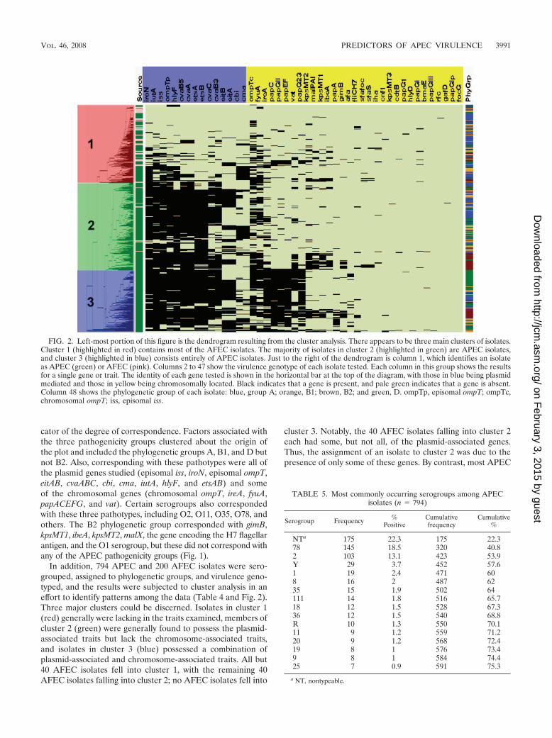
In addition, 794 APEC and 200 AFEC isolates were sero-grouped, assigned to phylogenetic groups, and virulence geno-typed, and the results were subjected to cluster analysis in aneffort to identify patterns among the data (Table 4 and Fig. 2).Three major clusters could be discerned. Isolates in cluster 1(red) generally were lacking in the traits examined, members ofcluster 2 (green) were generally found to possess the plasmid-associated traits but lack the chromosome-associated traits,and isolates in cluster 3 (blue) possessed a combination ofplasmid-associated and chromosome-associated traits. All but40 AFEC isolates fell into cluster 1, with the remaining 40AFEC isolates falling into cluster 2; no AFEC isolates fell into
cluster 3. Notably, the 40 AFEC isolates falling into cluster 2each had some, but not all, of the plasmid-associated genes.Thus, the assignment of an isolate to cluster 2 was due to thepresence of only some of these genes. By contrast, most APEC
FIG. 2. Left-most portion of this figure is the dendrogram resulting from the cluster analysis. There appears to be three main clusters of isolates.Cluster 1 (highlighted in red) contains most of the AFEC isolates. The majority of isolates in cluster 2 (highlighted in green) are APEC isolates,and cluster 3 (highlighted in blue) consists entirely of APEC isolates. Just to the right of the dendrogram is column 1, which identifies an isolateas APEC (green) or AFEC (pink). Columns 2 to 47 show the virulence genotype of each isolate tested. Each column in this group shows the resultsfor a single gene or trait. The identity of each gene tested is shown in the horizontal bar at the top of the diagram, with those in blue being plasmidmediated and those in yellow being chromosomally located. Black indicates that a gene is present, and pale green indicates that a gene is absent.Column 48 shows the phylogenetic group of each isolate: blue, group A; orange, B1; brown, B2; and green, D. ompTp, episomal ompT; ompTc,chromosomal ompT; iss, episomal iss.
TABLE 5. Most commonly occurring serogroups among APECisolates (n � 794)
Serogroup Frequency %Positive
Cumulativefrequency
Cumulative%
NTa 175 22.3 175 22.378 145 18.5 320 40.82 103 13.1 423 53.9Y 29 3.7 452 57.61 19 2.4 471 608 16 2 487 6235 15 1.9 502 64111 14 1.8 516 65.718 12 1.5 528 67.336 12 1.5 540 68.8R 10 1.3 550 70.111 9 1.2 559 71.220 9 1.2 568 72.419 8 1 576 73.49 8 1 584 74.425 7 0.9 591 75.3
a NT, nontypeable.
VOL. 46, 2008 PREDICTORS OF APEC VIRULENCE 3991
on February 3, 2015 by guest
http://jcm.asm
.org/D
ownloaded from

isolates contained the plasmid pathogenicity-associated island(PAI) genes that previously were identified as being highlyconserved among APEC isolates, including sitA, iutA, hlyF,episomal ompT, etsAB, episomal iss, iroN, and cvaABC (12, 13,22, 27, 36, 46, 48). In addition to these plasmid genes, membersof cluster 3, which were exclusively APEC isolates, also werecharacterized by the possession of certain chromosomal genes,including fyuA, ireA, the pap operon genes, vat, capsular bio-synthesis genes (K1 and K2 capsule types), and other PAImarkers (malX, ibeA, and gimB). The most commonly occur-ring serogroups among the APEC isolates examined were O78(18.5%) and O2 (13.1%) (Table 5). However, 22.3% of theisolates examined were nontypeable, and a high degree ofdiversity was found among the remaining isolates. Because68.4% of the APEC isolates examined did not belong to the O2or O78 serogroup and no other serogroup was prominent
among APEC isolates, no discernible patterns could be iden-tified with regard to serogroup and virulence potential.
An analysis of the distribution of virulence genes amongAPEC isolates of the four phylogenetic groups revealed thatmost of the genes were differentially distributed (Table 6). Infact, the only genes without significantly different distributionsacross phylogenetic groups (P � 0.05) were those that had verylow prevalence among all populations, such as cdtB, rfc, papG1,papG3, bmaE, gafD, kpsMT3, and hlyD. Among the AFECisolates examined, only nine genes displayed significant differ-ences across phylogenetic types. These included plasmid-asso-ciated genes, such as episomal iss and genes of the ColVoperon, and PAI-associated genes, such as fyuA, ibeA, and themal PAI marker (Table 7).
In an attempt to exploit these data to identify a minimumnumber of traits that could be used to distinguish an APEC
TABLE 6. Relationship between an APEC isolate’s (n � 794) phylogenetic group and gene prevalence
Gene (n � 44)No. of isolates in a phylogenetic group carrying (�) or lacking (�) the gene
ProbabilityA � A � B1 � B1 � B2 � B2 � D � D �
afa 9 274 9 126 1 143 24 208 �.0001cbi 94 189 86 49 36 108 62 170 �.0001cma 73 210 60 75 27 117 57 175 �.0001cvaB3 164 119 66 69 108 36 147 85 �.0001cvaC 151 132 74 61 106 38 154 78 �.0001etsA 171 112 101 34 111 33 178 54 �.0001etsB 167 116 101 34 114 30 178 54 �.0001fliCH7 4 279 6 129 25 119 2 230 �.0001fyuA 130 153 30 105 113 31 140 92 �.0001gimB 2 281 3 132 65 79 5 227 �.0001ibeA 9 274 1 134 68 76 16 216 �.0001ireA 75 208 44 91 68 76 165 67 �.0001Episomal iss 206 77 107 28 130 14 196 36 �.0001iutA 203 80 107 28 127 17 204 28 �.0001kpsMT1 13 270 6 129 91 53 15 217 �.0001kpsMT2 22 261 14 121 108 36 35 197 �.0001malX 9 274 7 128 97 47 12 220 �.0001Chromosomal ompT 140 143 56 79 111 33 190 42 �.0001papA 14 269 26 109 28 116 11 221 �.0001papC 68 215 54 81 47 97 120 112 �.0001papEF 62 221 54 81 41 103 113 119 �.0001papG23 61 222 39 96 43 101 113 119 �.0001papGII 69 214 49 86 55 89 113 119 �.0001sfaS 2 281 2 133 16 128 0 232 �.0001sfafoc 3 280 2 133 23 121 1 231 �.0001vat 12 271 8 127 107 37 135 97 �.0001iroN 221 62 116 19 129 15 212 20 0.0001cvaA 190 93 98 37 123 21 176 56 0.0005cvaB5 192 91 99 36 123 21 180 52 0.0006Episomal ompT 201 82 106 29 122 22 195 37 0.0008papG1 0 283 0 135 4 140 0 232 0.0019hlyF 202 81 113 22 116 28 191 41 0.0058iha 3 280 7 128 6 138 2 230 0.009cnf1 2 281 2 133 6 138 1 231 0.0228eitA 104 179 67 68 66 78 86 146 0.0282eitB 104 179 67 68 66 78 86 146 0.0282cdtB 1 282 1 134 4 140 1 231 0.0829papGI 2 281 0 135 0 144 5 227 0.1241rfc 1 282 0 135 2 142 0 232 0.1876bmaE 1 282 2 133 0 144 1 231 0.4303kpsMT3 4 279 1 134 3 141 1 231 0.4483papGIII 1 282 1 134 1 143 0 232 0.5078gafD 1 282 1 134 0 144 0 232 0.5353hlyD 1 282 1 134 2 142 1 231 0.5906
3992 JOHNSON ET AL. J. CLIN. MICROBIOL.
on February 3, 2015 by guest
http://jcm.asm
.org/D
ownloaded from

from an AFEC isolate, further LCA was done (Table 8). Thisanalysis identified a subset of genes, iutA, hlyF, episomal iss,iroN, and episomal ompT, which showed correspondence toAPEC pathotypes and appeared to be capable of discriminat-ing APEC from AFEC isolates to nearly the same degree asvirulence genotyping for 46 genes. Using this subset of genes,a pentaplex PCR procedure targeting these genes was designedand validated using E. coli strains known to lack or possessthese genes. In all cases, amplicons occurred as predicted, wereof the size predicted (Table 2), and were confirmed as to their
identities by DNA sequencing. Using this multiplex procedure,the 794 APEC and 200 AFEC isolates described above (Fig. 3)were analyzed, and the data generated were plotted in anothercluster diagram. As seen previously in the cluster diagramusing 46 genes, this cluster analysis showed a sharp demarca-tion between AFEC and APEC isolates, with the APEC iso-lates on average possessing 4.0 of the 5 genes and the AFECaveraging only 1.3 genes. These results suggest that screeningfor these genes is a useful tool in APEC diagnostics. Also,when the pentaplex results for the 200 AFEC and 124 APECisolates assigned to pathotypes were plotted against one an-other (Fig. 4), the average number of genes possessed de-creased from high (4.6) to medium (4.3) to low (3.9) for AFECgroups. However, despite strong differences in the distributionof these genes between APEC and AFEC, every gene studiedcould be found in both APEC and AFEC populations.
DISCUSSION
This study validates a refined multiplex PCR scheme to beused for the prediction of virulence of avian E. coli. This
TABLE 7. Relationship between an AFEC isolate’s (n � 200) phylogenetic group and gene prevalence
GeneaNo. of isolates in a phylogenetic group carrying (�) or lacking (�) the gene
Probabilityb
A � A � B1 � B1 � B2 � B2 � D � D �
fyuA 13 64 19 41 22 16 6 19 0.0001ibeA 5 72 5 55 11 27 0 25 0.0012malPAI 7 70 4 56 12 26 1 24 0.002Chromosomal ompT 10 67 13 47 17 21 7 18 0.0026cvaC 4 73 7 53 11 27 2 23 0.0048ireA 11 66 1 59 5 33 6 19 0.0061iroN 10 67 13 47 15 23 4 21 0.0148cvaB5 12 65 12 48 16 22 4 21 0.0159Episomal iss 20 57 16 44 19 19 5 20 0.033Episomal ompT 9 68 15 45 11 27 7 18 0.0592papG23 5 72 4 56 7 31 5 20 0.0649cvaA 12 65 12 48 14 24 4 21 0.0733etsB 11 66 14 46 13 25 6 19 0.1047kpsMT2 10 67 9 51 12 26 4 21 0.1078kpsMT1 8 69 6 54 10 28 3 22 0.1173gimB 0 77 1 59 2 36 0 25 0.1197papG1 19 58 6 54 6 32 3 22 0.1354etsA 12 65 13 47 13 25 5 20 0.1615papEF 6 71 2 58 6 32 2 23 0.1804papC 8 69 3 57 7 31 3 22 0.1996eitA 14 63 12 48 13 25 4 21 0.2257eitB 14 63 12 48 13 25 4 21 0.2257fliCH7 8 69 4 56 2 36 5 20 0.2281papGII 6 71 7 53 7 31 3 22 0.3843iha 2 75 0 60 0 38 1 24 0.4563hlyF 16 61 13 47 11 27 8 17 0.5609cdtB 0 77 1 59 0 38 0 25 0.615cma 6 71 6 54 3 35 4 21 0.6291cbi 7 70 9 51 5 33 2 23 0.6773cvaB3 11 66 11 49 8 30 3 22 0.7566papA 2 75 0 60 0 38 0 25 0.7678cnf1 4 73 3 57 1 37 0 25 0.8742sfaS 1 76 0 60 0 38 0 25 1iutA 20 57 16 44 29 9 6 19 �.0001vat 0 77 1 59 16 22 5 20 �.0001
a The probability column represents the P value for Fisher’s Exact test of the homogeneity of prevalence rates for each gene across the 4 phylogenetic groups. Notethat there are only 35 genes in the table, even though testing has been done for 46 genes; no occurrences of kpsMT3, bmaE, sfa/foc, papGIII, hlyD, rfc, papG I�, gafD,focG, papG1, and afa were found among these isolates.
b The probability column represents the P value for Fisher’s Exact test of the homogeneity of prevalence rates for each gene across the four phylogenetic groups.
TABLE 8. Genes useful in predicting APEC (n � 794) or AFEC(n � 200) membership
ModelNo. (%) correctly
identified asAPEC
No. (%) correctlyidentified as
AFEC
All 46 genes 685 (86.3) 168 (84.0)Stepwise (17-plex) 686 (86.4) 169 (84.5)Best fivea 678 (85.4) 158 (79.0)
a The genes in the best five model were iutA, hlyF, episomal iss, iroN, andepisomal ompT.
VOL. 46, 2008 PREDICTORS OF APEC VIRULENCE 3993
on February 3, 2015 by guest
http://jcm.asm
.org/D
ownloaded from

scheme is based on extensive virulence genotyping on a largenumber of isolates from a variety of sources; takes advantageof recent advances in plasmid genomics; and correlates thepresence of five genes with the ability of an APEC isolate tocause disease in 1-day-old chicks. A cluster analysis of multi-plex PCR results of nearly 1,000 isolates, screened for thepresence of more than 40 ExPEC-associated traits, showedthat the majority of APEC isolates fall into two distinct clus-ters: those with plasmid-associated virulence genes but lackingchromosome-associated virulence genes and those possessingboth plasmid- and chromosome-associated virulence genes.Although some APEC isolates lacked the plasmid genes stud-ied here, the vast majority of APEC isolates were distinguished
from AFEC isolates by their possession of plasmid-linked PAIgenes. In fact, the sharp demarcation between most APECisolates and most AFEC isolates due to their plasmid genecontent suggests that APEC isolates are well equipped for apathogenic lifestyle, which is contrary to the widely held beliefthat they are opportunistic pathogens. Perhaps APEC isolatesassigned to cluster 1, which are characterized by a dearth of thegenes tested, are opportunistic pathogens, while those of clus-ters 2 and 3 are frank pathogens. Indeed, an APEC isolate inthis study was defined as an E. coli strain isolated from thelesions of birds with colibacillosis with no regard to any hostfactors that might have predisposed the birds to infection,including infection with commensal strains of E. coli. Thus, wespeculate that APEC isolates falling into cluster 1 actually arecommensal E. coli strains taking advantage of an immunocom-promised host. A further examination of the APEC isolate ofthis cluster in a range of immunocompetent and immunocom-promised hosts would be helpful in resolving this issue. Re-gardless, it is evident from this study that a distinction can bemade between the majority of APEC and AFEC isolates ex-amined here by their possession of ColV virulence plasmids.
Besides confirming that virulence plasmids are a definingtrait of the APEC pathotype, these results help explain theassignment of APEC isolates to phylogenetic types that are nottypically associated with ExPEC isolates of human beings.While the majority of human ExPEC isolates belong to phylo-genetic type B2 and, to a lesser degree, D, the majority ofAPEC isolates belong to the A, B1, and D phylogenetic types(5, 36). Phylogenetic grouping, which relies on identifying cer-tain chromosomal markers, does not account for virulence dueto plasmid-mediated PAIs and other extrachromosomal andmobile elements. Since such extrachromosomally located PAIsare a defining trait of the APEC pathotype and appear to becritical to APEC virulence (8, 9, 42, 43, 45), PCR-based phy-logenetic typing is not a clear predictor of avian E. coli viru-lence. However, the literature provides evidence that no abso-lute definition of an APEC or a human ExPEC isolate ispossible (4, 13, 18, 19, 29, 31, 33). Overlap among all ExPECsubtypes in terms of serogroups, phylogenetic types, and viru-
FIG. 3. Results using the pentaplex panel for the 994 avian E. coliisolates. The left-most portion of this figure is the dendrogram result-ing from the cluster analysis. Just to the right of the dendrogram iscolumn 1, which identifies an isolate as APEC (green) or AFEC (pink).Columns 2 to 6 show the virulence genotype of each isolate tested foriroN, iutA, iss, ompTp, and hlyF. Each column in this group shows theresults for a single gene. Black indicates that the gene is present, andlight green indicates that the gene is absent.
FIG. 4. Histogram comparing the prevalence of the genes targetedin the pentaplex procedure among APEC isolates of known pathoge-nicity (APEC high, high pathogenicity [n � 73]; APEC int, interme-diate pathogenicity [n � 26]; APEC low, low pathogenicity [n � 25]).Also shown is the gene prevalence among 200 AFEC isolates. Lettersabove bars indicate levels of statistical significance according to Fish-er’s exact test (P � 0.05 is considered statistically significant).
3994 JOHNSON ET AL. J. CLIN. MICROBIOL.
on February 3, 2015 by guest
http://jcm.asm
.org/D
ownloaded from

lence genotypes exists to some degree (13, 33, 36). However,because most APEC isolates fall into phylogenetic groupsother than the B2 group and possess ColV or ColBM virulenceplasmid, one can conclude that these plasmids and/or someother genetic elements common to avian E. coli of the non-B2types provide these strains with an enhanced ability to causeavian colibacillosis.
In summary, plasmid-linked PAIs are common amongAPEC isolates and provide a useful target for identifying theseorganisms. By exploiting this characteristic trait of APEC, wehave developed and validated a pentaplex PCR panel that candistinguish most APEC isolates from AFEC isolates. SinceAPEC isolates remain an important concern for poultry pro-ducers and a potential one for public health professionals, sucha diagnostic tool may be used to detect APEC-like strainsoccurring in poultry production, along the food chain, and inhuman disease, helping to clarify the role of APEC in humandisease and identify targets for improved colibacillosis control.
ACKNOWLEDGMENTS
We gratefully acknowledge Chitrita Debroy (Pennsylvania StateUniversity) and Catherine Logue (North Dakota State University) forproviding strains used for this study.
This study was supported by Iowa State University’s College ofVeterinary Medicine.
REFERENCES
1. Arp, L. H. 1982. Effect of passive immunization on phagocytosis of blood-borne Escherichia coli in spleen and liver of turkeys. Am. J. Vet. Res.43:1034–1040.
2. Barnes, H. J., L. K. Nolan, and J. F. Vaillancourt. 2008. Colibacillosis, p.691–732. In Y. M. Saif et al., Diseases of poultry, 12th ed. Blackwell Pub-lishing, Arres, IA.
3. Bolin, C. A., and A. E. Jensen. 1987. Passive immunization with antibodiesagainst iron-regulated outer membrane proteins protects turkeys from Esch-erichia coli septicemia. Infect. Immun. 55:1239–1242.
4. Brzuszkiewicz, E., H. Bruggemann, H. Liesegang, M. Emmerth, T.Olschlager, G. Nagy, K. Albermann, C. Wagner, C. Buchrieser, L. Emody, G.Gottschalk, J. Hacker, and U. Dobrindt. 2006. How to become a uropatho-gen: comparative genomic analysis of extraintestinal pathogenic Escherichiacoli strains. Proc. Natl. Acad. Sci. USA 103:12879–12884.
5. Clermont, O., S. Bonacorsi, and E. Bingen. 2000. Rapid and simple deter-mination of the Escherichia coli phylogenetic group. Appl. Environ. Micro-biol. 66:4555–4558.
6. Cloud, S. S., J. K. Rosenberger, P. A. Fries, R. A. Wilson, and E. M. Odor.1985. In vitro and in vivo characterization of avian Escherichia coli. I. Sero-types, metabolic activity, and antibiotic sensitivity. Avian Dis. 29:1084–1093.
7. Deb, J. R., and E. G. Harry. 1978. Laboratory trials with inactivated vaccinesagainst Escherichia coli (O2:K1) infection in fowls. Res. Vet. Sci. 24:308–313.
8. Dozois, C. M., F. Daigle, and R. Curtiss III. 2003. Identification of pathogen-specific and conserved genes expressed in vivo by an avian pathogenic Esch-erichia coli strain. Proc. Natl. Acad. Sci. USA 100:247–252.
9. Dozois, C. M., M. Dho-Moulin, A. Bree, J. M. Fairbrother, C. Desautels, andR. Curtiss III. 2000. Relationship between the Tsh autotransporter andpathogenicity of avian Escherichia coli and localization and analysis of theTsh genetic region. Infect. Immun. 68:4145–4154.
10. Eisen, M. B., P. T. Spellman, P. O. Brown, and D. Botstein. 1998. Clusteranalysis and display of genome-wide expression patterns. Proc. Natl. Acad.Sci. USA 95:14863–14868.
11. Ewers, C., T. Janben, S. Kiebling, H. C. Philipp, and L. H. Wieler. 2005.Rapid detection of virulence-associated genes in avian pathogenic Esche-richia coli by multiplex polymerase chain reaction. Avian Dis. 49:269–273.
12. Ewers, C., T. Janssen, S. Kiessling, H. C. Philipp, and L. H. Wieler. 2004.Molecular epidemiology of avian pathogenic Escherichia coli (APEC) iso-lated from colisepticemia in poultry. Vet. Microbiol. 104:91–101.
13. Ewers, C., G. Li, H. Wilking, S. Kiessling, K. Alt, E. M. Antao, C. Laturnus,I. Diehl, S. Glodde, T. Homeier, U. Bohnke, H. Steinruck, H. C. Philipp, andL. H. Wieler. 2007. Avian pathogenic, uropathogenic, and newborn menin-gitis-causing Escherichia coli: how closely related are they? Int. J. Med.Microbiol. 297:163–176.
14. Foley, S. L., S. M. Horne, C. W. Giddings, M. Robinson, and L. K. Nolan.2000. Iss from a virulent avian Escherichia coli. Avian Dis. 44:185–191.
15. Huberty, C. J. 1994. Applied discriminant analysis. Wiley, New York, NY.
16. Johnson, J. R., O. Clermont, M. Menard, M. A. Kuskowski, B. Picard, andE. Denamur. 2006. Experimental mouse lethality of Escherichia coli isolates,in relation to accessory traits, phylogenetic group, and ecological source.J. Infect. Dis. 194:1141–1150.
17. Johnson, J. R., M. A. Kuskowski, K. Smith, T. T. O’Bryan, and S. Tatini.2005. Antimicrobial-resistant and extraintestinal pathogenic Escherichia coliin retail foods. J. Infect. Dis. 191:1040–1049.
18. Johnson, J. R., and T. A. Russo. 2002. Extraintestinal pathogenic Escherichiacoli: the other bad E. coli. J. Lab. Clin. Med. 139:155–162.
19. Johnson, J. R., and T. A. Russo. 2005. Molecular epidemiology of extraint-estinal pathogenic (uropathogenic) Escherichia coli. Int. J. Med. Microbiol.295:383–404.
20. Johnson, J. R., and A. L. Stell. 2000. Extended virulence genotypes ofEscherichia coli strains from patients with urosepsis in relation to phylogenyand host compromise. J. Infect. Dis. 181:261–272.
21. Johnson, T. J., S. J. Johnson, and L. K. Nolan. 2006. Complete DNAsequence of a ColBM plasmid from avian pathogenic Escherichia coli sug-gests that it evolved from closely related ColV virulence plasmids. J. Bacte-riol. 188:5975–5983.
22. Johnson, T. J., K. E. Siek, S. J. Johnson, and L. K. Nolan. 2006. DNAsequence of a ColV plasmid and prevalence of selected plasmid-encodedvirulence genes among avian Escherichia coli strains. J. Bacteriol. 188:745–758.
23. Johnson, T. J., J. Skyberg, and L. K. Nolan. 2004. Multiple antimicrobialresistance region of a putative virulence plasmid from an Escherichia coliisolate incriminated in avian colibacillosis. Avian Dis. 48:351–360.
24. Johnson, T. J., Y. M. Wannemuehler, and L. K. Nolan. 2008. Evolution ofthe iss gene in Escherichia coli. Appl. Environ. Microbiol. 74:2360–2369.
25. Kariyawasam, S., B. N. Wilkie, and C. L. Gyles. 2004. Construction, char-acterization, and evaluation of the vaccine potential of three geneticallydefined mutants of avian pathogenic Escherichia coli. Avian Dis. 48:287–299.
26. Kwaga, J. K., B. J. Allan, J. V. van der Hurk, H. Seida, and A. A. Potter. 1994.A carAB mutant of avian pathogenic Escherichia coli serogroup O2 is atten-uated and effective as a live oral vaccine against colibacillosis in turkeys.Infect. Immun. 62:3766–3772.
27. La Ragione, R. M., and M. J. Woodward. 2002. Virulence factors of Esche-richia coli serotypes associated with avian colisepticaemia. Res. Vet. Sci.73:27–35.
28. Lynne, A. M., S. L. Foley, and L. K. Nolan. 2006. Immune response torecombinant Escherichia coli Iss protein in poultry. Avian Dis. 50:273–276.
29. Maynard, C., S. Bekal, F. Sanschagrin, R. C. Levesque, R. Brousseau, L.Masson, S. Lariviere, and J. Harel. 2004. Heterogeneity among virulenceand antimicrobial resistance gene profiles of extraintestinal Escherichia coliisolates of animal and human origin. J. Clin. Microbiol. 42:5444–5452.
30. McLachlan, G. J. 1992. Discriminant analysis and statistical pattern recog-nition. Wiley, New York, NY.
31. Mokady, D., U. Gophna, and E. Z. Ron. 2005. Extensive gene diversity insepticemic Escherichia coli strains. J. Clin. Microbiol. 43:66–73.
32. Morales, C., M. D. Lee, C. Hofacre, and J. J. Maurer. 2004. Detection of anovel virulence gene and a Salmonella virulence homologue among Esche-richia coli isolated from broiler chickens. Foodborne Pathog. Dis. 1:160–165.
33. Moulin-Schouleur, M., M. Reperant, S. Laurent, A. Bree, S. Mignon-Gras-teau, P. Germon, D. Rasschaert, and C. Schouler. 2007. Extraintestinalpathogenic Escherichia coli strains of avian and human origin: link betweenphylogenetic relationships and common virulence patterns. J. Clin. Micro-biol. 45:3366–3376.
34. Peighambari, S. M., D. B. Hunter, P. E. Shewen, and C. L. Gyles. 2002.Safety, immunogenicity, and efficacy of two Escherichia coli cya crp mutantsas vaccines for broilers. Avian Dis. 46:287–297.
35. Pfaff-McDonough, S. J., S. M. Horne, C. W. Giddings, J. O. Ebert, C.Doetkott, M. H. Smith, and L. K. Nolan. 2000. Complement resistance-related traits among Escherichia coli isolates from apparently healthy birdsand birds with colibacillosis. Avian Dis. 44:23–33.
36. Rodriguez-Siek, K. E., C. W. Giddings, C. Doetkott, T. J. Johnson, M. K.Fakhr, and L. K. Nolan. 2005. Comparison of Escherichia coli isolates im-plicated in human urinary tract infection and avian colibacillosis. Microbi-ology 151:2097–2110.
37. Rodriguez-Siek, K. E., C. W. Giddings, C. Doetkott, T. J. Johnson, and L. K.Nolan. 2005. Characterizing the APEC pathotype. Vet. Res. 36:241–256.
38. Roland, K., K. Karaca, and D. Sizemore. 2004. Expression of Escherichia coliantigens in Salmonella typhimurium as a vaccine to prevent airsacculitis inchickens. Avian Dis. 48:595–605.
39. Rosenberger, J. K., P. A. Fries, and S. S. Cloud. 1985. In vitro and in vivocharacterization of avian Escherichia coli. III. Immunization. Avian Dis.29:1108–1117.
40. Rosenberger, J. K., P. A. Fries, S. S. Cloud, and R. A. Wilson. 1985. In vitroand in vivo characterization of avian Escherichia coli. II. Factors associatedwith pathogenicity. Avian Dis. 29:1094–1107.
41. Skyberg, J. A., S. M. Horne, C. W. Giddings, R. E. Wooley, P. S. Gibbs, andL. K. Nolan. 2003. Characterizing avian Escherichia coli isolates with multi-plex polymerase chain reaction. Avian Dis. 47:1441–1447.
42. Skyberg, J. A., T. J. Johnson, J. R. Johnson, C. Clabots, C. M. Logue, and
VOL. 46, 2008 PREDICTORS OF APEC VIRULENCE 3995
on February 3, 2015 by guest
http://jcm.asm
.org/D
ownloaded from

L. K. Nolan. 2006. Acquisition of avian pathogenic Escherichia coli plasmidsby a commensal E. coli isolate enhances its abilities to kill chicken embryos,grow in human urine, and colonize the murine kidney. Infect. Immun. 74:6287–6292.
43. Skyberg, J. A., T. J. Johnson, and L. K. Nolan. 2008. Mutational and tran-scriptional analyses of an avian pathogenic Escherichia coli ColV plasmid.BMC Microbiol. 8:24.
44. Snedecor, G. W., and W. G. Cochran. 1989. Statistical methods. Iowa StateUniversity Press, Ames, IA.
45. Tivendale, K. A., J. L. Allen, C. A. Ginns, B. S. Crabb, and G. F. Browning.2004. Association of iss and iucA, but not tsh, with plasmid-mediated viru-lence of avian pathogenic Escherichia coli. Infect. Immun. 72:6554–6560.
46. Vandekerchove, D., F. Vandemaele, C. Adriaensen, M. Zaleska, J. P. Her-
nalsteens, L. D. Baets, P. Butaye, F. V. Immerseel, P. Wattiau, H. Laevens,J. Mast, B. Goddeeris, and F. Pasmans. 2005. Virulence-associated traits inavian Escherichia coli: comparison between isolates from colibacillosis-af-fected and clinically healthy layer flocks. Vet. Microbiol. 108:75–87.
47. Yang, H., S. Chen, D. G. White, S. Zhao, P. McDermott, R. Walker, and J.Meng. 2004. Characterization of multiple-antimicrobial-resistant Escherichiacoli isolates from diseased chickens and swine in China. J. Clin. Microbiol.42:3483–3489.
48. Zhao, S., J. J. Maurer, S. Hubert, J. F. De Villena, P. F. McDermott, J.Meng, S. Ayers, L. English, and D. G. White. 2005. Antimicrobial suscepti-bility and molecular characterization of avian pathogenic Escherichia coliisolates. Vet. Microbiol. 107:215–224.
3996 JOHNSON ET AL. J. CLIN. MICROBIOL.
on February 3, 2015 by guest
http://jcm.asm
.org/D
ownloaded from