Yves Klein and Hysterical Marks of Authority - Monash University
Upload
khangminh22Category
view
0download
0
Downloaded from www.asmscience.org by
IP: 130.194.145.102
On: Thu, 20 Jun 2019 00:48:10
Virulence Plasmids of thePathogenic Clostridia
SARAH A. REVITT-MILLS, CALLUM J. VIDOR, THOMAS D. WATTS,DENA LYRAS, JULIAN I. ROOD, and VICKI ADAMS
Infection and Immunity Program, Biomedicine Discovery Institute and Department of Microbiology,Monash University, Clayton, Victoria 3800, Australia
ABSTRACT The clostridia cause a spectrum of diseases inhumans and animals ranging from life-threatening tetanus andbotulism, uterine infections, histotoxic infections and entericdiseases, including antibiotic-associated diarrhea, and foodpoisoning. The symptoms of all these diseases are the result ofpotent protein toxins produced by these organisms. These toxinsare diverse, ranging from a multitude of pore-forming toxinsto phospholipases, metalloproteases, ADP-ribosyltransferasesand large glycosyltransferases. The location of the toxin genesis the unifying theme of this review because with one or twoexceptions they are all located on plasmids or on bacteriophagethat replicate using a plasmid-like intermediate. Some of theseplasmids are distantly related whilst others share little or nosimilarity. Many of these toxin plasmids have been shown tobe conjugative. The mobile nature of these toxin genes givesa ready explanation of how clostridial toxin genes have beenso widely disseminated both within the clostridial genera aswell as in the wider bacterial community.
Many clostridial species are ubiquitous in the envi-ronment and in the intestinal tracts of birds, fish, andmammals. Commensal species are often carried asymp-tomatically within a host. However, if the immune sta-tus of the host is compromised, due to either age, illnessor a change in diet, disease can result from toxigenicstrains. Alternatively, some clostridial species or strainsdon’t require predisposing factors. They can cause dis-ease simply if they gain entry into the host either throughdamage to the skin or through the gastrointestinal tract,often via poorly prepared or incorrectly stored food.These clostridia then overgrow and cause cell and tis-sue damage. Diseases mediated by the clostridial spe-cies discussed in this review are predominantly mediatedby potent protein toxins, many of which are located
extrachromosomally. These toxins have diverse mecha-nisms of action and include pore-forming cytotoxins,phospholipases, metalloproteases, ADP-ribosyltransferasesand large glycosyltransferases. This review focuses onthese toxins and the elements that carry the toxin struc-tural genes. For ease of discussion it has been structuredon a bacterial species-specific basis.
PAENICLOSTRIDIUM (CLOSTRIDIUM)SORDELLIIVirulence Properties of P. sordelliiPaeniclostridium (formerly Clostridium) sordellii causesseveral severe diseases in both humans and animals.Most documented animal infections caused by P. sor-dellii are edemic or enterotoxic diseases of livestock
Received: 29 May 2018, Accepted: 16 August 2018,Published: 17 May 2019
Editors: Vincent A. Fischetti, The Rockefeller University, New York,NY; Richard P. Novick, Skirball Institute for Molecular Medicine, NYUMedical Center, New York, NY; Joseph J. Ferretti, Department ofMicrobiology & Immunology, University of Oklahoma HealthScience Center, Oklahoma City, OK; Daniel A. Portnoy, Departmentof Molecular and Cellular Microbiology, University of California,Berkeley, Berkeley, CA; Miriam Braunstein, Department ofMicrobiology and Immunology, University of North Carolina-ChapelHill, Chapel Hill, NC, and Julian I. Rood, Infection and ImmunityProgram, Monash Biomedicine Discovery Institute, MonashUniversity, Melbourne, Australia
Citation: Revitt-Mills SA, Vidor CJ, Watts TD, Lyras D, Rood JI, Adams V. . Virulence plasmids of the pathogenic clostridia. Microbiol Spectrum 7(3):GPP3-0034-2018. doi:10.1128/microbiolspec.GPP3- 0034-2018.
Correspondence: Vickie Adams, [email protected]
ASMscience.org/MicrobiolSpectrum 1
© 2019 American Society for Microbiology. All rights reserved.
2019
Downloaded from www.asmscience.org by
IP: 130.194.145.102
On: Thu, 20 Jun 2019 00:48:10
and poultry, but they also include rare myonecrotic in-fections of other animals (1). Human infections areinfrequent, but severe in nature, with mortality ratesapproaching 70% overall (2). The majority of P. sor-dellii human infections are soft tissue infections thatoccur after trauma or surgery (2). Sporadic, yet severecases of post-partum and post-medical abortion uter-ine P. sordellii infections have occurred over the last20 years, with almost every case resulting in the death ofthe patient (1, 2).
Due to the rare nature of P. sordellii infections, thepathogenesis of disease is still poorly understood. Themajor virulence factors involved in P. sordellii infection,however, are the production of two large clostridialtoxins (LCTs): TcsH (hemorrhagic toxin) and TcsL(lethal toxin) (1). The LCTs enter mammalian host cellsand glycosylate small GTPases such as Ras, leading to analtered cytoskeletal arrangement, cellular rounding andeventual death of the cell (3). Interestingly, while beinghighly potent, both P. sordellii LCTs are producedby only ∼5- to 13% of isolates tested (4–6), with themajority of toxigenic isolates producing only TcsL andcontaining a truncated tcsH gene (6). Despite thesefindings, TcsL has been shown to be the major virulencefactor responsible for the production of uterine-infectionrelated toxic shock using an animal model of infection(7). In P. sordellii, the LCTs are encoded within a regioncalled the pathogenicity locus (PaLoc), alongside genesthat are likely to be required for their regulation andrelease (8). A recent phylogenetic analysis found that inall toxigenic isolates the PaLoc is carried on a group ofrelated plasmids called the pCS1 family (6), which is themajor focus of this section. Other virulence factors ofP. sordellii include a pore-forming phospholipase SDL,the sialidase NanS, and the production of unique endo-spores (1, 9–11).
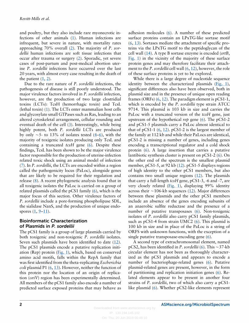
Bioinformatic Characterizationof Plasmids in P. sordelliiThe pCS1 family is a group of large plasmids carried byboth toxigenic and non-toxigenic P. sordellii isolates.Seven such plasmids have been identified to date (12).The pCS1 plasmids encode a putative replication initi-ation (Rep) protein (Fig. 1), which, based on conservedamino acid motifs, falls within the RepA family thatwas first identified from the theta-replicating Escherichiacoli plasmid P1 (6, 13). However, neither the function ofthis protein nor the location of an origin of replica-tion (oriV) region has been experimentally determined.All members of the pCS1 family also encode a number ofpredicted surface exposed proteins that may behave as
adhesion molecules (6). A number of these predictedsurface proteins contain an LPxTG-like sortase motif(6, 13). Sortases mediate the attachment of specific pro-teins via the LPxTG motif to the peptidoglycan of thecell wall (14). A type B sortase enzyme is encoded (srtB,Fig. 1) in the vicinity of the majority of these surfaceprotein genes and may therefore facilitate their attach-ment to the P. sordellii cell wall (6, 12), however, the roleof these surface proteins is yet to be explored.
While there is a large degree of nucleotide sequenceidentity between the characterized plasmids (Fig. 1),significant differences also have been observed, both inplasmid size and in the presence of unique open readingframes (ORFs) (6, 12). The paradigm element is pCS1-1,which is encoded by the P. sordellii type strain ATCC9714. This plasmid is 103 kb in size and carries thePaLoc with a truncated version of the tcsH gene, justupstream of the hypothetical rep gene (6). The pCS1-2and pCS1-5 plasmids carry a PaLoc almost identical tothat of pCS1-1 (6, 12). pCS1-2 is the largest member ofthe family at 112 kb and while their PaLocs are identical,pCS1-2 is missing a number of genes, including genesencoding a transcriptional regulator and a cold shockprotein (6). A large insertion that carries a putativelantibiotic synthesis cluster is present on pCS1-2 (6). Onthe other end of the spectrum is the smallest plasmidmember, pCS1-5, at 92 kb (12). pCS1-5 displays regionsof high identity to the other pCS1 members, but alsocontains two small unique regions (12). The plasmidsthat carry a full-length tcsH gene, pCS1-3, -6 and -7, arevery closely related (Fig. 1), displaying 99% identityacross their ∼106-kb sequences (12). Major differencesbetween these plasmids and the other pCS1 membersinclude an absence of the genes encoding subunits ofan anaerobic sulfite reductase and the presence of anumber of putative transposases (6). Non-toxigenicisolates of P. sordellii also carry pCS1 family plasmids,such as pCS1-4 from strain UMC2 (6). This plasmid is100 kb in size and in place of the PaLoc is a string ofORFS with unknown functions, with the exception of asingle putative transposase-encoding gene (6).
A second type of extrachromosomal element, namedpCS2, has been identified in P. sordellii (6). This∼37-kbcircular element has not been as thoroughly character-ized as the pCS1 plasmids and appears to encode anumber of bacteriophage-related genes (6). Putativeplasmid-related genes are present, however, in the formof partitioning and replication initiation genes (6). Re-lated elements appear to be present in another fourstrains of P. sordellii, two of which also carry a pCS1-like plasmid (6). Whether pCS2-like elements represent
2 ASMscience.org/MicrobiolSpectrum
Revitt-Mills et al.

Downloaded from www.asmscience.org by
IP: 130.194.145.102
On: Thu, 20 Jun 2019 00:48:10
FIGURE 1 The pCS1 family plasmids of P. sordellii. Shown is a visual representation of a blastn analysis comparing each of theseven sequenced pCS1 plasmids to the reference sequence of pCS1-3 from strain JGS6382. The third ring from the center (green)and the coordinates (in kb) correspond to pCS1-3. Plasmids displaying 70 to 100% identity to pCS1-3 at a particular locus areshown with a solid block of colour on their respective ring. Identity to pCS1-3 between 50 and 70% is represented as a pale blockof colour and if the identity is lower than 50% it is represented as a gap in the corresponding ring. Conserved loci on pCS1-3 areindicated as a gray arc on the outermost ring and labeled. Genes of interest are annotated as black arrows on the outermost ringand also labeled. Sequences analyzed and accession numbers: pCS1-1 (LN679999), pCS1-2 (LN681232), pCS1-3 (LN681235),pCS1-4 (LN681233), pCS1-5 (MG205643), pCS1-6 (MG205642), pCS1-7 (MG205641). Produced using BRIG (224).
ASMscience.org/MicrobiolSpectrum 3
Virulence Plasmids of the Pathogenic Clostridia
Downloaded from www.asmscience.org by
IP: 130.194.145.102
On: Thu, 20 Jun 2019 00:48:10
plasmids, cryptic plasmids, or phage is currently unclear,but no virulence genes have been identified within thisplasmid family to date.
Stability and Conjugative Transferof the pCS1 Family of PlasmidsA recent study has functionally characterized conservedgenes of the pCS1 family plasmids, using pCS1-1 as amodel (12). All pCS1 plasmids carry parA and parBgenes, which are putative members of a ParABS plasmidpartitioning system (6). Such systems are utilized by low-copy-number plasmids to ensure stable inheritance ofthe plasmid in each daughter cell upon cellular division(15). Stability assays conducted on a marked version ofpCS1-1 in different strain backgrounds showed that theplasmid is stable across ∼120 generations (12). Inde-pendent insertional inactivation mutants of the pCS1-1parB gene displayed gradual loss of the plasmid duringcontinual subculture, providing evidence that ParA andParB represent a true plasmid partitioning system (12).
Bioinformatic analysis of pCS1-1 also leads to theidentification of an ∼17.3-kb region, subsequentlynamed the cst (for C. sordellii transfer) locus (Fig. 1),that encoded proteins with similarity to componentsof a plasmid conjugation system (12). The conjugativetransfer of a marked derivative of pCS1-1, from typestrain ATCC 9714 to a P. sordellii isolate from a distantclade, was subsequently demonstrated (12). The cst lo-cus is conserved between all members of the pCS1 familyso far characterized (Fig. 1) (12). Encoded within the cstregion is a putative relaxase protein belonging to theMobMG family of relaxases, along with a cognate ori-gin of transfer (oriT) located upstream of the relaxasegene (12). A number of genes encoding components ofa type IV secretion system are also located within thecst locus. Two of these, the putative coupling protein,CstD4, and the putative ATPase, CstB4, were demon-strated experimentally to be required for conjugativetransfer, confirming the cst region to be a true conju-gation locus (12). Regions with significant similarityto the cst locus also have been identified in plasmidsof Clostridium perfringens and Clostridium botulinum,and within plasmid or conjugative transposon-like re-gions of the genomes of Clostridioides (previously Clos-tridium) difficile isolates, indicating that cst represents avariant of a common clostridial conjugation locus (12).
CLOSTRIDIUM PERFRINGENSC. perfringens is ubiquitously distributed among ani-mals, humans and the environment (16, 17), but is also
the causative agent of numerous opportunistic infections(16). The diseases caused by C. perfringens range fromtypically mild, self-limiting human food poisoning, tothe more severe pathologies of human gas gangrene,and enterotoxemia and necrotic enteritis in domesticlivestock (18). It is this broad spectrum of disease thatmakes this bacterium of particular interest to both vet-erinary and human medicine. Enteric disease caused byC. perfringens is estimated to cost the poultry industryin excess of $6 billion globally as well as $466 millionin human health service costs in the United States alone(19, 20). What makes C. perfringens such a success-ful pathogen is its ability to encode and produce up to20 toxins, many of which are associated with conju-gative plasmids (21, 22).
The C. perfringens Typing ToxinsStrains differ in their carriage and expression of toxins,and this forms the basis of the C. perfringens toxinotypeclassification scheme (23). This scheme previously cate-gorized C. perfringens strains into one of five toxino-types based on the expression of four lethal typingtoxins: alpha, beta, epsilon and iota (23). However, thisscheme recently was expanded to include two new typ-ing toxins, C. perfringens enterotoxin (CPE) and NetBtoxin (Table 1) (24). This toxinotype classification isunique because particular ”types” are often associ-ated with specific diseases in humans and animals, forexample; type A strains typically cause gas-gangrene,whereas type D strains are typically associated withenterotoxemia in sheep and goats (25).
Alpha toxin is encoded by all strains of C. perfringensand is the major toxin involved in the pathogenesis ofgas gangrene infections (26). In allC. perfringens strains,the alpha toxin structural gene, plc, is chromosomallyencoded with basal levels of plc mRNA always tran-scribed (27). The relative amount of toxin producedvaries based on the strain background, with type Astrains producing the highest levels of alpha toxin invitro (28, 29). This toxinotype-specific control of toxinexpression is said to be dependent on strain-specificglobal control elements, such as the two-componentregulatory system VirR/VirS, as well as the structureof the promoter sequences preceding the plc gene (27,28, 30, 31). Alpha toxin is a zinc-metalloenzyme thatpossesses both phospholipase C and sphingomyelinaseactivity, and functions by cleaving the phosphatidyl-choline and ceramide head groups from the componentsof membrane phospholipid bilayers (32, 33). It is thistargeted membrane disruption that leads to the severedisease often seen in gas gangrene infections (32, 34, 35).
4 ASMscience.org/MicrobiolSpectrum
Revitt-Mills et al.
Downloaded from www.asmscience.org by
IP: 130.194.145.102
On: Thu, 20 Jun 2019 00:48:10
Beta toxin, like many of the C. perfringens toxins, isa pore-forming toxin and is produced by both type Band C strains of C. perfringens (36). Beta toxin is re-sponsible for disease development in C. perfringenstype C infections; with beta toxin null mutant strainsdisplaying avirulent phenotypes in animal models ofenterotoxemia and necrotizing enteritis (37–39). Theprecise mechanism of action of beta toxin has not beenfully elucidated, but, the hypothesis is that the toxinforms pores in the membranes of susceptible cells lead-ing to cell lysis (40–42).
Beta toxin is encoded by the cpb gene, which is lo-cated on large conjugative plasmids (43–45). In type Bstains, cpb is encoded on one of two plasmids: either∼65 kb or ∼90 kb in size (44). More commonly the cpbgene is found on the larger ∼90-kb plasmid (44). Pre-ceding the cpb gene is the insertion sequence (IS) ele-ment, IS1151 (44, 45). The presence of IS elementsco-located in the toxin gene region may facilitate mo-bilization of these gene regions and may explain thepresence of this toxin gene onmultiple different plasmids(44, 45). The potential for mobilization is further sup-ported by the detection of toxin-encoding circularintermediates in these strains (44, 45). In type B strainsofC. perfringens there appears to be a strong associationbetween carriage of cpb and the gene encoding the LCT,TpeL, which has no demonstrated role in disease (44).This study found that in all type B strains examined,the tpeL gene was always encoded ∼3 kb after the cpbgene. The cpb plasmids of type B strains co-exist with atleast one other virulence plasmid, an epsilon toxin (etx)-encoding plasmid, and sometimes another accessorytoxin plasmid, encoding lambda toxin (lam) and a pu-tative urease operon (44). In these strains the size of theetx-encoding plasmid and other accessory plasmids isconsistent, with the only variable plasmid being the cpb-encoding plasmid (44). The limited variation of theseplasmid combinations may be due to incompatibility
issues with resident plasmids present in type B isolates.No type B plasmids have been identified that encodeboth the cpb and etx genes (44).
In contrast, the cpb-encoding plasmids of type Cstrains demonstrate much greater size variation (45). Inthese strains, the beta toxin gene is encoded on plasmidsranging in size from 65 kb to 110 kb (45). Restrictionanalysis suggests that some type C isolates may carrythe same cpb plasmids found in type B strains (90- and65-kb plasmids) (45). However, in addition to sizedifferences, the cpb-encoding plasmids of type C strainsalso differ in their carriage of accessory toxin genes (45).As in type B strains, cpb is often associated with tpeL(45), although tpeL and cpb are not always encoded onthe same plasmid (45). This result suggests potentialmovement of either of these IS-associated toxin genes(45). In addition to tpeL, cpb-plasmids can also encodeother toxin genes, such as the CPE structural gene, cpe(45).
Epsilon toxin is the most potent of all toxins producedby C. perfringens (40, 46). It is the third most potentbacterial toxin, after tetanus and botulinum toxins (47).During infection, epsilon toxin is secreted as a pro-toxinfrom type B and D C. perfringens strains and is pro-teolytically cleaved to produce the active toxin (48, 49).Activation of epsilon toxin can be facilitated by pro-teolytic cleavage catalyzed by the C. perfringens prote-ase lambda toxin (lam), but has been shown to be morecommonly activated by chymotrypsin, trypsin or othercarboxypeptidases encountered within the gut of thehost organism (49–52). Once active, epsilon toxin pri-marily targets endothelial cells near the intestinal border,forming a prepore complex before inserting itself intocell membrane regions following binding to specificcellular receptors (53, 54). The toxin oligomerizes,forming a pore in the cell wall and inducing cellulardamage (55). These damaged endothelial cells then al-low epsilon toxin to be absorbed, where it systemically
TABLE 1 The C. perfringens toxinotype classification scheme
type
Toxins Produceda
Associated DiseasesAlpha Beta Epsilon Iota CPE NetB
A + – – – – –Gas gangrene in humans and animalsCanine and equine enteric disease
B + + + – – – Lamb dysenteryC + + – – +/– – Necrotic enteritis in humans, sheep, horse, cattle and pigsD + – + – +/– – Enterotoxemia of sheep and goatsE + – – + +/– – Enterotoxemias of rabbits, canines and cattleF + – – – + – Food poisoning and nonfoodborne gastrointestinal disease in humansG + – – – – + Chicken necrotic enteritis
a+, Produced; –, not produced; +/–, as produced by some strains.
ASMscience.org/MicrobiolSpectrum 5
Virulence Plasmids of the Pathogenic Clostridia
Downloaded from www.asmscience.org by
IP: 130.194.145.102
On: Thu, 20 Jun 2019 00:48:10
targets organs such as the lungs, and kidneys andin the brain, where it induces the release of the excit-atory neurotransmitter, glutamate (47, 56, 57). Exces-sive glutamate release and subsequent overstimulationof neurons results in altered neurological capabilities,which are commonly observed in animals suffering fromepsilon toxin-induced enterotoxemia (47, 57, 58).
Epsilon toxin is encoded by the etx gene (57). The etxgene is harbored on conjugative plasmids found in bothtype B and D strains of C. perfringens (44, 59, 60). Theetx plasmids of type B strains again appear to have alimited degree of plasmid diversity, with the etx geneonly encoded on an ∼65-kb plasmid (44). In contrast,the etx plasmids of type D strains are more variable insize, ranging from ∼45 to ∼110 kb (60). In addition tothe size variability observed for type D etx plasmids,they are also more diverse in their toxin gene carriage(60). Some of the larger etx plasmids have been found toencode numerous other toxin genes such as cpe, lam,and cpb2, which encodes beta2 toxin (60). Whether thetoxin genotype of a type D strain is simple (carrying onlyplc and etx) or more complex (possessing other toxingenes such as cpe or cpb2) appears to delineate the sizeof the plasmid on which the etx gene is located (44).‘Simple’ strains tend to encode etx on plasmids rangingin size from∼45 to∼75 kb. Conversely, more ‘complex’strains tend to encode etx on larger plasmids of roughly75 to 110kb (44).
Iota toxin is a binary toxin encoded by type E strainsof C. perfringens, which infrequently cause entero-toxemia in rabbits, lambs and calves (61). It comprisestwo protein components: Ia -(the enzymatic component)and Ib -(the binding component) (62, 63). Each com-ponent alone is non-toxic, but when combined the intacttoxin produces rapid cytotoxic effects that often resultin death (64, 65).
Iota toxin is encoded by two genes, iap and ibp, whichform an operon (62). These genes are located on largevirulence plasmid that range in size from 65 kb to∼135 kb (66, 67). Like many other C. perfringenstoxins, the genes encoding iota toxin can be associatedwith the insertion sequence element, IS1151 (66), with acomplete element upstream of iap and ibp and a partialelement downstream (66). The association of the toxingenes with IS1151 may explain the existence of a highlyconserved silent cpe gene upstream of the iota toxinoperon in some strains (68). Current theories suggestthat an insertion event on a cpe-encoding plasmid withthe IS1151-associated iota toxin operon resulted in theformation of the type E iota toxin plasmids (66, 68).However, surveys of environmental strains and isolates
from healthy humans has indicated that some type Estrains derived from these sources encode an intact, butvariant cpe gene that is not associated with IS elements(67).
Many type E strains carry additional genes, such asthose encoding for urease and lambda toxin, on theiriota plasmid (66). The components of iota toxin, likeepsilon toxin, are activated by proteolytic processing(64), and the lambda toxin protease is known to processiota toxin into an active form (64, 69). The carriage ofboth toxin genes on a single plasmid would be consid-ered advantageous because it would be beneficial forcells expressing iota toxin to also encode its activator,lambda toxin (66). In all strains characterized both lamand iap/ibp were encoded on the same large conjugativeplasmid (66).
CPE is responsible for C. perfringens-mediated foodpoisoning in humans (70, 71) and is produced by thenewly designated C. perfringens type F strains duringsporulation in the small intestine, following the ingestionof a large number of C. perfringens cells from contam-inated food (24, 72). Once released, CPE binds to en-terocytes and forms a range of complexes on the cellsurface (70). These complexes then form pores in the cellmembrane, activating cell death pathways that result incellular destruction (71). Like epsilon and iota toxins,CPE requires proteolytic cleavage for activation (73). Itis encoded by the cpe gene, which can be located bothchromosomally and on conjugative plasmids (74, 75),which appears to play a role in the epidemiology ofdisease (75, 76). C. perfringens type F food poisoningisolates generally possess chromosomally-encoded cpegenes, whereas isolates from nonfood borne gastroin-testinal infections, such as antibiotic-associated diar-rhea, have plasmid-encoded cpe genes (75). In fact, thecpe encoding plasmid from strain CPF4969, pCPF4969,was the first toxin plasmid of C. perfringens demon-strated to be conjugative (74). It is thought that thecarriage of cpe on conjugative plasmids is a key factor inthe disease phenotype observed during nonfood borngastrointestinal infections (40). In this syndrome, unlikeC. perfringens-food poisoning, only a small numberof cpe-positive C. perfringens cells are required to beingested for disease establishment, which is postulated tobe facilitated by the conjugative transfer of the plasmidto commensal C. perfringens strains in the gastrointes-tinal tract (40, 74).
The cpe gene is encoded on numerous plasmids thatrange in size from ∼70 kb up to ∼110 kb (45, 60, 66,77). Plasmids encoding cpe have been found in type C,D, E and F strains, but not in any type B isolates (45, 60,
6 ASMscience.org/MicrobiolSpectrum
Revitt-Mills et al.
Downloaded from www.asmscience.org by
IP: 130.194.145.102
On: Thu, 20 Jun 2019 00:48:10
66, 77). In the newly designated type F strains (24),whether chromosomal or plasmid-borne, there is anIS1469 element directly upstream of the cpe gene (78).However, the sequence downstream of cpe differs greatly,depending on the different genetic locations (78). Chro-mosomal cpe loci possess flanking IS1470 sequences,which are proposed to comprise a larger mobile ele-ment (74, 77, 78). Plasmid-encoded cpe genes can haveeither a downstream IS1151 or IS1470-like element,which forms the basis of the two cpe plasmid families:pCPF4969-like (IS1470-like) and pCPF5603-like (IS1151and cpb2) (77). The cpe plasmids show significant di-versity and can differ in their carriage of other toxingenes (45, 60). In some type C strains cpe can eitherbe carried on the same plasmid as cpb or on a separateplasmid (45). The larger etx-encoding plasmids char-acterized in type D strains tend also to carry cpe genes(60).
NetB is a β-pore forming toxin which is the causativeagent of necrotic enteritis in chickens and forms thebasis for the new toxinotype G. NetB was discov-ered after C. perfringens strains isolated from necroticlesions were found to still induce disease after inactiva-tion of the plc gene, which previously was thought to bethe primary toxin involved in this syndrome (79, 80).Sequencing of these strains identified a putative toxinwhich was denoted NetB, Necrotic Enteritis Toxin B-like (80). Deletion of netB resulted in the formation of astrain that was unable to produce necrotic lesions inchickens (80). Complementation with the netB gene intrans restored the strain to wild-type virulence (80).NetB is produced by type G strains of C. perfringens andis encoded on large plasmids ranging in size from 82 to95 kb (81), which like pCPF4969, have been shown tobe conjugative (81, 82). Strains harboring netB plasmidscan also contain two other closely related conjugativeplasmids, encoding cpb2 and tetracycline resistance (80–82).
Other Plasmid-Encoded C. perfringens ToxinsC. perfringens strains can harbor and express numerousother toxins and extracellular enzymes. The toxins ofC. perfringens are functionally classified into four broadcategories: pore-forming toxins, intracellular toxins,membrane damaging enzymes and hydrolytic enzymes(36). With some exceptions, most of the toxins andhydrolytic enzymes produced by C. perfringens are en-coded on large conjugative plasmids (36, 83).
The beta2 toxin gene, cpb2, is possibly the mostpromiscuous of all toxin genes, because it can be foundon plasmids from all C. perfringens strain types (44, 45,
60, 66). Despite its name, beta2 toxin has little aminoacid sequence identity (less than 15%) to beta toxin (84).The involvement of beta2 toxin in disease is often de-bated because there is no direct evidence of beta2 toxin-induced virulence (85). However, there is a correlationbetween the prevalence of C. perfringens strains ex-pressing beta2 toxin among animals with enteritis, par-ticularly piglets, suggesting that it may play a role in thepathogenesis of this disease, but no genetic studies havebeen carried out (85).
The cpb2 gene is carried by numerous plasmids rang-ing in size from 45 to 90 kb (44, 45, 60, 81, 82). Theseplasmids can carry numerous other toxin genes such ascpe and etx, with one plasmid characterized in a type Dstrain carrying all three toxin genes (60). In many strainscpb2 is carried on a conjugative plasmid distinct fromother toxin plasmids, as is seen in many type B to Gstrains (44, 45, 66, 81, 82).
Delta toxin is another β-pore forming toxin producedby C. perfringens (86, 87). Although it has not beenshown to be involved in disease, it is thought that theremay be a synergistic effect between delta and beta toxins,because they are often produced by the same strains (88,89). Delta toxin is produced primarily by type C strainsand possibly some type B strains (89, 90). Delta toxin isencoded by the cpd gene, which has so far only beencharacterized from two type C strains, CP24-03 andNCTC8131 (88). Not much information regarding thegenetics of delta toxin is currently available, however,Southern blot analysis has shown that a cpd probehybridizes to both total DNA and plasmid DNA prepa-rations, suggesting that the cpd gene is likely carried on aplasmid (88). Further sequencing determined that cpdwas encoded within a region flanked by ORFs that couldmake up a mobile genetic element, as is seen for otherC. perfringens toxins (88).
NetE, NetF and NetG are putative β-pore form-ing toxins recently discovered in C. perfringens type Astrains from dogs and foals presenting with entericdisease (91). Sequencing of the strains identified threegenes—netE, netF, and netG—that were predicted toencode pore forming toxins with sequence similarity toNetB (91). In the first netE/F/G-positive type A straincharacterized, both netE and netF were found to beencoded on the same large conjugative plasmid (92, 93),whereas netG was located on a second large conjugativeplasmid that also carried the cpe gene (91). Sequencingof other netF-positive strains showed that co-carriage ofnetE and netF on the same plasmid remained consistent;however, carriage of the netG plasmid was variableamong these isolates (92, 93).
ASMscience.org/MicrobiolSpectrum 7
Virulence Plasmids of the Pathogenic Clostridia
Downloaded from www.asmscience.org by
IP: 130.194.145.102
On: Thu, 20 Jun 2019 00:48:10
NetF is cytolytic for equine ovary cells (91). Thereis also a strong association of netF-positive strains iso-lated from diseased dogs and foals which, along with thecytotoxicity assays, suggests that NetF is involved inthese diseases (91). Further genetic characterization ofnetF-positive strains has demonstrated that netE/netFand netG are encoded on two distinct and relativelylarge pathogenicity loci on their respective plasmids(92, 93).
TpeL is a large glycosylating toxin, closely relatedto the LCTs, toxin A and toxin B from C. difficile, aswell as TcsH and TcsL from P. sordellii. A role for TpeLin C. perfringens-associated disease has not yet been es-tablished (94). However, due to the presence of TpeLin some hypervirulent type G avian necrotic enteritisstrains it has been suggested that TpeL may play a syn-ergistic role with other toxins such as NetB (95). TpeLis encoded by the tpeL gene, which is located on aseries of large conjugative plasmids from type B, C andG strains (44, 45, 96, 97). To date, tpeL has not beenfound in any type D or type E strains (96). In type Bstrains, tpeL is consistently encoded on plasmids in alocation downstream of the cpb gene (44). In contrast,in type C strains that also carry the cpb and tpeL genesplasmid carriage is more diverse, and TpeL can be en-coded on plasmids with cpb, or cpb2 or on entirelyseparate plasmids (45).
Lambda toxin is a thermolysin-like metalloproteaseproduced by type B, D and E strains of C. perfringens(69). Lambda toxin possesses casein-hydrolyzing activ-ity and has been demonstrated to degrade biologicalsubstances such as fibrinogen, collagen and comple-ment component C3 (69). Purified lambda toxin in-creases vascular permeability, resulting in edema in anin vivo model, suggesting that the toxin is potentiallyinvolved in pathogenesis of C. perfringens infections(69). Lambda toxin is encoded by the lam gene, which islocated on large conjugative plasmids of varying size(44, 69). In many type B strains it is encoded with theurease genes on a plasmid of approximately 80 kb (44).The carriage of lambda toxin is not as prevalent in typeD strains, even though it has been shown experimentallythat lambda toxin can activate epsilon toxin, however,host proteases also can activate epsilon toxin (60). Themajority of type E strains carry lambda toxin on thesame plasmid as the iota toxin and urease genes (66).
Numerous type B, D and E strains possess the abilityto produce urease, which is encoded by a plasmid-encoded ureABC operon (98). These plasmids also maycarry toxin genes, such as cpe, lam, iap, ibp, and etx (44,60, 66, 98).
Toxin Association with Insertion SequencesA common theme among the toxin genes of C. per-fringens is the strong association with insertion sequenceelements even though none of these genes have beenshown to be located on a genetically confirmed trans-poson (60, 66). Frequently, toxin genes are located inclose proximity to elements such as IS1151, IS1470,IS1469, and IS406 (44, 45, 60, 68). The presence ofthese elements near toxin genes raises the possibilitythat toxin genes ofC. perfringens can be easily mobilizedand indeed circular DNA molecules that encompasstoxin genes have been detected (44, 60, 66, 74). Thistheory could explain why several toxin genes, such ascpb, are found on numerous diverse plasmids and whytoxin genes such as cpe are encoded both chromosom-ally and on plasmids (45, 74, 76).
Other C. perfringens PlasmidsPlasmids were first described in C. perfringens in 1973(99) with the identification of a small bacteriocin-encoding plasmid, pIP404, in a type A strain (100). Thisplasmid forms the basis for most of the current C.perfringens cloning vectors (101). Other studies thenfocused on the identification and characterization ofplasmids encoding antibiotic resistance determinantsand bacteriocins (102–105). The first conjugative plas-mid to be identified inC. perfringenswas the tetracyclineand chloramphenicol resistance plasmid pIP401 (102),in which the chloramphenicol resistance gene was latershown to be encoded on the integrative mobilizableelement Tn4451 (106, 107). Early studies also led to theidentification and characterization of the conjugativetetracycline resistance plasmid pCW3 (108) and a seriesof plasmids that are closely related to pCW3 and pIP401(109–112). Toxins were first associated with plasmidsafter a beta toxin-producing strain lost the ability toproduce toxin after the loss of an uncharacterized plas-mid (113).
Many of the antibiotic resistance and toxin plas-mids of C. perfringens were initially characterised bymethods such as pulsed-field gel electrophoresis, re-striction analysis and Southern hybridization (44, 45,60, 66, 105, 109–111, 114). It was through this analy-sis that the great diversity of toxin plasmids harboredby C. perfringens strains was demonstrated. The firstknown conjugative C. perfringens antibiotic resistanceplasmid to be sequenced was the 47.3 kb plasmid pCW3(115). Comparative analysis of this sequence revealedthat there was a region that was common to many largeC. perfringens plasmids (115). The first toxin plas-mids to be sequenced were the cpe-encoding plasmids,
8 ASMscience.org/MicrobiolSpectrum
Revitt-Mills et al.

Downloaded from www.asmscience.org by
IP: 130.194.145.102
On: Thu, 20 Jun 2019 00:48:10
pCPF4969 and pCPF5603, which were derived fromtwo type F food poisoning isolates (77). Sequence anal-ysis showed that the cpe plasmids had ∼35 kb of con-served sequence, indicating that these two plasmids mayhave been derived from a single common ancestor (77).Subsequently, several other toxin and antibiotic resis-tance plasmids of C. perfringens have been sequencedand characterized (74, 81, 82, 91, 97, 115, 116). As wasseen for the cpe-encoding plasmids, approximately 35 to40 kb of plasmid sequence remained highly conserved,despite these plasmids arising from different strain types.This analysis demonstrated that there was a significantsimilarity between the toxin plasmids and the tetracy-cline resistance plasmid, pCW3 (83, 115) and this plas-mid has become the paradigm for the genetic analysis ofthis family of toxin and antibiotic resistance plasmids(Fig. 2).
Sequencing of pCW3 identified novel replication andconjugation regions within the 35- to 40-kb conservedC. perfringens plasmid backbone (115). Within thisconserved backbone there were regions identified withsequence similarity to another C. perfringens plasmid,pCP13 (115, 117). pCP13 is a resident plasmid ofC. perfringens strain 13 and carries a cpb2 gene (117).Sequence analysis showed that these two plasmids areunrelated, apart from nine similar genes (115). Untilrecently pCP13 was not considered important to toxingene carriage, because the cpb2 gene located on thisplasmid was defective, due to the presence of a prema-ture stop codon (117). However, it is now apparent thatpCP13-like plasmids can encode other toxin genes, in-cluding a new C. perfringens binary toxin, BEC (alsocalled CPILE), which appears to be involved in non-CPEmediated human food poisoning in Japan (118, 119).Now, C. perfringens toxin plasmids can be broadlycategorized into two families: the well characterizedpCW3-like plasmids and the less well-characterizedpCP13-like plasmids. All the toxin plasmids mentionedso far, other than the BEC plasmids, belong to thepCW3-like plasmid family.
Comparative Analysis of pCW3-LikeAntibiotic Resistance and Toxin PlasmidsTwo key genetic loci were identified on the conservedbackbone of pCW3 following sequence analysis: theplasmid replication and maintenance region (81, 82,115, 120) and the putative conjugation locus, denotedas the transfer clostridial plasmid, or tcp locus (115).The tcp locus encodes a novel transfer mechanism thatis common to pCW3-like plasmids of C. perfringens(Fig. 2) (115, 121–127). To date, six large toxin plas-
mids (cpe, netB, cpb2, etx, net G, and the plasmid car-rying the netE, and netF genes) and many antibioticresistance plasmids have been shown experimentally tobe conjugative (59, 74, 82, 91, 97, 106, 108, 109, 111).Furthermore, sequencing, pulsed-field gel electrophore-sis, and subsequent Southern hybridization have dem-onstrated that virtually all large toxin plasmids carry atcp locus that is closely related to that of pCW3, albeitwith some minor variations in genetic organization andsequence, meaning that these plasmids are highly likelyto be conjugative (44, 59, 60, 66, 74, 77, 82, 116).
The biological importance of the toxins being en-coded on conjugative plasmids is the potential of C.perfringens strains, particularly non-toxigenic commen-sals, to be converted to virulent isolates following theacquisition of a toxin plasmid(s) (59, 74, 128). Recently,this concept was validated in vivo in the gastrointestinaltracts of chickens, whereby non-pathogenic strains ofC. perfringens were converted to disease causing iso-lates, following the horizontal transfer of a NetB toxin-encoding plasmid (129).
Current Model for Tcp-Mediated ConjugationpCW3 is the smallest sequenced conjugative plasmid inC. perfringens, it contains a complete tcp locus and itstransfer can easily be tracked by monitoring the acqui-sition of tetracycline resistance. Therefore, it has becomethe archetypal plasmid for conjugation studies in thisbacterium (115, 128). The tcp locus of pCW3 encodes11 genes, tcpM and tcpA to tcpJ (115). Many of thegenes within the tcp locus were predicted to encodefunctional or structural components of the mating pairformation or transferosome complex. By contrast, norelaxase or other relaxosome components were identi-fied from these initial investigations. Functional muta-genesis and complementation studies on each of thetcp encoded genes showed that most of these geneswere required for efficient pCW3 conjugation (115,121–127).
The pCW3 relaxosomePrior to transport into the recipient cell pCW3 DNAmust first be processed by the relaxosome. Typically,the relaxosome complex comprises a relaxase enzyme,which nicks DNA at the origin of transfer (oriT) of theplasmid to be transferred; in most cases, accessory pro-teins are also required (130). Initial analysis of thepCW3 tcp locus did not identify a typical relaxase gene,nor were any sequences identified with similarity toknown oriT sites (115). However, the first gene encodedin the putative tcp operon, tcpM, encodes a protein
ASMscience.org/MicrobiolSpectrum 9
Virulence Plasmids of the Pathogenic Clostridia
Downloaded from www.asmscience.org by
IP: 130.194.145.102
On: Thu, 20 Jun 2019 00:48:10
10 ASMscience.org/MicrobiolSpectrum
Revitt-Mills et al.
Downloaded from www.asmscience.org by
IP: 130.194.145.102
On: Thu, 20 Jun 2019 00:48:10
showing limited similarity to tyrosine recombinase en-zymes and was hypothesized to function as an atypicalrelaxase (83, 115, 127). Mutagenesis and complemen-tation studies have shown that TcpM is necessary forefficient transfer (127). Subsequent complementationwith site-directed mutants revealed that a single C-terminal tyrosine (Y259) residue was essential for TcpMfunction, in contrast to typical tyrosine recombinases,which require seven conserved arginine, histidine andtyrosine residues for catalytic function (127, 131). There-fore, it was concluded that TcpM was a unique relax-ase enzyme with Y259 postulated to be responsible forthe essential nucleophilic attack on the sugar-phosphatebackbone at the oriT site of pCW3 (127).
In many conjugation systems, the oriT site is locatedadjacent to genes involved in the relaxosome complex(132). It was postulated that the pCW3 oriTwas locatedwithin a 391-bp intergenic region located upstream oftcpM (127). Mobilization assays using the 391-bp re-gion cloned into a non-conjugative shuttle-vector con-firmed that the oriT sequence was located within thisregion and defined the minimal oriT site (127).
The pCW3 transferosomeTransferosomes are large multi-protein membrane-associated complexes, or type 4 secretion systems, thatphysically transfer the relaxase-plasmid DNA complexinto a recipient cell using ATPase activity to powerthe process (133). Initial bioinformatic analysis of thepCW3 tcp locus identified several putative proteinspredicted to assemble as part of the transferosome com-plex (115). TcpF is a putative hexameric ATPase (115);ATPases are common components of conjugation sys-tems, and are required to power DNA translocation andto assist in the formation of the transferosome complex(134, 135). TcpH is a putative structural protein witheight putative transmembrane domains; it is predictedto comprise a large proportion of the transmembranechannel that spans from the donor cell into the recipient(115). TcpF and TcpH were the first proteins tested fortheir involvement in pCW3 conjugation. Mutation byallelic exchange of the genes encoding these proteins andsubsequent complementation and conjugation analysis
demonstrated that the TcpF and TcpH proteins areessential for the conjugative transfer of pCW3 (115). Ina later study, functional domains of TcpH were deter-mined by deletion and site-directed mutagenesis. It wasconcluded that a region located between amino acids514 and 581 was essential for TcpH function in vivo(125). Despite being essential for pCW3 transfer, TcpFremains to be functionally characterized.
tcpG and tcpI are both predicted to encode pep-tidoglycan hydrolase enzymes, which are predicted tofunction by hydrolyzing local regions of the thick pep-tidoglycan layers of the donor and recipient cells to al-low for the formation of the transferosome. Mutationand complementation analysis determined that TcpG,but not TcpI, was required for efficient pCW3 transfer(121). Further analysis confirmed that TcpG had pepti-doglycan hydrolase activity (121).
tcpC, tcpD, tcpE, and tcpJ all encode hypotheticalproteins of unknown function (115, 123, 126). With theexception of tcpJ, they all are required for maximalpCW3 transfer. Analysis of the crystal structure of theC-terminal portion of TcpC (123) showed that althoughit had no conserved domains or similarity to other pro-tein sequences, TcpC had a structure similar to VirB8,a conserved structural protein of the Agrobacteriumtumefaciens type 4 secretion system (123). The struc-tural similarities between VirB8 and TcpC suggest thatTcpC plays a structural role in the pCW3 transfero-some (123). The small transmembrane proteins TcpDand TcpE are essential for pCW3 transfer, becauseplasmids with mutations in either of these genes are non-conjugative (126). Their function remains to be eluci-dated, but immunofluorescence imaging showed thatthese proteins localize to the cellular poles, such as TcpFand TcpH (125, 126). It is highly likely that TcpD andTcpE also compose part of the transferosome.
The second gene in the tcp operon, tcpA, encodes aputative integral membrane protein, with a conservedFtsK/SpoIIIE domain (122). Proteins within the FtsK/SpoIIIE family bind DNA in cellular processes suchas chromosomal segregation and the transfer of ge-netic material into the forespore during sporulation(136). Therefore, TcpA was postulated to function as
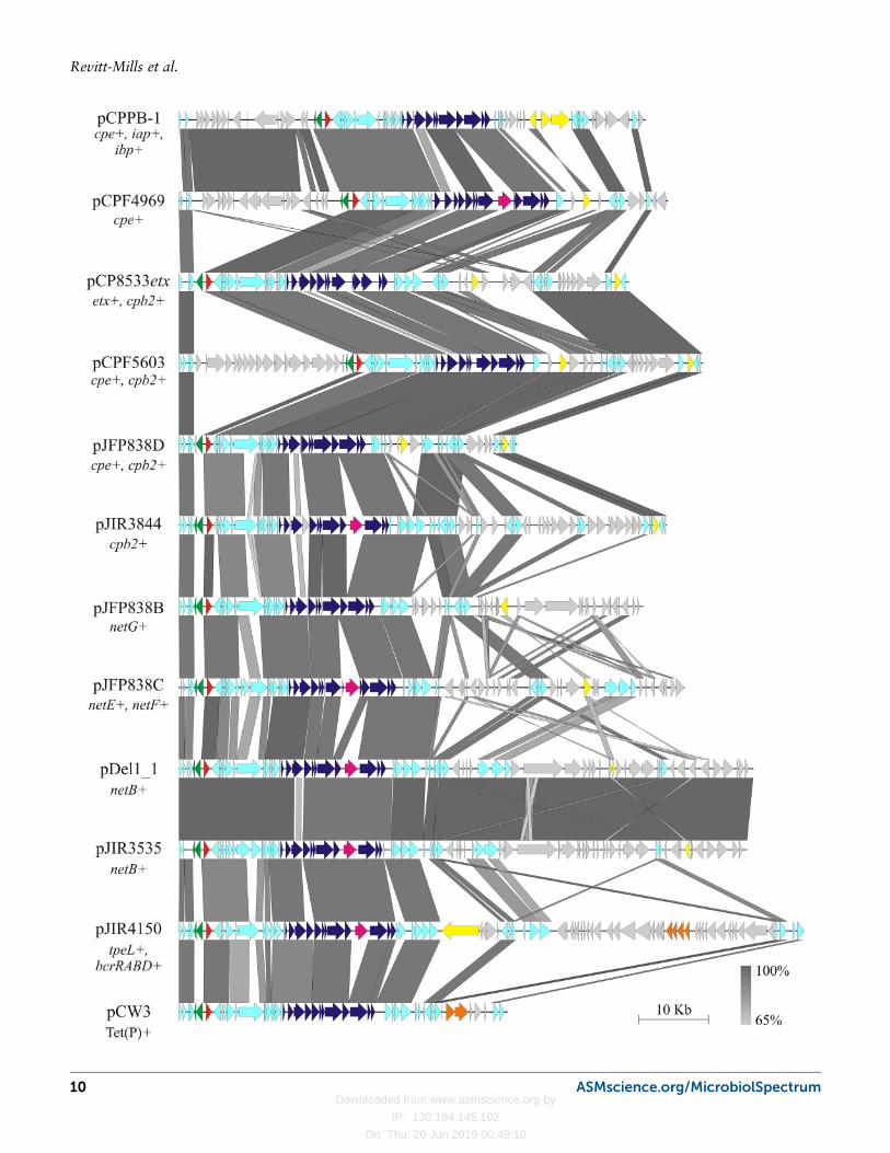
FIGURE 2 Nucleotide alignment of pCW3-like plasmids from C. perfringens. Full plasmidsequences were aligned using the blastn algorithm (225) and Easyfig for visualization (226).The plasmid names are noted on the left along with the toxin or antibiotic resistancedeterminants encoded within each plasmid sequence. Predicted (ORFs) are indicated byarrows with the following color code: conserved ORFs (light blue), tcp conjugation genes,dark blue; toxin genes, yellow; parMR partitioning genes, green; rep red; group II introns,pink; antibiotic resistance genes (orange) less-conserved ORFs, gray. The scale bar andkey for nucleotide identity are shown.
ASMscience.org/MicrobiolSpectrum 11
Virulence Plasmids of the Pathogenic Clostridia
Downloaded from www.asmscience.org by
IP: 130.194.145.102
On: Thu, 20 Jun 2019 00:48:10
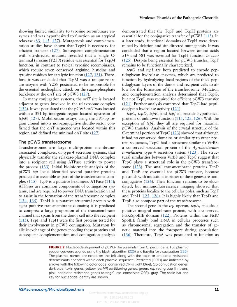
a coupling protein in pCW3 conjugative transfer (122).Subsequent mutation and complementation analysisconfirmed that TcpA was essential for pCW3 transfer(122). As the coupling protein, it was assumed thatTcpA mediated a range of protein-protein interactionswith other Tcp proteins in both the relaxosome andtransferosome complexes (124). Through chemical cross-linking and bacterial two-hybrid analysis, TcpA wasfound to self-associate, as well as to interact withthe transferosome components TcpC, TcpG and TcpH(124). At the time of this study no relaxosome com-
ponents had been identified, and thus, interactionswith pCW3 relaxosome components have not yet beenassessed (124). Based on these studies and other protein-protein interaction studies, including structural studies(121–125) a model for the conjugative apparatus uti-lized by pCW3 has been constructed (Fig. 3) (128).
Although a large body of work has been conductedon the tcp locus of pCW3, there are still large gaps inour knowledge and understanding of the structure andfunction of the conjugation apparatus. To date, it is stillnot certain whether pCW3 DNA transport occurs in a
FIGURE 3 Model of the pCW3 conjugation apparatus. The arrangement of the proteinswithin themodel is based on protein localization studies and bioinformatics analysis. Blackarrows indicate confirmed protein interactions between Tcp proteins and only Tcp pro-teins required for wild-type transfer of pCW3 are shown. Indicated within the membraneare the integral membrane proteins TcpH (brown), the peptidoglycan hydrolase TcpG(purple), the assembly factor TcpC (green; monomers as different shades), the proteins ofunknown function, TcpD (yellow) and TcpE (pink) and the putative coupling protein TcpA(orange). Within the cytoplasm are a putative ATPase TcpF (red) and the novel relaxaseTcpM (blue) in complex with the double stranded pCW3 oriT site. Dotted arrows indicateputative ATPase activity. Reproduced with permission from Wisniewski and Rood (2017).
12 ASMscience.org/MicrobiolSpectrum
Revitt-Mills et al.
Downloaded from www.asmscience.org by
IP: 130.194.145.102
On: Thu, 20 Jun 2019 00:48:10
single-stranded or double-stranded manner. Addition-ally, the precise processing reactions that are catalyzedby the relaxosome remain uncharacterized and the pro-tein composition of this structure remains to be elu-cidated. Finally, the mechanism of direct cell-to-cellcontact between C. perfringens cells remains elusive.These genetic and functional studies of pCW3 are im-portant because they serve as a model system for theanalysis of the pCW3-like toxin plasmids.
Multiple pCW3-Like Toxin PlasmidsAre Often Present in the Same StrainPlasmid incompatibility is defined as the inability of twoplasmids to coexist in the same cell without the appli-cation of external selection (137). Plasmid incompati-bility usually arises when two plasmids encode similaressential plasmid encoded factors such as replicationinitiation factors, replication control mechanisms orpartitioning machineries.
All plasmids must replicate and maintain a stablecopy number relative to the host chromosome to befaithfully inherited by the daughter cells at cell division(138). The copy number of a plasmid is related to theproperties of the replication machinery and other fac-tors. Initial attempts to identify the pCW3 replicationgene could not identify an ORF with significant simi-larity to any known plasmid replication proteins (115).Therefore, a functional genetics approach was employedto determine which plasmid encoded regions were in-volved in plasmid replication. A combination of deletionand transposon mutagenesis studies led to the identifi-cation of the rep gene, which was essential for the rep-lication of pCW3 (115). The intergenic region upstreamof the rep gene had five pairs of inverted repeats and aseries of conserved 17-bp direct repeats that were pos-tulated to act as iterons or as a centromeric binding sitefor partitioning proteins (115). Comparative bioinfor-matic analysis of the available C. perfringens conjuga-tive plasmid sequences revealed that the pCW3-likereplication protein is highly conserved (120).
As already mentioned, many strains of C. perfringenscarry more than one pCW3-like toxin or antibiotic re-sistance plasmid, each of which encodes a similar repli-cation protein (82, 115, 120). These plasmids are stablymaintained in the same cell, which is unexpected giventhe conventional relationship between plasmid incom-patibility and plasmid replication functions (137). Thebest studied example of this phenomenon is the aviannecrotic enteritis type G isolate, EHE-NE18. This strainstably harbours three highly similar pCW3-like conju-gative plasmids: a NetB plasmid, a beta2-toxin plasmid
and a tetracycline resistance plasmid. These plasmidseach encode an almost identical replication protein(≥ 98% amino acid sequence identity) (82, 120). Theapparent stability of this plasmid combination, andother toxin plasmid combinations in strains from othertoxinotypes, suggested that factors other than the rep-lication machinery were involved in the determination ofplasmid incompatibility in C. perfringens.
pCW3 is a low-copy number plasmid (∼5 plasmidcopies/chromosome) (T. Stent, X. Han, R. Moore, V.Adams and J. Rood, personal communication) andtherefore most likely requires an active maintenancesystem to ensure that it is inherited correctly (139).Closer inspection of the pCW3 sequence revealed twogenes that were adjacent to, but divergently transcribedfrom, the rep gene. These genes encoded putative par-titioning system homologues, ParM and ParR, and aputative parC centromere site (115).
Partitioning systems act as positioning systems toensure that sister plasmids are positioned at the cellpoles thus ensuring that the plasmid is maintained indaughter cells when cell division occurs (139). Type II orParMRC-like plasmid partitioning systems encode threecomponents: ParM, an actin-like ATPase that polymer-izes to form filaments, ParR, a DNA-binding adaptorprotein, and parC, a centromeric ParR binding regionthat usually comprises a series of direct repeats locatedupstream of the parM gene (140). ParMRC systemssegregate plasmids via a pushing mechanism in whichtwo sister plasmids are linked through a bundle of ParMfilaments that recognize and interact with ParR adaptorproteins bound to the parC centromere sites (140).C. perfringens ParMRC-like plasmid partitioning sys-tems have not been thoroughly investigated, although aparMRCB family partitioning system from the C. per-fringens isolate JGS1987 has been demonstrated to sta-bilize a mini-replicon in E. coli (141).
An extensive survey of sequenced pCW3-like plas-mids showed that there were at least 10 distinct familiesof parMRC-like partitioning systems (parMRCA-J) pres-ent in C. perfringens (81, 82, 115, 120, 142). Com-parative analysis showed that ParM homologues hadupwards of 90% amino acid sequence identity withina family, but only 15- to 54% identity between thevarious groups (120). ParR homologues clustered intothe same phylogenetic groups as the ParM homologues,but showed more amino acid sequence variation withina family (120). The parC centromere sites also clusteredwith their cognate ParM and ParR family groups, al-though the parC sites are very AT-rich and have a lotmore sequence variability between the 10 groups than
ASMscience.org/MicrobiolSpectrum 13
Virulence Plasmids of the Pathogenic Clostridia
Downloaded from www.asmscience.org by
IP: 130.194.145.102
On: Thu, 20 Jun 2019 00:48:10
the ParM and ParR components (120). It was observedthat C. perfringens strains that house multiple pCW3-like plasmids generally do not have plasmids that encodethe same parMRC allele (120). This observation sug-gested that parMRC partitioning families were respon-sible for maintenance within the same strain of multiplepCW3-like plasmids that encode a highly similar repli-cation region.
There now is experimental evidence to support thishypothesis (143). In this study different combinationsof genetically marked, parMRC-encoding, pCW3-likeplasmids were introduced into the same C. perfringensstrain. These strains were constructed so that theyhoused either two plasmids with the same parMRClocus, or two plasmids with parMRC loci from differentfamilies. Analysis of these isolates showed that plasmidswith the same parMRC families were unable to stablycoexist in the same cell, whereas plasmids with differentparMRC families could stably coexist even though theyencoded similar Rep proteins (143).
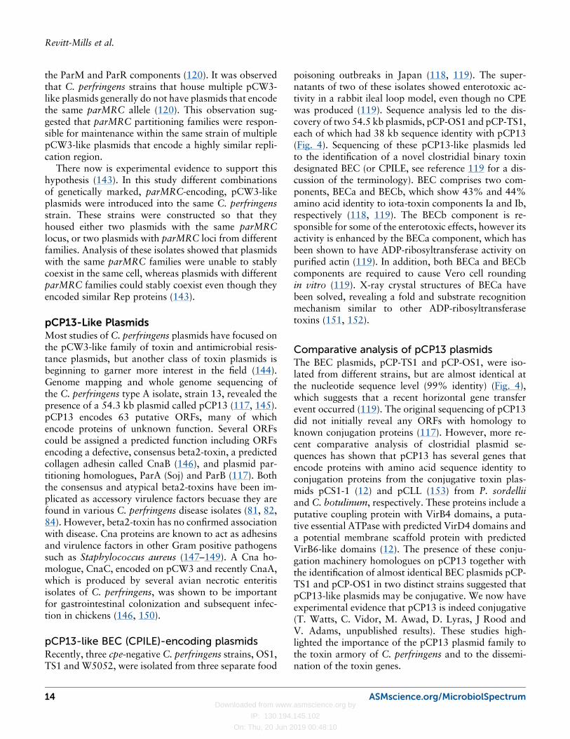
pCP13-Like PlasmidsMost studies of C. perfringens plasmids have focused onthe pCW3-like family of toxin and antimicrobial resis-tance plasmids, but another class of toxin plasmids isbeginning to garner more interest in the field (144).Genome mapping and whole genome sequencing ofthe C. perfringens type A isolate, strain 13, revealed thepresence of a 54.3 kb plasmid called pCP13 (117, 145).pCP13 encodes 63 putative ORFs, many of whichencode proteins of unknown function. Several ORFscould be assigned a predicted function including ORFsencoding a defective, consensus beta2-toxin, a predictedcollagen adhesin called CnaB (146), and plasmid par-titioning homologues, ParA (Soj) and ParB (117). Boththe consensus and atypical beta2-toxins have been im-plicated as accessory virulence factors becuase they arefound in various C. perfringens disease isolates (81, 82,84). However, beta2-toxin has no confirmed associationwith disease. Cna proteins are known to act as adhesinsand virulence factors in other Gram positive pathogenssuch as Staphylococcus aureus (147–149). A Cna ho-mologue, CnaC, encoded on pCW3 and recently CnaA,which is produced by several avian necrotic enteritisisolates of C. perfringens, was shown to be importantfor gastrointestinal colonization and subsequent infec-tion in chickens (146, 150).
pCP13-like BEC (CPILE)-encoding plasmidsRecently, three cpe-negative C. perfringens strains, OS1,TS1 and W5052, were isolated from three separate food
poisoning outbreaks in Japan (118, 119). The super-natants of two of these isolates showed enterotoxic ac-tivity in a rabbit ileal loop model, even though no CPEwas produced (119). Sequence analysis led to the dis-covery of two 54.5 kb plasmids, pCP-OS1 and pCP-TS1,each of which had 38 kb sequence identity with pCP13(Fig. 4). Sequencing of these pCP13-like plasmids ledto the identification of a novel clostridial binary toxindesignated BEC (or CPILE, see reference 119 for a dis-cussion of the terminology). BEC comprises two com-ponents, BECa and BECb, which show 43% and 44%amino acid identity to iota-toxin components Ia and Ib,respectively (118, 119). The BECb component is re-sponsible for some of the enterotoxic effects, however itsactivity is enhanced by the BECa component, which hasbeen shown to have ADP-ribosyltransferase activity onpurified actin (119). In addition, both BECa and BECbcomponents are required to cause Vero cell roundingin vitro (119). X-ray crystal structures of BECa havebeen solved, revealing a fold and substrate recognitionmechanism similar to other ADP-ribosyltransferasetoxins (151, 152).
Comparative analysis of pCP13 plasmidsThe BEC plasmids, pCP-TS1 and pCP-OS1, were iso-lated from different strains, but are almost identical atthe nucleotide sequence level (99% identity) (Fig. 4),which suggests that a recent horizontal gene transferevent occurred (119). The original sequencing of pCP13did not initially reveal any ORFs with homology toknown conjugation proteins (117). However, more re-cent comparative analysis of clostridial plasmid se-quences has shown that pCP13 has several genes thatencode proteins with amino acid sequence identity toconjugation proteins from the conjugative toxin plas-mids pCS1-1 (12) and pCLL (153) from P. sordelliiand C. botulinum, respectively. These proteins include aputative coupling protein with VirB4 domains, a puta-tive essential ATPase with predicted VirD4 domains anda potential membrane scaffold protein with predictedVirB6-like domains (12). The presence of these conju-gation machinery homologues on pCP13 together withthe identification of almost identical BEC plasmids pCP-TS1 and pCP-OS1 in two distinct strains suggested thatpCP13-like plasmids may be conjugative. We now haveexperimental evidence that pCP13 is indeed conjugative(T. Watts, C. Vidor, M. Awad, D. Lyras, J Rood andV. Adams, unpublished results). These studies high-lighted the importance of the pCP13 plasmid family tothe toxin armory of C. perfringens and to the dissemi-nation of the toxin genes.
14 ASMscience.org/MicrobiolSpectrum
Revitt-Mills et al.
Downloaded from www.asmscience.org by
IP: 130.194.145.102
On: Thu, 20 Jun 2019 00:48:10
The most recent addition to the pCP13-like family isthe bacteriocin-encoding plasmid pBCNF5603, a plas-mid found to coexist with a pCW3-like, CPE-plasmid inthe type F isolate F5603 (144). Bacteriocins are anti-bacterial factors produced by bacteria to inhibit thegrowth of similar bacterial strains (154). C. perfringensstrains often produce bacteriocins (155), some of whichhave been implicated in strain competition in the gas-trointestinal tracts of broiler chickens (129, 144, 156,157). pBCNF5603 was recently sequenced and found tohave genes with a high level of similarity to homologueson pCP13 and on the small bacteriocin plasmid pIP404,suggesting that pBCNF5603 arose from a recombina-tion event between these two C. perfringens plasmids(144) (Fig. 4). Sequencing of this plasmid has providedinsight into the replication functions of pCP13-likeplasmids by identifying two potential replication regions(144). The first replication region showed homology tothe rep and cop genes of pIP404, whereas the secondregion displayed similarity to the parA and parB generegions of pCP13, pCP-TS1 and pCP-OS1. The 5.5 kbgene cluster with similarity to pCP13 supported rep-lication of a recombinant plasmid in C. perfringens,whereas the replication region with similarity to pIP404did not (144). Analysis of subsequent deletion deriva-
tives showed that the homologue to PCP63 may be thepBCNF5603 Rep protein (144). The region upstreamof the putative rep gene was AT-rich, with a series ofinverted repeats that are likely to be important for rep-lication initiation.
THE TOXIN PLASMID OF C. TETANIThe neurotoxic clostridia comprise the C. botulinumcomplex, the causative agents of botulism, and C. tetani,which causes tetanus in both humans and animals. Boththe clostridial species that comprise the C. botulinumcomplex and C. tetani are renowned for their ability toproduce potent neurotoxins, botulinum toxin (BoNT)and tetanus toxin (TeNT), respectively (158). The vari-ous BoNT structural genes can be encoded on the chro-mosome, on a temperate bacteriophage or on a plasmid,whereas the TeNT structural gene is plasmid encoded(159).
TeNT is a zinc metalloprotease that acts on theSNARE protein VAMP/synaptobrevin at the synapticjunction of the nerve relaxation pathway in the spinalcord. Cleavage of VAMP/synaptobrevin means that it isunable to form a complex with other SNARE proteins,preventing fusion of the synaptic vesicle with the cellular
FIGURE 4 Sequence alignment of pCP13-like plasmids from C. perfringens. The plasmidsequences of pCP-TS1, pCP-OS1, pCP13 and pBCNF5603 were aligned using the Blastnalgorithm using Easyfig (226). The percentage identity is indicated by the scale bar at thebottom right and each sequence is compared separately to the sequences above andbelow. ORFs are indicated by arrows andORFs of particular interest are colored as follows:green, restriction modification systems; red, replication and maintenance; purple, toxingenes; yellow, transposase genes; dark blue, putative collagen adhesins; orange, putativerelaxase enzymes.
ASMscience.org/MicrobiolSpectrum 15
Virulence Plasmids of the Pathogenic Clostridia
Downloaded from www.asmscience.org by
IP: 130.194.145.102
On: Thu, 20 Jun 2019 00:48:10
membrane at the synaptic nerve junction and prevent-ing release of the neurotransmitter from the inhibitoryinterneurons to the motor neurons in the spinal cord.The net effect is inhibition of the muscle relaxationpathway and a rigid muscular paralysis that may be fatal(158, 160).
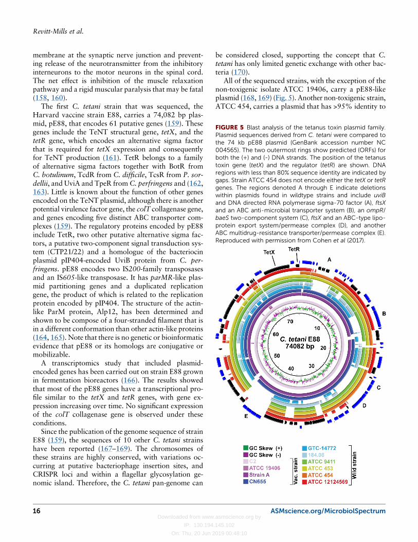
The first C. tetani strain that was sequenced, theHarvard vaccine strain E88, carries a 74,082 bp plas-mid, pE88, that encodes 61 putative genes (159). Thesegenes include the TeNT structural gene, tetX, and thetetR gene, which encodes an alternative sigma factorthat is required for tetX expression and consequentlyfor TeNT production (161). TetR belongs to a familyof alternative sigma factors together with BotR fromC. botulinum, TcdR from C. difficile, TcsR from P. sor-dellii, and UviA and TpeR fromC. perfringens and (162,163). Little is known about the function of other genesencoded on the TeNT plasmid, although there is anotherpotential virulence factor gene, the colT collagenase gene,and genes encoding five distinct ABC transporter com-plexes (159). The regulatory proteins encoded by pE88include TetR, two other putative alternative sigma fac-tors, a putative two-component signal transduction sys-tem (CTP21/22) and a homologue of the bacteriocinplasmid pIP404-encoded UviB protein from C. per-fringens. pE88 encodes two IS200-family transposasesand an IS605-like transposase. It has parMR-like plas-mid partitioning genes and a duplicated replicationgene, the product of which is related to the replicationprotein encoded by pIP404. The structure of the actin-like ParM protein, Alp12, has been determined andshown to be compose of a four-stranded filament that isin a different conformation than other actin-like proteins(164, 165). Note that there is no genetic or bioinformaticevidence that pE88 or its homologs are conjugative ormobilizable.
A transcriptomics study that included plasmid-encoded genes has been carried out on strain E88 grownin fermentation bioreactors (166). The results showedthat most of the pE88 genes have a transcriptional pro-file similar to the tetX and tetR genes, with gene ex-pression increasing over time. No significant expressionof the colT collagenase gene is observed under theseconditions.
Since the publication of the genome sequence of strainE88 (159), the sequences of 10 other C. tetani strainshave been reported (167–169). The chromosomes ofthese strains are highly conserved, with variations oc-curring at putative bacteriophage insertion sites, andCRISPR loci and within a flagellar glycosylation ge-nomic island. Therefore, the C. tetani pan-genome can
be considered closed, supporting the concept that C.tetani has only limited genetic exchange with other bac-teria (170).
All of the sequenced strains, with the exception of thenon-toxigenic isolate ATCC 19406, carry a pE88-likeplasmid (168, 169) (Fig. 5). Another non-toxigenic strain,ATCC 454, carries a plasmid that has >95% identity to
FIGURE 5 Blast analysis of the tetanus toxin plasmid family.Plasmid sequences derived from C. tetani were compared tothe 74 kb pE88 plasmid (GenBank accession number NC004565). The two outermost rings show predicted (ORFs) forboth the (+) and (-) DNA strands. The position of the tetanustoxin gene (tetX) and the regulator (tetR) are shown. DNAregions with less than 80% sequence identity are indicated bygaps. Strain ATCC 454 does not encode either the tetX or tetRgenes. The regions denoted A through E indicate deletionswithin plasmids found in wildtype strains and include uviBand DNA directed RNA polymerase sigma-70 factor (A), ftsXand an ABC anti-microbial transporter system (B), an ompR/baeS two-component system (C), ftsX and an ABC-type lipo-protein export system/permease complex (D), and anotherABC multidrug-resistance transporter/permease complex (E).Reproduced with permission from Cohen et al (2017).
16 ASMscience.org/MicrobiolSpectrum
Revitt-Mills et al.
Downloaded from www.asmscience.org by
IP: 130.194.145.102
On: Thu, 20 Jun 2019 00:48:10
pE88, but has a 20 kb deletion that encompasses boththe tetX and tetR genes (Fig. 5). The remaining plasmidshave considerable conservation as well as specific pE88regions that are not present (Fig. 5). The tetX genes arehighly conserved (99.3 to 99.4% identity) and the tetRgenes are all identical (169). The lack of genetic tools forthe analysis of C. tetani, combined with the inherentdifficulties in working with this anaerobic, very motileand highly toxigenic pathogen, have clearly limited thegenetic and functional studies that can be carried out onthese important plasmids.
BOTULINUM TOXIN-ENCODINGMOBILE GENETIC ELEMENTSThe production of BoNT neurotoxin is one of the de-fining features of the clostridial species C. botulinum.However, unlike TeNT-producing C. tetani, C. botuli-num constitutes a highly heterologous group of organ-isms (171–175). This collection of strains consists of sixgroups (I- to VI), with groups I- to III generally desig-nated C. botulinum (171, 172, 175). Groups IV, V, andVI consist of BoNT-producing strains of C. argenti-nense, C. baratii, and C. butyricum, respectively (175).This highly divergent spread of related toxin genessuggested early on that mobile genetic elements may beinvolved in toxin gene dissemination and subsequentstudies have demonstrated that multiple types of ele-ments are involved (171, 173).
All of these different species are able to produceBoNT, which is related to TeNT in both sequence,structure and function (176, 177). Both BoNT andTeNT are highly specific zinc metalloproteases. How-ever, unlike TeNT, BoNT associates with non-toxigenicproteins to form large complexes (178), which is relatedto the different routes of intoxication between TeNT andBoNT (wound versus intestinal absorption). The non-toxic components are involved in the protection andtransport of the active proteolytic component of BoNTfrom the gut to its site of action within the synapticvesicles of excitatory neurones, where it blocks the re-lease of neurotransmitters at the neuromuscular junction(179). The resultant flaccid paralysis can be fatal with-out appropriate supportive treatment (180). Many spe-cies, including humans, livestock, aquatic birds and fish,have been demonstrated to be susceptible to botulism(171).
The BoNT protein sequences have conserved func-tional domains, but are still highly variable (between29 and 64% amino acid sequence identity), and histori-cally have been separated into seven serologically dis-
tinct types (A- to G) (161, 179, 181). Many of the BoNTtypes have been further segregated into subtypes, des-ignated by a number (179, 182), for example, BoNT/F6.An eighth serotype designated H, was reported in 2014,but there is controversy about whether this serotypeconstitutes a chimeric protein between serotypes BoNT/F5 and BoNT/A or a distinct group (172, 183, 184).Subsequently, another BoNT type has been identifiedand named BoNT/X. This neurotoxin appears not to bea mosaic enzyme and constitutes a new class of BoNTs(185). A ninth class of BoNT, BoNT/En, has recentlybeen identified after genome sequencing of an Entero-coccus faecium isolate derived from a manure sample inSouth Carolina and appears most closely related toBoNT/X (38.7%) (181). The heterogeneity exhibited byBoNTs is intriguing because it has resulted in differencesin the complexes that are formed prior to cellular uptakeand in the mechanisms of transportation. Most impor-tantly, they have different host targets, catalyzing theproteolytic cleavage of different SNARE proteins, whichfacilitate neurotransmitter vesicle fusion and thereforeneurotransmitter release and propagation of the electri-cal signal from the nerve cell to the muscle cell (186). Inspite of this diversity, the result is the same: inhibition ofneurotransmitter vesicle fusion, leading to flaccid pa-ralysis (186).
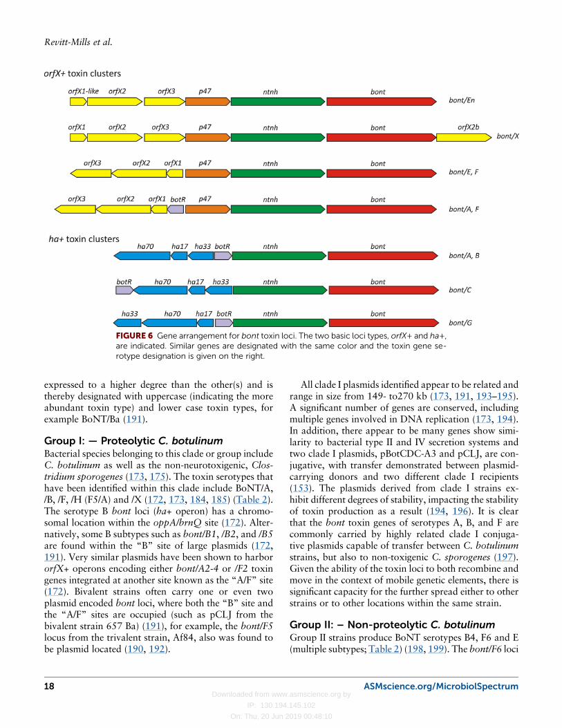
The BoNT structural (bont) genes are encoded withintwo distinct genetic regions, the orfX+ (function un-known) and ha+ (hemoglutinin) loci (Fig. 6) (173). Bothloci contain conserved ORF’s (orfs), including bont,found downstream, and in the same orientation, as theconserved ntnh (non-toxigenic, non-hemagglutinin) geneas well as the botR gene mentioned above (161, 178,187). All BotR-related proteins are responsible for pos-itively regulating the expression of toxin genes in theirnative hosts. Likewise, BotR has been shown to regulatetranscription of the bont gene (161, 162, 188).
The variable regions of the bont loci encode eitherthe orfX1-3 genes (orfX+ loci) or the ha17, -33, and-70 genes (ha+ loci; Fig. 6, Table 2). The organization ofthese two loci is largely conserved between differentstrains, the surrounding nucleotide sequences having co-evolved with the variable bont gene (173). In additionto the nine serological BoNT types, hybrid bont geneshave also been identified, such as serotype C/D and D/Cderivatives (189). Certain BoNT positive strains en-code multiple bont loci (173, 190), suggesting an easymechanism for genetic exchange and explaining howrecombination between bont loci could readily occur. Ithas been shown that when multiple bont loci are presentwithin the same strain, one particular BoNT toxin is
ASMscience.org/MicrobiolSpectrum 17
Virulence Plasmids of the Pathogenic Clostridia
Downloaded from www.asmscience.org by
IP: 130.194.145.102
On: Thu, 20 Jun 2019 00:48:10
expressed to a higher degree than the other(s) and isthereby designated with uppercase (indicating the moreabundant toxin type) and lower case toxin types, forexample BoNT/Ba (191).
Group I: — Proteolytic C. botulinumBacterial species belonging to this clade or group includeC. botulinum as well as the non-neurotoxigenic, Clos-tridium sporogenes (173, 175). The toxin serotypes thathave been identified within this clade include BoNT/A,/B, /F, /H (F5/A) and /X (172, 173, 184, 185) (Table 2).The serotype B bont loci (ha+ operon) has a chromo-somal location within the oppA/brnQ site (172). Alter-natively, some B subtypes such as bont/B1, /B2, and /B5are found within the “B” site of large plasmids (172,191). Very similar plasmids have been shown to harbororfX+ operons encoding either bont/A2-4 or /F2 toxingenes integrated at another site known as the “A/F” site(172). Bivalent strains often carry one or even twoplasmid encoded bont loci, where both the “B” site andthe “A/F” sites are occupied (such as pCLJ from thebivalent strain 657 Ba) (191), for example, the bont/F5locus from the trivalent strain, Af84, also was found tobe plasmid located (190, 192).
All clade I plasmids identified appear to be related andrange in size from 149- to270 kb (173, 191, 193–195).A significant number of genes are conserved, includingmultiple genes involved in DNA replication (173, 194).In addition, there appear to be many genes show simi-larity to bacterial type II and IV secretion systems andtwo clade I plasmids, pBotCDC-A3 and pCLJ, are con-jugative, with transfer demonstrated between plasmid-carrying donors and two different clade I recipients(153). The plasmids derived from clade I strains ex-hibit different degrees of stability, impacting the stabilityof toxin production as a result (194, 196). It is clearthat the bont toxin genes of serotypes A, B, and F arecommonly carried by highly related clade I conjuga-tive plasmids capable of transfer between C. botulinumstrains, but also to non-toxigenic C. sporogenes (197).Given the ability of the toxin loci to both recombine andmove in the context of mobile genetic elements, there issignificant capacity for the further spread either to otherstrains or to other locations within the same strain.
Group II: – Non-proteolytic C. botulinumGroup II strains produce BoNT serotypes B4, F6 and E(multiple subtypes; Table 2) (198, 199). The bont/F6 loci
FIGURE 6 Gene arrangement for bont toxin loci. The two basic loci types, orfX+ and ha+,are indicated. Similar genes are designated with the same color and the toxin gene se-rotype designation is given on the right.
18 ASMscience.org/MicrobiolSpectrum
Revitt-Mills et al.
Downloaded from www.asmscience.org by
IP: 130.194.145.102
On: Thu, 20 Jun 2019 00:48:10
are chromosomally located and found within what isthought to constitute a mobile genetic element that car-ries a recombinase gene, topB (172, 200). The bont/B4loci found within this clade are believed to be exclusivelyplasmid-borne (193, 198, 199). The first group II plas-mid sequenced is derived from strain Eklund 17B andcarries the bont/B4 locus on the 48-kb plasmid, pCLL(173, 174). Subsequent studies demonstrated that bont/B4 loci are carried by plasmids 47 to 63 kb in size thatare unrelated to group I plasmids (198). These plasmidsform two distinct plasmid groups, perhaps sharing acommon ancestor (198). One group (class 1) is relatedto pCLL and its members are of similar size, whereasclass 2 plasmids only have about 33% similarity to class1 plasmids; this similarity is limited to the large neuro-toxin gene cluster (198). Class 2 plasmids range in sizefrom 58 to 63 kb (198). A third plasmid appears to be ahybrid of the two other plasmid classes (198). All plas-mid types encoding bont/B4 loci were found only ingroup II strains and related plasmids that were non-toxigenic have subsequently been found by analysis ofgenome sequence data from other group II strains (199).
Serotype E strains belonging to group II had previ-ously been reported to encode only chromosomal bont/E loci, but recent pulsed-field gel electrophoresis studiesshowed that a proportion of type E strains (6-10%)carry bont/E loci on large plasmids in group II strains(199, 201). These plasmids range in size from 134 to144 kb and are closely related (199). FunctionalCRISPR systems were identified on plasmids carryingbont/E1 and bont/E10 loci, but not on plasmids carry-ing the bont/E3 loci (199). The type E toxigenicplasmids appear to be very low copy number as judgedfrom sequencing read abundance, approximately onecopy per chromosome (199). The bont/E plasmid lociare very similar to the chromosomal bont/E toxinclusters that have integrated into a chromosomal copyof the resolvase gene, rarA. The toxin loci in both casesconsists of a 24-kb cassette that include genes outsideof the normal toxin cluster. This region includes a sec-ond, related rarA gene that remains intact (199). Theplasmid-borne type E toxin cassette is completely con-served, but the integration site is different. The plas-mid gene that has been interrupted is a helicase gene andthe insertion site appears to be a 6-bp sequence that isconserved between the chromosomal rarA gene and theplasmid-encoded helicase gene (199).
All of the toxin plasmids so far identified from groupII organisms (class1 and 2 bont/B4 and bont/Eplasmids) have a significant number of ORFs whosepredicted function involves conjugative transfer (198,
TABLE
2Su
mmaryof
BoN
T-produ
cing
speciesan
dtoxingene
location
s
Bac
terial
clad
eor
speciesa
III
III
IVV
VI
Enteroco
ccus
faec
ium
BoNTtypeb
A(8)
B(8)
F(7)
HX
B(8)
E(12)
F(7)
CD
GF(7)
E(12)
En
bontlocu
scorfX
ha
orfX
orfX
orfX
ha
orfX
orfX
ha
ha
ha
ha
orfX
orfX
ha
Chromosomed
arsC
(orfX)
oppA/brnQ
(ha)
oppA/
brnQ
arsC
pulC
(F4)
IS110
flan
king
bont/H
Cys.b
iosyn.
Pathway
rarA(M
GE)
topB
(MGE)
(F6)
Strain
CDC27
41
IS1182flan
ksbont/F7
locu
sCompound
Transposon?
rarA
(MGE)
(E4,E
5)
Plasm
id15
0-2
70kb
A/F
site
E(orfX)
150-
270kb
Bsite
150-
270
kbA/F
site
47-63kb
(B4)133-144kb
Helicase
(E1,E3
,E10
)
140kb
Strain89G
207kb
Putative
Conjugative
Plasm
idBacterio
phag
een
coded
Yes
Yes
Speciese
Human
sChicke
ns
Human
sHorses
Cattle
Human
sHuman
(Bivalen
tB2h
)
Human
(Bivalent
B2x)
Human
Horses
Cattle
Human
Fish
Birds
Human
Birds
Cattle
Horses
Birds
Cattle
Human
Soil
Human
s?Human
sHuman
Cow
Man
ure
a Bacterial
clad
edesign
ation:
I-IIIC.bo
tulin
um,IV
C.argentinense,V
C.ba
ratii,VIC.bu
tyricum.
bDesigna
tion
ofserologically
distinct
BoN
Tproteins,thenu
mberof
toxinsubtyp
esarein
parentheses.
c Genes
encoding
BoN
Ttoxins
arefoun
dassociated
withon
eof
twodistinct
geneticloci;orfX
+or
theha
+loci
(see
Fig.
6).
dChrom
osom
allocation
ofbo
ntlocus,oftenassociated
withamob
ilegeneticelem
ent(M
GE).F6
isthebo
ntsubtyp
e.e The
vertebratespeciesinwhich
each
toxintype
ismostfrequ
ently
demon
stratedto
causeBotulism.C
lade
IVstrainsh
aveno
tbeendefinitiv
elyassociated
with
botulismbu
thavebeen
associated
with
multip
lesudd
endeaths
(199
).
ASMscience.org/MicrobiolSpectrum 19
Virulence Plasmids of the Pathogenic Clostridia
Downloaded from www.asmscience.org by
IP: 130.194.145.102
On: Thu, 20 Jun 2019 00:48:10
199). The smallest plasmid characterized so far, pCLL,which contains the least putative conjugation genes(198), is the only group II plasmid to have a demon-strated ability to transfer, remarkably, into a group Irecipient (153). No group II- like plasmids have beendetected in any other related strain and they show littleconservation between themselves or plasmids from othergroups, suggesting that these plasmids have a narrowhost range (199). pCLL has a low level of similarity topCS1-family plasmids from P. sordellii and pCP13-likeplasmids from C. perfringens (12). The plasmid locationof bont genes in this bacterial group explains how thebont genes have been so widely disseminated.
Group III: – Clostridium novyi sensu lato?The clade designated type III includes a number ofclostridial species and recently it was proposed that it bere-named from C. botulinum to C. novyi sensu lato(189). This designation includes three species dependingon the toxins that each strain produces (189): C novyi,Clostridium haemolyticum and C. botulinum. C. novyiproduces the lethal alpha toxin, which is related to thealpha toxin produced by Clostridium septicum. InC. novyi, the alpha toxin gene is located on a bacterio-phage (202) and any particular C. novyi strain may alsoproduce one of two phospholipases (beta and gamma)(189). C. haemolyticum produces a phospholipase se-rologically indistinguishable from the beta phospholi-pase of C. novyi (189). Group III C. botulinum strainsare associated with the expression of BoNT/C andBoNT/D and the chimeric derivatives, C/D and D/C(203) (Table 2). The location of the bont locus withingroup III strains of C. botulinum has been known formany decades to be within temperate bacteriophage,called pseudolysogens, that could be readily cured fromtoxigenic strains (203). The curing mechanism wasshown to be due to the unusual nature of the lysogenicstate— that of a replicating plasmid (203). For example,the bont-encoding bacteriophage from strain C-ST hasa large (186-kb) linear genome with 404-bp terminaldirect repeats when packaged within the phage parti-cle (203), but the genome is found as a circular, self-replicating molecule within the bacterial cell (189, 203).Many other plasmid and phage-like molecules can befound not just in C. botulinum group III, but in allC. novyi sensu lato strains, indicating that this group ishighly dependent on the accessory genome to determinethe virulence of any particular strain (189, 202–205).
Group IV: – Clostridium argentinenseThis group was renamed C. argentinense because it hadvery little similarity to other C. botulinum strains other
than the production of BoNT (171). Only BoNT/G hasbeen detected in C. argentinense and the bont/G toxingene was demonstrated to be located on large plasmidsin at least two strains (206) (Table 2). The first type Gstrain sequenced, CDC 2741, remains a draft assembly,but analysis of the sequence data indicated that the bont/G cluster was located on a large 1.1-Mb contig and wasthought to be chromosomally located (172, 207). Re-cently, another completed genome sequence for a BoNT/G-expressing strain was released, strain 89G (208), anda circular plasmid sequence was obtained and shownto carry the bont/G locus. This plasmid, pRSJ17_1,is 140 kb in size and, although no further analysis ofthis sequence has been published (208), it appears toshare little or no similarity to plasmids of group I or IIC. botulinum strains. However, the entire pRSJ17-1sequence appears to be located within the 1.1 Mb contigassembled from the genome sequence of strain CDC2741 so it is possible that either the bont/G plasmid hasbecome integrated into the chromosome in this strain orthat the CDC 2741 draft assembly is erroneous andCDC 2741 contains a 140-kb plasmid almost identicalto pRSJ17-1. Further analysis is required to confirm thetoxin cluster location in strain CDC 2741.
Group V: – Neurotoxigenic Clostridium baratiiType V BoNT-expressing strains are not designated asC.botulinum but are members of the non-neurotoxigenicspecies,C. baratii (172, 173). Again only one serotype ofBoNT has been detected from this species: BoNT/F7,and the location of this gene is chromosomal, where itis flanked by directly repeated IS1182 elements (207)(Table 2). The context of the bont/F7 locus in this strainsuggests that it has been acquired by horizontal genetransfer (207).
Group VI: – Neurotoxigenic ClostridiumbutyricumNeurotoxigenic C. butyricum strains have only beendemonstrated to carry bont/E genes within orfX+ loci(173) (Table 2). The neurotoxin gene cluster was ori-ginally thought to be plasmid determined rather thanchromosomal (209). However, more recent data haveclearly indicated that the bont/E genes in C. butyricumare chromosomally located (173, 210, 211). The rarAgene is the position of insertion for the bont/E locus inboth group II C. botulinum strains that carry bont/E aswell as neurotoxigenic C. butyricum strains (172, 173,199), which suggests that the bont/E locus has mostlikely been inserted via the same mechanism in bothorganisms, possibly via the recombinase RarA (172,173).
20 ASMscience.org/MicrobiolSpectrum
Revitt-Mills et al.
Downloaded from www.asmscience.org by
IP: 130.194.145.102
On: Thu, 20 Jun 2019 00:48:10
CLOSTRIDIOIDES (CLOSTRIDIUM)DIFFICILE VIRULENCE PLASMIDSC. difficile is a genomically diverse species, that consistsof eight phylogenomic clades (212), and it containsmany integrated and extrachromosomal genetic elements(213). The presence of numerous potentially mobile ge-netic elements suggests that horizontal gene transfer hasplayed an important role in the evolution of C. difficile.AlthoughC. difficile pathogenesis involvesmultiple factors(214), disease resulting from infection with this microbe isprimarily toxin-mediated, and strains can encode up tothree toxins. These toxins include toxin A (TcdA) andtoxin B (TcdB), which are both encoded within the Path-ogenicity Locus (PaLoc), and the C. difficile transferaseor binary toxin (CDT), encoded within the CDT locus(CdtLoc) (215). Both the PaLoc (216) and CdtLoc (217,218) were found in chromosomal locations when firstidentified. However, the chromosomal PaLoc is mobili-zable, and can be transferred to a non-toxigenic recipientthereby converting it to a toxigenic strain, but neither adefined mobilizable or conjugative PaLoc element nor themechanism of transfer have been determined (219).
Few studies have investigated plasmids in C. difficileand only a small number of native plasmids have beendescribed or characterized to date. Unlike many otherclostridial species, in which toxin genes are often en-coded on extrachromosomal elements, as discussedearlier in this review, C. difficile toxins appeared to beencoded exclusively on the host chromosome (219,220). However, a recent study using single-moleculereal-time sequencing technology showed that the ge-nome of a bacteriophage, designated phiSemix9P1, car-ries a complete functional binary toxin locus within itsgenome (221). The location of the CDT genes on this56.6 kb bacteriophage suggests that a transduction-mediated mechanism may be involved in transferringthese toxin genes across different C. difficile lineages. Ofrelevance to this review, the genome of phiSemix9P1was found to encode a putative ParA-like protein and agene for a protein that might play a role in the initiationof plasmid replication in addition to typical bacterio-phage-encoded genes involved in DNA packaging, headand tail morphogenesis or host cell lysis, suggesting thatthis element may represent a bacteriophage-plasmidhybrid (221). Such hybrid elements have recently beenidentified in C. difficile (221), and the presence ofthese elements can sometimes modulate the virulenceof strains by regulating the expression of the chromo-somally located toxin genes (222). Genome sequencingstudies are continuing to identify new and novel hybrids(223) which may impact phenotypes that influence
strain virulence or lateral gene transfer processes, andtherefore the evolution and dissemination of C. difficilevariant strains. The ubiquity of mobile elements inC. difficile suggests that horizontal gene transfer playsan important role in the movement of DNA betweenstrains, and the identification of plasmid-bacteriophagefusions may suggest that intra- and inter-species DNAexchange has occurred, probably within the diversebacterial populations found in the gastrointestinal en-vironment (213).
CONCLUSIONSThe importance of mobile genetic elements to toxin dis-semination within the clostridia is clearly evident. Thesegenera produce a large variety of protein toxins, in-cluding the three most potent toxins known to man:botulinum, tetanus and epsilon toxins. Many of thesetoxin genes are not only located on extrachromosomalelements, but also may be associated with mobile geneticelements that may facilitate their spread from one plas-mid or bacteriophage to another. The carriage of thesetoxins on extrachromosomal elements greatly facilitatesthe spread of toxin genes either within a species or be-tween species. No doubt the extent of the spread de-tected to date is limited only by our survey capabilitiesand the advent of next generation sequencing will fa-cilitate the identification of an even greater range ofelements that can facilitate the movement of the genesencoding these important, and sometimes devastating,toxins.
ACKNOWLEDGEMENTSResearch in JIR’s laboratory is supported by grants from theNational Health and Medical Research Council (GNT1082401,GNT1104502 and GNT1125349) and the Australian ResearchCouncil (DP160102680). Research in DL’s laboratory is sup-ported by grants from the National Health and Medical ResearchCouncil (GNT1107812, GNT1107969, GNT1103024 and GNT1145760) and the Australian Research Council (DP150104670and DP180102449).
REFERENCES1. Vidor C, Awad M, Lyras D. 2015. Antibiotic resistance, virulence fac-tors and genetics of Clostridium sordellii. Res Microbiol 166:368–374http://dx.doi.org/10.1016/j.resmic.2014.09.003.2. Aldape MJ, Bryant AE, Stevens DL. 2006. Clostridium sordellii infec-tion: epidemiology, clinical findings, and current perspectives on diagnosisand treatment. Clin Infect Dis 43:1436–1446 http://dx.doi.org/10.1086/508866.3. Jank T, Aktories K. 2008. Structure and mode of action of clostridialglucosylating toxins: the ABCD model. Trends Microbiol 16:222–229http://dx.doi.org/10.1016/j.tim.2008.01.011.4. Voth DE, Martinez OV, Ballard JD. 2006. Variations in lethal toxinand cholesterol-dependent cytolysin production correspond to differ-ences in cytotoxicity among strains of Clostridium sordellii. FEMS
ASMscience.org/MicrobiolSpectrum 21
Virulence Plasmids of the Pathogenic Clostridia
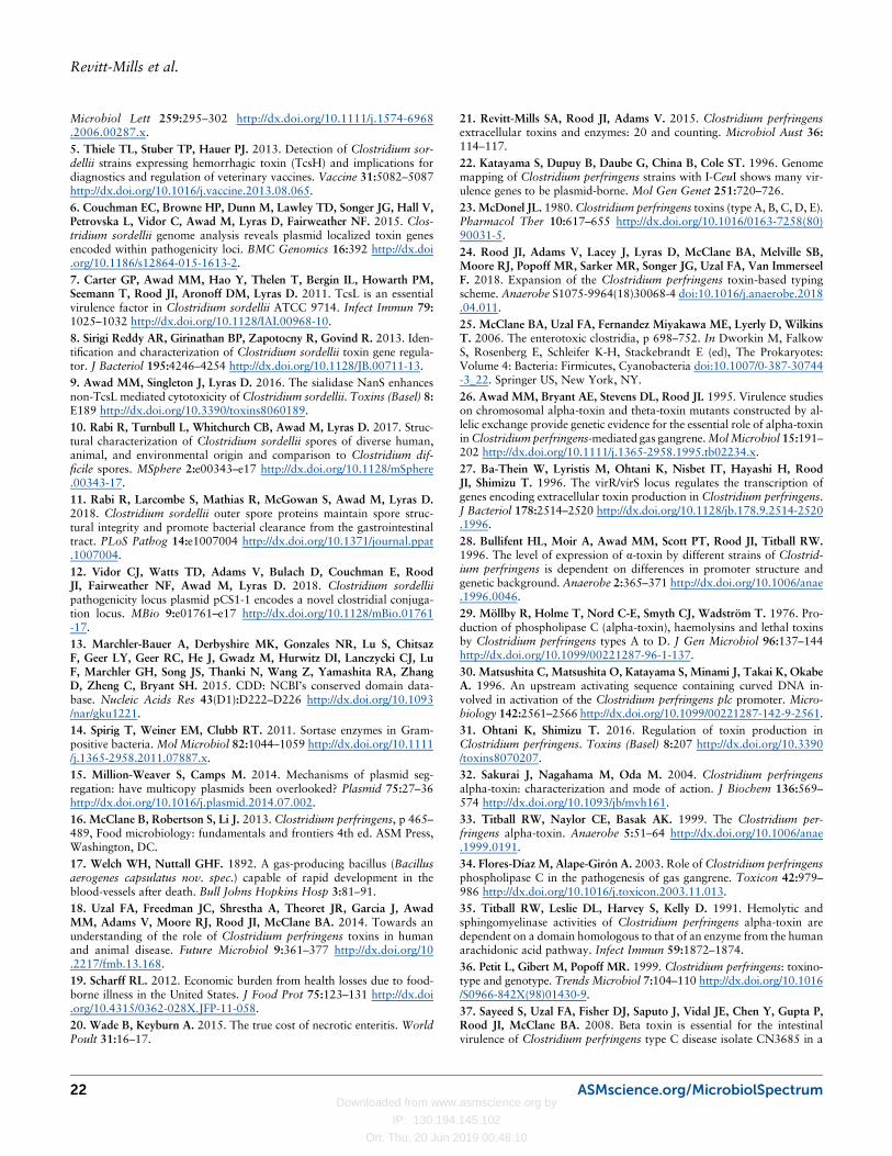
Downloaded from www.asmscience.org by
IP: 130.194.145.102
On: Thu, 20 Jun 2019 00:48:10
Microbiol Lett 259:295–302 http://dx.doi.org/10.1111/j.1574-6968.2006.00287.x.5. Thiele TL, Stuber TP, Hauer PJ. 2013. Detection of Clostridium sor-dellii strains expressing hemorrhagic toxin (TcsH) and implications fordiagnostics and regulation of veterinary vaccines. Vaccine 31:5082–5087http://dx.doi.org/10.1016/j.vaccine.2013.08.065.6. Couchman EC, Browne HP, Dunn M, Lawley TD, Songer JG, Hall V,Petrovska L, Vidor C, Awad M, Lyras D, Fairweather NF. 2015. Clos-tridium sordellii genome analysis reveals plasmid localized toxin genesencoded within pathogenicity loci. BMC Genomics 16:392 http://dx.doi.org/10.1186/s12864-015-1613-2.7. Carter GP, Awad MM, Hao Y, Thelen T, Bergin IL, Howarth PM,Seemann T, Rood JI, Aronoff DM, Lyras D. 2011. TcsL is an essentialvirulence factor in Clostridium sordellii ATCC 9714. Infect Immun 79:1025–1032 http://dx.doi.org/10.1128/IAI.00968-10.8. Sirigi Reddy AR, Girinathan BP, Zapotocny R, Govind R. 2013. Iden-tification and characterization of Clostridium sordellii toxin gene regula-tor. J Bacteriol 195:4246–4254 http://dx.doi.org/10.1128/JB.00711-13.9. Awad MM, Singleton J, Lyras D. 2016. The sialidase NanS enhancesnon-TcsL mediated cytotoxicity of Clostridium sordellii. Toxins (Basel) 8:E189 http://dx.doi.org/10.3390/toxins8060189.10. Rabi R, Turnbull L, Whitchurch CB, Awad M, Lyras D. 2017. Struc-tural characterization of Clostridium sordellii spores of diverse human,animal, and environmental origin and comparison to Clostridium dif-ficile spores. MSphere 2:e00343–e17 http://dx.doi.org/10.1128/mSphere.00343-17.11. Rabi R, Larcombe S, Mathias R, McGowan S, Awad M, Lyras D.2018. Clostridium sordellii outer spore proteins maintain spore struc-tural integrity and promote bacterial clearance from the gastrointestinaltract. PLoS Pathog 14:e1007004 http://dx.doi.org/10.1371/journal.ppat.1007004.12. Vidor CJ, Watts TD, Adams V, Bulach D, Couchman E, RoodJI, Fairweather NF, Awad M, Lyras D. 2018. Clostridium sordelliipathogenicity locus plasmid pCS1-1 encodes a novel clostridial conjuga-tion locus. MBio 9:e01761–e17 http://dx.doi.org/10.1128/mBio.01761-17.13. Marchler-Bauer A, Derbyshire MK, Gonzales NR, Lu S, ChitsazF, Geer LY, Geer RC, He J, Gwadz M, Hurwitz DI, Lanczycki CJ, LuF, Marchler GH, Song JS, Thanki N, Wang Z, Yamashita RA, ZhangD, Zheng C, Bryant SH. 2015. CDD: NCBI’s conserved domain data-base. Nucleic Acids Res 43(D1):D222–D226 http://dx.doi.org/10.1093/nar/gku1221.14. Spirig T, Weiner EM, Clubb RT. 2011. Sortase enzymes in Gram-positive bacteria. Mol Microbiol 82:1044–1059 http://dx.doi.org/10.1111/j.1365-2958.2011.07887.x.15. Million-Weaver S, Camps M. 2014. Mechanisms of plasmid seg-regation: have multicopy plasmids been overlooked? Plasmid 75:27–36http://dx.doi.org/10.1016/j.plasmid.2014.07.002.16. McClane B, Robertson S, Li J. 2013. Clostridium perfringens, p 465–489, Food microbiology: fundamentals and frontiers 4th ed. ASM Press,Washington, DC.17. Welch WH, Nuttall GHF. 1892. A gas-producing bacillus (Bacillusaerogenes capsulatus nov. spec.) capable of rapid development in theblood-vessels after death. Bull Johns Hopkins Hosp 3:81–91.18. Uzal FA, Freedman JC, Shrestha A, Theoret JR, Garcia J, AwadMM, Adams V, Moore RJ, Rood JI, McClane BA. 2014. Towards anunderstanding of the role of Clostridium perfringens toxins in humanand animal disease. Future Microbiol 9:361–377 http://dx.doi.org/10.2217/fmb.13.168.19. Scharff RL. 2012. Economic burden from health losses due to food-borne illness in the United States. J Food Prot 75:123–131 http://dx.doi.org/10.4315/0362-028X.JFP-11-058.20. Wade B, Keyburn A. 2015. The true cost of necrotic enteritis. WorldPoult 31:16–17.
21. Revitt-Mills SA, Rood JI, Adams V. 2015. Clostridium perfringensextracellular toxins and enzymes: 20 and counting. Microbiol Aust 36:114–117.22. Katayama S, Dupuy B, Daube G, China B, Cole ST. 1996. Genomemapping of Clostridium perfringens strains with I-CeuI shows many vir-ulence genes to be plasmid-borne. Mol Gen Genet 251:720–726.23. McDonel JL. 1980.Clostridium perfringens toxins (type A, B, C, D, E).Pharmacol Ther 10:617–655 http://dx.doi.org/10.1016/0163-7258(80)90031-5.24. Rood JI, Adams V, Lacey J, Lyras D, McClane BA, Melville SB,Moore RJ, Popoff MR, Sarker MR, Songer JG, Uzal FA, Van ImmerseelF. 2018. Expansion of the Clostridium perfringens toxin-based typingscheme. Anaerobe S1075-9964(18)30068-4 doi:10.1016/j.anaerobe.2018.04.011.25. McClane BA, Uzal FA, Fernandez Miyakawa ME, Lyerly D, WilkinsT. 2006. The enterotoxic clostridia, p 698–752. In Dworkin M, FalkowS, Rosenberg E, Schleifer K-H, Stackebrandt E (ed), The Prokaryotes:Volume 4: Bacteria: Firmicutes, Cyanobacteria doi:10.1007/0-387-30744-3_22. Springer US, New York, NY.26. Awad MM, Bryant AE, Stevens DL, Rood JI. 1995. Virulence studieson chromosomal alpha-toxin and theta-toxin mutants constructed by al-lelic exchange provide genetic evidence for the essential role of alpha-toxininClostridium perfringens-mediated gas gangrene.MolMicrobiol 15:191–202 http://dx.doi.org/10.1111/j.1365-2958.1995.tb02234.x.27. Ba-Thein W, Lyristis M, Ohtani K, Nisbet IT, Hayashi H, RoodJI, Shimizu T. 1996. The virR/virS locus regulates the transcription ofgenes encoding extracellular toxin production in Clostridium perfringens.J Bacteriol 178:2514–2520 http://dx.doi.org/10.1128/jb.178.9.2514-2520.1996.28. Bullifent HL, Moir A, Awad MM, Scott PT, Rood JI, Titball RW.1996. The level of expression of α-toxin by different strains of Clostrid-ium perfringens is dependent on differences in promoter structure andgenetic background. Anaerobe 2:365–371 http://dx.doi.org/10.1006/anae.1996.0046.29. Möllby R, Holme T, Nord C-E, Smyth CJ, Wadström T. 1976. Pro-duction of phospholipase C (alpha-toxin), haemolysins and lethal toxinsby Clostridium perfringens types A to D. J Gen Microbiol 96:137–144http://dx.doi.org/10.1099/00221287-96-1-137.30. Matsushita C, Matsushita O, Katayama S, Minami J, Takai K, OkabeA. 1996. An upstream activating sequence containing curved DNA in-volved in activation of the Clostridium perfringens plc promoter. Micro-biology 142:2561–2566 http://dx.doi.org/10.1099/00221287-142-9-2561.31. Ohtani K, Shimizu T. 2016. Regulation of toxin production inClostridium perfringens. Toxins (Basel) 8:207 http://dx.doi.org/10.3390/toxins8070207.32. Sakurai J, Nagahama M, Oda M. 2004. Clostridium perfringensalpha-toxin: characterization and mode of action. J Biochem 136:569–574 http://dx.doi.org/10.1093/jb/mvh161.33. Titball RW, Naylor CE, Basak AK. 1999. The Clostridium per-fringens alpha-toxin. Anaerobe 5:51–64 http://dx.doi.org/10.1006/anae.1999.0191.34. Flores-Díaz M, Alape-Girón A. 2003. Role of Clostridium perfringensphospholipase C in the pathogenesis of gas gangrene. Toxicon 42:979–986 http://dx.doi.org/10.1016/j.toxicon.2003.11.013.35. Titball RW, Leslie DL, Harvey S, Kelly D. 1991. Hemolytic andsphingomyelinase activities of Clostridium perfringens alpha-toxin aredependent on a domain homologous to that of an enzyme from the humanarachidonic acid pathway. Infect Immun 59:1872–1874.36. Petit L, Gibert M, Popoff MR. 1999. Clostridium perfringens: toxino-type and genotype. Trends Microbiol 7:104–110 http://dx.doi.org/10.1016/S0966-842X(98)01430-9.37. Sayeed S, Uzal FA, Fisher DJ, Saputo J, Vidal JE, Chen Y, Gupta P,Rood JI, McClane BA. 2008. Beta toxin is essential for the intestinalvirulence of Clostridium perfringens type C disease isolate CN3685 in a
22 ASMscience.org/MicrobiolSpectrum
Revitt-Mills et al.
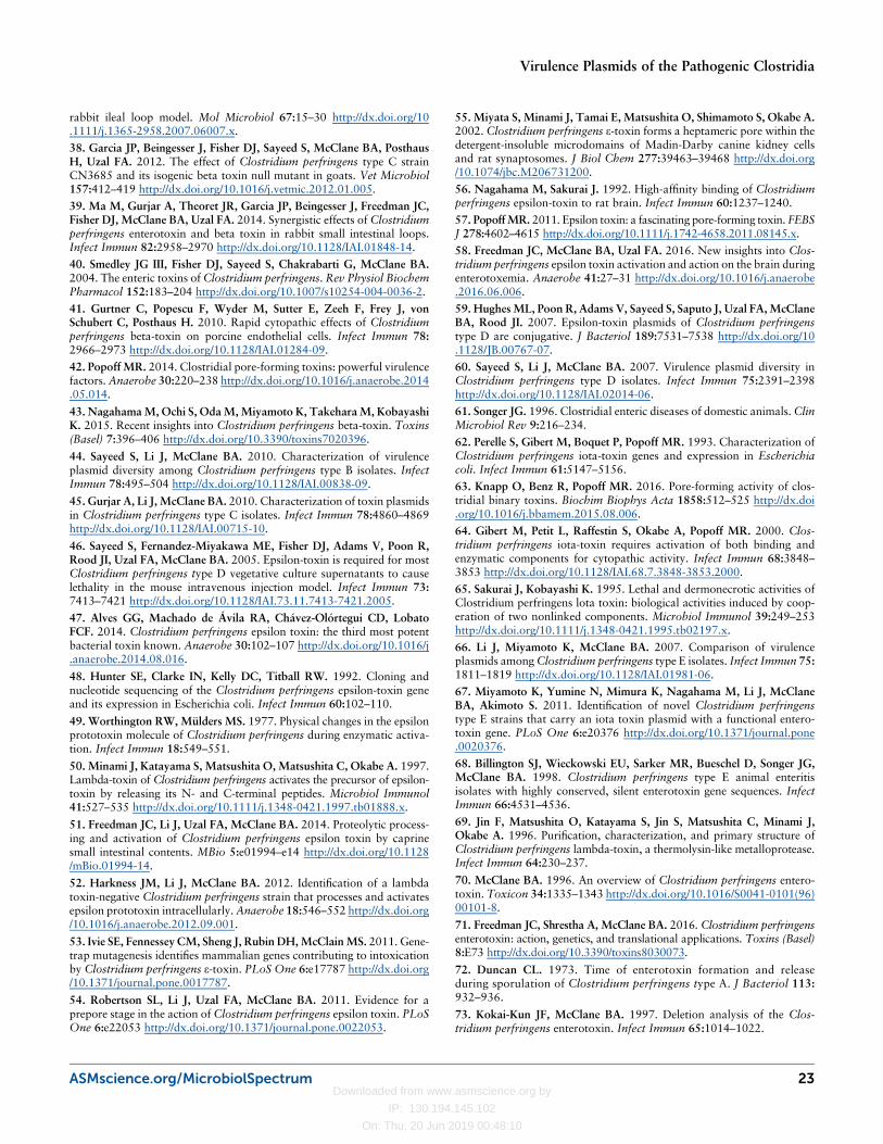
Downloaded from www.asmscience.org by
IP: 130.194.145.102
On: Thu, 20 Jun 2019 00:48:10
rabbit ileal loop model. Mol Microbiol 67:15–30 http://dx.doi.org/10.1111/j.1365-2958.2007.06007.x.
38. Garcia JP, Beingesser J, Fisher DJ, Sayeed S, McClane BA, PosthausH, Uzal FA. 2012. The effect of Clostridium perfringens type C strainCN3685 and its isogenic beta toxin null mutant in goats. Vet Microbiol157:412–419 http://dx.doi.org/10.1016/j.vetmic.2012.01.005.
39. Ma M, Gurjar A, Theoret JR, Garcia JP, Beingesser J, Freedman JC,Fisher DJ, McClane BA, Uzal FA. 2014. Synergistic effects of Clostridiumperfringens enterotoxin and beta toxin in rabbit small intestinal loops.Infect Immun 82:2958–2970 http://dx.doi.org/10.1128/IAI.01848-14.
40. Smedley JG III, Fisher DJ, Sayeed S, Chakrabarti G, McClane BA.2004. The enteric toxins of Clostridium perfringens. Rev Physiol BiochemPharmacol 152:183–204 http://dx.doi.org/10.1007/s10254-004-0036-2.
41. Gurtner C, Popescu F, Wyder M, Sutter E, Zeeh F, Frey J, vonSchubert C, Posthaus H. 2010. Rapid cytopathic effects of Clostridiumperfringens beta-toxin on porcine endothelial cells. Infect Immun 78:2966–2973 http://dx.doi.org/10.1128/IAI.01284-09.
42. Popoff MR. 2014. Clostridial pore-forming toxins: powerful virulencefactors. Anaerobe 30:220–238 http://dx.doi.org/10.1016/j.anaerobe.2014.05.014.
43. NagahamaM, Ochi S, OdaM,Miyamoto K, TakeharaM, KobayashiK. 2015. Recent insights into Clostridium perfringens beta-toxin. Toxins(Basel) 7:396–406 http://dx.doi.org/10.3390/toxins7020396.
44. Sayeed S, Li J, McClane BA. 2010. Characterization of virulenceplasmid diversity among Clostridium perfringens type B isolates. InfectImmun 78:495–504 http://dx.doi.org/10.1128/IAI.00838-09.
45. Gurjar A, Li J, McClane BA. 2010. Characterization of toxin plasmidsin Clostridium perfringens type C isolates. Infect Immun 78:4860–4869http://dx.doi.org/10.1128/IAI.00715-10.
46. Sayeed S, Fernandez-Miyakawa ME, Fisher DJ, Adams V, Poon R,Rood JI, Uzal FA, McClane BA. 2005. Epsilon-toxin is required for mostClostridium perfringens type D vegetative culture supernatants to causelethality in the mouse intravenous injection model. Infect Immun 73:7413–7421 http://dx.doi.org/10.1128/IAI.73.11.7413-7421.2005.
47. Alves GG, Machado de Ávila RA, Chávez-Olórtegui CD, LobatoFCF. 2014. Clostridium perfringens epsilon toxin: the third most potentbacterial toxin known. Anaerobe 30:102–107 http://dx.doi.org/10.1016/j.anaerobe.2014.08.016.
48. Hunter SE, Clarke IN, Kelly DC, Titball RW. 1992. Cloning andnucleotide sequencing of the Clostridium perfringens epsilon-toxin geneand its expression in Escherichia coli. Infect Immun 60:102–110.
49. Worthington RW, Mülders MS. 1977. Physical changes in the epsilonprototoxin molecule of Clostridium perfringens during enzymatic activa-tion. Infect Immun 18:549–551.
50. Minami J, Katayama S, Matsushita O, Matsushita C, Okabe A. 1997.Lambda-toxin of Clostridium perfringens activates the precursor of epsilon-toxin by releasing its N- and C-terminal peptides. Microbiol Immunol41:527–535 http://dx.doi.org/10.1111/j.1348-0421.1997.tb01888.x.
51. Freedman JC, Li J, Uzal FA, McClane BA. 2014. Proteolytic process-ing and activation of Clostridium perfringens epsilon toxin by caprinesmall intestinal contents. MBio 5:e01994–e14 http://dx.doi.org/10.1128/mBio.01994-14.
52. Harkness JM, Li J, McClane BA. 2012. Identification of a lambdatoxin-negative Clostridium perfringens strain that processes and activatesepsilon prototoxin intracellularly.Anaerobe 18:546–552 http://dx.doi.org/10.1016/j.anaerobe.2012.09.001.
53. Ivie SE, Fennessey CM, Sheng J, Rubin DH,McClainMS. 2011. Gene-trap mutagenesis identifies mammalian genes contributing to intoxicationby Clostridium perfringens ε-toxin. PLoS One 6:e17787 http://dx.doi.org/10.1371/journal.pone.0017787.
54. Robertson SL, Li J, Uzal FA, McClane BA. 2011. Evidence for aprepore stage in the action of Clostridium perfringens epsilon toxin. PLoSOne 6:e22053 http://dx.doi.org/10.1371/journal.pone.0022053.
55. Miyata S, Minami J, Tamai E, Matsushita O, Shimamoto S, Okabe A.2002. Clostridium perfringens ε-toxin forms a heptameric pore within thedetergent-insoluble microdomains of Madin-Darby canine kidney cellsand rat synaptosomes. J Biol Chem 277:39463–39468 http://dx.doi.org/10.1074/jbc.M206731200.
56. Nagahama M, Sakurai J. 1992. High-affinity binding of Clostridiumperfringens epsilon-toxin to rat brain. Infect Immun 60:1237–1240.
57. PopoffMR. 2011. Epsilon toxin: a fascinating pore-forming toxin. FEBSJ 278:4602–4615 http://dx.doi.org/10.1111/j.1742-4658.2011.08145.x.
58. Freedman JC, McClane BA, Uzal FA. 2016. New insights into Clos-tridium perfringens epsilon toxin activation and action on the brain duringenterotoxemia. Anaerobe 41:27–31 http://dx.doi.org/10.1016/j.anaerobe.2016.06.006.
59. HughesML, Poon R, Adams V, Sayeed S, Saputo J, Uzal FA,McClaneBA, Rood JI. 2007. Epsilon-toxin plasmids of Clostridium perfringenstype D are conjugative. J Bacteriol 189:7531–7538 http://dx.doi.org/10.1128/JB.00767-07.
60. Sayeed S, Li J, McClane BA. 2007. Virulence plasmid diversity inClostridium perfringens type D isolates. Infect Immun 75:2391–2398http://dx.doi.org/10.1128/IAI.02014-06.
61. Songer JG. 1996. Clostridial enteric diseases of domestic animals.ClinMicrobiol Rev 9:216–234.
62. Perelle S, Gibert M, Boquet P, Popoff MR. 1993. Characterization ofClostridium perfringens iota-toxin genes and expression in Escherichiacoli. Infect Immun 61:5147–5156.
63. Knapp O, Benz R, Popoff MR. 2016. Pore-forming activity of clos-tridial binary toxins. Biochim Biophys Acta 1858:512–525 http://dx.doi.org/10.1016/j.bbamem.2015.08.006.
64. Gibert M, Petit L, Raffestin S, Okabe A, Popoff MR. 2000. Clos-tridium perfringens iota-toxin requires activation of both binding andenzymatic components for cytopathic activity. Infect Immun 68:3848–3853 http://dx.doi.org/10.1128/IAI.68.7.3848-3853.2000.
65. Sakurai J, Kobayashi K. 1995. Lethal and dermonecrotic activities ofClostridium perfringens lota toxin: biological activities induced by coop-eration of two nonlinked components. Microbiol Immunol 39:249–253http://dx.doi.org/10.1111/j.1348-0421.1995.tb02197.x.
66. Li J, Miyamoto K, McClane BA. 2007. Comparison of virulenceplasmids amongClostridium perfringens type E isolates. Infect Immun 75:1811–1819 http://dx.doi.org/10.1128/IAI.01981-06.
67. Miyamoto K, Yumine N, Mimura K, Nagahama M, Li J, McClaneBA, Akimoto S. 2011. Identification of novel Clostridium perfringenstype E strains that carry an iota toxin plasmid with a functional entero-toxin gene. PLoS One 6:e20376 http://dx.doi.org/10.1371/journal.pone.0020376.
68. Billington SJ, Wieckowski EU, Sarker MR, Bueschel D, Songer JG,McClane BA. 1998. Clostridium perfringens type E animal enteritisisolates with highly conserved, silent enterotoxin gene sequences. InfectImmun 66:4531–4536.
69. Jin F, Matsushita O, Katayama S, Jin S, Matsushita C, Minami J,Okabe A. 1996. Purification, characterization, and primary structure ofClostridium perfringens lambda-toxin, a thermolysin-like metalloprotease.Infect Immun 64:230–237.
70. McClane BA. 1996. An overview of Clostridium perfringens entero-toxin. Toxicon 34:1335–1343 http://dx.doi.org/10.1016/S0041-0101(96)00101-8.
71. Freedman JC, Shrestha A, McClane BA. 2016. Clostridium perfringensenterotoxin: action, genetics, and translational applications. Toxins (Basel)8:E73 http://dx.doi.org/10.3390/toxins8030073.
72. Duncan CL. 1973. Time of enterotoxin formation and releaseduring sporulation of Clostridium perfringens type A. J Bacteriol 113:932–936.
73. Kokai-Kun JF, McClane BA. 1997. Deletion analysis of the Clos-tridium perfringens enterotoxin. Infect Immun 65:1014–1022.
ASMscience.org/MicrobiolSpectrum 23
Virulence Plasmids of the Pathogenic Clostridia
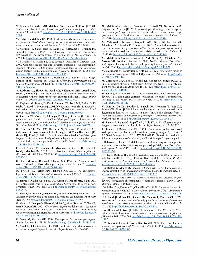
Downloaded from www.asmscience.org by
IP: 130.194.145.102
On: Thu, 20 Jun 2019 00:48:10
74. Brynestad S, Sarker MR, McClane BA, Granum PE, Rood JI. 2001.Enterotoxin plasmid from Clostridium perfringens is conjugative. InfectImmun 69:3483–3487 http://dx.doi.org/10.1128/IAI.69.5.3483-3487.2001.75. Collie RE, McClane BA. 1998. Evidence that the enterotoxin gene canbe episomal in Clostridium perfringens isolates associated with non-food-borne human gastrointestinal diseases. J Clin Microbiol 36:30–36.76. Cornillot E, Saint-Joanis B, Daube G, Katayama S, Granum PE,Canard B, Cole ST. 1995. The enterotoxin gene (cpe) of Clostridiumperfringens can be chromosomal or plasmid-borne. Mol Microbiol 15:639–647 http://dx.doi.org/10.1111/j.1365-2958.1995.tb02373.x.77. Miyamoto K, Fisher DJ, Li J, Sayeed S, Akimoto S, McClane BA.2006. Complete sequencing and diversity analysis of the enterotoxin-encoding plasmids in Clostridium perfringens type A non-food-bornehuman gastrointestinal disease isolates. J Bacteriol 188:1585–1598 http://dx.doi.org/10.1128/JB.188.4.1585-1598.2006.78. Miyamoto K, Chakrabarti G, Morino Y, McClane BA. 2002. Orga-nization of the plasmid cpe Locus in Clostridium perfringens type Aisolates. Infect Immun 70:4261–4272 http://dx.doi.org/10.1128/IAI.70.8.4261-4272.2002.79. Keyburn AL, Sheedy SA, Ford ME, Williamson MM, Awad MM,Rood JI, Moore RJ. 2006. Alpha-toxin of Clostridium perfringens is notan essential virulence factor in necrotic enteritis in chickens. Infect Immun74:6496–6500 http://dx.doi.org/10.1128/IAI.00806-06.80. Keyburn AL, Boyce JD, Vaz P, Bannam TL, Ford ME, Parker D, DiRubbo A, Rood JI, Moore RJ. 2008. NetB, a new toxin that is associatedwith avian necrotic enteritis caused by Clostridium perfringens. PLoSPathog 4:e26 http://dx.doi.org/10.1371/journal.ppat.0040026.81. Parreira VR, Costa M, Eikmeyer F, Blom J, Prescott JF. 2012. Se-quence of two plasmids from Clostridium perfringens chicken necroticenteritis isolates and comparison withC. perfringens conjugative plasmids.PLoS One 7:e49753 http://dx.doi.org/10.1371/journal.pone.0049753.82. Bannam TL, Yan XX, Harrison PF, Seemann T, Keyburn AL,Stubenrauch C, Weeramantri LH, Cheung JK, McClane BA, Boyce JD,Moore RJ, Rood JI. 2011. Necrotic enteritis-derived Clostridium per-fringens strain with three closely related independently conjugative toxinand antibiotic resistance plasmids. MBio 2:e00190–e11 http://dx.doi.org/10.1128/mBio.00190-11.83. Li J, Adams V, Bannam TL, Miyamoto K, Garcia JP, Uzal FA,Rood JI, McClane BA. 2013. Toxin plasmids of Clostridium perfringens.Microbiol Mol Biol Rev 77:208–233 http://dx.doi.org/10.1128/MMBR.00062-12.84. Gibert M, Jolivet-Reynaud C, Popoff MR. 1997. Beta2 toxin, a noveltoxin produced by Clostridium perfringens. Gene 203:65–73 http://dx.doi.org/10.1016/S0378-1119(97)00493-9.85. Tweten RK, Parker MW, Johnson AE. 2001. The cholesterol-dependent cytolysins. Curr Top Microbiol Immunol 257:15–33 http://dx.doi.org/10.1007/978-3-642-56508-3_2.86. Huyet J, Naylor CE, Savva CG, Gibert M, Popoff MR, Basak AK.2013. Structural insights into Clostridium perfringens delta toxin poreformation. PLoS One 8:e66673 http://dx.doi.org/10.1371/journal.pone.0066673.87. Seike S, Miyamoto K, Kobayashi K, TakeharaM, NagahamaM. 2016.Clostridium perfringens delta-toxin induces rapid cell necrosis. PLoS One11:e0147957 http://dx.doi.org/10.1371/journal.pone.0147957.88. ManichM, Knapp O, Gibert M, Maier E, Jolivet-Reynaud C, Geny B,Benz R, Popoff MR. 2008.Clostridium perfringens delta toxin is sequencerelated to beta toxin, NetB, and Staphylococcus pore-forming toxins,but shows functional differences. PLoS One 3:e3764 http://dx.doi.org/10.1371/journal.pone.0003764.89. Sterne M, Warrack GH. 1964. The types of Clostridium perfringens.J Pathol Bacteriol 88:279–283 http://dx.doi.org/10.1002/path.1700880135.90. Alouf JE, Jolivet-Reynaud C. 1981. Purification and characterizationof Clostridium perfringens delta-toxin. Infect Immun 31:536–546.
91. Mehdizadeh Gohari I, Parreira VR, Nowell VJ, Nicholson VM,Oliphant K, Prescott JF. 2015. A novel pore-forming toxin in type AClostridium perfringens is associated with both fatal canine hemorrhagicgastroenteritis and fatal foal necrotizing enterocolitis. PLoS One 10:e0122684 http://dx.doi.org/10.1371/journal.pone.0122684.92. Mehdizadeh Gohari I, Kropinski AM, Weese SJ, Parreira VR,Whitehead AE, Boerlin P, Prescott JF. 2016. Plasmid characterizationand chromosome analysis of two netF+ Clostridium perfringens isolatesassociated with foal and canine necrotizing enteritis. PLoS One 11:e0148344 http://dx.doi.org/10.1371/journal.pone.0148344.93. Mehdizadeh Gohari I, Kropinski AM, Weese SJ, Whitehead AE,Parreira VR, Boerlin P, Prescott JF. 2017. NetF-producing Clostridiumperfringens: clonality and plasmid pathogenicity loci analysis. Infect GenetEvol 49:32–38 http://dx.doi.org/10.1016/j.meegid.2016.12.028.94. Nagahama M, Kobayashi K, Oda M. 2012. Glycosylating toxin ofClostridium perfringens. INTECH Open Access Publisher. http://dx.doi.org/10.5772/48112.95. Coursodon CF, Glock RD, Moore KL, Cooper KK, Songer JG. 2012.TpeL-producing strains of Clostridium perfringens type A are highly vir-ulent for broiler chicks. Anaerobe 18:117–121 http://dx.doi.org/10.1016/j.anaerobe.2011.10.001.96. Chen J, McClane BA. 2015. Characterization of Clostridium per-fringens TpeL toxin gene carriage, production, cytotoxic contributions,and trypsin sensitivity. Infect Immun 83:2369–2381 http://dx.doi.org/10.1128/IAI.03136-14.97. Han X, Du XD, Southey L, Bulach DM, Seemann T, Yan XX,Bannam TL, Rood JI. 2015. Functional analysis of a bacitracin resistancedeterminant located on ICECp1, a novel Tn916-like element from aconjugative plasmid in Clostridium perfringens. Antimicrob Agents Che-mother 59:6855–6865 http://dx.doi.org/10.1128/AAC.01643-15.98. Dupuy B, Daube G, Popoff MR, Cole ST. 1997. Clostridium per-fringens urease genes are plasmid borne. Infect Immun 65:2313–2320.99. Ionesco H, Bouanchaud DH. 1973. [Bacteriocin production linkedto the presence of a plasmid, in Clostridium perfringens, type A].CRAcadSci Hebd Seances Acad Sci D 276:2855–2857. Bacteriocin productionlinked to the presence of a plasmid, in Clostridium perfringens, type A.100. Garnier T, Cole ST. 1988. Complete nucleotide sequence and geneticorganization of the bacteriocinogenic plasmid, pIP404, from Clostridiumperfringens. Plasmid 19:134–150 http://dx.doi.org/10.1016/0147-619X(88)90052-2.101. Lyras D, Rood JI. 2006. Clostridial genetics, p 682–687. In FischettiVA, Novick RP, Ferretti JJ, Portnoy DA, Rood JI (ed), Gram-PositivePathogens, 2nd ed. American Society forMicrobiology,Washington, D.C.http://dx.doi.org/10.1128/9781555816513.ch55102. Brefort G, Magot M, Ionesco H, Sebald M. 1977. Characterizationand transferability of Clostridium perfringens plasmids. Plasmid 1:52–66http://dx.doi.org/10.1016/0147-619X(77)90008-7.103. Magot M. 1984. Physical characterization of the Clostridium per-fringens tetracycline-chloramphenicol resistance plasmid pIP401. AnnMicrobiol (Paris) 135B:269–282.104. Mihelc VA, Duncan CL, Chambliss GH. 1978. Characterization of abacteriocinogenic plasmid in Clostridium perfringens CW55. AntimicrobAgents Chemother 14:771–779 http://dx.doi.org/10.1128/AAC.14.5.771.105. Rood JI, Maher EA, Somers EB, Campos E, Duncan CL. 1978.Isolation and characterization of multiply antibiotic-resistant Clostridumperfringens strains from porcine feces. Antimicrob Agents Chemother 13:871–880 http://dx.doi.org/10.1128/AAC.13.5.871.106. Abraham LJ, Rood JI. 1987. Identification of Tn4451 and Tn4452,chloramphenicol resistance transposons from Clostridium perfringens.J Bacteriol 169:1579–1584 http://dx.doi.org/10.1128/jb.169.4.1579-1584.1987.107. Adams V, Lyras D, Farrow KA, Rood JI. 2002. The clostridial mo-bilisable transposons. Cell Mol Life Sci 59:2033–2043 http://dx.doi.org/10.1007/s000180200003.
24 ASMscience.org/MicrobiolSpectrum
Revitt-Mills et al.
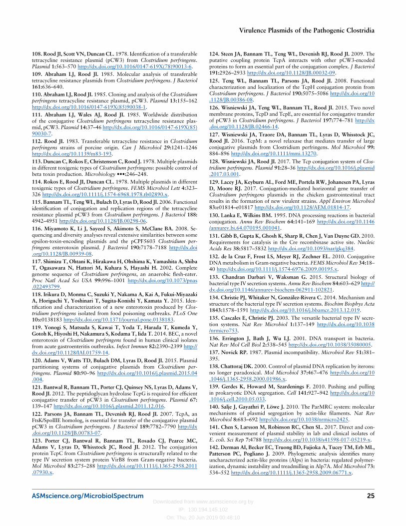
Downloaded from www.asmscience.org by
IP: 130.194.145.102
On: Thu, 20 Jun 2019 00:48:10
108. Rood JI, Scott VN, Duncan CL. 1978. Identification of a transferabletetracycline resistance plasmid (pCW3) from Clostridium perfringens.Plasmid 1:563–570 http://dx.doi.org/10.1016/0147-619X(78)90013-6.109. Abraham LJ, Rood JI. 1985. Molecular analysis of transferabletetracycline resistance plasmids from Clostridium perfringens. J Bacteriol161:636–640.110. Abraham LJ, Rood JI. 1985. Cloning and analysis of the Clostridiumperfringens tetracycline resistance plasmid, pCW3. Plasmid 13:155–162http://dx.doi.org/10.1016/0147-619X(85)90038-1.111. Abraham LJ, Wales AJ, Rood JI. 1985. Worldwide distributionof the conjugative Clostridium perfringens tetracycline resistance plas-mid, pCW3. Plasmid 14:37–46 http://dx.doi.org/10.1016/0147-619X(85)90030-7.112. Rood JI. 1983. Transferable tetracycline resistance in Clostridiumperfringens strains of porcine origin. Can J Microbiol 29:1241–1246http://dx.doi.org/10.1139/m83-193.113. Duncan C, Rokos E, Christenson C, Rood J. 1978.Multiple plasmidsin different toxigenic types of Clostridium perfringens: possible control ofbeta toxin production. Microbiology •••:246–248.114. Rokos E, Rood JI, Duncan CL. 1978. Multiple plasmids in differenttoxigenic types of Clostridium perfringens. FEMS Microbiol Lett 4:323–326 http://dx.doi.org/10.1111/j.1574-6968.1978.tb02890.x.115. BannamTL, TengWL, Bulach D, Lyras D, Rood JI. 2006. Functionalidentification of conjugation and replication regions of the tetracyclineresistance plasmid pCW3 from Clostridium perfringens. J Bacteriol 188:4942–4951 http://dx.doi.org/10.1128/JB.00298-06.116. Miyamoto K, Li J, Sayeed S, Akimoto S, McClane BA. 2008. Se-quencing and diversity analyses reveal extensive similarities between someepsilon-toxin-encoding plasmids and the pCPF5603 Clostridium per-fringens enterotoxin plasmid. J Bacteriol 190:7178–7188 http://dx.doi.org/10.1128/JB.00939-08.117. Shimizu T, Ohtani K, Hirakawa H, Ohshima K, Yamashita A, ShibaT, Ogasawara N, Hattori M, Kuhara S, Hayashi H. 2002. Completegenome sequence of Clostridium perfringens, an anaerobic flesh-eater.Proc Natl Acad Sci USA 99:996–1001 http://dx.doi.org/10.1073/pnas.022493799.118. Irikura D, Monma C, Suzuki Y, Nakama A, Kai A, Fukui-MiyazakiA, Horiguchi Y, Yoshinari T, Sugita-Konishi Y, Kamata Y. 2015. Iden-tification and characterization of a new enterotoxin produced by Clos-tridium perfringens isolated from food poisoning outbreaks. PLoS One10:e0138183 http://dx.doi.org/10.1371/journal.pone.0138183.
119. Yonogi S, Matsuda S, Kawai T, Yoda T, Harada T, Kumeda Y,Gotoh K, Hiyoshi H, Nakamura S, Kodama T, Iida T. 2014. BEC, a novelenterotoxin of Clostridium perfringens found in human clinical isolatesfrom acute gastroenteritis outbreaks. Infect Immun 82:2390–2399 http://dx.doi.org/10.1128/IAI.01759-14.
120. Adams V, Watts TD, Bulach DM, Lyras D, Rood JI. 2015. Plasmidpartitioning systems of conjugative plasmids from Clostridium per-fringens. Plasmid 80:90–96 http://dx.doi.org/10.1016/j.plasmid.2015.04.004.
121. Bantwal R, Bannam TL, Porter CJ, Quinsey NS, Lyras D, Adams V,Rood JI. 2012. The peptidoglycan hydrolase TcpG is required for efficientconjugative transfer of pCW3 in Clostridium perfringens. Plasmid 67:139–147 http://dx.doi.org/10.1016/j.plasmid.2011.12.016.
122. Parsons JA, Bannam TL, Devenish RJ, Rood JI. 2007. TcpA, anFtsK/SpoIIIE homolog, is essential for transfer of the conjugative plasmidpCW3 in Clostridium perfringens. J Bacteriol 189:7782–7790 http://dx.doi.org/10.1128/JB.00783-07.
123. Porter CJ, Bantwal R, Bannam TL, Rosado CJ, Pearce MC,Adams V, Lyras D, Whisstock JC, Rood JI. 2012. The conjugationprotein TcpC from Clostridium perfringens is structurally related to thetype IV secretion system protein VirB8 from Gram-negative bacteria.Mol Microbiol 83:275–288 http://dx.doi.org/10.1111/j.1365-2958.2011.07930.x.
124. Steen JA, Bannam TL, Teng WL, Devenish RJ, Rood JI. 2009. Theputative coupling protein TcpA interacts with other pCW3-encodedproteins to form an essential part of the conjugation complex. J Bacteriol191:2926–2933 http://dx.doi.org/10.1128/JB.00032-09.
125. Teng WL, Bannam TL, Parsons JA, Rood JI. 2008. Functionalcharacterization and localization of the TcpH conjugation protein fromClostridium perfringens. J Bacteriol 190:5075–5086 http://dx.doi.org/10.1128/JB.00386-08.
126. Wisniewski JA, Teng WL, Bannam TL, Rood JI. 2015. Two novelmembrane proteins, TcpD and TcpE, are essential for conjugative transferof pCW3 in Clostridium perfringens. J Bacteriol 197:774–781 http://dx.doi.org/10.1128/JB.02466-14.
127. Wisniewski JA, Traore DA, Bannam TL, Lyras D, Whisstock JC,Rood JI. 2016. TcpM: a novel relaxase that mediates transfer of largeconjugative plasmids from Clostridium perfringens. Mol Microbiol 99:884–896 http://dx.doi.org/10.1111/mmi.13270.
128. Wisniewski JA, Rood JI. 2017. The Tcp conjugation system of Clos-tridium perfringens. Plasmid 91:28–36 http://dx.doi.org/10.1016/j.plasmid.2017.03.001.
129. Lacey JA, Keyburn AL, Ford ME, Portela RW, Johanesen PA, LyrasD, Moore RJ. 2017. Conjugation-mediated horizontal gene transfer ofClostridium perfringens plasmids in the chicken gastrointestinal tractresults in the formation of new virulent strains. Appl Environ Microbiol83:e01814–e01817 http://dx.doi.org/10.1128/AEM.01814-17.
130. Lanka E, Wilkins BM. 1995. DNA processing reactions in bacterialconjugation. Annu Rev Biochem 64:141–169 http://dx.doi.org/10.1146/annurev.bi.64.070195.001041.
131. Gibb B, Gupta K, Ghosh K, Sharp R, Chen J, Van Duyne GD. 2010.Requirements for catalysis in the Cre recombinase active site. NucleicAcids Res 38:5817–5832 http://dx.doi.org/10.1093/nar/gkq384.
132. de la Cruz F, Frost LS, Meyer RJ, Zechner EL. 2010. ConjugativeDNAmetabolism in Gram-negative bacteria. FEMSMicrobiol Rev 34:18–40 http://dx.doi.org/10.1111/j.1574-6976.2009.00195.x.
133. Chandran Darbari V, Waksman G. 2015. Structural biology ofbacterial type IV secretion systems.Annu Rev Biochem 84:603–629 http://dx.doi.org/10.1146/annurev-biochem-062911-102821.
134. Christie PJ, Whitaker N, González-Rivera C. 2014. Mechanism andstructure of the bacterial type IV secretion systems. Biochim Biophys Acta1843:1578–1591 http://dx.doi.org/10.1016/j.bbamcr.2013.12.019.
135. Cascales E, Christie PJ. 2003. The versatile bacterial type IV secre-tion systems. Nat Rev Microbiol 1:137–149 http://dx.doi.org/10.1038/nrmicro753.
136. Errington J, Bath J, Wu LJ. 2001. DNA transport in bacteria.Nat Rev Mol Cell Biol 2:538–545 http://dx.doi.org/10.1038/35080005.137. Novick RP. 1987. Plasmid incompatibility. Microbiol Rev 51:381–395.138. Chattoraj DK. 2000. Control of plasmid DNA replication by iterons:no longer paradoxical. Mol Microbiol 37:467–476 http://dx.doi.org/10.1046/j.1365-2958.2000.01986.x.139. Gerdes K, Howard M, Szardenings F. 2010. Pushing and pullingin prokaryotic DNA segregation. Cell 141:927–942 http://dx.doi.org/10.1016/j.cell.2010.05.033.140. Salje J, Gayathri P, Löwe J. 2010. The ParMRC system: molecularmechanisms of plasmid segregation by actin-like filaments. Nat RevMicrobiol 8:683–692 http://dx.doi.org/10.1038/nrmicro2425.141. Chen S, Larsson M, Robinson RC, Chen SL. 2017. Direct and con-venient measurement of plasmid stability in lab and clinical isolates ofE. coli. Sci Rep 7:4788 http://dx.doi.org/10.1038/s41598-017-05219-x.142. Derman AI, Becker EC, Truong BD, Fujioka A, Tucey TM, ErbML,Patterson PC, Pogliano J. 2009. Phylogenetic analysis identifies manyuncharacterized actin-like proteins (Alps) in bacteria: regulated polymer-ization, dynamic instability and treadmilling in Alp7A.Mol Microbiol 73:534–552 http://dx.doi.org/10.1111/j.1365-2958.2009.06771.x.
ASMscience.org/MicrobiolSpectrum 25
Virulence Plasmids of the Pathogenic Clostridia
Downloaded from www.asmscience.org by
IP: 130.194.145.102
On: Thu, 20 Jun 2019 00:48:10
143.Watts TD, Johanesen PA, Lyras D, Rood JI, Adams V. 2017. Evidencethat compatibility of closely related replicons in Clostridium perfringensdepends on linkage to parMRC-like partitioning systems of different sub-families. Plasmid 91:68–75 http://dx.doi.org/10.1016/j.plasmid.2017.03.008.
144. Miyamoto K, Seike S, Takagishi T, Okui K, Oda M, TakeharaM, Nagahama M. 2015. Identification of the replication region inpBCNF5603, a bacteriocin-encoding plasmid, in the enterotoxigenicClostridium perfringens strain F5603. BMC Microbiol 15:118 http://dx.doi.org/10.1186/s12866-015-0443-3.
145. Shimizu T, Ohshima S, Ohtani K, Shimizu T, Hayashi H. 2001.Genomic map of Clostridium perfringens strain 13. Microbiol Immunol45:179–189 http://dx.doi.org/10.1111/j.1348-0421.2001.tb01278.x.
146. Wade B, Keyburn AL, Seemann T, Rood JI, Moore RJ. 2015. Bind-ing of Clostridium perfringens to collagen correlates with the ability tocause necrotic enteritis in chickens. Vet Microbiol 180:299–303 http://dx.doi.org/10.1016/j.vetmic.2015.09.019.
147. Rhem MN, Lech EM, Patti JM, McDevitt D, Höök M, JonesDB, Wilhelmus KR. 2000. The collagen-binding adhesin is a virulencefactor in Staphylococcus aureus keratitis. Infect Immun 68:3776–3779http://dx.doi.org/10.1128/IAI.68.6.3776-3779.2000.
148. Patti JM, Bremell T, Krajewska-Pietrasik D, Abdelnour A, TarkowskiA, Rydén C, Höök M. 1994. The Staphylococcus aureus collagen adhesinis a virulence determinant in experimental septic arthritis. Infect Immun62:152–161.
149. Xu Y, Rivas JM, Brown EL, Liang X, Höök M. 2004. Virulencepotential of the staphylococcal adhesin CNA in experimental arthritis isdetermined by its affinity for collagen. J Infect Dis 189:2323–2333 http://dx.doi.org/10.1086/420851.
150. Wade B, Keyburn AL, Haring V, Ford M, Rood JI, Moore RJ. 2016.The adherent abilities of Clostridium perfringens strains are critical for thepathogenesis of avian necrotic enteritis. Vet Microbiol 197:53–61 http://dx.doi.org/10.1016/j.vetmic.2016.10.028.
151. Kawahara K, Yonogi S, Munetomo R, Oki H, Yoshida T, KumedaY, Matsuda S, Kodama T, Ohkubo T, Iida T, Nakamura S. 2016. Crystalstructure of the ADP-ribosylating component of BEC, the binary entero-toxin of Clostridium perfringens. Biochem Biophys Res Commun 480:261–267 http://dx.doi.org/10.1016/j.bbrc.2016.10.042.152. Toniti W, Yoshida T, Tsurumura T, Irikura D, Monma C, KamataY, Tsuge H. 2017. Crystal structure and structure-based mutagene-sis of actin-specific ADP-ribosylating toxin CPILE-a as novel entero-toxin. PLoS One 12:e0171278 http://dx.doi.org/10.1371/journal.pone.0171278.153. Marshall KM, Bradshaw M, Johnson EA. 2010. Conjugative botu-linum neurotoxin-encoding plasmids in Clostridium botulinum. PLoSOne 5:e11087 http://dx.doi.org/10.1371/journal.pone.0011087.154. Riley MA, Wertz JE. 2002. Bacteriocins: evolution, ecology, andapplication. Annu Rev Microbiol 56:117–137 http://dx.doi.org/10.1146/annurev.micro.56.012302.161024.155. Mahony DE, Li A. 1978. Comparative study of ten bacteriocinsof Clostridium perfringens. Antimicrob Agents Chemother 14:886–892http://dx.doi.org/10.1128/AAC.14.6.886.156. Barbara AJ, Trinh HT, Glock RD, Glenn Songer J. 2008. Necroticenteritis-producing strains of Clostridium perfringens displace non-necroticenteritis strains from the gut of chicks. Vet Microbiol 126:377–382 http://dx.doi.org/10.1016/j.vetmic.2007.07.019.157. Tagg JR, Dajani AS, Wannamaker LW. 1976. Bacteriocins of gram-positive bacteria. Bacteriol Rev 40:722–756.158. Surana S, Tosolini AP, Meyer IFG, Fellows AD, Novoselov SS,Schiavo G. 2018. The travel diaries of tetanus and botulinum neurotoxins.Toxicon 147:58–67 http://dx.doi.org/10.1016/j.toxicon.2017.10.008.159. Brüggemann H, Bäumer S, Fricke WF, Wiezer A, Liesegang H,Decker I, Herzberg C, Martinez-Arias R, Merkl R, Henne A, GottschalkG. 2003. The genome sequence of Clostridium tetani, the causative agent
of tetanus disease. Proc Natl Acad Sci USA 100:1316–1321 http://dx.doi.org/10.1073/pnas.0335853100.
160. Rossetto O, ScorzetoM,Megighian A,Montecucco C. 2013. Tetanusneurotoxin. Toxicon 66:59–63 http://dx.doi.org/10.1016/j.toxicon.2012.12.027.
161. Connan C, Denève C, Mazuet C, Popoff MR. 2013. Regulation oftoxin synthesis in Clostridium botulinum and Clostridium tetani. Toxicon75:90–100 http://dx.doi.org/10.1016/j.toxicon.2013.06.001.
162. Raffestin S, Dupuy B, Marvaud JC, Popoff MR. 2005. BotR/A andTetR are alternative RNA polymerase sigma factors controlling the ex-pression of the neurotoxin and associated protein genes in Clostridiumbotulinum type A and Clostridium tetani. Mol Microbiol 55:235–249http://dx.doi.org/10.1111/j.1365-2958.2004.04377.x.
163. Carter GP, Larcombe S, Li L, Jayawardena D, Awad MM, SongerJG, Lyras D. 2014. Expression of the large clostridial toxins is controlledby conserved regulatory mechanisms. Int J Med Microbiol 304:1147–1159 http://dx.doi.org/10.1016/j.ijmm.2014.08.008.
164. Popp D, Narita A, Lee LJ, Ghoshdastider U, Xue B, Srinivasan R,Balasubramanian MK, Tanaka T, Robinson RC. 2012. Novel actin-likefilament structure from Clostridium tetani. J Biol Chem 287:21121–21129 http://dx.doi.org/10.1074/jbc.M112.341016.
165. Jiang S, Ghoshdastider U, Narita A, Popp D, Robinson RC.2016. Structural complexity of filaments formed from the actin and tu-bulin folds. Commun Integr Biol 9:e1242538 http://dx.doi.org/10.1080/19420889.2016.1242538.
166. Licona-Cassani C, Steen JA, Zaragoza NE, Moonen G, Moutafis G,Hodson MP, Power J, Nielsen LK, Marcellin E. 2016. Tetanus toxinproduction is triggered by the transition from amino acid consumptionto peptides. Anaerobe 41:113–124 http://dx.doi.org/10.1016/j.anaerobe.2016.07.006.167. Fournier PE, Levy PY, Million M, Croce O, Blanc-Tailleur C,Brouqui P, Raoult D. 2014. Genome of a chronic osteitis-causing Clos-tridium tetani. New Microbes New Infect 2:25–26 http://dx.doi.org/10.1002/2052-2975.27.168. Brüggemann H, Brzuszkiewicz E, Chapeton-Montes D, PlourdeL, Speck D, Popoff MR. 2015. Genomics of Clostridium tetani.Res Microbiol 166:326–331 http://dx.doi.org/10.1016/j.resmic.2015.01.002.169. Cohen JE,Wang R, Shen RF,WuWW, Keller JE. 2017. Comparativepathogenomics of Clostridium tetani. PLoS One 12:e0182909 http://dx.doi.org/10.1371/journal.pone.0182909.170. Rouli L, MBengue M, Robert C, Ndiaye M, La Scola B, Raoult D.2014. Genomic analysis of three African strains of Bacillus anthracisdemonstrates that they are part of the clonal expansion of an exclusivelypathogenic bacterium.NewMicrobes New Infect 2:161–169 http://dx.doi.org/10.1002/nmi2.62.171. Collins MD, East AK. 1998. Phylogeny and taxonomy of the food-borne pathogen Clostridium botulinum and its neurotoxins. J ApplMicro-biol 84:5–17 http://dx.doi.org/10.1046/j.1365-2672.1997.00313.x.172. Hill KK, Xie G, Foley BT, Smith TJ. 2015. Genetic diversity withinthe botulinum neurotoxin-producing bacteria and their neurotoxins.Toxicon 107(Pt A):2–8 http://dx.doi.org/10.1016/j.toxicon.2015.09.011.173. Hill KK, Xie G, Foley BT, Smith TJ, Munk AC, Bruce D, Smith LA,Brettin TS, Detter JC. 2009. Recombination and insertion events involv-ing the botulinum neurotoxin complex genes in Clostridium botulinumtypes A, B, E and F and Clostridium butyricum type E strains. BMC Biol7:66 http://dx.doi.org/10.1186/1741-7007-7-66.174. Stringer SC, Carter AT, Webb MD, Wachnicka E, CrossmanLC, Sebaihia M, Peck MW. 2013. Genomic and physiological variabilitywithin Group II (non-proteolytic) Clostridium botulinum. BMC Geno-mics 14:333 http://dx.doi.org/10.1186/1471-2164-14-333.175. Williamson CH, Sahl JW, Smith TJ, Xie G, Foley BT, Smith LA,Fernández RA, LindströmM, Korkeala H, Keim P, Foster J, Hill K. 2016.Comparative genomic analyses reveal broad diversity in botulinum-toxin-
26 ASMscience.org/MicrobiolSpectrum
Revitt-Mills et al.

Downloaded from www.asmscience.org by
IP: 130.194.145.102
On: Thu, 20 Jun 2019 00:48:10
producing Clostridia. BMC Genomics 17:180 http://dx.doi.org/10.1186/s12864-016-2502-z.
176. Schiavo G, Benfenati F, Poulain B, Rossetto O, Polverino de LauretoP, DasGupta BR, Montecucco C. 1992. Tetanus and botulinum-B neu-rotoxins block neurotransmitter release by proteolytic cleavage of syn-aptobrevin. Nature 359:832–835 http://dx.doi.org/10.1038/359832a0.
177. Hauser D, Gibert M, Marvaud JC, Eklund MW, Popoff MR. 1995.Botulinal neurotoxin C1 complex genes, clostridial neurotoxin homol-ogy and genetic transfer in Clostridium botulinum. Toxicon 33:515–526http://dx.doi.org/10.1016/0041-0101(94)00190-J.
178. Raffestin S, Marvaud JC, Cerrato R, Dupuy B, Popoff MR. 2004.Organization and regulation of the neurotoxin genes in Clostridium bot-ulinum and Clostridium tetani. Anaerobe 10:93–100 http://dx.doi.org/10.1016/j.anaerobe.2004.01.001.
179. Peck MW, Smith TJ, Anniballi F, Austin JW, Bano L, Bradshaw M,Cuervo P, Cheng LW, Derman Y, Dorner BG, Fisher A, Hill KK, Kalb SR,Korkeala H, Lindström M, Lista F, Lúquez C, Mazuet C, Pirazzini M,Popoff MR, Rossetto O, Rummel A, Sesardic D, Singh BR, Stringer SC.2017. Historical perspectives and guidelines for botulinum neurotoxinsubtype nomenclature. Toxins (Basel) 9:E38 http://dx.doi.org/10.3390/toxins9010038.
180. Fujinaga Y, Popoff MR. 2018. Translocation and dissemination ofbotulinum neurotoxin from the intestinal tract. Toxicon 147:13–18 http://dx.doi.org/10.1016/j.toxicon.2017.10.020.
181. Zhang S, Lebreton F, Mansfield MJ, Miyashita SI, Zhang J,Schwartzman JA, Tao L, Masuyer G, Martínez-Carranza M, Stenmark P,Gilmore MS, Doxey AC, Dong M. 2018. Identification of a botulinumneurotoxin-like toxin in a commensal strain of Enterococcus faecium. CellHost Microbe 23:169–176.e6 http://dx.doi.org/10.1016/j.chom.2017.12.018.
182. Hill KK, Smith TJ. 2013. Genetic diversity within Clostridium bot-ulinum serotypes, botulinum neurotoxin gene clusters and toxin subtypes.Curr Top Microbiol Immunol 364:1–20.
183. Barash JR, Arnon SS. 2014. A novel strain of Clostridium botulinumthat produces type B and type H botulinum toxins. J Infect Dis 209:183–191 http://dx.doi.org/10.1093/infdis/jit449.184. Dover N, Barash JR, Hill KK, Xie G, Arnon SS. 2014. Molecularcharacterization of a novel botulinum neurotoxin type H gene. J Infect Dis209:192–202 http://dx.doi.org/10.1093/infdis/jit450.185. Zhang S, Masuyer G, Zhang J, Shen Y, Lundin D, Henriksson L,Miyashita SI, Martínez-Carranza M, Dong M, Stenmark P. 2017. Iden-tification and characterization of a novel botulinum neurotoxin. NatCommun 8:14130 http://dx.doi.org/10.1038/ncomms14130.186. Popoff MR. 2014. Botulinum neurotoxins: more and more diverseand fascinating toxic proteins. J Infect Dis 209:168–169 http://dx.doi.org/10.1093/infdis/jit505.187. Dupuy B, Raffestin S, Matamouros S, Mani N, Popoff MR,Sonenshein AL. 2006. Regulation of toxin and bacteriocin gene expres-sion in Clostridium by interchangeable RNA polymerase sigma factors.Mol Microbiol 60:1044–1057 http://dx.doi.org/10.1111/j.1365-2958.2006.05159.x.188.Marvaud JC, Eisel U, Binz T, Niemann H, Popoff MR. 1998. TetR isa positive regulator of the tetanus toxin gene in Clostridium tetani and ishomologous to botR. Infect Immun 66:5698–5702.189. Skarin H, Håfström T,Westerberg J, Segerman B. 2011.Clostridiumbotulinum group III: a group with dual identity shaped by plasmids,phages and mobile elements. BMC Genomics 12:185 http://dx.doi.org/10.1186/1471-2164-12-185.190. Dover N, Barash JR, Hill KK, Davenport KW, Teshima H, Xie G,Arnon SS. 2013. Clostridium botulinum strain Af84 contains three neu-rotoxin gene clusters: bont/A2, bont/F4 and bont/F5. PLoS One 8:e61205http://dx.doi.org/10.1371/journal.pone.0061205.
191. Smith TJ, Hill KK, Foley BT, Detter JC, Munk AC, Bruce DC,Doggett NA, Smith LA, Marks JD, Xie G, Brettin TS. 2007. Analysis of
the neurotoxin complex genes in Clostridium botulinum A1-A4 and B1strains: BoNT/A3, /Ba4 and /B1 clusters are located within plasmids. PLoSOne 2:e1271 http://dx.doi.org/10.1371/journal.pone.0001271.
192. Raphael BH, BradshawM, Kalb SR, Joseph LA, Lúquez C, Barr JR,Johnson EA, Maslanka SE. 2014. Clostridium botulinum strains pro-ducing BoNT/F4 or BoNT/F5. Appl Environ Microbiol 80:3250–3257http://dx.doi.org/10.1128/AEM.00284-14.
193. Franciosa G, Maugliani A, Scalfaro C, Aureli P. 2009. Evidence thatplasmid-borne botulinum neurotoxin type B genes are widespread amongClostridium botulinum serotype B strains. PLoS One 4:e4829 http://dx.doi.org/10.1371/journal.pone.0004829.
194. Hosomi K, Sakaguchi Y, Kohda T, Gotoh K,Motooka D, NakamuraS, Umeda K, Iida T, Kozaki S, Mukamoto M. 2014. Complete nucleotidesequence of a plasmid containing the botulinum neurotoxin gene inClostridium botulinum type B strain 111 isolated from an infant patientin Japan. Mol Genet Genomics 289:1267–1274 http://dx.doi.org/10.1007/s00438-014-0887-4.
195. Marshall KM, Bradshaw M, Pellett S, Johnson EA. 2007. Plasmidencoded neurotoxin genes in Clostridium botulinum serotype A subtypes.Biochem Biophys Res Commun 361:49–54 http://dx.doi.org/10.1016/j.bbrc.2007.06.166.
196. Umeda K, Seto Y, Kohda T,MukamotoM, Kozaki S. 2012. Stabilityof toxigenicity in proteolytic Clostridium botulinum type B upon serialpassage. Microbiol Immunol 56:338–341 http://dx.doi.org/10.1111/j.1348-0421.2012.00441.x.
197. WeigandMR, Pena-Gonzalez A, Shirey TB, Broeker RG, Ishaq MK,Konstantinidis KT, Raphael BH. 2015. Implications of genome-baseddiscrimination between Clostridium botulinum group I and Clostridiumsporogenes strains for bacterial taxonomy. Appl Environ Microbiol 81:5420–5429 http://dx.doi.org/10.1128/AEM.01159-15.
198. Carter AT, Austin JW, Weedmark KA, Corbett C, Peck MW. 2014.Three classes of plasmid (47-63 kb) carry the type B neurotoxin genecluster of group II Clostridium botulinum. Genome Biol Evol 6:2076–2087 http://dx.doi.org/10.1093/gbe/evu164.
199. Carter AT, Austin JW, Weedmark KA, Peck MW. 2016. Evolutionof chromosomal Clostridium botulinum type E neurotoxin gene clusters:evidence provided by their rare plasmid-borne counterparts.Genome BiolEvol 8:540–555 http://dx.doi.org/10.1093/gbe/evw017.
200. Carter AT, Stringer SC, Webb MD, Peck MW. 2013. The typeF6 neurotoxin gene cluster locus of group II clostridium botulinum hasevolved by successive disruption of two different ancestral precursors.Genome Biol Evol 5:1032–1037 http://dx.doi.org/10.1093/gbe/evt068.
201. Zhang Z, Hintsa H, Chen Y, Korkeala H, Lindström M. 2013.Plasmid-borne type E neurotoxin gene clusters in Clostridium botulinumstrains. Appl Environ Microbiol 79:3856–3859 http://dx.doi.org/10.1128/AEM.00080-13.
202. Skarin H, Segerman B. 2014. Plasmidome interchange betweenClostridium botulinum, Clostridium novyi and Clostridium haemolyti-cum converts strains of independent lineages into distinctly differentpathogens. PLoS One 9:e107777 http://dx.doi.org/10.1371/journal.pone.0107777.
203. Sakaguchi Y, Hayashi T, Kurokawa K, Nakayama K, OshimaK, Fujinaga Y, Ohnishi M, Ohtsubo E, Hattori M, Oguma K. 2005. Thegenome sequence of Clostridium botulinum type C neurotoxin-convertingphage and the molecular mechanisms of unstable lysogeny. Proc Natl AcadSci USA 102:17472–17477 http://dx.doi.org/10.1073/pnas.0505503102.
204. Sakaguchi Y, Hayashi T, Yamamoto Y, Nakayama K, Zhang K, MaS, Arimitsu H, Oguma K. 2009. Molecular analysis of an extrachromo-somal element containing the C2 toxin gene discovered in Clostridiumbotulinum type C. J Bacteriol 191:3282–3291 http://dx.doi.org/10.1128/JB.01797-08.
205. Sakaguchi Y, Suzuki T, Yamamoto Y, Nishikawa A, Oguma K.2015. Genomics of Clostridium botulinum group III strains. Res Micro-biol 166:318–325 http://dx.doi.org/10.1016/j.resmic.2014.07.016.
ASMscience.org/MicrobiolSpectrum 27
Virulence Plasmids of the Pathogenic Clostridia
Downloaded from www.asmscience.org by
IP: 130.194.145.102
On: Thu, 20 Jun 2019 00:48:10
206. Zhou Y, Sugiyama H, Nakano H, Johnson EA. 1995. The genes forthe Clostridium botulinum type G toxin complex are on a plasmid. InfectImmun 63:2087–2091.
207. Smith TJ, Hill KK, Xie G, Foley BT, Williamson CHD, FosterJT, Johnson SL, Chertkov O, Teshima H, Gibbons HS, Johnsky LA,Karavis MA, Smith LA. 2015. Genomic sequences of six botulinumneurotoxin-producing strains representing three clostridial species il-lustrate the mobility and diversity of botulinum neurotoxin genes.Infect Genet Evol 30:102–113 http://dx.doi.org/10.1016/j.meegid.2014.12.002.
208. Halpin JL, Hill K, Johnson SL, Bruce DC, Shirey TB, Dykes JK,Lúquez C. 2017. Finished whole-genome sequence of Clostridium argen-tinense producing botulinum neurotoxin type G. Genome Announc 5:e00380–e17.
209. Hauser D, Gibert M, Boquet P, Popoff MR. 1992. Plasmid locali-zation of a type E botulinal neurotoxin gene homologue in toxigenicClostridium butyricum strains, and absence of this gene in non-toxigenicC. butyricum strains. FEMS Microbiol Lett 78:251–255 http://dx.doi.org/10.1111/j.1574-6968.1992.tb05576.x.
210. Franciosa G, Scalfaro C, Di Bonito P, Vitale M, Aureli P. 2011.Identification of novel linear megaplasmids carrying a ß-lactamase gene inneurotoxigenic Clostridium butyricum type E strains. PLoSOne 6:e21706http://dx.doi.org/10.1371/journal.pone.0021706.
211. Wang X, Maegawa T, Karasawa T, Kozaki S, Tsukamoto K, GyobuY, Yamakawa K, Oguma K, Sakaguchi Y, Nakamura S. 2000. Geneticanalysis of type E botulinum toxin-producing Clostridium butyricumstrains. Appl Environ Microbiol 66:4992–4997 http://dx.doi.org/10.1128/AEM.66.11.4992-4997.2000.
212. Janezic S, Potocnik M, Zidaric V, RupnikM. 2016. Highly divergentClostridium difficile strains isolated from the environment. PLoS One 11:e0167101 http://dx.doi.org/10.1371/journal.pone.0167101.213. Amy J, Johanesen P, Lyras D. 2015. Extrachromosomal and inte-grated genetic elements in Clostridium difficile. Plasmid 80:97–110 http://dx.doi.org/10.1016/j.plasmid.2015.04.006.214. Awad MM, Johanesen PA, Carter GP, Rose E, Lyras D. 2014.Clostridium difficile virulence factors: insights into an anaerobic spore-forming pathogen. Gut Microbes 5:579–593 http://dx.doi.org/10.4161/19490976.2014.969632.215. Smits WK, Lyras D, Lacy DB, Wilcox MH, Kuijper EJ. 2016.Clostridium difficile infection. Nat Rev Dis Primers 2:16020 http://dx.doi.org/10.1038/nrdp.2016.20.
216. Hundsberger T, Braun V, Weidmann M, Leukel P, Sauerborn M,von Eichel-Streiber C. 1997. Transcription analysis of the genes tcdA-E ofthe pathogenicity locus of Clostridium difficile. Eur J Biochem 244:735–742 http://dx.doi.org/10.1111/j.1432-1033.1997.t01-1-00735.x.217. Perelle S, Gibert M, Bourlioux P, Corthier G, Popoff MR. 1997.Production of a complete binary toxin (actin-specific ADP-ribosyltrans-ferase) by Clostridium difficile CD196. Infect Immun 65:1402–1407.218. Carter GP, Lyras D, Allen DL, Mackin KE, Howarth PM, O’ConnorJR, Rood JI. 2007. Binary toxin production in Clostridium difficile isregulated by CdtR, a LytTR family response regulator. J Bacteriol 189:7290–7301 http://dx.doi.org/10.1128/JB.00731-07.219. BrouwerMS, Roberts AP, Hussain H,Williams RJ, Allan E,MullanyP. 2013. Horizontal gene transfer converts non-toxigenic Clostridiumdifficile strains into toxin producers. Nat Commun 4:2601 http://dx.doi.org/10.1038/ncomms3601.220. Dingle KE, Elliott B, Robinson E, Griffiths D, Eyre DW, Stoesser N,Vaughan A, Golubchik T, FawleyWN,WilcoxMH, Peto TE, Walker AS,Riley TV, Crook DW, Didelot X. 2014. Evolutionary history of theClostridium difficile pathogenicity locus. Genome Biol Evol 6:36–52http://dx.doi.org/10.1093/gbe/evt204.221. Riedel T, Wittmann J, Bunk B, Schober I, Spröer C, Gronow S,Overmann J. 2017. A Clostridioides difficile bacteriophage genome en-codes functional binary toxin-associated genes. J Biotechnol 250:23–28http://dx.doi.org/10.1016/j.jbiotec.2017.02.017.222. Sekulovic O, Meessen-Pinard M, Fortier LC. 2011. Prophage-stimulated toxin production in Clostridium difficile NAP1/027 lysogens.J Bacteriol 193:2726–2734 http://dx.doi.org/10.1128/JB.00787-10.223. Ramírez-Vargas G, Goh S, Rodríguez C. 2018. The novel phagesphiCD5763 and phiCD2955 represent two groups of big plasmidialsiphoviridae phages of Clostridium difficile. Front Microbiol 9:26 http://dx.doi.org/10.3389/fmicb.2018.00026.224. Alikhan NF, Petty NK, Ben Zakour NL, Beatson SA. 2011.BLAST Ring Image Generator (BRIG): simple prokaryote genome com-parisons. BMC Genomics 12:402 http://dx.doi.org/10.1186/1471-2164-12-402.225. Altschul SF, Gish W, Miller W, Myers EW, Lipman DJ. 1990. Basiclocal alignment search tool. J Mol Biol 215:403–410 http://dx.doi.org/10.1016/S0022-2836(05)80360-2.226. Sullivan MJ, Petty NK, Beatson SA. 2011. Easyfig: a genome com-parison visualizer. Bioinformatics 27:1009–1010 http://dx.doi.org/10.1093/bioinformatics/btr039.
28 ASMscience.org/MicrobiolSpectrum
Revitt-Mills et al.
Copyright © 2022 FDOKUMEN