Viruses, plasmids and other genetic elements of thermophilic ...
12
MICROBIOLOGY REVIEWS ELSEVIER FEMS MicrobiologyReviews 18 (1996) 225-236 Viruses, plasmids and other genetic elements of thermophilic and hyperthermophilic Archaea Wolfram Zillig a,*, David Prangishvilli a, Christa Schleper b, Marieke Elferink a, Ingelore Holz a, Sonja Albers a, Davorin Janekovic a, Dorothee G6tz c a Max Planck-lnstitutfi~r Biochemie, 82152 Martinsried, Germany b Marine Sciences Institute, UniuersiO' of California, Santa Barbara, CA 93106, USA c Unieersity of Waikato, Hamilton, New Zealand Abstract We review and update the work on genetic elements, e.g., viruses and plasmids (exluding IS elements and transposons) in the kingdom Crenarchaeota (Thermoproteales and Sulfolobales) and the orders Thermococcales and Thermoplasmales in the kingdom Euryarchaeota of the archael domain, including unpublished data from our laboratory. The viruses of Crenarchaeota represent four novel virus families. The Fuselloviridae represented by SSVI of S. shibatae and relatives in other Sulfolobus strains have the form of a tailed spindle. The envelope is highly hydrophobic. The DNA is double-stranded and circular. Members of this group have also been found in Methanococcus and Haloarcula. The Lipothrixviridae (e.g., T TV 1 to 3) have the form of flexible filaments. They have a core containing linear double-stranded DNA and DNA-binding proteins which is wrapped into a lipid membrane. The 'Bacilloviridae' (e.g., TTV4 and SIRV) are stiff rods lacking this membrane, but also featuring linear double-stranded DNA and DNA-binding proteins. Both virus types carry on both ends structures involved in the attachment to receptors. Both types are represented in Thermoproteus and Sulfolobus. The droplet-formed novel Sulfolobus virus SNDV represents the 'Guttaviridae' containing circular double-stranded DNA. Though head and tail viruses distantly resembling T phages or lambdoid phages were seen electronmicroscopically in solfataric water samples, no such virus has so far been isolated. SSV1 is temperate, TTV1 causes lysis after induction, the other viruses found so far exist in cartier states. The hosts of all but TTVI survive virus production. We discuss the implications of the nature of these viruses for understanding virus evolution. The plasmids found so far range in size from 4.5 kb to about 40 kb. Most of them occur in high copy number, probably due to the way of their detection. Most are cryptic, pNOB8 is conjugative, the widespread pDLI0 alleviates in an unknown way autotrophic growth of its host Desulfurolobus by sulfur reduction. The plasmid pTIK4 appears to encode a killer function, pNOB8 has been used as a vector for the transfer of the lac S (fl-galactosidase) gene into a mutant of S. solfataricus. Keywords: Archaea, hyperthermophilic;Archaea, thermophilic; Virus; Crenarchaeota; Sulfolobales; Thermococcales; Thermoplasmales: Plasmid Corresponding author 0168-6445/96/$32.00 © 1996 Federationof European MicrobiologicalSocieties. All rights reserved PII $0168-6445(96)00014-9 Downloaded from https://academic.oup.com/femsre/article/18/2-3/225/479297 by guest on 11 February 2022
-
Upload
khangminh22 -
Category
Documents
-
view
1 -
download
0
Transcript of Viruses, plasmids and other genetic elements of thermophilic ...

MICROBIOLOGY REVIEWS
ELSEVIER FEMS Microbiology Reviews 18 (1996) 225-236
Viruses, plasmids and other genetic elements of thermophilic and hyperthermophilic Archaea
Wolfram Zillig a,*, David Prangishvilli a, Christa Schleper b, Marieke Elferink a, Ingelore Holz a, Sonja Albers a, Davorin Janekovic a, Dorothee G6tz c
a Max Planck-lnstitutfi~r Biochemie, 82152 Martinsried, Germany b Marine Sciences Institute, UniuersiO' of California, Santa Barbara, CA 93106, USA
c Unieersity of Waikato, Hamilton, New Zealand
Abstract
We review and update the work on genetic elements, e.g., viruses and plasmids (exluding IS elements and transposons) in the kingdom Crenarchaeota (Thermoproteales and Sulfolobales) and the orders Thermococcales and Thermoplasmales in the kingdom Euryarchaeota of the archael domain, including unpublished data from our laboratory. The viruses of Crenarchaeota represent four novel virus families. The Fuselloviridae represented by SSVI of S. shibatae and relatives in other Sulfolobus strains have the form of a tailed spindle. The envelope is highly hydrophobic. The DNA is double-stranded and circular. Members of this group have also been found in Methanococcus and Haloarcula. The Lipothrixviridae (e.g., T TV 1 to 3) have the form of flexible filaments. They have a core containing linear double-stranded DNA and DNA-binding proteins which is wrapped into a lipid membrane. The 'Bacilloviridae' (e.g., TTV4 and SIRV) are stiff rods lacking this membrane, but also featuring linear double-stranded DNA and DNA-binding proteins. Both virus types carry on both ends structures involved in the attachment to receptors. Both types are represented in Thermoproteus and Sulfolobus. The droplet-formed novel Sulfolobus virus SNDV represents the 'Guttaviridae' containing circular double-stranded DNA. Though head and tail viruses distantly resembling T phages or lambdoid phages were seen electronmicroscopically in solfataric water samples, no such virus has so far been isolated. SSV1 is temperate, TTV1 causes lysis after induction, the other viruses found so far exist in cartier states. The hosts of all but TTVI survive virus production. We discuss the implications of the nature of these viruses for understanding virus evolution. The plasmids found so far range in size from 4.5 kb to about 40 kb. Most of them occur in high copy number, probably due to the way of their detection. Most are cryptic, pNOB8 is conjugative, the widespread pDLI0 alleviates in an unknown way autotrophic growth of its host Desulfurolobus by sulfur reduction. The plasmid pTIK4 appears to encode a killer function, pNOB8 has been used as a vector for the transfer of the lac S (fl-galactosidase) gene into a mutant of S. solfataricus.
Keywords: Archaea, hyperthermophilic; Archaea, thermophilic; Virus; Crenarchaeota; Sulfolobales; Thermococcales; Thermoplasmales: Plasmid
Corresponding author
0168-6445/96/$32.00 © 1996 Federation of European Microbiological Societies. All rights reserved PII $0168-6445(96)00014-9
Dow
nloaded from https://academ
ic.oup.com/fem
sre/article/18/2-3/225/479297 by guest on 11 February 2022

226 W. Zillig et aL / F E M S Microbiology Reviews 18 (1996) 225-236
Contents
1. Introduction . . . . . . . . . . . . . . . . . . . . . . . . . . . . . . . . . . . . . . . . . . . . . . . . . . . . . . . . . . . . . . . 226
2. Viruses of Crenarchaeota . . . . . . . . . . . . . . . . . . . . . . . . . . . . . . . . . . . . . . . . . . . . . . . . . . . . . . 227 2.1. Nature of the viruses . . . . . . . . . . . . . . . . . . . . . . . . . . . . . . . . . . . . . . . . . . . . . . . . . . . . . . 227 2.2. Virus host interactions . . . . . . . . . . . . . . . . . . . . . . . . . . . . . . . . . . . . . . . . . . . . . . . . . . . . . 229 2.3. Plaque tests . . . . . . . . . . . . . . . . . . . . . . . . . . . . . . . . . . . . . . . . . . . . . . . . . . . . . . . . . . . . 230 2.4. Receptors . . . . . . . . . . . . . . . . . . . . . . . . . . . . . . . . . . . . . . . . . . . . . . . . . . . . . . . . . . . . . 232 2.5. Assembly . . . . . . . . . . . . . . . . . . . . . . . . . . . . . . . . . . . . . . . . . . . . . . . . . . . . . . . . . . . . . 232
3. Origin and phylogeny of viruses . . . . . . . . . . . . . . . . . . . . . . . . . . . . . . . . . . . . . . . . . . . . . . . . . . . 232
4. Plasmids . . . . . . . . . . . . . . . . . . . . . . . . . . . . . . . . . . . . . . . . . . . . . . . . . . . . . . . . . . . . . . . . . 233
5. 'Sulfolobicins' . . . . . . . . . . . . . . . . . . . . . . . . . . . . . . . . . . . . . . . . . . . . . . . . . . . . . . . . . . . . . 234
6. Construction of transforming (cloning) vectors based on genetic elements . . . . . . . . . . . . . . . . . . . . . . . . . . 235
Acknowledgements . . . . . . . . . . . . . . . . . . . . . . . . . . . . . . . . . . . . . . . . . . . . . . . . . . . . . . . . . . . . 235
References . . . . . . . . . . . . . . . . . . . . . . . . . . . . . . . . . . . . . . . . . . . . . . . . . . . . . . . . . . . . . . . . . 235
1. Introduction
Genetic elements, e.g., viruses and plasmids may be considered 'fragments of life'. They constitute an important fraction of biodiversity which is not con- gruous to and appears to be largely independent of that of their hosts and they have certainly partici- pated in generating the variety of extant organisms via recombination processes. Studying emergence and scope of this diversity should help in understand- ing origin and evolution of life. Moreover, such elements supply small and thus comprehensible genomes which have served in studying mechanisms and controls of DNA replication and gene expression in Bacteria and Eucarya. Because of their capability to be maintained and multiplied in their hosts, they have been utilized for developing transformation vectors.
The third domain of life, Archaea, has been divided into two kingdoms, Crenarchaeota and Eur- yarchaeota [1]. A number of viruses and plasmids have been described for Euryarchaeota, especially Halobacteriales (the viruses are listed in [2], earlier reviews are [3] and [4]). Some of these, e.g., Halobacteriophage qbH [5] and a large plasmid of
Halobacterium halobium, pHH1 [6] have been stud- ied in molecular genetic detail. In the lowest branch of the Euryarchaeota, the Thermococcales, harbor- ing the hyperthermophilic genera Thermococcus and Pyrococcus, several plasmids including pGT5 [7] have been found recently, but no viruses are known so far. In the second lowest branch of this kingdom, the Thermoplasmales, viruses have not been de- scribed to date, but several plasmids were found recently. The Pyrococcus plasmid pGT5 has been utilized as basis for the construction of a shuttle vector for E. coli, Sulfolobus and Pyrococcus (Aagaard and Garrett, personal communication). In Crenarchaeota, four viruses of Thermoproteus tenax (Thermoproteales) TTV1 to 3, representing the novel virus family Lipothrixviridae, and TI'V4, represent- ing another, not yet validated virus family, the 'Bacilloviridae', have been isolated (see [3] and [4]). Only TTVI has been investigated in more detail, including sequencing of 85% of its genome (see [8]). In the other order of the Crenarchaeota, Sulfolob- ales, the Sulfolobus virus SSVI, again representing a novel virus family, the Fuselloviriodae, has been studied extensively, including complete sequencing [9], mapping of transcripts and promoters, and analy-
Dow
nloaded from https://academ
ic.oup.com/fem
sre/article/18/2-3/225/479297 by guest on 11 February 2022

W. Zill ig et al. / F E M S M i c r o b i o l o g y Re~'iews 18 (1996) 2 2 5 - 2 3 6 227
sis of lysogeny and of induction of virus production. Two novel Sulfolobus viruses, SIRV and DAFV have been found in isolates from Icelandic solfataras [10]. SIRV another representative of the 'Bacillo- L, iridae' has been partially sequenced and character- ized (D. Prangishvilli and D. G~Stz, unpublished work from this laboratory). Our knowledge of DAFV of Desulfurolobus ambivalens and of a novel Sul- .[blobus virus from New Zealand, SNDV (W. Zillig, D. G~Stz, D. Janekovic and D. Langer, unpublished work from this laboratory) is, however, superficial.
Some l0 plasmids, most of them cryptic, but one conjugative, another one encoding a killer function and a third involved in autotrophic growth by sulfur reduction, have been found in Sulfolobales, the latter in Desulfurolobus, the others in heterotrophic Sul- folobus strains from Iceland [10], Japan (Hokkaido [1 l]) and New Zealand (see below).
Here we present a short resum6 and an update of the state of the field, including unpublished material from our laboratory.
2. Viruses of Crenarchaeota
2. I. Nature of the t'iruses
The viruses of extremely halophilic and methanogenic Euryarchaeota are so far, with two exceptions (see below), of the same head and tail type as T phages and lambdoid phages of Bacteria (see [2] and Table 1). A striking example is the temperate halobacteriophage q)H, which resembles
E. coli phage P1 not only in structure and replication of its DNA but also in the type of the lysogeny conferred to its host by a circular prophage. In view of the incompatibility of the transcription systems of Bacteria and Archaea [12], of the entirely different lifestyles of the hosts - life at low ionic strength for E. coli but at extreme salinity for Halobacterium - and of the complexity of the viral genomes, stepwise surmounting of the host range barriers between these distant phyla during evolution appears highly im- probable such that we feel forced to assume this virus type existed already before the separation of the bacterial from the archaeal lineage, which is a very early event in the evolution of organisms [13,14].
In contrast, to the euryarchaeotal viruses, those of Crenarchaeota all represent previously unknown virus families. The spindle-like virus SSVI of Sul- folobus shibatae, solfataricus and islandicus repre- sents the Fuselloviridae which are characterized by containing about 16 kb circular double-stranded DNA, a DNA-binding protein and a lipid membrane made up of viral hydrophobic coat proteins and host lipids. A short tail is visible but has not been anal- ysed in detail. At least 7 viruses very similar in dimensions and shape and containing DNA of the same size which cross-hybridized with that of SSVI, but was different in restriction patterns have been found in novel Sulfolobus isolates from Icelandic solfataras (D. Prangishvilli and I. Holz, unpublished work from this laboratory). A virus-like particle of Methanococcus t~oltae, VLP J2 [ 15] and two viruses, Hisl and His2 of Haloarcula hispanica (C. Bath and M. Dyall Smith, personal communication) appear to
Table 1 Viruses of crenarcaeota
Designation Shape Size(length/width, rim) Type DNA size (kb) Sequence known Proteins Membrane Host Virus type
SSVI spindle 100/60 ccc 15.463 total >_ 3 + S. so i l Fuse l lor i r idae
TTV 1 flex. ill. 400/40 lin. ds 16 85% >_ 4 + T. tenax. LipothrixtJiridae
TTV2 flex. ill. 1200/20 lin. ds 16 - > 1 + T. tenax. Lipothrix t ' i r idae
TTV3 flex. ill. 2500/30 lin. ds 27 - > 1 + T. tenax. L ipothr ixc i r idae
DAFV flex. ill. 2200/27 lin. ds 56 - > 3 + D. arab. L ipothr ixc ir idae
TTV4 stiff rod 500/30 lin. ds 17 - > 1 - T. t enax "Bacillot, iridae"
SIRV stiffrod 950/26 lin. ds 33 15% >_ 2 - S. isl. "Baci l locir idae"
SNDV droplet 180/80 ccc 20 - > 16 ? S. spec. novel
Dow
nloaded from https://academ
ic.oup.com/fem
sre/article/18/2-3/225/479297 by guest on 11 February 2022

228 W. Zillig et aL/ FEMS Microbiology ReNews 18 (1996) 225-236
be of the same type, which thus occurs in both archaeal kingdoms but not outside the archaeal do- main.
A second novel family of viruses, the 'Bacillo- viridae'(provisional name), has so far only been found in Crenarchaeota, TTV4 in T. tenax (see [3], [4] and [16]) and SIRV (S. islandicus rod shaped virus) in S. islandicus [10]. These viruses are stiff rods formed by a double helix consisting of linear double-stranded DNA and DNA-binding protein(s), wound up to a tube terminally plugged and equipped with tail fibers for adsorption, in the case of SIRV to
pili of the host. They lack a membrane or hydropho- bic coat. Early after discovery our 'isolate' of SIRV turned out to be a mixture of variants which differed by restriction patterns of their DNAs but could not be distinguished by shape, size and SDS-PAGE pat- tern of viral proteins. Stable strains were obtained by cloning variants via single plaques. Though the nu- cleotide sequences of corresponding genes of differ- ent variants (DNA-binding protein and dUTPase) showed up to 10% difference, the protein sequences were almost identical (D. Prangishvilli and D. Gt~tz, unpublished work from this laboratory).
Fig. 1. Virus-like particles in PEG precipitates of supernatants of liquid samples from Icelandic solfataras, in some cases attached to cells of Thermoproteus sp. Negatively stained.
Dow
nloaded from https://academ
ic.oup.com/fem
sre/article/18/2-3/225/479297 by guest on 11 February 2022
w. Zillig et al./ FEMS Microbiology Reviews 18 (1996) 225-236 229
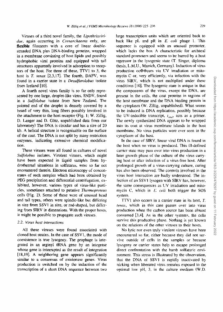
Viruses of a third novel family, the Lipothrixviri- dae, again occurring in Crenarchaeota only, are flexible filaments with a core of linear double- stranded DNA plus DNA-binding proteins, wrapped in a membrane consisting of host lipids and possibly hydrophobic viral proteins and equipped with tail structures apparently involved in adsorption to recep- tors of the host. For three of these, TTV1 to 3, the host is T. tenax [2,3,17]. The fourth, DAFV, was found in a carrier state in a Desulfurolobus isolate from Iceland [10].
A fourth novel virus family is so far only repre- sented by one large, droplet-like virus, SNDV, found in a Sulfolobus isolate from New Zealand. The pointed end of the droplet is densely covered by a beard of very thin, long fibers possibly involved in the attachment to the host receptor (Fig. 1; W. Zillig, D. Langer and D. Gtitz, unpublished data from our laboratory) The DNA is circular and has a size of 20 kb. A helical structure is recognizable on the surface of the coat. The DNA is not split by many restriction enzymes, indicating extensive chemical modifica- tion.
These viruses were all found in cultures of novel Sulfolobus isolates. Virulent viruses, which might have been expected in liquid samples from hy- drothermal situations in solfataras, were so far not encountered therein. Electron microscopy of concen- trates of such samples which had been obtained by PEG precipitation and differential centrifugation, ex- hibited, however, various types of virus-like parti- cles, sometimes attached to putative Thermoproteus cells (Fig. 2). Some of these were of unusual head and tail types, others were spindle-like but differing in size from SSV1 in size, or rod-shaped, but differ- ing from SIRV in dimensions. With the proper hosts, it might be possible to propagate such viruses.
2.2. Virus host interactions
All these viruses were found associated with cloned host strains. In the case of SSV1, the mode of coexistence is true lysogeny. The prophage is inte- grated in an arginyl tRNA gene by an integrase whose gene is interrupted as the result of integration [18,19]. A neighboring gene appears significantly similar to a consensus of exisionase genes. Virus production is switched on by the induction of the transcription of a short DNA sequence between two
large transcription units which are oriented back to back like pL and pR in E. coli phage 1. This sequence is equipped with an unusual promoter, which lacks the box A characteristic for archaeal standard promoters and seems to be barred by a host repressor in the lysogenic state (T. Singer, diploma thesis, L.M.U., Munich, Germany). Induction of virus production oc/I~K~curs via UV irradiation or mito- mycin C or, very efficiently, via infection with the virus SIRV, which is not multiplied under these conditions [10]. The lysogenic state is unique in that the components of the virus, except the DNA, are present in the ceils, the coat proteins in regions of the host membrane and the DNA binding protein in the cytoplasm (W. Zillig, unpublished). What seems to be induced is DNA replication, possibly because the UV-inducible transcript, lind, acts as a primer. The newly synthesized DNA appears to be wrapped into its coat at virus membrane islands in the host membrane. No virus particles were ever seen in the cytoplasm of the host.
In the case of SIRV, linear viral DNA is found in the host when no virus is produced. This ill-defined carrier state may pass over into virus production in a later growth phase of the culture of the virus carry- ing host or after infection of a virus-free host. After prolonged growth of a virus-carrying culture, curing has also been observed. The controls involved in the virus host interaction are badly understood. The in- fection of an SSV l lysogen with SIRV has, however, the same consequences as UV irradiation and mito- mycin C, which in E. coli both trigger the SOS system.
TTV1 also occurs in a carder state in its host, T. tenax, which in this case passes over into virus production when the carbon source has been almost consumed [3,4]. As in the other systems, the cells survive this productive phase. Nothing is yet known on the relations of the other viruses to their hosts.
No lytic nor even truly virulent viruses have been encountered so far, either because they did not sur- vive outside of cells in the samples or because lysogeny or carrier states help to escape prolonged direct confrontation with the harsh solfataric envi- ronment. This stress is illustrated by the observation, that the DNA of SSV1 is rapidly inactivated by nicking when liberated virus remains exposed to the optimal low pH, 3, in the culture medium (W.D.
Dow
nloaded from https://academ
ic.oup.com/fem
sre/article/18/2-3/225/479297 by guest on 11 February 2022

230 W. Zillig et al. / FEMS Microbiology Reviews 18 (1996) 225-236
Reiter, diploma thesis, L.M.U. Munich Germany). The pH of typical Sulfolobus habitats in solfataric fields is as low as 1.7 to 2.5.
2.3. Plaque tests
Plaque tests, which are instrumental for molecular genetic virus work are not available for the viruses of the obligatory strict anaerobe T. tenax, which requires elemental sulfur for growth and could not yet be plated as a lawn. Moreover, the necessity to handle this host in the absence of oxygen hinders the investigation of the interaction of virus and host. This was the major reason to concentrate our efforts on screening for genetic elements of aerobic, prefer- ably heterotrophic Sulfolobus strains. Virus-free het- erotrophic Sulfolobus species, e.g., S. solfataricus and S. acidocaldarius yield good lawns when plated in soft layers of about 0.2% gelrite, but not on surfaces of 0.6 to 0.8% gelrite gels [20].
Good plaque tests have been established for SSVI [20], with S. solfataricus strain P1 as host and for SIRV, with S. islandicus strain RN2H 1 as host [ 10]. Because these viruses are not lytic, the plaques, which are due to growth inhibition, are more or less turbid. In case of SSV1, they do not show sharp edges. The quality of the plaques depends on the growth state of the lawns which can be manipulated by the seeding density.
From a single colony of a Sulfolobus strain from New Zealand, STH3/1, which carried the novel virus SNDV, a cured daughter strain was obtained which upon infection produced this virus. No plaques were, however, recognized after infection of this host with the virus, which is propagated in the late expo- nential growth phase, where the lawn has almost reached its final density (W. Zillig, unpublished).
Fig. 2. Viruses of Thermoproteus tenax and Sulfolobus sp. Nega- tively stained. (a) TTV1, (b) TTV2, (c) TTV3, (d) TTV4, (e) DAFV, (f) DAFV within and getting out of cell of D. ambivalens, (g) SIRV (courtesy of D. Typke and W. Baumeister), (h) particles of SIRV spanning two pili of S. islandicus, (i) SNDV, (k) SSV1, single particles and particles attached to membrane vesicle, (1) same in the course of liberation from cell of S. shibatae, (m) SNDV particles within cell of Sulfolobus sp.
o,s, urn
Dow
nloaded from https://academ
ic.oup.com/fem
sre/article/18/2-3/225/479297 by guest on 11 February 2022

r~
i !
Dow
nloaded from https://academ
ic.oup.com/fem
sre/article/18/2-3/225/479297 by guest on 11 February 2022

232 w. Zillig et al. / FEMS Microbiology Reviews 18 (1996) 225-236
2.4. Receptors
Both TTV 1 and SIRV attach to pili of their hosts (see Fig. 1), the former to the tips, the latter laterally. Both of these oblong viruses are on both ends equipped with structures allowing attachment to their receptors. As a consequence, SIRV can form 'string ladders' by forming bridges between two pili.
SSV1 attaches to the membranes of vesicles de- void of an S layer. Its receptor appears, therefore, to be situated in the membrane. Infection of a host with an intact S layer, e.g., S. solfataricus, thus poses a problem. It might occur at perturbations of S layer structure which are responsible for the characteristic edged or angular appearance of Sulfolobus cells.
2.5. Assembly
The occurrence of complete virus particles in the cytoplasm indicates that T r v 1 and DAFV are as- sembled within the cell. In contrast, massive extru- sion of SSV1 occurs in regions of the membrane which are not or only loosely covered by an S layer (Fig. 1) and no virus was ever seen within cells. We have shown, that in SSV1 lysogens the highly hy- drophobic viral coat proteins VP1 and VP3 are mem- brane proteins of the host while the DNA-binding protein VP2 occurs in a particulate fraction of the cytoplasm (W. Zillig, unpublished). VP2 has been shown to bind to circular DNA, especially viral DNA in a cooperative manner (W.D. Reiter, diploma thesis, L.M.U., Munich, Germany). Taken together, these observations indicate that VP2 is somehow stored inside the lysogenic cell, and forms a core particle with viral DNA after DNA replication has been induced. The latter then diffuses to islands of virus membrane in the cell membrane and is wrapped there into its coat. Nothing is known yet on the integration of tail structures into the virus particle nor on the assembly of the other viruses listed here.
3. Orion and phylogeny of viruses
It is usually assumed that viruses are fragments of life which are composed of elements derived from organisms. This claim is based on several arguments. One is, that many viral genes have, indeed, high
similarity to cellular genes and thus very probably share a common origin with the latter. Another, more formal one is, that viruses by definition require hosts providing essential functions for propagation. Both arguments are not compelling. At least in the case of rep proteins involved in RC type replication it ap- pears, that plasmid and viral genes branch off before the separation of Bacteria and Archaea [24]. Many viral components do not find homologs in genomes of organisms, e.g., viral envelopes, RNA replicases of RNA viruses and reverse transcriptases of retro- viruses etc. All components packed into the first common ancestor of cellular life must have been created in the course of prebiotic or chemical evolu- tion and thus probably evolved there in correspon- dence with each other and with many molecules and systems which were not integrated into the first autonomous entity. Some of these could have formed partially autonomous assemblies, with small genomes as essential components, depending on the metabolic potential of the 'primordial soup' as 'superhost' in much the same way as viruses on their hosts. After the onset of cellular life such assemblies had sur- vived when they were able to replace the metabolic potential of the primordial soup (or whatever the scene of chemical evolution was) by that of a host. Viral genes derived from cellular homologs could have been added to viral genomes in the long period of interactive evolution of viruses and hosts.
As already discussed above, the similarities be- tween the archaeal virus ~ H and the bacterial phage P1 strongly indicate that this temperate phage type already existed before the separation of the Archaea from the Bacteria, which was the first documented lineage diversion in cellular evolution [13,14]. Further striking similarities between corre- sponding features of an archaeal and a bacterial virus host system are, (1) the close back to back arrangement of the promoters of the two large early transcription units both in the archaeal virus SSV1 (where they encode the early transcpts t5 and t6) and the bacterial phage 1 (where they also code for the two major early transcripts [9]); (2) the position of the UV inducible genes controlling virus production, the tin d gene in SSV1 and the C1 gene in phage P1, between these promoters [9]; and (3) the site specific integration of the genomes of both viruses into the host chromosome involving an integrase and an exci-
Dow
nloaded from https://academ
ic.oup.com/fem
sre/article/18/2-3/225/479297 by guest on 11 February 2022
W. Zillig et al. / FEMS Microbiology Reviews 18 (1996) 225-236 233
sionase gene [19]. The resemblance of the latter feature is even higher between SSVI and those temperate bacteriophages and other bacterial genetic elements in which integration occurs in an arginyl tRNA gene of the host [ 18].
A particularly interesting hint about the early evolution of viruses is the finding that the circular genome of the Sulfolobus virus SSV1 consists of two parts of almost equal size, one containing cys- teine codons in its ORFs, the other completely de- void of these, the former encompassing the early transcription units for t5 and t6, the latter containing the gene cluster encoding some viral structural pro- teins and thus possibly other late functions too. These two functionally different halves of the SSV1 genome, one using cysteine, the other not at all, appear to constitute 'modules' [21] of different ori- gin, the combination of which created the viral genome. When that half which encodes similarities to functions of bacterial phages originated from an ancestral situation already existing when Archaea and Bacteria parted (see above) the other one, not yet utilizing cysteine, could even have a more primeval, possibly prebiotic origin. Because it con- tains the genes encoding structural proteins of the virus, e.g., the viral envelope, which so far does not find homologies in organismic structures, viruses might indeed have existed before organismic life started.
On the other hand, representatives of the Fuselloviridae, e.g., SSVI, have only been found in
Archaea and were thus possibly conserved only in that lineage or were in some way excluded from the other domains. Lipothrixviridae and 'Bacilloviridae' have so far only been found in crenarchaeota and are therefore possibly younger than the deepest division within the Archaea.
Unfortunately, the fast evolution of viruses im- peding significant sequence comparison, and the ex- cessive exchange of modules [21] between different viruses prohibit construction of (possibly indepen- dent) phylogenetic dendrograms for these genetic elements.
4. Plasmids
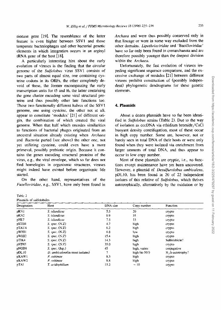
About a dozen plasmids have so far been identi- fied in Sulfolobus strains (Table 2). Due to the way of isolation as cccDNA via ethidium bromide/CsCi buoyant density centrifugation, most of these occur in high copy number. Some are, however, not or barely seen in total DNA of their hosts or were only found when they were isolated via enrichment from larger amounts of total DNA, and thus appear to occur in low copy number.
Most of these plasmids are cryptic, i.e., no func- tions except maintenance have yet been uncovered. However, a plasmid of Desulfurolobus ambivalens, pDLI0, has been found in 20 of 22 independent isolates of this relative of Sulfolobus, which thrives autotrophically, alternatively by the oxidation or by
Table 2 Plasmids of sulfolobales
Designation Host DNA size Copy number Function
pRNI S. islandicus 5.5 20 crypto pRN2 S. islandicus 6.9 35 crypto pilE7 S. islandicus 7.5 15 crypto pSTH4 S. spec. (N.Z) 4.7 high crypto pTAU4 S. spec. (N.Z) 6.2 high crypto pWHII S. spec. (N.Z) 6.8 low crypto pWHI2 S. spec. (N.Z) 15.4 high crypto pTIK4 S. spec. (N.Z) 14.3 high Sulfolobicin? pSTH3 S. spec. (N.Z) 35.0 high crypto pNOB8 S. spec. (Jap.) 45 high, varies conjugative pDL10 D. ambivalens(in most isolates) 7 high (to 50?) H2S-autotrophy? pKAW 1 P. oshimae 8.3 high crypto pKAW2 P. oshimae 8.8 high crypto pTAI T. acidophilium 15.2 ~ 10 crypto
Dow
nloaded from https://academ
ic.oup.com/fem
sre/article/18/2-3/225/479297 by guest on 11 February 2022

234 W. Zillig et al. / FEMS Microbiology Reviews 18 (1996) 225-236
the reduction of sulfur. Its copy number is strongly enhanced during growth by sulfur reduction though the plasmid is not absolutely required for this mode of existence. The widespread occurrence of this ele- ment indicates the existence of a spreading mecha- nism while the linkage between copy number and mode of energy metabolism points at some func- tional, though not stringent, involvement, in growth by sulfur reduction [22].
Another plasmid, pTIK4 from New Zealand, en- ables its host, to outgrow or kill competing Sul- folobus strains without producing a diffusible toxic agent, possibly by cell contact. Colonies of this isolate spread fast on gelrite plates by swarming (W. Zillig, unpublished).
The conjugative plasmid pNOB8 from Japan (Hokkaido) is efficiently transferred from donor to recipient cells via cell to cell contacts [11]. The transfer mechanism is unknown. After transfer, the copy number (per host chromosome) is high, but colonies obtained by plating on gelrite yield liquid cultures with low copy number. The mechanism of copy number control is not understood but the low copy number state can also be approached by serial transfer of the transcipient. We have been able to obtain Sulfolobus strains containing both the cryptic plasmid pilE7 and the conjugative plasmid pNOB8 by electroporating a recipient already carrying the former plasmid with the latter, followed by allowing conjugative spread of pNOB8 in the culture contain- ing the primary transcipients. Immediately after spreading, the copy numbers of both plasmids were high. In strains obtained from single colonies on gelrite plates, they were so low that the plasmids could only be recognized by Southern hybridization and one or the other had been lost in some of these strains. The replication of both plasmids appears thus to be controlled in the same way. When a donor containing both pNOB8 and the small cryptic plas- mid pilE7 was mated with S. solfataricus PHI as a fl-galactosidase negative recipient containing neither plasmid, pNOB8 spread in the culture but transfer of pilE7 was not observed. This indicates specific transfer of pNOB8 (S. Albers and I. Holz, unpub- lished work from our laboratory) rather than trans- port through a cytoplasmatic bridge as in a mating mode observed with Haloferax volcanii [23].
Only two of all these plasmids, pRN1 [23] and the
Pyrococcus element pGT5 [24] have been sequenced so far. Each of these sequences contains two ORFs one of which is large and has significant similarity with rep genes involved in the maintenance, e.g., replication of such elements in the cell via a rolling circle mechanism. This notion is supported by the occurrence of origin motifs known to be used in RC replication [24].
Two plasmids, one of 8.3, the other of 8.8 kb, have been found in two different strains of the moderately thermophilic hyperacidophile Pi- crophilus oshimae, a relative of Thermoplasma aci- dophilum from a Japanese (Hokkaido) solfatara [25]. They are both contained in the strain DSM 9789 kept by the Deutsche Sammlung von Mikroorganismen, the larger one as major, the smaller as minor compo- nent. The two plasmids share yet unmapped cross- hybridizing sequences. A plasmid of 15.2 kb has recently been found in Thermoplasma acidophilum [26].
5. 'Sulfolobicins'
About 30 of some 300 glycerol conserves of novel Sulfolobus strains from Iceland which had been cloned from single colonies formed sharp-edged halos upon dotting on lawns of selected virus-free hosts (D. Prangishvilli, unpublished results from our laboratory). These halos resulted from complete inhi- bition of the growth of the lawn around the dot. No virus particle nor infectious agent responsible for this phenomenon could be discovered. Instead, a dif- fusible macromolecular, apparently non proteina- ceous cytotoxic agent was concentrated from the cell-free supernatant of the culture of one of the strains. The toxin producing strain was resistant against its own product. The chemical nature of the agent and the mode of its action are unknown. But it suggests itself that these agents could be means of defense in the competition with other Sulfolobus strains. All isolates from an enrichment culture giv- ing rise to one such strain indeed shared the capacity to excrete such toxins. Since toxin production is a unique property - the high frequency of observing it results probably from selection during enrichment - genes for toxin synthesis and insensitivity towards toxin action might be encoded on plasmids.
Dow
nloaded from https://academ
ic.oup.com/fem
sre/article/18/2-3/225/479297 by guest on 11 February 2022

W. Zillig et a l . / FEMS Microbiology ReL'iews 18 (1996) 225-236 235
6. Construction of transforming (cloning) vectors based on genetic elements
The conjugative 45-kb plasmid pNOB8 suffers extensive variation by insertions, deletions and other recombinatory events upon transfer to recipients. The transfer and propagation of variants containing dele- tions indicates the deleted sequence is not required for the functions involved. We therefore inserted a lacS (/3-galactosidase) gene from S. solfataricus [27] provided with the strong promoter of the gene of ribosomal protein S 12 of S. acidocaldarius into such a non essential sequence and electroporated this con- struct into a lacS negative mutant of S. solfataricus, PHI, in which an IS element of 1150 kb has been stably integrated into the gene thus destroying its function. Almost complete spreading was then achieved by allowing conjugation. The success could be monitored by the X-Gal reaction in which fl- galactosidase-positive colonies turn blue. Many blue colonies were obtained, however all in close contact with white colonies of the non transformed recipient or with eachother. The total DNA of the transformed culture contained at least 20 copies of the wild-type gene in the vector per inactive gene containing the IS element in the recipient chromosome. This is to be expected when the efficiency of transformation is about 50% or more. The weakness of this system is the inability of transcipients to form isolated colonies on gelrite plates due to the burden imposed on growth by the high copy number of the large plas- mid. We hope to overcome this problem by reducing the copy number with the help of a not fully under- stood copy number control system [11].
A region in the sequence of the cryptic plasmid pRN1 appears to be non essential because it contains a series of transcription terminators. We therefore introduced the lacS-gene there and obtained blue colonies after electroporetic transfer of this vector and X-Gal staining of colonies on gelrite plates (M.G.L. Elferink and D. Hoogstraten, unpublished work from this laboratory). Attempts to construct shuttle vectors on the basis of the genome of SSVI were so far unsuccessful, probably because the unique restriction sites used for this purpose were situated in essential regions. A useful construct was obtained by Cannio, Rossi and Bartolucci (personal communica- tion), who utilized a region of SSV1 containing the
putative origin of replication and thus the UV-in- ducible gene encoding the transcript ti, d, combining it with the E. coli vector pGEM5Zf- and introducing an alcohol dehydrogenase gene as selection marker against poisoning by benzyl alcohol.
Another shuttle vector was constructed by Aagard el al. from the Pyrococcus plasmid pGT5 and the E. coli plasmid pUC19 [28]. This recombinant vector is maintained in Pyrococcus as well as in Sulfolobus.
Acknowledgements
We are indebted to Tairo Oshima for his invalu- able help in organizing sampling ventures in Japan and Jakob Kristjansson of the Ice Tec at Reykjavik, Iceland, whose hospitality enabled us (I.H., D.P. and W.Z.), to run a sampling and isolation laboratory in Iceland in the summer of 1995.
References
[1] Woese, C.R., O. Kandler and M.L. Wheelis (1990) Towards a natural system of organisms: Proposal for the domains. Proc. Natl. Adad. Sci. USA 87, 4576-4579.
[2] Stolt, P. and W. Zillig (1994) Archaebacterial bacterio- phages, in: Encyclopaedia of Virology, Academic Press Ltd., pp. 50-58.
[3] Reiter, W.P., W. Zillig and D. Palm (1988) Archaebacterial viruses, in: Advances in Virus Research (K. Marmarosch, F.A. Purphy and A.J. Shatking, Eds.), Vol. 34, pp. 143-188. Academic Press Inc., Orlando, FL.
[4] Zillig, W., W.-D. Reiter, P. Palm, F. Gropp, H. Neumann and M. Rettenberger (1988) Viruses of archaebacteria, in: The Bacteriophages (R. Calendar, Ed.), a volume of 'The Viruses' (H. Fr~inkel Conrat and R.R. Wagner, Eds.), Vol. 1, pp. 517-558. Plenum Press, New York.
[5] Stolt, P. and W. Zillig (1994) Gene regulation in halophage qOH; more than promoters. Syst. Appl. Microbiol. 16, 591- 596.
[6] Pfeifer, F. (1988) Genetics of halobacteria, in: Halophile Bacteria (F. Rodriguez-Valera, Ed.), Vol. 2, pp. 113-115. CRC Press.
[7] Erauso, G., S. Marsin, N. Benzouzid-Rollet, M.F. Baucher, T. Barbeyron, Y. Zivanovic, D. Prieur and P. Forterre (1996) Sequence of the plasmid pGT5 from the archaeon Pyrococ- cus abvssi: evidences for rolling-circle replication in a hy- perthermophile, J. Bacteriol., submitted.
[8] Neumann, H. and W. Zillig (1990) Structural variability in the genome of the Thermoproteus tenax virus TTV1, Mol. Gen. Genet. 222, 435-437.
Dow
nloaded from https://academ
ic.oup.com/fem
sre/article/18/2-3/225/479297 by guest on 11 February 2022

236 W. Zillig et al. / FEMS Microbiology Reviews 18 (1996) 225-236
[9] Palm, P., C. Schleper, B. Grampp, S. Yeats, P. McWilliam, W.-D. Reiter and W. Zillig (1991) Complete nucleotide sequence of the virus SSV1 of the archaebacterium Sul- folobus shibatae, Virology 185, 242-250.
[10] Zillig, W., A. Kletzin, C. Schleper, I. Holz, D. Janekovic, J. Hain, M. Lanzend/Srfer and J.K. Kristjansson (1994) Screen- ing for Sulfolobales, their plasmids and their viruses in Icelandic solfataras, System. Appl. Microbiol. 16, 609-628.
[11] Schleper, C., I. Holz, D. Janekovic, J. Murphy and W. Zillig (1995) A multicopy plasmid of the extremely thermophilic arcaeon Sulfolobus effects its transfer to recipients by mat- ing, J. Bacteriol. 177, 4417-4426.
[12] Langer, D., J. Hain, P. Thuriaux and W. Zillig (1995) Transcription in Arcaea" similarity to that in Eucarya, Proc. Natl. Acad. Sci. USA 92, 5768-5772.
[13] Gogarten, J.P., H. Kibak, P. Dittrich, P. Taiz, E.J. Bowman, M.F. Manolson, P.J. Poole, T. Data, T. Oshima, J. Konishi, K. Denca and M. Yoshida (1989) Evolutionary relationship of archaebacteria, eubacteria and eukaryotes inferred from phylogentic trees of duplicated genes, Proc. Natl. Acad. Sci. USA 86, 9355-9359.
[14] lwabe, N., K. Kuma, M. Hasegawa, S. Osawa and T. Miyata (1989) Evolution of RNA polymerases and branching pat- terns of the three major groups of archaebacteria, J. Mol. Evol. 32, 70-78.
[15] Wood, A.G., W.B. Whitman and J. Konisky (1989) Isolation and characterization of an archaebacterial virus-like particle from Methanococcus voltae A3, J. Bacteriol. 171, 93-98.
[16] Rettenberger, M. (1990) Das Virus TTV1 des extrem ther- mophilen Schwefel-Archaebakteriums Thermoproteus tenax: Zusammensetzung und Struktur, Diss. LMU Munich.
[17] Janekovic, D., S. Wunderl, I. Holz, W. Zillig, A. Gierl and H. Neumann (1983) T r v I , "I~V2 and TYV3, a family of viruses fo the extremely thermophilic, anaerobic, sulfur re- ducing archaebacterium Thermoproteus tenax. Mol. Gen. Genet. 192, 39-45.
[18] Reiter, W.-D., P. Palm and S. Yeats (1989) Transfer RNA genes frequently serve as integration sites for prokaryotic genetic elements, Nucleic Acids Res. 17, 1907-1914.
[19] Muskhelishvili, G., P. Palm and W. Zillig (1993) SSVI-en- coded site-specific recombination system in Sulfolobus shi- batae, Mol. Gen. Genet. 237, 334-342.
[20] Schleper, C., K. Kubo and W. Zillig (1992) The particle SSV1 from the extremely thermophilic arcaeon Sulfolobus is a virus: Demonstration of inectivity and of transfection with viral DNA. Proc. Natl. Acad. Sci. USA 89, 7645-7649.
[21] Campbell, A. (1988) Phage evolution and speciation, in: The Bacteriophages (R. Calendar, Ed.), a volume of 'The Viruses' (H. Fr~tkel Conrat and R.R. Wagner, Eds.), Vol. 1, pp. 1-14. Plenum Press, New York,
[22] Zillig, W., S. Yeats, I. Holz, A. BiSck, E. Gropp, M. Retten- berger and S. Lutz (1985) Plasmid-related anaerobic autotro- phy of the novel archaebacterium Sulfolobus ambivalens, Nature 313, 789-791.
[23] Mevarech, M. and R. Werczberger (1985) Genetic transfer in Halobacterium volcanii, J. Bacteriol. 162, 461-462.
[24] Keeling, P.J., H,-P. Klenk, R.K. Singh, O. Feeley, C. Schleper, W. Zillig, W.F. Doolittle and C.W. Sensen (1996) Complete nucleotide sequence of the Sulfolobus islandicus multicopy plasmid pRN1, Plasmid, in press.
[25] Schleper, C., G. Puehler, I. Holz, A. Gambacorta, D. Janekovic, U. Santarias, H.-P. Klenk and W. Zillig (1995) Picrophilus Gen. Nov. Fam. Nov.: A novel aerobic, het- erotrophic, thermoacidophilic genus and family comprising archaea capable of grwoth around pH Zero. J. Bacteriol. 177, 7050-7059.
[26] Yasuda, M., A. Yamagishi, T. Oshima (1995) The plasmids found in isolates of the acidothermophilic archaebacterium Thermoplasma acidophilum, FEMS Microbiol. Lett. 128, 157-161.
[27] Elferink, M.G.L., C. Schleper and W. Zillig (1996) Transfor- mation of the extremely thermoacidophilic archaeon Sul- folobus solfatiricus via a self-spreading vector, FEMS Mi- crobiol. Lett. 137, 31-35.
[28] Aagaard, C., I. Leviev, R.N. Aravalli, P. Forterre, D. Prieur and R.A. Garret (1996) General vectors for archaeal hyper- thermophiles: Strategies based on a mobile intron and a plasmid, FEMS Microbiol. Rev. 18, 93-104.
Dow
nloaded from https://academ
ic.oup.com/fem
sre/article/18/2-3/225/479297 by guest on 11 February 2022