Topically applied EGF and PDGFs affect positively the co-ordinate expression of EGF and PDGF...
10
RESEARCH ARTICLES CURRENT SCIENCE, VOL. 92, NO. 5, 10 MARCH 2007 618 *For correspondence. (e-mail: [email protected]) Topically applied EGF and PDGFs affect positively the co-ordinate expression of EGF and PDGF receptor genes during acute cutaneous wound-healing process Mohan L. Gope 1 and Rajalakshmi Gope 2, * 1 Department of Biotechnology, City College, Bangalore 560 070, India 2 Department of Human Genetics, National Institute of Mental Health and Neurosciences, Bangalore 560 029, India Daily topical application of epidermal growth factor (EGF) and platelet-derived growth factors (PDGFs) individually resulted in approximately two- to three- fold increase in EGF-R and α- and β-PDGF-R mRNA levels, and combinations of these growth factors pro- duced a 2.5 to four-fold increase. Rapid attainment of maximum level of receptor mRNA on day one and its maintenance at comparatively higher level till day three appears to be essential for faster and better healing. PDGF-AA + EGF inhibited EGF-mediated increase in receptor mRNA levels when applied pre- mixed or within 30 min following EGF application. The results are also suggestive of the possible role of PDGF-AA during later stages of wound healing. Keywords: Epidermal growth factor, platelet-derived growth factors, receptor mRNA, wound healing. GROWTH factors are essential to the complex process of wound repair and they have become targets for therapeu- tic interventions. Various growth factors such as epidermal growth factor (EGF), platelet-derived growth factors (PDGFs), transforming growth factors (TGF) α and β, and insulin-like growth factor-1 (IGF-1), either alone or in combination, have been implicated in the wound healing process in various tissues 1–5 . The regulatory role of EGF in gene expression and development has been well estab- lished 2,6 . It has been shown that EGF is necessary for re- epithelialization during the wound-repair process 6 . EGF is known to stimulate keratinocyte migration, granulation, tissue development and mitogenesis of keratinocytes and fibroblasts 7 . Some of these studies were performed with single or multiple application of growth factors 6–8 . Studies have shown that topical application of PDGF enhances wound repair, especially in animals with healing defects 9–16 . Reduced expressions of PDGFs and PDGF-R- α were observed during early phase of impaired wound healing 10,11,13,14 . Previous studies have shown that PDGF- treated wounds closed by re-epithelialization and the filling- in with scar, whereas control wounds healed by contrac- tion 17–20 . We have previously reported that growth factor- treated wounds healed with fine scars compared to control 20 . The growth factor-treated wounds healed in ap- proximately 5 to 7 days compared to controls, which took 10 to 15 days 20 . The wound-healing process was further accelerated with the application of a combination of growth factors (EGF + PDGF-BB or PDGF-AB), which took only 3 to 5 days for healing 20 . Recent reports using gene-transfer techniques where the PDGF-B gene was transferred into cultured dermal fibroblasts from normal and diabetic mice, indicated that the PDGF-B gene en- hanced healing of dermal fibroblast 14,15,17 . Topical appli- cation of PDGF-gels healed wounds with decreased wound depth compared to antibiotic-treated wounds 18 . Addition of exogenous PDGF in problematic wounds resulted in increased fibroblasts and collagen content, suggesting that cytokines such as EGF and PDGFs are pivotal mole- cules that may be deficient in some wound conditions 2,16 . Using the mouse model system previously we have shown that EGF, PDGF-AB and PDGF-BB, when topi- cally applied either alone or in combination, accelerated wound healing by 50 to 66% faster with less scar tissue compared to the control 20 . The best healing effect was ob- tained with a combination of EGF plus PDGF-BB. PDGF-AA, either alone or in combination with EGF, did not enhance the wound-repair process 20 . The present study was carried out to determine the effect of optimum concentrations of purified EGF, PDGF-AB, PDGF-BB and PDGF-AA, individually and in various combinations, on the expression of their receptor mRNAs during the acute cutaneous wound-healing process. Materials and methods Animals The experimental protocol was cleared by the institutional Animal Ethics Committee. Random bred male Swiss al-
Transcript of Topically applied EGF and PDGFs affect positively the co-ordinate expression of EGF and PDGF...

RESEARCH ARTICLES
CURRENT SCIENCE, VOL. 92, NO. 5, 10 MARCH 2007 618
*For correspondence. (e-mail: [email protected])
Topically applied EGF and PDGFs affect positively the co-ordinate expression of EGF and PDGF receptor genes during acute cutaneous wound-healing process Mohan L. Gope1 and Rajalakshmi Gope2,* 1Department of Biotechnology, City College, Bangalore 560 070, India 2Department of Human Genetics, National Institute of Mental Health and Neurosciences, Bangalore 560 029, India
Daily topical application of epidermal growth factor (EGF) and platelet-derived growth factors (PDGFs) individually resulted in approximately two- to three-fold increase in EGF-R and α- and β-PDGF-R mRNA levels, and combinations of these growth factors pro-duced a 2.5 to four-fold increase. Rapid attainment of maximum level of receptor mRNA on day one and its maintenance at comparatively higher level till day three appears to be essential for faster and better healing. PDGF-AA + EGF inhibited EGF-mediated increase in receptor mRNA levels when applied pre-mixed or within 30 min following EGF application. The results are also suggestive of the possible role of PDGF-AA during later stages of wound healing.
Keywords: Epidermal growth factor, platelet-derived growth factors, receptor mRNA, wound healing.
GROWTH factors are essential to the complex process of wound repair and they have become targets for therapeu-tic interventions. Various growth factors such as epidermal growth factor (EGF), platelet-derived growth factors (PDGFs), transforming growth factors (TGF) α and β, and insulin-like growth factor-1 (IGF-1), either alone or in combination, have been implicated in the wound healing process in various tissues1–5. The regulatory role of EGF in gene expression and development has been well estab-lished2,6. It has been shown that EGF is necessary for re-epithelialization during the wound-repair process6. EGF is known to stimulate keratinocyte migration, granulation, tissue development and mitogenesis of keratinocytes and fibroblasts7. Some of these studies were performed with single or multiple application of growth factors6–8. Studies have shown that topical application of PDGF enhances wound repair, especially in animals with healing defects9–16. Reduced expressions of PDGFs and PDGF-R-α were observed during early phase of impaired wound healing10,11,13,14. Previous studies have shown that PDGF-
treated wounds closed by re-epithelialization and the filling-in with scar, whereas control wounds healed by contrac-tion17–20. We have previously reported that growth factor-treated wounds healed with fine scars compared to control20. The growth factor-treated wounds healed in ap-proximately 5 to 7 days compared to controls, which took 10 to 15 days20. The wound-healing process was further accelerated with the application of a combination of growth factors (EGF + PDGF-BB or PDGF-AB), which took only 3 to 5 days for healing20. Recent reports using gene-transfer techniques where the PDGF-B gene was transferred into cultured dermal fibroblasts from normal and diabetic mice, indicated that the PDGF-B gene en-hanced healing of dermal fibroblast14,15,17. Topical appli-cation of PDGF-gels healed wounds with decreased wound depth compared to antibiotic-treated wounds18. Addition of exogenous PDGF in problematic wounds resulted in increased fibroblasts and collagen content, suggesting that cytokines such as EGF and PDGFs are pivotal mole-cules that may be deficient in some wound conditions2,16. Using the mouse model system previously we have shown that EGF, PDGF-AB and PDGF-BB, when topi-cally applied either alone or in combination, accelerated wound healing by 50 to 66% faster with less scar tissue compared to the control20. The best healing effect was ob-tained with a combination of EGF plus PDGF-BB. PDGF-AA, either alone or in combination with EGF, did not enhance the wound-repair process20. The present study was carried out to determine the effect of optimum concentrations of purified EGF, PDGF-AB, PDGF-BB and PDGF-AA, individually and in various combinations, on the expression of their receptor mRNAs during the acute cutaneous wound-healing process.
Materials and methods
Animals
The experimental protocol was cleared by the institutional Animal Ethics Committee. Random bred male Swiss al-

RESEARCH ARTICLES
CURRENT SCIENCE, VOL. 92, NO. 5, 10 MARCH 2007 619
bino mice were used as a model system. Three-week-old mice were obtained from the Central Animal Research Facility, National Institute of Mental Health and Neuro-sciences (NIMHANS), Bangalore and kept for an addi-tional two to three weeks before use. This time period assured that the animals were free from diseases. All the animals were provided ample pelleted animal food (Godrej India Ltd, Mumbai) and water and normal day–night light cycles.
Wounding of mice
The dorso-lateral sides of mice were cleaned with disin-fectant and approximately 5 mm × 1.5 cm area was cleared of all hair. Two full-thickness wounds of approximately 1 cm long and 0.3 to 0.4 cm deep were created on the dorso-lateral sides of each mouse, as described previ-ously20.
Number of mice used and collection of tissues
The experiments were repeated thrice to obtain statisti-cally significant data. Fifty mice were kept as untreated external controls where wounds on both sides were left untreated. In the second set of 50 mice, one wound from each mice was untreated and the other wound was treated with diluent alone and these acted as external controls. In the third set of 50 mice, wound on one side was treated with growth factors and the other wound was treated with diluent alone which acted as internal control. Thus, a group of 50 mice were used for each of the following growth factor treatments, either alone or in combination: (1) EGF, (2) PDGF-AB, (3) PDGF-BB, (4) PDGF-AA, (5) EGF + PDGF-BB, (6) EGF + PDGF-AB or (7) EGF + PDGF-AA. The newly formed tissues at the wound site were collected and pooled from 8 to 10 mice for each time period, i.e. day 1, 3, 5 and 7. After collecting tissues at the specified time points (40 mice) the mice were sacri-ficed and disposed. The remaining ten mice were kept under observation for approximately 30 days for any ab-normal growth or rash. Thus, for each experiment a total of 100 mice were used as external control and 350 mice were used for growth-factor application. Overall, 1350 mice were used for three separate sets of experiment.
Growth factors
Recombinant mouse EGF and recombinant human PDGF-AB, PDGF-BB and PDGF-AA were obtained from Sigma–Aldrich (Saint Louis, USA) as lyophilized powders and diluted according to the manufacturer’s instructions in a diluent solution containing 4 mM HCl and 0.1% (w/v) bovine serum albumin (Sigma–Aldrich). Aliquots of ap-propriate volumes were stored at –20°C and they were thawed only once for each use.
Application of growth factors
The day of creating the wound was counted as day 0 and the next day as day 1. Optimum concentrations of the growth factors in diluent (EGF, 10 ng; PDGF-AB, 15 ng; PDGF-BB, 15 ng or PDGF-AA, 20 ng) were applied di-rectly to the wounds each day20 starting from day 0 to day 7. The growth factors were applied with a micropipette as previously described20. For the studies using combina-tions of growth factors, 10 ng of EGF was applied first followed by application of 15 ng of PDGF-AB, 15 ng PDGF-BB or 20 ng PDGF-AA within 30 min after EGF application. Alternatively, the growth factors, EGF plus PDGF-AB, EGF plus PDGF-BB and EGF plus PDGF-AA, were premixed and applied to the wounds as descri-bed previously20.
Monitoring
The wound-repair process was monitored visually. The time taken for wound repair was scored visually and pho-tographically using the following parameters: (a) wound closure and (b) scar-tissue formation (fine or raised), and these data have been already published20. The newly formed irregular masses of granular tissues were col-lected and pooled from the wound sites of control and growth factor-treated mice. The pooled granular tissues from approximately 8 to 10 mice were stored at –70°C till they were further processed. The total RNAs were ex-tracted from these granular tissues and used for Northern blot analysis21.
Extraction of total RNA
The granular tissues were homogenized in commercially available Tri-reagent (Sigma–Aldrich) and the total RNAs were isolated according to the protocol supplied by the manufacturer.
Northern blot analysis
Equal quantities of approximately 80 µg of total RNAs from the controls and growth factor-treated wounds were denatured in a buffer containing 50% formaldehyde, size-separated on a 1.2% (w/v) denaturing agarose–formalde-hyde gel and transferred to nylon membrane (Sigma–Aldrich)21. The filters were baked at 80°C for 2 h and stored at 4°C until hybridized21.
Probes
Human EGF-R and β-PDGF-R cDNA probes were ob-tained from American Type Culture Collection (ATCC,
RESEARCH ARTICLES
CURRENT SCIENCE, VOL. 92, NO. 5, 10 MARCH 2007 620
USA). The EGF-R cDNA probe recognized the expected two bands of 10.0 and 5.5 kb. The β-PDGF-R probe rec-ognized the expected two bands of 6.4 kb and a 5.3 kb corresponding to α-PDGF-R and β-PDGF-R respectively, under low-stringency washing condition. However, under high-stringency washing condition, this probe recognized only the 5.3 kb β-PDGF-R mRNA. The internal control probe, β-actin cDNA, was also obtained from ATCC.
Probe labelling and detection
The cDNA probes were labelled with North2South Direct HRP Labelling and Detection kit (Pierce, USA). The Northern filters were probed with chemiluminescent-labelled cDNA probes. The solutions supplied by the manufacturer (Pierce) were used for labelling of the probe, pre-hybridization and hybridization, and the manu-facturer’s protocol was followed. Pre-hybridization and hybridization were done at 55°C. The filters were first hybridized with chemiluminescent-labelled β-PDGF-R cDNA probe. These filters were washed under low-stringency condi-tion, and after developing and visualizing the α-PDGF-R and β-PDGF-R bands, the same filters were washed un-der high-stringency condition and developed again to visualize the β-PDGF-R bands. For low-stringency condi-tions, the filters were washed in 2X SSC/0.1% (v/v) SDS, three times for 5 min each at 50°C. Then the filters were washed in 2X SSC three times for 5 min each at room temperature. For high-stringency washing conditions the solutions were the same, but the temperature was in-creased to the hybridization temperature of 55°C. Then the filters were washed three times at room temperature as described. The filters were developed with Luminol (Pierce) and the bands were visualized by exposing the filters to X-ray films according to the manufacturer’s (Pierce) protocol. After stripping, these filters were hy-bridized with EGF-R cDNA probe, washed under a high-stringency condition at 55°C and the bands were visualized as previously described. Finally, the same Northern filters were stripped and re-probed with β-actin cDNA probe and washed under high-stringency wash condition at 55°C.
Quantitation
The levels of mRNAs were calculated by measuring the receptor band intensities in Bio-Rad gel documentation system (Bio-Rad, USA) using Bio-Rad quantity one software. The background values on a blank lane were subtracted from these values. The values were then com-pared to the corresponding β-actin mRNA levels in the same lanes. The effect of no treatment or diluent treatment on the level of receptor mRNA expression was taken as a base value and then the effect of exogenous addition of
growth factors on the level of mRNA was calculated and is reported here as fold increase. To calculate the level of EGF-R mRNA, intensities of both 10.0 and 5.5 kb mRNA species were measured and the combined value is repor-ted here.
Statistical analysis
Statistical analysis was performed using SPSS software version 13.0. Student’s t test, $2 test and odds ratio were compared. P < 0.05 was considered statistically signifi-cant. The following parameters were used for statistical correlation: (a) total number of experiments conducted; (b) total number of mice used for each experiment; (c) fold induction of receptor mRNA level to the wound-repair process and (d) standard deviation between the three separate experimental results.
Results
All the internal and external controls showed consistently similar levels of growth factor receptor mRNAs in all the three separate sets of experiment.
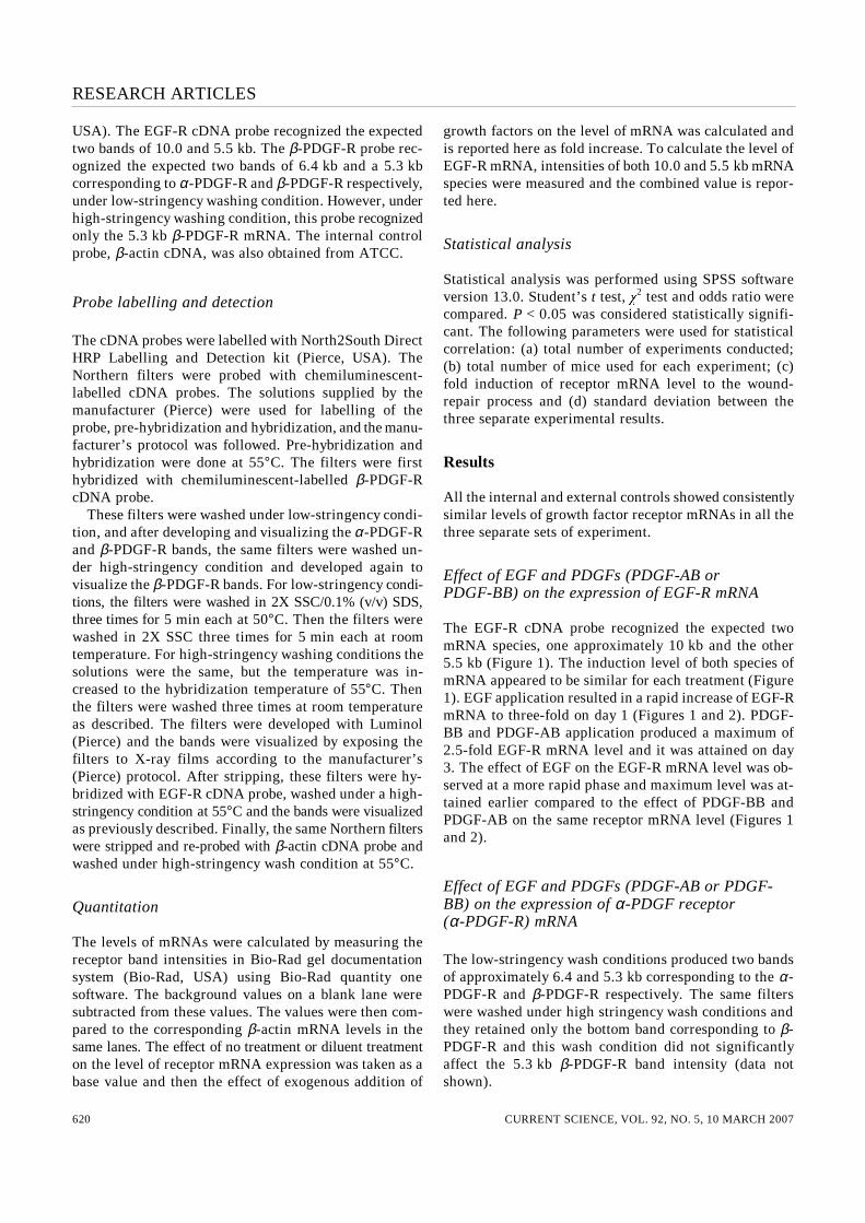
Effect of EGF and PDGFs (PDGF-AB or PDGF-BB) on the expression of EGF-R mRNA
The EGF-R cDNA probe recognized the expected two mRNA species, one approximately 10 kb and the other 5.5 kb (Figure 1). The induction level of both species of mRNA appeared to be similar for each treatment (Figure 1). EGF application resulted in a rapid increase of EGF-R mRNA to three-fold on day 1 (Figures 1 and 2). PDGF-BB and PDGF-AB application produced a maximum of 2.5-fold EGF-R mRNA level and it was attained on day 3. The effect of EGF on the EGF-R mRNA level was ob-served at a more rapid phase and maximum level was at-tained earlier compared to the effect of PDGF-BB and PDGF-AB on the same receptor mRNA level (Figures 1 and 2).
Effect of EGF and PDGFs (PDGF-AB or PDGF-BB) on the expression of α-PDGF receptor (α-PDGF-R) mRNA
The low-stringency wash conditions produced two bands of approximately 6.4 and 5.3 kb corresponding to the α-PDGF-R and β-PDGF-R respectively. The same filters were washed under high stringency wash conditions and they retained only the bottom band corresponding to β-PDGF-R and this wash condition did not significantly affect the 5.3 kb β-PDGF-R band intensity (data not shown).

RESEARCH ARTICLES
CURRENT SCIENCE, VOL. 92, NO. 5, 10 MARCH 2007 621
Daily topical application of EGF or PDGFs (PDGF-AB or PDGF-BB) increased the level of 6.4 kb mRNA corre-sponding to α-PDGF-R (top band, Figure 3), but the maximum level (Figure 4) was slightly lower than that of EGF-R (Figure 2) and β-PDGF-R (Figure 5) mRNA levels. Application of PDGF-BB resulted in a rapid increase in the level of α-PDGF-R to a maximum of 2.3-fold on day 1 (Figures 3 and 4). However, application of PDGF-AB and EGF caused a gradual increase in the α-PDGF-R re-ceptor mRNA level, reaching a maximum of approxi-mately two- to 2.3-fold on day 3 (Figures 3 and 4).
Effect of EGF and PDGFs (PDGF-AB or PDGF-BB) on the expression of β-PDGF-receptor (β-PDGF-R) mRNA
Daily topical application of the growth factors resulted in the induction of the 5.3 kb mRNA corresponding to β-PDGF-R (bottom band, Figure 3). Application of PDGF-AB or PDGF-BB resulted in rapid induction of β-PDGF-R to three-fold on day 1, and the level was approxi-mately the same till day 3 (Figures 3 and 5). However,
Figure 1. Effect of EGF and PDGFs on the expression of EGF-R mRNA level. EGF-R mRNA levels were compared between the growth factor-treated wounds and the controls. The levels of EGF-R mRNAs were calculated by measuring the band intensities of both the 10.0 kb (top arrow) and 5.5 kb bands (bottom arrow). Details are described in the text. C, Untreated external control; D, Diluent-treated external con-trol. The growth factors used are marked above the lanes. –, Diluent-treated internal control; +, Growth factor treatment. Days of growth factor treatment are marked on the left.
EGF application produced a gradual increase of β-PDGF-R mRNA reaching a maximum level of three-fold on day 3 (Figures 3 and 5).
Figure 2. Level of EGF-R expression with EGF and PDGFs. Days of treatment are marked on the X-axis, and the levels of receptor mRNAs are marked on the Y-axis. C, Untreated external control; D, Diluent-treated internal control. The levels of EGF-R mRNA given are the combined value of both 10.0 kb plus 5.5 kb bands. The levels of recep-tor are the average from three independent experiments.
Figure 3. Effect of EGF and PDGFs on the expression of α-PDGF-R (top arrow) and β-PDGF-R (bottom arrow) mRNA levels. The proce-dures are given in the text. C, Untreated external control; D, Diluent-treated external control; –, Diluent-treated internal control; +, Growth factor treatment. Days of treatment are shown on the left. Top arrow, 6.4 kb α-PDGF-R mRNA; bottom arrow, 5.3 kb β-PDGF-R mRNA.

RESEARCH ARTICLES
CURRENT SCIENCE, VOL. 92, NO. 5, 10 MARCH 2007 622
Effect of PDGF-AA on the expression of α-PDGF-R, β-PDGF-R and EGF-R mRNA levels
Daily topical application of 20 ng of PDGF-AA per wound produced little increase in the levels of all three receptor mRNAs. However, on day 5, a slight increase in the levels of all three receptor mRNAs was observed (Figures 1–5).
Figure 4. Level of α-PDGF-R mRNA expression in growth factor-treated wounds. Days of growth factor treatment are shown on the X-axis and the levels of receptor mRNA are shown on the Y-axis. C, Un-treated external control; D, Diluent-treated internal control. The level of receptor mRNA is the average of three separate experiments.
Figure 5. Level of β-PDGF-R mRNA expression in growth factor-treated wounds. Days of growth factor treatment are shown on the X-axis and the receptor mRNA levels are shown on the Y-axis. C, Un-treated external control; D, Diluent-treated internal control. The level of receptor mRNA is the average of three separate experiments.
Control
Re-probing the Northern filters with β-actin cDNA probe confirmed that increase in the level of growth factor re-ceptor mRNAs was not due to variation in the loading of the RNA gels (Figure 6).
Effect of combination of EGF plus PDGF-AB and EGF plus PDGF-BB on the level of expression of EGF-R mRNAs
Combination of EGF plus PDGF-BB showed rapid induc-tion of EGF-R mRNA level, reaching a maximum of 3.5-fold on day 1 followed by a slight decrease to three- and 2.8-fold on day 3 and day 5 respectively (Figures 7 and 8). A combination of EGF plus PDGF-AB also resulted in rapid induction of EGF-R mRNA on day 1, but this com-bination led to only a maximum of 2.8-fold increase. The maximum level of the EGF-R mRNA was not additive with the combination of growth factors to that obtained with individual growth factors (Figures 1–5).
Effect of combination of EGF plus PDGF-AB or EGF plus PDGF-BB on the expression of α-PDGF-R mRNA level
With the combination of EGF + PDGF-AB or EGF + PDGF-BB, the level of α-PDGF-R mRNA also increased
Figure 6. Level of control β-actin mRNA. C, Untreated external con-trol; D, Diluent-treated external control; –, Diluent-treated internal con-trol; +, Growth factor treatment. Days of treatment are shown on the left. Arrows indicates β-actin mRNA.

RESEARCH ARTICLES
CURRENT SCIENCE, VOL. 92, NO. 5, 10 MARCH 2007 623
Figure 7. Effect of combination of EGF plus PDGFs on the expres-sion of EGF-R. Arrows indicate EGF-R mRNA. Days of growth factor treatment are given on the left. C, Untreated external control; D, Dilu-ent-treated internal control. As the diluent-treated internal control showed similar results in all the blots, only one control is shown in this blot. Treatment with growth factor as indicated above the lanes. The top (10.0 kb) and bottom arrow (5.5 kb) indicate the two species of EGF-R mRNA band. Days of growth factor treatment are indicated on the left.
Figure 8. Levels of EGF-R mRNAs in growth factor-treated wounds. Days of growth factor treatments are given on the X-axis and EGF-R mRNA levels are given on the Y-axis. C, Untreated external control; D, Diluent-treated internal control. The levels shown here are the com-bined values of both 10.0 and 5.5 kb EGF-R mRNA bands. The level of receptor mRNA shown here is the average from three separate experi-ments.
rapidly, reaching a maximum of 3.5- to four-fold on day 1 (top arrow, Figures 9 and 10) and the maximum levels with these combinations were not additive compared to the individual growth factors.
Effect of combination of EGF plus PDGF-AB or EGF plus PDGF-BB on the expression of β-PDGF-R mRNA level
Maximum level of 3.5- to four-fold β-PDGF-R mRNAs was attained rapidly on day 1 with the application of EGF + PDGF-AB or EGF + PDGF-BB (bottom arrow, Figures 9 and 11). The maximum levels were not additive compared to the levels obtained with individual growth factors.
Effect of combination of EGF plus PDGF-AA on the level of PDGF-R and EGF-R mRNA expression
Combination of EGF plus PDGF-AA produced little ef-fect on the levels of EGF-R (Figures 7 and 8) or PDGF-R
Figure 9. Effect of combination of EGF plus PDGFs on the expres-sion of α-PDGF-R (top arrow) and β-PDGF-R (bottom arrow) mRNA levels. C, Untreated external control; D, Diluent-treated internal con-trol. All diluent-treated internal controls showed similar results, there-fore only one lane is loaded with this sample; Treatment with growth factors as indicated above the lanes. Days of treatment are shown on the left.

RESEARCH ARTICLES
CURRENT SCIENCE, VOL. 92, NO. 5, 10 MARCH 2007 624
(Figures 9–11) mRNAs. PDGF-AA in combination with EGF repressed the EGF-mediated increase in the EGF-R and PDGF-R mRNA expression (Figures 7–11). This re-pression was observed when PDGF-AA was applied within 30 min after the application of EGF. Similar results were obtained when these growth factors were applied pre-mixed (data not shown). However, application of PDGF-AA approximately 30 min after the application of EGF did not affect the EGF-mediated induction of EGF-R or the pleiotropic effect of EGF observed in our ex-periments (data not shown).
Control
The observed increase in the growth factor receptor mRNAs was not due to unequal loading of the Northern gels as confirmed by re-probing the filters with β-actin cDNA probe (Figure 12).
Statistical analysis
The number of mice used in each experiment was statisti-cally significant. There was a positive significant correla-tion between the day of attainment of peak level of the receptor mRNA to the rate of wound healing and quality of the scar tissues (p < 0.05). In addition, similar positive correlation was also observed between the maximum level of receptor mRNA to that of wound healing and
Figure 10. Levels of α-PDGF-R mRNA in growth factor-treated wounds. Days of growth factor treatment are given on the X-axis and levels of α-PDGF-R mRNA are shown on the Y-axis. C, Untreated ex-ternal control; D, Diluent-treated internal control. The levels of recep-tor mRNA shown here are the average from three separate experiments.
scar tissue. The standard deviations for the levels of rece-ptor mRNAs between the three different sets of experi-ments were less than 0.05, confirming the statistical significance of the results from these experiments.
Discussion
A previous report had shown that one to two days after injury, epidermal cells at the wound margin begin to pro-liferate behind actively migrating cells22. A sustained daily release of 10 to 20 µg of partially purified, biologi-cally active EGF was found22 to increase the extent and organization of the granulation tissue at day 7. The stim-uli for the migration and proliferation of epidermal cells during re-epithelialization have not been determined, but it has been suggested that the presence of growth factor EGF and its receptor may stimulate these processes5,22. We have previously shown that daily topical application of optimum concentration of 10 ng EGF enhanced the acute cutaneous wound-healing process, where the wounds healed in 5 to 7 days compared to the controls which took approximately twice as long, i.e. 10 to 15 days to heal20. Among individual growth factors, the best healing was observed with PDGF-BB and among the combinations EGF + PDGF-BB produced the best results20. This is con-sistent with other reports for the positive role of EGF dur-ing wound healing22, as EGF promoted cell motility5 and maximally stimulated keratinocytes and epithelial cells at
Figure 11. Levels of β-PDGF-R mRNA in growth factor-treated wounds. Days of growth factor treatment are given on the X-axis and levels of β-PDGF-R mRNA are marked on the Y-axis. C, Untreated ex-ternal control; D, Diluent-treated internal control. The levels of recep-tor mRNA shown here are the average from three separate experiments.

RESEARCH ARTICLES
CURRENT SCIENCE, VOL. 92, NO. 5, 10 MARCH 2007 625
Figure 12. Levels of control β-actin mRNA. C, Untreated external control; D, Diluent-treated internal control. Days of growth factor treatment are given on the left. Arrows indicate β-actin mRNA.
25 ng and 5 ng/ml respectively7,8. The optimum concen-tration required for EGF to produce its effect on various cell types could depend on the purity of the growth factor and type of wound7,8,20. In addition to inducing the level of its own receptor mRNA (Figures 1 and 2), EGF also induced α-PDGF-R and β-PDGF-R mRNA levels (Fig-ures 3–5), indicating the pleiotropic effect of EGF during the wound-healing process. Delayed appearance of EGF and EGF-R in incisional wounds of aged mice has been reported by other groups16,23. Our results show that in re-sponse to daily EGF treatment, the maximum EGF-R level was reached rapidly on day 1 (Figure 2). Thus, it is conceivable that optimum concentration of growth factors that could produce peak levels of receptor mRNAs rap-idly could be essential for enhancement of cutaneous wound repair (Figures 3–5). Similar to EGF, PDGF-AB and PDGF-BB also pro-duced pleiotropic effect by inducing EGF-R mRNA level in addition to inducing PDGF-R mRNAs levels (Figures 1–5). Rapid induction of peak level of β-PDGF-R mRNA by PDGF-BB on day 1 and its maintenance till day 3 (Figures 1 and 5) could be correlated with published data on its better healing ability compared to other growth fac-tors18–20. No significant induction of PDGFs was reported in normal mice after skin injury and their expression lev-els were found to be similar at all stages of the repair
process24. Our data confirm this report, where the control wounds showed only a slight increase in the receptor mRNA levels (Figures 1–5), perhaps due to the lower levels of PDGFs24. The prolonged time taken for the heal-ing of control wounds20 could also be due to the presence of sub-optimal levels of receptor mRNAs as seen in the present study. Glucocorticoid treatment has been reported to cause severe defect in wound repair and it resulted in decreased α-PDGF-R and β-PDGF-R in the early phase of wound healing24. Reports have shown that daily appli-cation of lower doses of 5 to 10 ng of PDGF-AB in rat25 and mouse20 failed to significantly enhance wound repair. In the present study, we found that rapid attainment of peak value of the receptor mRNA expression could be achieved only with the optimum concentrations of growth factors (Figures 1–5). Therefore, the beneficial effect of topically applied PDGF observed by others18,19 and by us20 could be due to sustained expression of maximum level of PDGF-R mRNAs at the very early stages (Fig-ures 1, 3 and 4). Combinations of growth factors were found to be more effective in the wound-healing process compared to sin-gle growth factors, perhaps due to their pleiotropic effects on the expression of various genes involved in the regen-eration mechanism1,5,8,13,20,26,27. It has been suggested that the levels of receptor could be critical for faster and bet-ter wound healing27,28. Statistical analysis of our data showed significant positive correlation between the rapid attainment of peak levels of receptor mRNAs (Figures 1–5 and 7–11) and the time taken for healing and the quality of healed wound (scar) that we reported previously20. PDGF-AA does not accelerate the rat cutaneous wounds compared to PDGF-AB25, but helped healing of the tympanic membrane29. This is suggestive of the abil-ity of PDGF-AA to heal only certain types of wounds and not others. Our previous study20 and data from the present study suggest that EGF is essential at early stages of wound healing and the long form of PDGF-AA could an-tagonize the EGF-mediated healing (Figures 7–11). Dif-ferential physiologic functions of various forms of PDGFs have been reported previously30,31. The PDGF-B chain (c-sis proto-oncogene) homodimer (PDGF-BB) and v-sis, its viral counterpart, were known to activate both α-PDGF-R and β-PDGF-R and mediate anchorage-inde-pendent phenotypic transformation in NIH 3T3 cells30. In contrast, the PDGF-A chain homodimer (PDGF-AA) was known to activate α-PDGF-R only, and it failed to induce phenotypic transformation30. β-PDGF-R activation alone is reported to be sufficient for PDGF-BB-mediated anchorage-independent cell growth and inhibition of α-PDGF-R signalling enhanced PDGF-BB-mediated pheno-typic transformations, suggesting that α-PDGF-R antago-nized β-PDGF-R-induced transformation30. Further, α- and β-PDGF-R are known to differentially regulate Ras-mito-gen-activated protein kinase pathway, and transforma-tion-suppressing activity of α-PDGF-R involves JNK-1

RESEARCH ARTICLES
CURRENT SCIENCE, VOL. 92, NO. 5, 10 MARCH 2007 626
activation30. The role of PDGF-AA was reported to be different from that of PDGF-BB during mitogenesis31. It would be interesting to see if the transformation ability is essential for the PDGF-induced wound-healing process. In such a case, it would be crucial to distinguish if lack of such ability reported for PDGF-AA could be deleterious to the repair process of certain types of wounds. Alterna-tively, PDGF-AA might require other growth factor(s) to function during the cutaneous wound repair. PDGF is re-ported32 to inhibit angiogenic properties of basic FGF through α-PDGF-R. PDGF-AA is known to bind only to α-PDGF-R and angiogenesis is essential for wound re-pair. It has been suggested30,31 that PDGF-AA might act through a different molecular pathway compared to PDGF-BB and PDGF-AB. We found a slight increase in the levels of receptor mRNAs with the application of PDGF-AA (Figures 1–11). Also, it might not be suffi-cient to enhance or accelerate the cutaneous wound repair as it appears that the sustained optimum levels of these mRNAs are essential for the same (Figures 1–11)20. Al-ternately, the slight increase in the receptor mRNA levels at day 5 produced by PDGF-AA (Figures 1–5) could be indicative of a possible role of PDGF-AA during later stages of wound healing. It has been shown that PDGF-BB, PDGF-AA and foetal bovine serum were mitogenic, and anti-apoptotic, but only PDGF-BB could induce cell migration33. Therefore, it appears that PDGF-AA lacks the ability to induce cell migration which has been re-ported to be essential for normal wound healing33,34. Re-cent reports indicate that various PDGFs act through different signalling pathways that are mediated by vari-ous cell types35. It is conceivable that some of these cell types required for the PDGF-AA mediated signalling pathway could be absent at the wound site during the ini-tial stages of wound healing.
1. Steed, D. L., Modifying the wound healing response with exoge-nous growth factors. Clin. Plast. Surg., 1998, 25, 397–405.
2. Babu, M. and Wells, A., Dermal and epidermal communication in wound healing. Wounds, 2001, 13, 183–189.
3. Declair, V., The importance of growth factors in wound healing. Ostomy-Wound-Manage., 1999, 45, 64–68; 70–72; 74.
4. Heldin, C.-H. and Westermark, B., Role of platelet derived growth factor in vivo. In The Molecular and Cellular Biology of Wound Repair (ed. Clark, R. A. F.), Plenum, New York, 1996, 2nd edn, pp. 249–273.
5. Kurten, R. C. et al., Coordinating epidermal growth factor-indu-ced motility promotes efficient wound closure. Am. J. Physiol. Cell. Physiol., 2005, 288, C109–C121.
6. Nanney, L. B., Epidermal and dermal effects of epidermal growth factor during wound repair. J. Invest. Dermatol., 1990, 94, 624– 629.
7. McCawley, L. J., O’Brien, P. and Hudson, L. G., Overexpression of the epidermal growth factor receptor contributes to enhanced ligand-mediated motility in keratinocyte cell lines. Endocrinology, 1997, 138, 121–127.
8. Haber, M., Cao, Z., Panjwani, N., Bedenice, D., Li, W. W. and Provost, P. J., Effect of growth factors (EGF, PDGF-BB and TGF-
beta 1) on cultured equine epithelial cells and keratocytes: Impli-cations for wound healing. Vet. Ophthalmol., 2003, 6, 211–217.
9. Bohling, T., Hatva, E., Kujala, M., Claesson-Welsh, L., Alitalo, K. and Haltia, M., Expression of growth factors and growth factor re-ceptors in capillary hemangioblastoma. J. Neuropathol. Exp. Neu-rol., 1996, 55, 522–527.
10. Beer, H. D., Longaker, M. T. and Werner, S., Reduced expression of PDGF and PDGF receptors during impaired wound healing. J. Invest. Dermatol., 1997, 109, 132–138.
11. Uhl, E., Rosken, F., Sirsjo, A. and Messmer, K., Influence of platelet-derived growth factor on microcirculation during normal and impaired wound healing. Wound Repair Regen., 2003, 11, 361–367.
12. Piazuelo, E., Jimenez, P., Lanas, A., Garcia, A., Esteva, F. and Sainz, R., Platelet-derived growth factor and epidermal growth factor play a major role in human colonic fibroblast repair activi-ties. Eur. Surg. Res., 2000, 32, 191–196.
13. Leaper, D., Paggi, B., Compton, G. A., Orsted, H., Teot, L. and Doz, Dr. med., Growth factors and interactive dressings in wound repair. EWMA J., 2002, 2, 17–23.
14. Lee, J. A., Conejero, J. A., Mason, J. M., Parrett, B. M., Wear-Maggitti, K. D., Grant, R. T. and Breitbart, A. S., Lentiviral trans-fection with the PDGF-B gene improves diabetic wound healing. Plast. Reconstr. Surg., 2005, 116, 532–538.
15. Keswani, S. G. et al., Adenoviral mediated gene transfer of PDGF-B enhances wound healing in type I and type II diabetic wounds. Wound Repair Regen., 2004, 12, 497–504.
16. Werner, S. and Grose, R., Regulation of wound healing by growth factors and cytokines. Physiol. Rev., 2003, 83, 835–870.
17. Breitbart, A. S., Laser, J., Parrett, B., Porti, D., Grant, R. T., Grande, D. A. and Mason, J. M., Accelerated diabetic wound heal-ing using cultured dermal fibroblasts retrovirally transduced with the platelet-derived growth factor B gene. Ann. Plast. Surg., 2003, 51, 409–414.
18. Cohen, M. A. and Eaglstein, W. H., Recombinant human platelet-derived growth factor gel speeds healing of acute full-thickness punch biopsy wounds. J. Am. Acad. Dermatol., 2001, 45, 857–862.
19. Ehrlich, H. P. and Freedman, B. M., Topical platelet-derived growth factor in patients enhances wound closure in the absence of wound contraction. Cytokines Cell. Mol. Ther., 2002, 7, 85–90.
20. Gope, R., The effect of epidermal growth factor and platelet-derived growth factors on wound healing process. Indian J. Med. Res., 2002, 116, 201–206.
21. Sambrook, J., Fritsch, E. F. and Maniatis, T., In Molecular Clon-ing: A Laboratory Manual, Cold Spring Harbor Laboratory Press, New York, 1989, 2nd edn.
22. Buckley, A., Davidson, J. M., Kamerath, C. D., Wolt, T. B. and Woodward, S. C., Sustained release of epidermal growth factor accelerates wound repair. Proc. Natl. Acad. Sci. USA, 1985, 82, 7340–7344.
23. Shiraha, H., Gupta, K., Drabik, K. and Wells, A., Aging fibro-blasts presence reduced epidermal growth factor (EGF) respon-siveness due to preferential loss of EGF receptors. J. Biol. Chem., 2000, 275, 19343–19351.
24. Antoniades, H. N., Galanopoulos, T., Neville-Golden, J., Kiristy, C. P. and Lynch, S. E., Injury induces in vivo expression of plate-let-derived growth factor (PDGF) and PDGF-receptor mRNAs in skin epithelial cells and PDGF mRNA in connective tissue fibro-blasts. Proc. Natl. Acad. Sci. USA, 1991, 88, 565–569.
25. Lepisto, J., Kujari, H., Niinikoski, J. and Laato, M., Effects of het-erodimeric isoform of platelet-derived growth factor PDGF-AB on wound healing in the rat. Eur. Surg. Res., 1994, 26, 267–272.
26. Loot, M. A., Kenter, S. B., Au, F. L., van Galen, W. J., Middel-koop, E., Bos, J. D. and Mekkes, J. R., Fibroblasts derived from chronic diabetic ulcers differ in their response to stimulation with EGF, IGF-I, bFGF and PDGF-AB compared to controls. Eur. J. Cell Biol., 2002, 81, 153–160.

RESEARCH ARTICLES
CURRENT SCIENCE, VOL. 92, NO. 5, 10 MARCH 2007 627
27. Doxey, D. L., Ng, M. C., Dill, R. E. and Iacopino, A. M., Platelet-derived growth factor levels in wounds of diabetic rats. Life Sci., 1995, 57, 1111–1123.
28. Chandler, L. A. and Sosnowski, B. A., Gene therapy for cutaneous wound repair. Wounds, 2004, 16, 23–33.
29. Yeo, S. W., Kim, S. W., Suh, B. D. and Cho, S. H., Effect of platelet-derived growth factor-AA on the healing process of tym-panic membrane perforation. Am. J. Otolaryngol., 2000, 21, 153–160.
30. Yu, J., Deuel, T. F. and Kim, H. R., Platelet-derived growth factor (PDGF) receptor-alpha-activates c-Jun NH2-terminal kinase-1 and antagonizes PDGF receptor-beta-induced phenotypic transforma-tion. J. Biol. Chem., 2000, 275, 19076–19082.
31. Pestana, I. A., Vazquez-Padron, R. I., Aitouche, A. and Pham, S. M., Nicotinic and PDGF-receptor function are essential for nico-tine-stimulated mitogenesis in human vascular smooth muscle cells. J. Cell Biochem., 2005, 96, 986–995.
32. De Marchis, F. et al., Platelet-derived growth factor inhibits basic fibroblast growth factor angiogenic properties in vitro and in vivo through its α-receptor. Blood, 2002, 99, 2045–2053.
33. Gao, Z. et al., Deletion of the PDGF-R-beta gene affects key fibroblast functions important for wound healing. J. Biol. Chem., 2005, 280, 9375–9389.
34. Li, W., Fan, J., Chen, M., Guan, S., Sawcer, D., Bokoch, G. M. and Woodley, D. T., Mechanism of human dermal fibroblast migration driven by type I collagen and platelet-derived growth factor-BB. Mol. Biol. Cell, 2004, 15, 294–309.
35. Schneider, L., Clement, C. A., Teilmann, S. C., Pazour, G. J., Hoffmann, E. K., Satir, P. and Christensen, S. T., PDGFRαα sig-naling is regulated through the primary cilium in fibroblasts. Curr. Biol., 2005, 15, 1861–1866.
ACKNOWLEDGEMENTS. This work was fully supported by a grant from the Life Sciences Research Board, DRDO, Government of India (project number DBA/48222/C/RD-81). We thank Drs J. Suresh Chandra, R. Ramachandran and K. Manolingam, Central Animal Research Faci-lity, NIMHANS, Bangalore, and AHPP Dayalan for help. J. Mathiva-nan, K. Rohini and R. Thomas helped in preparation of the manuscript. Received 4 April 2006; revised accepted 6 September 2006
MEETINGS/SYMPOSIA/SEMI NARS IX Convention of Mineralogical Society of India (MSI-2007) and National Seminar on Exploration for Platinum Group Elements (PGE), Gold and Diamonds in India Date: 19–20 July 2007 Place: Hyderabad Topics include: PGE in the ultramafic flows of Deccan Vol-canic Province (DVP); PGE fractionation in mantle sulfide phases: Finger printing of metasomatic processes; Are all man-tle plumes equal in Ni & PGE potential?; Noble metal minerali-zation in non-traditional geological formations; Determination of PGE, Au and other trace metals in geological samples; Au–Pb–Zn and other base metal mineralizations; Volcanic hosted massive sulfide (VHMS) deposits; Evolution and metallogeny of greenstone belts; Shear zone-related minealization; Conti-nental crust and associated mineralization; Diamond and asso-ciated indicator minerals; Role of trace elements in diamond exploration; Mantle xenoliths in kimberlites; Kimberlites and lamproites as probes of the deep crust/lithosphere; Surface geo-chemical prospecting; Geochemical baseline mapping; Marine mineral resources. Contact: Dr V. Balaram Convener (MSI-2007) Geochemistry Division National Geophysical Research Institute Hyderabad 500 007 Tel: 040-23434607; 040-23434700 Ext. 2434/2458 Fax: 040-23434651/040-27171564 Email: [email protected] Web: www.ngri.res.in
Fifth All India Summer Research Training Programme on Molecular Techniques Date: 16–30 May 2007 Place: Tiruchengode Summer school on: Genomics, enzymology and enzyme tech-nology, Electrophoretic techniques, Immunotechnology, poly-merase chain reaction (PCR), Animal biotechnology, Plant biotechnology, Methods of virus cultivation, Bioinformatics, Plant tissue culture, Bioautography, Gel documentation. Contact: Dr A. Mohankumar Director School of Biological Sciences Sengunthar Arts and Science College (Campus) Tiruchengode 637 205 Tel: 04288-283545 (Extn: 218) Fax: 04288-284344 (Campus) Mobile: (0) 98427-91195 Email: [email protected]