Mechanism for Activation of the EGF Receptor Catalytic Domain by the Juxtamembrane Segment
15
Mechanism for Activation of the EGF Receptor Catalytic Domain by the Juxtamembrane Segment Natalia Jura, 1,3 Nicholas F. Endres, 1,3 Kate Engel, 1,3 Sebastian Deindl, 1,3 Rahul Das, 1,3 Meindert H. Lamers, 1,3 David E. Wemmer, 2,3,5 Xuewu Zhang, 6,7 and John Kuriyan 1,2,3,4,5, * 1 Department of Molecular and Cell Biology 2 Department of Chemistry 3 California Institute for Quantitative Biosciences 4 Howard Hughes Medical Institute University of California, Berkeley, Berkeley, CA 94720, USA 5 Physical Biosciences Division, Lawrence Berkeley National Laboratory, Berkeley, CA 94720, USA 6 Department of Pharmacology 7 Department of Biochemistry University of Texas Southwestern Medical Center, Dallas, TX 75390, USA *Correspondence: [email protected] DOI 10.1016/j.cell.2009.04.025 SUMMARY Signaling by the epidermal growth factor receptor requires an allosteric interaction between the kinase domains of two receptors, whereby one activates the other. We show that the intracellular juxtamembrane segment of the receptor, known to potentiate kinase activity, is able to dimerize the kinase domains. The C-terminal half of the juxtamembrane segment latches the activated kinase domain to the activator, and the N-terminal half of this segment further poten- tiates dimerization, most likely by forming an antipar- allel helical dimer that engages the transmembrane helices of the activated receptor. Our data are consistent with a mechanism in which the extracel- lular domains block the intrinsic ability of the trans- membrane and cytoplasmic domains to dimerize and activate, with ligand binding releasing this block. The formation of the activating juxtamembrane latch is prevented by the C-terminal tails in a structure of an inactive kinase domain dimer, suggesting how alternative dimers can prevent ligand-independent activation. INTRODUCTION Intercellular signaling in animals relies on receptor tyrosine kinases that consist of an extracellular ligand binding domain and a cytoplasmic kinase domain (Hubbard and Miller, 2007). A distinct subfamily of these receptors includes the epidermal growth factor receptor (EGFR, also known as ErbB1 or Her1) and its three homologs in humans: ErbB2/Her2 and ErbB4/ Her4, which are kinase active, and ErbB3/Her3, which is not (Landau and Ben-Tal, 2008; Schlessinger, 2002; Yarden and Sliwkowski, 2001). The ligand-dependent dimerization of various combinations of EGFR family members results in phosphoryla- tion of their C-terminal tail segments, which is crucial for cellular proliferation and survival. Activation of the catalytic domain of EGFR family members is controlled primarily by an allosteric interaction between two protein kinase domains in an asymmetric dimer, rather than by phosphorylation (Gotoh et al., 1992; Stamos et al., 2002; Zhang et al., 2006). The kinase domain of one receptor molecule (the activator) plays a role analogous to that of a cyclin bound to a cyclin-dependent protein kinase and activates the kinase domain of a second receptor (the receiver)(Zhang et al., 2006) (Figure 1A). The formation of the asymmetric dimer appears to underlie the activation of all EGFR family members (Qiu et al., 2008; Zhang et al., 2006). Upon binding ligand, the extracellular domains of EGFR family members dimerize such that their C-terminal ends are brought close together at the junction with the transmembrane segments (Burgess et al., 2003). The transmembrane segments connect to the cytoplasmic juxtamembrane segments of the receptor. The role of the juxtamembrane segment of EGFR family members is distinct from that of typical receptor tyrosine kinases because it activates, rather than inhibits, the kinase domain (Thiel and Carpenter, 2007). The nature of this coupling between the juxta- membrane segment and the kinase domain is not understood. The juxtamembrane segment of human EGFR spans residues 645 to 682, and we refer to the N-terminal half (residues 645 to 663) as JM-A and the C-terminal half (residues 664 to 682) as JM-B (Figure 1B). An examination of crystal lattice contacts in a previously reported structure of the Her4 kinase domain (Wood et al., 2008) reveals that the JM-B portion of the juxta- membrane segment forms a clamp that reaches across from the N-terminal lobe of the receiver kinase domain in an asym- metric dimer to engage the C-terminal lobe of the activator kinase domain. The significance of this interaction has not been noted previously, but we demonstrate that this interface Cell 137, 1293–1307, June 26, 2009 ª2009 Elsevier Inc. 1293
Transcript of Mechanism for Activation of the EGF Receptor Catalytic Domain by the Juxtamembrane Segment
Mechanism for Activationof the EGF Receptor Catalytic Domainby the Juxtamembrane SegmentNatalia Jura,1,3 Nicholas F. Endres,1,3 Kate Engel,1,3 Sebastian Deindl,1,3 Rahul Das,1,3 Meindert H. Lamers,1,3
David E. Wemmer,2,3,5 Xuewu Zhang,6,7 and John Kuriyan1,2,3,4,5,*1Department of Molecular and Cell Biology2Department of Chemistry3California Institute for Quantitative Biosciences4Howard Hughes Medical Institute
University of California, Berkeley, Berkeley, CA 94720, USA5Physical Biosciences Division, Lawrence Berkeley National Laboratory, Berkeley, CA 94720, USA6Department of Pharmacology7Department of Biochemistry
University of Texas Southwestern Medical Center, Dallas, TX 75390, USA
*Correspondence: [email protected] 10.1016/j.cell.2009.04.025
SUMMARY
Signaling by the epidermal growth factor receptorrequires an allosteric interaction between the kinasedomains of two receptors, whereby one activates theother. We show that the intracellular juxtamembranesegment of the receptor, known to potentiate kinaseactivity, is able to dimerize the kinase domains. TheC-terminal half of the juxtamembrane segmentlatches the activated kinase domain to the activator,and the N-terminal half of this segment further poten-tiates dimerization, most likely by forming an antipar-allel helical dimer that engages the transmembranehelices of the activated receptor. Our data areconsistent with a mechanism in which the extracel-lular domains block the intrinsic ability of the trans-membrane and cytoplasmic domains to dimerizeand activate, with ligand binding releasing this block.The formation of the activating juxtamembrane latchis prevented by the C-terminal tails in a structure ofan inactive kinase domain dimer, suggesting howalternative dimers can prevent ligand-independentactivation.
INTRODUCTION
Intercellular signaling in animals relies on receptor tyrosine
kinases that consist of an extracellular ligand binding domain
and a cytoplasmic kinase domain (Hubbard and Miller, 2007).
A distinct subfamily of these receptors includes the epidermal
growth factor receptor (EGFR, also known as ErbB1 or Her1)
and its three homologs in humans: ErbB2/Her2 and ErbB4/
Her4, which are kinase active, and ErbB3/Her3, which is not
(Landau and Ben-Tal, 2008; Schlessinger, 2002; Yarden and
Sliwkowski, 2001). The ligand-dependent dimerization of various
combinations of EGFR family members results in phosphoryla-
tion of their C-terminal tail segments, which is crucial for cellular
proliferation and survival.
Activation of the catalytic domain of EGFR family members is
controlled primarily by an allosteric interaction between two
protein kinase domains in an asymmetric dimer, rather than by
phosphorylation (Gotoh et al., 1992; Stamos et al., 2002; Zhang
et al., 2006). The kinase domain of one receptor molecule (the
activator) plays a role analogous to that of a cyclin bound to a
cyclin-dependent protein kinase and activates the kinase
domain of a second receptor (the receiver) (Zhang et al., 2006)
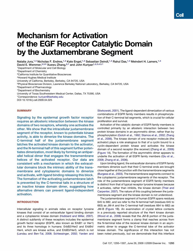
(Figure 1A). The formation of the asymmetric dimer appears to
underlie the activation of all EGFR family members (Qiu et al.,
2008; Zhang et al., 2006).
Upon binding ligand, the extracellular domains of EGFR family
members dimerize such that their C-terminal ends are brought
close together at the junction with the transmembrane segments
(Burgess et al., 2003). The transmembrane segments connect to
the cytoplasmic juxtamembrane segments of the receptor. The
role of the juxtamembrane segment of EGFR family members
is distinct from that of typical receptor tyrosine kinases because
it activates, rather than inhibits, the kinase domain (Thiel and
Carpenter, 2007). The nature of this coupling between the juxta-
membrane segment and the kinase domain is not understood.
The juxtamembrane segment of human EGFR spans residues
645 to 682, and we refer to the N-terminal half (residues 645 to
663) as JM-A and the C-terminal half (residues 664 to 682) as
JM-B (Figure 1B). An examination of crystal lattice contacts
in a previously reported structure of the Her4 kinase domain
(Wood et al., 2008) reveals that the JM-B portion of the juxta-
membrane segment forms a clamp that reaches across from
the N-terminal lobe of the receiver kinase domain in an asym-
metric dimer to engage the C-terminal lobe of the activator
kinase domain. The significance of this interaction has not
been noted previously, but we demonstrate that this interface
Cell 137, 1293–1307, June 26, 2009 ª2009 Elsevier Inc. 1293
A
B
Figure 1. Schematic Diagrams of EGFR
(A) Activation of EGFR by EGF results in the formation of an asymmetric kinase domain dimer.
(B) Domains of EGFR. Residue numbering corresponds to human EGFR, excluding the signal sequence.
involving JM-B, which we refer to as the ‘‘juxtamembrane latch,’’
is crucial for receptor activation.
We show that JM-A segments on the receiver and the activator
are both required for dimerization and activation, and we
propose that the two JM-A segments in an asymmetric kinase
domain dimer form short a helices that are likely to interact in
an antiparallel manner and connect to the C-terminal ends of
the dimeric form of the transmembrane helices. This allows
a model for the entire activated receptor to be built, in which
ligand engagement by the extracellular domains stabilizes the
formation of the JM-A helical dimer, which in turn stabilizes the
asymmetric kinase domain dimer, resulting in activation.
We have determined a structure of the EGFR kinase core in
which formation of the juxtamembrane latch is blocked by the
C-terminal tails of the receptor. This structure forms a symmet-
rical dimer of inactive kinase domains and suggests a potential
mechanism whereby alternative dimers can prevent ligand-inde-
pendent activation.
1294 Cell 137, 1293–1307, June 26, 2009 ª2009 Elsevier Inc.
RESULTS
The Juxtamembrane Segment Activates the KinaseDomain in SolutionThe core kinase domain has low activity when measured in solu-
tion with purified protein (the catalytic efficiency, kcat/KM, is
0.0049 ± 0.0005 s�1mM�1, Figure 2A). Introduction of the
L834R mutation, commonly found in lung cancer patients,
increases the activity of the kinase core by �14-fold, consistent
with previous results (Yun et al., 2007; Zhang et al., 2006)
(Figure 2A). Attachment of the juxtamembrane segment to the
wild-type kinase domain results in substantially greater activity.
The value of kcat/KM for the JM-kinase construct (0.33 ±
0.02 s�1mM�1) is �70-fold greater than that for the kinase core
alone. The activity of the kinase core is increased �20-fold by
concentrating it on lipid vesicles (Zhang et al., 2006), but the addi-
tionof the juxtamembranesegment results in greater catalytic effi-
ciency in solution, without concentration on vesicles (Figure 2A).
A B
D
C
Figure 2. The Effect of the Juxtamembrane Segment on Activity
(A) Catalytic efficiency (kcat/KM) of the kinase core (residues 672–998) in solution (yellow) and on vesicles (blue), compared to the catalytic efficiency in solution
of constructs that include the full juxtamembrane segment (JM-A and JM-B, residues 645–998) or only JM-B (residues 658–998). The values of kcat/KM were
obtained from the linear dependence of reaction velocity on substrate concentration at low substrate concentration, and the error bars are derived from the linear
fit (Zhang et al., 2006).
(B) The activity of constructs that are either receiver impaired (restricted to serve as activators, with the I682Q mutation) or activator impaired (restricted to serve
as receivers, with the V924R mutation). The values of kcat/KM were obtained from the linear dependence of reaction velocity on substrate concentration at low
substrate concentration, and the error bars are derived from the linear fit.
(C) Concentration-dependent change in specific activity, in solution, for the JM-kinase construct (containing both JM-A and JM-B, residues 645–998) and
a construct containing JM-B but lacking JM-A (residues 658–998). Data shown are mean values from two independent experiments ±STD.
(D) EGFR constructs were immunoprecipitated from cell lysates with an anti-FLAG antibody, and EGFR autophosphorylation was examined by immunobloting
with an anti-phosphotyrosine antibody (anti-pTyr).
Dimerization and Activation Requires IntactJuxtamembrane Segments on Both the Receiverand the Activator Kinase DomainsDeletion of the JM-A segment reduces catalytic efficiency by
10-fold, to 0.032 ± 0.003 s�1mM�1 (Figure 2A), showing that
an intact juxtamembrane segment is required for full activation.
In order to determine whether the JM-A segment is required on
both the activator and receiver, we made mutant kinases that
can only take the activator or the receiver position. We refer
to kinases with an I682Q mutation, which can take only the
Cell 137, 1293–1307, June 26, 2009 ª2009 Elsevier Inc. 1295
A
D
C
E
B
F
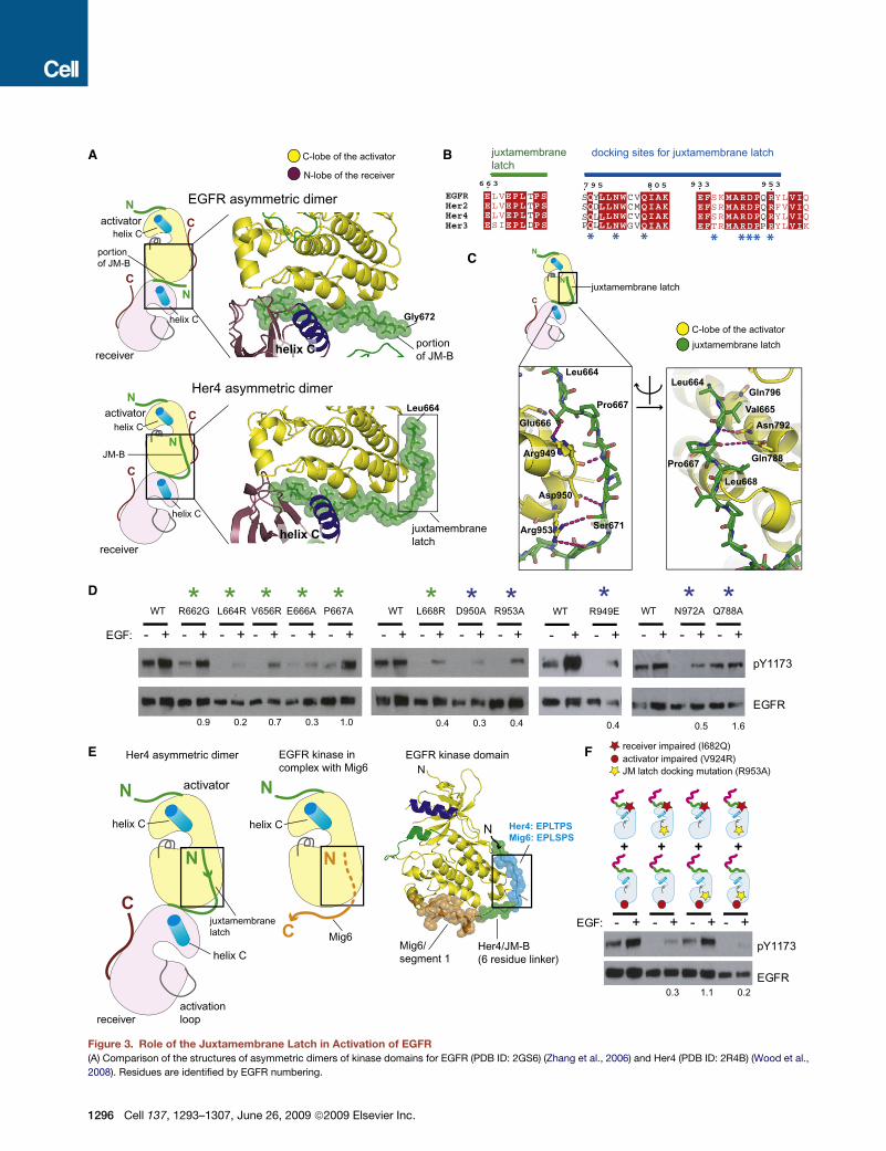
Figure 3. Role of the Juxtamembrane Latch in Activation of EGFR
(A) Comparison of the structures of asymmetric dimers of kinase domains for EGFR (PDB ID: 2GS6) (Zhang et al., 2006) and Her4 (PDB ID: 2R4B) (Wood et al.,
2008). Residues are identified by EGFR numbering.
1296 Cell 137, 1293–1307, June 26, 2009 ª2009 Elsevier Inc.
activator position, as ‘‘receiver impaired’’ (Zhang et al., 2006).
Kinases with a V924R mutation cannot be activated and are
referred to as ‘‘activator impaired.’’ Both the activator-impaired
and receiver-impaired JM-kinase constructs have low activity
alone, but mixing these constructs recovers�50% of the activity
of wild-type JM-kinase constructs in solution, as observed in cell
transfection studies (Thiel and Carpenter, 2007) (Figure 2B). In
contrast, activity is reduced significantly if the JM-A segment
on either the activator-impaired or the receiver-impaired kinase
is missing (Figure 2B). Thus, both JM-A and JM-B segments
are required on both the activator and the receiver kinases for
robust stimulation of activity.
The core kinase domain is a monomer, based on static light
scattering at a concentration of 150 mM, whereas the JM-kinase
construct is predominantly a dimer with no detectable monomer
(Figure S1 available online). The specific activity of the JM-kinase
construct increases with concentration, and the dissociation
constant (KD) for dimerization is estimated to be �200 nM
(Figure 2C). Deletion of the JM-A segment increases the
midpoint value of the concentration dependence curve to at least
�8 mM (Figure 2C). The >40-fold increase in the estimated values
for KD for dimerization upon deletion of the JM-A segment
provides strong evidence for a role of the JM-A segment in
dimerization.
Activation by the Transmembrane and JuxtamembraneSegments Is Comparable to the Effects of EnforcedDimerizationWe used cell-based assays to compare the activities of various
constructs containing the JM-kinase portion of the receptor.
One construct includes the transmembrane and intracellular
domains of EGFR (TM-ICD). The other construct is a fusion of
the intracellular domains to 29 residues of the coiled-coil portion
of the transcriptional activator GCN4 (O’Shea et al., 1991), which
is expected to enforce constitutive dimerization on the intracel-
lular domains.
We transfected COS7 cells with an �5-fold lower level of DNA
than used in the previous study (Thiel and Carpenter, 2007)
(Figure 2D). Under these conditions, the intracellular domains
alone display very low levels of autophosphorylation in an immu-
noprecipitation assay. In contrast, a construct that contains the
transmembrane segment, but without the extracellular segment
(TM-ICD), shows high activity. The activity of the TM-ICD
construct is about the same as that for the GCN4-ICD construct,
indicating that the effect of the juxtamembrane segment, when
fused to the transmembrane domain and localized to
membranes, is comparable to that of enforced dimerization.
This result is consistent with previous experiments showing that
removal of the extracellular domains activates the kinase domains
of EGFR (Chantry, 1995; Nishikawa et al., 1994; Zhu et al., 2003).
Many kinases are activated by imposed dimerization, where
the effect could simply be due to enhancement of transphos-
phorylation. Introduction of the V924R mutation in the GCN4-
ICD construct, which is expected to disrupt the asymmetric
dimer interface, results in a complete loss of activity (Figure 2D).
Thus, even when dimerization is enforced by GCN4, formation of
the asymmetric dimer appears to be essential for activity.
A Structure of the Her4 Kinase Domain Suggestsa Latching Function for the JM-B Segmentof the Juxtamembrane DomainA crystal structure of the Her4 kinase domain bound to a cova-
lently linked inhibitor (PDB ID: 2R4B) (Wood et al., 2008) provides
insight into the structural basis for the role of the juxtamembrane
segment. Crystallization of the Her4-inhibitor complex utilized a
construct that includes the JM-B portion of the juxtamembrane
segment (Wood et al., 2008). Although the authors do not
comment on this fact, the kinase domains in their crystal struc-
ture form an asymmetric dimer that is similar in general terms
to that seen in other crystal structures for active forms of EGFR
and Her4. The kinase domain of Her4 in this complex is in an
inactive conformation, which is required for accommodation of
the covalently bound inhibitor (Wood et al., 2008). Nevertheless,
the crystal lattice generates a ‘‘daisy chain’’ of kinase domains in
which each molecule in the chain is docked on the next one, as in
crystals of the active kinase domain.
The last ten residues of JM-B (corresponding to Gly 672 to Ile
682 in EGFR) have been visualized previously at the asymmetric
dimer interface, and are critical for EGFR activation (Zhang et al.,
2006). What is new in the Her4-inhibitor complex is that the rest
of the JM-B segment, provided by the kinase domain in the
receiver position, latches two kinase domains together by
running along the surface of the C-lobe of the activator kinase
domain (Figure 3A).
Formation of the juxtamembrane latch involves residues in the
receiver and activator kinases that are conserved in EGFR family
members (Figure 3B). The interaction involves several hydrogen
bonds and hydrophobic contacts (see Figure 3C). Mutations in
C-lobe residues that anchor the JM-B region (e.g., Asn 972,
Arg 949, Asp 950, Arg 953 in the C-lobe) have substantial inhib-
itory effects on EGFR autophosphorylation in cell-based assays
(Figure 3D). Mutation of Glu 666 in the JM-B region, which forms
an ion pair with Arg 949, is also inhibitory. Three hydrophobic
residues in the JM-B segment (Leu 664, Val 656, Leu 668) are
also essential for EGFR activation (Figure 3D). Leu 668 packs
against Pro 951 in the C-lobe of the kinase domain. Leu 664
and Val 665 are located near the junction of JM-B with the
N-terminal JM-A segment.
A segment spanning residues 315–374 of the EGFR inhibitor
Mig6 blocks asymmetric dimer formation (Zhang et al., 2007).
(B) Sequence conservation in the juxtamembrane latch/C-lobe interface. Residues forming the juxtamembrane latch are indicated by asterisks.
(C) Detailed view of the structure of the juxtamembrane latch in the Her4 structure (PDB ID: 2R4B), with residues identified by EGFR numbering.
(D) Effect of mutating residues involved in formation of the juxtamembrane latch. The level of EGF-stimulated phosphorylation on Tyr 1173 relative to the
wild-type, after normalizing for EGFR levels, is shown below each lane.
(E) Comparison of the juxtamembrane latch with the docking of the EGFR inhibitor, Mig6 (PDB ID: 2RFE).
(F) The effect of a mutation that prevents docking of the juxtamembrane latch (R953A). The results of cotransfection experiments with full-length EGFR receptor
variants that are receiver impaired (I682Q) or activator impaired (V924R) are shown. The level of EGF-stimulated phosphorylation relative to I682Q and V924R
cotransfection in the wild-type background, after normalizing for EGFR levels, is shown below each lane.
Cell 137, 1293–1307, June 26, 2009 ª2009 Elsevier Inc. 1297
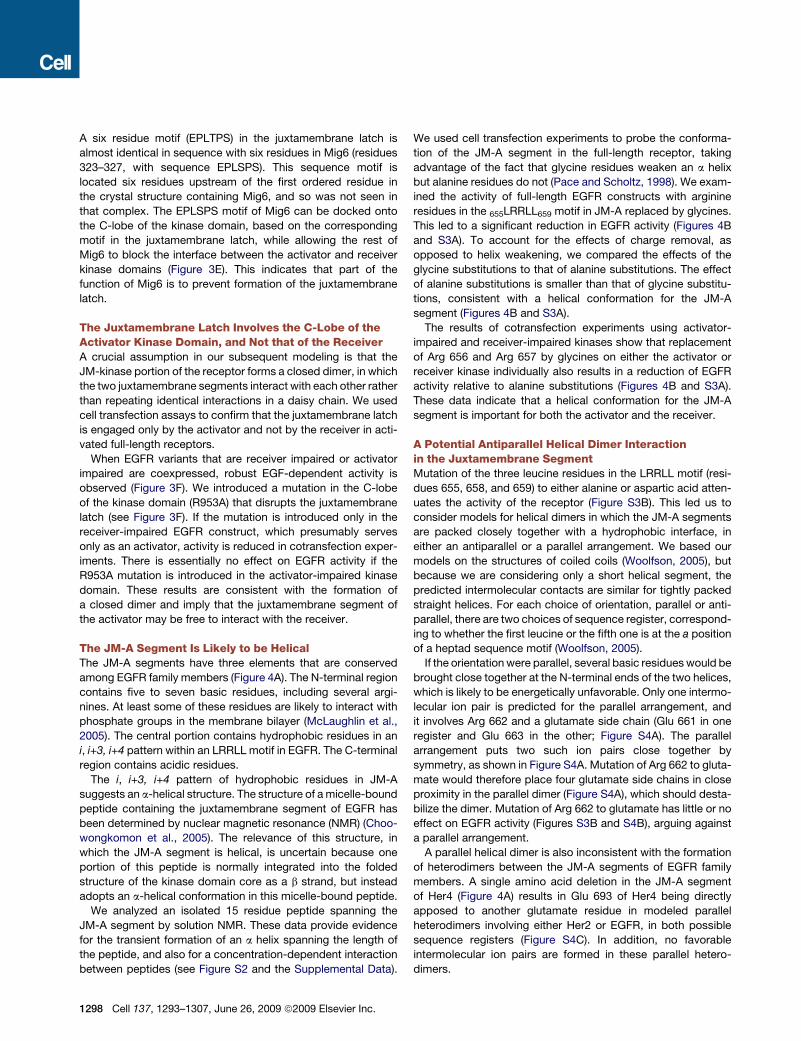
A six residue motif (EPLTPS) in the juxtamembrane latch is
almost identical in sequence with six residues in Mig6 (residues
323–327, with sequence EPLSPS). This sequence motif is
located six residues upstream of the first ordered residue in
the crystal structure containing Mig6, and so was not seen in
that complex. The EPLSPS motif of Mig6 can be docked onto
the C-lobe of the kinase domain, based on the corresponding
motif in the juxtamembrane latch, while allowing the rest of
Mig6 to block the interface between the activator and receiver
kinase domains (Figure 3E). This indicates that part of the
function of Mig6 is to prevent formation of the juxtamembrane
latch.
The Juxtamembrane Latch Involves the C-Lobe of theActivator Kinase Domain, and Not that of the ReceiverA crucial assumption in our subsequent modeling is that the
JM-kinase portion of the receptor forms a closed dimer, in which
the two juxtamembrane segments interact with each other rather
than repeating identical interactions in a daisy chain. We used
cell transfection assays to confirm that the juxtamembrane latch
is engaged only by the activator and not by the receiver in acti-
vated full-length receptors.
When EGFR variants that are receiver impaired or activator
impaired are coexpressed, robust EGF-dependent activity is
observed (Figure 3F). We introduced a mutation in the C-lobe
of the kinase domain (R953A) that disrupts the juxtamembrane
latch (see Figure 3F). If the mutation is introduced only in the
receiver-impaired EGFR construct, which presumably serves
only as an activator, activity is reduced in cotransfection exper-
iments. There is essentially no effect on EGFR activity if the
R953A mutation is introduced in the activator-impaired kinase
domain. These results are consistent with the formation of
a closed dimer and imply that the juxtamembrane segment of
the activator may be free to interact with the receiver.
The JM-A Segment Is Likely to be HelicalThe JM-A segments have three elements that are conserved
among EGFR family members (Figure 4A). The N-terminal region
contains five to seven basic residues, including several argi-
nines. At least some of these residues are likely to interact with
phosphate groups in the membrane bilayer (McLaughlin et al.,
2005). The central portion contains hydrophobic residues in an
i, i+3, i+4 pattern within an LRRLL motif in EGFR. The C-terminal
region contains acidic residues.
The i, i+3, i+4 pattern of hydrophobic residues in JM-A
suggests an a-helical structure. The structure of a micelle-bound
peptide containing the juxtamembrane segment of EGFR has
been determined by nuclear magnetic resonance (NMR) (Choo-
wongkomon et al., 2005). The relevance of this structure, in
which the JM-A segment is helical, is uncertain because one
portion of this peptide is normally integrated into the folded
structure of the kinase domain core as a b strand, but instead
adopts an a-helical conformation in this micelle-bound peptide.
We analyzed an isolated 15 residue peptide spanning the
JM-A segment by solution NMR. These data provide evidence
for the transient formation of an a helix spanning the length of
the peptide, and also for a concentration-dependent interaction
between peptides (see Figure S2 and the Supplemental Data).
1298 Cell 137, 1293–1307, June 26, 2009 ª2009 Elsevier Inc.
We used cell transfection experiments to probe the conforma-
tion of the JM-A segment in the full-length receptor, taking
advantage of the fact that glycine residues weaken an a helix
but alanine residues do not (Pace and Scholtz, 1998). We exam-
ined the activity of full-length EGFR constructs with arginine
residues in the 655LRRLL659 motif in JM-A replaced by glycines.
This led to a significant reduction in EGFR activity (Figures 4B
and S3A). To account for the effects of charge removal, as
opposed to helix weakening, we compared the effects of the
glycine substitutions to that of alanine substitutions. The effect
of alanine substitutions is smaller than that of glycine substitu-
tions, consistent with a helical conformation for the JM-A
segment (Figures 4B and S3A).
The results of cotransfection experiments using activator-
impaired and receiver-impaired kinases show that replacement
of Arg 656 and Arg 657 by glycines on either the activator or
receiver kinase individually also results in a reduction of EGFR
activity relative to alanine substitutions (Figures 4B and S3A).
These data indicate that a helical conformation for the JM-A
segment is important for both the activator and the receiver.
A Potential Antiparallel Helical Dimer Interactionin the Juxtamembrane SegmentMutation of the three leucine residues in the LRRLL motif (resi-
dues 655, 658, and 659) to either alanine or aspartic acid atten-
uates the activity of the receptor (Figure S3B). This led us to
consider models for helical dimers in which the JM-A segments
are packed closely together with a hydrophobic interface, in
either an antiparallel or a parallel arrangement. We based our
models on the structures of coiled coils (Woolfson, 2005), but
because we are considering only a short helical segment, the
predicted intermolecular contacts are similar for tightly packed
straight helices. For each choice of orientation, parallel or anti-
parallel, there are two choices of sequence register, correspond-
ing to whether the first leucine or the fifth one is at the a position
of a heptad sequence motif (Woolfson, 2005).
If the orientation were parallel, several basic residues would be
brought close together at the N-terminal ends of the two helices,
which is likely to be energetically unfavorable. Only one intermo-
lecular ion pair is predicted for the parallel arrangement, and
it involves Arg 662 and a glutamate side chain (Glu 661 in one
register and Glu 663 in the other; Figure S4A). The parallel
arrangement puts two such ion pairs close together by
symmetry, as shown in Figure S4A. Mutation of Arg 662 to gluta-
mate would therefore place four glutamate side chains in close
proximity in the parallel dimer (Figure S4A), which should desta-
bilize the dimer. Mutation of Arg 662 to glutamate has little or no
effect on EGFR activity (Figures S3B and S4B), arguing against
a parallel arrangement.
A parallel helical dimer is also inconsistent with the formation
of heterodimers between the JM-A segments of EGFR family
members. A single amino acid deletion in the JM-A segment
of Her4 (Figure 4A) results in Glu 693 of Her4 being directly
apposed to another glutamate residue in modeled parallel
heterodimers involving either Her2 or EGFR, in both possible
sequence registers (Figure S4C). In addition, no favorable
intermolecular ion pairs are formed in these parallel hetero-
dimers.
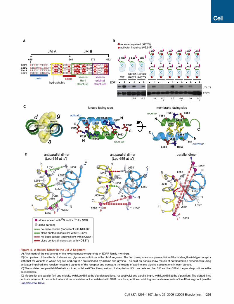
A B
C
D
Figure 4. A Helical Dimer in the JM-A Segment
(A) Alignment of the sequences of the juxtamembrane segments of EGFR family members.
(B) Comparison of the effects of alanine and glycine substitutions in the JM-A segment. The first three panels compare activity of the full-length wild-type receptor
with that for variants in which Arg 656 and Arg 657 are replaced by alanine and glycine. The next six panels show results of cotransfection experiments using
activator-impaired and receiver-impaired variants of the receptor and compare the results of alanine and glycine substitutions in each variant.
(C) The modeled antiparallel JM-A helical dimer, with Leu 655 at the d position of a heptad motif in one helix and Leu 658 and Leu 659 at the g and a positions in the
second helix.
(D) Models for antiparallel (left and middle, with Leu 655 at the d and a positions, respectively) and parallel (right, with Leu 655 at the d position). The dotted lines
indicate interatomic contacts that are either consistent or inconsistent with NMR data for a peptide containing two tandem repeats of the JM-A segment (see the
Supplemental Data).
Cell 137, 1293–1307, June 26, 2009 ª2009 Elsevier Inc. 1299
In an antiparallel coiled coil, residues at the a and d positions of
the heptad repeat in one helix interact with the residues at the
d and a positions, respectively, in the other (Woolfson, 2005).
Additional interhelical interactions can be made by the side
chains at the e or g positions. The LxxLL motif in EGFR JM-A
fits into this pattern if the first leucine is at either the a position
or the d position, with the second two leucines at the d and e posi-
tions or the g and a positions, respectively. We favor placement of
the first leucine at the d position because it provides a role for both
arginine residues in the LRRLL motif, consistent with the strong
effects of mutating these residues (Figures 4B and S3B). In this
pairing, Leu 658 and Leu 659 at the g and a positions in each helix
form a V-shaped crevice into which the side chain of Leu 655 at
the d position from the other helix is inserted (Figure 4C).
An antiparallel dimer leads naturally to interhelical ion pairing
between arginine or lysine side chains at the N-terminal end of
each helix with the acidic side chains located at the C-terminal
end of the partner helix, and so we favor this arrangement for
the helical dimer over a parallel one (Figures 4C and S5). The
pattern of interhelical ion pairs predicted in this way for EGFR
is also consistent with the formation of relevant heterodimer
pairs (Figure S6).
Additional support for the antiparallel model is provided by
NMR measurements on a 35 residue peptide containing two
copies of the JM-A segment of EGFR with a five residue flexible
spacer (see the Supplemental Data and Figure S7A). The first
and last leucine residues in the LRRLL motif in the first JM-A
segment (Leu 655 and Leu 659) are labeled with 15N. The second
glutamate in the first segment (Glu 663) is labeled with 15N and13C. As for the 15 residue JM-A peptide, NMR data for the tan-
demly linked JM-A segments provide evidence for transient
rather than stable adoption of helical structure in both JM-A
segments under the conditions of the NMR experiment (see
the Supplemental Data). Despite the transient nature of these
helices, the NMR data demonstrate that the two JM-A segments
in the peptide interact with each other.
Data from Nuclear Overhauser Effect (NOE) experiments are
consistent with an antiparallel rather than parallel orientation of
two JM-A helices. This is not surprising, since an antiparallel
interaction could occur in an intramolecular fashion within this
peptide, whereas a parallel interaction would require dimeriza-
tion. Nevertheless, the significance of these NMR data arises
from the evidence that they provide for a specific register in
the antiparallel interaction, in which the first leucine of the LRRLL
motif is at the d position of a heptad repeat, with the second two
leucines at the g and a positions, respectively (Figure 4D and the
Supplemental Data).
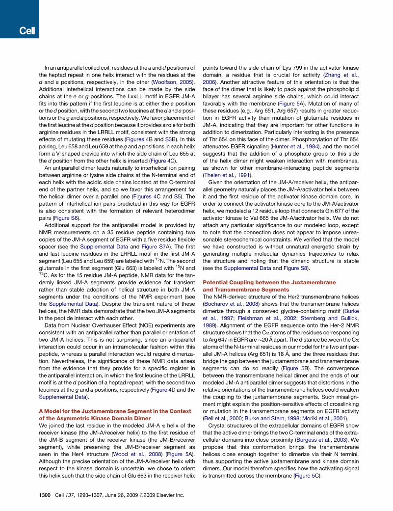
A Model for the Juxtamembrane Segment in the Contextof the Asymmetric Kinase Domain DimerWe joined the last residue in the modeled JM-A a helix of the
receiver kinase (the JM-A/receiver helix) to the first residue of
the JM-B segment of the receiver kinase (the JM-B/receiver
segment), while preserving the JM-B/receiver segment as
seen in the Her4 structure (Wood et al., 2008) (Figure 5A).
Although the precise orientation of the JM-A/receiver helix with
respect to the kinase domain is uncertain, we chose to orient
this helix such that the side chain of Glu 663 in the receiver helix
1300 Cell 137, 1293–1307, June 26, 2009 ª2009 Elsevier Inc.
points toward the side chain of Lys 799 in the activator kinase
domain, a residue that is crucial for activity (Zhang et al.,
2006). Another attractive feature of this orientation is that the
face of the dimer that is likely to pack against the phospholipid
bilayer has several arginine side chains, which could interact
favorably with the membrane (Figure 5A). Mutation of many of
these residues (e.g., Arg 651, Arg 657) results in greater reduc-
tion in EGFR activity than mutation of glutamate residues in
JM-A, indicating that they are important for other functions in
addition to dimerization. Particularly interesting is the presence
of Thr 654 on this face of the dimer. Phosphorylation of Thr 654
attenuates EGFR signaling (Hunter et al., 1984), and the model
suggests that the addition of a phosphate group to this side
of the helix dimer might weaken interaction with membranes,
as shown for other membrane-interacting peptide segments
(Thelen et al., 1991).
Given the orientation of the JM-A/receiver helix, the antipar-
allel geometry naturally places the JM-A/activator helix between
it and the first residue of the activator kinase domain core. In
order to connect the activator kinase core to the JM-A/activator
helix, we modeled a 12 residue loop that connects Gln 677 of the
activator kinase to Val 665 the JM-A/activator helix. We do not
attach any particular significance to our modeled loop, except
to note that the connection does not appear to impose unrea-
sonable stereochemical constraints. We verified that the model
we have constructed is without unnatural energetic strain by
generating multiple molecular dynamics trajectories to relax
the structure and noting that the dimeric structure is stable
(see the Supplemental Data and Figure S8).
Potential Coupling between the Juxtamembraneand Transmembrane SegmentsThe NMR-derived structure of the Her2 transmembrane helices
(Bocharov et al., 2008) shows that the transmembrane helices
dimerize through a conserved glycine-containing motif (Burke
et al., 1997; Fleishman et al., 2002; Sternberg and Gullick,
1989). Alignment of the EGFR sequence onto the Her-2 NMR
structure shows that the Ca atoms of the residues corresponding
to Arg 647 in EGFR are�20 A apart. The distance between the Ca
atoms of the N-terminal residues in our model for the two antipar-
allel JM-A helices (Arg 651) is 18 A, and the three residues that
bridge the gap between the juxtamembrane and transmembrane
segments can do so readily (Figure 5B). The convergence
between the transmembrane helical dimer and the ends of our
modeled JM-A antiparallel dimer suggests that distortions in the
relative orientations of the transmembrane helices could weaken
the coupling to the juxtamembrane segments. Such misalign-
ment might explain the position-sensitive effects of crosslinking
or mutation in the transmembrane segments on EGFR activity
(Bell et al., 2000; Burke and Stern, 1998; Moriki et al., 2001).
Crystal structures of the extracellular domains of EGFR show
that the active dimer brings the two C-terminal ends of the extra-
cellular domains into close proximity (Burgess et al., 2003). We
propose that this conformation brings the transmembrane
helices close enough together to dimerize via their N termini,
thus supporting the active juxtamembrane and kinase domain
dimers. Our model therefore specifies how the activating signal
is transmitted across the membrane (Figure 5C).
A
B
C
Figure 5. Structural Coupling between the Extracellular and Intracellular Domains in Active EGFR
(A) A model for the JM-A helical dimer in the context of the asymmetric dimer of kinase domains. In the exploded view, arginine side chains that face the
membrane are shown.
(B) Structure of the transmembrane domain dimer of Her2 (PDB ID: 2JWA), (Bocharov et al., 2008) and the modeled JM-A dimer. Positively charged side chains
that face the membrane are in blue.
(C) A model for the activated EGF receptor. Two liganded EGFR extracellular domains are shown in an active dimeric assembly (PDB ID: 1IVO) (Ogiso et al., 2002),
with domain IV based on the structure of the inactive EGFR extracellular domain (PDB ID: 1NQL) (Ferguson et al., 2003). This arrangement is compatible with the
transmembrane domain dimer and couples to the asymmetric kinase domain dimer via the dimeric JM-A helices and the juxtamembrane latch.
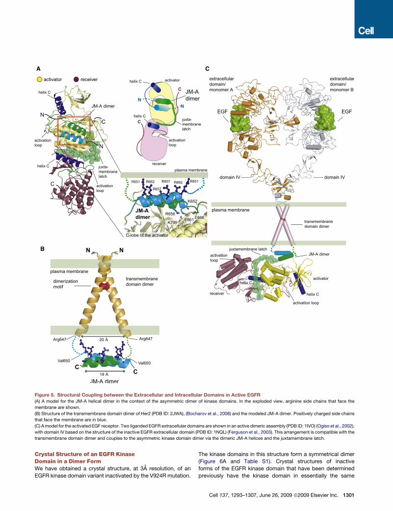
Crystal Structure of an EGFR KinaseDomain in a Dimer FormWe have obtained a crystal structure, at 3A resolution, of an
EGFR kinase domain variant inactivated by the V924R mutation.
The kinase domains in this structure form a symmetrical dimer
(Figure 6A and Table S1). Crystal structures of inactive
forms of the EGFR kinase domain that have been determined
previously have the kinase domain in essentially the same
Cell 137, 1293–1307, June 26, 2009 ª2009 Elsevier Inc. 1301
A
C
B
E
D
Figure 6. A Symmetric Inactive Dimer of the EGFR Kinase Domain
(A) Overview of the crystal structure of the symmetric inactive dimer.
(B) Detailed view of the hydrophobic packing between the C-terminal AP-2 helix of monomer B and the N-lobe of monomer A.
(C) Exploded view of the electrostatic hook formed between the C-terminal tail (residues 979–990) of EGFR and the hinge region in the kinase domain.
(D) Effect of mutations in the electrostatic hook on autophosphorylation of full-length EGFR in COS7 cells.
(E) Alignment of the sequences of EGFR family members in the C-terminal tail regions encompassing residues in the electrostatic hook and AP-2 helix.
conformation as in our structure, but do not show extensive
contacts within symmetrical dimers (Wood et al., 2004; Xu
et al., 2008; Zhang et al., 2006). These structures were deter-
mined in the presence of salts that might disrupt the electrostatic
interactions that are at the center of the dimer interface of our
crystal form, which is obtained under low-salt conditions (see
the Experimental Procedures). A symmetrical EGFR dimer
1302 Cell 137, 1293–1307, June 26, 2009 ª2009 Elsevier Inc.
described previously has the kinase domains in an active confor-
mation (Landau et al., 2004).
Dimer formation in this crystal form is mediated principally by
the C-terminal tail of the kinase core (Figure 6A). There are four
independent molecules in the asymmetric unit, designated A,
B, C, and D, which form two nearly identical dimers (A:B and
C:D). In one molecule (A), electron density for the C-terminal
segment is visualized up to Asp 990. The residues between Ser
967 and Met 978 form an a helix (the AP-2 helix; see below),
which is followed by a five residue turn spanning Asp 979 to
Met 983. The last seven residues in the structural model, Asp
984 to Asp 990, run along the surface of the C-lobe of the kinase
domain in an extended conformation. In molecule D, there is no
electron density for the extended strand (residues 984 to 990),
and this region is blocked by crystal contacts. Electron density
for the extended strand is present but weak in molecules B
and C. Subsequent discussion of the dimer interactions is
focused on the A:B dimer. The portion of the C-terminal tail (resi-
dues 967 to 983) that is of interest here is mainly disordered in
structures of the active conformation of the EGFR kinase domain
(Stamos et al., 2002) and is partially ordered but in a different
conformation in the other inactive structures (Wood et al.,
2004; Zhang et al., 2007).
The Inactive Kinase Domain Dimer May SuppressActivity Prior to EGF BindingSeveral studies have shown that EGFR dimerizes prior to EGF
binding (Clayton et al., 2008; Sako et al., 2000). Although these
preformed dimers are likely to involve the intracellular domain
(Yu et al., 2002), the orientation of the kinase domains in these
preformed dimers is unknown. Our inactive dimer has several
features, which suggests that it could play a role in the inhibition
of kinase activity.
Each AP-2 helix in the C-terminal tail of one kinase subunit
interacts with the other subunit, burying �1400A2 of surface
area at each interface. The helix encompasses residues 973 to
977, which form the recognition element in EGFR for the AP-2
clathrin adaptor protein (Sorkin et al., 1996). The recognition of
AP-2 by EGFR is dependent on activation by EGF (Sorkin and
Carpenter, 1993), and our structure shows how an inactive
form of EGFR can sequester the AP-2 recognition motif. The
interactions made by the AP-2 helix are reminiscent of those
made by the SH2-kinase linker in inactive Src family kinases
(Figure 6B) (Sicheri et al., 1997; Xu et al., 1997). In particular,
the engagement of the N-lobe of the adjacent kinase domain
by the side chain of Phe 973 in the C-terminal tail of EGFR is anal-
ogous to interactions made by the side chain of Leu 255 in the
SH2-kinase linker of c-Src (Xu et al., 1997).
Acidic side chains (Asp 979, Glu 980, Glu 981) in the turn after
the AP-2 helix form ion pairs with residues in the kinase domain
(His 749, His 826, Lys 828, and Lys 822) (Figure 6C). The turn,
referred to as an ‘‘electrostatic hook,’’ is located near the hinge
region of the kinase domain and near the aC/b4 loop. In
ZAP-70 and in certain other tyrosine kinases, the formation of
a hydrogen-bonded network similar to that seen here in EGFR
has been correlated with the inhibition of kinase activity (Chen
et al., 2007; Deindl et al., 2007).
The conformation of the portion of the C-terminal tail that
follows the electrostatic hook (residues 982 to 990) and runs along
the surface of the C-lobe of the kinasemimics the manner in which
the JM-B segment of the receiver kinase domain engages the
same surface of the activator kinase domain when forming the
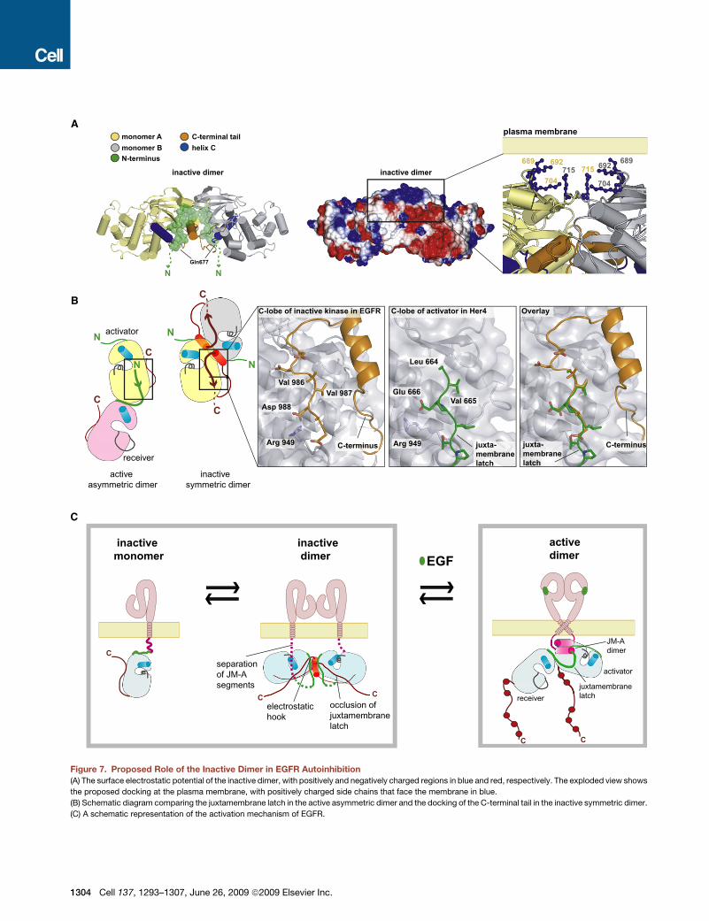
juxtamembrane latch (Figure 7B). Thus, formation of the inactive
dimer blocks formation of the activating juxtamembrane latch, in
a manner similar to that postulated for Mig6 (Figure 3E).
The key residues in the AP-2 helix, the electrostatic hook, and
the region of the C-terminal tail that interacts with the JM-B
binding interface, as well as the basic residues that interact
with the electrostatic hook, are conserved between EGFR and
its kinase-active homologs (Her2 and Her4) (Figure 6E). The resi-
dues that form the electrostatic hook are absent in Her3, as are
two of the three basic residues in the kinase domain. Presum-
ably, the lack of kinase activity in Her3 renders inhibition of
kinase activity unnecessary.
The surface electrostatic potential of the inactive dimer is
strongly polarized (Figures 7A and S9). Eight lysine residues
(residues 689, 692, 704, and 715 in each subunit) are clustered
together on the face of the dimer that is opposite to the internally
engaged position of the C-terminal tail, which is a region of nega-
tive electrostatic potential. The lysine residues are conserved
among EGFR family members. We speculate that the inactive
dimer might be oriented with respect to the membrane such
that the lysine residues can interact with negatively charged lipid
head groups. The juxtamembrane segments would then be
located on the far side of the dimer with respect to the membrane
and dimerization of the JM-A segments would be prevented
(Figure 7A).
Effect of Mutations in the C-Terminal Tailon Activity of EGFRCell transfection experiments show that mutations in the electro-
static hook result in substantial activation of EGFR in the
absence of EGF, as well an increased response to EGF, consis-
tent with a role for this region in inhibiting EGFR kinase activity
(Figure 6D). Notably, several insertions in exon 20 that drive
constitutive EGFR activation are detected in lung cancer patients
(Shigematsu et al., 2005; Greulich et al., 2005). These insertions
are in the b4/aC loop and are likely to disturb the electrostatic
hook. Mutation of Lys 828, located within the electrostatic
hook, increases the basal level of EGFR autophosphorylation
(Zhang et al., 2006).
Surprisingly, deletion of the AP-2 helix or mutations of residues
within this helix resulted consistently in impaired EGFR function
in the cell-based assay (Figures S10A and S10B). These muta-
tions result in activation in vitro (Figure S10C), demonstrating
that the AP-2 helix is not required for the integrity of the catalytic
domain. Mutation of residues in the AP-2 recognition motif does
not lead to an obvious change in kinetics of EGFR internalization,
presumably because of redundancy in endocytic targeting
signals (Sorkin et al., 1996). The observed reduction in activity
in cell-based assays suggests that the AP-2 helix plays a role
in the activation mechanism, perhaps by presenting the
C-terminal tails for phosphorylation.
DISCUSSION
We propose a structural mechanism for the activation of the
EGFR receptor by integrating our model for the juxtamembrane
segment with the separately determined structures of the trans-
membrane and extracellular domains (Figure 5C). In this model,
the proximity of the C-terminal ends of the activated extracellular
domains (Burgess et al., 2003) stabilizes the dimer of transmem-
brane helices observed in the Her2 NMR structure (Bocharov
Cell 137, 1293–1307, June 26, 2009 ª2009 Elsevier Inc. 1303
A
C
B
Figure 7. Proposed Role of the Inactive Dimer in EGFR Autoinhibition
(A) The surface electrostatic potential of the inactive dimer, with positively and negatively charged regions in blue and red, respectively. The exploded view shows
the proposed docking at the plasma membrane, with positively charged side chains that face the membrane in blue.
(B) Schematic diagram comparing the juxtamembrane latch in the active asymmetric dimer and the docking of the C-terminal tail in the inactive symmetric dimer.
(C) A schematic representation of the activation mechanism of EGFR.
1304 Cell 137, 1293–1307, June 26, 2009 ª2009 Elsevier Inc.
et al., 2008). This provides the proper geometry for the formation
of the helical JM-A dimer and the juxtamembrane latch, which
activate the kinase domain by stabilizing the asymmetric dimer.
Our model suggests that an important role for the extracellular
domain is to inhibit the formation of the activated kinase domain
dimer prior to ligand binding, as proposed earlier (Chantry, 1995;
Nishikawa et al., 1994; Zhu et al., 2003). Our model also provides
an explanation for why the insertion of a flexible linker between
the extracellular domain and the transmembrane segment acti-
vates EGFR in the absence of ligand (Sorokin, 1995), since
such a linker would decouple the conformation of the extracel-
lular domains from the transmembrane helices. It appears that
merely allowing the transmembrane segments to interact in
a manner consistent with dimeric engagement of the JM-A
helices suffices for activation.
In the absence of ligand, the extracellular domains of EGFR
adopt a compact conformation that converts to an extended
conformation upon ligand binding (Burgess et al., 2003). Conver-
sion of the extracellular domain to the active conformation
without dimerization is by itself insufficient for activation (Mat-
toon et al., 2004). It appears that the extracellular segments
either prevent interaction in the absence of ligand or else
dimerize in a way that keeps their C-terminal ends at a distance
that is sufficient to prevent the transmembrane segments from
stabilizing the dimeric interaction between the juxtamembrane
helices. Separation of the C-terminal ends of the extracellular
domains in an inactive dimer would be consistent with coupling
to the inactive kinase domain dimer that we have described here.
Our structure of an inactive kinase domain dimer reveals that
the C-terminal tails are central to the extensive inactive dimer
interface that blocks formation of the juxtamembrane latch.
Analysis of EGFR mutations in cancer patients has shown that
deletion of the C-terminal tail can drive constitutive EGFR activa-
tion (Frederick et al., 2000). The formation of preformed EGFR
dimers has been documented (Clayton et al., 2008; Sako et al.,
2000), and the intracellular domain is implicated in formation of
these dimers (Yu et al., 2002). Thus, inactive kinase dimers
may constitute a key autoinhibitory mechanism that prevents
ligand-independent activation. The extracellular domains deter-
mine the balance between inactive and active dimeric states of
the kinase domain, with inactive monomers and dimers predom-
inating prior to EGF stimulation, and active dimers, or higher-
order assemblies, predominating after stimulation (Clayton
et al., 2008) (Figure 7C). In this model, both the extracellular
domains and the inactive dimer work synergistically to prevent
the high local concentration of receptors at the membrane
from generating spurious signals through uncontrolled trans-
phosphorylation. Determining the mechanism by which this
balance is achieved remains an important focus for further study.
EXPERIMENTAL PROCEDURES
Protein Expression and Purification
Human EGFR kinase core constructs (residues 672:998) were expressed and
purified as described (Zhang et al., 2006). JM-kinase constructs (residues
645–998 and 658–998) were expressed and purified with the same protocol.
All protein constructs contained N-terminal 6His tags (Zhang et al., 2006).
Mutations were introduced with the Quick-Change kit (Stratagene) and
confirmed by DNA sequencing.
In Vitro Kinase Assays
Kinase activity was measured with a continuous enzyme-coupled kinase
assay in solution and on vesicles as described (Zhang et al., 2006) with the
substrate peptide corresponding to the Tyr 1173 phosphorylation site in
EGFR (TAENAEYLRVAPQ). The details of the reaction conditions are given
in Table S2. The concentration dependence of activity was measured with
poly-4Glu:Tyr used as a substrate (Sigma), at a substrate concentration signif-
icantly above the KM value (3 mg/ml).
Crystallization and Structure Determination
The crystal form containing the EGFR inactive symmetric dimer was obtained
during an attempt to crystallize a complex of the EGFR kinase core (V924R)
with the kinase domain of Her3. Crystals (space group P21, a = 61.9 A, b =
72.5 A, c = 143.5 A, b = 101.7�) were obtained from solutions containing
a 1:1 mixture of the EGFR and Her3 kinase domains at 6 mg/ml, 5 mM
AMP-PNP, 2 mM MgCl2, 0.1 M Bis-Tris (pH 5.5), 0.1 M ammonium acetate,
and 17% w/v PEG 10,000. Refinement of the structure to 3 A with PHENIX
(Adams et al., 2002) showed unambiguously that the structure was that of
an EGFR dimer and that the crystals did not contain Her3. We then crystallized
the EGFR kinase domain alone, obtaining the same crystal form (space group
P21, a = 62.6 A, b = 73.3 A, c = 144.5 A, b = 102.0�), containing the same pair of
dimers, with essentially the same structure. The resolution limit for these data
is slightly lower (3.2 A), and our analysis is based on the structure obtained at
higher resolution. Data collection and refinement statistics are in the Supple-
mental Data.
Mammalian Cell-Based Assays
DNA encoding the full-length human EGFR gene with an N-terminal FLAG tag
in pcDNA3.1 mammalian expression vector was used in cell-based assays, as
described (Zhang et al., 2006). The AP-2 helix deletion was constructed by
deletion of residues 973–978 in full-length EGFR. The JM-kinase and TM-
JM-kinase constructs were cloned into pcDNA3.1 (Invitrogen), and a FLAG
tag was inserted N-terminal to the EGFR sequence. The GCN4-JM-kinase
construct was generated by insertion of the GCN4 sequence (RVKQLEDK
VEELLSKNAHLENEVARLKKL) N-terminal to the JM-kinase constructs.
COS7 cells were cultured in Dulbecco’s Modified Eagle’s Medium supple-
mented with 10% fetal bovine serum (FBS) and streptomycin/penicillin. Cells
were transiently transfected with Fugene 6 (Roche) and cultured for 24 hr after
transfection, followed by 12 hr serum starvation. Cells were treated with EGF
(50 ng/ml, PeproTech) for 5 min at 37�C and lysed in lysis buffer (50 mM Tris
[pH 7.5], 150 mM NaCl, 1 mM EDTA, 1 mM Na3VO4, 1 mM NaF, 1% Triton
X-100, and a protease inhibitor cocktail [Roche]). Cell lysates were subjected
to western blot analyses with the following primary antibodies: anti-EGFR,
sc-03, polyclonal (Santa Cruz), anti-phosphoTyr 4G10 (Upstate), and EGFR
site-specific phosphorylation antibodies, Tyr1173 (Santa Cruz), Tyr974 (Cell
Signaling), Tyr992 (Cell Signaling), Tyr1045 (Cell Signaling), and Tyr1068
(Cell Signaling). Secondary antibodies used were either goat anti-mouse (GE
Healthcare) or goat anti-rabbit (Cell Signaling) coupled to horseradish peroxi-
dase. For immunoprecipitation assay, cells were lysed in the lysis buffer,
cleared by centrifugation, and incubated with anti-FLAG monoclonal antibody
M2 (Sigma-Aldrich) followed by incubation with 1:1 protein A-sepharose slurry
(Sigma-Aldrich). The immunoprecipitates were analyzed by western blotting
with the indicated antibodies.
ACCESSION NUMBERS
The coordinates for structure of the inactive EGFR dimer solved in this study
were deposited in the Protein Data Bank with ID code 3GT8.
SUPPLEMENTAL DATA
Supplemental Data include Supplemental Results, Supplemental Experi-
mental Procedures, ten figures, and two tables and can be found with this
article online at http://www.cell.com/supplemental/S0092-8674(09)00450-4.
Cell 137, 1293–1307, June 26, 2009 ª2009 Elsevier Inc. 1305
ACKNOWLEDGMENTS
We thank Deborah Makino for help with structure determination, Xiaoxian Cao
for Sf9 cell culture, David King for peptide synthesis, and Jeffrey Pelton for help
with NMR experiments. We thank Tony Hunter, Nir Ben-Tal, Alexander Sorkin,
Susan Marqusee, and members of the Kuriyan laboratory for helpful discus-
sions. This work is supported in part by grant from the National Cancer Institute
to J.K. (RO1 CA96504-06). N.E. is a fellow of the Leukemia and Lymphoma
Society. We thank the staff at the Advanced Light Source, which is supported
by U.S. Department of Energy under contract DE-AC03-76SF00098 at the
Lawrence Berkeley National Laboratory. NMR instrumentation and operation
were supported by NIH-GM 68933.
Received: December 14, 2008
Revised: March 9, 2009
Accepted: April 2, 2009
Published: June 25, 2009
REFERENCES
Adams, P.D., Grosse-Kunstleve, R.W., Hung, L.W., Ioerger, T.R., McCoy, A.J.,
Moriarty, N.W., Read, R.J., Sacchettini, J.C., Sauter, N.K., and Terwilliger, T.C.
(2002). PHENIX: building new software for automated crystallographic struc-
ture determination. Acta Crystallogr. D Biol. Crystallogr. 58, 1948–1954.
Bell, C.A., Tynan, J.A., Hart, K.C., Meyer, A.N., Robertson, S.C., and
Donoghue, D.J. (2000). Rotational coupling of the transmembrane and kinase
domains of the Neu receptor tyrosine kinase. Mol. Biol. Cell 11, 3589–3599.
Bocharov, E.V., Mineev, K.S., Volynsky, P.E., Ermolyuk, Y.S., Tkach, E.N.,
Sobol, A.G., Chupin, V.V., Kirpichnikov, M.P., Efremov, R.G., and Arseniev,
A.S. (2008). Spatial structure of the dimeric transmembrane domain of the
growth factor receptor ErbB2 presumably corresponding to the receptor
active state. J. Biol. Chem. 283, 6950–6956.
Burgess, A.W., Cho, H.S., Eigenbrot, C., Ferguson, K.M., Garrett, T.P., Leahy,
D.J., Lemmon, M.A., Sliwkowski, M.X., Ward, C.W., and Yokoyama, S. (2003).
An open-and-shut case? Recent insights into the activation of EGF/ErbB
receptors. Mol. Cell 12, 541–552.
Burke, C.L., Lemmon, M.A., Coren, B.A., Engelman, D.M., and Stern, D.F.
(1997). Dimerization of the p185neu transmembrane domain is necessary
but not sufficient for transformation. Oncogene 14, 687–696.
Burke, C.L., and Stern, D.F. (1998). Activation of Neu (ErbB-2) mediated by
disulfide bond-induced dimerization reveals a receptor tyrosine kinase dimer
interface. Mol. Cell. Biol. 18, 5371–5379.
Chantry, A. (1995). The kinase domain and membrane localization determine
intracellular interactions between epidermal growth factor receptors. J. Biol.
Chem. 270, 3068–3073.
Chen, H., Ma, J., Li, W., Eliseenkova, A.V., Xu, C., Neubert, T.A., Miller, W.T.,
and Mohammadi, M. (2007). A molecular brake in the kinase hinge region regu-
lates the activity of receptor tyrosine kinases. Mol. Cell 27, 717–730.
Choowongkomon, K., Carlin, C.R., and Sonnichsen, F.D. (2005). A structural
model for the membrane-bound form of the juxtamembrane domain of the
epidermal growth factor receptor. J. Biol. Chem. 280, 24043–24052.
Clayton, A.H., Orchard, S.G., Nice, E.C., Posner, R.G., and Burgess, A.W.
(2008). Predominance of activated EGFR higher-order oligomers on the cell
surface. Growth Factors 26, 316–324.
Deindl, S., Kadlecek, T.A., Brdicka, T., Cao, X., Weiss, A., and Kuriyan, J.
(2007). Structural basis for the inhibition of tyrosine kinase activity of
ZAP-70. Cell 129, 735–746.
Ferguson, K.M., Berger, M.B., Mendrola, J.M., Cho, H.S., Leahy, D.J., and
Lemmon, M.A. (2003). EGF activates its receptor by removing interactions
that autoinhibit ectodomain dimerization. Mol. Cell 11, 507–517.
Fleishman, S.J., Schlessinger, J., and Ben-Tal, N. (2002). A putative molecular-
activation switch in the transmembrane domain of erbB2. Proc. Natl. Acad.
Sci. USA 99, 15937–15940.
1306 Cell 137, 1293–1307, June 26, 2009 ª2009 Elsevier Inc.
Frederick, L., Wang, X.Y., Eley, G., and James, C.D. (2000). Diversity and
frequency of epidermal growth factor receptor mutations in human glioblas-
tomas. Cancer Res. 60, 1383–1387.
Gotoh, N., Tojo, A., Hino, M., Yazaki, Y., and Shibuya, M. (1992). A highly
conserved tyrosine residue at codon 845 within the kinase domain is not
required for the transforming activity of human epidermal growth factor
receptor. Biochem. Biophys. Res. Commun. 186, 768–774.
Greulich, H., Chen, T.H., Feng, W., Janne, P.A., Alvarez, J.V., Zappaterra, M.,
Bulmer, S.E., Frank, D.A., Hahn, W.C., Sellers, W.R., and Meyerson, M. (2005).
Oncogenic transformation by inhibitor-sensitive and -resistant EGFR mutants.
PLoS Med. 2, e313.
Hubbard, S.R., and Miller, W.T. (2007). Receptor tyrosine kinases: mecha-
nisms of activation and signaling. Curr. Opin. Cell Biol. 19, 117–123.
Hunter, T., Ling, N., and Cooper, J.A. (1984). Protein kinase C phosphorylation
of the EGF receptor at a threonine residue close to the cytoplasmic face of the
plasma membrane. Nature 311, 480–483.
Landau, M., and Ben-Tal, N. (2008). Dynamic equilibrium between multiple
active and inactive conformations explains regulation and oncogenic muta-
tions in ErbB receptors. Biochim. Biophys. Acta 1785, 12–31.
Landau, M., Fleishman, S.J., and Ben-Tal, N. (2004). A putative mechanism for
downregulation of the catalytic activity of the EGF receptor via direct contact
between its kinase and C-terminal domains. Structure 12, 2265–2275.
Mattoon, D., Klein, P., Lemmon, M.A., Lax, I., and Schlessinger, J. (2004). The
tethered configuration of the EGF receptor extracellular domain exerts only
a limited control of receptor function. Proc. Natl. Acad. Sci. USA 101, 923–928.
McLaughlin, S., Smith, S.O., Hayman, M.J., and Murray, D. (2005). An electro-
static engine model for autoinhibition and activation of the epidermal growth
factor receptor (EGFR/ErbB) family. J. Gen. Physiol. 126, 41–53.
Moriki, T., Maruyama, H., and Maruyama, I.N. (2001). Activation of preformed
EGF receptor dimers by ligand-induced rotation of the transmembrane
domain. J. Mol. Biol. 311, 1011–1026.
Nishikawa, R., Ji, X.D., Harmon, R.C., Lazar, C.S., Gill, G.N., Cavenee, W.K.,
and Huang, H.J. (1994). A mutant epidermal growth factor receptor common
in human glioma confers enhanced tumorigenicity. Proc. Natl. Acad. Sci.
USA 91, 7727–7731.
Ogiso, H., Ishitani, R., Nureki, O., Fukai, S., Yamanaka, M., Kim, J.H., Saito, K.,
Sakamoto, A., Inoue, M., Shirouzu, M., and Yokoyama, S. (2002). Crystal
structure of the complex of human epidermal growth factor and receptor extra-
cellular domains. Cell 110, 775–787.
O’Shea, E.K., Klemm, J.D., Kim, P.S., and Alber, T. (1991). X-ray structure of
the GCN4 leucine zipper, a two-stranded, parallel coiled coil. Science 254,
539–544.
Pace, C.N., and Scholtz, J.M. (1998). A helix propensity scale based on exper-
imental studies of peptides and proteins. Biophys. J. 75, 422–427.
Qiu, C., Tarrant, M.K., Choi, S.H., Sathyamurthy, A., Bose, R., Banjade, S., Pal,
A., Bornmann, W.G., Lemmon, M.A., Cole, P.A., and Leahy, D.J. (2008). Mech-
anism of activation and inhibition of the HER4/ErbB4 kinase. Structure 16,
460–467.
Sako, Y., Minoghchi, S., and Yanagida, T. (2000). Single-molecule imaging of
EGFR signalling on the surface of living cells. Nat. Cell Biol. 2, 168–172.
Schlessinger, J. (2002). Ligand-induced, receptor-mediated dimerization and
activation of EGF receptor. Cell 110, 669–672.
Shigematsu, H., Takahashi, T., Nomura, M., Majmudar, K., Suzuki, M., Lee, H.,
Wistuba, I.I., Fong, K.M., Toyooka, S., Shimizu, N., et al. (2005). Somatic muta-
tions of the HER2 kinase domain in lung adenocarcinomas. Cancer Res. 65,
1642–1646.
Sicheri, F., Moarefi, I., and Kuriyan, J. (1997). Crystal structure of the Src family
tyrosine kinase Hck. Nature 385, 602–609.
Sorkin, A., and Carpenter, G. (1993). Interaction of activated EGF receptors
with coated pit adaptins. Science 261, 612–615.
Sorkin, A., Mazzotti, M., Sorkina, T., Scotto, L., and Beguinot, L. (1996).
Epidermal growth factor receptor interaction with clathrin adaptors is
mediated by the Tyr974-containing internalization motif. J. Biol. Chem. 271,
13377–13384.
Sorokin, A. (1995). Activation of the EGF receptor by insertional mutations in its
juxtamembrane regions. Oncogene 11, 1531–1540.
Stamos, J., Sliwkowski, M.X., and Eigenbrot, C. (2002). Structure of the
epidermal growth factor receptor kinase domain alone and in complex with
a 4-anilinoquinazoline inhibitor. J. Biol. Chem. 277, 46265–46272.
Sternberg, M.J., and Gullick, W.J. (1989). Neu receptor dimerization. Nature
339, 587.
Thelen, M., Rosen, A., Nairn, A.C., and Aderem, A. (1991). Regulation by phos-
phorylation of reversible association of a myristoylated protein kinase C
substrate with the plasma membrane. Nature 351, 320–322.
Thiel, K.W., and Carpenter, G. (2007). Epidermal growth factor receptor
juxtamembrane region regulates allosteric tyrosine kinase activation. Proc.
Natl. Acad. Sci. USA 104, 19238–19243.
Wood, E.R., Shewchuk, L.M., Ellis, B., Brignola, P., Brashear, R.L., Caferro,
T.R., Dickerson, S.H., Dickson, H.D., Donaldson, K.H., Gaul, M., et al. (2008).
6-Ethynylthieno[3,2-d]- and 6-ethynylthieno[2,3-d]pyrimidin-4-anilines as
tunable covalent modifiers of ErbB kinases. Proc. Natl. Acad. Sci. USA 105,
2773–2778.
Wood, E.R., Truesdale, A.T., McDonald, O.B., Yuan, D., Hassell, A., Dickerson,
S.H., Ellis, B., Pennisi, C., Horne, E., Lackey, K., et al. (2004). A unique struc-
ture for epidermal growth factor receptor bound to GW572016 (Lapatinib):
relationships among protein conformation, inhibitor off-rate, and receptor
activity in tumor cells. Cancer Res. 64, 6652–6659.
Woolfson, D.N. (2005). The design of coiled-coil structures and assemblies.
Adv. Protein Chem. 70, 79–112.
Xu, G., Searle, L.L., Hughes, T.V., Beck, A.K., Connolly, P.J., Abad, M.C.,
Neeper, M.P., Struble, G.T., Springer, B.A., Emanuel, S.L., et al. (2008).
Discovery of novel 4-amino-6-arylaminopyrimidine-5-carbaldehyde oximes
as dual inhibitors of EGFR and ErbB-2 protein tyrosine kinases. Bioorg.
Med. Chem. Lett. 18, 3495–3499.
Xu, W., Harrison, S.C., and Eck, M.J. (1997). Three-dimensional structure of
the tyrosine kinase c-Src. Nature 385, 595–602.
Yarden, Y., and Sliwkowski, M.X. (2001). Untangling the ErbB signalling
network. Nat. Rev. Mol. Cell Biol. 2, 127–137.
Yu, X., Sharma, K.D., Takahashi, T., Iwamoto, R., and Mekada, E. (2002).
Ligand-independent dimer formation of epidermal growth factor receptor
(EGFR) is a step separable from ligand-induced EGFR signaling. Mol. Biol.
Cell 13, 2547–2557.
Yun, C.H., Boggon, T.J., Li, Y., Woo, M.S., Greulich, H., Meyerson, M., and
Eck, M.J. (2007). Structures of lung cancer-derived EGFR mutants and inhib-
itor complexes: mechanism of activation and insights into differential inhibitor
sensitivity. Cancer Cell 11, 217–227.
Zhang, X., Gureasko, J., Shen, K., Cole, P.A., and Kuriyan, J. (2006). An allo-
steric mechanism for activation of the kinase domain of epidermal growth
factor receptor. Cell 125, 1137–1149.
Zhang, X., Pickin, K.A., Bose, R., Jura, N., Cole, P.A., and Kuriyan, J. (2007).
Inhibition of the EGF receptor by binding of MIG6 to an activating kinase
domain interface. Nature 450, 741–744.
Zhu, H.J., Iaria, J., Orchard, S., Walker, F., and Burgess, A.W. (2003).
Epidermal growth factor receptor: association of extracellular domain nega-
tively regulates intracellular kinase activation in the absence of ligand. Growth
Factors 21, 15–30.
Note Added in Proof
A structure of the kinase domain of EGFR with the juxtamembrane segment
has been obtained in a related study: Brewer, M.R., Choi, S.H., Alvarado, D.,
Moravcevic, K., Pozzi, A., Lemmon, M.A., and Carpenter, G. (2009). The juxta-
membrane region of the EGF receptor functions as an activation domain. Mol.
Cell 34, in press. 10.1016/j.molcel.2009.04.034.
Cell 137, 1293–1307, June 26, 2009 ª2009 Elsevier Inc. 1307