MDI 301, a nonirritating retinoid, improves abrasion wound healing in damaged/atrophic skin
18
MDI 301, a nonirritating retinoid, improves abrasion wound healing in damaged/atrophic skin Roscoe L. Warner, PhD 1 , Narasimharao Bhagavathula, PhD 1 , Kamalakar Nerusu, MD 1 , Andrew Hanosh, BS 1 , Shannon D. McClintock, BS 1 , Madhav K. Naik 1 , Kent J. Johnson, MD 1 , Isaac Ginsburg, PhD 2 , and James Varani, PhD 1 1 Department of Pathology, University of Michigan, Ann Arbor, Michigan 2 Department of Oral Biology, Hadassah School of Dental Medicine, Hebrew University, Jerusalem, Israel Abstract MDI 301 is a picolinic acid-substituted ester of 9-cis retinoic acid. It has been shown in the past that MDI 301 increases epidermal thickness, decreases matrix metalloproteinase (MMP) activity, and increases procollagen synthesis in organ-cultured human skin. Unlike all-trans retinoic acid (RA), MDI 301 does not induce expression of proinflammatory cytokines or induce expression of leukocyte adhesion molecules in human skin. In the present study we examined topical MDI 301 treatment for ability to improve the structure and function of skin in three models of skin damage in rodents and for ability to improve abrasion wound healing in these models. MDI 301 was applied daily to the skin of rats treated with the potent corticosteroid, clobetasol propionate, to the skin of diabetic rats (8 weeks posttreatment with streptozotocin) and to the skin of aged (14–16-month-old) rats. In all three models, subsequently induced abrasion wounds healed more rapidly in the retinoid-treated animals than in vehicle-treated controls. Immediately after complete wound closure, tissue from the wound site (as well as from a control site) was put into organ culture and maintained for 3 days. At the end of the incubation period, culture fluids were assessed for soluble type I collagen and for MMPs-2 and -9. In all three models, the level of type I collagen was increased and MMP levels were decreased by MDI 301. In all three models, skin irritation during the retinoid-treatment phase was virtually nonexistent. Minor abrasions that occur in healthy skin are expected to heal without incident. Interventions are designed primarily to prevent infection and to provide support for the body’s own regenerative mechanisms. In contrast, wounds in chronically damaged and atrophic skin often go on to form nonhealing ulcers with devastating consequences. Diabetes alone is a contributing factor in up to 70% of the > 55,000 amputations that occur annually. 1–4 Skin that has become atrophic as a consequence of the aging process also demonstrates impaired wound healing, 5–7 as does skin that has been damaged as a result of extended corticosteroid use. 8– 10 The majority of wound-healing research is directed toward understanding the patho- physiology of impaired wound healing and identifying interventions that can mitigate the critical patho-physiological events. Several past studies have demonstrated the efficacy of all-trans retinoic acid (RA) and its parent compound all-trans retinol (ROL, vitamin A) in wound healing. Although most studies have focused on wounds in the skin, 11–17 retinoid efficacy has also been demonstrated in healing of wounds in other tissues (bone, cornea, respiratory tract, upper digestive system, and gut). Reprint requests: James Varani, PhD, Department of Pathology, The University of Michigan, 1301 Catherine Road/PO Box 0602, Ann Arbor, MI 48109. Tel: 734 615 0298; Fax: 734 763 6476; [email protected]. NIH Public Access Author Manuscript Wound Repair Regen. Author manuscript; available in PMC 2010 May 6. Published in final edited form as: Wound Repair Regen. 2008 ; 16(1): 117–124. doi:10.1111/j.1524-475X.2007.00338.x. NIH-PA Author Manuscript NIH-PA Author Manuscript NIH-PA Author Manuscript
-
Upload
independent -
Category
Documents
-
view
4 -
download
0
Transcript of MDI 301, a nonirritating retinoid, improves abrasion wound healing in damaged/atrophic skin
MDI 301, a nonirritating retinoid, improves abrasion woundhealing in damaged/atrophic skin
Roscoe L. Warner, PhD1, Narasimharao Bhagavathula, PhD1, Kamalakar Nerusu, MD1,Andrew Hanosh, BS1, Shannon D. McClintock, BS1, Madhav K. Naik1, Kent J. Johnson,MD1, Isaac Ginsburg, PhD2, and James Varani, PhD11 Department of Pathology, University of Michigan, Ann Arbor, Michigan2 Department of Oral Biology, Hadassah School of Dental Medicine, Hebrew University, Jerusalem,Israel
AbstractMDI 301 is a picolinic acid-substituted ester of 9-cis retinoic acid. It has been shown in the past thatMDI 301 increases epidermal thickness, decreases matrix metalloproteinase (MMP) activity, andincreases procollagen synthesis in organ-cultured human skin. Unlike all-trans retinoic acid (RA),MDI 301 does not induce expression of proinflammatory cytokines or induce expression of leukocyteadhesion molecules in human skin. In the present study we examined topical MDI 301 treatment forability to improve the structure and function of skin in three models of skin damage in rodents andfor ability to improve abrasion wound healing in these models. MDI 301 was applied daily to theskin of rats treated with the potent corticosteroid, clobetasol propionate, to the skin of diabetic rats(8 weeks posttreatment with streptozotocin) and to the skin of aged (14–16-month-old) rats. In allthree models, subsequently induced abrasion wounds healed more rapidly in the retinoid-treatedanimals than in vehicle-treated controls. Immediately after complete wound closure, tissue from thewound site (as well as from a control site) was put into organ culture and maintained for 3 days. Atthe end of the incubation period, culture fluids were assessed for soluble type I collagen and forMMPs-2 and -9. In all three models, the level of type I collagen was increased and MMP levels weredecreased by MDI 301. In all three models, skin irritation during the retinoid-treatment phase wasvirtually nonexistent.
Minor abrasions that occur in healthy skin are expected to heal without incident. Interventionsare designed primarily to prevent infection and to provide support for the body’s ownregenerative mechanisms. In contrast, wounds in chronically damaged and atrophic skin oftengo on to form nonhealing ulcers with devastating consequences. Diabetes alone is acontributing factor in up to 70% of the > 55,000 amputations that occur annually.1–4 Skin thathas become atrophic as a consequence of the aging process also demonstrates impaired woundhealing,5–7 as does skin that has been damaged as a result of extended corticosteroid use.8–10 The majority of wound-healing research is directed toward understanding the patho-physiology of impaired wound healing and identifying interventions that can mitigate thecritical patho-physiological events.
Several past studies have demonstrated the efficacy of all-trans retinoic acid (RA) and its parentcompound all-trans retinol (ROL, vitamin A) in wound healing. Although most studies havefocused on wounds in the skin,11–17 retinoid efficacy has also been demonstrated in healingof wounds in other tissues (bone, cornea, respiratory tract, upper digestive system, and gut).
Reprint requests: James Varani, PhD, Department of Pathology, The University of Michigan, 1301 Catherine Road/PO Box 0602, AnnArbor, MI 48109. Tel: 734 615 0298; Fax: 734 763 6476; [email protected].
NIH Public AccessAuthor ManuscriptWound Repair Regen. Author manuscript; available in PMC 2010 May 6.
Published in final edited form as:Wound Repair Regen. 2008 ; 16(1): 117–124. doi:10.1111/j.1524-475X.2007.00338.x.
NIH
-PA Author Manuscript
NIH
-PA Author Manuscript
NIH
-PA Author Manuscript

18–25 While retinoids have the potential of improving skin structure and function, aconsequence of topical retinoid use is skin irritation. 26 Irritated skin is characterized byredness, dryness, and flaking at the treated site. At the histological level, one sees a perivascularaccumulation of mononuclear cells, with neutrophils and monocytes scattered throughout thedermis and occasional micro-abscesses in the dermis or epidermis. ROL tends to be lessirritating than RA in most cases, but even with this agent, significant irritation is observed inmany individuals.27 Likewise, skin irritation is also a complication with synthetic retinoidalagents currently on the market.28 Irritation is a major cause of non-compliance among retinoidusers. In addition, excessive irritation may counteract the beneficial effects of retinoid use oractually predispose the tissue to abrasion wounding.
MDI 301 is a picolinic acid-substituted 9-cis retinoic acid ester. In a recent study, this agentwas shown to increase epidermal proliferation, decrease matrix metalloproteinase (MMP)activity and increase procollagen synthesis in organ-cultured human skin.29 Unlike RA, MDI301 did not induce expression of proinflammatory cytokines or lead to up-regulation ofleukocyte adhesion molecules in the treated skin. When applied topically to the skin of hairlessmice, essentially no skin irritation was observed.30 In the present study we examined MDI 301for ability to improve healing of superficial abrasion wounds in three models of delayed woundhealing in rodents. MDI 301 was applied daily to the skin of rats treated with the potentcorticosteroid, clobetasol propionate (Temovate), to the skin of rats made diabetic by treatmentwith streptozotocin (STZ) or to the skin of aged rats. In all three models, subsequently inducedabrasion wounds healed more rapidly in the MDI 301-treated animals than in vehicle-treatedcontrols. In all three models, skin irritation during the retinoid-treatment phase was virtuallynonexistent.
MATERIALS AND METHODSMDI 301
MDI 301 is a 9-cis RA derivative in which the terminal carboxylic acid group has been replacedby a picolinic ester. MDI 301 was synthesized as described in the original patent (US Patent5,837,728; Molecular Design International, Memphis, TN) and our previous report.30 Fortopical treatment, MDI 301 was dissolved in dimethyl sulfoxide (DMSO) at a 1.0%concentration and frozen at −80 °C protected from light. RA was purchased from SigmaChemical Company (St. Louis, MO) and handled exactly as with MDI 301.
Hairless mice and hairless ratsFour- to 6-week-old male HRLS strain hairless mice were obtained from Taconic Farms Inc.,Hudson, NY. Male CD hairless rats (250–300 g) were purchased from Charles RiverLaboratories, Portage, MI. Mice and rats were housed in temperature and light-controlledrooms and given water and feed ad libitum. All studies involving animals were approved bythe University Committee on Use and Care of Animals.
Corticosteroid-induced skin damageTemovate (0.05% solution of clobetasol propionate in cream base) was obtained from GlaxoSmith Kline (Philadelphia, PA). For 15 consecutive days, animals were treated withapproximately 0.1mL (mice) or 0.5 mL (rats) of Temovate cream, applied to the back andflanks of the animals. At the end of the treatment period, mice were sacrificed and skin fromthe animals was used for histology or for organ culture as described below. In experimentswith rats, the animals were subdivided into groups and used in wounding studies as describedbelow.
Warner et al. Page 2
Wound Repair Regen. Author manuscript; available in PMC 2010 May 6.
NIH
-PA Author Manuscript
NIH
-PA Author Manuscript
NIH
-PA Author Manuscript
Streptozotocin (STZ)-induced diabetesRats were rendered diabetic by a single intraperitoneal injection of STZ (70 mg/kg of bodyweight). For study inclusion, diabetic rats had a nonfasting blood glucose concentration of >250 mg/dL in tail vein blood (measured by Glucometer; LifeScan Inc., Milpitas, CA) at 72hours after STZ injection. Animals were individually housed with ad libitum access to waterand suitable rat chow for the next 8 weeks. Weight and capillary glucose values were checkedon a weekly basis until the end of the 8-week period. Diabetic rats were given small daily doses(0.5–1.0 U/day) of protamine zinc insulin (Aventis Pharmaceuticals, Kansas City, MO) asnecessary to maintain blood glucose levels at approximately 300–350 mg/dL. At the end ofthe treatment period, animals were subdivided into groups and used in wounding studies asdescribed below. Our previous studies have demonstrated that 8 weeks is sufficient for deficitsin the skin to develop in STZ-treated animals.17
Aged skin modelAged rats were purchased from Charles River Laboratories as retired breeders at 10–12 monthsof age and then aged for an additional 4 months in housing at the Unit for Laboratory animalMedicine (University of Michigan). At that time, the animals had reached a weight ofapproximately 600 g. Animals were then randomly assigned to groups and topically treatedand used in abrasion wound studies as described below. Young (4–6-week-old) animals wereused as controls. They were purchased immediately before use.
Skin irritation and epidermal thickness in hairless miceDilutions of the 1.0% solution of MDI 301 were prepared in DMSO. Once daily for 14 days,hairless mice were topically treated over the back and flank with 100 μL of the retinoidsolutions. Some mice were treated with a 100 μL of a 0.1% solution of RA and others received100 μL of DMSO alone. Each day during the retinoid-treatment phase, the animals wereexamined for signs of skin irritation. Irritation was characterized by redness, dryness (withsmall cracks developing in the skin), and epidermal flaking. On day 15, the animals werecarefully evaluated for these features and given an overall “irritation score” of 1+ to 4+. A 1+score indicated no difference between the retinoid-treated animals and the vehicle-treatedcontrols; a score of 4+ indicated maximal irritation.
One day after the last treatment (i.e., on day 15), animals were sacrificed and skin samplesfrom the treated site removed. One piece of skin from each animal was immediately fixed in10% buffered formalin and used for histological evaluation. Epidermal thickness was assessedquantitatively in the skin samples following sectioning and staining with hematoxylin andeosin. A second piece of skin from each animal was cut into pieces approximately 2mm on aside. Four–five such tissue pieces were incubated for 3 days in 0.5mL of a culture mediumconsisting of growth factor-free, serum-free keratinocyte basal medium (Cambrex Inc., EastRutherford, NJ). Before use, the culture medium was supplemented with Ca2+ to a finalconcentration of 1.4mM. At the end of the 3-day incubation period, the organ culture fluid wascollected and assayed for soluble type I collagen, MMPs-2 and -9 as described below.
Abrasion wound modelAbrasion wounding was performed in hairless rats as described previously.17 Briefly, undergeneral anesthesia (ketamine/xylazine), paravertebral skin from the back and flanks wascleaned with 70% ethanol. A premeasured circular area, approximately 2 in. in diameter wasscrubbed with a stiff-nylon bristle brush lightly wetted with acetone and concomitantly abradedwith a piece of course sanding sponge. Abrasion was sufficient to remove the thin epidermis,the basement membrane and the upper most part of the subepithelial stroma. Oozing of fluid(with a small amount of blood) into the abraded area indicated that the appropriate degree of
Warner et al. Page 3
Wound Repair Regen. Author manuscript; available in PMC 2010 May 6.
NIH
-PA Author Manuscript
NIH
-PA Author Manuscript
NIH
-PA Author Manuscript
abrasion was achieved. The degree of injury approximates that commonly occurring after aminor scrape. Wounding was performed under sterile conditions in a laminar flow hood.Wound size was determined daily by measuring the X and Y-axis of the scabbed wound withcalipers and calculating the area of the remaining scab. At the time of wound closure, animalswere sacrificed and skin samples were removed and processed as described above for hairlessmice. One piece of tissue from each site was fixed in 10% buffered formalin and used forhistology. A second piece was incubated in organ culture for 3 days. At the end of the incubationperiod, the culture fluid was assayed for soluble type I collagen and MMPs as indicated below.
Soluble type I collagenCulture fluids were assayed for type I collagen by Western blotting. Briefly, organ culturefluids representing equal quantity of protein were resolved using 8% sodium do-decyl sulphate-polyacrylamide gel electrophoresis (SDS-PAGE) under nonreducing conditions andtransferred to nitrocellulose membranes. The membranes were blocked with 5% nonfat milksolution in Tris-buffered saline with 0.1% Tween (TTBS) for 1 hour at room temperature.Following this, they were incubated overnight with a rabbit antibody to rodent type I collagen(1: 10,000 dilution) (Abcam Inc., Cambridge, MA) in the same buffer at 4 °C. The membraneswere then washed with TTBS and bound antibody was detected using the Phototope-HRPWestern detection kit (Cell Signaling Technologies Inc., Danvers, MA). Images were scanned,digitized, and quantified using NIH image analysis software.
MMP productionSubstrate embedded enzymography (zymography) was used to assess levels of latent and activeMMPs-2 and -9 in organ culture fluids. As described previously,17 SDS-PAGE gels wereprepared with the incorporation of gelatin (1 mg/mL) at the time of casting. Afterelectrophoresis under nonreducing conditions to separate proteins and overnight incubation toallow for substrate digestion, zones of hydrolysis were identified as “holes” in the stained gelsand quantified. Values for latent and active MMPs-2 and -9 bands were obtained followingdigitization.
Statistical analysisData were analyzed using one-way analysis of variance (ANOVA) followed by the Bonferroniposttest for selected pairs (Graph-Pad Prism version 4.00 for Windows, Graph-Pad Software,San Diego, CA). For experiments in which there were only two groups, the Student t-test wasused to assess statistical significance of the differences. Data were considered significant atp < 0.05.
RESULTSTopical treatment with MDI 301 induces epidermal hyperplasia but not irritation in hairlessmice
In the first series of experiments, hairless mice were treated topically with concentrations ofMDI 301 ranging from 0.01 to 1.0%. Vehicle-treated mice and animals treated with 0.1% RAserved as controls. One day after the last treatment (day 15), mice were evaluated grossly forsigns of irritation. As shown in Figure 1A, concentrations as high as 0.3% MDI 301 producedno evidence of irritation while RA was (as expected) irritating at 0.1%. Additional mice weretreated with MDI 301 at a concentration of 1.0%, and this concentration also induced minimalirritation (Figure 1A).
Mice were sacrificed after gross evaluation for irritation and tissue from treated sites evaluatedhistologically for epidermal hyperplasia (Figure 1B). Topical treatment of hairless mice with
Warner et al. Page 4
Wound Repair Regen. Author manuscript; available in PMC 2010 May 6.
NIH
-PA Author Manuscript
NIH
-PA Author Manuscript
NIH
-PA Author Manuscript
either retinoid induced a robust hyperplasia. With MDI 301, increased epidermal thickeningcould be seen at a concentration of 0.1% and was statistically significant at concentrations of0.3% or higher. As expected, RA induced epidermal hyperplasia at 0.1%.
Topical treatment with MDI 301 stimulates collagen production in hairless miceIn the next experiments, hairless mice were treated daily for 15 days with Temovate and duringthe last 12 days with 0.3 or 1.0% MDI 301. As expected, the combination of Temovate andMDI 301 produced no evidence of skin irritation (not shown). At the end of the treatmentperiod, skin samples from the treated site were obtained for histological evaluation. Additionalskin samples were incubated in organ culture for 3 days and then analyzed for soluble type Icollagen. As seen in Figure 2A, both concentrations of MDI 301 induced epidermal thickeningin the Temovate-treated skin. Both concentrations also stimulated type I collagen production(Figure 2B). To establish that the ability of MDI 301 to induce epidermal thickening andcollagen production (without irritation) was not unique to mice, a small cohort of CD hairlessrats (three rats per group) were also treated with the combination of Temovate (15 days) andMDI-301 (0.3%) during the final 12 days. Similar to what was observed in hairless mice,epidermal thickening and collagen production were both stimulated by MDI 301 in Temovate-treated rats (not shown). Based on these consistent findings in control and Temovate-treatedhairless mice and rats, we undertook subsequent wound healing experiments in hairless rats,using MDI 301 at a concentration of 0.3%.
Healing of superficial abrasion wounds in corticosteroid-treated skin and in diabetic skin:stimulation of wound healing by MDI 301
Skin atrophy was induced in hairless rats by treatment for 15 days with Temovate exactly asdescribed above. Some of the animals were also treated with 0.3% MDI 301 once daily duringthe final 12 days. Others were treated with vehicle (DMSO) alone. At the end of the treatmentphase, abrasion wounds were induced in the vehicle-treated and MDI 301-treated animals (aswell as in hairless rats that had not been exposed to the steroid) and healing times assessed.The data from these studies are presented in Figure 3A. Wound closure was slower in thesteroid-treated animals than in non-steroid–treated controls. Wound closure was significantlyimproved in those steroid-treated rats that also received MDI 301 as compared with steroid-treated rats that received vehicle alone (i.e., time to complete closure was reduced in theretinoid-treated animals). Differences could be seen by days 3 and 4, difference betweenvehicle-treated and MDI 301-treated animals was statistically significant (Figure 3A).
Figure 3B summarizes findings from studies in which superficial abrasion wounds were createdin the skin of nondiabetic and STZ-diabetic hairless rats. For this study, rats were made diabeticwith a single does of STZ. Beginning 8 weeks after STZ injection, the animals were treatedfor 14 days with vehicle alone or with MDI-301. Abrasion wounding was done at the end ofthe retinoid-treatment phase. Consistent with past results,17 wound closure was slower in thediabetic rats than in controls. It can be seen from Figure 3B that in diabetic rats pretreated for14 days with 0.3% MDI 301, the average time to wound-closure time was reduced as comparedwith that seen in diabetic rats treated with vehicle alone. Differences between retinoid-treatedand vehicle-treated rats were evident by day 3 and statistically significant by day 5.
Figure 3C compares healing of superficial skin wounds In young rats and aged rats treated for14 days with 0.3% MDI 301 or with vehicle alone. The findings from this study are similar tofindings from the other two models. Abrasion wound healing was significantly slower in skinof aged rats than in the skin of young controls. As in the other two models, topical pretreatmentof the aged rats with MDI 301 substantially improved healing rates.
Warner et al. Page 5
Wound Repair Regen. Author manuscript; available in PMC 2010 May 6.
NIH
-PA Author Manuscript
NIH
-PA Author Manuscript
NIH
-PA Author Manuscript
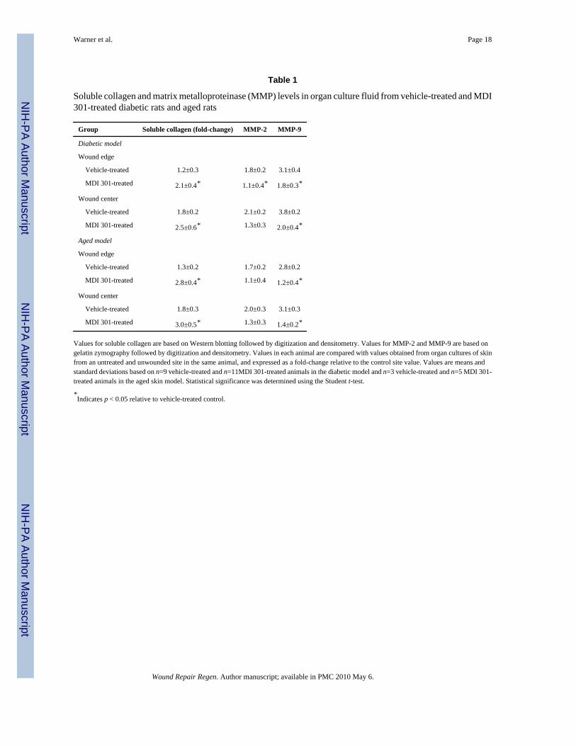
Type I collagen and MMP-2/MMP-9 were assessed in skin biopsies taken at the time of woundclosure in all three models. With corticosteroid treated rats, skin biopsies were taken from foursites. Biopsy site one was from the area just beyond the initial wound margin; sites two andthree were within the initial area encompassed by the abrasion wound and site four was froman area not exposed to Temovate or MDI 301 (and not abraded). Figure 4 compares levels ofsoluble type I collagen in organ culture fluids (corticosteroid-treated animals) and levels of thetwo MMPs are shown in Figure 5. Collagen levels were higher and MMP levels were lowerin the culture fluids from biopsies of the retinoid-treated skin than in culture fluids from vehicle-treated animals. Table 1 summarizes the effects of MDI 301 on type I collagen and MMP levelsin skin taken from control and treated diabetic rats and from control and treated aged rats.Consistent with what was seen in the corticosteroid model (Figures 4 and 5), organ culturefluids from MDI 301-treated skin had higher collagen levels and lower MMP levels than didculture fluid from vehicle-treated control skin.
DISCUSSIONIn the present study, MDI 301, an experimental retinoid, was applied daily to the skin of ratstreated with the potent corticosteroid, Temovate (clobetasol propionate), to the skin of diabeticrats (8 weeks posttreatment with STZ) and to the skin of aged rats (14–16 months old). In allthree models, subsequently induced abrasion wounds healed more rapidly in the retinoid-treated animals than in vehicle-treated controls. In all three models, type I collagen levelsincreased and MMP levels decreased in response to MDI 301. Skin irritation during theretinoid-treatment phase was virtually nonexistent.
The biologically active retinoids have effects on the dermis that delay or prevent atrophy.Topical retinoid use induces the synthesis of types I and III procollagen31–33 and suppressesthe major collagen-degrading enzymes in the skin.34,35 These retinoid effects in the skin reflectmultiple mechanisms. Retinoids directly influence gene transcription and alter signalingcascades that regulate gene transcription. 36 In the case of MMP reduction, RA not only down-regulates enzyme production at the molecular level 34,35 but also up-regulates tissue inhibitorof metalloproteinases-1 (TIMP-1),37 the major MMP inhibitor in the skin.38 Along with thesespecific effects on collagen metabolism, retinoid treatment also stimulates proliferation ofdermal fibroblasts, thus fostering additional collagen production. Although it is generallybelieved that improved wound healing in retinoid-treated skin reflects these dermal activities,it is important to keep in mind that retinoids are broad-acting molecules that influence thefunctioning of other elements in the skin, as well. Retinoid treatment increases keratinocyteproliferation and motility, 26,27,39 properties essential for rapid wound closure. Given theregenerative effects of retinoid use on both the dermal and epidermal component of the skin,it is not surprising that retinoids should be considered for use as wound-preventative or wound-healing agents. Several past studies have, in fact, demonstrated beneficial effects of retinoiduse in wound healing. Although most of these studies have focused on wounds in the skin,11–17 retinoid efficacy has also been demonstrated in healing of wounds in other tissues—bone,cornea, respiratory tract, upper digestive system, and gut.18–25 Although retinoids havebeneficial activities, skin irritation is commonly seen following topical application of RA.26–28 If skin irritation is too severe, retinoid use will simply be discontinued by most users. Equallyimportant, excessive irritation may negate beneficial effects that would otherwise be seen, oractually increase sensitivity to wound formation.
In past studies, MDI 301 (a synthetic retinoid) has been demonstrated to increase epidermalthickness, induce collagen production and inhibit the major collagen-degrading enzymes inorgan cultures of human skin.29 In these regards, MDI 301 and RA are similar. With thesynthetic retinoid, however, surrogate markers of inflammation that are up-regulated by RAin human skin organ culture are not increased.29 Given this profile of activities, the use of MDI
Warner et al. Page 6
Wound Repair Regen. Author manuscript; available in PMC 2010 May 6.
NIH
-PA Author Manuscript
NIH
-PA Author Manuscript
NIH
-PA Author Manuscript

301 in topical preparations should, in theory, have significant advantages over RA and otherretinoids that are currently available as skin-repair agents. In the present study we demonstratethat MDI 301 does, in fact, have wound healing potential. Using three different models ofdelayed (impaired) skin wound healing in hairless rats, pretreatment of the rats for 12–14 dayswith MDI 301 before wounding was shown to decrease the time to wound closure as comparedwith vehicle-treated control animals. In all three models, biopsies from the wound site in MDI301-treated animals (taken at the time of wound closure) produced higher levels of type Icollagen and lower amounts of MMPs than did wound site biopsies from vehicle-treatedanimals. The ability of topical MDI 301 to improve healing of superficial wounds is similar towhat we reported earlier with RA (diabetic model only).17 The ability of the synthetic retinoidto improve superficial wound healing in at-risk skin but without the irritation associated withRA and other currently available agents provides a strong rational for further development ofthis agent as a wound preventative.
AcknowledgmentsThis study was supported in part by NIH grants AR49621 and GM77724 from the USPHS.
References1. NIH. NIH. Bethesda, MD: NIH; 1980. A report of the National Diabetes Advisory Board.2. Diabetes in the United States: A strategy for prevention. US Department of Health and Human Services,
National Center for Chronic Disease Prevention and Health Promotion, Division of DiabetesTranslation; 1992.
3. Edmonds, ME. Experience in a multidisciplinary diabetic foot clinic. In: Connor, H.; Boulton, AJM.;Ward, JD., editors. The foot in diabetes: Proceedings of the First National Conference on the DiabeticFoot. Chicester, UK: John Wiley; 1987. p. 121-34.
4. Pecoraro RE, Reiber GE, Burgess EM. Pathways to diabetic limb amputation: basis for prevention.Diabetes Care 1990;13:13–21.
5. Strigini L, Ryan T. Wound healing in elderly human skin. Clin Dermatol 1996;14:197–206. [PubMed:9117986]
6. Ashcroft G, Mills SJ, Ashworth JJ. Ageing and wound healing. Biogerentology 2002;3:337–45.7. Brem H, Tomic-Canic M, tarnovskaya A, Gill K, Ehrlich HP, Carasa M, Weinberger S, Baskin-Bey
E, Entero H. Healing of elderly patients with diabetic foot ulcers, venous stasis ulcers and pressureulcers. Surg Technol Int 2003;11:161–7. [PubMed: 12931298]
8. Anstead GM. Steroids, retinoids, and wound healing. Adv Wound Care 1998;11:277–85. [PubMed:10326344]
9. Ehrlich HP, Hunt TK. Effect of cortisone and vitamin A on wound healing. Ann Surg 1968;167:32.10. Wicke C, Halliday B, Allen D, Roche NS, Scheuenstuhl H, Spencer MM, Roberts AB, Hunt TK.
Effects of steroids and retinoids on wound healing. Arch Surg 2000;135:1265–70. [PubMed:11074878]
11. Seifter E, Rettura G, Padawer J, Stratford F, Kambosos D, Levenson SM. Impaired wound healingin streptozotocin diabetes. Ann Surg 1981;194:42–50. [PubMed: 6454399]
12. Frosch PJ, Czarnetzki BM. Effect of retinoids on wound healing in diabetic rats. Arch Dermatol Res1989;281:424–6. [PubMed: 2596868]
13. Popp C, Kligman AM, Stoudemayer TJ. Pretreatment of photoaged forearm skin with topical tretinoinaccelerates healing of full-thickness wounds. Br J Dermatol 1995;132:46–53. [PubMed: 7756151]
14. Otley CC, Gayner SM, Ahmed I, Moore EJ, Roenigk RK, Sherris DA. Preoperative and postoperativetopical tretinoin on high-tension excisional wounds and full-thickness skin grafts in a porcine model:a pilot study. Dermatol Surg 1999;25:716–21. [PubMed: 10491064]
15. Kitano Y, Yoshimura K, Uchida G, Sato K, Harii K. Pretreatment with topical all-trans retinoic acidis beneficial for wound healing in genetically diabetic mice. Arch Dermatol Res 2001;293:512–21.
Warner et al. Page 7
Wound Repair Regen. Author manuscript; available in PMC 2010 May 6.
NIH
-PA Author Manuscript
NIH
-PA Author Manuscript
NIH
-PA Author Manuscript
16. Basak PY, Eroglu E, Altuntas I, Agalar F, Basak K, Sutcu R. Comparison of the effects of tretinoin,adapalene and collagenase in an experimental model of wound healing. Eur J Dermatol 2002;12:145–8. [PubMed: 11872410]
17. Lateef H, Abatan OI, Aslam MN, Stevens MJ, Varani J. Pretreatment of diabetic rats with all-transretinoic acid increases healing of subsequently-induced abrasion wounds. Diabetes 2005;54:855–61.[PubMed: 15734865]
18. Hatchell DL, Ubels JL, Stekiel T, Hatchell MC. Corneal epithelial wound healing in normal anddiabetic rabbits treated with tretinoin. Arch Ophthalmol 1985;103:98–100. [PubMed: 3977683]
19. de Waard JW, Wobbes T, van der Linden CJ, Hendriks T. Retinol may promote fluorouracil-suppressed healing of experimental intestinal anastomoses. Arch Surg 1995;130:959–65. [PubMed:7661680]
20. MacDonald IM, Peters C, Chen MH. Effect of retinoic acid on wound healing of laser burns to porcineretinal pigment epithelium. Can J Ophthalmol 1996;3:175–8. [PubMed: 8804754]
21. Johansen S, Heegaard S, Prause JU, Rask-Pedersen E. The healing effect of all-trans retinoic acid onepithelial corneal abrasions in rabbits. Acta Ophthalmologica Scandinavica 1998;76:401–4.[PubMed: 9716324]
22. Sela J, Kaufman D, Shoshan S, Shani J. Retinoic acid enhances the effect of collagen on bone union,following induced non-union defect in guinea pig ulna. In am Res 2000;49:679–83.
23. Maccabee MS, Trune DR, Hwang PH. Paranasal sinus mucosal regeneration: the effect of topicalretinoic acid. Am J Rhinol 2003;17:133–7. [PubMed: 12862400]
24. Talas DU, Nayci A, Atis S, Comelekoglu U, Polat A, Bagdatoglu C, Renda N. The effects ofcorticosteroids and vitamin A on the healing of tracheal anastomoses. Int J Ped Otorhinolaryngol2003;67:109–16.
25. Yuen DE, Stratford AF. Vitamin A activation of transforming growth factor-beta1 enhances porcineileum wound healing in vitro. Ped Res 2004;55:935–9.
26. Griffiths CE, Kang S, Ellis CN, Kim KJ, Finkel LJ, Ortiz-Ferrer LC, White GM, Hamilton TA,Voorhees JJ. Two concentrations of topical tretinoin (retinoic acid) cause similar improvement ofphotoaging but different degrees of irritation. A double-blind, vehicle-controlled comparison of 0.1%and 0.025% tretinoin cream. Arch Dermatol 1995;131:1037–44. [PubMed: 7544967]
27. Kang S, Duell EA, Fisher GJ, Datta SC, Wang Z-Q, Reddy AP, Tavakkol A, Voorhees JJ. Applicationof retinol to human skin in vivo induces epidermal hyperplasia and cellular retinoid-binding proteinscharacteristic of retinoic acid but without measurable retinoic acid levels or irritation. J InvestDermatol 1995;105:549–56. [PubMed: 7561157]
28. Phillips TJ, Gottlieb AB, Leyden JJ, Lowe NJ, Lew-Kaya DA, Sefton J, Walker PS, Gibson JR.Efficacy of 0.1% Tazarotene cream for the treatment of photodamage. Arch Dermatol2002;138:1486–93. [PubMed: 12437455]
29. Varani J, Fay K, Perone P. MDI-301: a non-irritating retinoid, induces changes in organ-culturedhuman skin that underlie repair. Arch Dermatol Res 2007;298:439–48. [PubMed: 17146625]
30. Varani J, Fligiel H, Zhang J, Aslam MN, Lu Y, Dehne LA, Keller ET. Separation of retinoid-inducedepidermal and dermal thickening from skin irritation. Arch Dermatol Res 2003;295:255–62.[PubMed: 14564458]
31. Varani J, Warner RL, Phan SH, Datta SC, Fisher GJ, Voorhees JJ. Vitamin A antagonizes decreasedcell growth, and elevated collagen-degrading matrix metalloproteinases and stimulates collagenaccumulation in naturally-aged human skin. J Invest Dermatol 2000;114:480–6. [PubMed:10692106]
32. Griffiths CEM, Russman G, Majmudar G, Singer RS, Hamilton TA, Voorhees JJ. Restoration ofcollagen formation in photodamaged human skin by tretinoin (retinoic acid). N Engl J Med1993;329:530–4. [PubMed: 8336752]
33. Talwar HS, Griffiths CEM, Fisher GJ, Hamilton TA, Voorhees JJ. Reduced type I and type IIIprocollagens in photodamaged adult human skin. J Invest Dermatol 1995;105:285–90. [PubMed:7543550]
34. Fisher GJ, Datta SC, Talwar HS, Wang ZQ, Varani J, Kang S, Voorhees JJ. The molecular basis ofsun-induced premature skin ageing and retinoid antagonism. Nature (London) 1996;379:335–8.[PubMed: 8552187]
Warner et al. Page 8
Wound Repair Regen. Author manuscript; available in PMC 2010 May 6.
NIH
-PA Author Manuscript
NIH
-PA Author Manuscript
NIH
-PA Author Manuscript

35. Fisher GJ, Wang Z-Q, Datta SC, Varani J, Kang S, Voorhees JJ. Pathophysiology of premature skinaging induced by ultraviolet light. New Engl J Med 1997;337:1419–28. [PubMed: 9358139]
36. Fisher GJ, Datta S, Wang ZQ, Li X-Y, Quan T, Chung JH, Kang S, Voorhees JJ. C-Jun-dependentinhibition of cutaneous procollagen transcription following ultraviolet irradiation is reversed by all-trans retinoic acid. J Clin Invest 2000;106:663–70. [PubMed: 10974019]
37. Lateef H, Sevens M, Varani J. All-trans retinoic acid suppresses matrix metalloproteinase production/activation and increases collagen synthesis in diabetic skin in organ culture. Am J Pathol2004;165:167–74. [PubMed: 15215172]
38. Varani J, Hattori Y, Chi Y, Schmidt T, Perone P, Zeigler ME, Fader DJ, Johnson TJ. Elaboration ofcollagenolytic and gelatinolytic matrix metalloproteinases and their inhibitors by basal cellcarcinomas of skin: comparison with normal skin. Br J Cancer 2000;82:657–65. [PubMed:10682680]
39. Varani J, Zeigler M, Dame MK, Kang S, Fisher GJ, Voorhees JJ, Stoll SW, Elder JT. Heparin-bindingepidermal growth factor activation of keratinocyte ErbB receptors mediates epidermal hyperplasia,a prominent side-effect of retinoid therapy. J Invest Dermatol 2001;117:1335–41. [PubMed:11886492]
Warner et al. Page 9
Wound Repair Regen. Author manuscript; available in PMC 2010 May 6.
NIH
-PA Author Manuscript
NIH
-PA Author Manuscript
NIH
-PA Author Manuscript
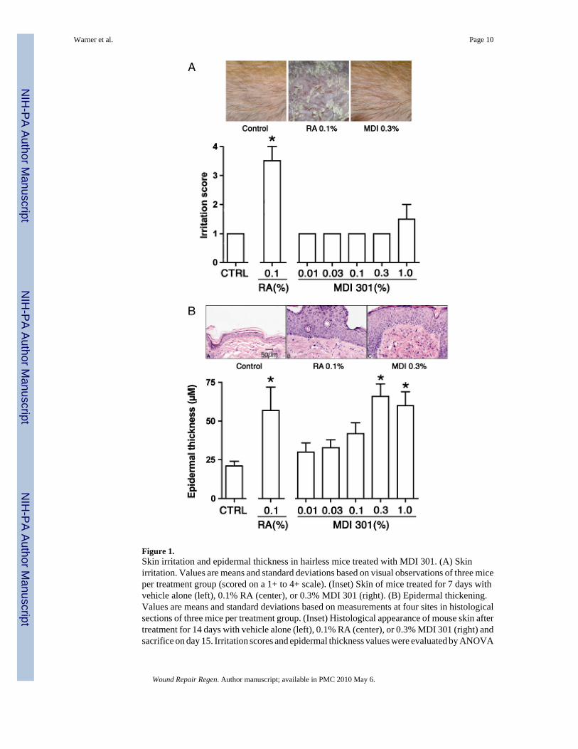
Figure 1.Skin irritation and epidermal thickness in hairless mice treated with MDI 301. (A) Skinirritation. Values are means and standard deviations based on visual observations of three miceper treatment group (scored on a 1+ to 4+ scale). (Inset) Skin of mice treated for 7 days withvehicle alone (left), 0.1% RA (center), or 0.3% MDI 301 (right). (B) Epidermal thickening.Values are means and standard deviations based on measurements at four sites in histologicalsections of three mice per treatment group. (Inset) Histological appearance of mouse skin aftertreatment for 14 days with vehicle alone (left), 0.1% RA (center), or 0.3% MDI 301 (right) andsacrifice on day 15. Irritation scores and epidermal thickness values were evaluated by ANOVA
Warner et al. Page 10
Wound Repair Regen. Author manuscript; available in PMC 2010 May 6.
NIH
-PA Author Manuscript
NIH
-PA Author Manuscript
NIH
-PA Author Manuscript

followed by paired group comparisons. *Statistically significant difference from the controlgroup at p < 0.05.
Warner et al. Page 11
Wound Repair Regen. Author manuscript; available in PMC 2010 May 6.
NIH
-PA Author Manuscript
NIH
-PA Author Manuscript
NIH
-PA Author Manuscript

Figure 2.Epidermal thickness and soluble collagen elaboration in skin of control mice and mice treatedwith a combination of Temovate and MDI 301. (A) Epidermal thickness. Values are meansand standard deviations based on measurements at four sites in histological sections of threemice per treatment group. (B) Soluble type I collagen. Values are means and standarddeviations based on organ culture fluids from four animals per group. (Inset) Western blotexamples of soluble collagen elaborated by organ-cultured skin from control mice (lane 1),mice treated with Temovate alone (lane 2), Temovate plus 0.3% MDI 301 (lane 3) or Temovateplus 1.0% MDI 301 (lane 4). Epidermal thickness values and soluble collagen values wereevaluated by ANOVA followed by paired group comparisons. *Statistically significant
Warner et al. Page 12
Wound Repair Regen. Author manuscript; available in PMC 2010 May 6.
NIH
-PA Author Manuscript
NIH
-PA Author Manuscript
NIH
-PA Author Manuscript
difference from the control group at p < 0.05; ** Statistically significant difference from theTemovate alone group at p < 0.05.
Warner et al. Page 13
Wound Repair Regen. Author manuscript; available in PMC 2010 May 6.
NIH
-PA Author Manuscript
NIH
-PA Author Manuscript
NIH
-PA Author Manuscript
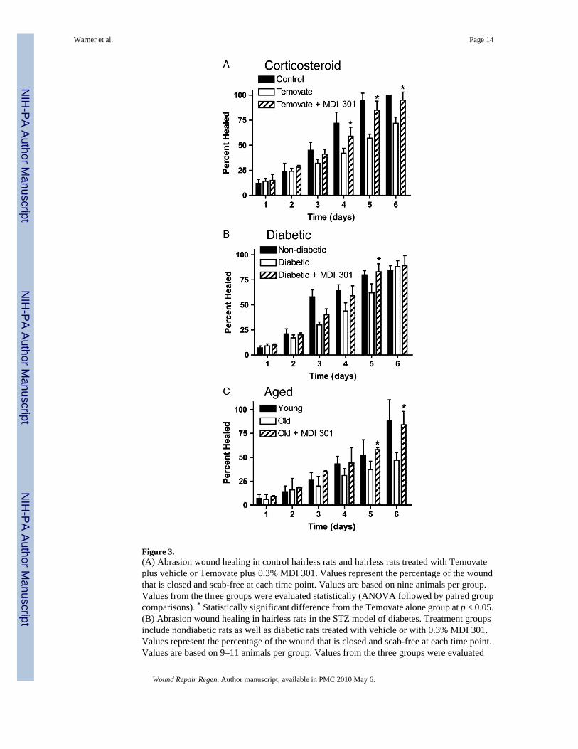
Figure 3.(A) Abrasion wound healing in control hairless rats and hairless rats treated with Temovateplus vehicle or Temovate plus 0.3% MDI 301. Values represent the percentage of the woundthat is closed and scab-free at each time point. Values are based on nine animals per group.Values from the three groups were evaluated statistically (ANOVA followed by paired groupcomparisons). * Statistically significant difference from the Temovate alone group at p < 0.05.(B) Abrasion wound healing in hairless rats in the STZ model of diabetes. Treatment groupsinclude nondiabetic rats as well as diabetic rats treated with vehicle or with 0.3% MDI 301.Values represent the percentage of the wound that is closed and scab-free at each time point.Values are based on 9–11 animals per group. Values from the three groups were evaluated
Warner et al. Page 14
Wound Repair Regen. Author manuscript; available in PMC 2010 May 6.
NIH
-PA Author Manuscript
NIH
-PA Author Manuscript
NIH
-PA Author Manuscript

statistically (ANOVA followed by paired group comparisons). * Statistically significantdifference from the diabetic alone group at p < 0.05. (C) Abrasion wound healing in hairlessaged rats. Treatment groups include young rats as well as aged rats treated with vehicle or with0.3% MDI 301. Values represent the percentage of the wound that is closed and scab-free ateach time point. Values are based on three to five animals per group. Values from the threegroups were evaluated statistically (ANOVA followed by paired group comparisons). *Statistically significant difference from the aged animal group at p < 0.05.
Warner et al. Page 15
Wound Repair Regen. Author manuscript; available in PMC 2010 May 6.
NIH
-PA Author Manuscript
NIH
-PA Author Manuscript
NIH
-PA Author Manuscript
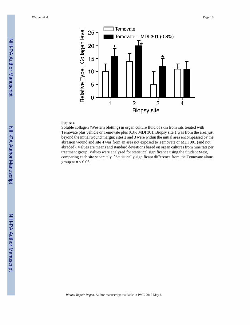
Figure 4.Soluble collagen (Western blotting) in organ culture fluid of skin from rats treated withTemovate plus vehicle or Temovate plus 0.3% MDI 301. Biopsy site 1 was from the area justbeyond the initial wound margin; sites 2 and 3 were within the initial area encompassed by theabrasion wound and site 4 was from an area not exposed to Temovate or MDI 301 (and notabraded). Values are means and standard deviations based on organ cultures from nine rats pertreatment group. Values were analyzed for statistical significance using the Student t-test,comparing each site separately. *Statistically significant difference from the Temovate alonegroup at p < 0.05.
Warner et al. Page 16
Wound Repair Regen. Author manuscript; available in PMC 2010 May 6.
NIH
-PA Author Manuscript
NIH
-PA Author Manuscript
NIH
-PA Author Manuscript
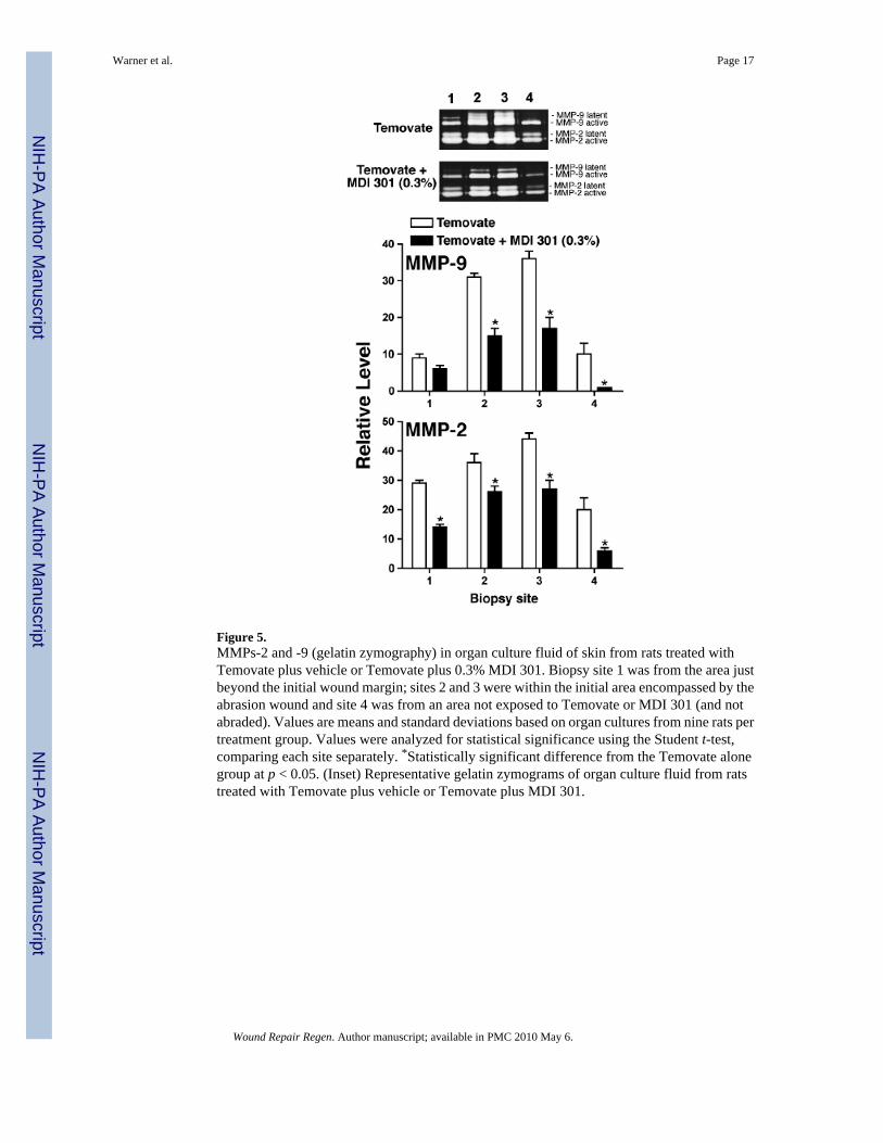
Figure 5.MMPs-2 and -9 (gelatin zymography) in organ culture fluid of skin from rats treated withTemovate plus vehicle or Temovate plus 0.3% MDI 301. Biopsy site 1 was from the area justbeyond the initial wound margin; sites 2 and 3 were within the initial area encompassed by theabrasion wound and site 4 was from an area not exposed to Temovate or MDI 301 (and notabraded). Values are means and standard deviations based on organ cultures from nine rats pertreatment group. Values were analyzed for statistical significance using the Student t-test,comparing each site separately. *Statistically significant difference from the Temovate alonegroup at p < 0.05. (Inset) Representative gelatin zymograms of organ culture fluid from ratstreated with Temovate plus vehicle or Temovate plus MDI 301.
Warner et al. Page 17
Wound Repair Regen. Author manuscript; available in PMC 2010 May 6.
NIH
-PA Author Manuscript
NIH
-PA Author Manuscript
NIH
-PA Author Manuscript
NIH
-PA Author Manuscript
NIH
-PA Author Manuscript
NIH
-PA Author Manuscript
Warner et al. Page 18
Table 1
Soluble collagen and matrix metalloproteinase (MMP) levels in organ culture fluid from vehicle-treated and MDI301-treated diabetic rats and aged rats
Group Soluble collagen (fold-change) MMP-2 MMP-9
Diabetic model
Wound edge
Vehicle-treated 1.2±0.3 1.8±0.2 3.1±0.4
MDI 301-treated 2.1±0.4* 1.1±0.4* 1.8±0.3*
Wound center
Vehicle-treated 1.8±0.2 2.1±0.2 3.8±0.2
MDI 301-treated 2.5±0.6* 1.3±0.3 2.0±0.4*
Aged model
Wound edge
Vehicle-treated 1.3±0.2 1.7±0.2 2.8±0.2
MDI 301-treated 2.8±0.4* 1.1±0.4 1.2±0.4*
Wound center
Vehicle-treated 1.8±0.3 2.0±0.3 3.1±0.3
MDI 301-treated 3.0±0.5* 1.3±0.3 1.4±0.2*
Values for soluble collagen are based on Western blotting followed by digitization and densitometry. Values for MMP-2 and MMP-9 are based ongelatin zymography followed by digitization and densitometry. Values in each animal are compared with values obtained from organ cultures of skinfrom an untreated and unwounded site in the same animal, and expressed as a fold-change relative to the control site value. Values are means andstandard deviations based on n=9 vehicle-treated and n=11MDI 301-treated animals in the diabetic model and n=3 vehicle-treated and n=5 MDI 301-treated animals in the aged skin model. Statistical significance was determined using the Student t-test.
*Indicates p < 0.05 relative to vehicle-treated control.
Wound Repair Regen. Author manuscript; available in PMC 2010 May 6.