Cognitive sequencing impairment in patients with focal or atrophic cerebellar damage
12
Cognitive sequencing impairment in patients with focal or atrophic cerebellar damage M. G. Leggio, 1,2 A. M. Tedesco, 1,2 F. R. Chiricozzi, 1,2 S. Clausi, 1,2 A. Orsini 1 and M. Molinari 2 1 Department of Psychology, University of Rome ‘La Sapienza’ and 2 Ataxia Lab, I.R.C.C.S. Santa Lucia Foundation, Rome, Italy Correspondence to: Maria G. Leggio, MD, PhD, Associate Professor of Psychophysiology, Head Ataxia Lab Santa Lucia Foundation, Department of Psychology, University of Rome ‘La Sapienza’, Via dei Marsi 78, 00185 Roma, Italy E-mail: [email protected] Although cognitive impairment after cerebellar damage has been widely reported, the mechanisms of cerebro- cerebellar interactions are still a matter of debate. The cerebellum is involved in sequence detection and production in both motor and sensory domains, and sequencing has been proposed as the basic mechanism of cerebellar functioning. Furthermore, it has been suggested that knowledge of sequencing mechanisms may help to define cerebellar predictive control processes. In spite of its recognized importance, cerebellar sequencing has seldom been investigated in cognitive domains. Cognitive sequencing functions are often analysed by means of action/script elaboration. Lesion and activation studies have localized this function in frontal cortex and basal ganglia circuits. The present study is the first to report deficits in script sequencing after cerebellar damage.We employed a card-sequencing test, developed ad hoc, to evaluate the influence of the content to be sequenced. Stimuli consisted of sets of sentences that described actions with a precise logical and temporal sequence (Verbal Factor), sets of cartoon-like drawings that reproduced behavioural sequences (Behavioural Factor) or abstract figures (Spatial Factor). The influence of the lesion characteristics was analysed by grouping patients according to lesion-type (focal or atrophic) and lesion-side (right or left). The results indicated that patients with cerebellar damage present a cognitive sequencing impairment independently of lesion type or localization. A correlation was also shown between lesion side and characteristics of the material to be sequenced. Namely, patients with left lesions perform defectively only on script sequences based on pictorial material and patients with right lesions only on script sequences requiring verbal elaboration.The present data support the hypothesis that sequence processing is the cerebellar mode of operation also in the cognitive domain. In addition, the pre- sence of right/left and pictorial/verbal differences is in agreement with the idea that cerebro-cerebellar interac- tions are organized in segregated cortico-cerebellar loops in which specificity is not related to the mode of functioning, but to the characteristics of the information processed. Keywords: cerebellum; executive function; picture arrangement; script Abbreviations: MRI = magnetic resonance imaging; TIQ = total intelligence quotient; WAIS-r = Wechsler Adult Intelligence Scale Revised Received July 24, 2007. Revised and Accepted February 14, 2008. Advance Access publication March 11, 2008 Introduction Anatomical, experimental and functional neuroimaging and clinical data stress the importance of cortico-cerebellar interactions in a variety of non-motor functions such as cognition, emotion and affective processing (Timmann and Daum, 2007). This cerebellar revolution makes a complete reconsideration of cortico-cerebellar interrelationships man- datory in order to discover the mechanisms through which the cerebellum exerts its influence on the cerebral cortex. Among the different theories on cerebellar functions (Bower and Parsons, 2003; Ito, 2006), a cerebellar role in sequencing incoming sensory patterns and outgoing responses has been proposed (Braitenberg et al., 1997; Ivry, 1997; Mauk, et al., 2000). Visuo-spatial implicit learning of sequences in patients with cerebellar lesions has been analysed in different experimental paradigms and cerebellar patients have been consistently reported impaired (Pascual-Leone et al., 1993; Molinari et al., 1997; Doyon et al., 1998; Gomez-Beldarrain et al., 1998). Functional magnetic resonance data in healthy subjects are controversial in discriminating the cerebellar involvement in sequence learning or in motor adaptation (Doyon et al., 1998; Seidler et al., 2002; Parsons et al., 2005). doi:10.1093/brain/awn040 Brain (2008), 131 , 1332^1343 ß The Author (2008). Published by Oxford University Press on behalf of the Guarantors of Brain. All rights reserved. For Permissions, please email: [email protected] by guest on June 27, 2016 http://brain.oxfordjournals.org/ Downloaded from
Transcript of Cognitive sequencing impairment in patients with focal or atrophic cerebellar damage
Cognitive sequencing impairment in patients withfocal or atrophic cerebellar damageM.G. Leggio,1,2 A. M.Tedesco,1,2 F. R. Chiricozzi,1,2 S. Clausi,1,2 A.Orsini1 and M. Molinari2
1Department of Psychology, University of Rome ‘La Sapienza’ and 2Ataxia Lab, I.R.C.C.S. Santa Lucia Foundation, Rome, Italy
Correspondence to: Maria G. Leggio, MD, PhD, Associate Professor of Psychophysiology, Head Ataxia Lab Santa LuciaFoundation, Department of Psychology, University of Rome ‘La Sapienza’, Via dei Marsi 78, 00185 Roma, ItalyE-mail: [email protected]
Although cognitive impairment after cerebellar damage has been widely reported, the mechanisms of cerebro-cerebellar interactions are still a matter of debate. The cerebellum is involved in sequence detection andproduction in both motor and sensory domains, and sequencing has been proposed as the basic mechanism ofcerebellar functioning. Furthermore, it has been suggested that knowledge of sequencing mechanismsmay helpto define cerebellar predictive control processes. In spite of its recognized importance, cerebellar sequencinghas seldom been investigated in cognitive domains.Cognitive sequencing functions are often analysed bymeansof action/script elaboration. Lesion and activation studies have localized this function in frontal cortex and basalganglia circuits.The present study is the first to report deficits in script sequencing after cerebellar damage.Weemployed a card-sequencing test, developed ad hoc, to evaluate the influence of the content to be sequenced.Stimuli consisted of sets of sentences that described actions with a precise logical and temporal sequence(Verbal Factor), sets of cartoon-like drawings that reproduced behavioural sequences (Behavioural Factor) orabstract figures (Spatial Factor). The influence of the lesion characteristics was analysed by grouping patientsaccording to lesion-type (focal or atrophic) and lesion-side (right or left).The results indicated that patients withcerebellar damage present a cognitive sequencing impairment independently of lesion type or localization.A correlation was also shown between lesion side and characteristics of the material to be sequenced. Namely,patients with left lesions perform defectively only on script sequences based on pictorial material and patientswith right lesions only on script sequences requiring verbal elaboration.The present data support the hypothesisthat sequence processing is the cerebellar mode of operation also in the cognitive domain. In addition, the pre-sence of right/left and pictorial/verbal differences is in agreement with the idea that cerebro-cerebellar interac-tions are organized in segregated cortico-cerebellar loops in which specificity is not related to the mode offunctioning, but to the characteristics of the information processed.
Keywords: cerebellum; executive function; picture arrangement; script
Abbreviations: MRI=magnetic resonance imaging; TIQ=total intelligence quotient; WAIS-r=Wechsler Adult IntelligenceScale Revised
Received July 24, 2007. Revised and Accepted February 14, 2008. Advance Access publication March 11, 2008
IntroductionAnatomical, experimental and functional neuroimaging andclinical data stress the importance of cortico-cerebellarinteractions in a variety of non-motor functions such ascognition, emotion and affective processing (Timmann andDaum, 2007). This cerebellar revolution makes a completereconsideration of cortico-cerebellar interrelationships man-datory in order to discover the mechanisms through whichthe cerebellum exerts its influence on the cerebral cortex.Among the different theories on cerebellar functions (Bowerand Parsons, 2003; Ito, 2006), a cerebellar role in sequencing
incoming sensory patterns and outgoing responses has beenproposed (Braitenberg et al., 1997; Ivry, 1997; Mauk, et al.,2000). Visuo-spatial implicit learning of sequences in patientswith cerebellar lesions has been analysed in differentexperimental paradigms and cerebellar patients have beenconsistently reported impaired (Pascual-Leone et al., 1993;Molinari et al., 1997; Doyon et al., 1998; Gomez-Beldarrainet al., 1998). Functional magnetic resonance data in healthysubjects are controversial in discriminating the cerebellarinvolvement in sequence learning or in motor adaptation(Doyon et al., 1998; Seidler et al., 2002; Parsons et al., 2005).
doi:10.1093/brain/awn040 Brain (2008), 131, 1332^1343
� The Author (2008). Published by Oxford University Press on behalf of the Guarantors of Brain. All rights reserved. For Permissions, please email: [email protected]
by guest on June 27, 2016http://brain.oxfordjournals.org/
Dow
nloaded from
Conversely, neurophysiological studies in healthy volunteersor in patients with focal cerebellar damage indicated a role ofthe cerebellum in sequence acquisition/detection (Molinariet al., 1997; Restuccia et al., 2007).Acquiring and acting upon a serial order of events is a
fundamental ability that can lead to sequence structureknowledge either incidentally through experience (implicitlearning) or intentionally through explicit effort (declar-ative learning). To recognize that stimuli are presented ina given order, the sensory information pertaining to onestimulus must be kept active in a working memorysystem and compared with subsequent stimuli. Procedurallearning can be achieved only if the correct sequence ofevents (sensory or motor) is acquired implicitly orexplicitly. Thus, a disruption of ‘sequence in’ processingof stimuli could be responsible for the implicit learningimpairment.The severity of cerebellar patients’ difficulty in the serial
reaction time task in detecting a visuo-spatial sequence,indicates a prevalent role of cerebellar circuitry inrecognizing event sequences, rather than in planning andexecuting them (Molinari et al., 1997). Tesche and Karhu(2000), with a somatosensory evoked paradigm, analysedthe neural signal generated in the cortex and in thecerebellum during the presentation of somatosensorysequences perturbed by random stimulus omissions.While the response in the somatosensory cortex was closelylinked to the actual presentation of the stimulus, cerebellaractivity was particularly evident when the expected stimuluswas omitted (Tesche and Karhu, 2000). As stated by Ivry(2000), this finding provides experimental evidence of acerebellar role ‘as detector of change or deviation in thesequence of sensory events’.To verify whether cerebellar processing affects the ability
to recognize the similarity/diversity of incoming sequenceinputs, the somatosensory mismatch negativity (S-MMN)component of event-related potentials (ERPs) was recentlyanalysed in six patients with unilateral cerebellar lesions(Restuccia et al., 2007). In all subjects analysed, MMN wasclearly abnormal in the cerebral hemisphere contralateral tothe cerebellar damage. This evidence identifies the cerebel-lum as the ideal structure for detecting discordancesbetween the input from the deviant event and the sensorymemory representation of the regular aspects of sequencestimulation.Support for a cerebellar role in the acquisition of
procedural sequences also derives from animal data. In aseries of studies based on surgical lesions it was shown thatcerebellar damage impairs the acquisition of the spatialprocedural sequences required for Morris water maze testin rats (Petrosini et al., 1998). The good performances ofanimals, that have acquired the correct competence beforethe lesion, underline the specificity of the cerebellum inacquisition rather than execution (Leggio et al., 1999).Furthermore, evidence that the cerebellar lesioned rats wereimpaired not only in learning through direct execution but
also in learning through observation of conspecificbehaviour, provides additional proof of the importance ofthe cerebellum in sensory processing (Leggio et al., 2000a;Graziano et al., 2002).
Thus, detecting and generating sequences might be a keyfor understanding the basic cerebellar function in differentdomains. If so, the ability to detect and generate sequencesshould represent an operational mode also in the cognitivedomain. To investigate this topic (Experiment 1) we firstretrospectively investigated the performances of patientswith cerebellar lesions on the Picture Arrangement subtest(PAs) of the Italian version of the Wechsler AdultIntelligence Scale Revised (WAIS-r) (Orsini and Laicardi,1997); and second (Experiment 2) we analysed whether thecharacteristics of the material processed influencedsequence detection performances of cerebellar-damagedsubjects.
Experiment 1çWAIS-r PictureArrangement subtestThe PAs of the WAIS-r mainly investigates sequentialthinking. To solve the task correctly visual material has tobe analysed, understood and integrated (Lezak, 1995). Thecorrect logical sequence is reconstructed by identifyingrelations between events, deciding priority and orderingthese events in chronological order (Orsini and Laicardi,1997). In Experiment 1, PAs subtest performances ofpatients affected by pathologies exclusively confined to thecerebellar structures, who were admitted to the IRCCSSanta Lucia Foundation rehabilitation hospital between2003 and 2006, were retrospectively reviewed.
SubjectsBased on lesion lateralization and focal or degenerativeaetiology, 77 right-handed patients, i.e. 44 males and 33females were divided into the following groups: patientsaffected by focal cerebellar lesions on the right side (Table 1:RCb—n.21); patients affected by focal cerebellar lesions onthe left side (Table 1: LCb—n.21); and patients affected bycerebellar atrophy. These latter patients were grouped eitherconsidering all subjects independently from aetiologies(CA—n.35 Experiment 1 Supplementary Material Table 1)or considering only subjects with idiopathic cerebellar ataxia(Table 1: ICA—n.18). Therefore according to the groupingmethods the total number of cerebellar subjects was 77(Cbt group Experiment 1 Supplementary Material Table 1) or60 (Cb group Table 1).
Focal cerebellar lesions consisted of ischemic or haemor-rhagic stroke or surgical ablation due to arteriovenousmalformations or tumours. Lesion characteristics werereconstructed from the written reports of the charts. Noclinical or radiological evidence of extracerebellar patholo-gies were reported, with the exception of one subject thatpresented an involvement of the brainstem. One subject
Cerebellar cognitive sequencing impairment Brain (2008), 131, 1332^1343 1333
by guest on June 27, 2016http://brain.oxfordjournals.org/
Dow
nloaded from
had a temporary, moderate, increase of the volume of theventricles not requiring surgical derivation and notassociated with comatose conditions.Of the patients with cerebellar atrophy 12 had a genet-
ically determined diagnosis (2: ataxia-oculomotor apraxiatype 2, 5: Friedreich ataxia, 2: spino-cerebellar ataxia type 2,and 3: spino-cerebellar ataxia type 1), 1 presented atrophyas sequaele of a cerebellitis and 22 presented idiopathicforms. Of the idiopathic forms, 17 subjects presented purecerebellar syndromes, four subjects presented additionalextracerebellar atrophy (3: brainstem atrophy and 1:bilateral posterior parietal atrophy), one subject presentedspastic paraparesis beside cerebellar deficits. The diagnosisof ICA was based on clinical indications of a purelycerebellar syndrome and on magnetic resonance imaging(MRI) evidence of atrophic pathology restricted to thecerebellum.Differences in the grouping of atrophic subjects might
influence the results; therefore we run statistics followingboth grouping methods. Since no differences were evi-denced, in the results section only data from the moreselective series of patients with ICA will be presented. Dataon the statistical comparisons considering the entire groupof subjects with degenerative pathologies (CA) are reportedin Supplementary Material (Filename: Experiment 1—Testwith CA group).Some of these patients had already participated in
previous studies (Leggio et al., 2000b; Molinari et al.,2004, 2005; Restuccia et al., 2007). All patients underwent aneurological examination and their motor impairment wasquantified using a modified version of the cerebellar motordeficit scale, proposed by Appollonio et al. (1993), whichranges from 0 (absence of any deficit) to 42 (presence of alldeficits to the highest degree) and evaluates eight cerebellarsigns (dysarthria, limb tone, postural tremor, upper andlower limb ataxia, standing balance, gait ataxia and ocularmovements). See Table 1 for group characteristics. Thecontrol group consisted of 69 subjects who had no historyof neurological or psychiatric illness (Table 1: C—n.69).Mean age and education of control subjects is reported inTable 1. t-Test for independent samples confirmed thatcerebellar patients and control subjects were well-matched
for age (P=n.s.) and education (P=n.s.). Furthermore aone-way ANOVA failed to reveal any differences inage [F(3,125) = 2.59, P=n.s.] or years of education[F(3,125) = 0.58, P=n.s.] among C group and cerebellarsubgroups. Experimental procedures were approved by theethical committee of the IRCCS Santa Lucia Foundation;written consent for anonymous use of clinical data wasobtained from each subject.
Neuropsychological assessmentThe patients’ general cognitive profile was assessedfrom data available on their charts. In particular, weconsidered the following data: WAIS-r total intelligencequotient (TIQ) values, immediate and delayed recall ofRey’s 15 words (Rey, 1958), immediate visual memory(Carlesimo et al., 1996), forward and backward digit spanand forward and backward Corsi Test (Corsi, 1972),Raven’s 47 progressive matrices (Raven, 1949), freehandcopying of drawings (Gainotti et al., 1977), copyingdrawings with landmarks (Gainotti et al., 1977), temporalrules induction (Villa et al., 1990) and word fluency(Borkowsky et al., 1967). The same Italian version of theWAIS-r reported in the charts was employed to test controlsubjects.
The picture arrangement subtestof theWAIS-rThe PAs consists of 10 sets of cartoon pictures that tellstories. Each set, comprised of three to six pictures, ispresented to the subject in scrambled order with instruc-tions to rearrange the pictures to make the most sensiblestory. The PAs was administered and scored according tothe Italian version of the WAIS-r (Wechsler, 1981; Orsiniand Laicardi, 1997, 2003).
Statistical analysisStudent’s t-test for independent samples was used to detectdifferences between cerebellar patients and control subjects.Metric units were compared by one-way ANOVA. Whensignificant differences were found, post-hoc comparisons
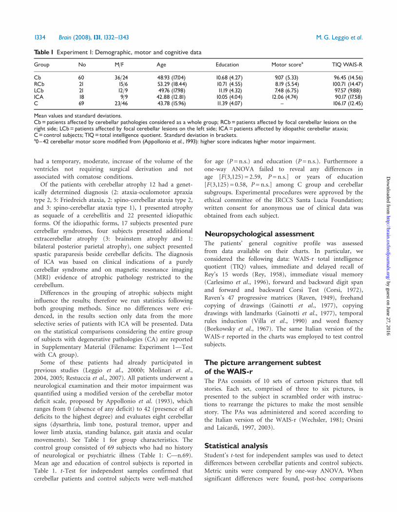
Table 1 Experiment 1: Demographic, motor and cognitive data
Group No M/F Age Education Motor scorea TIQ WAIS-R
Cb 60 36/24 48.93 (17.04) 10.68 (4.27) 9.07 (5.33) 96.45 (14.56)RCb 21 15/6 53.29 (18.44) 10.71 (4.55) 8.19 (5.54) 100.71 (14.47)LCb 21 12/9 49.76 (17.98) 11.19 (4.32) 7.48 (6.75) 97.57 (9.88)ICA 18 9/9 42.88 (12.81) 10.05 (4.04) 12.06 (4.74) 90.17 (17.58)C 69 23/46 43.78 (15.96) 11.39 (4.07) ^ 106.17 (12.45)
Mean values and standard deviations.Cb=patients affected by cerebellar pathologies considered as a whole group; RCb=patients affected by focal cerebellar lesions on theright side; LCb=patients affected by focal cerebellar lesions on the left side; ICA=patients affected by idiopathic cerebellar ataxia;C=control subjects; TIQ=total intelligence quotient. Standard deviation in brackets.a0^42 cerebellar motor score modified from (Appollonio et al., 1993): higher score indicates higher motor impairment.
1334 Brain (2008), 131, 1332^1343 M.G. Leggio et al.
by guest on June 27, 2016http://brain.oxfordjournals.org/
Dow
nloaded from
among groups were assessed with the Bonferroni post hoctest; Bonferroni adjusted P-values (PBonf) are reported.Pearson correlations among motor scores and PAs scoreswere calculate to verify possible relations between motorperformances and cognitive performances.
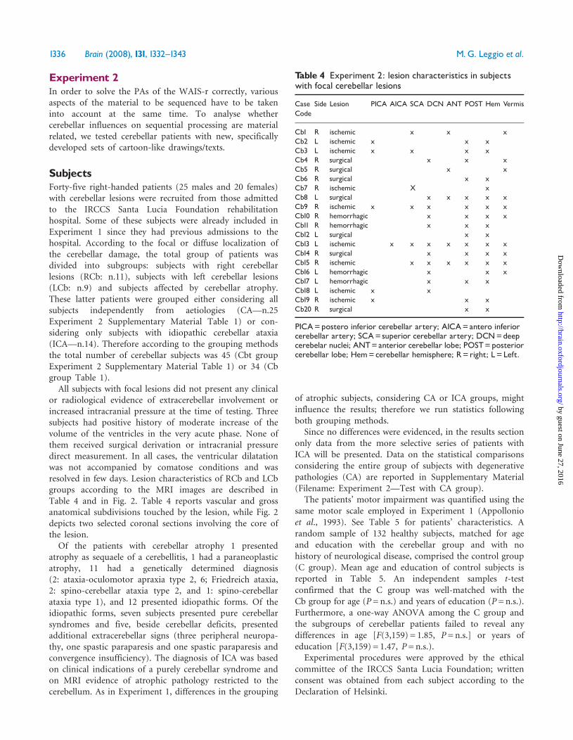
ResultsCerebellar patients showed no obvious deficits in thegeneral neuropsychological assessment. The TIQ values ofcerebellar patients and control subjects (Table 1) as well asthe scores of the neuropsychological assessment of cere-bellar patients (Table 2) were within the normal rangeexcept for the forward Corsi test, which was just above thecut-off. TIQ scores were employed to compare cognitivelevels among groups; significant differences were presentamong the control subjects and the three subgroups ofcerebellar patients [One-way ANOVA: F(3,125) = 7.89,P= 0.001]; Bonferroni post hoc comparisons showed thatICA group had scores significantly lower than controls(PBonf = 0.000).Regarding the PAs, all experimental groups scored within
the normal range (10� 3) (Wechsler, 1981; Orsini andLaicardi, 1997, 2003). However, all cerebellar group scoreswere lower than C group scores (Fig. 1). An independentsamples t-test demonstrated that the difference between theperformances of Cb and C groups was significant(P50.001). This finding was further confirmed by amultiple comparison among the three subgroups ofcerebellar patients and the C group [One-way Anova:F(3,125) = 10.97, P50.001]. Post hoc analyses (BonferroniTest) demonstrated that all patient subgroups performedworse than control subjects (RCb: PBonf = 0.011; LCb:PBonf = 0.002; ICA: PBonf = 0.000), while no difference wasdetected between subgroups of cerebellar patients. Pearsoncorrelation results did not highlight any relation betweenataxia and PAs scores (Table 3).Cerebellar subjects as a group and considering type and
side of lesion presented a preserved general cognitivepattern. The lack of deficits of clinical relevance is not
completely surprising. In different domains such aslanguage, working memory and visuo-spatial abilities, justto name a few, cerebellar deficits have been evidenced onlyin ad hoc testing conditions (Silveri et al., 1998; Leggioet al., 2000b; Molinari et al., 2004; Justus, 2004; Restucciaet al., 2007). Although on the PAs cerebellar patients’performances were within the normal range, they wereclearly defective when compared to control group perfor-mances. As stated in the ‘Introduction’ section, differentlines of reasoning prompted us to hypothesize a sequencingdeficit after cerebellar damage.
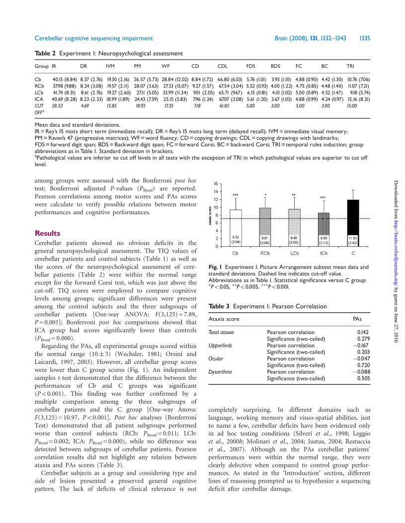
Table 2 Experiment 1: Neuropsychological assessment
Group IR DR IVM PM WF CD CDL FDS BDS FC BC TRI
Cb 40.15 (8.84) 8.37 (2.76) 19.30 (2.16) 26.57 (5.73) 28.84 (12.02) 8.84 (1.72) 66.80 (6.03) 5.76 (1.01) 3.93 (1.10) 4.88 (0.90) 4.42 (1.30) 10.76 (7.06)RCb 37.98 (9.88) 8.24 (3.08) 19.57 (2.11) 28.07 (3.63) 27.33 (15.07) 9.27 (1.57) 67.54 (3.04) 5.52 (0.93) 4.00 (1.22) 4.75 (0.85) 4.48 (1.40) 11.07 (7.21)LCb 41.74 (8.31) 8.61 (2.76) 19.27 (2.60) 27.11 (5.05) 33.99 (11.34) 9.01 (2.05) 65.71 (9.67) 6.15 (0.81) 4.10 (1.02) 5.00 (0.89) 4.52 (1.47) 9.18 (5.74)ICA 40.69 (8.28) 8.23 (2.53) 18.99 (1.89) 24.43 (7.59) 25.15 (5.83) 7.96 (1.24) 67.07 (3.08) 5.61 (1.20) 3.67 (1.03) 4.88 (0.99) 4.24 (0.97) 12.16 (8.21)CUTOFFa
28.53 4.69 13.85 18.93 17.35 7.18 61.85 5.00 3.00 5.00 3.00 15.00
Mean data and standard deviations.IR=Rey’s 15 mots short term (immediate recall); DR=Rey’s 15 mots long term (delayed recall); IVM= immediate visual memory;PM=Raven’s 47 (progressive matrices); WF=word fluency; CD=copying drawings; CDL=copying drawings with landmarks;FDS= forward digit span; BDS=Backward digit span; FC= forward Corsi; BC=backward Corsi; TRI= temporal rules induction; groupabbreviations as inTable 1. Standard deviation in brackets.aPathological values are inferior to cut off levels in all tests with the exception of TRI in which pathological values are superior to cut offlevel.
Fig. 1 Experiment 1. Picture Arrangement subtest mean data andstandard deviations. Dashed line indicates cut-off value.Abbreviations as inTable 1. Statistical significance versus C group:�P50.05, ��P50.005, ���P50.001.
Table 3 Experiment 1: Pearson Correlation
Ataxia score PAs
Total ataxia Pearson correlation 0.142Significance (two-tailed) 0.279
Upperlimb Pearson correlation �0.167Significance (two-tailed) 0.203
Ocular Pearson correlation �0.047Significance (two-tailed) 0.720
Dysarthria Pearson correlation �0.088Significance (two-tailed) 0.505
Cerebellar cognitive sequencing impairment Brain (2008), 131, 1332^1343 1335
by guest on June 27, 2016http://brain.oxfordjournals.org/
Dow
nloaded from
Experiment 2In order to solve the PAs of the WAIS-r correctly, variousaspects of the material to be sequenced have to be takeninto account at the same time. To analyse whethercerebellar influences on sequential processing are materialrelated, we tested cerebellar patients with new, specificallydeveloped sets of cartoon-like drawings/texts.
SubjectsForty-five right-handed patients (25 males and 20 females)with cerebellar lesions were recruited from those admittedto the IRCCS Santa Lucia Foundation rehabilitationhospital. Some of these subjects were already included inExperiment 1 since they had previous admissions to thehospital. According to the focal or diffuse localization ofthe cerebellar damage, the total group of patients wasdivided into subgroups: subjects with right cerebellarlesions (RCb: n.11), subjects with left cerebellar lesions(LCb: n.9) and subjects affected by cerebellar atrophy.These latter patients were grouped either considering allsubjects independently from aetiologies (CA—n.25Experiment 2 Supplementary Material Table 1) or con-sidering only subjects with idiopathic cerebellar ataxia(ICA—n.14). Therefore according to the grouping methodsthe total number of cerebellar subjects was 45 (Cbt groupExperiment 2 Supplementary Material Table 1) or 34 (Cbgroup Table 1).All subjects with focal lesions did not present any clinical
or radiological evidence of extracerebellar involvement orincreased intracranial pressure at the time of testing. Threesubjects had positive history of moderate increase of thevolume of the ventricles in the very acute phase. None ofthem received surgical derivation or intracranial pressuredirect measurement. In all cases, the ventricular dilatationwas not accompanied by comatose conditions and wasresolved in few days. Lesion characteristics of RCb and LCbgroups according to the MRI images are described inTable 4 and in Fig. 2. Table 4 reports vascular and grossanatomical subdivisions touched by the lesion, while Fig. 2depicts two selected coronal sections involving the core ofthe lesion.Of the patients with cerebellar atrophy 1 presented
atrophy as sequaele of a cerebellitis, 1 had a paraneoplasticatrophy, 11 had a genetically determined diagnosis(2: ataxia-oculomotor apraxia type 2, 6; Friedreich ataxia,2: spino-cerebellar ataxia type 2, and 1: spino-cerebellarataxia type 1), and 12 presented idiopathic forms. Of theidiopathic forms, seven subjects presented pure cerebellarsyndromes and five, beside cerebellar deficits, presentedadditional extracerebellar signs (three peripheral neuropa-thy, one spastic paraparesis and one spastic paraparesis andconvergence insufficiency). The diagnosis of ICA was basedon clinical indications of a purely cerebellar syndrome andon MRI evidence of atrophic pathology restricted to thecerebellum. As in Experiment 1, differences in the grouping
of atrophic subjects, considering CA or ICA groups, mightinfluence the results; therefore we run statistics followingboth grouping methods.
Since no differences were evidenced, in the results sectiononly data from the more selective series of patients withICA will be presented. Data on the statistical comparisonsconsidering the entire group of subjects with degenerativepathologies (CA) are reported in Supplementary Material(Filename: Experiment 2—Test with CA group).
The patients’ motor impairment was quantified using thesame motor scale employed in Experiment 1 (Appollonioet al., 1993). See Table 5 for patients’ characteristics. Arandom sample of 132 healthy subjects, matched for ageand education with the cerebellar group and with nohistory of neurological disease, comprised the control group(C group). Mean age and education of control subjects isreported in Table 5. An independent samples t-testconfirmed that the C group was well-matched with theCb group for age (P=n.s.) and years of education (P=n.s.).Furthermore, a one-way ANOVA among the C group andthe subgroups of cerebellar patients failed to reveal anydifferences in age [F(3,159) = 1.85, P=n.s.] or years ofeducation [F(3,159) = 1.47, P=n.s.).
Experimental procedures were approved by the ethicalcommittee of the IRCCS Santa Lucia Foundation; writtenconsent was obtained from each subject according to theDeclaration of Helsinki.
Table 4 Experiment 2: lesion characteristics in subjectswith focal cerebellar lesions
CaseCode
Side Lesion PICA AICA SCA DCN ANT POST Hem Vermis
Cb1 R ischemic x x xCb2 L ischemic x x xCb3 L ischemic x x x xCb4 R surgical x x xCb5 R surgical x xCb6 R surgical x xCb7 R ischemic X xCb8 L surgical x x x x xCb9 R ischemic x x x x x xCb10 R hemorrhagic x x x xCb11 R hemorrhagic x x xCb12 L surgical x xCb13 L ischemic x x x x x x xCb14 R surgical x x x xCb15 R ischemic x x x x x xCb16 L hemorrhagic x x xCb17 L hemorrhagic x x xCb18 L ischemic x xCb19 R ischemic x x xCb20 R surgical x x
PICA=postero inferior cerebellar artery; AICA=antero inferiorcerebellar artery; SCA=superior cerebellar artery; DCN=deepcerebelar nuclei; ANT=anterior cerebellar lobe; POST=posteriorcerebellar lobe; Hem=cerebellar hemisphere; R=right; L=Left.
1336 Brain (2008), 131, 1332^1343 M.G. Leggio et al.
by guest on June 27, 2016http://brain.oxfordjournals.org/
Dow
nloaded from

Fig. 2 Experiment 2. Subjects with focal lesions: lesion extent in two representative coronal sections for each individual. Lesion ispresented as overlaid on coronal T1-weighted template from (Schmahmann et al., 2000). Lesion extensions were assessed on the3D-T1-MPRAGEs after spatial normalization. Case codes as inTable 4.
Table 5 Experiment 2: Demographic, motor and cognitive data
Group No M/F AGE Education Motor scorea Raven’s 47
Cb 34 18/16 51.94 (14.77) 11.32 (4.41) 10.38 (7.32) 27.05 (6.32)RCb 11 6/5 48.18 (20.72) 13.73 (4.41) 7.89 (7.14) 28.50 (3.52)LCb 9 2/7 60.63 (7.42) 10.75 (4.20) 7.66 (6.80) 29.60 (3.56)ICA 14 5/9 49.36 (11.08) 10.00 (4.14) 14.37 (6.85) 24.74 (8.23)C 132 57/75 47.02 (17.33) 12.80 (4.41) ^ 29.50 (2.52)
Mean values and standard deviations.Abbreviations as in table 1. Standard deviation in brackets.a0^42 cerebellar motor score modified from (Appollonio et al., 1993).
Cerebellar cognitive sequencing impairment Brain (2008), 131, 1332^1343 1337
by guest on June 27, 2016http://brain.oxfordjournals.org/
Dow
nloaded from
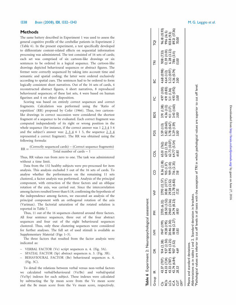
MethodsThe same battery described in Experiment 1 was used to assess the
general cognitive profile of the cerebellar patients in Experiment 2
(Table 6). In the present experiment, a test specifically developed
to differentiate content-related effects on sequential information
processing was administered. The test consisted of 16 sets of cards;
each set was comprised of six cartoon-like drawings or six
sentences to be ordered in a logical sequence. The cartoon-like
drawings depicted behavioural sequences or abstract figures. The
former were correctly sequenced by taking into account time and
semantic and spatial coding; the latter were ordered exclusively
according to spatial cues. The sentences had to be ordered to form
logically consistent short narratives. Out of the 16 sets of cards, 4
reconstructed abstract figures, 4 short narratives, 8 reproduced
behavioural sequences; of these last sets, 4 were based on human
figurines and 4 on object disposition.Scoring was based on entirely correct sequences and correct
fragments. Calculation was performed using the ‘Ratio of
repetition’ (RR) proposed by Cofer (1966). Thus, two cartoon-
like drawings in correct succession were considered the shortest
fragment of a sequence to be evaluated. Each correct fragment was
computed independently of its right or wrong position in the
whole sequence (for instance, if the correct answer was 1 2 3 4 5 6
and the subject’s answer was: 2 3 4 6 1 5, the sequence 2 3 4
represented a correct fragment). The RR was obtained using the
following formula:
RR ¼ðCorrectly sequenced cardsÞ � ðCorrect sequence fragmentsÞ
Total number of cards� 1
Thus, RR values run from zero to one. The task was administered
without a time limit.Data from the 132 healthy subjects were pre-processed for item
analysis. This analysis excluded 5 out of the 16 sets of cards. To
analyse whether the performances on the remaining 11 sets
clustered, a factor analysis was performed. Analysis of the principal
component, with extraction of the three factors and an oblique
rotation of the axis, was carried out. Since the intercorrelation
among factors resulted lower than 0.18, confirming the hypothesis of
the independence among factors, we executed an analysis of the
principal component with an orthogonal rotation of the axis
(Varimax). The factorial saturation of the rotated solution is
reported in Table 7.Thus, 11 out of the 16 sequences clustered around three factors.
All four sentence sequences, three out of the four abstract
sequences and four out of the eight behavioural sequences
clustered. Thus, only these clustering sequences were considered
for further analyses. The full set of used stimuli is available as
Supplementary Material (Figs 1–3).The three factors that resulted from the factor analysis were
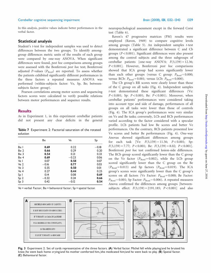
indicated as:
– VERBAL FACTOR (Ve) script sequences n. 4. (Fig. 3A).– SPATIAL FACTOR (Sp) abstract sequences n. 3. (Fig. 3B).– BEHAVIOURAL FACTOR (Be) behavioural sequences n. 4.
(Fig. 3C).
To detail the relations between verbal versus non-verbal factors
we calculated verbal/behavioural (Ve/Be) and verbal/spatial
(Ve/Sp) indexes for each subject. These indexes were calculated
by subtracting the Sp mean score from the Ve mean score
and the Be mean score from the Ve mean score, respectively. Table6
Experiment2:
Neuropsycho
logicala
ssessm
ent
Group
IRDR
IVM
PMWF
CD
CDL
FDS
BDS
FCBC
TRI
TQI
Cb
43.37(7.07)
9.14(2.38)
19.47(1.99
)27.05(6.32)
27.93(12.57)
8.36
(2.18
)65.01(7.62)
5.59
(1.13)
3.91
(1.38)
4.97
(1.00
)4.68
(0.98)
9.08(7.33)
96.18
(15.93)
RCb
44.33(7.24)
9.37(2.50)
19.28(1.95)
28.50(3.52)
30.81(17.7
7)9.15(1.37)
67.13
(2.70)
5.82
(0.60)
4.36
(1.12)
4.91
(0.83)
4.64
(1.12)
12.59(11.01)
99.6(16.5)
LCb
43.33(7.87)
8.95
(3.25)
20.46(1.30)
29.60(3.56)
29.28(3.52)
9.19(2.10)
62.73(13
.07)
5.78
(0.67)
4.11(1.17)
5.11(1.36)
5.22
(0.97)
6.28
(3.22)
103.8(9.3)
ICA
42.74(6.84)
9.07(1.52)
18.98(2.27)
24.74(8.23)
22.78(6.83)
7.16(2.35)
64.77(5.54)
5.29
(1.59)
3.43
(1.60
)4.93
(0.92)
4.36
(0.74)
7.86(3.11)
89.21(17.3
6)CU
TOFF
a28.53
4.69
13.85
18.93
17.35
7.18
61.85
5.00
3.00
5.00
3.00
15.00
70.00
Means
andstandard
deviations.
Abbreviations
asin
tables
1and2.
Standard
deviationin
brackets.
a Patho
logicalv
alue
sareinferior
tocutofflevelsin
alltests
withtheexceptionof
TRIinwhich
pathologicalvalues
aresuperior
tocutofflevel.
1338 Brain (2008), 131, 1332^1343 M.G. Leggio et al.
by guest on June 27, 2016http://brain.oxfordjournals.org/
Dow
nloaded from
In this analysis, positive values indicate better performances in theverbal factor.
Statistical analysisStudent’s t-test for independent samples was used to detectdifferences between the two groups. To identify among-group differences metric units of the results of each groupwere compared by one-way ANOVA. When significantdifferences were found, post hoc comparisons among groupswere assessed with the Bonferroni post hoc test; Bonferroniadjusted P-values (PBonf) are reported. To assess whetherthe patients exhibited significantly different performances inthe three factors a repeated measures ANOVA wasperformed (within-subjects factor: Ve, Sp, Be; between-subjects factor: group).Pearson correlations among motor scores and sequencing
factors scores were calculated to verify possible relationsbetween motor performances and sequence results.
ResultsAs in Experiment 1, in this experiment cerebellar patientsdid not present any clear deficits in the general
neuropsychological assessment except in the forward Corsitest (Table 6).
Raven’s 47 progressive matrices (PM) results wereemployed (Raven, 1949) to compare cognitive levelsamong groups (Table 5). An independent samples t-testdemonstrated a significant difference between C and Cbgroups (P50.001). Significant differences were also presentamong the control subjects and the three subgroups ofcerebellar patients [one-way ANOVA: F(3,159) = 12.36;P50.001]. However, Bonferroni post hoc comparisonsshowed that ICA group had scores significantly lowerthan each other groups (versus C group: PBonf = 0.000;versus RCb: PBonf = 0.001; versus LCb: PBonf = 0.000).
The Cb group’s RR scores were clearly lower than thoseof the C group on all tasks (Fig. 4). Independent samplest-test demonstrated these significant differences (Ve:P50.001; Sp: P50.001; Be: P50.001). Moreover, whencerebellar patients’ performances were considered takinginto account type and side of damage, performances of allgroups on all tasks were lower than those of controls(Fig. 4). The ICA group’s performances were very similaron Ve and Be tasks; conversely, LCb and RCb performancesvaried according to the factor considered with a specularprofile. LCb patients had low Be scores and better Veperformances. On the contrary, RCb patients presented lowVe scores and better Be performances (Fig. 4). One-wayAnovas showed significant differences among groupsfor each task [Ve: F(3,159) = 11.56; P50.001; Sp:F(3,159) = 7.77; P50.001; Be: F(3,159) = 8.02; P50.001].Bonferroni post hoc test confirmed lesion-side differences.The RCb group scored significantly lower than the C groupon the Ve factor (PBonf = 0.002), while the LCb groupscored significantly lower than the C group on the Be(PBonf = 0.013) and Sp factors (PBonf = 0.019). The ICAgroup’s scores were significantly lower than the C group’sscores on all factors (Ve Factor: PBonf = 0.000; Be Factor:PBonf = 0.001; Sp Factor: PBonf = 0.006). A repeated measuresAnova confirmed the differences among groups [between-subjects effect: F(3,159) = 2191.185; P50.001] and also
Fig. 3 Experiment 2. Set of cards representative of the three factors. (A) Verbal factor. Michel fell while playing/and he bruised hisknee:/he went back home crying/and his mother comforted him,/she medicated him/and he went back to play. (B) Spatial factor.(C) Behavioural factor.
Table 7 Experiment 2: Factorial saturation of the rotatedsolution
Be Ve Sp
Be 1 0.69 0.22 �0.18Be 2 0.66 0.29 0.20Be 3 0.54 0.21 0.33Be 4 0.69 �0.23 0.06Ve 1 0.07 0.58 0.07Ve 2 �0.16 0.77 0.03Ve 3 0.35 0.50 0.01Ve 4 0.27 0.44 0.25Sp 1 0.14 0.08 0.69Sp 2 �0.32 0.24 0.66Sp 3 0.42 �0.12 0.54
Ve=verbal Factor; Be=behavioral factor; Sp= spatial factor.
Cerebellar cognitive sequencing impairment Brain (2008), 131, 1332^1343 1339
by guest on June 27, 2016http://brain.oxfordjournals.org/
Dow
nloaded from
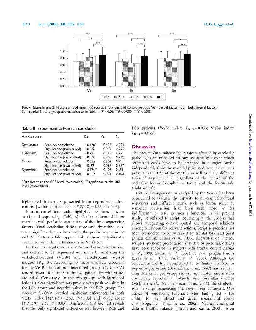
highlighted that groups presented factor dependent perfor-mances [within-subjects effect: F(2,318) = 4.35; P50.05].Pearson correlation results highlighted relations between
ataxia and sequencing (Table 8). Ocular subscore did notcorrelate with performances in any of the three sequencingfactors. Total cerebellar deficit score and dysarthria sub-score significantly correlated with the performances in Beand Ve factors while upper limb subscore significantlycorrelated with the performances in Ve factor.Further investigation of the relations between lesion side
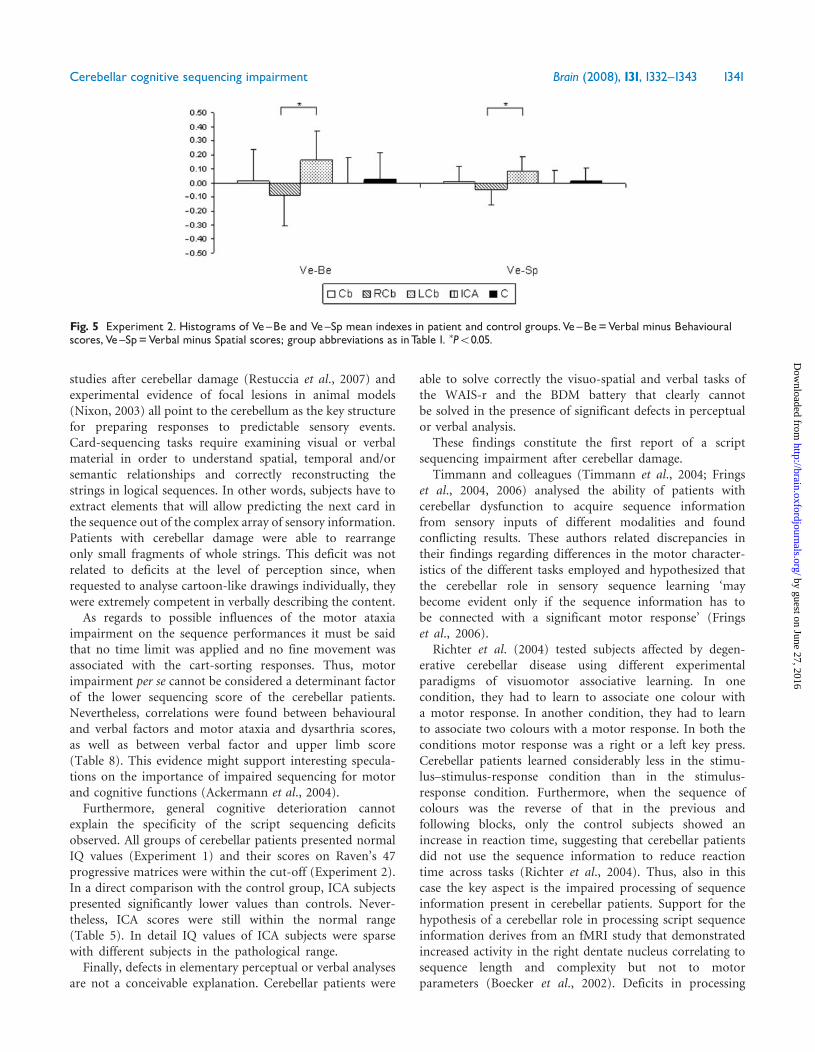
and content to be sequenced was made by analysing theverbal/behavioural (Ve/Be) and verbal/spatial (Ve/Sp)indexes (Fig. 5). According to these analyses, especiallyfor the Ve–Be data, all non-lateralized groups (C, Cb, CA)tended toward a balance in the two parameters with valuesaround 0. Conversely, in the two groups with lateralizedlesions a clear prevalence was present with positive values inthe LCb group and negative values in the RCb group. Theone-way ANOVA revealed significant differences for bothVe/Be index [F(3,159) = 2.67, P50.05] and Ve/Sp index[F(3,159) = 2.68, P50.05]. Bonferroni post hoc test revealsthat the only significant difference was between RCb and
LCb patients (Ve/Be index: PBonf = 0.035; Ve/Sp index:PBonf = 0.035).
DiscussionThe present data indicate that subjects affected by cerebellarpathologies are impaired on card-sequencing tests in whichscrambled cards have to be arranged in a logical orderindependently from the material processed. Impairment waspresent in the PAs of the WAIS-r as well as in the differenttasks of Experiment 2, regardless of the nature of thecerebellar lesion (atrophic or focal) and the lesion side(right or left).
Picture Arrangement, as analysed by the WAIS, has beenconsidered to evaluate the capacity to process behaviouralsequences and different terms, such as action script orsemantic sequencing, have been used more or lessindifferently to refer to such a function. In the presentstudy, we referred to script sequencing as the process thatallows recognizing correct spatial and temporal relationsamong behaviourally relevant actions. Script sequencing hasbeen considered to be sustained by frontal lobe and basalganglia circuits (Tinaz et al., 2006). Regardless of whetherscript-sequencing presentation is verbal or pictorial, deficitshave been reported in subjects with frontal cortex (Siriguet al., 1998; Zanini et al., 2002) or basal ganglia lesions(Zalla et al., 1998; Tinaz et al., 2008). Although thecerebellum has been considered to be highly involved insequence processing (Braitenberg et al., 1997) and sequen-cing deficits in processing sensory and motor informationare widely reported in subjects with cerebellar damage(Molinari et al., 1997; Timmann et al., 2004), the cerebellarrole in script sequencing has never been addressed. Oneaspect of sequencing functions often highlighted is theability to plan ahead and order meaningful eventschronologically (Tinaz et al., 2006). Neurophysiologicaldata in healthy subjects (Tesche and Karhu, 2000), lesion
Table 8 Experiment 2: Pearson correlation
Ataxia score Be Ve Sp
Total ataxia Pearson correlation �0.420� �0.423� 0.224Significance (two-tailed) 0.019 0.018 0.225
Upperlimb Pearson correlation �0.299 �0.375� 0.221Significance (two-tailed) 0.102 0.038 0.232
Ocular Pearson correlation �0.258 �0.303 0.101Significance (two-tailed) 0.162 0.097 0.587
Dysarthria Pearson correlation �0.474�� �0.405� 0.189Significance (two-tailed) 0.007 0.024 0.308
�Significant at the 0.05 level (two-tailed); ��significant at the 0.01level (two-tailed).
Fig. 4 Experiment 2. Histograms of mean RR scores in patient and control groups.Ve=verbal factor; Be=behavioural factor;Sp= spatial factor; group abbreviation as inTable 1. �P50.05, ��P50.005, ���P50.001.
1340 Brain (2008), 131, 1332^1343 M.G. Leggio et al.
by guest on June 27, 2016http://brain.oxfordjournals.org/
Dow
nloaded from
studies after cerebellar damage (Restuccia et al., 2007) andexperimental evidence of focal lesions in animal models(Nixon, 2003) all point to the cerebellum as the key structurefor preparing responses to predictable sensory events.Card-sequencing tasks require examining visual or verbalmaterial in order to understand spatial, temporal and/orsemantic relationships and correctly reconstructing thestrings in logical sequences. In other words, subjects have toextract elements that will allow predicting the next card inthe sequence out of the complex array of sensory information.Patients with cerebellar damage were able to rearrangeonly small fragments of whole strings. This deficit was notrelated to deficits at the level of perception since, whenrequested to analyse cartoon-like drawings individually, theywere extremely competent in verbally describing the content.As regards to possible influences of the motor ataxia
impairment on the sequence performances it must be saidthat no time limit was applied and no fine movement wasassociated with the cart-sorting responses. Thus, motorimpairment per se cannot be considered a determinant factorof the lower sequencing score of the cerebellar patients.Nevertheless, correlations were found between behaviouraland verbal factors and motor ataxia and dysarthria scores,as well as between verbal factor and upper limb score(Table 8). This evidence might support interesting specula-tions on the importance of impaired sequencing for motorand cognitive functions (Ackermann et al., 2004).Furthermore, general cognitive deterioration cannot
explain the specificity of the script sequencing deficitsobserved. All groups of cerebellar patients presented normalIQ values (Experiment 1) and their scores on Raven’s 47progressive matrices were within the cut-off (Experiment 2).In a direct comparison with the control group, ICA subjectspresented significantly lower values than controls. Never-theless, ICA scores were still within the normal range(Table 5). In detail IQ values of ICA subjects were sparsewith different subjects in the pathological range.Finally, defects in elementary perceptual or verbal analyses
are not a conceivable explanation. Cerebellar patients were
able to solve correctly the visuo-spatial and verbal tasks ofthe WAIS-r and the BDM battery that clearly cannotbe solved in the presence of significant defects in perceptualor verbal analysis.
These findings constitute the first report of a scriptsequencing impairment after cerebellar damage.
Timmann and colleagues (Timmann et al., 2004; Fringset al., 2004, 2006) analysed the ability of patients withcerebellar dysfunction to acquire sequence informationfrom sensory inputs of different modalities and foundconflicting results. These authors related discrepancies intheir findings regarding differences in the motor character-istics of the different tasks employed and hypothesized thatthe cerebellar role in sensory sequence learning ‘maybecome evident only if the sequence information has tobe connected with a significant motor response’ (Fringset al., 2006).
Richter et al. (2004) tested subjects affected by degen-erative cerebellar disease using different experimentalparadigms of visuomotor associative learning. In onecondition, they had to learn to associate one colour witha motor response. In another condition, they had to learnto associate two colours with a motor response. In both theconditions motor response was a right or a left key press.Cerebellar patients learned considerably less in the stimu-lus–stimulus-response condition than in the stimulus-response condition. Furthermore, when the sequence ofcolours was the reverse of that in the previous andfollowing blocks, only the control subjects showed anincrease in reaction time, suggesting that cerebellar patientsdid not use the sequence information to reduce reactiontime across tasks (Richter et al., 2004). Thus, also in thiscase the key aspect is the impaired processing of sequenceinformation present in cerebellar patients. Support for thehypothesis of a cerebellar role in processing script sequenceinformation derives from an fMRI study that demonstratedincreased activity in the right dentate nucleus correlating tosequence length and complexity but not to motorparameters (Boecker et al., 2002). Deficits in processing
Fig. 5 Experiment 2. Histograms of Ve^Be and Ve^Sp mean indexes in patient and control groups.Ve^Be=Verbal minus Behaviouralscores,Ve^Sp=Verbal minus Spatial scores; group abbreviations as inTable 1. �P50.05.
Cerebellar cognitive sequencing impairment Brain (2008), 131, 1332^1343 1341
by guest on June 27, 2016http://brain.oxfordjournals.org/
Dow
nloaded from
sequential information have also been reported in differentexperimental models in rats. Gaytan-Tocaven and Olvera-Cortes reported that bilateral lesions of the dentate nucleusimpair the acquisition of a ‘new’ sequential egocentric-based task (Gaytan-Tocaven and Olvera-Cortes, 2004); in aseries of studies, Petrosini and co-workers demonstrateddeficits in the acquisition of sequential procedures afterhemicerebellectomy (Petrosini et al., 1998; Leggio et al.,1999, 2000a).Shin and Ivry (2003) investigated the role of the
cerebellum and the basal ganglia in learning spatial andtemporal sequences and in integrating them when they weresimultaneously present. Unlike Parkinson’s disease patients,who were unable to learn the relationship between the twosequences but acquired the spatial and temporal sequencesindividually, cerebellar patients failed to show any evidenceof sequence detection and acquisition, indicating that thecerebellum plays a central role in sequence learning ingeneral (Shin and Ivry, 2003).In the present work we specifically analysed the perfor-
mances of cerebellar patients in script sequencing and insequencing non-behaviourally relevant abstract figures.Script sequencing requires using both spatial and temporalinformation while abstract figure sequencing can relyexclusively on spatial information. Subjects with cerebellarlesions were impaired in both conditions. These dataindicate that cerebellar processing is required in both scriptand spatial sequencing and, together with previous data oncerebellar sequencing functions, support the hypothesis of acentral role of cerebellar circuits in sequence processingregardless of whether the material processed is sensory(Bower, 1997), motor (Thach et al., 1992) or behavioural(present work).Within this general framework supporting the wide-
spread influence of the cerebellum on sequencing, indica-tions of a more selective role emerged from the present dataon patients with unilateral cerebellar damage. Statisticalevaluation of performances on the different card-sequen-cing tasks demonstrated significant differences betweensubjects with right and left focal lesions. Indeed, patientswith lesions of the left hemicerebellum performed defec-tively on script sequences based on pictorial material.Conversely, patients with lesions of the right hemicerebel-lum were impaired, exclusively on script sequences requir-ing verbal elaboration. The relation between right cerebellarhemisphere and verbal processing appears stronger than therelation between left hemisphere and non-verbal processing.Specificity of the cortico-cerebellar interactions or differ-ences in the two patient groups’ characteristics mightexplain the observed variability. These data not onlydemonstrate that the cerebellum has a specific role inelaborating sequential information pertaining to cognitivedomains, but also that the ability to integrate differentinformation in correct logical sequences is linked to thespecific characteristic of the material to be processed. Thus,sequencing in general requires cerebellar processing and
different cerebro-cerebellar circuits might be engageddepending on the material to be sequenced. This hypothesisis in agreement with the existence of a crossed cerebello-cortical loop organized in segregated channels that reachspecific cortical zones (Schmahmann and Pandya, 1997;Middleton and Strick, 2000; Giannetti and Molinari, 2002).Different authors have stressed that this precise topographycould represent the hardware that allows the cerebellum tointervene in many functions pertaining to motor control aswell as to cognition (Molinari et al., 2002; Schmahmann,2004; Ito, 2005, 2006).
Supplementary materialSupplementary material is available at Brain online.
AcknowledgementsThe continuous encouragement and support of ProfessorCarlo Caltagirone is gratefully acknowledged. The profes-sional English style editing of Claire Montagna and thestatistical expert support of Alessia Mammone are alsogratefully acknowledged. The present work was in partsupported by MURST, and Italian Ministry of Health grantsto M.M. and M.G.L.
ReferencesAckermann H, Mathiak K, Ivry RB. Temporal organization of ‘‘internal
speech’’ as a basis for cerebellar modulation of cognitive functions.
Behav Cogn Neurosci Rev 2004; 3: 14–22.
Appollonio IM, Grafman J, Schwartz V, Massaquoi S, Hallett M. Memory
in patients with cerebellar degeneration. Neurology 1993; 43: 1536–44.
Boecker H, Ceballos AO, Bartenstein P, et al. A H215O positron emission
tomography study on mental imagery of movement sequences – the
effect of modulating sequence length and direction. NeuroImage 2002;
17: 999–1009.
Borkowsky JG, Benton AL, Spreen O. Word fluency and brain-damage.
Neuropsychologia 1967; 5: 135–40.
Bower JM. Control of sensory data acquisition. Int Rev Neurobiol 1997;
41: 489–513.
Bower JM, Parsons LM. Rethinking the ‘‘lesser brain’’. Sci Am 2003; 289:
50–7.
Braitenberg V, Heck D, Sultan F. The detection and generation of
sequences as a key to cerebellar function: experiments and theory. Behav
Brain Sci 1997; 20: 229–77.
Carlesimo GA, Caltagirone C, Gainotti G. The mental deterioration
battery: Normative data, diagnostic reliability and qualitative analyses of
cognitive impairment. Eur Neurol 1996; 36: 378–84.
Cofer CN, Bruce DR, Reicher GM. Clustering in free recall as a function of
certain methodological variations. J Exp Psychol 1966; 71: 858–66.
Corsi PM. Human memory and the medial temporal regions of the brain.
Dissertation Abstracts International, 34 (02), 891B. (University
Microfilms No. AAI05-77717). Mc Gill University; 1972.
Doyon J, Laforce R, Bouchard G, et al. Role of the striatum, cerebellum
and frontal lobes in the automatization of a repeated visuomotor
sequence of movements. Neuropsychologia 1998; 36: 625–41.
Frings M, Boenisch R, Gerwig M, Diener HC, Timmann D. Learning of
sensory sequences in cerebellar patients. Learn Mem 2004; 11: 347–55.
Frings M, Maschke M, Gerwig M, Diener HC, Timmann D. Acquisition of
simple auditory and visual sequences in cerebellar patients. Cerebellum
2006; 5: 206–11.
1342 Brain (2008), 131, 1332^1343 M.G. Leggio et al.
by guest on June 27, 2016http://brain.oxfordjournals.org/
Dow
nloaded from
Gainotti G, Miceli G, Caltagirone C. Constructional apraxia in left brain-
damage patients: a planning disorder? Cortex 1977; 13: 109–18.
Gaytan-Tocaven L, Olvera-Cortes ME. Bilateral lesion of the cerebellar-
dentate nucleus impairs egocentric sequential learning but not
egocentric navigation in the rat. Neurobiol Learn Mem 2004; 82: 120–7.
Giannetti S, Molinari M. Cerebellar input to the posterior parietal cortex
in the rat. Brain Res Bull 2002; 58: 481–9.
Gomez-Beldarrain XXXX, Garcia-Monco JC, Rubio B, Pascual-Leone A.
Effect of focal cerebellar lesions on procedural learning in the serial
reaction time task. Exp Brain Res 1998; 120: 25–30.
Graziano A, Leggio MG, Mandolesi L, Neri P, Molinari M, Petrosini L.
Learning power of single behavioral units in acquisition of a complex
spatial behavior: an observational learning study in cerebellar-lesioned
rats. Behav Neurosci 2002; 116: 116–25.
Ito M. Bases and implications of learning in the cerebellum–adaptive
control and internal model mechanism. Prog Brain Res 2005; 148:
95–109.
Ito M. Cerebellar circuitry as a neuronal machine. Prog Neurobiol 2006;
78: 272–303.
Ivry R. Cerebellar timing systems. Int Rev Neurobiol 1997; 41: 555–73.
Ivry R. Exploring the role of the cerebellum in sensory anticipation and
timing: commentary on Tesche and Karhu. Hum Brain Mapp 2000; 9:
115–8.
Justus T. The cerebellum and English grammatical morphology: evidence
from production, comprehension, and grammaticality judgments.
J Cogn Neurosci 2004; 16: 1115–30.
Leggio MG, Molinari M, Neri P, Graziano A, Mandolesi L, Petrosini L.
Representation of actions in rats: the role of cerebellum in learning
spatial performances by observation. Proc Natl Acad Sci USA 2000a; 29:
5–2320.
Leggio MG, Neri P, Graziano A, Mandolesi L, Molinari M, Petrosini L.
Cerebellar contribution to spatial event processing: characterization of
procedural learning. Exp Brain Res 1999; 127: 1–11.
Leggio MG, Silveri MC, Petrosini L, Molinari M. Phonological grouping
is specifically affected in cerebellar patients: a verbal fluency study.
J Neurol Neurosurg Psychiatry 2000b; 69: 102–6.
Lezak MD. Neuropychological assessment. New York: Oxford University
Press; 1995.
Mauk MD, Medina JF, Nores WL, Ohyama T. Cerebellar function:
coordination, learning or timing? Curr Biol 2000; 10: 522–5.
Middleton FA, Strick PL. Basal ganglia output and cognition: evidence
from anatomical, behavioral, and clinical studies. Brain Cogn 2000; 42:
183–200.
Molinari M, Filippini V, Leggio MG. Neuronal plasticity of interrelated
cerebellar and cortical networks. Neuroscience 2002; 111: 863–70.
Molinari M, Leggio MG, Filippini V, Gioia MC, Cerasa A, Thaut MH.
Sensorimotor transduction of time information is preserved in subjects
with cerebellar damage. Brain Res Bull 2005; 67: 448–58.
Molinari M, Leggio MG, Solida A, et al. Cerebellum and procedural
learning: evidence from focal cerebellar lesions. Brain 1997; 120:
1753–62.
Molinari M, Petrosini L, Misciagna S, Leggio MG. Visuospatial abilities in
cerebellar disorders. J Neurol Neurosurg Psychiatry 2004; 75: 235–40.
Nixon PD. The role of the cerebellum in preparing responses to
predictable sensory events. Cerebellum 2003; 2: 114–22.
Orsini A, Laicardi C. Wais-r. Contributo alla taratura italiana. Firenze:
Organizzazioni Speciali; 1997.
Orsini A, Laicardi C. Wais-r e terza eta. Firenze: Organizzazioni Speciali;
2003.
Parsons MW, Harrington DL, Rao SM. Distinct neural system underline
learning visuomtor and spatial representations of motor skills. Hum
Brain Mapp 2005; 24: 229–47.
Pascual-Leone A, Grafman J, Clark K, et al. Procedural learning in
Parkinson’s disease and cerebellar degeneration. Ann Neurol 1993; 34:
594–602.
Petrosini L, Leggio MG, Molinari M. The cerebellum in the spatial
problem solving: a co-star or a guest star? Prog Neurobiol 1998; 56:
191–210.
Raven JC. Progressive matrices (1947). Set A, Ab, B: board and book form.
London: H.K. Lewis; 1949.
Restuccia D, Della MG, Valeriani M, Leggio MG, Molinari M. Cerebellar
damage impairs detection of somatosensory input changes. A somato-
sensory mismatch-negativity study. Brain 2007; 130: 276–87.
Rey A. Memorisation d’une serie de 15 mots en 5 repetitions. In: Rey A,
editor. L’examen clinique en psychologie. Paris: Presses Universiteries de
France; 1958.
Richter S, Matthies K, Ohede T, et al. Stimulus-response versus stimulus-
stimulus-response learning in cerebellar patients. Exp Brain Res 2004;
158: 438–49.
Schmahmann JD. Disorders of the cerebellum: ataxia, dysmetria of
thought, and the cerebellar cognitive affective syndrome.
J Neuropsychiatry Clin Neurosci 2004; 16: 367–78.
Schmahmann JD, Pandya DN. The cerebrocerebellar system. Int Rev
Neurobiol 1997; 41: 31–60.
Schmahmann JD, Doyon J, Toga AW, Petrides M, Evans AC. MRI Atlas of
the human cerebellum. San Diego: Academic Press; 2000.
Seidler RD, Purushotham A, Kim SG, Ugurbil K, Willingham D, Ashe J.
Cerebellum activation associated with performance change but not
motor learning. Science 2002; 296: 2043–6.
Shin JC, Ivry RB. Spatial and temporal sequence learning in patients with
Parkinson’s disease or cerebellar lesions. J Cogn Neurosci 2003; 15:
1232–43.
Silveri MC, Di Betta AM, Filippini V, Leggio MG, Molinari M. Verbal
short-term store-rehearsal system and the cerebellum. Evidence from a
patient with a right cerebellar lesion. Brain 1998; 121 (Pt 11): 2175–87.
Sirigu A, Cohen L, Zalla T, et al. Distinct frontal regions
for processing sentence syntax and story grammar. Cortex 1998; 34:
771–8.
Tesche CD, Karhu JT. Anticipatory cerebellar responses during somato-
sensory omission in man. Hum Brain Mapp 2000; 9: 119–42.
Thach WT, Goodkin HP, Keating JG. The cerebellum and the
adaptive coordination of movement. Annu Rev Neurosci 1992; 15:
403–42.
Timmann D, Drepper J, Calabrese S, et al. Use of sequence information in
associative learning in control subjects and cerebellar patients.
Cerebellum 2004; 3: 75–82.
Timmann D, Daum I. Cerebellar contributions to cognitive functions: a
progress report after two decades of research. Cerebellum 2007; 6:
159–62.
Tinaz S, Schendan HE, Schon K, Stern CE. Evidence for the importance of
basal ganglia output nuclei in semantic event sequencing: an fMRI
study. Brain Res 2006; 1067: 239–49.
Tinaz S, Schendan HE, Stern CE. Fronto-striatal deficit in Parkinson’s
disease during semantic event sequencing. Neurobiol Aging 2008; 29:
397–407.
Villa G, Gainotti G, De Bonis C, Marra C. Double dissociation between
temporal and spatial pattern processing in patients with frontal and
parietal damage. Cortex 1990; 26: 399–407.
Wechsler D. Wais-r. Wechsler Adult Intelligence Scale Revised. Firenze:
Organizzazioni Speciali; 1981.
Zalla T, Sirigu A, Pillon B, Dubois B, Grafman J, Agid Y. Deficit in
evaluating pre-determined sequences of script events in patients with
Parkinson’s disease. Cortex 1998; 34: 621–7.
Zanini S, Rumiati RI, Shallice T. Action sequencing deficit following
frontal lobe lesion. Neurocase 2002; 8: 88–99.
Cerebellar cognitive sequencing impairment Brain (2008), 131, 1332^1343 1343
by guest on June 27, 2016http://brain.oxfordjournals.org/
Dow
nloaded from