An ear punch model for studying the effect of radiation on wound healing
20
An ear punch model for studying the effect of radiation on wound healing DIVINO DeOLIVEIRA 1,* , YIQUN JIAO 1,* , JOEL R. ROSS 1 , KAYLA CORBIN 1 , QIZHEN XIAO 1 , GRETA TONCHEVA 2 , COLIN ANDERSON-EVANS 2 , TERRY T. YOSHIZUMI 2,3 , BENNY J. CHEN 1 , and NELSON J. CHAO 1,4 1 Department of Medicine, Duke University Medical Center, Durham, North Carolina, USA 2 Department of Radiation Safety, Duke University Medical Center, Durham, North Carolina, USA 3 Department of Radiology, Duke University Medical Center, Durham, North Carolina, USA 4 Department of Immunology, Duke University Medical Center, Durham, North Carolina, USA Abstract Purpose—Radiation and wound combined injury represents a major clinical challenge because of the synergistic interactions that lead to higher morbidity and mortality than either insult would produce singly. The purpose of this study was to develop a mouse ear punch model to study the physiological mechanisms underlying radiation effects on healing wounds. Materials and methods—Surgical wounds were induced by a 2 mm surgical punch in the ear pinnae of MRL/MpJ mice. Photographs of the wounds were taken and the sizes of the ear punch wounds were quantified by image analysis. Local radiation to the ear was delivered by orthovoltage X-ray irradiator using a specially constructed jig that shields the other parts of body. Results—Using this model, we demonstrated that local radiation to the wound area significantly delayed the healing of ear punch wounds in a dose-dependent fashion. The addition of sublethal whole body irradiation (7 Gy) further delayed the healing of ear punch wounds. These results were replicated in C57BL/6 mice; however, wound healing in MRL/MpJ mice was accelerated. Conclusions—These data indicate that the mouse ear punch model is a valuable model to study radiation and wound combined injury. Keywords radiation; wound healing; combined injury; ear punch model; MRL/MpJ mice; in vivo imaging Introduction In healthy individuals, exposure to radiation doses as low as 0.5 Gy can lead to detectable changes in circulating lymphocyte counts and doses of 6 Gy can be fatal even with aggressive treatment (Gale 1987). Radiation doses of 2 Gy or more can lead to acute © 2011 Informa UK, Ltd. Correspondence: Nelson J. Chao, Duke University Medical Center, 2400 Pratt Street, Suite 1100, Box 3961, Durham, NC 27705, USA. Tel: 1 (919) 668 1010. Fax: +1 (919) 668 1091. [email protected]; or Benny J. Chen, Duke University Medical Center, Box 3289, 247A Carl Building, Durham, NC 27710, USA. Tel: +1 (919) 681-3021. Fax: +1 (919) 681-7060. [email protected]. * These authors contribute equally to this work. Declaration of interest: The authors report no conflicts of interest. The authors alone are responsible for the content and writing of the paper. NIH Public Access Author Manuscript Int J Radiat Biol. Author manuscript; available in PMC 2012 June 1. Published in final edited form as: Int J Radiat Biol. 2011 August ; 87(8): 869–877. doi:10.3109/09553002.2011.568575. NIH-PA Author Manuscript NIH-PA Author Manuscript NIH-PA Author Manuscript
-
Upload
independent -
Category
Documents
-
view
2 -
download
0
Transcript of An ear punch model for studying the effect of radiation on wound healing
An ear punch model for studying the effect of radiation onwound healing
DIVINO DeOLIVEIRA1,*, YIQUN JIAO1,*, JOEL R. ROSS1, KAYLA CORBIN1, QIZHEN XIAO1,GRETA TONCHEVA2, COLIN ANDERSON-EVANS2, TERRY T. YOSHIZUMI2,3, BENNY J.CHEN1, and NELSON J. CHAO1,4
1Department of Medicine, Duke University Medical Center, Durham, North Carolina, USA2Department of Radiation Safety, Duke University Medical Center, Durham, North Carolina, USA3Department of Radiology, Duke University Medical Center, Durham, North Carolina, USA4Department of Immunology, Duke University Medical Center, Durham, North Carolina, USA
AbstractPurpose—Radiation and wound combined injury represents a major clinical challenge becauseof the synergistic interactions that lead to higher morbidity and mortality than either insult wouldproduce singly. The purpose of this study was to develop a mouse ear punch model to study thephysiological mechanisms underlying radiation effects on healing wounds.
Materials and methods—Surgical wounds were induced by a 2 mm surgical punch in the earpinnae of MRL/MpJ mice. Photographs of the wounds were taken and the sizes of the ear punchwounds were quantified by image analysis. Local radiation to the ear was delivered byorthovoltage X-ray irradiator using a specially constructed jig that shields the other parts of body.
Results—Using this model, we demonstrated that local radiation to the wound area significantlydelayed the healing of ear punch wounds in a dose-dependent fashion. The addition of sublethalwhole body irradiation (7 Gy) further delayed the healing of ear punch wounds. These results werereplicated in C57BL/6 mice; however, wound healing in MRL/MpJ mice was accelerated.
Conclusions—These data indicate that the mouse ear punch model is a valuable model to studyradiation and wound combined injury.
Keywordsradiation; wound healing; combined injury; ear punch model; MRL/MpJ mice; in vivo imaging
IntroductionIn healthy individuals, exposure to radiation doses as low as 0.5 Gy can lead to detectablechanges in circulating lymphocyte counts and doses of 6 Gy can be fatal even withaggressive treatment (Gale 1987). Radiation doses of 2 Gy or more can lead to acute
© 2011 Informa UK, Ltd.Correspondence: Nelson J. Chao, Duke University Medical Center, 2400 Pratt Street, Suite 1100, Box 3961, Durham, NC 27705,USA. Tel: 1 (919) 668 1010. Fax: +1 (919) 668 1091. [email protected]; or Benny J. Chen, Duke University Medical Center,Box 3289, 247A Carl Building, Durham, NC 27710, USA. Tel: +1 (919) 681-3021. Fax: +1 (919) 681-7060. [email protected].*These authors contribute equally to this work.Declaration of interest: The authors report no conflicts of interest. The authors alone are responsible for the content and writing of thepaper.
NIH Public AccessAuthor ManuscriptInt J Radiat Biol. Author manuscript; available in PMC 2012 June 1.
Published in final edited form as:Int J Radiat Biol. 2011 August ; 87(8): 869–877. doi:10.3109/09553002.2011.568575.
NIH
-PA Author Manuscript
NIH
-PA Author Manuscript
NIH
-PA Author Manuscript
radiation syndrome (ARS) but would not be considered life threatening unless that exposureoccurred with simultaneous physical trauma or burns (Gale 1987, DiCarlo et al. 2008). Eventhough the radiation exposure in therapeutic procedures is carefully controlled, the net effecton the hematopoietic system would be similar in the case of accidental exposure.Radiotherapy in combination with surgical resection has been used for the treatment of avariety of malignancies. It is now well recognised that radiation therapy has both acute andlong-term effects on skin and connective tissues (Wang et al. 2006). A single dose of 3–8Gy can cause capillary congestion and dilation in the upper dermis leading to a transienterythema (Charles 2004).
Radiation effects on wound healing either from accidental radiation exposure, orcombination of radiotherapy and surgical resection, effects a great challenge. Data from avariety of animal models of combined injury including mice (Ledney et al. 1981), rats(Alpen and Sheline 1954, Valeriote and Baker 1964), guinea pigs (Korlof 1956), dogs(Brooks et al. 1952) and swine (Baxter et al. 1953) demonstrate that burns and woundsusually increase mortality after otherwise nonlethal radiation exposures (Meineke 2005,Meineke and Fliedner 2005, Peter 2005; DiCarlo et al. 2008).
Although the combination of wound or/and burns with radiation can be studied in animals(Brooks et al. 1952, Baxter et al. 1953, Alpen and Sheline 1954, Korlof 1956, Valeriote andBaker 1964), only a few models allow for repetitive studies of combined radiation injury.Moreover, most of these models do not allow quantifiable measurement of wound healing.In addition, selective shielding to wounded area is hard to achieve using the currentlyavailable models. A model that can address these problems will definitely facilitate theresearch in this area.
In this study, we aimed to develop a mouse model that would allow us to study the effect ofradiation on wound healing. We constructed a custom jig that allows selective irradiation ofthe ear. Using this custom jig, we demonstrated that the effects of local and systemicradiation on wound healing can be differentially studied in both MRL/MpJ and C57BL/6mouse ear punch wound models.
Material and methodsMice
C57BL/6 and MRL/MpJ mice were purchased from The Jackson Laboratories (Bar Harbor,ME, USA). Eight- to 12-week old female mice were used in this study. The mice werehoused in a specific pathogen-free facility throughout the study. The study was performedunder a protocol approved by Duke University Institutional Animal Care and UseCommittee.
IrradiationIrradiation was performed using a 320 X-ray Irradiator (Precision X-ray, Inc, NorthBranford, CT, USA) under the following conditions: 320 kVp, 10 mA, Filter 4 (0.1 mm Cu+ 2.5 mm Al). Mice receiving local irradiation to the ear were placed in a custom-designedirradiation jig and given specified doses of radiation. As shown in Figure 1, this jig was builtusing a 5 mm thick lead cylinder, allowing the delivery of radiation to the ear selectively.
Histology and immunohistochemistrySkin tissues were fixed in 10% formalin, embedded in paraffin and cut into 5-μm-thicksections. The sections were stained with hematoxylin-eosin for morphological evaluation.For better appreciation of collagen deposition, Masson's trichrome stain (Electron
DeOLIVEIRA et al. Page 2
Int J Radiat Biol. Author manuscript; available in PMC 2012 June 1.
NIH
-PA Author Manuscript
NIH
-PA Author Manuscript
NIH
-PA Author Manuscript
Microscopy Sciences, Hatfield, PA, USA) was used. For immunohistochemistry, theparaffin sections were deparaffinised and were subjected to heat-induced antigen retrieval byincubation in 1 mm ethylenediaminetetraacetic acid (pH 8.0) solution for 40 min in asteamer (IHC-Tek™ Epitope Retrieval Steamer, IHC World, LLC, Woodstock, MD, USA).After incubation with 10% normal goat serum for 1 h, the sections were exposedsequentially to rabbit anti-caspase-3 antiserum (1:200, Cell Signaling Technology, Danvers,MA, USA) at 4°C overnight, or anti-CD31 antibody (1:100, Abcam, Cambridge, MA, USA)1 h at room temperature, biotinylated goat anti-rabbit IgG (1:200; Vector Laboratories,Burlingame, CA, USA) for 1 h, then avidin-biotin-horseradish peroxidase (Vectastain®Elite ABC kit; Vector Laboratories) for 1 h. Colour was developed using 3,3′-diaminobenzidine (DAB) substrate kit (Vector Laboratories).
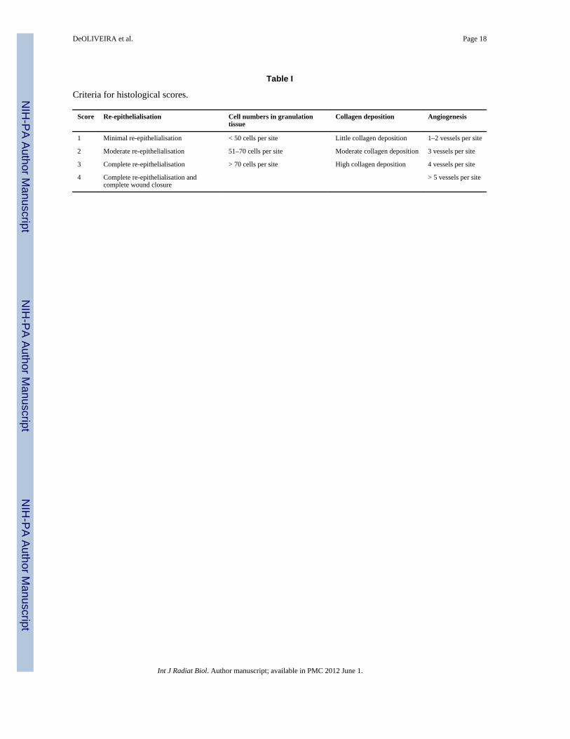
To quantify the histological changes, we employed a semi-quantitative scoring systemmodified from previously published reports (Wu et al. 2007, Thomson et al. 2010). Slideswere scored on the basis of granulation tissue formation, collagen deposition,reepithelialisation, and angiogenesis. The criteria for histological scores are detailed in TableI. Scores for each category were added to provide a total score for each specimen. Minimumscore was 4 and maximum score was 14.
Statistical analysisObserved measurements were summarised as mean ± standard deviation (SD). Comparisonbetween groups was performed by Student's t-test or analysis of variance (ANOVA). Whena difference was detected by ANOVA among the groups, Fisher's protected least significantdifference test was used to determine the significance of pairwise differences. The datashown in Figures 3–5 were further analysed by repeated measures ANOVA. All statisticalanalyses were performed using StatView software (SAS institute, Cary, NC, USA) orMicrosoft Excel (Microsoft, Seattle, WA, USA). P values less than 0.05 were consideredsignificant.
ResultsConfirmation of dosimetry with metal oxide semiconductor field effect transistors(MOSFET)
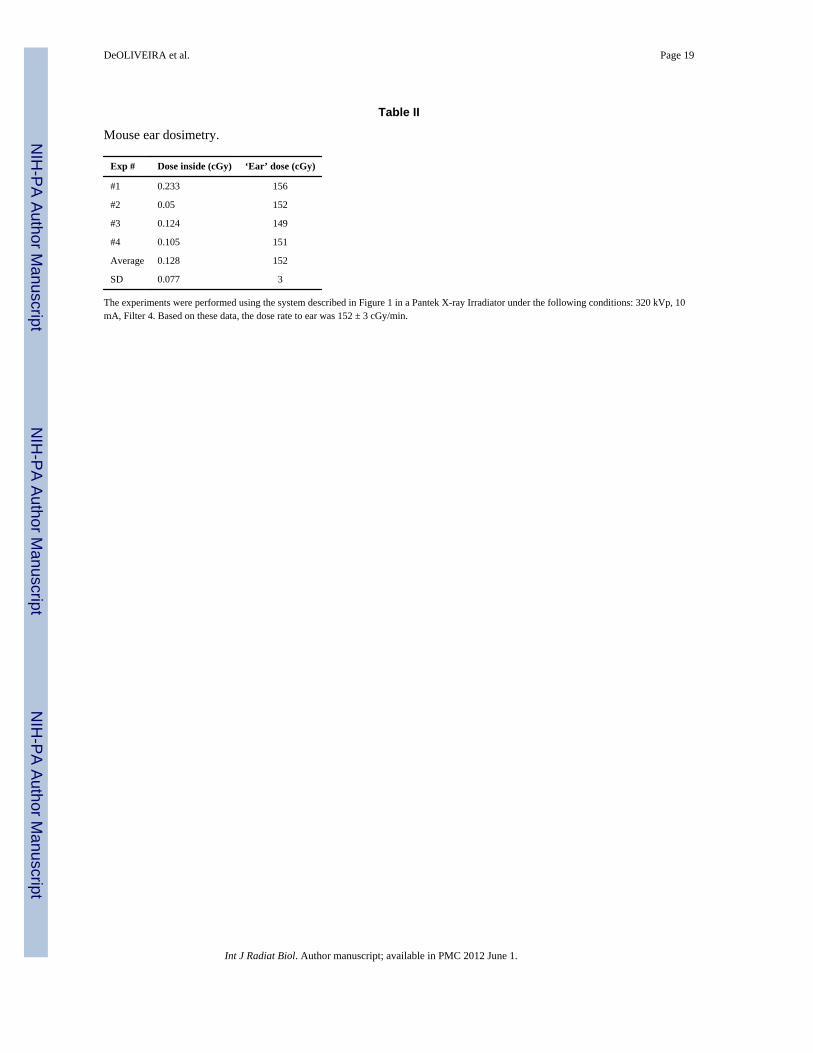
In order to deliver radiation to the ear selectively without irradiating the other parts of thebody, we first constructed an irradiation jig (Figure 1). To confirm that this custom jig coulddeliver proper radiation to the ear selectively, we performed a dosimetry experiment. A sizeappropriate phantom was constructed and MOSFET were placed inside the jig to confirmshielding properties and at the exposed ear position to confirm dose and rate of irradiation(Figure 2). As shown in Table II, radiation could be delivered selectively to ear with lessthan 0.01% of leakage to the other parts of the body. Based on the data from thisexperiment, the dose rate to the ear was 152 ± 3 cGy/min.
Development of an ear punch model for wound healingIn order to study the effect of radiation on wound healing, we developed an ear punchmodel. This model was adapted from a published study (Rajnoch et al. 2003). Briefly, a 2-mm diameter hole was made in the center of each ear by using a clinical biopsy punch. Afterwounding, photos were taken using a camera mounted to a dissecting microscope. An imageof a stage micrometer was also taken using the same settings and used to calibrate thewound area in the analysis. The distance between the camera and mouse ear wasstandardised throughout the experiments. Total area of the wounds was then measured usingPhotoshop software (Adobe Systems, San Jose, CA, USA). The results were represented as% healing. Percent healing was calculated by the following formula:
DeOLIVEIRA et al. Page 3
Int J Radiat Biol. Author manuscript; available in PMC 2012 June 1.
NIH
-PA Author Manuscript
NIH
-PA Author Manuscript
NIH
-PA Author Manuscript
Effect of local radiation on wound healingTo determine the effect of local radiation on wound healing, a 2-mm diameter hole was firstpunched in each ear. The ear was then irradiated with several different doses of radiation (7,10, 15 Gy) using the jig described in Figure 1. Photos of the wounded area were taken overtime and the wound area was then determined. As shown in Figure 3, it took 21 days fornon-irradiated MRL/MpJ mice to close 90% of the 2-mm diameter hole. After beingirradiated locally in the wounded sites at 7, 10, 15 Gy, the healing of the wound wassignificantly delayed (P < 0.01, Figure 3, days to 90% healing for 7, 10, 15 Gy were 28, 70,and > 100 days, respectively). Time required for complete closure of the punch was alsoradiation dose dependent (35, 70, and > 100 days for 7, 10, and 15 Gy, respectively, ascompared to 28 days for non-irradiated mice).These data clearly demonstrate that localirradiation significantly delays the wound healing process. Because the radiation dose of 10Gy significantly delayed the wound healing process and the irradiated wound was still ableto heal completely, this dose was chosen for the subsequent studies in the MRL/MpJ model.
To make sure that these results were not strain specific, we repeated the experiments usingC57BL/6 mice. As demonstrated in Figure 4, it took much longer for non-irradiated C57BL/6 mice to close 90% of the 2-mm diameter hole compared with MRL/MpJ mice (14 vs. 56days). Irradiation with 3 Gy slightly affected the wound healing, but the difference was notstatistically significant. By contrast, higher doses of irradiation (7 and 10 Gy) significantlydelayed the closure of the punch holes (P < 0.05). The wounded area was not closed to >90% of initial size within 100 days post irradiation in the mice irradiated with both 7 and 10Gy. It is of note that all locally irradiated animals including MRL/MpJ and C57BL/6 micesurvived during the entire period of study.
Effect of systemic radiation on wound healingTo study the effect of systemic radiation on wound healing, a 2-mm diameter hole waspunched in the ear of MRL/MpJ mice. The mice then received whole body irradiation with 7Gy followed by additional 3 Gy to the ear. The total dose for mice in this ‘local + systemic’group was 7 Gy of whole body radiation and 10 Gy to the ear. The ‘local’ group received 10Gy of radiation in the ear only. As demonstrated in Figure 5, similar to the data shown inFigure 3, 10 Gy of local radiation significantly prolonged the wound healing (days to 90%healing were 42 vs. 28 days). Additional whole body radiation of 7 Gy further delayed thehealing of the ear punch (day to 90% healing was 70 days). The time required for completeclosure of the 2-mm diameter punch was also at least 14 days longer for the ‘local +systemic’ group compared with the ‘local’ group (> 70 days vs. 56 days). It is of note that allanimals received local radiation survived during the period of study.
Histological changesTo characterise the cellular features of radiation induced alterations to normal woundhealing, we sacrificed the animals and obtained biopsies from the wounded area at day 1, 3,5, 7, 14, and 28 days after wounding. The mice that we analysed included non-irradiatednormal MRL/MpJ mice, mice receiving 10 Gy of local radiation (the ‘local’ group) and 7Gy of whole body radiation and 10 Gy to the ear (the ‘local + systemic’ group). Since therewere no significant differences between different groups as revealed by this analysis at earlytime points, only the data from day +7, +14, +21, and +28 are presented. As shown in Figure6 A–C, the wound in the non-irradiated ears healed normally. At day 7 after wounding,blastema-like structure consisting mostly fibroblasts and blood vessels was evident. At day
DeOLIVEIRA et al. Page 4
Int J Radiat Biol. Author manuscript; available in PMC 2012 June 1.
NIH
-PA Author Manuscript
NIH
-PA Author Manuscript
NIH
-PA Author Manuscript
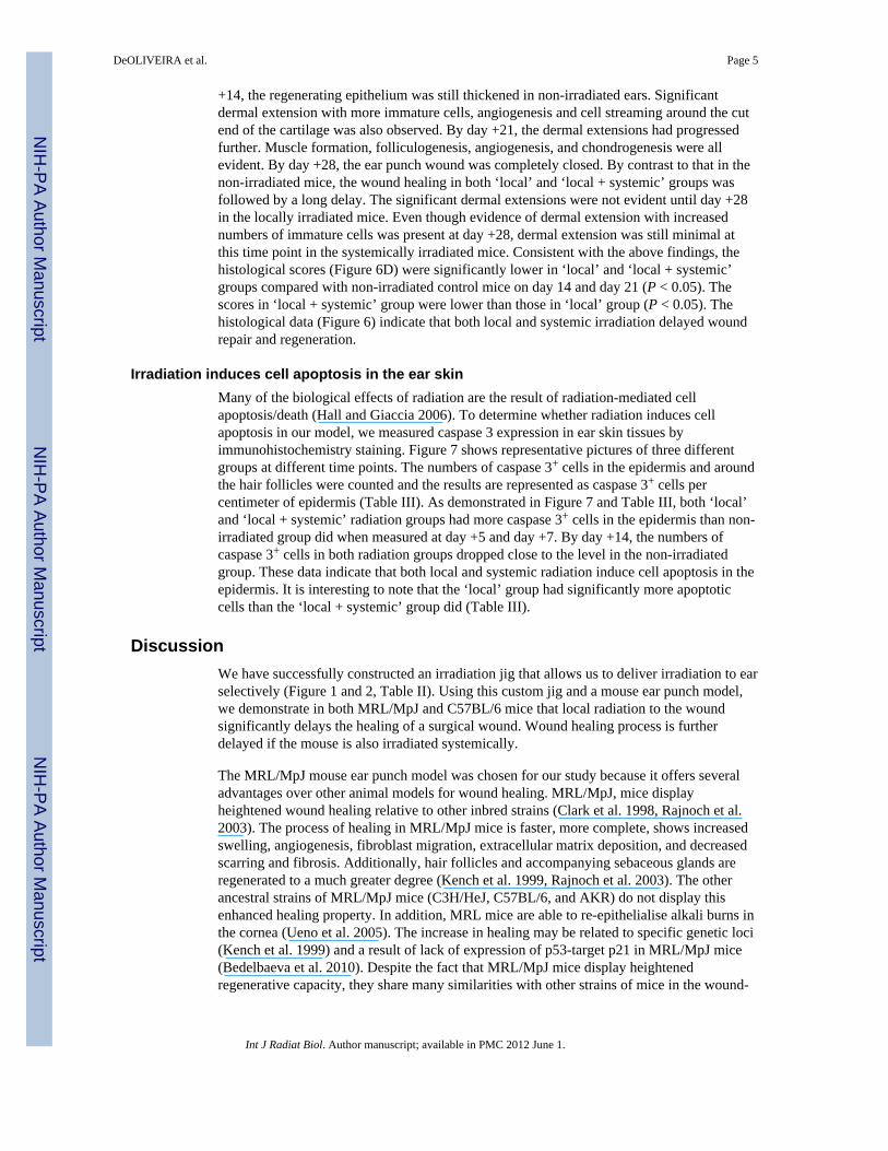
+14, the regenerating epithelium was still thickened in non-irradiated ears. Significantdermal extension with more immature cells, angiogenesis and cell streaming around the cutend of the cartilage was also observed. By day +21, the dermal extensions had progressedfurther. Muscle formation, folliculogenesis, angiogenesis, and chondrogenesis were allevident. By day +28, the ear punch wound was completely closed. By contrast to that in thenon-irradiated mice, the wound healing in both ‘local’ and ‘local + systemic’ groups wasfollowed by a long delay. The significant dermal extensions were not evident until day +28in the locally irradiated mice. Even though evidence of dermal extension with increasednumbers of immature cells was present at day +28, dermal extension was still minimal atthis time point in the systemically irradiated mice. Consistent with the above findings, thehistological scores (Figure 6D) were significantly lower in ‘local’ and ‘local + systemic’groups compared with non-irradiated control mice on day 14 and day 21 (P < 0.05). Thescores in ‘local + systemic’ group were lower than those in ‘local’ group (P < 0.05). Thehistological data (Figure 6) indicate that both local and systemic irradiation delayed woundrepair and regeneration.
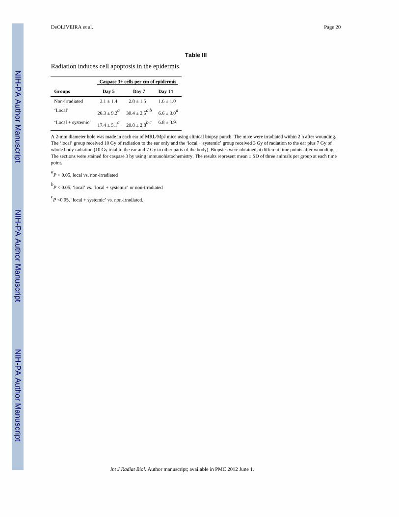
Irradiation induces cell apoptosis in the ear skinMany of the biological effects of radiation are the result of radiation-mediated cellapoptosis/death (Hall and Giaccia 2006). To determine whether radiation induces cellapoptosis in our model, we measured caspase 3 expression in ear skin tissues byimmunohistochemistry staining. Figure 7 shows representative pictures of three differentgroups at different time points. The numbers of caspase 3+ cells in the epidermis and aroundthe hair follicles were counted and the results are represented as caspase 3+ cells percentimeter of epidermis (Table III). As demonstrated in Figure 7 and Table III, both ‘local’and ‘local + systemic’ radiation groups had more caspase 3+ cells in the epidermis than non-irradiated group did when measured at day +5 and day +7. By day +14, the numbers ofcaspase 3+ cells in both radiation groups dropped close to the level in the non-irradiatedgroup. These data indicate that both local and systemic radiation induce cell apoptosis in theepidermis. It is interesting to note that the ‘local’ group had significantly more apoptoticcells than the ‘local + systemic’ group did (Table III).
DiscussionWe have successfully constructed an irradiation jig that allows us to deliver irradiation to earselectively (Figure 1 and 2, Table II). Using this custom jig and a mouse ear punch model,we demonstrate in both MRL/MpJ and C57BL/6 mice that local radiation to the woundsignificantly delays the healing of a surgical wound. Wound healing process is furtherdelayed if the mouse is also irradiated systemically.
The MRL/MpJ mouse ear punch model was chosen for our study because it offers severaladvantages over other animal models for wound healing. MRL/MpJ, mice displayheightened wound healing relative to other inbred strains (Clark et al. 1998, Rajnoch et al.2003). The process of healing in MRL/MpJ mice is faster, more complete, shows increasedswelling, angiogenesis, fibroblast migration, extracellular matrix deposition, and decreasedscarring and fibrosis. Additionally, hair follicles and accompanying sebaceous glands areregenerated to a much greater degree (Kench et al. 1999, Rajnoch et al. 2003). The otherancestral strains of MRL/MpJ mice (C3H/HeJ, C57BL/6, and AKR) do not display thisenhanced healing property. In addition, MRL mice are able to re-epithelialise alkali burns inthe cornea (Ueno et al. 2005). The increase in healing may be related to specific genetic loci(Kench et al. 1999) and a result of lack of expression of p53-target p21 in MRL/MpJ mice(Bedelbaeva et al. 2010). Despite the fact that MRL/MpJ mice display heightenedregenerative capacity, they share many similarities with other strains of mice in the wound-
DeOLIVEIRA et al. Page 5
Int J Radiat Biol. Author manuscript; available in PMC 2012 June 1.
NIH
-PA Author Manuscript
NIH
-PA Author Manuscript
NIH
-PA Author Manuscript
healing process. In fact, histologically, they heal ear wounds in a similar manner comparedwith other strains of mice such as C57BL/6 and BALB/c mice (Rajnoch et al. 2003).
The heightened wound healing in MRL/MpJ mice allows for an easily quantifiablemeasurement of wound healing over a relatively short time frame. These characteristicsmake this an ideal model to study the effects of radiation on wound healing and to studypotential therapeutic agents. Use of selective shielding of the wounded area, which is easilyachievable in this model, will allow for dissection of the local effects of radiation onwounded tissue vs. the systemic effects of radiation on inflammatory cascades and theavailability of local or circulating stem cells. Understanding the mechanisms that cause theincreased morbidity and mortality in combined injuries is crucial to developing effectiveclinical responses to such injuries. Secondarily, it may help with planning for surgery in thepost radiation therapy setting.
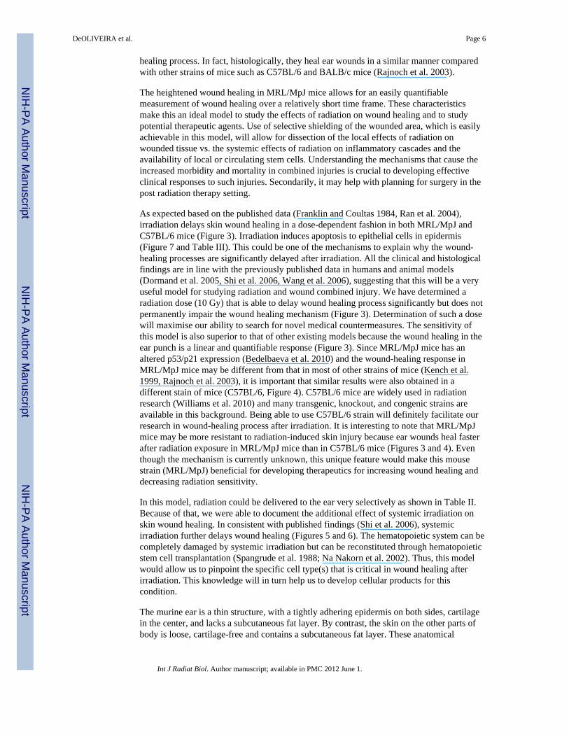
As expected based on the published data (Franklin and Coultas 1984, Ran et al. 2004),irradiation delays skin wound healing in a dose-dependent fashion in both MRL/MpJ andC57BL/6 mice (Figure 3). Irradiation induces apoptosis to epithelial cells in epidermis(Figure 7 and Table III). This could be one of the mechanisms to explain why the wound-healing processes are significantly delayed after irradiation. All the clinical and histologicalfindings are in line with the previously published data in humans and animal models(Dormand et al. 2005, Shi et al. 2006, Wang et al. 2006), suggesting that this will be a veryuseful model for studying radiation and wound combined injury. We have determined aradiation dose (10 Gy) that is able to delay wound healing process significantly but does notpermanently impair the wound healing mechanism (Figure 3). Determination of such a dosewill maximise our ability to search for novel medical countermeasures. The sensitivity ofthis model is also superior to that of other existing models because the wound healing in theear punch is a linear and quantifiable response (Figure 3). Since MRL/MpJ mice has analtered p53/p21 expression (Bedelbaeva et al. 2010) and the wound-healing response inMRL/MpJ mice may be different from that in most of other strains of mice (Kench et al.1999, Rajnoch et al. 2003), it is important that similar results were also obtained in adifferent stain of mice (C57BL/6, Figure 4). C57BL/6 mice are widely used in radiationresearch (Williams et al. 2010) and many transgenic, knockout, and congenic strains areavailable in this background. Being able to use C57BL/6 strain will definitely facilitate ourresearch in wound-healing process after irradiation. It is interesting to note that MRL/MpJmice may be more resistant to radiation-induced skin injury because ear wounds heal fasterafter radiation exposure in MRL/MpJ mice than in C57BL/6 mice (Figures 3 and 4). Eventhough the mechanism is currently unknown, this unique feature would make this mousestrain (MRL/MpJ) beneficial for developing therapeutics for increasing wound healing anddecreasing radiation sensitivity.
In this model, radiation could be delivered to the ear very selectively as shown in Table II.Because of that, we were able to document the additional effect of systemic irradiation onskin wound healing. In consistent with published findings (Shi et al. 2006), systemicirradiation further delays wound healing (Figures 5 and 6). The hematopoietic system can becompletely damaged by systemic irradiation but can be reconstituted through hematopoieticstem cell transplantation (Spangrude et al. 1988; Na Nakorn et al. 2002). Thus, this modelwould allow us to pinpoint the specific cell type(s) that is critical in wound healing afterirradiation. This knowledge will in turn help us to develop cellular products for thiscondition.
The murine ear is a thin structure, with a tightly adhering epidermis on both sides, cartilagein the center, and lacks a subcutaneous fat layer. By contrast, the skin on the other parts ofbody is loose, cartilage-free and contains a subcutaneous fat layer. These anatomical
DeOLIVEIRA et al. Page 6
Int J Radiat Biol. Author manuscript; available in PMC 2012 June 1.
NIH
-PA Author Manuscript
NIH
-PA Author Manuscript
NIH
-PA Author Manuscript
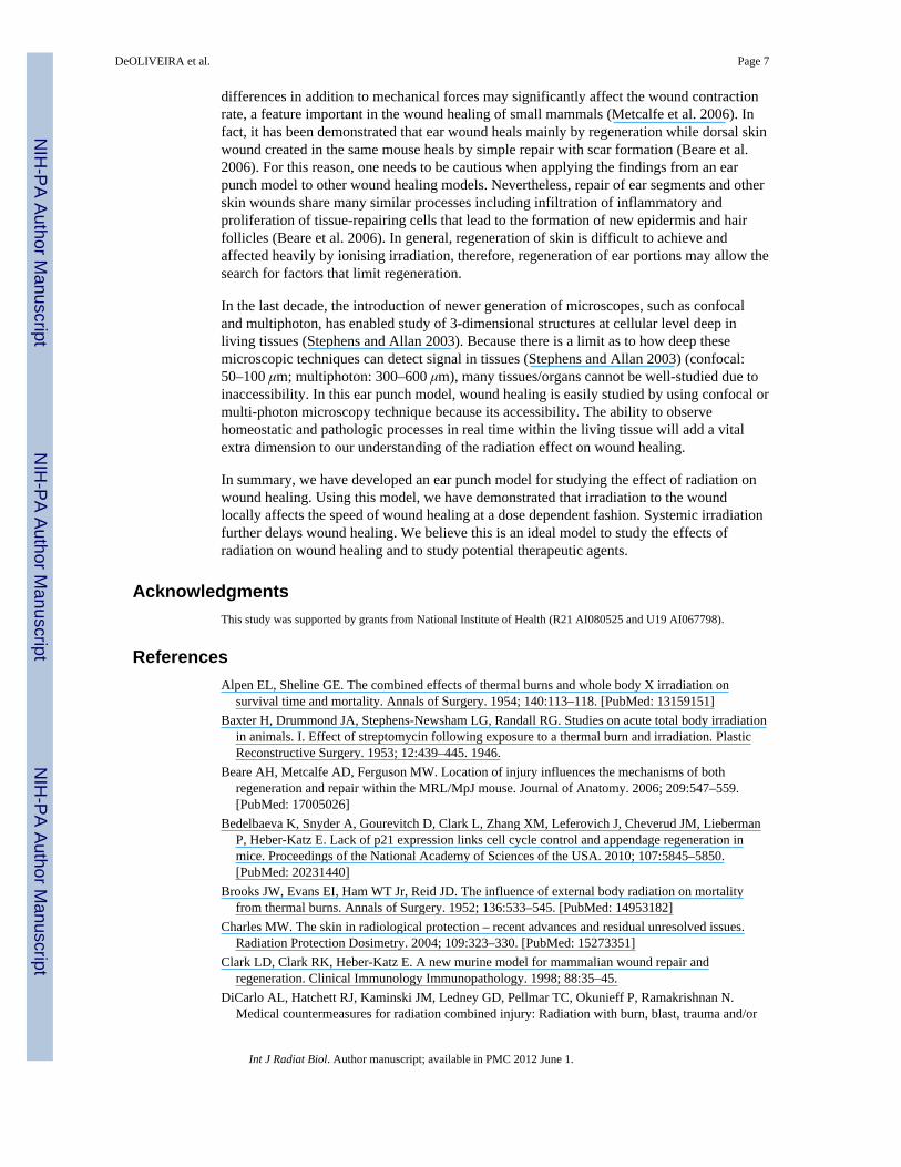
differences in addition to mechanical forces may significantly affect the wound contractionrate, a feature important in the wound healing of small mammals (Metcalfe et al. 2006). Infact, it has been demonstrated that ear wound heals mainly by regeneration while dorsal skinwound created in the same mouse heals by simple repair with scar formation (Beare et al.2006). For this reason, one needs to be cautious when applying the findings from an earpunch model to other wound healing models. Nevertheless, repair of ear segments and otherskin wounds share many similar processes including infiltration of inflammatory andproliferation of tissue-repairing cells that lead to the formation of new epidermis and hairfollicles (Beare et al. 2006). In general, regeneration of skin is difficult to achieve andaffected heavily by ionising irradiation, therefore, regeneration of ear portions may allow thesearch for factors that limit regeneration.
In the last decade, the introduction of newer generation of microscopes, such as confocaland multiphoton, has enabled study of 3-dimensional structures at cellular level deep inliving tissues (Stephens and Allan 2003). Because there is a limit as to how deep thesemicroscopic techniques can detect signal in tissues (Stephens and Allan 2003) (confocal:50–100 μm; multiphoton: 300–600 μm), many tissues/organs cannot be well-studied due toinaccessibility. In this ear punch model, wound healing is easily studied by using confocal ormulti-photon microscopy technique because its accessibility. The ability to observehomeostatic and pathologic processes in real time within the living tissue will add a vitalextra dimension to our understanding of the radiation effect on wound healing.
In summary, we have developed an ear punch model for studying the effect of radiation onwound healing. Using this model, we have demonstrated that irradiation to the woundlocally affects the speed of wound healing at a dose dependent fashion. Systemic irradiationfurther delays wound healing. We believe this is an ideal model to study the effects ofradiation on wound healing and to study potential therapeutic agents.
AcknowledgmentsThis study was supported by grants from National Institute of Health (R21 AI080525 and U19 AI067798).
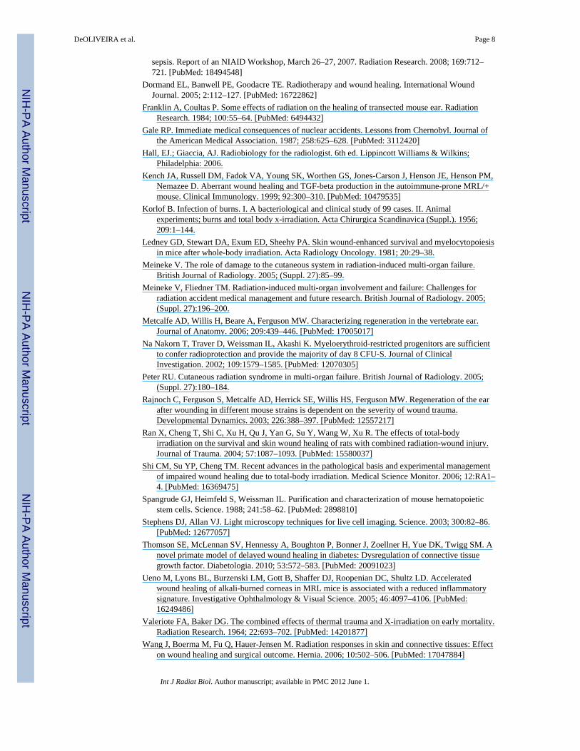
ReferencesAlpen EL, Sheline GE. The combined effects of thermal burns and whole body X irradiation on
survival time and mortality. Annals of Surgery. 1954; 140:113–118. [PubMed: 13159151]Baxter H, Drummond JA, Stephens-Newsham LG, Randall RG. Studies on acute total body irradiation
in animals. I. Effect of streptomycin following exposure to a thermal burn and irradiation. PlasticReconstructive Surgery. 1953; 12:439–445. 1946.
Beare AH, Metcalfe AD, Ferguson MW. Location of injury influences the mechanisms of bothregeneration and repair within the MRL/MpJ mouse. Journal of Anatomy. 2006; 209:547–559.[PubMed: 17005026]
Bedelbaeva K, Snyder A, Gourevitch D, Clark L, Zhang XM, Leferovich J, Cheverud JM, LiebermanP, Heber-Katz E. Lack of p21 expression links cell cycle control and appendage regeneration inmice. Proceedings of the National Academy of Sciences of the USA. 2010; 107:5845–5850.[PubMed: 20231440]
Brooks JW, Evans EI, Ham WT Jr, Reid JD. The influence of external body radiation on mortalityfrom thermal burns. Annals of Surgery. 1952; 136:533–545. [PubMed: 14953182]
Charles MW. The skin in radiological protection – recent advances and residual unresolved issues.Radiation Protection Dosimetry. 2004; 109:323–330. [PubMed: 15273351]
Clark LD, Clark RK, Heber-Katz E. A new murine model for mammalian wound repair andregeneration. Clinical Immunology Immunopathology. 1998; 88:35–45.
DiCarlo AL, Hatchett RJ, Kaminski JM, Ledney GD, Pellmar TC, Okunieff P, Ramakrishnan N.Medical countermeasures for radiation combined injury: Radiation with burn, blast, trauma and/or
DeOLIVEIRA et al. Page 7
Int J Radiat Biol. Author manuscript; available in PMC 2012 June 1.
NIH
-PA Author Manuscript
NIH
-PA Author Manuscript
NIH
-PA Author Manuscript
sepsis. Report of an NIAID Workshop, March 26–27, 2007. Radiation Research. 2008; 169:712–721. [PubMed: 18494548]
Dormand EL, Banwell PE, Goodacre TE. Radiotherapy and wound healing. International WoundJournal. 2005; 2:112–127. [PubMed: 16722862]
Franklin A, Coultas P. Some effects of radiation on the healing of transected mouse ear. RadiationResearch. 1984; 100:55–64. [PubMed: 6494432]
Gale RP. Immediate medical consequences of nuclear accidents. Lessons from Chernobyl. Journal ofthe American Medical Association. 1987; 258:625–628. [PubMed: 3112420]
Hall, EJ.; Giaccia, AJ. Radiobiology for the radiologist. 6th ed. Lippincott Williams & Wilkins;Philadelphia: 2006.
Kench JA, Russell DM, Fadok VA, Young SK, Worthen GS, Jones-Carson J, Henson JE, Henson PM,Nemazee D. Aberrant wound healing and TGF-beta production in the autoimmune-prone MRL/+mouse. Clinical Immunology. 1999; 92:300–310. [PubMed: 10479535]
Korlof B. Infection of burns. I. A bacteriological and clinical study of 99 cases. II. Animalexperiments; burns and total body x-irradiation. Acta Chirurgica Scandinavica (Suppl.). 1956;209:1–144.
Ledney GD, Stewart DA, Exum ED, Sheehy PA. Skin wound-enhanced survival and myelocytopoiesisin mice after whole-body irradiation. Acta Radiology Oncology. 1981; 20:29–38.
Meineke V. The role of damage to the cutaneous system in radiation-induced multi-organ failure.British Journal of Radiology. 2005; (Suppl. 27):85–99.
Meineke V, Fliedner TM. Radiation-induced multi-organ involvement and failure: Challenges forradiation accident medical management and future research. British Journal of Radiology. 2005;(Suppl. 27):196–200.
Metcalfe AD, Willis H, Beare A, Ferguson MW. Characterizing regeneration in the vertebrate ear.Journal of Anatomy. 2006; 209:439–446. [PubMed: 17005017]
Na Nakorn T, Traver D, Weissman IL, Akashi K. Myeloerythroid-restricted progenitors are sufficientto confer radioprotection and provide the majority of day 8 CFU-S. Journal of ClinicalInvestigation. 2002; 109:1579–1585. [PubMed: 12070305]
Peter RU. Cutaneous radiation syndrome in multi-organ failure. British Journal of Radiology. 2005;(Suppl. 27):180–184.
Rajnoch C, Ferguson S, Metcalfe AD, Herrick SE, Willis HS, Ferguson MW. Regeneration of the earafter wounding in different mouse strains is dependent on the severity of wound trauma.Developmental Dynamics. 2003; 226:388–397. [PubMed: 12557217]
Ran X, Cheng T, Shi C, Xu H, Qu J, Yan G, Su Y, Wang W, Xu R. The effects of total-bodyirradiation on the survival and skin wound healing of rats with combined radiation-wound injury.Journal of Trauma. 2004; 57:1087–1093. [PubMed: 15580037]
Shi CM, Su YP, Cheng TM. Recent advances in the pathological basis and experimental managementof impaired wound healing due to total-body irradiation. Medical Science Monitor. 2006; 12:RA1–4. [PubMed: 16369475]
Spangrude GJ, Heimfeld S, Weissman IL. Purification and characterization of mouse hematopoieticstem cells. Science. 1988; 241:58–62. [PubMed: 2898810]
Stephens DJ, Allan VJ. Light microscopy techniques for live cell imaging. Science. 2003; 300:82–86.[PubMed: 12677057]
Thomson SE, McLennan SV, Hennessy A, Boughton P, Bonner J, Zoellner H, Yue DK, Twigg SM. Anovel primate model of delayed wound healing in diabetes: Dysregulation of connective tissuegrowth factor. Diabetologia. 2010; 53:572–583. [PubMed: 20091023]
Ueno M, Lyons BL, Burzenski LM, Gott B, Shaffer DJ, Roopenian DC, Shultz LD. Acceleratedwound healing of alkali-burned corneas in MRL mice is associated with a reduced inflammatorysignature. Investigative Ophthalmology & Visual Science. 2005; 46:4097–4106. [PubMed:16249486]
Valeriote FA, Baker DG. The combined effects of thermal trauma and X-irradiation on early mortality.Radiation Research. 1964; 22:693–702. [PubMed: 14201877]
Wang J, Boerma M, Fu Q, Hauer-Jensen M. Radiation responses in skin and connective tissues: Effecton wound healing and surgical outcome. Hernia. 2006; 10:502–506. [PubMed: 17047884]
DeOLIVEIRA et al. Page 8
Int J Radiat Biol. Author manuscript; available in PMC 2012 June 1.
NIH
-PA Author Manuscript
NIH
-PA Author Manuscript
NIH
-PA Author Manuscript
Williams JP, Brown SL, Georges GE, Hauer-Jensen M, Hill RP, Huser AK, Kirsch DG, Macvittie TJ,Mason KA, Medhora MM, et al. Animal models for medical countermeasures to radiationexposure. Radiation Research. 2010; 173:557–578. [PubMed: 20334528]
Wu Y, Chen L, Scott PG, Tredget EE. Mesenchymal stem cells enhance wound healing throughdifferentiation and angiogenesis. Stem Cells. 2007; 25:2648–2659. [PubMed: 17615264]
DeOLIVEIRA et al. Page 9
Int J Radiat Biol. Author manuscript; available in PMC 2012 June 1.
NIH
-PA Author Manuscript
NIH
-PA Author Manuscript
NIH
-PA Author Manuscript
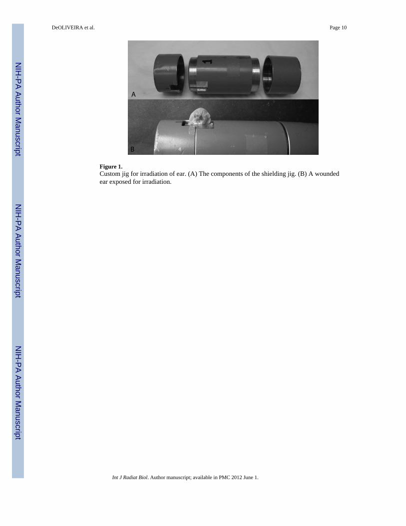
Figure 1.Custom jig for irradiation of ear. (A) The components of the shielding jig. (B) A woundedear exposed for irradiation.
DeOLIVEIRA et al. Page 10
Int J Radiat Biol. Author manuscript; available in PMC 2012 June 1.
NIH
-PA Author Manuscript
NIH
-PA Author Manuscript
NIH
-PA Author Manuscript
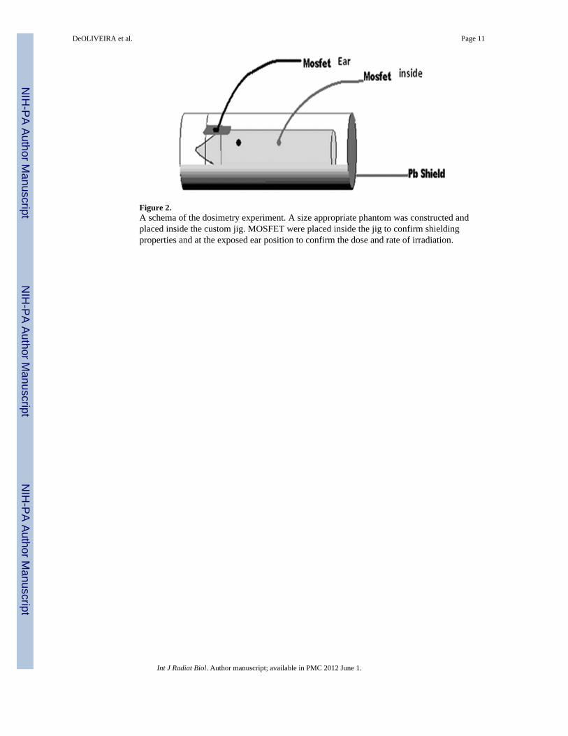
Figure 2.A schema of the dosimetry experiment. A size appropriate phantom was constructed andplaced inside the custom jig. MOSFET were placed inside the jig to confirm shieldingproperties and at the exposed ear position to confirm the dose and rate of irradiation.
DeOLIVEIRA et al. Page 11
Int J Radiat Biol. Author manuscript; available in PMC 2012 June 1.
NIH
-PA Author Manuscript
NIH
-PA Author Manuscript
NIH
-PA Author Manuscript
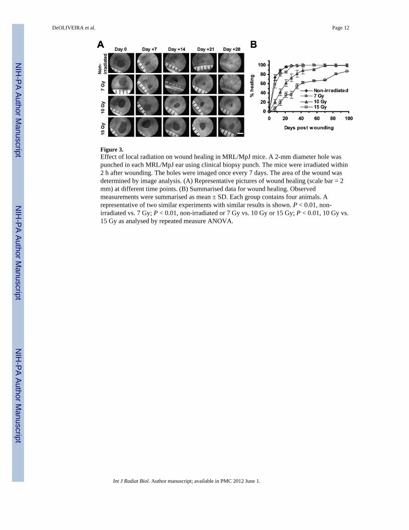
Figure 3.Effect of local radiation on wound healing in MRL/MpJ mice. A 2-mm diameter hole waspunched in each MRL/MpJ ear using clinical biopsy punch. The mice were irradiated within2 h after wounding. The holes were imaged once every 7 days. The area of the wound wasdetermined by image analysis. (A) Representative pictures of wound healing (scale bar = 2mm) at different time points. (B) Summarised data for wound healing. Observedmeasurements were summarised as mean ± SD. Each group contains four animals. Arepresentative of two similar experiments with similar results is shown. P < 0.01, non-irradiated vs. 7 Gy; P < 0.01, non-irradiated or 7 Gy vs. 10 Gy or 15 Gy; P < 0.01, 10 Gy vs.15 Gy as analysed by repeated measure ANOVA.
DeOLIVEIRA et al. Page 12
Int J Radiat Biol. Author manuscript; available in PMC 2012 June 1.
NIH
-PA Author Manuscript
NIH
-PA Author Manuscript
NIH
-PA Author Manuscript
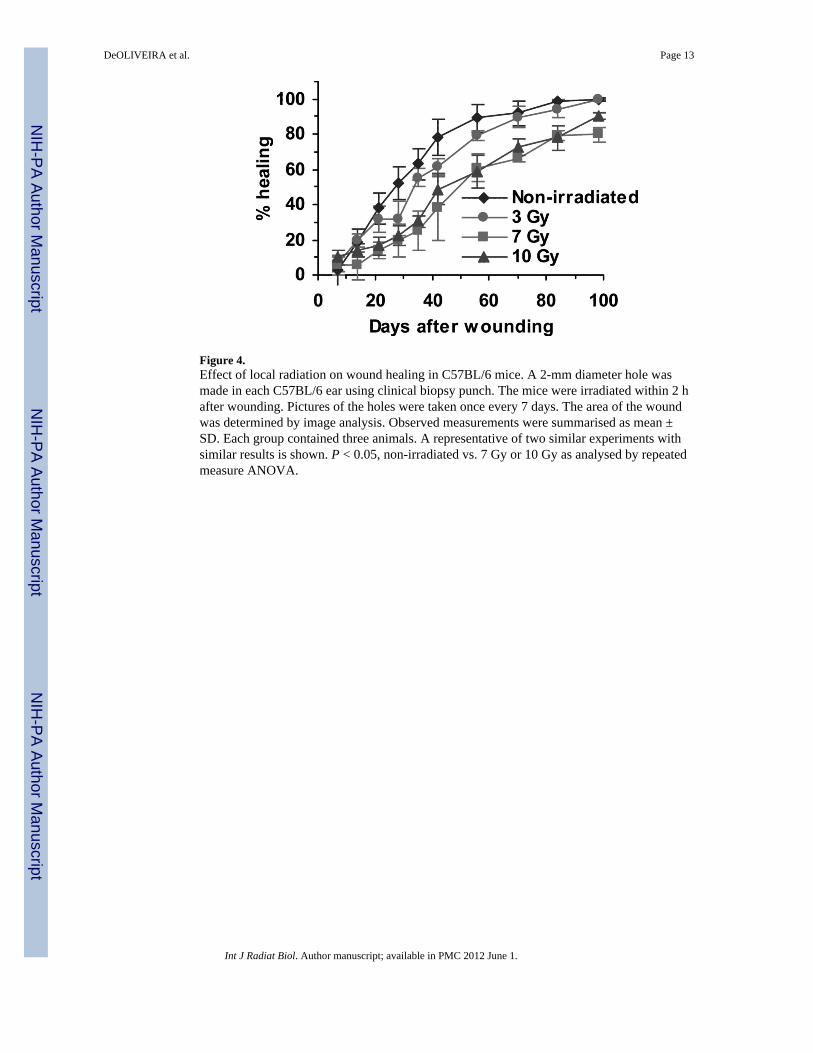
Figure 4.Effect of local radiation on wound healing in C57BL/6 mice. A 2-mm diameter hole wasmade in each C57BL/6 ear using clinical biopsy punch. The mice were irradiated within 2 hafter wounding. Pictures of the holes were taken once every 7 days. The area of the woundwas determined by image analysis. Observed measurements were summarised as mean ±SD. Each group contained three animals. A representative of two similar experiments withsimilar results is shown. P < 0.05, non-irradiated vs. 7 Gy or 10 Gy as analysed by repeatedmeasure ANOVA.
DeOLIVEIRA et al. Page 13
Int J Radiat Biol. Author manuscript; available in PMC 2012 June 1.
NIH
-PA Author Manuscript
NIH
-PA Author Manuscript
NIH
-PA Author Manuscript
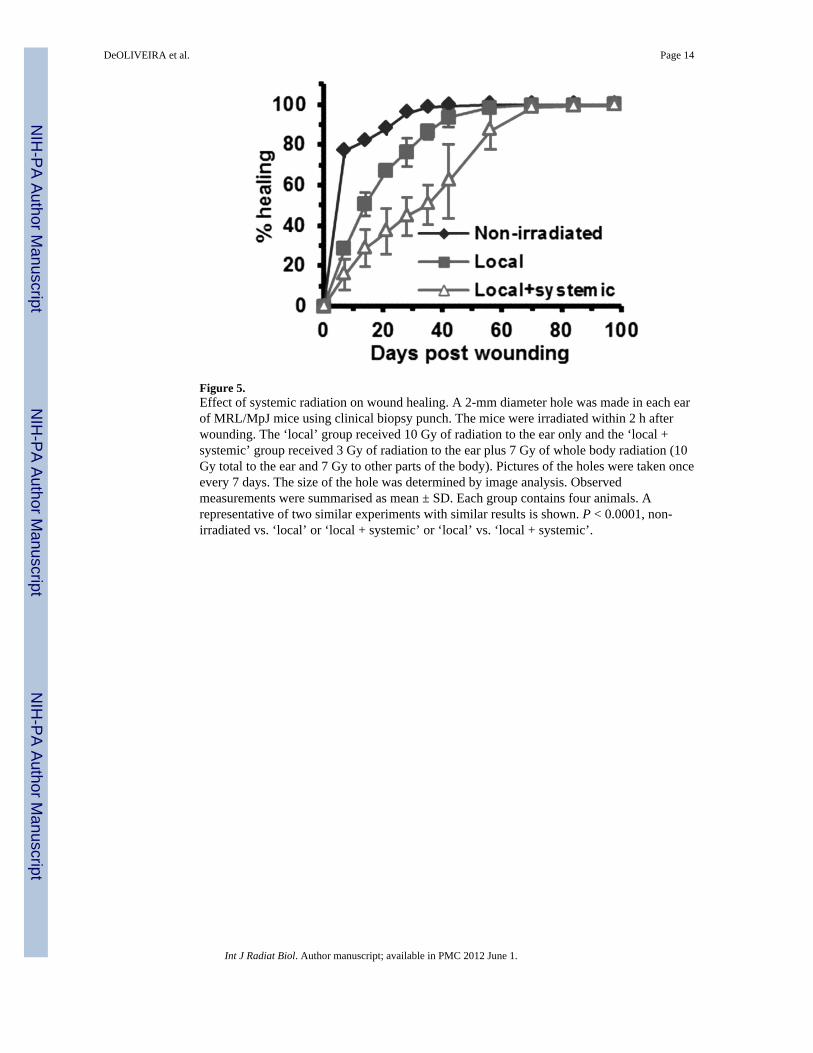
Figure 5.Effect of systemic radiation on wound healing. A 2-mm diameter hole was made in each earof MRL/MpJ mice using clinical biopsy punch. The mice were irradiated within 2 h afterwounding. The ‘local’ group received 10 Gy of radiation to the ear only and the ‘local +systemic’ group received 3 Gy of radiation to the ear plus 7 Gy of whole body radiation (10Gy total to the ear and 7 Gy to other parts of the body). Pictures of the holes were taken onceevery 7 days. The size of the hole was determined by image analysis. Observedmeasurements were summarised as mean ± SD. Each group contains four animals. Arepresentative of two similar experiments with similar results is shown. P < 0.0001, non-irradiated vs. ‘local’ or ‘local + systemic’ or ‘local’ vs. ‘local + systemic’.
DeOLIVEIRA et al. Page 14
Int J Radiat Biol. Author manuscript; available in PMC 2012 June 1.
NIH
-PA Author Manuscript
NIH
-PA Author Manuscript
NIH
-PA Author Manuscript
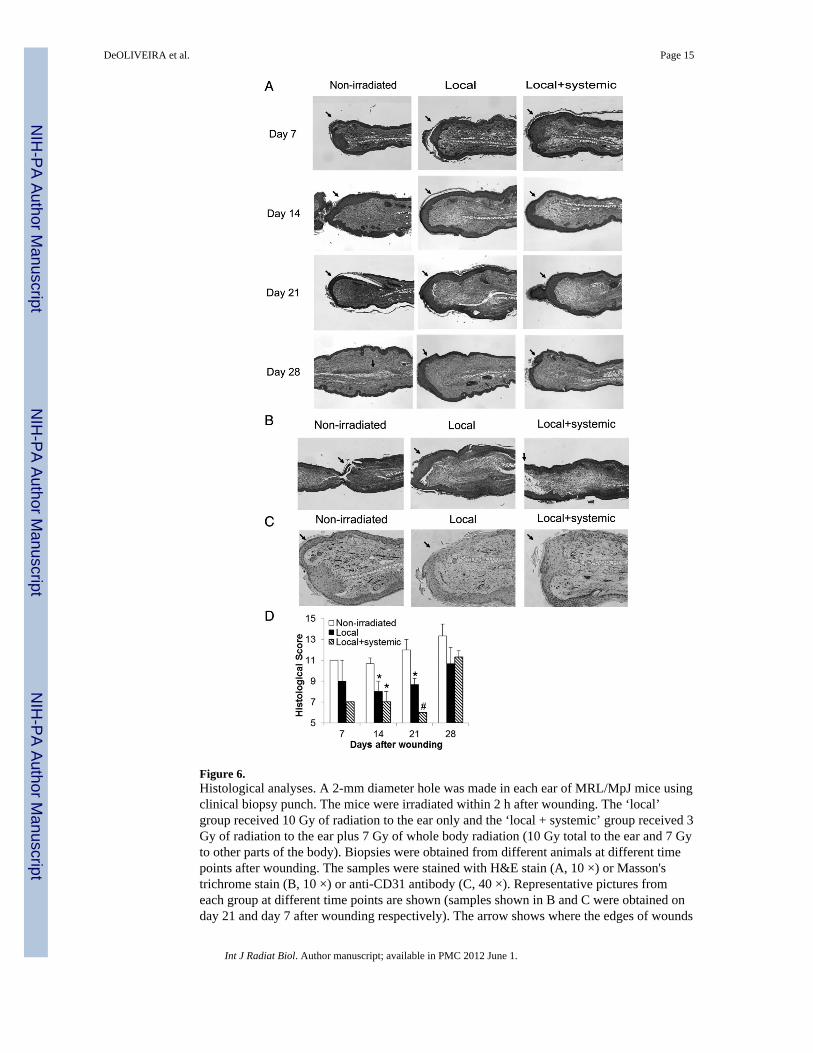
Figure 6.Histological analyses. A 2-mm diameter hole was made in each ear of MRL/MpJ mice usingclinical biopsy punch. The mice were irradiated within 2 h after wounding. The ‘local’group received 10 Gy of radiation to the ear only and the ‘local + systemic’ group received 3Gy of radiation to the ear plus 7 Gy of whole body radiation (10 Gy total to the ear and 7 Gyto other parts of the body). Biopsies were obtained from different animals at different timepoints after wounding. The samples were stained with H&E stain (A, 10 ×) or Masson'strichrome stain (B, 10 ×) or anti-CD31 antibody (C, 40 ×). Representative pictures fromeach group at different time points are shown (samples shown in B and C were obtained onday 21 and day 7 after wounding respectively). The arrow shows where the edges of wounds
DeOLIVEIRA et al. Page 15
Int J Radiat Biol. Author manuscript; available in PMC 2012 June 1.
NIH
-PA Author Manuscript
NIH
-PA Author Manuscript
NIH
-PA Author Manuscript
were located. The samples obtained on day 21 after wounding were shown. Histologicalscores (Mean ± SD of three animals per group per time point) are summarised in D. *P <0.05, compared with non-irradiated group; #P < 0.05, compared with both non-irradiatedand local groups.
DeOLIVEIRA et al. Page 16
Int J Radiat Biol. Author manuscript; available in PMC 2012 June 1.
NIH
-PA Author Manuscript
NIH
-PA Author Manuscript
NIH
-PA Author Manuscript
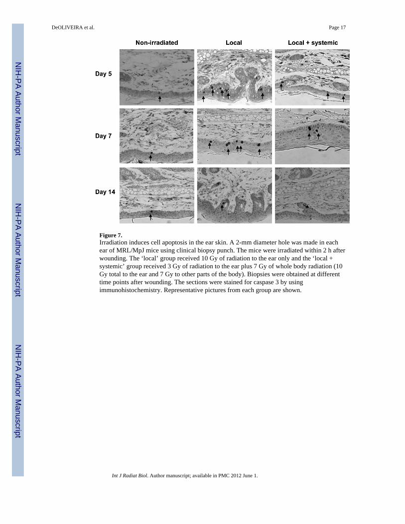
Figure 7.Irradiation induces cell apoptosis in the ear skin. A 2-mm diameter hole was made in eachear of MRL/MpJ mice using clinical biopsy punch. The mice were irradiated within 2 h afterwounding. The ‘local’ group received 10 Gy of radiation to the ear only and the ‘local +systemic’ group received 3 Gy of radiation to the ear plus 7 Gy of whole body radiation (10Gy total to the ear and 7 Gy to other parts of the body). Biopsies were obtained at differenttime points after wounding. The sections were stained for caspase 3 by usingimmunohistochemistry. Representative pictures from each group are shown.
DeOLIVEIRA et al. Page 17
Int J Radiat Biol. Author manuscript; available in PMC 2012 June 1.
NIH
-PA Author Manuscript
NIH
-PA Author Manuscript
NIH
-PA Author Manuscript
NIH
-PA Author Manuscript
NIH
-PA Author Manuscript
NIH
-PA Author Manuscript
DeOLIVEIRA et al. Page 18
Table I
Criteria for histological scores.
Score Re-epithelialisation Cell numbers in granulationtissue
Collagen deposition Angiogenesis
1 Minimal re-epithelialisation < 50 cells per site Little collagen deposition 1–2 vessels per site
2 Moderate re-epithelialisation 51–70 cells per site Moderate collagen deposition 3 vessels per site
3 Complete re-epithelialisation > 70 cells per site High collagen deposition 4 vessels per site
4 Complete re-epithelialisation andcomplete wound closure
> 5 vessels per site
Int J Radiat Biol. Author manuscript; available in PMC 2012 June 1.
NIH
-PA Author Manuscript
NIH
-PA Author Manuscript
NIH
-PA Author Manuscript
DeOLIVEIRA et al. Page 19
Table II
Mouse ear dosimetry.
Exp # Dose inside (cGy) ‘Ear’ dose (cGy)
#1 0.233 156
#2 0.05 152
#3 0.124 149
#4 0.105 151
Average 0.128 152
SD 0.077 3
The experiments were performed using the system described in Figure 1 in a Pantek X-ray Irradiator under the following conditions: 320 kVp, 10mA, Filter 4. Based on these data, the dose rate to ear was 152 ± 3 cGy/min.
Int J Radiat Biol. Author manuscript; available in PMC 2012 June 1.
NIH
-PA Author Manuscript
NIH
-PA Author Manuscript
NIH
-PA Author Manuscript
DeOLIVEIRA et al. Page 20
Table III
Radiation induces cell apoptosis in the epidermis.
Caspase 3+ cells per cm of epidermis
Groups Day 5 Day 7 Day 14
Non-irradiated 3.1 ± 1.4 2.8 ± 1.5 1.6 ± 1.0
‘Local’ 26.3 ± 9.2a 30.4 ± 2.5a,b 6.6 ± 3.0a
‘Local + systemic’ 17.4 ± 5.1c 20.8 ± 2.8b,c 6.8 ± 3.9
A 2-mm diameter hole was made in each ear of MRL/MpJ mice using clinical biopsy punch. The mice were irradiated within 2 h after wounding.The ‘local’ group received 10 Gy of radiation to the ear only and the ‘local + systemic’ group received 3 Gy of radiation to the ear plus 7 Gy ofwhole body radiation (10 Gy total to the ear and 7 Gy to other parts of the body). Biopsies were obtained at different time points after wounding.The sections were stained for caspase 3 by using immunohistochemistry. The results represent mean ± SD of three animals per group at each timepoint.
aP < 0.05, local vs. non-irradiated
bP < 0.05, ‘local’ vs. ‘local + systemic’ or non-irradiated
cP <0.05, ‘local + systemic’ vs. non-irradiated.
Int J Radiat Biol. Author manuscript; available in PMC 2012 June 1.