Patterning and cell fate in ear development
12
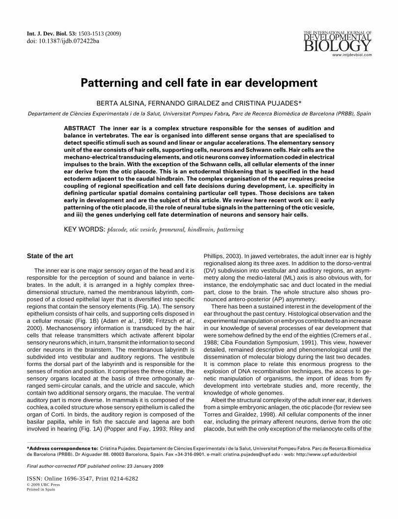
Patterning and cell fate in ear development BERTA ALSINA, FERNANDO GIRALDEZ and CRISTINA PUJADES* Departament de Ciències Experimentals i de la Salut, Universitat Pompeu Fabra, Parc de Recerca Biomèdica de Barcelona (PRBB), Spain ABSTRACT The inner ear is a complex structure responsible for the senses of audition and balance in vertebrates. The ear is organised into different sense organs that are specialised to detect specific stimuli such as sound and linear or angular accelerations. The elementary sensory unit of the ear consists of hair cells, supporting cells, neurons and Schwann cells. Hair cells are the mechano-electrical transducing elements, and otic neurons convey information coded in electrical impulses to the brain. With the exception of the Schwann cells, all cellular elements of the inner ear derive from the otic placode. This is an ectodermal thickening that is specified in the head ectoderm adjacent to the caudal hindbrain. The complex organisation of the ear requires precise coupling of regional specification and cell fate decisions during development, i.e. specificity in defining particular spatial domains containing particular cell types. Those decisions are taken early in development and are the subject of this article. We review here recent work on: i) early patterning of the otic placode, ii) the role of neural tube signals in the patterning of the otic vesicle, and iii) the genes underlying cell fate determination of neurons and sensory hair cells. KEY WORDS: placode, otic vesicle, proneural, hindbrain, patterning State of the art The inner ear is one major sensory organ of the head and it is responsible for the perception of sound and balance in verte- brates. In the adult, it is arranged in a highly complex three- dimensional structure, named the membranous labyrinth, com- posed of a closed epithelial layer that is diversified into specific regions that contain the sensory elements (Fig. 1A). The sensory epithelium consists of hair cells, and supporting cells disposed in a cellular mosaic (Fig. 1B) (Adam et al., 1998; Fritzsch et al., 2000). Mechanosensory information is transduced by the hair cells that release transmitters which activate afferent bipolar sensory neurons which, in turn, transmit the information to second order neurons in the brainstem. The membranous labyrinth is subdivided into vestibular and auditory regions. The vestibule forms the dorsal part of the labyrinth and is responsible for the senses of motion and position. It comprises the three cristae, the sensory organs located at the basis of three orthogonally ar- ranged semi-circular canals, and the utricle and saccule, which contain two additional sensory organs, the maculae. The ventral auditory part is more diverse. In mammals it is composed of the cochlea, a coiled structure whose sensory epithelium is called the organ of Corti. In birds, the auditory region is composed of the basilar papilla, while in fish the saccule and lagena are both involved in hearing (Fig. 1A) (Popper and Fay, 1993; Riley and Int. J. Dev. Biol. 53: 1503-1513 (2009) doi: 10.1387/ijdb.072422ba THE INTERNATIONAL JOURNAL OF DEVELOPMENTAL BIOLOGY www.intjdevbiol.com *Address correspondence to: Cristina Pujades. Departament de Ciències Experimentals i de la Salut, Universitat Pompeu Fabra. Parc de Recerca Biomèdica de Barcelona (PRBB). Dr Aiguader 88. 08003 Barcelona, Spain. Fax +34-316-0901. e-mail: [email protected] - web: http://www.upf.edu/devbiol Final author-corrected PDF published online: 23 January 2009 ISSN: Online 1696-3547, Print 0214-6282 © 2009 UBC Press Printed in Spain Phillips, 2003). In jawed vertebrates, the adult inner ear is highly regionalised along its three axes. In addition to the dorso-ventral (DV) subdivision into vestibular and auditory regions, an asym- metry along the medio-lateral (ML) axis is also obvious with, for instance, the endolymphatic sac and duct located in the medial part, close to the brain. The whole structure also shows pro- nounced antero-posterior (AP) asymmetry. There has been a sustained interest in the development of the ear throughout the past century. Histological observation and the experimental manipulation on embryos contributed to an increase in our knowledge of several processes of ear development that were somehow defined by the end of the eighties (Cremers et al., 1988; Ciba Foundation Symposium, 1991). This view, however detailed, remained descriptive and phenomenological until the dissemination of molecular biology during the last two decades. It is common place to relate this enormous progress to the explosion of DNA recombination techniques, the access to ge- netic manipulation of organisms, the import of ideas from fly development into vertebrate studies and, more recently, the knowledge of whole genomes. Albeit the structural complexity of the adult inner ear, it derives from a simple embryonic anlagen, the otic placode (for review see Torres and Giraldez, 1998). All cellular components of the inner ear, including the primary afferent neurons, derive from the otic placode, but with the only exception of the melanocyte cells of the
Transcript of Patterning and cell fate in ear development
Patterning and cell fate in ear development
BERTA ALSINA, FERNANDO GIRALDEZ and CRISTINA PUJADES*
Departament de Ciències Experimentals i de la Salut, Universitat Pompeu Fabra, Parc de Recerca Biomèdica de Barcelona (PRBB), Spain
ABSTRACT The inner ear is a complex structure responsible for the senses of audition and
balance in vertebrates. The ear is organised into different sense organs that are specialised to
detect specific stimuli such as sound and linear or angular accelerations. The elementary sensory
unit of the ear consists of hair cells, supporting cells, neurons and Schwann cells. Hair cells are the
mechano-electrical transducing elements, and otic neurons convey information coded in electrical
impulses to the brain. With the exception of the Schwann cells, all cellular elements of the inner
ear derive from the otic placode. This is an ectodermal thickening that is specified in the head
ectoderm adjacent to the caudal hindbrain. The complex organisation of the ear requires precise
coupling of regional specification and cell fate decisions during development, i.e. specificity in
defining particular spatial domains containing particular cell types. Those decisions are taken
early in development and are the subject of this article. We review here recent work on: i) early
patterning of the otic placode, ii) the role of neural tube signals in the patterning of the otic vesicle,
and iii) the genes underlying cell fate determination of neurons and sensory hair cells.
KEY WORDS: placode, otic vesicle, proneural, hindbrain, patterning
State of the art
The inner ear is one major sensory organ of the head and it isresponsible for the perception of sound and balance in verte-brates. In the adult, it is arranged in a highly complex three-dimensional structure, named the membranous labyrinth, com-posed of a closed epithelial layer that is diversified into specificregions that contain the sensory elements (Fig. 1A). The sensoryepithelium consists of hair cells, and supporting cells disposed ina cellular mosaic (Fig. 1B) (Adam et al., 1998; Fritzsch et al.,2000). Mechanosensory information is transduced by the haircells that release transmitters which activate afferent bipolarsensory neurons which, in turn, transmit the information to secondorder neurons in the brainstem. The membranous labyrinth issubdivided into vestibular and auditory regions. The vestibuleforms the dorsal part of the labyrinth and is responsible for thesenses of motion and position. It comprises the three cristae, thesensory organs located at the basis of three orthogonally ar-ranged semi-circular canals, and the utricle and saccule, whichcontain two additional sensory organs, the maculae. The ventralauditory part is more diverse. In mammals it is composed of thecochlea, a coiled structure whose sensory epithelium is called theorgan of Corti. In birds, the auditory region is composed of thebasilar papilla, while in fish the saccule and lagena are bothinvolved in hearing (Fig. 1A) (Popper and Fay, 1993; Riley and
Int. J. Dev. Biol. 53: 1503-1513 (2009)doi: 10.1387/ijdb.072422ba
THE INTERNATIONAL JOURNAL OF
DEVELOPMENTAL
BIOLOGYwww.intjdevbiol.com
*Address correspondence to: Cristina Pujades. Departament de Ciències Experimentals i de la Salut, Universitat Pompeu Fabra. Parc de Recerca Biomèdicade Barcelona (PRBB). Dr Aiguader 88. 08003 Barcelona, Spain. Fax +34-316-0901. e-mail: [email protected] - web: http://www.upf.edu/devbiol
Final author-corrected PDF published online: 23 January 2009
ISSN: Online 1696-3547, Print 0214-6282© 2009 UBC PressPrinted in Spain
Phillips, 2003). In jawed vertebrates, the adult inner ear is highlyregionalised along its three axes. In addition to the dorso-ventral(DV) subdivision into vestibular and auditory regions, an asym-metry along the medio-lateral (ML) axis is also obvious with, forinstance, the endolymphatic sac and duct located in the medialpart, close to the brain. The whole structure also shows pro-nounced antero-posterior (AP) asymmetry.
There has been a sustained interest in the development of theear throughout the past century. Histological observation and theexperimental manipulation on embryos contributed to an increasein our knowledge of several processes of ear development thatwere somehow defined by the end of the eighties (Cremers et al.,1988; Ciba Foundation Symposium, 1991). This view, howeverdetailed, remained descriptive and phenomenological until thedissemination of molecular biology during the last two decades.It is common place to relate this enormous progress to theexplosion of DNA recombination techniques, the access to ge-netic manipulation of organisms, the import of ideas from flydevelopment into vertebrate studies and, more recently, theknowledge of whole genomes.
Albeit the structural complexity of the adult inner ear, it derivesfrom a simple embryonic anlagen, the otic placode (for review seeTorres and Giraldez, 1998). All cellular components of the innerear, including the primary afferent neurons, derive from the oticplacode, but with the only exception of the melanocyte cells of the

1504 B. Alsina et al.
secretory epithelium and Schwann cells of the ganglia, which areof neural crest origin (D’Amico-Martel and Noden, 1983, Rubeland Fritzsch, 2002). Cranial placodes are specialized areas ofectoderm outside the neural plate that contribute to all the cranialpaired sensory organs and most of the sensory neurons from theperipheral nervous system of the head. The current view is thatplacodes share a common developmental origin, the preplacodalregion (PPR), a horseshoe band encircling the neural plate fromwhich individual placodes emerge (Jacobson, 1966; Torres andGiraldez, 1998; Streit, 2007). Olfactory, profundal and trigeminal,otic, lateral line, and epibranchial are all neurogenic placodes thatgive rise to sensory neurons among other specialized cell-types(Adam et al., 1998; Ma et al., 2000; Schlosser and Northcutt,2000; Andermann et al., 2002; Begbie et al., 2002).
The complex organisation of the ear requires precise couplingbetween regional specification and cell fate decisions, that is,specificity in defining particular spatial domains with particular celltypes. We shall review some of the recent work done on that issue,and discuss first, the early steps of regional specification of the
otic placode, then patterning of the otic vesicle, and finally, thegenetic networks underlying cell fate decisions and sensoryorgan development.
The regional specification of the otic vesicle: the pat-terning of the ear
The regional (or axial) polarity needed to develop the membra-nous labyrinth of the ear has long been recognised as providingthe basis for ear function. Classical transplantation experimentsshowed that rotated otic placodes produce enantiomorphic twins(Harrison, 1945; Yntema, 1955), which somewhat resemble thesymmetric ear of the hagfish. The early patterning of the ear is alsoset, at least in part, by interactions with surrounding tissues,particularly the neural tube. The first sign of otic regionalisation isthat of the establishment of the otic neural and non-neural fields(Fig. 2). In the following, we review the molecular properties ofthese two domains and discuss the possible models by which thisearly specification is set in place. However, axial polarity extends
Fig. 1. The structure of the inner ear. (A) The membranous labyrinth in threevertebrate species. From left to right: zebrafish,chicken and mouse. The vestibular(dorsal) part of the membranous labyrinth contains five sensory organs: the threecristae (blue) located at the basis of the three semicircular canals, and the utricular andsaccular maculae (orange), surrounded by otoliths. The ventral, auditory part of theinner ear (grey) is highly variable in morphology and complexity in different vertebrates.In the mouse, the cochlear duct, acoiled structure, contains a finely patterned sensoryorgan, the organ of Corti. In chicken, the auditory organ, the basilar papilla, is alsocontained in the cochlear duct. In zebrafish, there is no ventral cochlear duct and theauditory function is carried by the saccular and lagenar maculae. ac: anterior crista; asc:anterior semicircular canal; cd:cochlear duct; hsc: horizontal semicircular canal; l:lagena; lc: lateral crista; pc:posterior crista; psc: posterior semicircular canal; s:saccule; u: utricle. Anterior is to theright ans dorsal to the top. (B) The functional unitof the ear. The four basic cellular elements of the functional unit are depicted: hair cells(red), supporting cells (orange), neurons (blue) and Schwann cells (white).
B
A
to further complexity in the regionalisation of the oticvesicle, when it becomes necessary to establish thedifferent domains of the inner ear. Inductive processesextend further into the development of the otic vesicle,and the neural tube seems to play an important role inestablishing the final axial pattern of the ear (for latereviews see Choo, 2007; Schneider-Maunoury andPujades, 2007; Whitfield and Hammond, 2007). Weshall examine first, the establishment of the neuraldomain of the otic placode, including the role of Notchsignalling in this process and, secondly, the function ofthe neural tube as a source of signals for theregionalisation of the otic vesicle.
The establishment of the otic neural competentdomain
Current understanding of specification of the oticplacode involves a two-stage mechanism by whichfirst, an extended multipotent pre-placodal domain isspecified at the head ectoderm, and then individualplacode identities are specified (Jacobson, 1966; Streit,2007). An initial set of genes (Foxi, Msx and Dlx)identify an ectodermal domain between the neuralplate and the epidermis from which the preplacodaldomain is segregated from the neural crest. Both thepositioning of the preplacodal ectoderm and its capac-ity to express the specific Six/Eya/Dach cassette seemto require interactions between the presumptive ecto-derm and the surrounding tissues (Streit 2007). Howdoes the preplacodal ectoderm transit from a pluripo-tent ground state to one in which otic fate is specified?This apparently requires another round of interactionsthat position and specify the fate of individual placodes(Ohyama et al., 2007; Streit, 2007; Jayasena et al.,2008). This notion of sequential rounds of interactionswas anticipated by the classical studies of Yntema,1955 and Jacobson, 1966.
Recent work has shown that the ear primordium isalready patterned at the time of the development of theotic placode (Alsina et al., 2004; Vázquez-Echeverría
Ear patterning and cell fate 1505
et al., 2008; Bell et al., 2008). This early regionalisation is relatedto the establishment of two complementary neural and non-neuralterritories in the otic placode and otic vesicle (Fig. 2). In the chickembryo, otic cell types emerge sequentially during development.First the neuroblasts are specified in the anterior otic cup, asrevealed by Neurog1-positive cells (Fig. 2A). Mechanosensoryhair cells do so later in development within the domains of thedifferent sense organs (Fig. 1A). The activation of proneuralgenes is one of the firsts signs of neural determination, and in thecase of the development of sensory neurons, Neurog1 has beenshown to be sufficient for the acquisition of neuronal fate. Over-expression of Neurog1 drives formation of ectopic neurons (Per-ron et al., 1999; Kim et al., 2001), while targeted inactivation ofNeurog1 results in severe loss of proximal cranial sensory gangliaand hair cells (Ma et al., 2000). This suggests that Neurog1 isrelated to the development of a common pro-neural and -sensoryfield that first gives rise to neurons and later on to hair cells, oralternatively to a common neural-competent field (Raft et al.,2007). In chick, a territory expressing Neurog1, Delta1 and LFngemerges as a triangle in the anterior half of the flat otic placode.Initially the neural domain is the anterior-medial aspect of the oticplacode to end up, after invagination, in an anterior-medial andventral position of the otic vesicle (Alsina et al., 2004). In mam-mals, expression of Neurog1 appears in a more lateral position,
detected as an antero-ventro-lateral quadrant at otic cup stage, toextend also medially as development proceeds (Fig. 2B and Maet al., 1998; Vázquez-Echeverría et al., 2008). A recent analysisby Cre-loxP fate mapping in mouse shows that part of thevestibular sensory hair cells derive from a neurogenic region, andthe study of mouse mutants provides evidences that a mutualantagonism between Neurog1 and Math1 regulates the transitionto sensory cell production (Raft et al., 2007).
Experiments of otic cup rotation have suggested that APpatterning is not fixed until late otic cup stage (Bok et al., 2005),suggesting that the first signs of otic regionalisation detected bygene expression patterns require posterior signals to stabilize theneural/non-neural patterning.
But is the expression of Neurog1 the first sign of neuralcommitment within the otic placode? Recent studies of indicatethat Sox3 is required early in otic neural development (Khatri andAlsina, unpublished results). Sox3 is expressed before the oticplacode is morphologically visible, within a broad band thatcontains the otic and epibranchial territories, to get restricted lateron in development to the proneural region of the otic placode (Fig.3). This suggests that neural fate specification takes place priorthe formation of the otic placode, within a broad territory thatcontains the antero-medial otic region and the lateral epibranchialterritory. Previous work has indicated that FGF signalling is
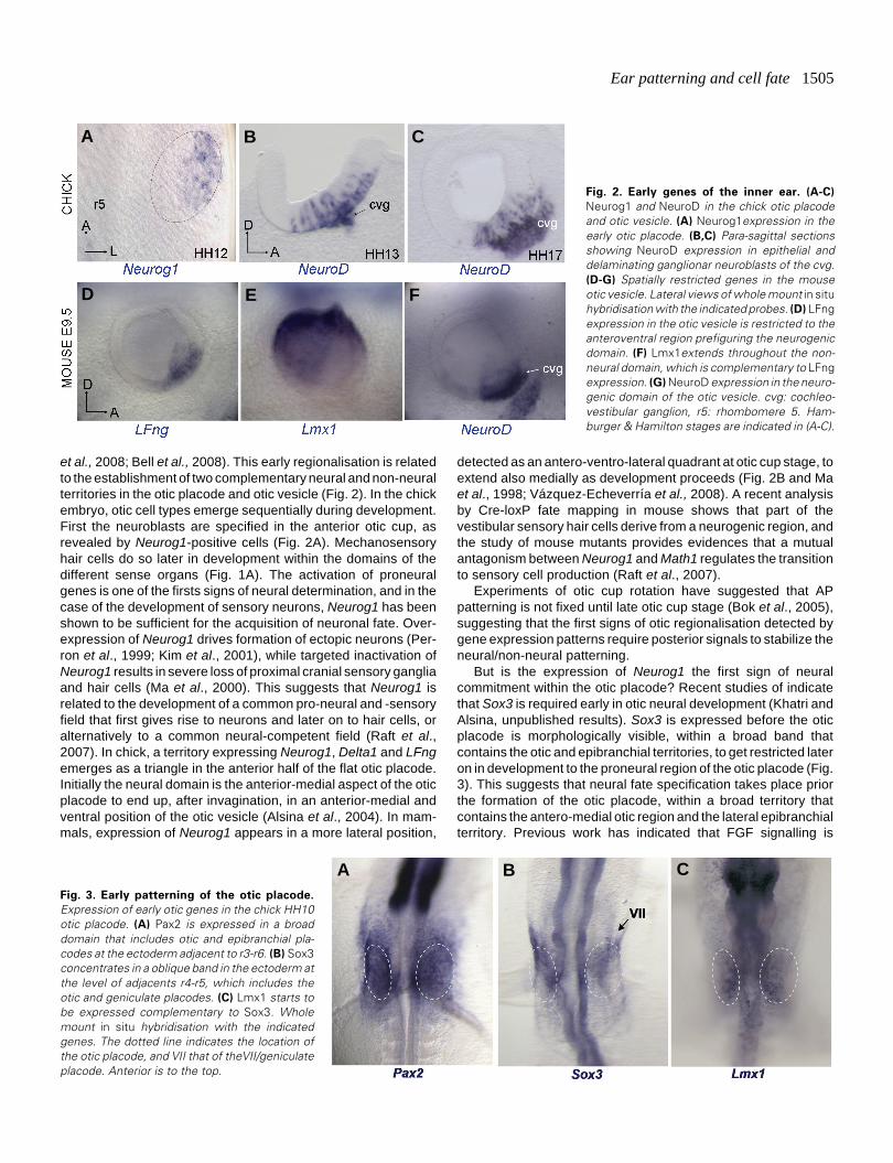
Fig. 2. Early genes of the inner ear. (A-C)
Neurog1 and NeuroD in the chick otic placodeand otic vesicle. (A) Neurog1expression in theearly otic placode. (B,C) Para-sagittal sectionsshowing NeuroD expression in epithelial anddelaminating ganglionar neuroblasts of the cvg.(D-G) Spatially restricted genes in the mouseotic vesicle. Lateral views of whole mount in situhybridisation with the indicated probes. (D) LFngexpression in the otic vesicle is restricted to theanteroventral region prefiguring the neurogenicdomain. (F) Lmx1extends throughout the non-neural domain, which is complementary to LFngexpression. (G) NeuroD expression in the neuro-genic domain of the otic vesicle. cvg: cochleo-vestibular ganglion, r5: rhombomere 5. Ham-burger & Hamilton stages are indicated in (A-C).
B C
D E F
A
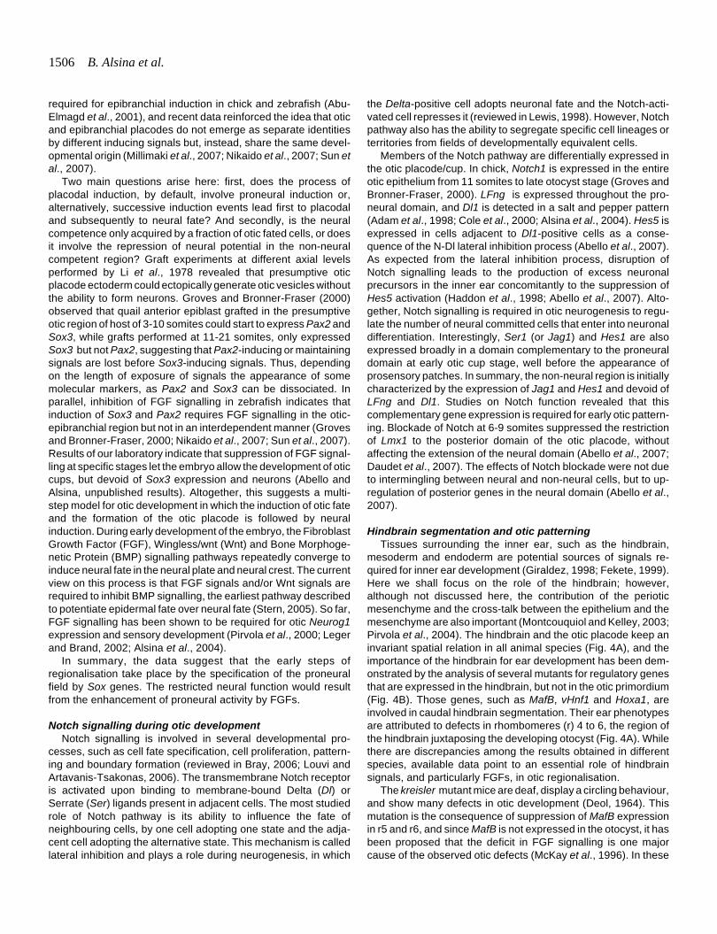
Fig. 3. Early patterning of the otic placode.
Expression of early otic genes in the chick HH10otic placode. (A) Pax2 is expressed in a broaddomain that includes otic and epibranchial pla-codes at the ectoderm adjacent to r3-r6. (B) Sox3concentrates in a oblique band in the ectoderm atthe level of adjacents r4-r5, which includes theotic and geniculate placodes. (C) Lmx1 starts tobe expressed complementary to Sox3. Wholemount in situ hybridisation with the indicatedgenes. The dotted line indicates the location ofthe otic placode, and VII that of theVII/geniculateplacode. Anterior is to the top.
B CA
1506 B. Alsina et al.
required for epibranchial induction in chick and zebrafish (Abu-Elmagd et al., 2001), and recent data reinforced the idea that oticand epibranchial placodes do not emerge as separate identitiesby different inducing signals but, instead, share the same devel-opmental origin (Millimaki et al., 2007; Nikaido et al., 2007; Sun etal., 2007).
Two main questions arise here: first, does the process ofplacodal induction, by default, involve proneural induction or,alternatively, successive induction events lead first to placodaland subsequently to neural fate? And secondly, is the neuralcompetence only acquired by a fraction of otic fated cells, or doesit involve the repression of neural potential in the non-neuralcompetent region? Graft experiments at different axial levelsperformed by Li et al., 1978 revealed that presumptive oticplacode ectoderm could ectopically generate otic vesicles withoutthe ability to form neurons. Groves and Bronner-Fraser (2000)observed that quail anterior epiblast grafted in the presumptiveotic region of host of 3-10 somites could start to express Pax2 andSox3, while grafts performed at 11-21 somites, only expressedSox3 but not Pax2, suggesting that Pax2-inducing or maintainingsignals are lost before Sox3-inducing signals. Thus, dependingon the length of exposure of signals the appearance of somemolecular markers, as Pax2 and Sox3 can be dissociated. Inparallel, inhibition of FGF signalling in zebrafish indicates thatinduction of Sox3 and Pax2 requires FGF signalling in the otic-epibranchial region but not in an interdependent manner (Grovesand Bronner-Fraser, 2000; Nikaido et al., 2007; Sun et al., 2007).Results of our laboratory indicate that suppression of FGF signal-ling at specific stages let the embryo allow the development of oticcups, but devoid of Sox3 expression and neurons (Abello andAlsina, unpublished results). Altogether, this suggests a multi-step model for otic development in which the induction of otic fateand the formation of the otic placode is followed by neuralinduction. During early development of the embryo, the FibroblastGrowth Factor (FGF), Wingless/wnt (Wnt) and Bone Morphoge-netic Protein (BMP) signalling pathways repeatedly converge toinduce neural fate in the neural plate and neural crest. The currentview on this process is that FGF signals and/or Wnt signals arerequired to inhibit BMP signalling, the earliest pathway describedto potentiate epidermal fate over neural fate (Stern, 2005). So far,FGF signalling has been shown to be required for otic Neurog1expression and sensory development (Pirvola et al., 2000; Legerand Brand, 2002; Alsina et al., 2004).
In summary, the data suggest that the early steps ofregionalisation take place by the specification of the proneuralfield by Sox genes. The restricted neural function would resultfrom the enhancement of proneural activity by FGFs.
Notch signalling during otic developmentNotch signalling is involved in several developmental pro-
cesses, such as cell fate specification, cell proliferation, pattern-ing and boundary formation (reviewed in Bray, 2006; Louvi andArtavanis-Tsakonas, 2006). The transmembrane Notch receptoris activated upon binding to membrane-bound Delta (Dl) orSerrate (Ser) ligands present in adjacent cells. The most studiedrole of Notch pathway is its ability to influence the fate ofneighbouring cells, by one cell adopting one state and the adja-cent cell adopting the alternative state. This mechanism is calledlateral inhibition and plays a role during neurogenesis, in which
the Delta-positive cell adopts neuronal fate and the Notch-acti-vated cell represses it (reviewed in Lewis, 1998). However, Notchpathway also has the ability to segregate specific cell lineages orterritories from fields of developmentally equivalent cells.
Members of the Notch pathway are differentially expressed inthe otic placode/cup. In chick, Notch1 is expressed in the entireotic epithelium from 11 somites to late otocyst stage (Groves andBronner-Fraser, 2000). LFng is expressed throughout the pro-neural domain, and Dl1 is detected in a salt and pepper pattern(Adam et al., 1998; Cole et al., 2000; Alsina et al., 2004). Hes5 isexpressed in cells adjacent to Dl1-positive cells as a conse-quence of the N-Dl lateral inhibition process (Abello et al., 2007).As expected from the lateral inhibition process, disruption ofNotch signalling leads to the production of excess neuronalprecursors in the inner ear concomitantly to the suppression ofHes5 activation (Haddon et al., 1998; Abello et al., 2007). Alto-gether, Notch signalling is required in otic neurogenesis to regu-late the number of neural committed cells that enter into neuronaldifferentiation. Interestingly, Ser1 (or Jag1) and Hes1 are alsoexpressed broadly in a domain complementary to the proneuraldomain at early otic cup stage, well before the appearance ofprosensory patches. In summary, the non-neural region is initiallycharacterized by the expression of Jag1 and Hes1 and devoid ofLFng and Dl1. Studies on Notch function revealed that thiscomplementary gene expression is required for early otic pattern-ing. Blockade of Notch at 6-9 somites suppressed the restrictionof Lmx1 to the posterior domain of the otic placode, withoutaffecting the extension of the neural domain (Abello et al., 2007;Daudet et al., 2007). The effects of Notch blockade were not dueto intermingling between neural and non-neural cells, but to up-regulation of posterior genes in the neural domain (Abello et al.,2007).
Hindbrain segmentation and otic patterningTissues surrounding the inner ear, such as the hindbrain,
mesoderm and endoderm are potential sources of signals re-quired for inner ear development (Giraldez, 1998; Fekete, 1999).Here we shall focus on the role of the hindbrain; however,although not discussed here, the contribution of the perioticmesenchyme and the cross-talk between the epithelium and themesenchyme are also important (Montcouquiol and Kelley, 2003;Pirvola et al., 2004). The hindbrain and the otic placode keep aninvariant spatial relation in all animal species (Fig. 4A), and theimportance of the hindbrain for ear development has been dem-onstrated by the analysis of several mutants for regulatory genesthat are expressed in the hindbrain, but not in the otic primordium(Fig. 4B). Those genes, such as MafB, vHnf1 and Hoxa1, areinvolved in caudal hindbrain segmentation. Their ear phenotypesare attributed to defects in rhombomeres (r) 4 to 6, the region ofthe hindbrain juxtaposing the developing otocyst (Fig. 4A). Whilethere are discrepancies among the results obtained in differentspecies, available data point to an essential role of hindbrainsignals, and particularly FGFs, in otic regionalisation.
The kreisler mutant mice are deaf, display a circling behaviour,and show many defects in otic development (Deol, 1964). Thismutation is the consequence of suppression of MafB expressionin r5 and r6, and since MafB is not expressed in the otocyst, it hasbeen proposed that the deficit in FGF signalling is one majorcause of the observed otic defects (McKay et al., 1996). In these
Ear patterning and cell fate 1507
mice, dorsomedial markers such as Gbx2 and Wnt2b are lost,while the ventral Otx2 domain is expanded, suggesting a role ofthe hindbrain in the specification of dorsomedial structures of theinner ear (Choo et al., 2006). The similarity of this phenotype withthe observed in Gbx2 null mutants (Lin et al., 2005) suggestedthat Gbx2 was a target of hindbrain signalling in the otocyst. Onthe other hand, kreisler mutants display an expansion of the oticneurogenic region as revealed by the complementary changes inthe early expression of patterning genes LFng and Lmx1 (Vázquez-Echeverría et al., 2008). Thus, in addition to the DV patterningdefects, kreisler mutants display an early AP patterning defect,affecting mainly the neurogenic/non-neurogenic fate decision. Asfor Hoxa1 mutants in which ear patterning defects are correlatedto the loss of Fgf3 expression in the hindbrain (Pasqualetti et al.,2001), kreisler mutants fail to upregulate Fgf3 and Fgf10 in r5 andr6 (Vázquez-Echeverría et al., 2008).
The MafB/val mutation in zebrafish results mainly in AP pat-terning defects (Kwak et al., 2002). Anterior markers such ashmx3 are expanded posteriorly, while the expression of caudalmarkers is reduced or absent. val mutants also present an excess
of hair cells, ectopically produced be-tween the anterior and posterior macu-lae (Kwak et al., 2002). The val muta-tion results in a posterior expansion offgf3 expression in the hindbrain. Re-duction of fgf3 RNA levels in valmutants using morpholinos rescuessome of the otic defects, strongly sug-gesting that, in zebrafish as well as inmouse, FGF3 is a major signal in-volved in ear patterning, downstreamof MafB (Kwak et al., 2002).
The analysis of the zebrafish vhnf1mutant has added more complexity tothe picture. vhnf1 positively controlsval expression in the hindbrain(Wiellette and Sive, 2003; Hernandezet al., 2004). As expected, AP pat-terning phenotypes are observed inthe inner ear of vhnf1 mutants, whichdisplay an expansion or a duplicationof the expression of anterior otic genessuch as hmx3, fgf8 and pax5. How-ever, vhnf1 mutants also show DVpatterning defects, and a dorsal shiftof intermediate markers such asatoh1a, which marks the future macu-lae (Lecaudey et al., 2007). val andvhnf1 mutants display hair cells atectopic positions all along the AP axisof the otic vesicle, suggesting thateither an intact r5 identity or r5-sig-nals are essential to restrict early haircell specification to the otic regionlateral to r4 and r6. There is a strikingdifference between mouse andzebrafish. Although for years it wasthought that the main defects seen inmice are along the DV axis, while in
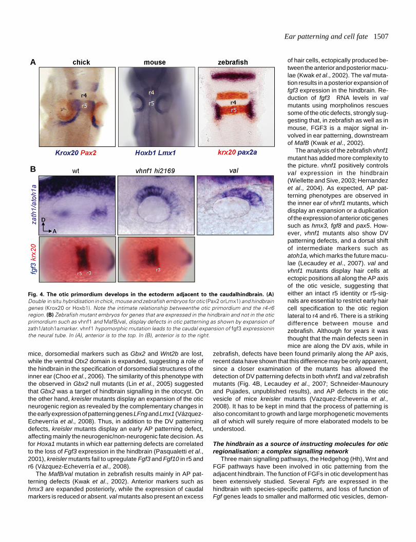
Fig. 4. The otic primordium develops in the ectoderm adjacent to the caudalhindbrain. (A)
Double in situ hybridisation in chick, mouse and zebrafish embryos for otic (Pax2 orLmx1) and hindbraingenes (Krox20 or Hoxb1). Note the intimate relationship betweenthe otic primordium and the r4-r6region. (B) Zebrafish mutant embryos for genes that are expressed in the hindbrain and not in the oticprimordium such as vhnf1 and MafB/val, display defects in otic patterning as shown by expansion ofzath1/atoh1amarker. vhnf1 hypomorphic mutation leads to the caudal expansion of fgf3 expressioninthe neural tube. In (A), anterior is to the top. In (B), anterior is to the right.
B
A
zebrafish, defects have been found primarily along the AP axis,recent data have shown that this difference may be only apparent,since a closer examination of the mutants has allowed thedetection of DV patterning defects in both vhnf1 and val zebrafishmutants (Fig. 4B, Lecaudey et al., 2007; Schneider-Maunouryand Pujades, unpublished results), and AP defects in the oticvesicle of mice kreisler mutants (Vazquez-Echeverria et al.,2008). It has to be kept in mind that the process of patterning isalso concomitant to growth and large morphogenetic movementsall of which will surely require of more elaborated models to beunderstood.
The hindbrain as a source of instructing molecules for oticregionalisation: a complex signalling network
Three main signalling pathways, the Hedgehog (Hh), Wnt andFGF pathways have been involved in otic patterning from theadjacent hindbrain. The function of FGFs in otic development hasbeen extensively studied. Several Fgfs are expressed in thehindbrain with species-specific patterns, and loss of function ofFgf genes leads to smaller and malformed otic vesicles, demon-
1508 B. Alsina et al.
strating a role for this signalling pathway in otic induction (forreview see Schimmang, 2007). In zebrafish, this function isattributed mainly to FGF signals coming from the hindbrain, whilein amniotes, other surrounding tissues such as the mesenchymeand endoderm are also sources of FGFs (Ladher et al., 2005;Freter et al., 2008). FGF target genes are expressed in the oticepithelium, suggesting a direct effect of this signalling pathway(Chambers et al., 2000; Raible and Brand, 2001; Aragon andPujades, unpublished results). The redundancy between differ-ent FGFs and their role in otic induction have hampered theanalysis of their role in otocyst regionalisation (for review seeSchimmang, 2007).
Shh signalling from the notochord and floor plate is essentialfor ear patterning in mice. The study of Shh mutants shows thatthis pathway is required for the formation of the cochlea, sinceventral Otx1/2 expression is reduced and dorsal Dlx5 expressionis expanded. While sensory specification is not affected, proneu-ral gene expression is strongly reduced, and the SAG is absent.
genes in these experimental contexts, a direct effect of Hhsignalling on posterior otic cells is proposed (Hammond et al.,2003). Given that the relevant receptors are expressed uniformlyalong the AP axis, how can we explain the different responses toHh signalling of ventral otic cells along this axis? One possibilityis that FGFs locally restrict the response to Hh signalling of ventralprogenitors. Recent results in zebrafish vhnf1 hypomorphs showthat although these embryos present an expansion of Fgf3 in thecaudal hindbrain, they do not display any defects in the Hhpathway elements in the otic vesicle (Sapède and Pujades,unpublished data). Moreover, since Fgf3 expression in the neuraltube of the Shh-/- mice is not affected (Riccomagno et al., 2002),it will be interesting to explore the crosstalk between Shh andother pathways such retinoic acid.
The role of canonical Wnt signalling from the dorsal neural tubehas been studied in mouse (Riccomagno et al., 2005). Surpris-ingly, while Wnt-responsive cells are distributed along thedorsomedial otic cup and later confined to the dorsal aspect of the
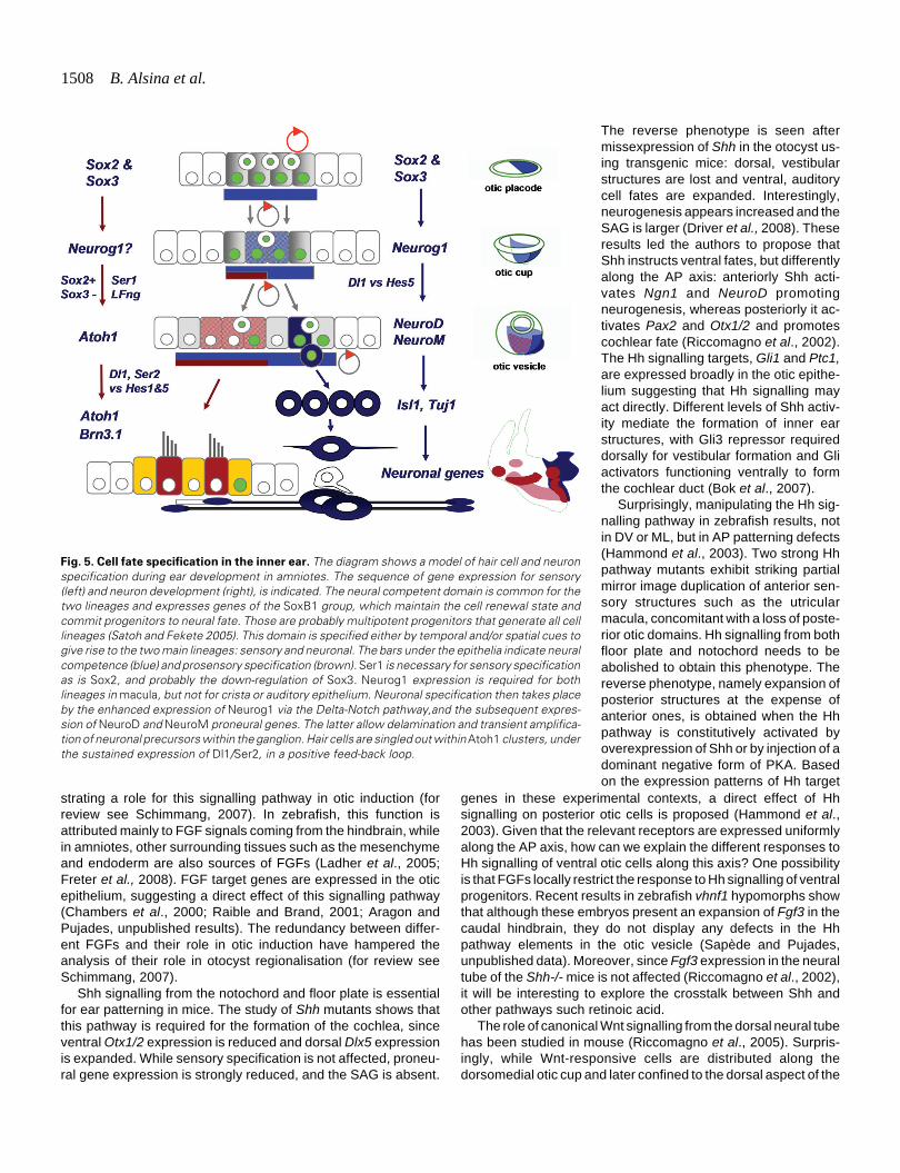
Fig. 5. Cell fate specification in the inner ear. The diagram shows a model of hair cell and neuronspecification during ear development in amniotes. The sequence of gene expression for sensory(left) and neuron development (right), is indicated. The neural competent domain is common for thetwo lineages and expresses genes of the SoxB1 group, which maintain the cell renewal state andcommit progenitors to neural fate. Those are probably multipotent progenitors that generate all celllineages (Satoh and Fekete 2005). This domain is specified either by temporal and/or spatial cues togive rise to the two main lineages: sensory and neuronal. The bars under the epithelia indicate neuralcompetence (blue) and prosensory specification (brown). Ser1 is necessary for sensory specificationas is Sox2, and probably the down-regulation of Sox3. Neurog1 expression is required for bothlineages in macula, but not for crista or auditory epithelium. Neuronal specification then takes placeby the enhanced expression of Neurog1 via the Delta-Notch pathway,and the subsequent expres-sion of NeuroD and NeuroM proneural genes. The latter allow delamination and transient amplifica-tion of neuronal precursors within the ganglion. Hair cells are singled out within Atoh1 clusters, underthe sustained expression of Dl1/Ser2, in a positive feed-back loop.
The reverse phenotype is seen aftermissexpression of Shh in the otocyst us-ing transgenic mice: dorsal, vestibularstructures are lost and ventral, auditorycell fates are expanded. Interestingly,neurogenesis appears increased and theSAG is larger (Driver et al., 2008). Theseresults led the authors to propose thatShh instructs ventral fates, but differentlyalong the AP axis: anteriorly Shh acti-vates Ngn1 and NeuroD promotingneurogenesis, whereas posteriorly it ac-tivates Pax2 and Otx1/2 and promotescochlear fate (Riccomagno et al., 2002).The Hh signalling targets, Gli1 and Ptc1,are expressed broadly in the otic epithe-lium suggesting that Hh signalling mayact directly. Different levels of Shh activ-ity mediate the formation of inner earstructures, with Gli3 repressor requireddorsally for vestibular formation and Gliactivators functioning ventrally to formthe cochlear duct (Bok et al., 2007).
Surprisingly, manipulating the Hh sig-nalling pathway in zebrafish results, notin DV or ML, but in AP patterning defects(Hammond et al., 2003). Two strong Hhpathway mutants exhibit striking partialmirror image duplication of anterior sen-sory structures such as the utricularmacula, concomitant with a loss of poste-rior otic domains. Hh signalling from bothfloor plate and notochord needs to beabolished to obtain this phenotype. Thereverse phenotype, namely expansion ofposterior structures at the expense ofanterior ones, is obtained when the Hhpathway is constitutively activated byoverexpression of Shh or by injection of adominant negative form of PKA. Basedon the expression patterns of Hh target
Ear patterning and cell fate 1509
otic vesicle, both vestibular and cochlear structures are reducedin double Wnt1/Wnt3a mutants (Riccomagno et al., 2005). Toexplain these conflicting observations, lineage studies using aninducible genetic marker of Wnt-responsive cells were performed.These studies show that progenitors of the cochlea received Wntsignalling, suggesting that ventral cells of the otic placode origi-nate from the dorsomedial part of the otic cup. This study under-lines the contribution of cell migration and morphogenetic move-ments to otic patterning processes: otic cell groups originallylocated close to the dorsal neural tube end up at the ventral aspectof the otocyst after otic invagination and morphogenesis. Gain-of-function studies confirmed the role of canonical Wnt pathway investibular formation and showed a mutual repression betweenWnt and Shh pathways in ear DV patterning. However, Wntsignals cannot be the only cues involved in auditory fate specifi-cation since ventral otic determinants are appropriately expressedin double mutants for Wnt1 and Wnt3a (Riccomagno et al., 2005).Other dorsal secreted cues, such as BMPs, could play a role inthis process.
Cell fate specification of the neural elements of theear: the components of the mechanotransducing unit
The elementary sensory unit of the ear – the sensory patch-consists of: i) hair cells, which are the sensory receptor cellsthat contain the mechano-electrical transducing machinery, ii)supporting cells that hold and space the hair cells in a precisepattern, and collaborate to their maintenance, iii) otic neuronsthat innervate the sensory patches and connect the hair cellswith the brain, and iv) glial-Schwann cells that enwrap neuronsand their axons (see Fig. 1B). As mentioned above, with theexception of most Schwann cells that are of neural crest origin,all cell types derive form the otic placode (D’Amico-Martel andNoden, 1983).
The expression of proneural genes confers to cells thepotential to become neural precursors, the ability to differenti-ate into neural elements, and in some instances they specifyparticular cellular identities (Bertrand et al., 2002). Proneuralgenes were related to the proneural achaete-scute complex(ASC) in flies (Garcia-Bellido, 1979), and the analysis of thecomplex lead to the identification of four genes (Ghysen andDambly-Chaudiere, 2000). The vertebrate counterparts wereunveiled by screening for homologous sequences in mouse(Bertrand et al., 2002). A further Drosophila proneural gene,atonal, was isolated later in a PCR-based screen to identifygenes containing bHLH sequences. The orthologs of this genesubfamily have been shown by loss-of-function analysis to becritical for ear development (Jarman et al., 1993; Ma et al.,1998; Bermingham et al., 1999; Kim et al., 2001). It is now clearthat Neurog1, NeuroD1 and Atoh1 are at the core of theproneural function in the ear. They are necessary and sufficientto promote neuronal and hair cell fates, respectively (reviewedby Kelley, 2007). The diagram in Fig. 5 summarises the se-quence of cellular states that generate neurons and hair cellsfrom neural competent epithelium. NeuroD acts after Neurog1and drives neuronal differentiation, and Atoh1 is a proneuralgene that confers competence to a prosensory cell cluster todevelop into hair- or supporting cells and which persistencedirects development towards hair cell fate. This decision re-
quires lateral inhibition through the Delta-Notch mechanismand results in the characteristic cellular pattern of ear sensoryepithelia (Whitfield et al., 1997).
Hair cell fate specification is concomitant with the morphoge-netic process that foreshadows the appearance of the sensoryorgans (see diagram at the right in Fig. 5). They emerge as groupsof sensory fated cells regionally restricted in what is called thesensory patches. It is still unclear how sensory patches emergewithin the otocyst. Recent work suggests that sensory organs andtheir innervating neurons are spatially segregated in the oticplacode (Bell et al., 2008). The transition between the proneuraldomain, which is clearly defined at the otic vesicle stage and theprosensory patches, which are identifiable later on in develop-ment, has not yet been resolved unambiguously. In amniotes, thisoccurs after the otic vesicle is formed and it has been difficult toassess whether it is the result of the development of a commondomain, or the result of the emergence of different, perhapsoverlapping, independent prosensory patches. Some genes ex-pressed in the neurogenic domain, like LFng and Fgf10, persist inthe prospective sensory patches, during the stages of sensoryorgan development (Cole et al., 2000; Pauley et al., 2003;Pujades et al., 2006). Other genes, like Bmp4 are absent from theinitial proneural domain, but thereafter they foreshadow thesensory domains and precede Atoh1 expression at the sensorypatches (Pujades et al., 2006). Ser1 is probably accompanyingthe prospective sensory domain since very initial steps of speci-fication, and functional studies have shown that it is required forthe development of sensory organs (Cole et al., 2000; Daudet andLewis, 2005; Brooker et al., 2006; Kiernan et al., 2006). A recentstudy suggests that the macula emerge from a Neurog1 positivedomain that is common to the neurogenic domain, whereas cristaand the auditory epithelium derive from other independent re-gions (Raft et al., 2007).
Sox genes in neurosensory fateSox genes contain an HMG-box closely related to that of the
mammalian testes-determining gene Sry, and are highly con-served throughout evolution. The C-terminal region of the SOXprotein carries a cryptic transactivating domain that uncovers onlyafter specific interaction with partner factors. To date, twenty fourvertebrate Sox genes have been identified and are classified intoseven subgroups (A–G) based on sequence identity, and at leasttwelve members of the Sox gene family are expressed in thenervous system (Pevny and Placzek, 2005; Wegner and Stolt,2005). Throughout evolution, the expression of the SoxB1 genes(Sox1, Sox2 and Sox3), directly correlates with: i) ectodermalcells that are competent to acquire neural fate; and ii) the commit-ment of cells to a neural fate (Rex et al., 1997; Pevny and Placzek,2005). The Drosophila SoxNeuro, a putative ortholog of thevertebrate Sox1-3 genes, is one of the earliest transcription factorto be expressed pan-neuroectodermally (Cremazy et al., 2000),and it acts upstream and in parallel with the achaete-scute genes.Interestingly, in Drosophila, SoxNeuro is only involved in centralbut not in peripheral nervous system development, suggestingthat recruitment of SOX proteins into placode development is anovelty of craniates in order to rapidly expand the ectodermalanlage (Fritzsch et al., 2006).
SOX2 is expressed in multipotent neural stem cells at allstages during mouse ontogeny (Wegner and Stolt, 2005). Sox2

1510 B. Alsina et al.
expression in the early embryonic Central Nervous System (CNS)is pieced together by separate enhancers with distinct spatio-temporal specificities, and the enhancers driving expression ofSox2 to the lens and nasal/otic placodes have been identified(Uchikawa et al., 1999). Sox2 belongs to the stem-cell cassettethat maintains the self renewal state and pluripotency of progeni-tors (Takahashi and Yamanaka, 2006). Sox1-3 interact withvarious partner transcription factors, and participate in definingdistinct cell states that depend on the partner factors -Pax6 forlens differentiation, Oct3/4 for establishing the epiblast/ES cellstate and, Brn2 for the neural primordia. Sox1-3 are co-expressedin proliferating neural progenitors of the embryonic and adultCNS. The SoxB2 subgroup of Sox factors, including Sox14 andSox21, are very similar to SoxB1 in their HMG-DNA bindingdomain, but act as transrepression domains. A key commonfeature of SoxB1, SoxB2 and SoxE, however, is their ability tomaintain neural progenitor or stem cell identity (for a reviewWegner and Stolt, 2005). Studies in the chick embryo haveprovided evidence that neural inducing signals directly regulateSOX2 expression in the neural tube, and that SOX2 is responsible
for commitment of actively proliferating cells to neural fate (Rex etal., 1997; Bylund et al., 2003; Graham et al., 2003). As mentionedbefore, recent studies also revealed that Sox2-regulatory regioncontains a domain that responds directly to neural inducingsignals, which is conserved across diverse animal species(Takemoto et al., 2006).
Sox2 and Sox3 are expressed in the early proneural domain ofthe otic placode and otic vesicle (Fig. 5 and Fig. 6A and B, see alsoUchikawa et al., 1999; Abello et al., 2007; Neves et al., 2007).Later in development, SOX2 expression foreshadows theprosensory patches and is expressed in all sensory organs(Kiernan et al., 2005; Neves et al., 2007). Two Sox2-deficientmice, light coat and circling (Lcc) and yellow submarine (Ysb),show hearing and balance impairment. Lcc/Lcc mutant mice failto establish a prosensory domain and as a result of this, neitherhair cells nor supporting cells differentiate (Kiernan et al., 2005).Ysb/Ysb mice show abnormal development with disorganizedand fewer hair cells. These phenotypes are a direct consequenceof the absence or reduced expression of the transcription factorSOX2 in the developing inner ear (Kiernan et al., 2005). More-over, mutations of Sox2 in humans cause anophthalmia, senso-rineural hearing loss and global brain defects (Hagstrom et al.,2005) and regulates retinal neural progenitor competence(Taranova et al., 2006).
The role of SoxB1 genes in cell fate specification in the ear isa subject of intense work. Otic neurons and hair cells are neuralcell types in strict sense and both are born upon activation ofproneural bHLH genes. The outcome of the terminal division ofear proneural progenitors is the withdrawal of the cell cycle andthe expression of proneural differentiation genes, NeuroD forneurons (Alsina et al., 2004) and Atoh1 for sensory cells (Pujadeset al., 2006). This links SOX2 function with cell fate acquisition inthe way it has been illustrated in the neural tube, where SOX2maintains to repress the activity of proneural genes until cell cyclewithdrawal, and the expression of the SoxB2 gene group counter-acting this effect (Bylund et al., 2003). Hence, there seems to besome general principle for shifting the balance between two cellstates: on one side a state where cells are committed, butmaintain the capacity for self-renewal; on the other, a state of celldetermination where cells make their terminal division and be-come determined to a particular fate. As discussed by Fritzsch etal. (2006), the vertebrate sensory organ requires a mechanism forrapidly expanding the basic sensory unit, so that placodal epithe-lial cells bear characteristics of stem cells. Therefore, it is ex-pected that they express typical genes of the stem cell cassette(Takahashi and Yamanaka, 2006). The expression of SOX2 in theear is reminiscent of this general stem-cell function, but restrictedto neural committed progenitors. Early in development, duringotic vesicle stages, SOX2 and SOX3 are found in proliferatingcells, but only within the proneural domain of the early otocyst.SOX2 and SOX3 are expressed during the generation of neurons,but only SOX2 remains during sensory organ formation. Theconcomitant expression of SOX2 and SOX3 only during neurongeneration suggests the possibility that at a given stage ofdevelopment, SOX3 expression would be switched off and thepersistent SOX2 expression would result in sensory cell genera-tion. The possibility of a phenotypic switch of cycling neuralprogenitors from neuron to hair cell fate has been suggested tooccur in the Neurog1 null-mice (Matei et al., 2005).
Fig. 6. SOX2 and SOX3 expression in the developing inner ear. (A,B)
SOX2 and SOX3 detected by immunofluorescence (red) and HNK1surface epitope (green) in a HH18 chick otic vesicle. SOX2 and 3 overlapin the proneural domain of the otic vesicle and SOX3 seems to be moreintense at the posterior pole of the otic vesicle. (C) SOX2 but not SOX3maintains its expression in the supporting cells of the sensory patchesafter sensory organ formation. SOX2 immunofluorescence (red) and Tuj1(green) are shown in a confocal section of the macula utriculi from an E6chick embryo. In (A,B), anterior to the top.
B
C
A

Ear patterning and cell fate 1511
Sox2 and 3 are expressed within ear domains that are also thedomain of expression of Notch signalling pathway genes (Abelloet al., 2007). A potential link between Sox2 and Notch signallingis suggested by the observation that Sox2 expression is missingin Jagged1 conditional mutants and after Notch inhibition (Kiernanet al., 2006; Daudet et al., 2007), which would indicate that Ser1(Jagged1 in mammals) is upstream of Sox2 in the specification ofthe prosensory field. On the other hand, the loss of Sox2 in theinner ear results in the loss of p27kip1, a regulator of terminaldivision in the cochlea (Kiernan et al., 2005). These results areconsistent with a function of Sox genes in maintaining the self-renewal state along with a state of neural commitment, perhapsthe latter being restricted by other patterning signals.
Concluding remarks
The question of coupling patterning and cell fate determinationis central to development. Recent work has shed light into howthose processes take place during ear development. The earlyspecification of the neural competent domain, which ultimatelygives rise to neurons and hair cells, seems to occur very early andconcomitantly to the specification of the otic fate. It requires theactivity of the Notch signalling pathway for maintenance, but notfor its establishment, which involves FGF signals and the rein-forced expression of SoxB1 genes. Further work is required tounderstand these very initial steps of regionalisation and theirgenetic relation with the acquisition of the otic fate by thepreplacodal ectoderm. Further regionalisation of the otic vesiclegives rise to the topological organisation of the ear and the role ofthe neural tube in this process has been studied extensively. Atleast three signalling systems, FGF, Wnt and Hh are known tocontribute to pattern the otic vesicle, and two of them (FGF andWnt) depend on hindbrain signals. The analysis of hindbrain genemutants, such MafB, Hoxa1 and vHnf1, is helping to dissect thegenetic pathways that link hindbrain segmentation and otic pat-terning. Finally, the allocation of specific neural competent pre-cursors to the neural domain of the otic placode and otic vesicleallows the development of neurons and hair cells. The geneticcassette involved in the expansion of these progenitor cells, andthe one that further leads to determination of neurons and haircells is starting to be unveiled. The SoxB1 gene group and theproneural genes Neurog1, NeuroD and Atoh1 appear as majorcell fate determination factors. Precise genetic networks, cellularinteractions and the interplay with signalling mechanisms are thesubject of current studies in several laboratories, and a subject ofgreat interest not only for the understanding of the developmentof the ear but for generating tools for ear repair.
AcknowledgementsWe wish to thank Gina Abelló, Safia Khatri, Marija Radosevic, Citlali
Vázquez-Echeverría, Ferran Aragón, Dora Sapède, Andrés Kamaid andJoana Neves for sharing unpublished results and for comments. The workwas supported by grants BFU2005-03045 to BA, and BFU2006-05604 toC.P, MEC, Spain.
References
ABELLO, G., KHATRI, S., GIRALDEZ, F., and ALSINA, B. (2007). Earlyregionalization of the otic placode and its regulation by the notch signalingpathway. Mech Dev. 124: 631-645.
ABELLO, G. and ALSINA, B. (2007). Establishment of a proneural field in the innerear. Int J Dev Biol. 51: 483-493 (DOI: 10.1387/ijdb.072343ga).
ABU-ELMAGD, M., ISHII, Y., CHEUNG, M., REX, M., LE ROUEDEC, D. andSCOTTING, P.J. (2001). cSox3 expression and neurogenesis in the epibranchialplacodes. Dev Biol. 237: 258-269.
ADAM, J., MYAT, A., LE ROUX, I., EDDISON, M., HENRIQUE, D., ISH-HOROWICZ,D. and LEWIS, J. (1998). Cell fate choices and the expression of notch, deltaand serrate homologues in the chick inner ear: Parallels with Drosophila sense-organ development. Development 125: 4645-4654.
ALSINA, B., ABELLO, G., ULLOA, E., HENRIQUE, D., PUJADES, C. and GIRALDEZ,F. (2004). FGF signaling is required for determination of otic neuroblasts in thechick embryo. Dev Biol. 267: 119-134.
ANDERMANN, P., UNGOS, J. and RAIBLE, D.W. (2002). Neurogenin1 defineszebrafish cranial sensory ganglia precursors. Dev Biol. 251: 45-58.
BEGBIE, J., BALLIVET, M. and GRAHAM, A. (2002). Early steps in the productionof sensory neurons by the neurogenic placodes. Mol Cell Neurosci. 21: 502-511.
BELL, D., STREIT, A., GOROSPE, I., VARELA-NIETO, I., ALSINA, B. andGIRALDEZ, F. (2008). Spatial and temporal segregation of auditory andvestibular neurons in the otic placode. Dev Biol. 322:109-120.
BERMINGHAM, N.A., HASSAN, B.A., PRICE, S.D., VOLLRATH, M.A., BEN-ARIE,N., EATOCK, R.A., BELLEN, H.J., LYSAKOWSKI, A. and ZOGHBI, H.Y. (1999).Math1: an essential gene for the generation of inner ear hair cells. Science. 284:1837-1841.
BERTRAND, N., CASTRO, D.S. and GUILLEMOT, F. (2002). Proneural genes andthe specification of neural cell types. Nat Rev Neurosci. 3: 517-530.
BOK, J., BRONNER-FRASER, M. and WU, D.K. (2005). Role of the hindbrain indorsoventral but not anteroposterior axial specification of the inner ear. Devel-opment 132: 2115-2124.
BOK, J., DOLSON, D.K., HILL, P., RUTHER, U., EPSTEIN, D.J. and WU, D.K.(2007). Opposing gradients of gli repressor and activators mediate shh signal-ing along the dorsoventral axis of the inner ear. Development 134: 1713-1722.
BRAY, S.J. (2006). Notch signalling: A simple pathway becomes complex. Nat RevMol Cell Biol. 7: 678-689.
BROOKER, R., HOZUMI, K. AND LEWIS, J. (2006). Notch Ligands with Contrast-ing Functions: Jagged1 and Delta1 in the Mouse Inner Ear. Development 133:1277-1286.
BYLUND, M., ANDERSSON, E., NOVITCH, B.G. and MUHR, J. (2003). Vertebrateneurogenesis is counteracted by Sox1-3 activity. Nat Neurosci. 6: 1162-1168.
CHAMBERS, D., MEDHURST, A.D., WALSH, F.S., PRICE, J. and MASON, I.(2000). Differential display of genes expressed at the midbrain - hindbrainjunction identifies sprouty2: An FGF8-inducible member of a family of intracel-lular FGF antagonists. Mol Cell Neurosci. 15: 22-35.
CHANG, W., BRIGANDE, J. V., FEKETE, D. M. AND WU, D. K. (2004). TheDevelopment of Semicircular Canals in the Inner Ear: Role of FGFs in SensoryCristae. Development 131: 4201-4211.
CHOO, D., WARD, J., REECE, A., DOU, H., LIN, Z. and GREINWALD, J. (2006).Molecular mechanisms underlying inner ear patterning defects in kreislermutants. Dev Biol. 289: 308-317.
CHOO, D. (2007). The Role of the Hindbrain in Patterning of the Otocyst. Dev. Biol.308: 257-265.
CIBA FOUNDATION SYMPOSIUM (1991). In: Regeneration of vertebrate sensoryreceptor cells. Ciba Found Symp. 160; Wiley and Chicester, Eds.
COLE, L.K., LE, R.,I., NUNES, F., LAUFER, E., LEWIS, J. and WU, D.K. (2000).Sensory organ generation in the chicken inner ear: Contributions of bonemorphogenetic protein 4, serrate1, and lunatic fringe. J Comp Neurol. 424: 509-520.
CREMAZY, F., BERTA, P. and GIRARD, F. (2000). Sox neuro, a new Drosophilasox gene expressed in the developing central nervous system. Mech Dev. 93:215-219.
CREMERS, C.W.R.J., GRAHAM, J., PARVING, A., and RUBEN, R.J. (1988).Biology of sensorineural hearing loss in children. Report of the Holte Sympo-sium. Int J Pediatric Otorhynolayrngology. 15: 1-15.
D’AMICO-MARTEL, A. and NODEN, D.M. (1983). Contributions of placodal andneural crest cells to avian cranial peripheral ganglia. Am J Anat. 166: 445-468.

1512 B. Alsina et al.
DAUDET, N. and LEWIS, J. (2005). Two contrasting roles for notch activity in chickinner ear development: Specification of prosensory patches and lateral inhibi-tion of hair-cell differentiation.nDevelopment 132: 541-551.
DAUDET, N., ARIZA-MCNAUGHTON, L. and LEWIS, J. (2007). Notch signalling isneeded to maintain, but not to initiate, the formation of prosensory patches in thechick inner ear. Development 134: 2369-2378.
DEOL, M.S. (1964). The abnormalities of the inner ear in kreisler mice. J EmbryolExp Morphol. 12: 475-490.
DRIVER, E.C., PRYOR, S.P., HILL, P., TURNER, J., RÜTHER, U., BIESECKER,L.G., GRIFFITH, A.J. and KELLEY, M.W. (2008). Hedgehog signaling regulatessensory cell formation and auditory function in mice and humans. J Neurosci.,28: 7350-7358.
FEKETE, D.M. (1999). Development of the vertebrate ear: Insights from knockoutsand mutants. Trends Neurosci. 22: 263-269.
FRETER, S., MUTA, Y., MAK, S.S., RINKWITZ, S. and LADHER, R.K. (2008).Progressive restriction of otic fate: the role of FGF and Wnt in resolving inner earpotential. Development. 135: 3415-3424.
FRITZSCH, B., BEISEL, K. W. AND BERMINGHAM, N. A. (2000). DevelopmentalEvolutionary Biology of the Vertebrate Ear: Conserving Mechanoelectric Trans-duction and Developmental Pathways in Diverging Morphologies.nNeuroreport11: R35-44.
FRITZSCH, B., BEISEL, K.W. and HANSEN, L.A. (2006). The molecular basis ofneurosensory cell formation in ear development: A blueprint for hair cell andsensory neuron regeneration? Bioessays 28: 1181-1193.
GARCIA-BELLIDO, A. (1979). Genetic analysis of the achaete-scute system ofDrosophila melanogaster. Genetics. 91: 491-520.
GHYSEN, A. and DAMBLY-CHAUDIERE, C. (2000). A genetic programme forneuronal connectivity. Trends Genet. 16: 221-226.
GIRALDEZ, F. (1998). Regionalized organizing activity of the neural tube revealedby the regulation of lmx1 in the otic vesicle. Dev Biol. 203: 189-200.
GRAHAM, V., KHUDYAKOV, J., ELLIS, P. and PEVNY, L. (2003). SOX2 functionsto maintain neural progenitor identity. Neuron 39: 749-765.
GROVES, A.K. and BRONNER-FRASER, M. (2000). Competence, specificationand commitment in otic placode induction.£Development 127: 3489-3499.
HADDON, C., JIANG, Y.J., SMITHERS, L. and LEWIS, J. (1998). Delta-notchsignalling and the patterning of sensory cell differentiation in the zebrafish ear:Evidence from the mind bomb mutant. Development 125: 4637-4644.
HAGSTROM, S.A., PAUER, G.J., REID, J., SIMPSON, E., CROWE, S., MAUMENEE,I.H. and TRABOULSI, E.I. (2005). SOX2 mutation causes anophthalmia,hearing loss, and brain anomalies. Am J Med Genet A. 138: 95-98.
HAMMOND, K.L., LOYNES, H.E., FOLARIN, A.A., SMITH, J. and WHITFIELD, T.T.(2003). Hedgehog signalling is required for correct anteroposterior patterning ofthe zebrafish otic vesicle. Development 130: 1403-1417.
HARRISON, R.G. (1945). Relations of symmetry in the developing embryo. TransConn Acad Arts Sci USA. 22: 238-247.
HATCH, E. P., NOYES, C. A., WANG, X., WRIGHT, T. J. AND MANSOUR, S. L.(2007). Fgf3 is Required for Dorsal Patterning and Morphogenesis of the InnerEar Epithelium. Development 134: 3615-3625.
HERNANDEZ, R.E., RIKHOF, H.A., BACHMANN, R. and MOENS, C.B. (2004).vhnf1 integrates global RA patterning and local FGF signals to direct posteriorhindbrain development in zebrafish. Development 131: 4511-4520.
JACOBSON, A.G. (1966). Inductive processes in embryonic development. Science152: 25-34.
JARMAN, A.P., GRAU, Y., JAN, L.Y. and JAN, Y.N. (1993). Atonal is a proneuralgene that directs chordotonal organ formation in the Drosophila peripheralnervous system. Cell 73: 1307-1321.
JAYASENA, C.S., OHYAMA, T., SEGIL, N. and GROVES, A.K. (2008). Notchsignaling augments the canonical Wnt pathway to specify the size of the oticplacode. Development. 135: 2251-2261.
KELLEY, M.W. (2007). Cellular commitment and differentiation in the organ of corti.Int J Dev Biol. 51: 571-583 (doi: 10.1387/ijdb.072388mk).
KIERNAN, A.E., PELLING, A.L., LEUNG, K.K., TANG, A.S., BELL, D.M., TEASE,C., LOVELL-BADGE, R., STEEL, K.P. and CHEAH, K.S. (2005). Sox2 isrequired for sensory organ development in the mammalian inner ear. Nature434: 1031-1035.
KIERNAN, A.E., XU, J. and GRIDLEY, T. (2006). The notch ligand JAG1 is requiredfor sensory progenitor development in the mammalian inner ear. PLoS Genet.2, e4.
KIM, W.Y., FRITZSCH, B., SERLS, A., BAKEL, L.A., HUANG, E.J., REICHARDT,L.F., BARTH, D.S. and LEE, J.E. (2001). NeuroD-null mice are deaf due to asevere loss of the inner ear sensory neurons during development. Development128: 417-426.
KWAK, S.J., PHILLIPS, B.T., HECK, R. and RILEY, B.B. (2002). An expandeddomain of fgf3 expression in the hindbrain of zebrafish valentino mutants resultsin mis-patterning of the otic vesicle. Development 129: 5279-5287.
LADHER, R.K., WRIGHT, T.J., MOON, A.M., MANSOUR, S.L. and SCHOENWOLF,G.C. (2005). FGF8 initiates inner ear induction in chick and mouse. Genes Dev.19: 603-613.
LECAUDEY, V., ULLOA, E., ANSELME, I., STEDMAN, A., SCHNEIDER-MAUNOURY, S. and PUJADES, C. (2007). Role of the hindbrain in patterningthe otic vesicle: A study of the zebrafish vhnf1 mutant. Dev Biol. 303: 134-143.
LEGER, S.and BRAND, M. (2002). Fgf8 andnFgf3 are required for zebrafish earplacode induction, maintenance and inner ear patterning. Mech Dev. 119: 91-108.
LEWIS, J. (1998). Notch signalling and the control of cell fate choices in vertebrates.Semin Cell Dev Biol. 9: 583-589.
LI, C.W., VAN DE WATER, T.R. and RUBEN, R.J. (1978). The fate mapping of theeleventh and twelfth day mouse otocyst: An in vitro study of the sites of originof the embryonic inner ear sensory structures. J Morphol. 157: 249-267.
LIN, Z., CANTOS, R., PATENTE, M. and WU, D.K. (2005). Gbx2 is required for themorphogenesis of the mouse inner ear: A downstream candidate of hindbrainsignaling. Development 132: 2309-2318.
LOUVI, A. and ARTAVANIS-TSAKONAS, S. (2006). Notch signalling in vertebrateneural development. Nat Rev Neurosci. 7: 93-102.
MA, Q., CHEN, Z., DEL BARCO BARRANTES, I., DE LA POMPA, J.L. andANDERSON, D.J. (1998). Neurogenin1 is essential for the determination ofneuronal precursors for proximal cranial sensory ganglia. Neuron 20, : -482.
MA, Q., ANDERSON, D.J. and FRITZSCH, B. (2000). Neurogenin 1 null mutantears develop fewer, morphologically normal hair cells in smaller sensoryepithelia devoid of innervation. J Assoc Res Otolaryngol. 1: 129-143.
MATEI, V., PAULEY, S., KAING, S., ROWITCH, D., BEISEL, K.W., MORRIS, K.,FENG, F., JONES, K., LEE, J. and FRITZSCH, B. (2005). Smaller inner earsensory epithelia in neurog 1 null mice are related to earlier hair cell cycle exit.Dev Dyn. 234: 633-650.
MCKAY, I.J., LEWIS, J. and LUMSDEN, A. (1996). The role of FGF-3 in early innerear development: An analysis in normal and kreisler mutant mice. Dev Biol. 174:370-378.
MILLIMAKI, B.B., SWEET, E.M., DHASON, M.S. and RILEY, B.B. (2007). Zebrafishatoh1 genes: Classic proneural activity in the inner ear and regulation by fgf andnotch. Development 134: 295-305.
MONTCOUQUIOL, M. AND KELLEY, M. W. (2003). Planar and Vertical SignalsControl Cellular Differentiation and Patterning in the Mammalian Cochlea. J.Neurosci. 23: 9469-9478.
NEVES, J., KAMAID, A., ALSINA, B. and GIRALDEZ, F. (2007). Differentialexpression of Sox2 and Sox3 in neuronal and sensory progenitors of thedeveloping inner ear of the chick. J Comp Neurol. 503: 487-500.
NIKAIDO, M., DOI, K., SHIMIZU, T., HIBI, M., KIKUCHI, Y. and YAMASU, K. (2007).Initial specification of the epibranchial placode in zebrafish embryos depends onthe fibroblast growth factor signal. Dev Dyn. 236: 564-571.
OHYAMA, T., GROVES, A.K. and MARTIN, K. (2007). The first steps towardshearing: Mechanisms of otic placode induction. Int J Dev Biol. 51: 463-472 (DOI10.1387/ijdb.072320to).
PAULEY, S., WRIGHT, T. J., PIRVOLA, U., ORNITZ, D., BEISEL, K. ANDFRITZSCH, B. (2003). Expression and Function of FGF10 in Mammalian InnerEar Development. Dev. Dyn. 227: 203-215.
PASQUALETTI, M., NEUN, R., DAVENNE, M. and RIJLI, F.M. (2001). Retinoic acidrescues inner ear defects in Hoxa1 deficient mice. Nat Genet. 29: 34-39.
PERRON, M., OPDECAMP, K., BUTLER, K., HARRIS, W.A. and BELLEFROID,E.J. (1999). X-ngnr-1 and Xath3 promote ectopic expression of sensory neuronmarkers in the neurula ectoderm and have distinct inducing properties in the
Ear patterning and cell fate 1513
retina. Proc Natl Acad Sci U S A. 96: 14996-15001.
PEVNY, L. and PLACZEK, M. (2005). SOX genes and neural progenitor identity.Curr Opin Neurobiol. 15: 7-13.
PIRVOLA, U., SPENCER-DENE, B., XING-QUN, L., KETTUNEN, P., THESLEFF,I., FRITZSCH, B., DICKSON, C. and YLIKOSKI, J. (2000). FGF/FGFR-2(IIIb)signaling is essential for inner ear morphogenesis. J Neurosci. 20: 6125-6134.
PIRVOLA, U., ZHANG, X., MANTELA, J., ORNITZ, D. M. AND YLIKOSKI, J. (2004).Fgf9 Signaling Regulates Inner Ear Morphogenesis through Epithelial-Mesen-chymal Interactions. Dev. Biol. 273: 350-360.
POPPER, A. N. AND FAY, R. R. (1993). Sound Detection and Processing by Fish:Critical Review and Major Research Questions. Brain Behav. Evol. 41: 14-38.
PUJADES, C., KAMAID, A., ALSINA, B. and GIRALDEZ, F. (2006). BMP-signalingregulates the generation of hair-cells. Dev Biol. 292: 55-67.
RAFT, S., KOUNDAKJIAN, E. J., QUINONES, H., JAYASENA, C. S., GOODRICH,L. V., JOHNSON, J. E., SEGIL, N. AND GROVES, A. K. (2007). Cross-Regulation of Ngn1 and Math1 Coordinates the Production of Neurons andSensory Hair Cells during Inner Ear Development. Development 134: 4405-4415.
RAIBLE, F. and BRAND, M. (2001). Tight transcriptional control of the ETS domainfactors erm and Pea3 by fgf signaling during early zebrafish development. MechDev. 107: 105-117.
REX, M., ORME, A., UWANOGHO, D., TOINTON, K., WIGMORE, P.M., SHARPE,P.T. and SCOTTING, P.J. (1997). Dynamic expression of chicken Sox2 andSox3 genes in ectoderm induced to form neural tissue. Dev Dyn. 209: 323-332.
RICCOMAGNO, M.M., TAKADA, S. and EPSTEIN, D.J. (2005). Wnt-dependentregulation of inner ear morphogenesis is balanced by the opposing andsupporting roles of shh. Genes Dev. 19: 1612-1623.
RICCOMAGNO, M.M., MARTINU, L., MULHEISEN, M., WU, D.K. and EPSTEIN,D.J. (2002). Specification of the mammalian cochlea is dependent on sonichedgehog. Genes Dev. 16: 2365-2378.
RILEY, B. B. AND PHILLIPS, B. T. (2003). Ringing in the New Ear: Resolution ofCell Interactions in Otic Development. Dev. Biol. 261: 289-312.
RUBEL, E. W. AND FRITZSCH, B. (2002). Auditory System Development: PrimaryAuditory Neurons and their Targets. Annu. Rev. Neurosci. 25: 51-101.
SATOH, T. and FEKETE, D.M. (2005). Clonal analysis of the relationships betweenmechanosensory cells and the neurons that innervate them in the chicken ear.Development 132, 1687-1697.
SCHIMMANG, T. (2007). Expression and functions of FGF ligands during EarlyOticdevelopment induction. Int J Dev Biol. 51: 473-481. (DOI: 10.1387/ijdb.072334ts).
SCHLOSSER, G. and NORTHCUTT, R.G. (2000). Development of neurogenicplacodes in Xenopus laevis. J Comp Neurol. 418: 121-146.
SCHNEIDER-MAUNOURY, S. and PUJADES, C. (2007). Hindbrain signals in oticregionalization: Walk on the wild side. Int J Dev Biol. 51: 495-506. (DOI:10.1387/ijdb.072345ss).
STERN, C.D. (2005). Neural induction: Old problem, new findings, yet morequestions. Development 132: 2007-2021.
STREIT, A. (2007). The preplacodal region: An ectodermal domain with multipoten-tial progenitors that contribute to sense organs and cranial sensory ganglia. IntJ Dev Biol. 51: 447-461 (doi: 10.1387/ijdb.072327as).
SUN, S.K., DEE, C.T., TRIPATHI, V.B., RENGIFO, A., HIRST, C.S. and SCOT-TING, P.J. (2007). Epibranchial and otic placodes are induced by a common fgfsignal, but their subsequent development is independent. Dev Biol. 303: 675-686.
TAKAHASHI, K. and YAMANAKA, S. (2006). Induction of pluripotent stem cellsfrom mouse embryonic and adult fibroblast cultures by defined factors. Cell 126:663-676.
TAKEMOTO, T., UCHIKAWA, M., KAMACHI, Y. and KONDOH, H. (2006). Conver-gence of wnt and FGF signals in the genesis of posterior neural plate throughactivation of the Sox2 enhancer N-1. Development 133: 297-306.
TARANOVA, O.V., MAGNESS, S.T., FAGAN, B.M., WU, Y., SURZENKO, N.,HUTTON, S.R. and PEVNY, L.H. (2006). SOX2 is a dose-dependent regulatorof retinal neural progenitor competence. Genes Dev. 20: 1187-1202.
TORRES, M. and GIRALDEZ, F. (1998). The development of the vertebrate innerear. Mech Dev. 71: 5-21.
UCHIKAWA, M., KAMACHI, Y. and KONDOH, H. (1999). Two distinct subgroupsof group B sox genes for transcriptional activators and repressors: Theirexpression during embryonic organogenesis of the chicken. Mech Dev. 84: 103-120.
VÁZQUEZ-ECHEVERRÍA, C., DOMINGUEZ-FRUTOS, E., CHARNAY, P.,SCHIMMANG, T. and PUJADES, C. (2008). Analysis of mouse kreisler mutantsreveals new roles of hindbrain-derived signals in the establishment of the oticneurogenic domain.Dev Biol. 322: 167-78.
WEGNER, M. and STOLT, C.C. (2005). From stem cells to neurons and glia: Asoxist’s view of neural development. Trends Neurosci. 28, 583-588.
WHITFIELD, T., HADDON, C. and LEWIS, J. (1997). Intercellular signals and cell-fate choices in the developing inner ear: Origins of global and of fine-grainedpattern. Semin Cell Dev Biol. 8: 239-247.
WHITFIELD, T. and HAMMOND, K.L. (2007). Axial patterning in the vertebrate oticvesicle. Int J Dev Biol. 51: 507-520. (DOI: 10.1387/ijdb.072380tw).
WIELLETTE, E.L. and SIVE, H. (2003). vhnf1 and fgf signals synergize to specifyrhombomere identity in the zebrafish hindbrain. Development 130: 3821-3829.
YNTEMA, C.L. (1955). An analysis of induction of the ear from foreign ectoderm inthe salamander embryo. J Exp Zool. 113: 211-244.
1514 B. Alsina et al.
Further Related Reading, published previously in the Int. J. Dev. Biol.
See our Special Issue Ear Development edited by Fernando Giraldez and Bernd Fritzsch at:http://www.ijdb.ehu.es/web/contents.php?vol=51&issue=6-7
See our recent Special Issue Fertilization, in honor of David L. Garbers and edited by Paul M. Wassarman and Victor D. Vacquier at:http://www.ijdb.ehu.es/web/contents.php?vol=52&issue=5-6
Patterning and morphogenesis of the vertebrate inner earJinwoong Bok, Weise Chang and Doris K. WuInt. J. Dev. Biol. (2007) 51: 521-533
Axial patterning in the developing vertebrate inner earTanya T. Whitfield and Katherine L. HammondInt. J. Dev. Biol. (2007) 51: 507-520
Establishment of a proneural field in the inner earGina Abelló and Berta AlsinaInt. J. Dev. Biol. (2007) 51: 483-493
The first steps towards hearing: mechanisms of otic placode inductionTakahiro Ohyama, Andrew K. Groves and Kareen MartinInt. J. Dev. Biol. (2007) 51: 463-472
In pursuit of communication. An interview with Bob RubenFernando Giraldez and Bernd FritzschInt. J. Dev. Biol. (2007) 51: 439-445
Hindbrain signals in otic regionalization: Walk on the wild side.Schneider-Maunoury, S. and Pujades, C.(2007).Int J Dev Biol. 51: 495-506.
Cell proliferation during the early compartmentalization of the Xenopus laevisinner earQuincy A. Quick and Elba E. SerranoInt. J. Dev. Biol. (2007) 51: 201-210
Genetic control of dorsoventral patterning and neuroblast specification in theDrosophila Central Nervous SystemGuoyan Zhao, Scott R. Wheeler and James B. SkeathInt. J. Dev. Biol. (2007) 51: 107-115
Head-tail patterning of the vertebrate embryo: one, two or many unresolvedproblems?Claudio D. Stern, Jeroen Charité, Jacqueline Deschamps, Denis Duboule, Anthony J.Durston, Marie Kmita, Jean-François Nicolas, Isabel Palmeirim, Jim C. Smith and LewisWolpertInt. J. Dev. Biol. (2006) 50: 3-15
Patterning a multi-headed mutant in Hydractinia: enhancement of head formationand its phenotypic normalization.Werner A Müller, Regina Teo and Frank MöhrlenInt. J. Dev. Biol. (2004) 48: 9-15
5 yr ISI Impact Factor (2008) = 3.271