Macroscopic anatomy of the eye and ear
41
Macroscopic anatomy of the eye and ear Dr. Sándor Katz Ph.D.
-
Upload
khangminh22 -
Category
Documents
-
view
0 -
download
0
Transcript of Macroscopic anatomy of the eye and ear
Macroscopic anatomy of the eye and ear
Dr. Sándor Katz Ph.D.
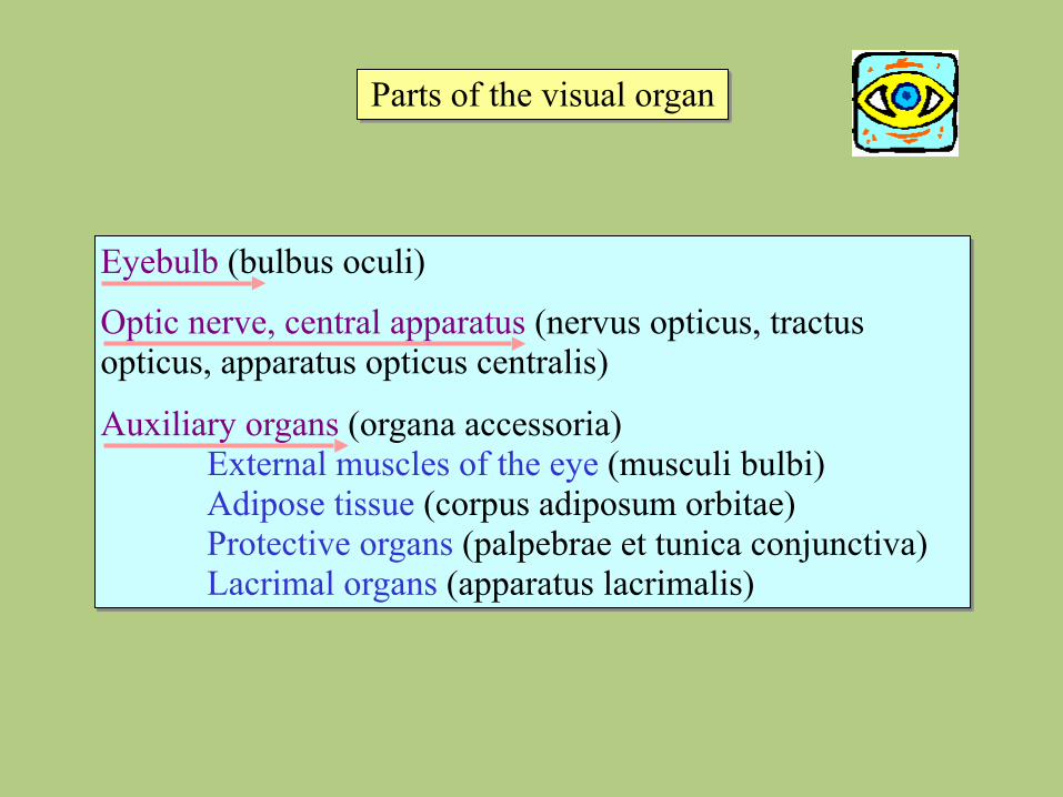
Parts of the visual organ
Eyebulb (bulbus oculi)
Optic nerve, central apparatus (nervus opticus, tractus opticus, apparatus opticus centralis)
Auxiliary organs (organa accessoria) External muscles of the eye (musculi bulbi) Adipose tissue (corpus adiposum orbitae) Protective organs (palpebrae et tunica conjunctiva) Lacrimal organs (apparatus lacrimalis)
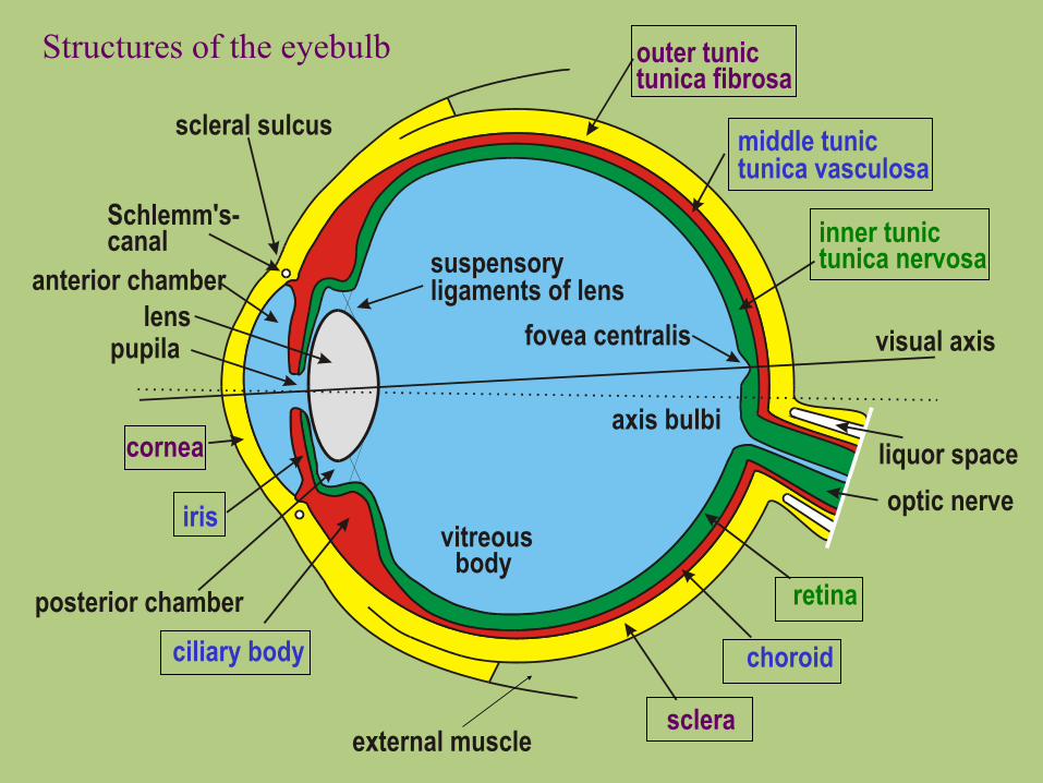
scleral sulcus
Schlemm's-canal
pupila
iris
cornea
ciliary body
sclera
choroid
retina
optic nerveliquor space
outer tunictunica fibrosa
middle tunictunica vasculosa
inner tunictunica nervosa
vitreous body
lens
axis bulbi
visual axis
suspensoryligaments of lens
fovea centralis
external muscle
anterior chamber
posterior chamber
Structures of the eyebulb
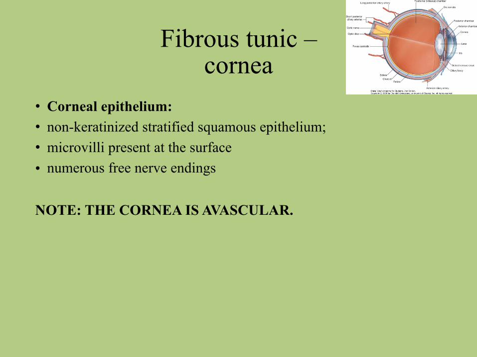
Fibrous tunic – cornea
• Corneal epithelium: • non-keratinized stratified squamous epithelium; • microvilli present at the surface • numerous free nerve endings
NOTE: THE CORNEA IS AVASCULAR.
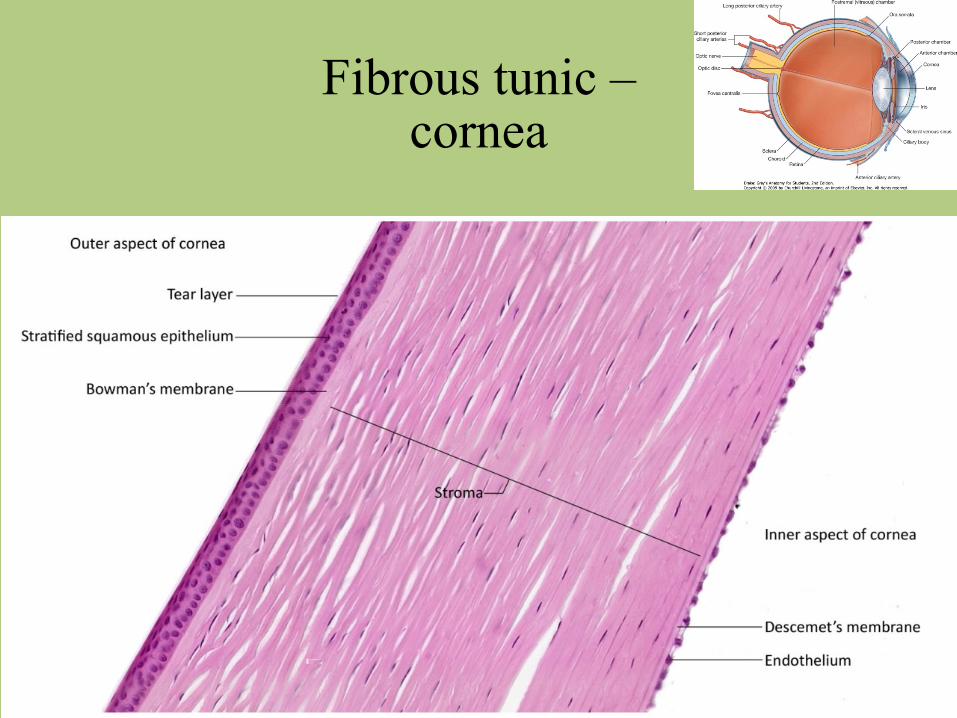
Fibrous tunic – cornea
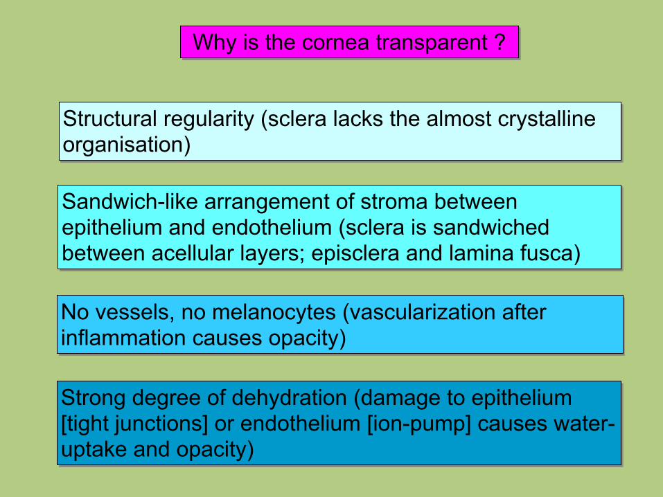
Why is the cornea transparent ?
Strong degree of dehydration (damage to epithelium [tight junctions] or endothelium [ion-pump] causes water-uptake and opacity)
Structural regularity (sclera lacks the almost crystalline organisation)
Sandwich-like arrangement of stroma between epithelium and endothelium (sclera is sandwiched between acellular layers; episclera and lamina fusca)
No vessels, no melanocytes (vascularization after inflammation causes opacity)
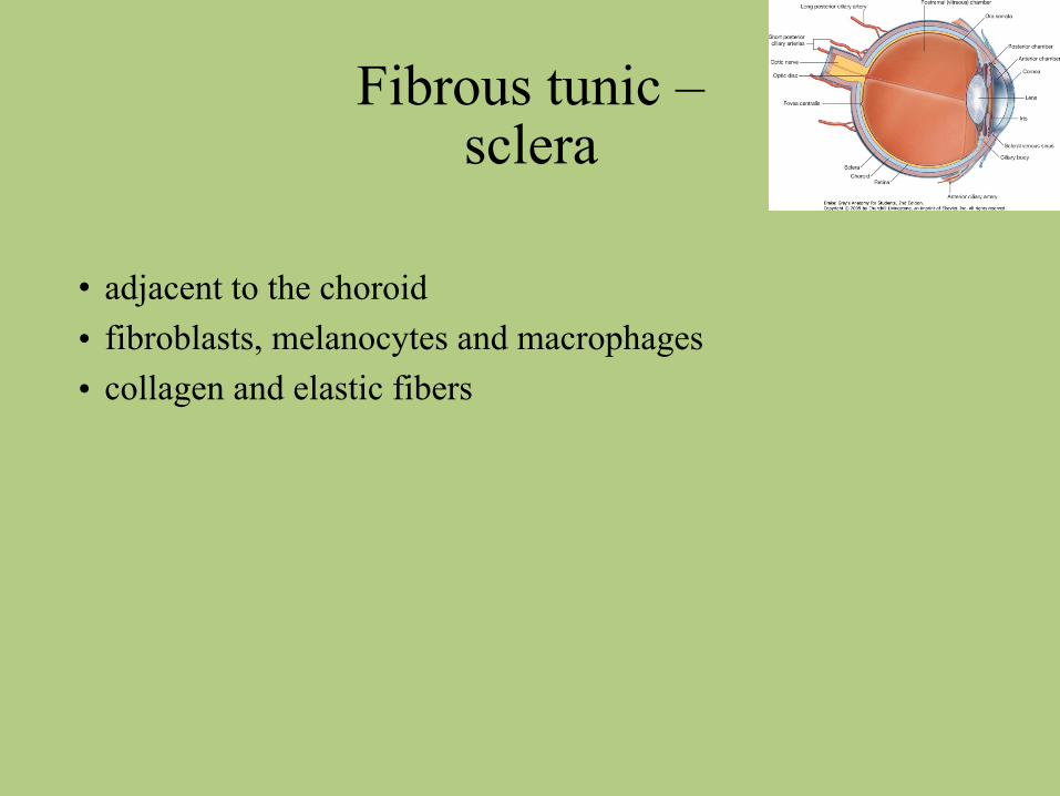
Fibrous tunic – sclera
• adjacent to the choroid • fibroblasts, melanocytes and macrophages • collagen and elastic fibers
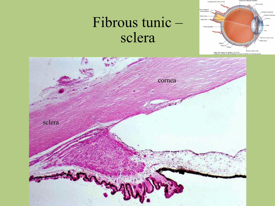
Fibrous tunic – sclera
cornea
sclera
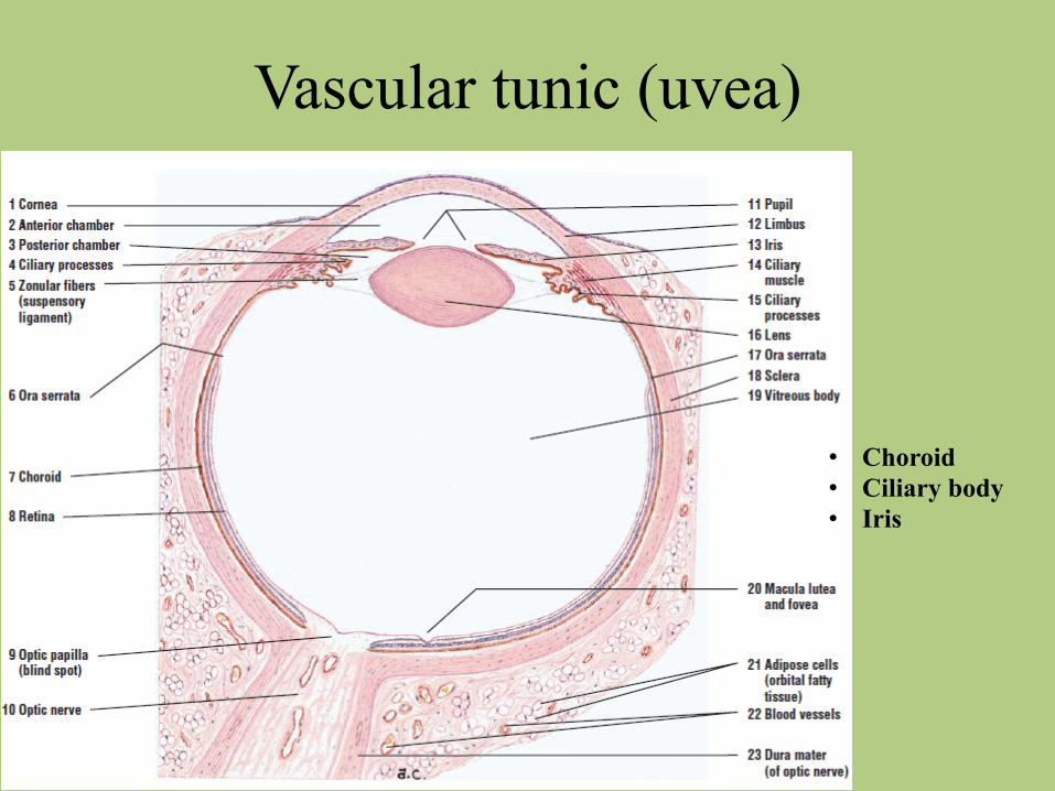
Vascular tunic (uvea)
• Choroid • Ciliary body • Iris
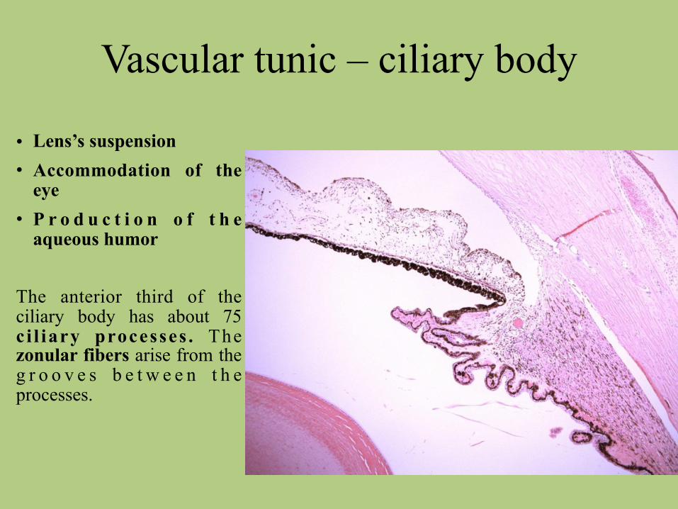
Vascular tunic – ciliary body
• Lens’s suspension • Accommodation of the
eye • P r o d u c t i o n o f t h e
aqueous humor
The anterior third of the ciliary body has about 75 c i l iary processes . The zonular fibers arise from the g r o o v e s b e t w e e n t h e processes.
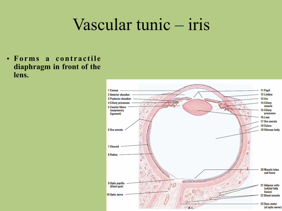
Vascular tunic – iris
• Forms a contract i l e diaphragm in front of the lens.
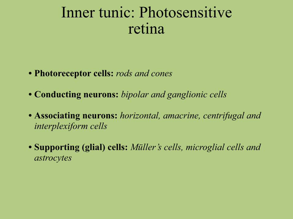
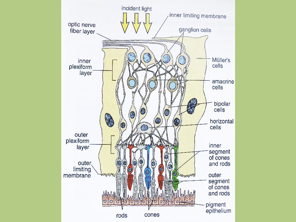
Inner tunic: Photosensitive retina
• Photoreceptor cells: rods and cones
• Conducting neurons: bipolar and ganglionic cells
• Associating neurons: horizontal, amacrine, centrifugal and interplexiform cells
• Supporting (glial) cells: Müller’s cells, microglial cells and astrocytes
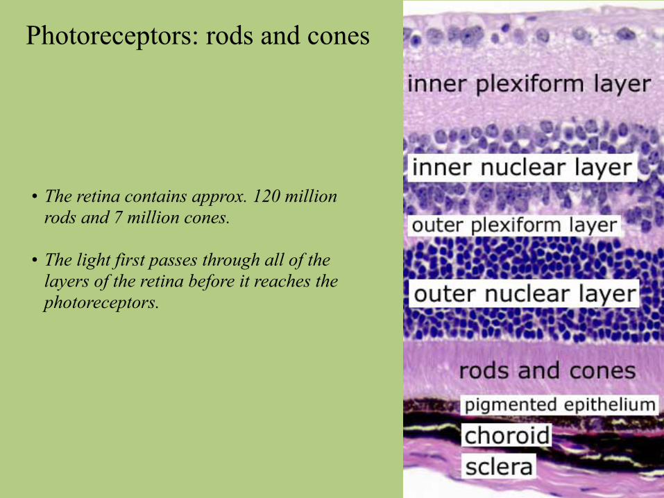
Photoreceptors: rods and cones
• The retina contains approx. 120 million rods and 7 million cones.
• The light first passes through all of the layers of the retina before it reaches the photoreceptors.
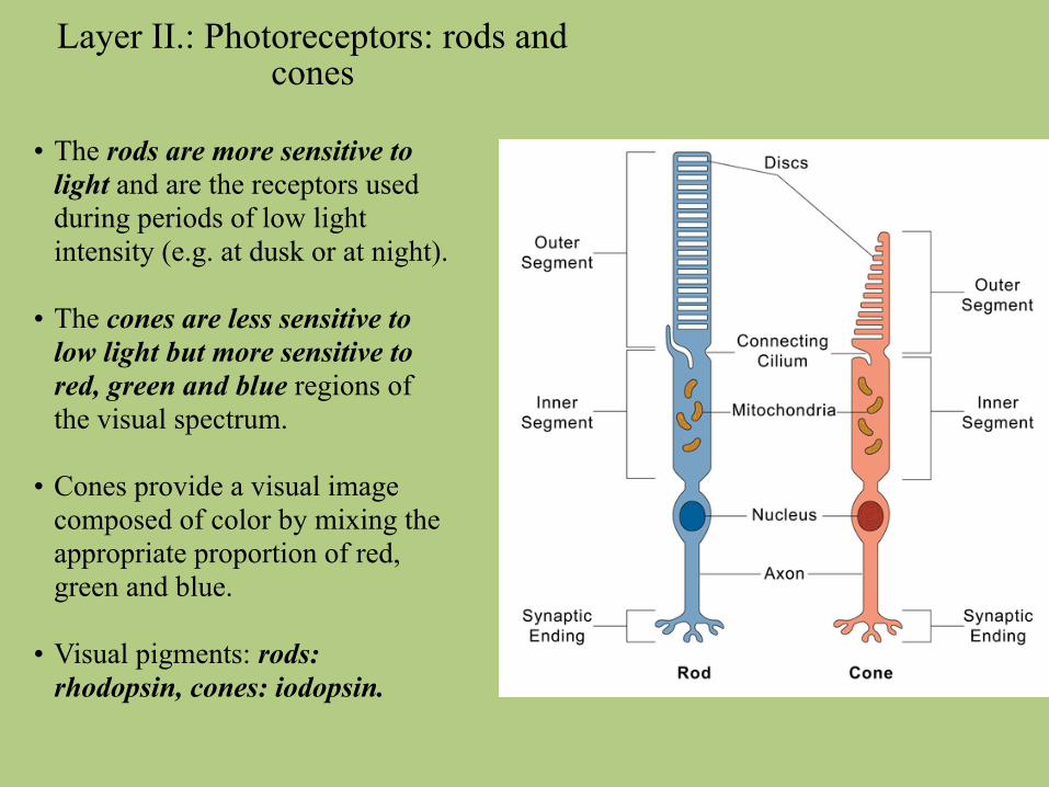
Layer II.: Photoreceptors: rods and cones
• The rods are more sensitive to light and are the receptors used during periods of low light intensity (e.g. at dusk or at night).
• The cones are less sensitive to low light but more sensitive to red, green and blue regions of the visual spectrum.
• Cones provide a visual image composed of color by mixing the appropriate proportion of red, green and blue.
• Visual pigments: rods: rhodopsin, cones: iodopsin.
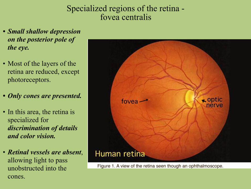
Specialized regions of the retina - fovea centralis
• Small shallow depression on the posterior pole of the eye.
• Most of the layers of the retina are reduced, except photoreceptors.
• Only cones are presented.
• In this area, the retina is specialized for discrimination of details and color vision.
• Retinal vessels are absent, allowing light to pass unobstructed into the cones.
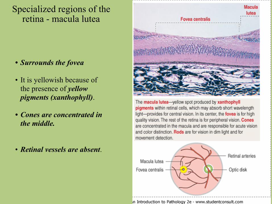
Specialized regions of the retina - macula lutea
• Surrounds the fovea
• It is yellowish because of the presence of yellow pigments (xanthophyll).
• Cones are concentrated in the middle.
• Retinal vessels are absent.
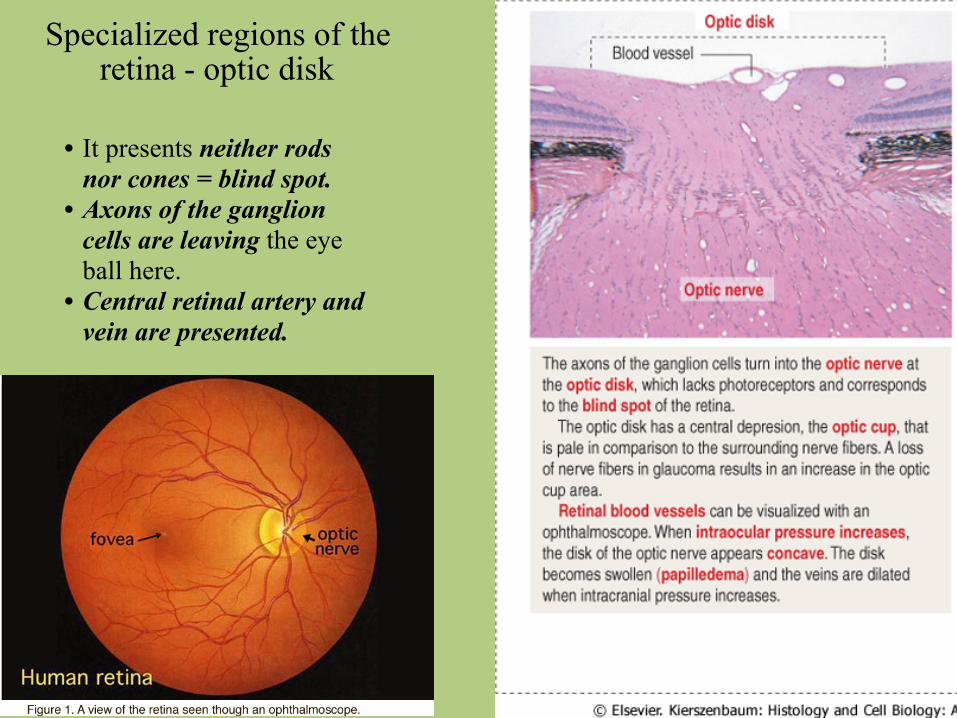
Specialized regions of the retina - optic disk
• It presents neither rods nor cones = blind spot.
• Axons of the ganglion cells are leaving the eye ball here.
• Central retinal artery and vein are presented.
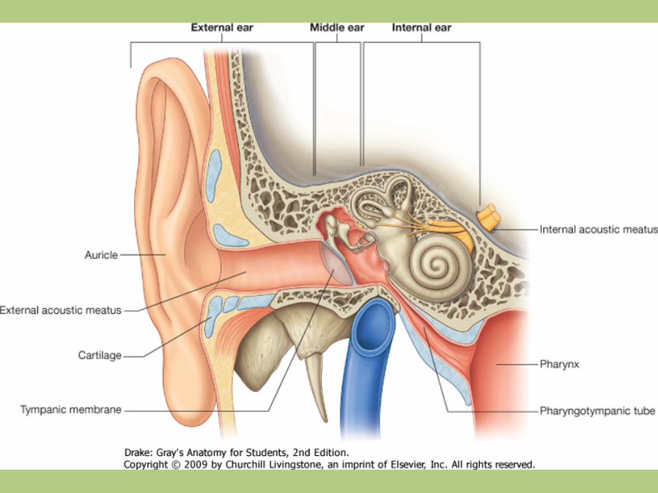
Gross anatomy of the ear
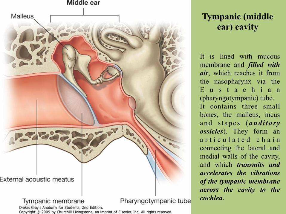
It is lined with mucous membrane and filled with air, which reaches it from the nasopharynx via the E u s t a c h i a n (pharyngotympanic) tube. It contains three small bones, the malleus, incus a n d s t a p e s ( a u d i t o r y ossicles). They form an a r t i c u l a t e d c h a i n connecting the lateral and medial walls of the cavity, and which transmits and accelerates the vibrations of the tympanic membrane across the cavity to the cochlea.
Tympanic (middle ear) cavity
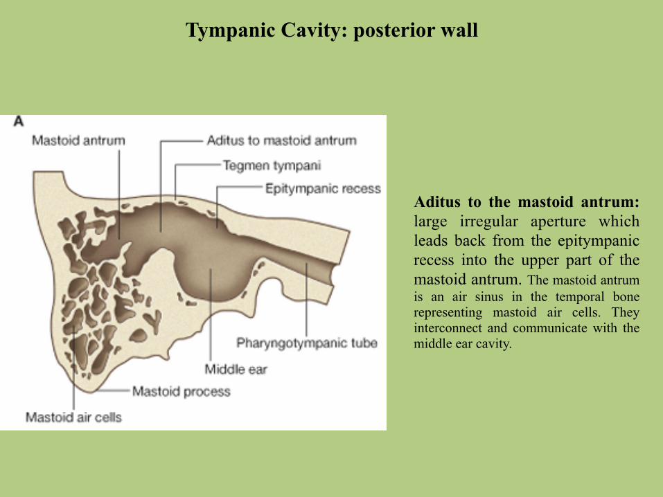
Tympanic Cavity: posterior wall
Aditus to the mastoid antrum: large irregular aperture which leads back from the epitympanic recess into the upper part of the mastoid antrum. The mastoid antrum is an air sinus in the temporal bone representing mastoid air cells. They interconnect and communicate with the middle ear cavity.
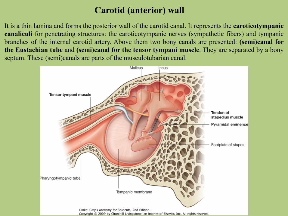
Carotid (anterior) wallIt is a thin lamina and forms the posterior wall of the carotid canal. It represents the caroticotympanic canaliculi for penetrating structures: the caroticotympanic nerves (sympathetic fibers) and tympanic branches of the internal carotid artery. Above them two bony canals are presented: (semi)canal for the Eustachian tube and (semi)canal for the tensor tympani muscle. They are separated by a bony septum. These (semi)canals are parts of the musculotubarian canal.

Labyrinthine (medial) wallIt forms the lateral boundary of the inner ear. The promontory is the projection of the basal turn of the cochlea and represents the tympanic plexus. The oval window (fenestra vestibuli) is a kidney-shaped opening situated above the promontory, and leading from the tympanic cavity to the vestibule of the inner ear. It is occupied by the footplate of the stapes, which is attached to the margin by an anular ligament.
in the oval window
The round window (fenestra cochleae) is situated below and a little behind the oval w i n d o w . I n d r i e d specimens it opens into the scala tympani of the cochlea, but in life it is closed by the secondary tympanic membrane. The prominence of the facial nerve crosses the medial wall from the cochleariform process to the posterior wall. It contains the facial nerve.
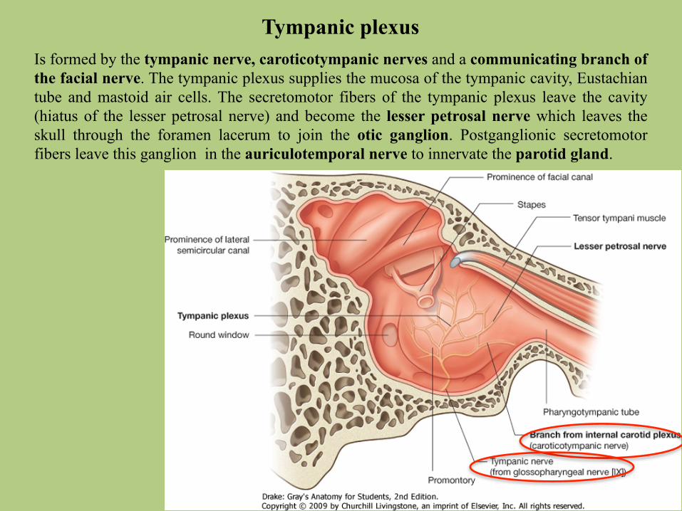
Tympanic plexusIs formed by the tympanic nerve, caroticotympanic nerves and a communicating branch of the facial nerve. The tympanic plexus supplies the mucosa of the tympanic cavity, Eustachian tube and mastoid air cells. The secretomotor fibers of the tympanic plexus leave the cavity (hiatus of the lesser petrosal nerve) and become the lesser petrosal nerve which leaves the skull through the foramen lacerum to join the otic ganglion. Postganglionic secretomotor fibers leave this ganglion in the auriculotemporal nerve to innervate the parotid gland.
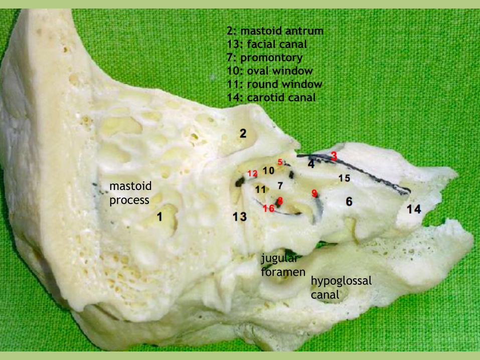
mastoid process
2: mastoid antrum 13: facial canal 7: promontory 10: oval window 11: round window 14: carotid canal
jugular foramen
hypoglossal canal
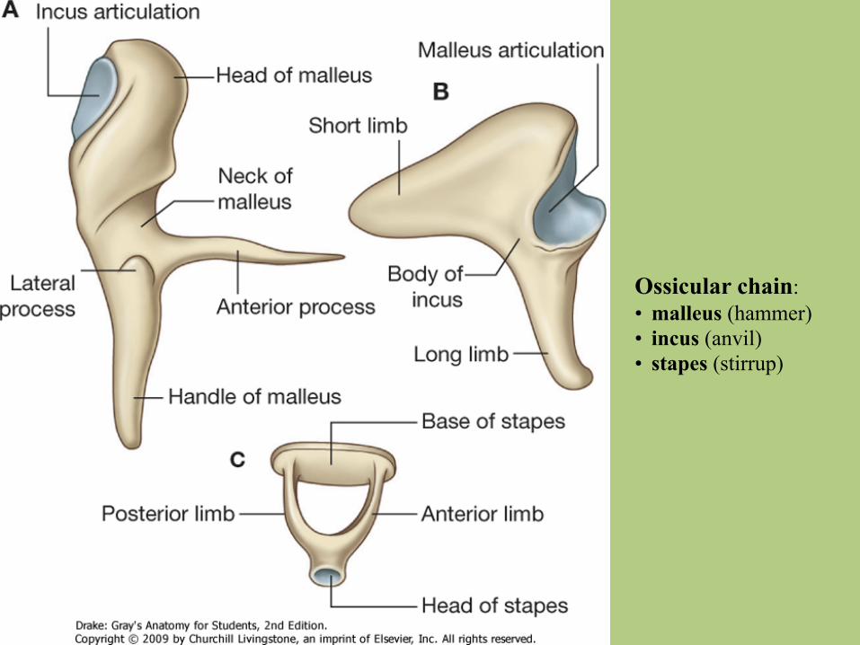
Ossicular chain: • malleus (hammer) • incus (anvil) • stapes (stirrup)

Ossicular chain: • malleus (hammer) • incus (anvil) • stapes (stirrup)
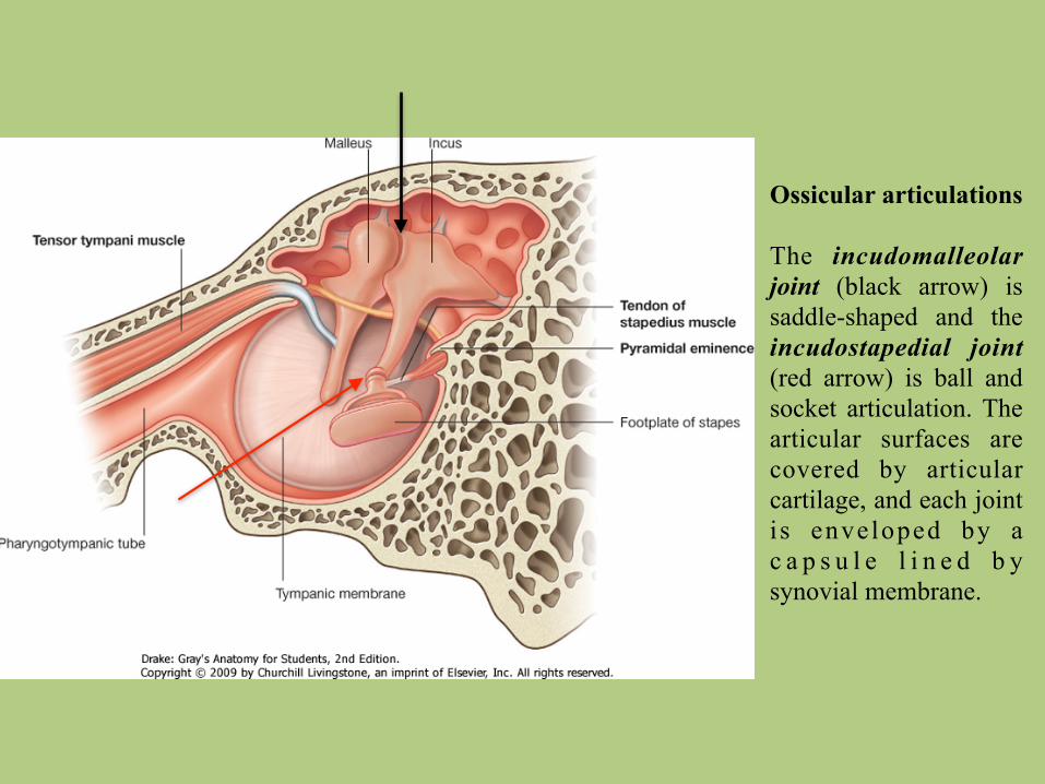
Ossicular articulations
The incudomalleolar joint (black arrow) is saddle-shaped and the incudostapedial joint (red arrow) is ball and socket articulation. The articular surfaces are covered by articular cartilage, and each joint i s enveloped by a c a p s u l e l i n e d b y synovial membrane.
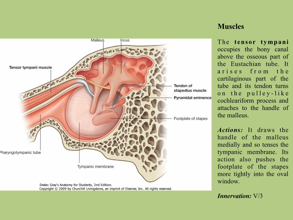
Muscles
The tensor tympani occupies the bony canal above the osseous part of the Eustachian tube. It a r i s e s f r o m t h e cartilaginous part of the tube and its tendon turns o n t h e p u l l e y - l i k e cochleariform process and attaches to the handle of the malleus.
Actions: It draws the handle of the malleus medially and so tenses the tympanic membrane. Its action also pushes the footplate of the stapes more tightly into the oval window.
Innervation: V/3
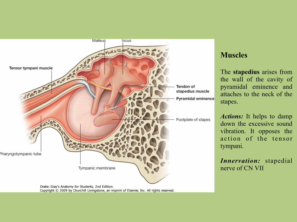
Muscles
The stapedius arises from the wall of the cavity of pyramidal eminence and attaches to the neck of the stapes.
Actions: It helps to damp down the excessive sound vibration. It opposes the ac t ion o f the t ensor tympani.
Innervation: stapedial nerve of CN VII
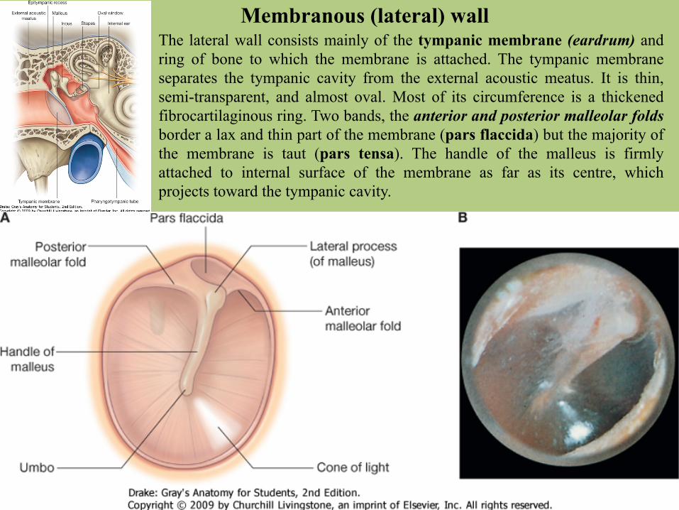
Membranous (lateral) wallThe lateral wall consists mainly of the tympanic membrane (eardrum) and ring of bone to which the membrane is attached. The tympanic membrane separates the tympanic cavity from the external acoustic meatus. It is thin, semi-transparent, and almost oval. Most of its circumference is a thickened fibrocartilaginous ring. Two bands, the anterior and posterior malleolar folds border a lax and thin part of the membrane (pars flaccida) but the majority of the membrane is taut (pars tensa). The handle of the malleus is firmly attached to internal surface of the membrane as far as its centre, which projects toward the tympanic cavity.
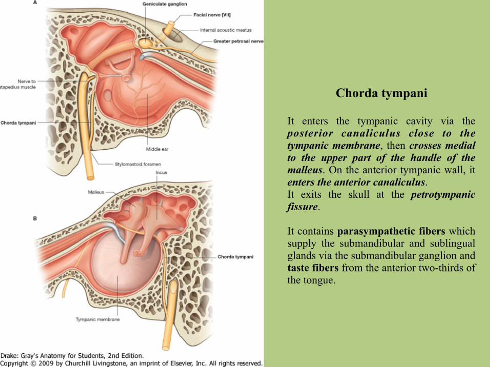
Chorda tympani
It enters the tympanic cavity via the posterior canaliculus close to the tympanic membrane, then crosses medial to the upper part of the handle of the malleus. On the anterior tympanic wall, it enters the anterior canaliculus. It exits the skull at the petrotympanic fissure.
It contains parasympathetic fibers which supply the submandibular and sublingual glands via the submandibular ganglion and taste fibers from the anterior two-thirds of the tongue.
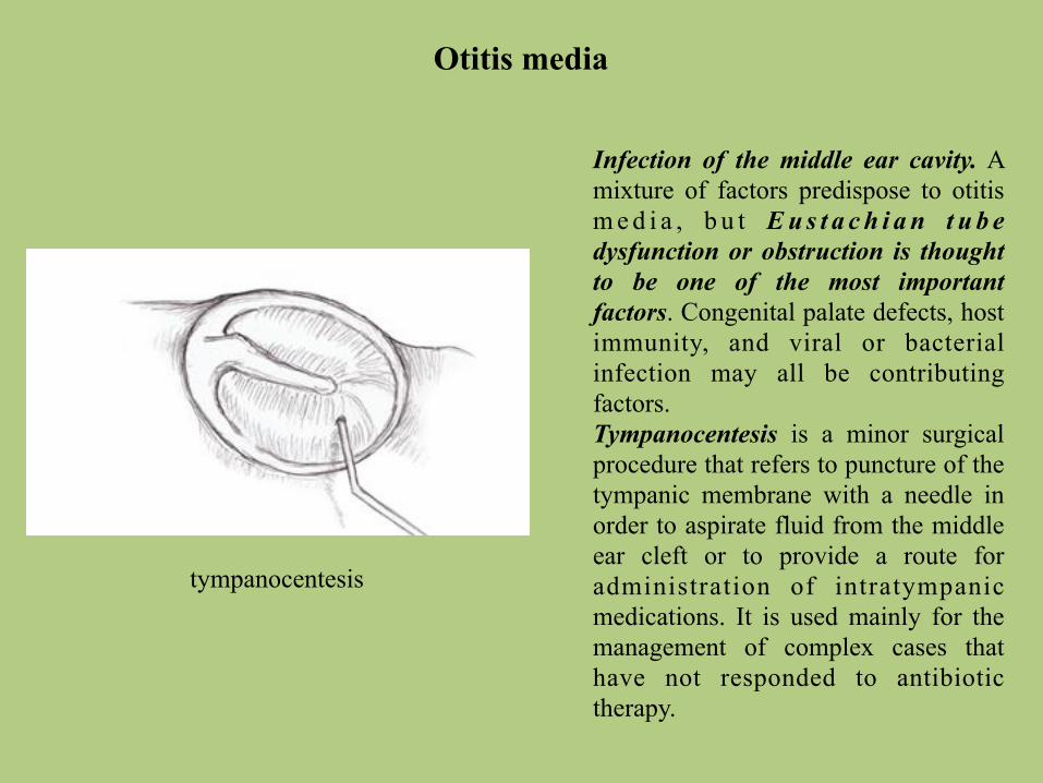
Otitis media
Infection of the middle ear cavity. A mixture of factors predispose to otitis m e d i a , b u t E u s t a c h i a n t u b e dysfunction or obstruction is thought to be one of the most important factors. Congenital palate defects, host immunity, and viral or bacterial infection may all be contributing factors. Tympanocentesis is a minor surgical procedure that refers to puncture of the tympanic membrane with a needle in order to aspirate fluid from the middle ear cleft or to provide a route for administration of intratympanic medications. It is used mainly for the management of complex cases that have not responded to antibiotic therapy.
tympanocentesis
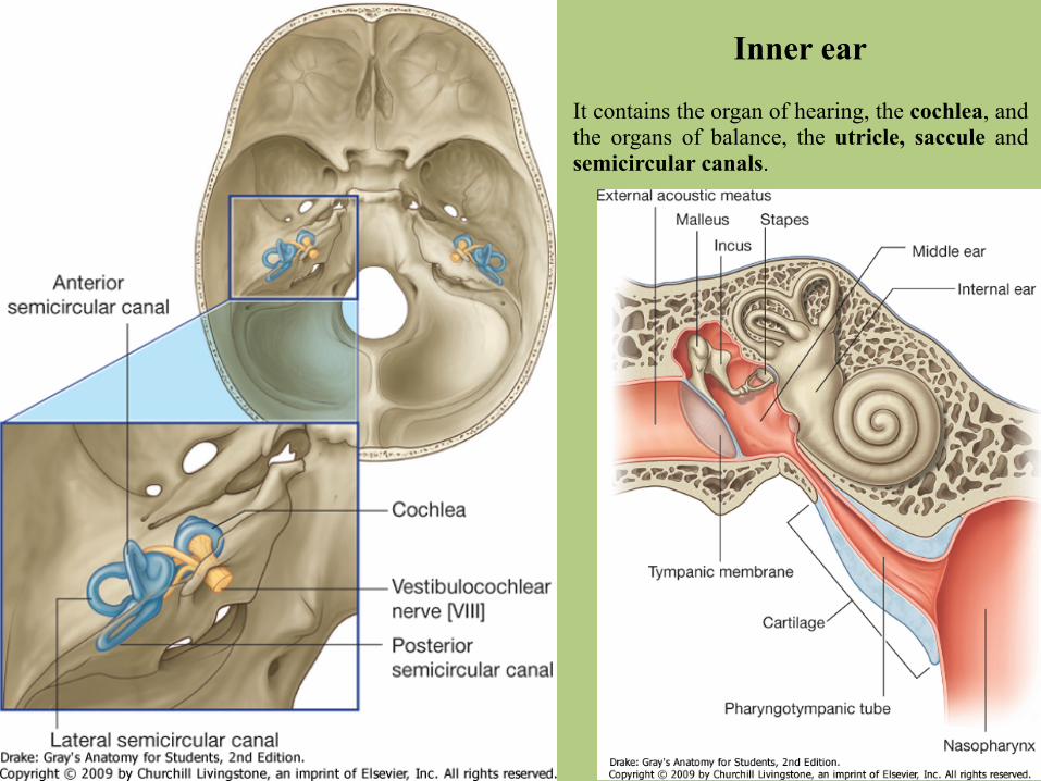
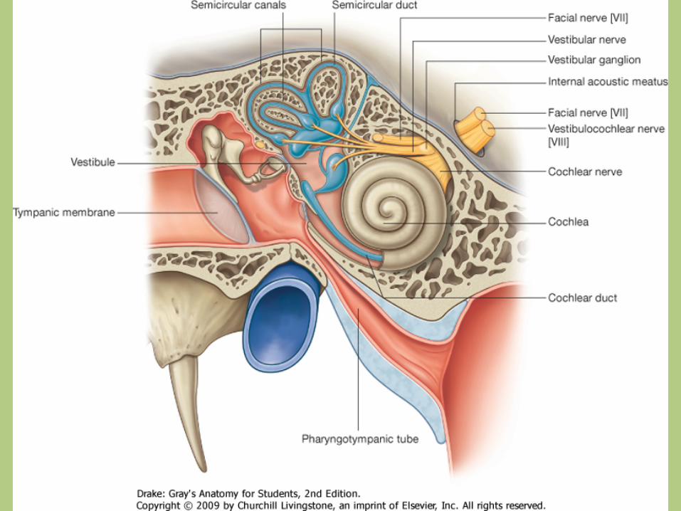
Inner ear
It contains the organ of hearing, the cochlea, and the organs of balance, the utricle, saccule and semicircular canals.
Inner ear
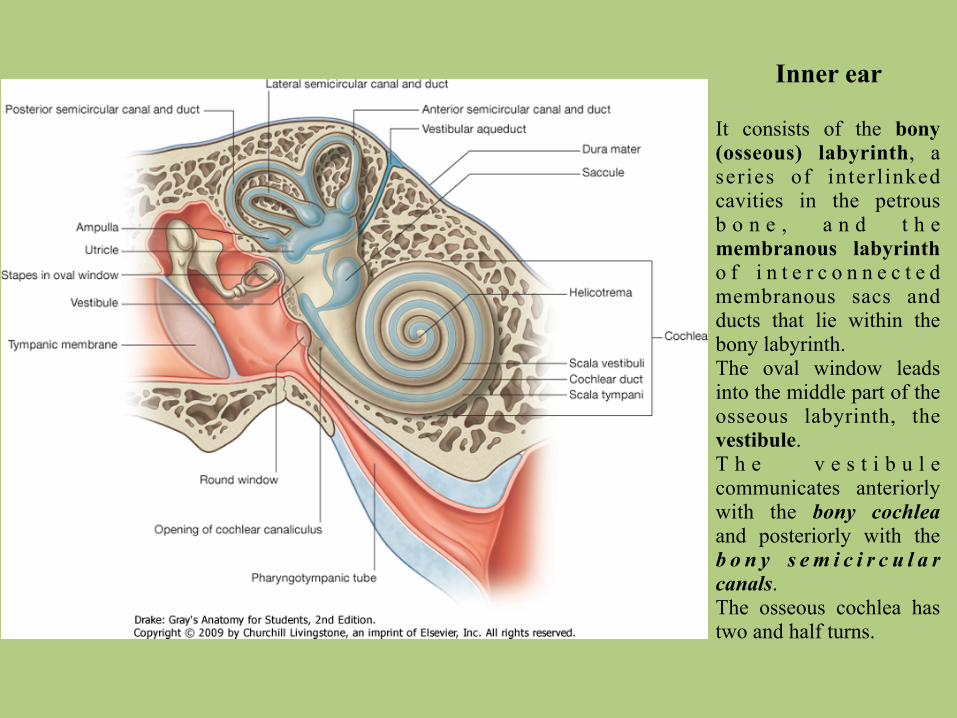
It consists of the bony (osseous) labyrinth, a series of interl inked cavities in the petrous b o n e , a n d t h e membranous labyrinth o f i n t e r c o n n e c t e d membranous sacs and ducts that lie within the bony labyrinth. The oval window leads into the middle part of the osseous labyrinth, the vestibule. T h e v e s t i b u l e communicates anteriorly with the bony cochlea and posteriorly with the b o n y s e m i c i r c u l a r canals. The osseous cochlea has two and half turns.
Inner ear
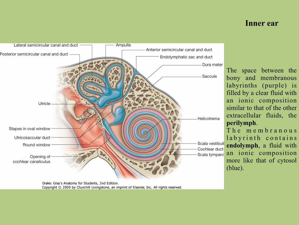
The space between the bony and membranous labyrinths (purple) is filled by a clear fluid with an ionic composition similar to that of the other extracellular fluids, the perilymph. T h e m e m b r a n o u s l a b y r i n t h c o n t a i n s endolymph, a fluid with an ionic composition more like that of cytosol (blue).
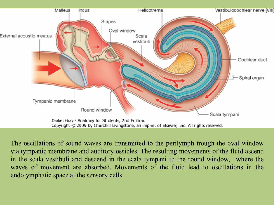
The oscillations of sound waves are transmitted to the perilymph trough the oval window via tympanic membrane and auditory ossicles. The resulting movements of the fluid ascend in the scala vestibuli and descend in the scala tympani to the round window, where the waves of movement are absorbed. Movements of the fluid lead to oscillations in the endolymphatic space at the sensory cells.
Inner ear
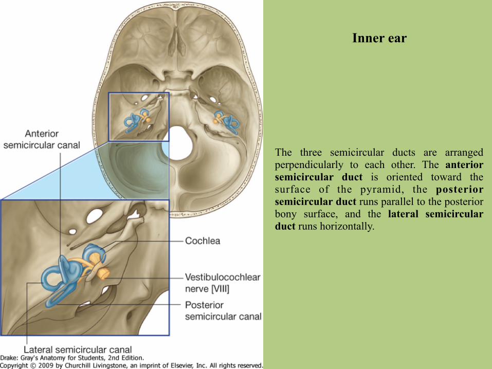
The three semicircular ducts are arranged perpendicularly to each other. The anterior semicircular duct is oriented toward the surface of the pyramid, the posterior semicircular duct runs parallel to the posterior bony surface, and the lateral semicircular duct runs horizontally.

Thank you for your attention.References: Prof. Dr. Ágoston Szél’s lectures (2006, 2007)
Gray’s Anatomy for Students Gray’s Anatomy, The Anatomical Basis of Clinical Practice Thieme, Atlas of Anatomy, Head, Neck and Neuroanatomy Thieme, Flexibook, Color Atlas of Human Anatomy, Nervous System and Sensory Organs
radiopaedia.com