Hyaluronate derivatives and their application to wound healing: Preliminary observations
Fourteenth Annual Meeting and Exhibition of the WoundHealing Society
Sheraton Atlanta HotelAtlanta, GeorgiaMay 23–26, 2004
Summary Program
Sunday, May 23, 2004
18:00 Keynote Address – Robert Nerem, Ph.D., GeorgiaInstitute of TechnologyBlue Ribbon Poster Presentation (Abstracts 1–10)Opening Ceremony
Monday, May 24, 2004
08:30 Opening RemarksAnnouncement – Blue Ribbon Industrial PosterAward Winners
General Session: The Inflammatory Response
09:00 The Macrophage and Coordination of theInflammatory Response – John Christman,M.D., Vanderbilt University
09:30 Inflammatory Cytokines – Anne Richmond,Ph.D., Vanderbilt University
10:00 Refreshment Break
Breakout Sessions
10:30 Inflammation/Cytokines (Abstracts 11–17)Clinical Studies (Abstracts 18–24)Growth Factors (Abstracts 25–31)
12:15 Symposium Lunch
General Session: Young Investigator Awards
14:00 Young Investigator Award Presentations(Abstracts 32–37)
15:30 Refreshment Break16:00 Young Investigator Award Presentations
(Abstracts 38–41)17:00 Poster Reception: Wine & Cheese
Tuesday, May 25, 2004
07:00 Continental Breakfast
Breakout Sessions
08:30 Gene Therapy (Abstracts 42–48)Oxygen & Oxidative Stress (Abstracts 49–55)Wound Infection (Abstracts 56–62)
10:15 Refreshment Break
General Session: Strategies for Tissue Repair
11:00 Regeneration of the Damaged Heart – PieroAnversa, M.D., New York Medical College
11:35 Recruitment of Endothelial Precursor Cells toSites of Tissue Repair – Timothy Crombleholme,M.D., Cincinnati Children’s Hospital and theUniversity of Cincinnati School of Medicine
12:30 Symposium Lunch
Dual Track Plenary: Acute Wounds
14:00 The Effect of the Perioperative Environment onAcute Wound Healing – Andrea Kurz, Universityof Bern
14:30 Nitric Oxide and Acute Wound Healing – AdrianBarbul, M.D., Sinai Hospital/Johns HopkinsUniversity
15:00 Ventral Hernias – Michael Franz, M.D., FACS,University of Michigan Health System and theVA Ann Arbor Health Care System
15:30 Smoking (Cessation) and Surgical Wound Com-plication – Lars Tue Sorensen, M.D., Universityof Copenhagen
Dual Track Plenary: Oxidative Stress/Ischemia
14:00 Role of Reactive Oxgyen Species in WoundHealing – Chandan Sen, Ph.D., The Ohio StateUniversity Medical Center
14:30 Stress, Oxygen, Nitric Oxide, and WoundHealing – Phillip Marucha, D.D.S., University ofIllinois at Chicago
Abstracts
A1
15:00 HIF-1a and its Downstream Targets – WayneS. Zundel, Ph.D., University of Colorado HealthSciences Center
15:30 Hypoxic Interactions – Thomas Mustoe, M.D.,Northwestern University Medical School
16:00 Refreshment Break
General Session
16:15 3M Award PresentationBusiness Meeting
Wednesday, May 26, 2004
07:00 Continental Breakfast
Dual Track Plenary Session: Cell–Matrix Interactions
08:30 Mechanisms of Feedback Regulation byCollagen-Binding Integrins – Thomas Krieg,M.D., University of Cologne
09:00 Integrin-Mediated Regulation of Generators ofAngiogenesis – Ambra Pozzi, Ph.D., VanderbiltUniversity School of Medicine
09:30 A Reevaluation of Integrins as Mediators ofAngiogensis – Dwayne G. Stupack, Ph.D., TheScripps Research Institute
Dual Track Plenary: Diabetic Wounds
08:30 Off-Loading – Lawrence Harkless, D.P.M.,University of Texas Health Science Centerat San Antonio
09:00 Debridement – Christopher Attinger, M.D.,Georgetown University
09:30 Control of Diabetic Foot Infections – PeterSheehan, M.D., Hospital for Joint Diseases
10:00 Refreshment Break
Breakout Sessions
10:30 Biomaterials/Bioengineering (Abstracts 63–69)Epithelization (Abstracts 70–76)Stem Cells (Abstracts 77–83)
12:15 Symposium Lunch
General Session: Bioengineering
14:00 Endothelial Responses to Inflammation –Robert A. Swerlick, M.D., Emory UniversitySchool of Medicine
14:30 Biomaterials for Vascular Repair – ElliottL. Chaikof, M.D., Ph.D., Emory University Schoolof Medicine
15:00 Refreshment Break
General Session: Engineering of the Repair Process
15:15 Tissue Engineering of Bone – Barbara Boyan,Ph.D., Georgia Institute of Technology
15:45 Neural Tissue Engineering –Michelle Laplaca,Georgia Institute of Technology
16:15 Signaling at the Cell–Materials Interface –Andres Garcia, Georgia Institute of Technology
16:45 Neurite Extension on Biomaterials – RaviBellakonda, Georgia Institute of Technology
Program subject to change
All abstracts for presentation at the Annual Meeting areincluded in the following compilation. Abstracts arenumbered and categorized according to the meetingschedule.
Abstracts No. Session Name
1–10 Blue Ribbon Industrial Posters*11–17 Inflammation/Cytokines18–24 Clinical Studies25–31 Growth Factors32–41 Young Investigator Awards42–48 Gene Therapy49–55 Oxygen & Oxidative Stress56–62 Wound Infection63–69 Biomaterials/Bioengineering70–76 Epithelization77–83 Stem Cells84–153 Posters
* Blue Ribbon Industrial Posters 2, 3, 4, and 5 will alsobe presented in podium sessions as presentations 76,26, 28, and 81, respectively.
WOUND REPAIR AND REGENERATIONA2 ABSTRACTS MARCH–APRIL 2004
BLUE RIBBON INDUSTRIAL POSTER SESSION
001
A NOVEL MODEL FOR THE ASSESSMENT OF FORMULATED ENZYMATICDEBRIDING AGENTS
Lei Shi, Justin Keen, Braham Shroot
DPT Laboratories, San Antonio, TX USA
In order to closely mimic the actual mechanism of wound debridement by an enzymaticdebriding agent, a synthetic wound eschar substrate (SWES) has been developed. The SWESis prepared by the enzymatic conversion of fibrinogen to fibrin in the presence of fibrousbovine collagen and elastin, to form an insoluble planar matrix composed of the three majorproteins present in wound eschar. Each of the proteinaceous components is uniquely labeledprior to formation of SWES with different dye, and the debridement test is conducted using aFranz Diffusion System. The debriding agent is applied to the SWES mounted on the donorcompartment of the cell, and the digested hydrolysate containing dye-labeled peptide frag-ments is sampled from the receptor compartment. Accordingly, the progress of digestion isobtained in real time. Since the three proteins are labeled with spectrally distinct dyes, dataon the hydrolysis of the substrates can be collected simultaneously. This method facilitatesthe testing of formulated debriding products in a manner that closely mimics application to awound and provides a powerful tool for the evaluation of formulated enzymatic debridingagents.
002
AUSTRALIAN TAIPAN SNAKE VENOM TO SYNTHETIC OXYNOR FORWOUND HEALING
Binie V. Lipps,
Ophidia Products, Inc., 11320 South Post Oak #203, Houston, Texas 77035
At Ophidia Products, Inc. we have discovered Oxynor a synthetic therapeutic from snakevenom for wound healing. The venom of Australian taipan snake (Oxyuranus s. scutellatus)is extremely potent due to the presence of taipoxin. The intact complex molecule of taipoxinhaving molecular weight 45.6 kDa is composed of three subunits a, b and g. Fractionation ofcrude venom by High pressure chromatography (HPLC) using ion exchange column permitsseparation of the subunits. The mitogenic activity was revealed in non toxic b taipoxin. Nontoxic b taipoxin showed mitogenic activity on variety of eukaryotic cells. Its activity as amitogen extended to wound healing in experimental animals. We identified the activedomain for wound healing in b taipoxin. after trypsin digestion were separated on HPLC,resolved into 11 different fragments. Each fragment was tested on PC12 cells to testmitogenic/neurotrophic activity. The fragment which showed the highest activity consistedof 21 amino acids. A synthetic peptide consisting of ten amino acids, from the N-terminal ––was named Oxynor.Oxynor mimics the biological properties of natural b taipoxin when tested in vitro and in vivosystems. The biological results showed that Oxynor was not toxic at the concentration of100mg/ml for PC12 cells and 500mg/adult mouse. Oxynor was mitogenic to various eukar-yotic cells. Oxynor is neurotrophic showing neurite outgrowth on PC12 cells. Growth ofhuman skin fibroblast cells indicates that Oxynor has keratinous activity. Experimentallyproduced 4 mm punched wounds on the back of mice were treated with b taipoxin 100mg/mouse or Oxynor at 500mg/mouse for seven consecutive days. The results showed completeclosure of the wounds after six days while the wounds of control treated with PBS wereopen. Histology examination of the skin around the wounds revealed that treated miceshowed re-epithelization of the epidermis, which looked close to the normal mouse skinbiopsy. Whereas the controls showed distortion of epithelium. b taipoxin and Oxynor weretested for in vivo at the concentration of 100mg/ml in hydrogel vehicle applied once daily tohelp heal 6 mm ischemic skin wounds in rats. At days 7 and 14 wound treated with b taipoxinwere about 15% smaller than the wounds treated with Oxynor. This shows that syntheticOxynor is as efficacious as natural b taipoxin under the conditions of the test. On increasingthe concentration of Oxynor to 500mg/ml should give equivalent wound closure to 100mg/mlof b taipoxin. In 1997 the US FDA approved for sale the first recombinant human platelet-derived growth factor (rhPDGF) for treatment of human non-healing wounds. Oxynor will benext therapeutic for wound healing Currently, Oxynor is in clinical trials, it has passedtoxicity tests.
003
a1-ANTICHYMOTRYPSIN – A HUMAN SERPIN WITH THE POTENTIAL TOHEAL DIABETIC ULCERS
A. Goppelt1, W. Hans1, Y. Shi2, M. Bittner1, J.-P. Halle1, P. Hof1, J.M. Davidson2,3.1SWITCH Biotech AG, Floriansbogen 2–4, D-82061 Neuried, Germany 2Department of Path-ology, Vanderbilt University, 3 VA Medical Center, Nashville, TN 37212, USA
a1-Antichymotrypsin (a1-ACT) is a secreted serine proteinase inhibitor of the serpin family.It is strongly upregulated during the inflammatory phase of wound repair to control theundesirable destructive side effects of cathepsin G which is released from infiltratingneutrophils. We have shown that a1-ACT and its mouse homologue spi2 exhibit a classicacute phase response after cutaneous injury in murine and human skin. The induction of spi2gene expression following wounding is significantly less pronounced in diabetic mice.Transient overexpression of spi2 or a1-ACT significantly increased wound tensile strengthin two independent diabetic models: in genetically diabetic mice, gene gun mediated deliveryof spi2 or a1-ACT cDNA increased the average wound strength by 42% (P¼ 0.001) and 21%(P¼ 0.013) at d5 post-wounding respectively. In a STZ induced diabetic rat model thebreaking strength of adenovirally spi2- or a1-ACT-infected wounds increased by 20%(P¼ 0.049) and 23% (P¼ 0.004) at d7 after injection respectively. Moreover, the topicalapplication of human a1-ACT protein to wounds of diabetic mice had a significant anddose dependent effect on tensile strength at d5 post-wounding (21% increase, P¼ 0.003).Histological analyses of these wounds indicated less infiltration of neutrophils in the treatedwounds. a1-ACT appears to be a key mediator between proteolysis and cytokine agonismand antagonism. Our data indicate that this balance is disturbed in diabetic wounds and thata1-ACT might act as a switch to initiate wound healing in diabetic ulcers.
(Supported by SWITCH Biotech AG)
004
NOVEL PLATELET CONCENTRATE PREPARATION SYSTEM FOR GROWTHFACTOR MEDIATED WOUND HEALING
Sukavaneshvar S1, Zheng Y1, Mohammad SF1
1 Hemogenesis, Salt Lake City, UT, USA.
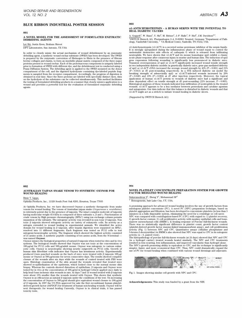
A promising approach for advanced wound healing involves the use of growth factors fromautologous platelet concentrate (PC). A novel PC (NPC) preparation technique, based onplatelet aggregation and filtration, has been developed to concentrate platelets (in less than 5minutes) in a fully disposable system, eliminating the need for a centrifuge or cell saver.NPC was compared with centrifugation-based PC (CPC) with regard to: 1) platelet recovery,2) growth factor content, 3) cell proliferative activity with monkey epithelial cells and bone-marrow mesenchymal cells (BMMC), 4) healing response of Porcine full-thickness wounds.There was no statistically significant difference in platelet count, growth factor content(platelet-derived growth factor enzyme-linked immunosorbent assay), and cell proliferationactivity (Fig. 1) between NPC and CPC. Quantitative assays (Alkaline phosphatase and5-bromo-2-deoxyuridine) in BMMCs also showed that NPC was equal to CPC in its mitogenicactivity (n¼ 6, paired t-test).The histopathology of porcine full-thickness wounds (at 24 days) showed that NPC and CPC(in a hydrogel carrier) treated wounds healed similarly. The NPC and CPC treatmentsresulted in less scarring, less inflammation, and improved vascularity than hydrogel alone.The NPC’s growth promoting ability is equivalent to CPC, and the technique is significantlysimpler, faster, and more economical than CPC. Thus, NPC could dramatically expand theuse of PC for wound healing when combined with various wound dressings and adjuvants.
Acknowledgements: This study was funded by a grant from the NIH.
Fig. 1. Images showing similar cell growth with NPC and CPC.
WOUND REPAIR AND REGENERATIONVOL. 12, NO. 2 ABSTRACTS A3
005
CYCLODEXTRIN POLYMER BASED BIOCOMPATIBLE MATRIX FOR LOCALGENE DELIVERY
D.W. Kang1, N.C. Bellocq2, T. Schluep1, L.D. Looper1, D-L. Gu1, E.M. Quijano1, S.F. Wen1,
X. Wang2, G.S. Jensen2, S.H. Pun2, M.L. Zepeda1, M.E. Davis3
1Canji Inc., San Diego, CA USA; 2Insert Therapeutics Inc., Pasadena, CA USA; 3ChemicalEngineering, Caltech, Pasadena, CA USA
Biocompatible matrices, such as bovine collagen, have demonstrated usefulness in deliveringgene therapy vectors that express growth factors to local environments for tissue repair.Unlike animal derived matrices, we have developed a new synthetic matrix consisting of alinear cyclodextrin-polyethyleneglycol co-polymer that is non-covalently cross-linked withdi-adamantane-polyethyleneglycol via inclusion complex formation between adamantaneand cyclodextrin (CD-Ada). We performed both in vitro and in vivo experiments forbiocompatibility and localized transgene expression using a recombinant adenovirus (rAd)vector containing either the reporter gene, GFP, or the therapeutic gene, PDGF-B. In vitro
results demonstrated cell migration, adenoviral transduction, and gene expression with novisible signs of toxicity in human skin fibroblasts. Qualitative gene expression from the CD-Ada containing rAd was delayed by approximately two days when compared to collagen, butthe level of expression was greater over a longer period of time. In vivo experimentsdemonstrated gene expression after local delivery to mouse skin using rAd-GFP in CD-Ada. Again, the expression was slightly delayed but duration of expression was comparableto collagen. Expression studies using rAd-PDGF-B, were performed in the rat polyvinylalcohol sponge model and showed comparable quantitative DNA and RNA levels betweenCD-Ada and collagen (DNA: 4.1· 1010 and 4.5 · 1010 MEQ of PDGF-B/assayed sponge, respec-tively; RNA: 7.0· 108 and 3.2· 108 MEQ of PDGF-B/assayed sponge, respectively). Addition-ally, we explored the use of plasmid DNA with the CD-Ada matrix and observed PDGF-Bexpression in vivo. Our results show that this new delivery system provides a safe, efficient,and adaptable medium for both viral and non-viral gene delivery.
006
DEVELOPMENT OF A NOVEL MATRIX METALLOPROTEINASE-INHIBITING WOUND DRESSING
Gary A. Skarja, Maud B. Gorbet, Rebecca K. Lawson-Smith, Michael H. May, Michael
V. Sefton*Rimon Therapeutics Ltd., Toronto, Ontario, Canada, *Institute of Biomaterials and Bio-medical Engineering, University of Toronto, Ontario, Canada
Rimon Therapeutics develops synthetic therapeutic polymers (TheramersTM) that combinebioactivity with a broad range of desirable mechanical and physical properties. The MITheramerTM is a polymer that modulates the activity of matrix metalloproteinases (MMPs)..Excessive proteolytic cleavage of extracellular matrix and growth factors resulting fromelevated levels of MMPs is believed to contribute to the impaired wound healing associatedwith chronic, non-healing wounds. MI TheramerTM beads suspended in ThermaGelTM (athermoreversible gel) make up MI-GelTM Dressing. The dressing may be applied as a liquidthat is poured into a wound, where it will gel on contact. The dressing can be reliquified andremoved easily without re-injuring the wound by cooling it.In vitro inhibition of collagenase type IV (equivalent to MMP-2) from clostridium histolyti-cum (Molecular Probes) was measured. A dose-dependent inhibition of MMP-2 activity wasmeasured for MI TheramerTM beads alone and suspended in ThermaGelTM indicating thatMMP-2 is able to diffuse into the gel and bind to the beads suspended therein. In vivo
efficacy was demonstrated by observing the degradation of glutaraldehyde cross-linkedgelatin tubes using a mouse subcutaneous pouch model. Gelatin zymography on extractscollected from the gelatin tubes co-implanted with MI TheramerTM beads also showedreduced MMP-2 and MMP-9 activity in comparison to controls.The insoluble MI TheramerTM beads were able to modulate MMP activity both in vitro andin vivo. The beads retained their inhibitory activity when they were suspended in a novelthermoreversible gel (ThermaGelTM). The combined MI-GelTM Dressing may have promise asa novel chronic wound healing product.
We would like to thank Ping Xu for his assistance in the collection of the gelatin implantationstudy data.
007
REBALANCING WOUND BIOCHEMISTRY IMPROVES HEALING: ACLINICAL STUDY EXAMINING EFFECT OF PROMOGRAN
B. Cullen, L. Kemp, A. Essler, K. Wallenfang-Sohle, R. Stadler
Johnson & Johnson Wound Management, Gargrave, N. Yorkshire, UKDept. Dermatology, Minden Hospital, Germany
Normal wound healing is a carefully controlled balance of new tissue formation and destruc-tive processes necessary to remove damaged tissue. Within this complex environment thereare many points of regulation, which control the biological processes necessary to achievewound repair. An alteration in any of these processes can result in an imbalance of thebiochemical components, which ultimately results in delayed wound closure and the forma-tion of a chronic wound. Therefore, we propose that an effective therapeutic approach wouldmodify this hostile environment and redress this imbalance.In this study we have evaluated the effect of PROMOGRAN in vivo, in patients with venousleg ulcers. Wound fluid samples were collected and analyzed from a number of patients priorto and during treatment with PROMOGRAN.Our results have shown that wounds, which respond to PROMOGRAN treatment, have alsoshown a decrease in protease activity in the corresponding wound fluid samples. Whilst it isimpossible to determine whether this reduction in protease levels is responsible for healingor merely symptomatic of other changes occurring within the wound, we have shown that adecrease in proteolytic activity is concomitant with healing.This study provides clinical evidence that PROMOGRAN can rebalance the chronic woundenvironment in situ and thereby promotes wound repair.
Type acknowledgements here.
008
CROSS – TALK BETWEEN FIBROBLASTS AND KERATINOCYTES IN 3-DFIBRIN CLOTS. IN VITRO STUDIES
N. Sese, M. Cole, and B. Tawil
Baxter BioScience, BioSurgery, Westlake Village, CA USA
Fibrin Sealant products such as TisseelR have been used over the last 25 years to reachhemostasis during surgery or to seal tissues. Moreover, there were many studies on the useof fibrin sealant in cell and bioactive substance delivery. Our In Vitro and In Vivo studies,over the last 2 years, have focused on the use of fibrin sealant to deliver human fibroblasts orkeratinocytes to overcome the healing deficiency in chronic wounds. We have shown thatsome fibrin formulations supported a high fibroblast proliferation and other fibrin formula-tions supported a high proliferation of human keratinocytes. In this study, we examined theuse of fibrin sealant in the co-delivery of both human – derived keratinocytes and fibroblasts.The study report examined the cell proliferation of these two cell types in various formula-tions of fibrin sealant. Fibroblasts and keratinocytes were mixed with various dilutions ofthe Sealer Protein solution and added to culture plates before adding the Thrombin solutionto form fibrin clots containing both cell types. We found that a low to medium sealer proteinconcentration (1–34 mG/mL) and a very low thrombin concentration (1 U/mL) in the finalfibrin clots provided for an optimal cell proliferation for both cell types within these fibrinclots. This profile of proliferation was different from that seen when keratinocytes alone orfibroblasts alone were incorporated in the fibrin clots. Morphologically, it was difficult todetermine the cell type from examining cell morphology, thus, it was difficult to determinethe cell distribution. In conclusion, we found that various modified formulations of fibrinsealant may be chosen when co- delivering fibroblasts and keratinocytes. Moreover, thereseems to be a positive feedback between keratinocytes and fibroblasts when they wereco – introduced in the fibrin clots. This feedback could be carried by growth factors. Futurestudies will determine his signaling process.
WOUND REPAIR AND REGENERATIONA4 ABSTRACTS MARCH–APRIL 2004
009
EVALUATION OF AN OZONE-BASED WOUND MANAGEMENT SYSTEM
A.J. Denvir1, A.C. Blankenburg1, I.A. Holder2
1Lynntech Inc., College Station, TX, USA 2Shriners Hospital for Children, Cincinnati, OH,USA
The onset of infection can lead rapidly to sepsis, septic shock, and eventually death. Con-sidering the high costs of hospitalization and the added trauma and discomfort to the patient,improved methods for infection control are needed. Ozone offers a specific solution to theproblem of effective management of microbial wound contamination. Despite the advan-tages, several technical barriers have prevented ozone-based disinfection treatments frombecoming more widely investigated. Negative press from years of unsubstantiated medicalsuccesses has made the medical community wary of even legitimate ozone technologies.Because there has been no research into using ozone in infection prevention, much of thehardware has not been developed to the stage where the technology can be used in clinicaltrials. Lynntech Inc., over the past 5 years, has been working to develop a pre-clinical ozone-based wound management system. This new hardware uses an electrochemical ozonegenerator that can deliver a predetermined quantity of ozone gas to a specific wound site.Electrochemically generated ozone has been shown to be effective against a range of gramnegative and gram positive bacteria and our studies with various mimetic wound systemshave shown that with controlled delivery, ozone can be utilized as either a disinfectant or asa biostat. The device is well suited for hospital use as it operates of low voltage powersupplies and unlike other ozone generation technologies will not interfere with other elec-tronic equipment while it is in operation. This paper outlines both the advantages andlimitations of using an ozone based system as a wound management tool and looks to thefuture research that needs to be performed to substantiated the initial research findings.
This work was funded through NIH
010
STRUCTURAL CHARACTERIZATION OF SMALL INTESTINAL SUBMUCOSA(SIS) USING CRYO AND TRADITIONAL SCANNING ELECTRONMICROSCOPY (SEM)
A.D. Janis1, D.M. Sherman2
1Cook Biotech Incorporated, West Lafayette IN 2Life Science Microscopy Facility, PurdueUniversity, West Lafayette IN
Composition and structure determine the fate of biomaterials in the body, encouraging ordiscouraging acceptance, cellular infiltration, and degradation or incorporation into hosttissue. Most biomaterials research has been focused on composition, while structure remainsa relatively uninvestigated factor in biomaterial success. Naturally derived biomaterials mayprovide optimal structure for cellular repopulation in certain applications. Porcine smallintestinal submucosa (SIS) is an acellular, naturally occurring extracellular matrix used inthe clinical treatment of soft tissue injury, including pressure, venous, and diabetic ulcers.Numerous studies have demonstrated that SIS induces host tissue infiltration, tissue-specificremodeling, and repair. SIS consists primarily of collagen, although the presence of otherproteins, growth factors, glycosaminoglycans, and proteoglycans has been reported. SEMwas used to characterize the cellular environment provided by this biomaterial. Cryo-SEMwas used to image mechanically isolated, oxidatively disinfected, lyophilized, sterilized, andrehydrated SIS. The fractured cross-sectional faces of intact intestine, oxidatively disinfectedSIS, and lyophilized SIS were imaged with traditional SEM. Comparison of the imagesutilizing cryo and traditional preparation techniques show that SIS is a heterogeneous matrixconsisting of textured fibers of mixed size. There are differences in the structure of themucosal and serosal surfaces. Although the matrix architecture shows signs of collapse anddelamination following lyophilization, much of the native structure is preserved. Thesechanges are preserved following rehydration. The effects of these structural changes oncellular reaction and infiltration are unknown, but the naturally complex architecture-clearlyfacilitates host interaction on thecellular and subcellular levels. This work was supported byCook Biotech Incorporated.
INFLAMMATION/CYTOKINES
011
INFLAMMATION-INDUCED ANGIOGENESIS: POTENTIAL ROLES OF IL-8.AND VEGF
M. Yao, M. Dueck, and M. Martins-Green
University of California, Riverside, CA USA
Inflammation is invariably accompanied by angiogenesis; both are important in woundhealing. However, it is not known how angiogenic factors that are present during theinflammatory phase of wound healing are involved in inflammation-induced angiogenesis.In this study, we address the contribution of two such factors, interleukin-8 (IL-8/CXCL8)and VEGF, in inflammation-induced angiogenesis. We determined the relative levels of thesemolecules during the first two weeks of the wound healing process in partial-thickness skinwounds of rabbits. IL-8 levels increase rapidly after wounding, reaching maximal levels after24 hr; in contrast, VEGF levels peak at 3 days. We also determined the time course of theinfiltration of various inflammatory cells and production of new blood vessels after wound-ing. Neutrophils were markedly increased 4 hr after wounding, and their level peaked at24 hr. Monocyte infiltration peaked 48 hr after wounding and decreased by day 3. Thenumber of blood vessels was significantly increased by day 3 after wounding and continuedto increase through days 5 and 7.In order to determine the contribution of IL-8 and VEGF to the angiogenic process, weperformed the wounding experiments in the presence and absence of IL-8 and VEGF anti-bodies. Treatment with IL-8 antibodies blocked the early stage of blood vessel formation butdid not alter the late stage; in contrast, VEGF antibodies blocked the late stage withoutaltering the early stage. Currently, we are performing studies using inhibitors against IL-8 andVEGF receptors to determine whether we observe similar results; we are also beginning todecipher molecular mechanisms involved in this process. This study suggests that IL-8 andVEGF play complementary roles in stimulating angiogenesis during wound healing.
This work was funded in part by AHA and TRDRP
012
MOLECULAR MECHANISMS OF THROMBIN-INDUCED INTERLEUKIN-8EXPRESSION IN HUMAN MACROPHAGES
L. Zheng; M. Martins-Green;
Cell Biology and Neuroscience Department; University of California, Riverside, CA
Wound healing involves a series of overlapping, highly coordinated events. Macrophages playmultiple crucial roles in these processes including the secretion of chemokines. Interleukin-8(IL-8) is an inducible chemokine that is expressed in a variety of cell types present at thewound site, including macrophages, and is known to contribute to chemotaxis, angiogenesis,and tissue remodeling during wound healing. One of the potent natural inducers of IL-8 isthrombin, a multifunctional enzyme that is released upon wounding. Little is known abouthow this enzyme stimulates IL-8 in macrophages. Therefore, we used a variety of cellular andmolecular approaches to investigate the signal transduction mechanism of thrombin-inducedIL-8 expression in THP-1-differentiated macrophages. We show that stimulation of IL-8 bythrombin occurs in a rapid manner that is time- and dose-dependent and can be abolished byaddition of hirudin, a specific inhibitor of thrombin. At the mRNA level, IL-8 peaks *4 hrsafter treatment and declines by 9 hrs. A variety of inhibitors were used to delineate the signaltransduction pathways leading to IL-8 expression. Application of AG1478, a potent EGFRinhibitor, abolishes thrombin-induced IL-8 production suggesting EGFR transactivation. Wefurther show that MEK1/2 and p38 are important signal mediators; their inhibition abolishesIL-8 production. Contrarily, PKC inhibitors do not abolish, but rather enhance production ofIL-8, whereas activators inhibit the thrombin stimulation, suggesting that PKC is a negativeregulator of IL-8 expression. In summary, these results suggest that thrombin stimulates IL-8expression by transactivating the EGFR leading to activation of MEK1/2 and p38, and thatPKC is a negative regulator of this stimulation process.
Supported by AHA and TRDRP.
WOUND REPAIR AND REGENERATIONVOL. 12, NO. 2 ABSTRACTS A5
013
VARIABLE P53 RESPONSE IN A 3D MATRIX
M.A. Carlson1 and M.T. Longaker1University of Nebraska Medical Center, Omaha, NE; 2Stanford University, Stanford, CA
Detachment of an attached (stressed) fibroblast-populated collagen matrix (FPCM) inducesfibroblast apoptosis; this may be secondary to increased p53 after detachment. We havenoted, however, that the p53 response after detachment of an attached FPCM (i.e., stress-release) varies among cells strains. We hypothesized that modulation of p53 after FPCMstress-release may be related to the fibroblast population density at the time of release.Human foreskin fibroblasts (initially seeded at 0.5 · 106/ml in 0.2 ml) were cultured for 48 hrin attached collagen matrices prior to detachment for 0–6 hr; p53 levels and lysate DNAconcentration (used as a measure of fibroblast population density) were determined withimmunoblot densitometry and a spectrophotometer, respectively. The experiment was per-formed with 15 different strains of fibroblasts. The p53 level increased (defined as �100%increase in baseline p53 level) during the 6 hr detachment period in 6/15 (40%) of fibroblaststrains (responders), and downregulated or unchanged in the remaining strains(nonresponders). The mean DNA concentration in the responders vs. nonresponders was1.56mg/ml (median 1.63, range 0.89–2.25) vs. 2.57 (median 1.94, range 1.38–5.09), respectively(p < 0.05, Wilcoxon rank sum test). Stress-release of the FPCM resulted in a variable p53response which appeared to have a relationship with the fibroblast population density; alower density was associated with p53 upregulation after detachment. This variable p53response may in turn be related to varying proliferative capacity in the collagen matrix, asthere was a broad range (nearly an order of magnitude) of DNA concentration among the 15cell strains after 48 hr of attached FPCM cuture.
Supported by a grant from the NIH (K08 GM00703).
014
MACROPHAGE SUSPENSIONS TREATMENT OF INFECTED STERNALWOUNDS FOLLOWING CABG SURGERY
David Danon1, Adi Zuloff-Shani1, Erez Kachel2, Raphael Mohr4, Eilat Shinar1 and Arie
Orenstein3
1Research and Development unit, M.D. A national Blood Services, 2Department of CardiacSurgery, 3Department of Plastic Surgery, Sheba Medical Center, 4Thoracic and Cardio-vascular Surgery, Sourasky Medical Center, ISRAEL
Macrophages serve as the coordinators of the wound healing process. Since 1998 followingthe Israeli Ministry of health authorization, macrophage suspensions have been used for thetreatment of ulcers, in more than 800 elderly and paraplegic patients suffering from decubitalchronic ulcers.As previously published, a significant number of genes showed increased levels of expres-sion in hypo-osmotic shock activated cells, using DNA microarrays technique. The majorityof these genes are considered to be directly involved in the macrophage function and in thewound healing process.Macrophge suspensions are prepared from a whole blood unit of healthy, young volunteerblood donors in a closed, sterile system, as previously described. The activated cells areapplied to the wounds either by local injection or by direct deposition to the wound.Between January 2000 and October 2003, 112 patients with postoperative sternal woundinfection were treated with macrophage suspension. Full closure of the wounds wasachieved in 104 (93%) of the patients.
No side effects were noted.The use of macrophage suspension is a safe and effective therapeutic strategy that reducesrisk of complications and morbidity and improves the quality of life for long -sufferingpatients. Length and cost of hospital stay may be reduced, as the treatment requires nohospitalization.
015
IMPACT OF HYDRATION ON MMP-ACTIVITY
Andrea A. Tandara, Thomas A. Mustoe, Jon E. Mogford
Wound Healing Research Laboratory, Division of Plastic and Reconstructive Surgery, North-western University, Chicago, IL 60611
Hydration of keratinocytes modifies the levels of cytokines they secrete, which in turnimpacts the secretory behaviour of dermal fibroblasts. In an in vitro coculture model,conditioned media (CM) collagen content was decreased 44% when keratinocytes werehydrated. We hypothesized that this is partly due to increased MMP-activity. We used thesame coculture model to study changes in MMP-activity and TIMP secreted by keratinocytesas well as by fibroblasts in monoculture and in coculture in relation to air-treatment orhydration of keratinocytes. Stratified human epidermal keratinocytes (HEK) and confluenthuman dermal fibroblasts (HDF) were cocultured for 72 h under serum-free conditions. HEKwere either kept at the air-interface or hydrated. CM was assayed for MMP-1, -2, -9, TIMP-1and �2 were assayed using zymograms, western blotting, and ELISA. MMP-1, secreted by bothcell types, increased significantly in cocultures compared to monocultures (4-fold in the air-treated group, 26-fold in the hydrated group). MMP-2, secreted mainly by HDFs, was sig-nificantly increased by coculture (hydration: 2.4-fold, air: 2.8-fold). MMP-9, predominantlysecreted by air-treated HEKs and was significantly decreased in hydrated monoculture (76%)and coculture. HEK-monoculture hydration also significant decreased MMP-1 (86%) andMMP-2 (81%) activity. HDF-secreted TIMP-1 expression was significantly increased bycoculture and was unaffected by hydration. Our findings demonstrate that paracrine inter-actions between HEK and HDF modify MMP activity and that HEK hydration significantlyeffects on MMP activity. The findings provide insight into the role of hydration on HEK andHDF ctivity during the wound healing process.
016
TNF-ALPHA MEDIATED INDUCTION OF MMP-9 IS MODULATED BY P21-ACTIVATED KINASE (PAK)
Yuan-Ping Han, Ling Zhou and Warren L. Garner
Department of SurgeryThe Keck School of Medicine, University of Southern California, CA 90033
Matrix metalloproteinase-9 (MMP-9) transiently expresses in acute wound. In non-healedwounds, MMP-9 together with other proteinases persistently elevate, which may lead exces-sive ECM degradation and failure of wound closure. To understand the molecular regulationof MMP-9 we investigated the signal transduction for TNF-alpha mediated induction of MMP-9 by dermal fibroblasts. TNF-alpha initiates three major signal pathways including NF-11B,JUN N-terminal kinase (JNK), and p38 MAPK. On the other hand, Rho-GTPase plays animportant role in a variety of cellular functions including cell morphogenesis, motility,survival, angiogenesis, and mitosis. It remains unknown if the ‘‘cross talk’’ of these signalshaving a role in regulation of matrix metalloproteinases (MMPs). In this study we found thatover expression of the p21-activated kinase (PAK) specifically attenuates TNF-alphamediated induction of MMP-9. However, TNF-alpha mediated induction of MMP-3 andproMMP-2 activation was intact. NF-kB signal is regarded as a common pathway for manyMMPs. Indeed, PAK did not affect TNF-alpha mediated degradation of Ikappa B, suggestingadditional signal is targeted by PAK. In contrast, MMP-3 but not MMP-9 expression isspecifically blocked by p38 MAK. Thus TNF-alpha induced expression of multiple MMPs inwound healing may utilize different intracellular signal pathways.
Original wound surface area 8–175 cm2 (mean 93)Days until treatment 6–180 (mean 47)Days until 50% closure 6–60 (mean 21)Days until full closure 10–138 (mean 49)
WOUND REPAIR AND REGENERATIONA6 ABSTRACTS MARCH–APRIL 2004
017
LOWER EXPRESSION OF P-SELECTIN MAY MODULATE THE FETALINFLAMMATORY RESPONSE:
X. Zhu1, D. Cass1,2, C.W. Smith2, O.O. Olutoye1,2
Baylor College of Med., Depts. of Surgery1 & Peds2, Houston, Texas.
Fetal dermal wound healing is characterized by a minimal inflammatory response andabsence of scar. In order to determine the factors contributing to the minimal inflammatoryresponse in the fetus, the role of P-selectin in the interaction and transmigration of leuko-cytes through vascular endothelium was investigated.Methods: Primary endothelial monolayers were established from adult porcine centralveins and from umbilical veins of mid-gestation porcine fetuses. The ability of the endothelialcells to capture and enable transmigration of adult leukocytes was studied under flowconditions of 4 dynes/sec, with and without prior stimulation of the endothelial cells withTNF-a or IL-1b. In addition, P-selectin mRNA expression was determined by quantitative realtime PCR. All data were analyzed by ANOVA.Results: In response to IL-1b 10 ng/ml stimulation, adult endothelial cells manifested a 10fold increase in P-selectin mRNA expression while fetal endothelial cells mounted only a 3.5fold increase. 100 ng/ml was required to mount a 10 fold increase in fetal P-selectin expres-sion while no further increase in adult P-selectin expression was noted at this dose. Leuko-cyte capture, demonstrated by rolling of neutrophils over the endothelial surface, was moreefficient on adult monolayers compared to the fetus. This is inversely related to the rollingvelocity which was significantly slower in the adult (5.3� 0.6 vs 12.4� 1.0mm/mm2). Ulti-mately, the number of neutrophils transmigrating across adult endothelial monolayers underflow conditions was significantly higher compared to the fetal monolayer (199� 18 vs 72� 9cells/mm2).Conclusion: Lower P-selectin expression on fetal endothelial cells may account, in part, forthe minimal inflammatory response noted following fetal dermal injury.
Support: RWJF- MMFDP #43485 and Curtis Hankamer Fund (OOO)
CLINICAL STUDIES
018
HEALING RATE FOR DIABETIC NEUROPATHIC FOOT ULCER
Margolis DJ, Allen-Taylor L, Hoffstad O, Berlin, JA
Department of Biostatistics and Epidemiology, University of Pennsylvania School of Medi-cine
The goal of this study was to benchmark by year the likelihood that an individual with adiabetic neuropathic foot ulcer (DNFU) will heal. We observed these rates over a more than10 year period (1988–2000). For this study, we designed a cohort study within the multi-center Curative Health System wound care network of diabetics who have DNFU andwhether they healed by the 20th week of care stratified by calendar year.In total, we evaluated 27, 193 individuals with a DNFU. For, example, between 1988 and 1990approximately 66% of patients did not heal. By 1999 this percentage had decreased to 49%.Most of the change in improvement occurred before 1998. We noted that the change in therate of failure to heal was very closely associated with an increase over time in the propor-tion of patients seen with wounds identified as prognostically favorable as determined by apreviously published prognostic model (i.e., individuals with wounds� 2 cm2, wound less �2months old, and wound of grade less� 2). Among those most likely to heal, the likelihood offailing to heal went from 62% in 1990 to 32% in 2000. Therefore, the overall decrease inlikelihood that a patient with a DNFU would not heal is not just due to the enrollment of lesssevere cases. There was, however, no real improvement in the likelihood that those withwounds that were unlikely to heal over time and fewer patients with these wounds soughtcare over time.In conclusion, those with a DFFU seeking care are more likely to heal today than 10 yearsago. This improvement is related to the fact that individuals are seeking care when theirwounds are most easily treated and these wounds are now more likely to heal. Unfortu-nately, those with the most severe wounds are not anymore likely to heal today than theywere 10 years ago.
This work was supported by grants DK59154 and AR02212 from the National Institutes ofHealth
019
EFFECTIVENESS OF OASIS�
WOUND MATRIX VERSUS REGRANEX�
INTREATING DIABETIC WOUNDS
J. A. Niezgoda
St. Luke’s Medical Center, Milwaukee, WI USA
Background: Current care for chronic diabetic wounds involves maintaining wound hydra-tion, providing protection from infection through regular dressing changes, and providingappropriate pressure relief. Even with the best care available, such therapies lead to effectivewound healing in only 24% of ulcers within 12 weeks. Treatment of chronic ulcers withrecombinant growth factors only moderately improves 12-week healing rates, to 34%. A new,bioactive material, the Oasis� Wound Matrix, comprises a full complement of structuralextracellular matrix components and associated growth factors and may be more effective inmanaging chronic wounds than other current alternatives.Method: Interim data from 88 evaluable patients in this multicenter, prospective, random-ized clinical trial are presented. Patients diagnosed with chronic, full-thickness, diabeticulcers were enrolled and randomly assigned to receive either weekly treatments of OasisWound Matrix or daily treatments of Regranex� becaplermin gel. All wounds were carefullycleaned and irrigated weekly for the duration of the study, with debridement performed asneeded; wound areas were recorded to track the rate of wound closure. Incidence ofcomplete wound healing by 12 weeks was evaluated.Results: Study groups were balanced with respect to patient age and gender. Currently, 53%(24/45) of patients receiving Oasis wound matrix are considered healed versus 35% (15/43) ofpatients receiving treatment with Regranex.Conclusion: These data clearly demonstrate Oasis wound matrix is at least as effective asbecaplermin gel in managing chronic, full-thickness, diabetic ulcers.
This trial was supported by Cook Biotech Incorporated.
020
VENOUS LEG WOUNDS MANAGED WITH COMPLEX EXTRACELLULARMATRIX
Eliot N. Mostow, MD, MPHAkron General Medical Center, Akron, OH USA
Although collagen wound dressings have been used clinically for many years, wound matrixmaterials comprising the full complement of extracellular matrix molecules have not. Oasis�
Wound Matrix (Oasis), a complex, growth factor-containing matrix derived from the intest-inal submucosa of pigs, has shown promise as a treatment to manage full-thickness, hard toheal wounds. A multi-center, prospective, randomized clinical trial was conducted to com-pare the effectiveness of Oasis to a standard of care (SOC) regimen consisting of compres-sion therapy, weekly dressing changes and debridement (as needed) for the treatment ofvenous leg ulcers. Incidence of healing within 12 weeks, as defined by full epithelializationwithout drainage, was the primary outcome measure. Significance was determined by theproportion of ulcers healed using Fisher’s Exact Test. This interim data included 84 evalu-able patients (45 Oasis, 39 SOC). Incidence of healing at 12 weeks was 71% in the Oasis groupversus 46% in the SOC group. Fisher’s Exact Test yields a significant p-value of 0.018. Theseresults clearly demonstrate that the healing of these hard-to-heal wounds is markedlyimproved when Oasis wound matrix is used as part of the wound treatment program ascompared to standard of care alone.
This trial was supported by Cook Biotech Incorporated.
WOUND REPAIR AND REGENERATIONVOL. 12, NO. 2 ABSTRACTS A7
021
CONTACT SENSITIVITY IN PATIENTS WITH LEG ULCERATIONS: ANORTH AMERICAN STUDY
L. Saap1, S. Fahim2, E. Arsenault, 1, M. Pratt2, T. Pierscianowski2, V. Falanga1·3, A. Pedvis-Leftick1
1Roger Williams Medical Center, Providence, RI USA; 2University of Ottawa, Ontario,Canada; 3Boston University, Boston, MA USA
Background: Over the last two decades, there have been a number of studies in Europe oncontact sensitivity in patients with chronic leg ulcerations with a frequency of positive patchtest results ranging from 40 to 82.5%. The prevalence of sensitization has not been studied inNorth America. Furthermore, many of the newer dressings and wound care products in themarket have not been studied for contact sensitivity in patients with chronic wounds.Objectives: 1) To determine the prevalence of allergen sensitivity in patients with history ofleg ulcers in two North American study centers, 2) to compare our results to the Europeanstudies and to the North American Contact Dermatitis Group (NACDG) database and 3) tohelp delineate a standard battery of allergens for patch testing in North American leg ulcerpatientsMethods: 54 patients with an active or past leg ulcer were prospectively entered in thestudy. The patients were patch tested to both the NACDG Standard series, as well as, acomprehensive supplemental series of 48 allergens including wound care medicaments anddressings.Results: 63% of patients were sensitized to at last one allergen. The most common allergenswere Balsam of Peru (29.6%), bacitracin (24.1%), fragrance mix (20.4%), wood tar mix(20.4%), propylene glycol (13.5%), neomycin sulfate (13%), benzalkonium chloride (13%),carba mix (11.1%), nickel sulfate (11.1%) and Duoderm CGF (11.1%). Duoderm CGF wasthe most allergenic dressing in our study group.Conclusion: There is a high incidence of positive patch tests in patients with past or currentleg ulcerations. Using a modified leg ulcer series along with the standard NACDG series isvery important in evaluating patients with leg ulcers.
022
WARMING SURGICAL WOUNDS: EFFECTS ON HEALING AND WOUNDCOMPLICATIONS
J.D. Whitney, E. P. Dellinger, M. Wickline
University of Washington. Seattle, WA, USA
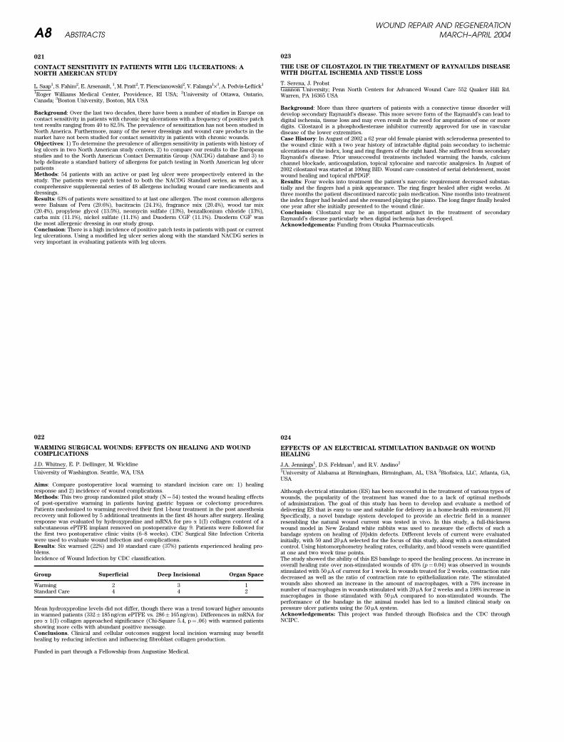
Aims: Compare postoperative local warming to standard incision care on: 1) healingresponse and 2) incidence of wound complications.Methods: This two group randomized pilot study (N¼ 54) tested the wound healing effectsof post-operative warming in patients having gastric bypass or colectomy procedures.Patients randomized to warming received their first 1-hour treatment in the post anesthesiarecovery unit followed by 5 additional treatments in the first 48 hours after surgery. Healingresponse was evaluated by hydroxyproline and mRNA for pro a 1(I) collagen content of asubcutaneous ePTFE implant removed on postoperative day 9. Patients were followed forthe first two postoperative clinic visits (6–8 weeks). CDC Surgical Site Infection Criteriawere used to evaluate wound infection and complications.Results: Six warmed (22%) and 10 standard care (37%) patients experienced healing pro-blems.Incidence of Wound Infection by CDC classification.
Group Superficial Deep Incisional Organ Space
Warming 2 3 1Standard Care 4 4 2
Mean hydroxyproline levels did not differ, though there was a trend toward higher amountsin warmed patients (332� 185 ng/cm ePTFE vs. 286� 165 ng/cm). Differences in mRNA forpro a 1(I) collagen approached significance (Chi-Square 5.4, p¼ .06) with warmed patientsshowing more cells with abundant positive message.Conclusions. Clinical and cellular outcomes suggest local incision warming may benefithealing by reducing infection and influencing fibroblast collagen production.
Funded in part through a Fellowship from Augustine Medical.
023
THE USE OF CILOSTAZOL IN THE TREATMENT OF RAYNAULDS DISEASEWITH DIGITAL ISCHEMIA AND TISSUE LOSS
T. Serena, J. ProbstGannon University; Penn North Centers for Advanced Wound Care 552 Quaker Hill Rd.Warren, PA 16365 USA
Background: More than three quarters of patients with a connective tissue disorder willdevelop secondary Raynauld’s disease. This more severe form of the Raynauld’s can lead todigital ischemia, tissue loss and may even result in the need for amputation of one or moredigits. Cilostazol is a phosphodiesterase inhibitor currently approved for use in vasculardisease of the lower extremities.Case History: In August of 2002 a 62 year old female pianist with scleroderma presented tothe wound clinic with a two year history of intractable digital pain secondary to ischemiculcerations of the index, long and ring fingers of the right hand. She suffered from secondaryRaynauld’s disease. Prior unsuccessful treatments included warming the hands, calciumchannel blockade, anticoagulation, topical xylocaine and narcotic analgesics. In August of2002 cilostazol was started at 100mg BID. Wound care consisted of serial debridement, moistwound healing and topical rhPDGF.Results: Four weeks into treatment the patient’s narcotic requirement decreased substan-tially and the fingers had a pink appearance. The ring finger healed after eight weeks. Atthree months the patient discontinued narcotic pain medication. Nine months into treatmentthe index finger had healed and she resumed playing the piano. The long finger finally healedone year after she initially presented to the wound clinic.Conclusion: Cilostazol may be an important adjunct in the treatment of secondaryRaynauld’s disease particularly when digital ischemia has developed.Acknowledgements: Funding from Otsuka Pharmaceuticals.
024
EFFECTS OF AN ELECTRICAL STIMULATION BANDAGE ON WOUNDHEALING
J.A. Jennings1, D.S. Feldman1, and R.V. Andino2
1University of Alabama at Birmingham, Birmingham, AL, USA 2Biofisica, LLC, Atlanta, GA,USA
Although electrical stimulation (ES) has been successful in the treatment of various types ofwounds, the popularity of the treatment has waned due to a lack of optimal methodsof administration. The goal of this study has been to develop and evaluate a method ofdelivering ES that is easy to use and suitable for delivery in a home-health environment.[0]Specifically, a novel bandage system developed to provide an electric field in a mannerresembling the natural wound current was tested in vivo. In this study, a full-thicknesswound model in New Zealand white rabbits was used to measure the effects of such abandage system on healing of [0]skin defects. Different levels of current were evaluatedinitially, with 50 and 20 mA selected for the focus of this study, along with a non-stimulatedcontrol. Using histomorphometry healing rates, cellularity, and blood vessels were quantifiedat one and two week time points.The study showed the ability of this ES bandage to speed the healing process. An increase inoverall healing rate over non-stimulated wounds of 45% (p¼ 0.04) was observed in woundsstimulated with 50mA of current for 1 week. In wounds treated for 2 weeks, contraction ratedecreased as well as the ratio of contraction rate to epithelialization rate. The stimulatedwounds also showed an increase in the amount of macrophages, with a 79% increase innumber of macrophages in wounds stimulated with 20mA for 2 weeks and a 198% increase inmacrophages in those stimulated with 50 mA compared to non-stimulated wounds. Theperformance of the bandage in the animal model has led to a limited clinical study onpressure ulcer patients using the 50 mA system.Acknowledgements: This project was funded through Biofisica and the CDC throughNCIPC.
WOUND REPAIR AND REGENERATIONA8 ABSTRACTS MARCH–APRIL 2004
GROWTH FACTORS
025
MODULATION OF WOUND REPAIR IN THE OBESE DIABETIC MOUSE: AROLE FOR VEGF GENE TRANSFER
SB Schwartz, B McCampbell, R Garone, Z Vodslon, E Tokarcsik, J Coico, L Staiano-Coico
Identifying molecular loci of impaired cutaneous healing in diabetes with an eye towardsdeveloping targeted therapy to ameliorate dysrepair continues to evolve as a promising areaof study. By using an excisional wound model produced on the dorsum of female diabeticC57BL/KsJ dbþ /dbþ mice as well as their normal (WT) & heterozygous (HZ) littermates, westudied the effects of peri-wound intradermal injection of adeno-associated viral vector(AAV) expressing the 165-amino acid isoform of human vascular endothelial growth factor(VEGF) on the following: kinetics of re-epithelialization, neoangiogenesis and granulationtissue formation, matrix remodelling, collagen deposition, and maturation. One sq. in. fullthickness excisional wound was created in the mid-upper back, rendering half of the woundas either right or left paravertebral. Animals were randomized to receive 1 of 3 treatments viaintradermal injection: 1)VEGF (AAV) vector; 2)Adnull vector; 3)PBS. Postoperatively,wounds were examined & photographed on Days 3, 7, 10, 14, 21 & 28. Also, tissue washarvested for histology & immunohistochemistry (PECAM), and snap frozen for protein &RNA analysis. A scoring system was used to grade re-epithelialization, granulation tissuethickness, matrix density, inflammation, vascular density, epithelial maturity. AAV-VEGFexerted minimal effect on repair in WT and HZ mice. However, pronounced neovasculariza-tion, thickened granulation tissue & increased matrix deposition was noted after VEGFtreatment in the db/db mice compared to those that received PBS or adnull vector at alltimepoints. While the induction of angiogenesis in VEGF treated db/db mice lagged behindthe unimpaired mice by 5–7 days, a global improvement in wound healng was observed.
R Crystal, Dir Inst Genetic Medicine, Weill Med College-Cornell Univ
027
FIBRIN SEALANT COMBINED WITH FIBROBLASTS AND PDGF ENHANCEWOUND HEALING IN EXCISIONAL WOUNDS
J. E. Mogford1, B. Tawil2 and T. A. Mustoe1
1Feinberg School of Medicine, Northwestern University, Chicago, IL USA 2Baxter BioScience,BioSurgery Westlake Village, CA USA
A number of strategies have been explored for the treatment of cutaneous defects such asprotein growth factor or gene therapy approaches. Alternative approaches include dermal ordermal-epidermal skin substitutes or clot substitutes such as fibrin sealants (FS’s). Here wetest the fibrinogen-thrombin formulation of fibrin sealant combined with fibroblasts andPDGF in wound healing models. Four formulations varying in fibrinogen and thrombinconcentration were applied to full-thickness biopsy wounds in the rabbit ear cutaneouswound-healing model with or without cultured rabbit dermal fibroblasts (RDFs; 3· 105
cells/wound) embedded in the fibrinogen component. At post-wounding day 7, there wasno difference in the diluted vs. non-diluted formulations for either the promotion of granula-tion tissue coverage of the open wounds or total granulation tissue area when tested withoutembedded cells. Including the RDFs, the highest degree of wound coverage by granulationtissue was observed in the combined dilution formulation (17.3 mg/ml fibrinogen, 167 U/mlthrombin; n¼ 10) that was 167% (p < 0.05) of the non-diluted FS containing cells (50 mg/mlfibrinogen, 250 U/ml thrombin; n¼ 10). Inclusion of fibroblasts increased granulation tissuearea within the wounds vs. FS alone (p < 0.05) for each diluted formulation although nodifferences in this parameter was observed within each group (FS alone or with embeddedcells). However, addition of the vulnerary growth factor PDGF-B (3mg; n¼ 4) with theembedded RDFs in the combined dilution formulation increased granulation tissue areaover 2-fold (p < 0.01). Additionally, the presence of the RDFs promoted incorporation of thegranulation tissue with and epithelial migration over the FS suggesting an active interactionbetween cells delivered to the wound by FS and the host repair cells. The findings suggestthe progress of cutaneous defect repair can be enhanced by ex vivo cell delivery in FibrinSealant that is adjusted to balance hemostasis with promotion of wound healing processes.
029
DIFFERENTIAL ANGIOGENIC RESPONSES IN ORAL MUCOSAL ANDCUTANEOUS WOUNDS
A. M. Szpaderska, C. G. Walsh, M. J. Steinberg, L. A. DiPietro
Loyola University Medical Center, Maywood, IL
Wound healing in adults is a multi-step process that concludes with scar formation. Acommon clinical observation is that intra-oral wounds demonstrate privileged healingwhen compared to wounds at extra-oral sites. Oral mucosa heals faster and with less scarformation than skin does. We have shown that oral mucosal wounds exhibit decreasedinflammation and faster reepithelialization than skin wounds. The present study comparesangiogenesis in oral mucosal and skin wounds, and examines in vitro VEGF production byoral and epidermal keratinocytes. Two 1 mm diameter excisional full-thickness wounds wereplaced on lateral sides of the tongue and on the dorsal skin of anesthetized BALB/C mice. At0, 2, 4, 12, 24, 36 hours and 2, 3, 5, and 7 days after injury, wound tissue was harvested foranalysis. VEGF and FGF-2 levels were measured by ELISA, and normalized for proteincontent. The level of VEGF in oral mucosal wounds was less than that of cutaneous woundsat all time points. At 24 hours, VEGF levels peaked in oral wounds, yet were much lower thancomparable skin wounds (106.6þ/- 19 vs. 194.0þ/- 23.6 pg/mg protein, p < 0.001). At 36 hours,wound VEGF levels had declined in oral mucosa, while reaching peak levels in skin (82.7þ/-8.9 vs. 374.2þ/- 27 pg/mg protein, p < 0.001). No differences in FGF-2 levels were seen. Therelative increase in tissue vascularization during wound repair, analyzed by CD 31 immuno-staining, was lower in oral mucosal wounds than in skin (3.4þ/- 0.2% vs. 11.5þ/- 0.5%,p < 0.001). For in vitro experiments, normal human epidermal and oral mucosal keratino-cytes were grown for 12 h under hpoxic conditions. In response to hypoxia, VEGF proteinproduction, determined by ELISA, increased 2.3-fold in oral keratinocytes vs. 3.3-fold inepidermal keratinocytes. RT-PCR analysis revealed that relative VEGF mRNA expressionincreased 2-fold in oral vs. 4-fold in epidermal keratinocytes. This study demonstrates thatthe differential repair of skin and oral mucosal wounds involves distinct patterns of angio-genesis, and that keratinocytes exhibit intrinsic site-specific differences in VEGF production.
Supported by NIH GM-55238 (LAD) and NIH T32AI07508(AMS).
030
A RABBIT EAR EXCESSIVE SCARRING MODEL USING GROWTH FACTORSTIMULATORS
D-L. Gu, D.W. Kang, D. Looper, D. Maneval, M. Zepeda
Department of Pharmacology, Canji, Inc., San Diego, CA 92121
Research on abnormal scarring, such as excessive scars, has been hampered by the lack ofrelevant animal models. Recent studies suggest that TGF-b1, PDGF-BB and FGF-2 arestimulators of fibrosis or scarring. We developed a rabbit ear cutaneous excessive scarringmodel using the growth factor stimulators TGF- b1 (0.25, 0.5 or 1mg per wound), PDGF-BB(1, 2 or 3mg per wound) and FGF-2 (0.5, 1, 3 or 5mg per wound). Three 8-mm diameterwounds were created on each ear in NZW rabbits (n¼ 6 wounds). The test growth factorswere injected intradermally immediately after wounding. Controls consisted of PBS, BSAand wounding only without injections. Scar thickness of wounds was measured with amicrometer at days 14 and continued twice a week thereafter until sacrifice at day 28.Results showed that the greatest measurement of elevated scars was observed at day 14.Significant differences of scar thickness were observed in TGF- b1 at a dose of 0.25mg,PDGF-BB at a dose of 3mg, and FGF-2 at a dose of 3 mg per wound groups when compared toPBS and wounding only groups (p < 0.05). Interestingly, BSA, a protein stabilizer for TGF- b1and FGF-2 proteins, also significantly elevated scar thickness at day 14 over PBS alone(p < 0.05). Trichrome stained tissue sections on day 28 showed an increase in cellularity andthicker epithelium in all doses of TGF- b1, PDGF-BB and FGF-2 protein treated groups whencompared to wounding only, PBS or BSA treatments. In general, FGF-2 treated woundsshowed more vascularity than TGF- b1 and PDGF-BB treated wounds. By contrast, PDGF-BBtreated wounds showed richer collagen deposition than TGF- b1 and FGF-2 treated wounds.Results from these studies demonstrated that TGF- b1, PDGF-BB and FGF-2 contributed tothe scar formation in the rabbit ear scar model. BSA may contribute in stimulating scarformation in the early stage of wound healing in this model. Enhancing elevated scarformation in the rabbit ear model will be useful in evaluating anti-scarring agents forexcessive scars such as hypertrophic scars and keloids.
WOUND REPAIR AND REGENERATIONVOL. 12, NO. 2 ABSTRACTS A9
031
IGF-I STIMULATES VEGF PRODUCTION IN ENDOTHELIAL CELLS BYINHIBITING POLY(ADP-RIBOSE)-POLYMERASE
S. Beckert, H. Scheuenstuhl, F. Farrahi, R. Aslam, TK. Hunt, Z. Hussain.
Department of Surgery, University of California, San Francisco
Introduction: Vascular Endothelial Growth Factor (VEGF) mediated angiogenesis plays akey role in wound healing. Insulin-like Growth Factor I (IGF-I) has been reported to beangiogenic. However, the mechanism is not known. Recently, a link between transcriptionalactivity and inhibition of poly(ADP-Ribose)polymerase (PARP) has been reported. We inves-tigated whether IGF-I increases VEGF expression and whether this effect is regulated by theinhibition of PARP.Material and methods: Subconfluent monolayers of human umbilical vein cells werecultured and serum starved. Cultures were treated with Long-R3-IGF-I for 20 h. VEGF inthe supernatant was measured by ELISA and lactate by a lactate analyser. PARP activity wasassessed by measuring the incorporation of 14C-radiolabeled NADþ. All experiments wereperformed in triplicate. Results are given as percent change compared to control�SD;p < 0.05 calculated by Student‘s t-test was considered significant.Results: IGF-I increased both VEGF (20� 10%, 50� 16% (p¼ 0.01) and 79� 13%(p¼ 0.0003)) and lactate production (12� 11%, 20� 12% and 48� 16% (p¼ 0.01)) in a dosedependent manner (25, 50 and 100 ng/ml). Blocking glucose utilization by 2-desoxyglucose,decreased lactate by 70� 11% (p¼ 0.0001), but not VEGF expression. Inhibitors of MAP-kinase (PD 98059) and Proteinkinase C (Staurosporine) reduced the IGF-I effect on VEGFexpression by 40� 6% (p¼ 0.0003) and 30� 7% (p¼ 0.01). 3-Aminobenzamide and nicotin-amide alone, inhibitors of PARP, stimulated VEGF production by 66� 5% (p¼ 0.0003) and32� 8% (p¼ 0.002), respectively. IGF-I inhibited PARP by 44� 3% (p¼ 0.01).Conclusion: IGF-I enhances VEGF protein expression in endothelial cells. This is mediatedthrough signal transduction and by inhibition of PARP.
Supported by NIGMS grant GM 27345
YOUNG INVESTIGATOR AWARD
032
TRANSCRIPTIONAL PROFILING OF WOUND HEALING PROGRESSION
A. Murad1 and M.R. Sierra-Honigmann1
1Cedars-Sinai Medical Center and University of Southern California Keck School of Medicine,Los Angeles, CA
The complexity of cellular and molecular events that occur during wound healing is given bythe rapid and continuous changes of matrix structure, cell population, local cytokine com-position, nutrient and oxygen availabilities and overall metabolic activity. Angiogenesis is anessential event in the repair of injured tissue. Transcriptional events leading to woundneovascularization begin immediately after the injury has occurred and continue throughoutthe process until scar remodeling becomes quiescent. This study investigated progression ofwound angiogenesis by quantitatively measuring transcripts of multiple genes that partici-pate in wound angiogenesis. The studies were done using a murine model of bilateral 3 mmexcision wounds. Wound tissues were collected from 6 hours up to 10 days post-wounding.Each time point consisted of 8 wounds pooled from four different mice. After RNA purifica-tion, the samples were analyzed by quantitative real-time RT-PCR using specific TaqManprobe/primer sets for selected transcripts that included the following groups: a) angiogenicfactors; b) anti-angiogenic factors; c) receptors; d) cytokines and enzymes; e) matrix pro-teins; f) signaling molecules and transcription factors; g) apoptosis and proliferation mar-kers; and h) housekeeping genes. A total of 58 genes were profiled including fourhousekeeping genes. The transcripts revealed several patterns of expression. Some genessuch as angiopoietin 1(Ang1), e-selecting, placental growth factor (PlGF), MMP-9 andthrombospondin 1(TSP1) had a clear biphasic pattern where early peaks were detectedwithin the first 24 hours after wounding and were followed by dowregulation and a secondincrease in transcription after several days. Other transcripts appeared early and had asustained expression and they include fibroblast growth factor two (FGF2), FGF7, hypoxiainducile factor alpha (HIF-1a) and STAT3. Examples of late transcripts included HOXD3 andalpha smooth muscle actin. Results derived from this study largely confirm previous studiesprofiling a small number of individual genes such as VEGF, TSP1 and 2 and others. However,the present study integrates complete transcriptional profiles of large number of genesknown to have a critical role in healing progression with a particular emphasis on revascu-larization. A more detailed model of event sequences that lead to tissue reperfusion afterinjury is derived from the results.
Funded by a Cedars-Sinai grant from the Skirball-Kenis Center for Plastic and ReconstructiveSurgery and RO1GM066292 to MRSH.
033
REGULATION OF TGF-b RESPONSES BY A NOVEL ACCESSORY TGF-bRECEPTOR IN HUMAN KERATINOCYTES
A. Marcoux, K. Finnson and A. Philip
Division of Plastic Surgery, Department of Surgery, McGill University, Montreal, Quebec,CanadaMake sure to underline the presenting author’s name
The potential of TGF-b isoforms to regulate wound healing and scarring is well documentedin animal models. Since TGF-b action is likely to be regulated at the level of its cell surfacereceptors, we analyzed TGF-b receptor profiles and regulation of TGF-b signaling in humankeratinocytes. We identified a novel cell surface TGF-b1 binding protein of 150 kDa (r150) onhuman keratinocytes that interacts with the TGF-b signaling receptors. Further characteriza-tion of r150 demonstrated that it is a GPI-anchored protein, that it can be released from thecell surface by an endogenous phospholipase C, and that the released form can bind to TGF-b1. Recent cloning of r150 revealed it to be a novel protein of 1428 amino acids. Our objectivewas to determine the functional significance of r150 in regulating TGF-b responses inkeratinocytes.Affinity labeling of keratinocytes overexpressing r150 cDNA and immunoprecipitation stud-ies using anti-r150 antibodies show that the cloned cDNA represents r150. Importantly, overexpression of r150 results in inhibition of TGF-b1-induced Smad 2 and Smad 3 phosphoryla-tion, gene transcriptional activity, and keratinocyte migration. In contrast, loss of r150function using antisense morpholino oligos of r150 leads to enhanced Smad 2 and Smad 3phosphorylation, gene transcriptional activity and proliferation of keratinocytes. In sum-mary, our results demonstrate that r150 is a potent inhibitor of TGF-b signaling in keratino-cytes, and that it may have potential therapeutic value in modulating TGF-b action in humandiseases where TGF-b plays a pathophysiological role. As such, r150 may be of use inreducing hypertrophic scarring, and an r150 antagonist may promote wound healing.
This work was supported by a ‘Canadian Institutes of Health Research’ grant and a ‘Fonds dela Recherche en Sante du Quebec’ award to AP
034
LIGHT-EMITTING DIODE THERAPY INCREASES VEGF AND NOPRODUCTION IN RAW 264.7 CELLS
M. Leong, J. Zhao, L.J. Gould
The University of Texas Medical Branch, Galveston, TX USA
Introduction: Low-energy laser photostimuation has been reported to affect the prolifera-tive phase of wound healing and stimulate macrophages. Light-emitting diodes (LED) are anefficient, inexpensive source of low energy photons. We propose that LED therapy willstimulate macrophage production of VEGF and NO.Methods: Serum-starved RAW 264.7 cells (5 · 105cells/ml) were treated with or without LPS(10 ng/ml) and IFN-gamma (100 Units/ml) for 24 hours. Cells were divided into groups:1) control – no LED, 2) 670 nm, 3) 730 nm, 4) 880 nm, and 5) combination (880 nm/730 nm/670 nm) at 4 J/cm2 per wavelength. Twenty-four hours post-LED, conditioned media wasanalyzed for VEGF (ELISA) and nitrites (Greiss assay). Proliferation was assessed usingBrDU incorporation. Data were analyzed using one-way ANOVA with Tukey post-test.Results: In serum-starved cells, proliferation was unchanged by LED. VEGF and nitritelevels were increased in groups 4 (VEGF¼ 1057.1þ/- 100.3 pg/ml; nitrites¼ 11.13þ/-0.79 uM) and 5 (VEGF¼ 1148.9þ/- 88.5 pg/ml; nitrites¼ 10.72þ/- 1.03 uM) compared to con-trol (VEGF¼ 812.2þ/- 94.8 pg/ml; nitrites¼ 7.32þ/- 1.92 uM). LPS/IFN decreased proliferation25 percent at all wavelengths compared to controlþLPS/IFN (p < 0.01). Nitrite and VEGFlevels were markedly elevated in all five LPS/IFN-treated groups with no appreciable inter-group differences.Conclusions: LPS/IFN stimulation resulted in maximally elevated levels of nitrites andVEGF, possibly masking the effects of LED. Serum-starved RAW 264.7 cells responded toLED treatment at 880 nm and combined wavelengths with increased VEGF and NO produc-tion. These results suggest that LED treatment at 880 nm and 880 nm/730 nm/670 nm mayupregulate the production of pro-angiogenic factors by macrophages in the wound bed.
WOUND REPAIR AND REGENERATIONA10 ABSTRACTS MARCH–APRIL 2004
035
LEPTIN PLAYS A KEY ROLE IN THE MODULATION OF WOUNDANGIOGENESIS
A.K. Nath1, A. Murad2, S.T. Cha2, and M.R. Sierra-Honigmann2
1Yale University School of Medicine, New Haven, CT 2Cedars-Sinai Medical Center andUniversity of Southern California Keck School of Medicine, Los Angeles, CA
Leptin is a pleiotropic cytokine constitutively expressed in adipose tissue and upregulated byhypoxia in sites of tissue injury and in the placenta. Leptin has been demonstrated to play arole in normal and pathological wound healing and it is gene is acutely expressed inexperimental wounds. Angiogenesis is among the most salient and well-documented bio-logical actions of leptin. Therefore it was hypothesized that leptin may play in the modulationof wound neovascularization. Using a murine model of experimental wounds the modulationof wound angiogenesis was investigated by treatment of the wounds to either recombinantleptin or a neutralizing anti-leptin antibody. The parameters measured were: a) wound blood
flow using laser Doppler imaging; b) vessel density by counting the number of CD-31 positivevessels in wound sections; c) angiogenic transcriptional profiling using quantitative real-time RT-PCR; and d) validation of transcriptional profiling by immunohistochemistry andimmunoblotting. The treatment of wounds with leptin had a marked effect in wound bloodflow that was most significant at 24hours post-wounding with average increase of 55% withrespect to the controls. The effect diminished significantly by 72 hours (24%) and becamenegligible thereafter. Leptin also accelerate wound neovascularization by 24 hours where thenumber of small caliber vessels averaged 30/mm2 in leptin treated vessels and 13/mm2 in thecontrols (n¼ 10; P < 0.005). However, the number of vessels measured by 72 hours notsignificantly different between controls and leptin-treated wounds. In addition transcrip-tional profiling reveled changes in gene expression of molecules related to angiogenesissuch as VEGF, PlGF, Tie-1, Tie-2, Flt-1, Flk-1, FGF-2 and endothelial markers like CD-31,endoglin and eNOS. Overall, time course analysis throughout the healing process showedthat leptin treatment induced an early shift in the expression of many of the analyzedtranscripts. The most salient changes could be observed as early as 6 hours post-woundingand included: TSP-1 (þ8.7); FLK-1(þ9); Ang-2 (þ18); CD31(þ5.3); endoglin (þ7.6); eNOS(þ4.2); FGF-2 (þ4.3); etc. Leptin mRNA was also significantly increased (þ10) as well as thelong-form of its receptor ObR-b (þ9). Conversely, when wounds were exposed to anti-leptinantibodies, healing progression was impaired and changes in the transcriptional profileshowed diminished expression of several genes related to wound neovascularization. Someexamples by 24 hours: Endoglin(-2.5); Flk-1(-2.8); eNOS (-5.1); Ang-2 (-5.1); CD31(-3); VEGF-A (-3.6). In conclusion, leptin had and early and significant effect in modulating woundneovascularization. The effect of treatment of wounds with exogenous leptin did not elicithypervascularization of the wound and appeared to only accelerate what appeared to be aself-limited process of wound angiogenesis.
Funded by a Cedars-Sinai grant from the Skirball-Kenis Center for Plastic and ReconstructiveSurgery.
036
OVEREXPRESSION OF SMAD7 IN KERATINOCYTES ACCELERATESCUTANEOUS WOUND HEALING
G. Han, A. G. Li, P. Owens, X-J Wang
Oregon Health & Science University, Portland, OR USA
Transforming growth factor b (TGFb) has both positive and negative effects on cutaneouswound healing. Smad7 acts as a major downstream antagonist of TGFb signaling in keratino-cytes and its role in wound healing has not been defined. We have established a Smad7transgenic mouse line using a keratin 5 (K5) promoter (K5.Smad7) which expresses Smad7transgene at a mild level (*2 fold of the endogenous Smad7 in the skin). These mice did nothave overt skin defects as shown from our previous Smad7 transgenic mice expressing muchhigher levels of the Smad7 transgene (EMBO J 2002, 21:2580–90). K5.Smad7 mice from theabove low expressor line and non-transgenic littermates were subject to 6-mm full-thicknessexcisional wounding. K5.Smad7 mice exhibited early scab rejection, reduced inflammation,and accelerated re-epithelialization as compared to non-transgenic mice. To further deter-mine the stage-specific effects of Smad7 on wound healing, we generated a transgenic modelin which Smad7 transgene expression can be induced in the epidermis and hair follicles(gene-switch-Smad7) by topically RU486 application. Smad7 induction from day 3 to day 7after excisional wounding reduced inflammatory responses through suppressing expressionof a variety of inflammatory cytokines/chemokines in gene-switch-Smad7 mice as comparedto control mice. Meanwhile, overexpression of Smad7 exhibited accelerated re-epithelializa-tion, which correlated with increased expression of metalloproteinases and elevated Erk(extracellular signal-regulated kinases) signaling in the leading epidermal edges in gene-switch-Smad7 wounds compared to control wounds. Smad7 induction from day 7 to day 20after excisional wounding reduced dermal fibrotic response and angiogenesis in the dermis,resulting in a better tissue repair. We conclude that the effectsof Smad7 on wound healingare likely due to blocking the negative effects of TGFb on cutaneous wound healing.
037
FIBROBLAST GROWTH FACTOR – BINDING PROTEIN CDNA ANDTRUNCATED VARIANTS ARE ACTIVE IN DIABETIC WOUND HEALING
Jayasri Dasgupta*, Michaela Bittner{, Andreas Goppelt{, Sabine Werner**, Hans-Dietmar
Beer**, and Jeffrey M. Davidson*.*Dept. of Pathology, Vanderbilt Univ. and VA Med Ctr., Nashville, TN; **ETH, Zurich;{SWITCH Biotech AG, D-82152 Neuried, Germany ([email protected])
Fibroblast growth factor – binding protein (FGF-BP) is a secreted protein that appears tofunction as a low affinity heparin –binding protein. FGF-BP binds to FGF-1 and -2 in a non-covalent, reversible manner to mobilize and solubilize these growth factors from theirstorage sites in the extracellular matrix. FGF-BP is involved in both developmental andadult tissue homeostasis as well as in angiogenesis and tumorogenesis involving FGF-1/2.FGF-BP is overexpressed in several tumor types: head and neck, skin, cervical, and lungcancer, squamous cell carcinoma, and colon and breast adenocarcinoma. To establish theeffect of FGF-BP on wound healing, several forms of FGF-BP cDNA were administered byparticle-mediated gene transfer into various animal wound models using the gene gun (Bio-Rad). In a rat incisional wound model, gene gun cDNA delivery of full length FGF-BP at thetime of surgery produced a 117% increase of wound strength in diabetic rats at 10d, althoughthe relative increase did not reach statistical significance (P < 0.08). Two truncated variantsof FGF-BP (pFGFbp10 and 17) were also administered in the rat incisional wound model bygene gun technique. pFGFbp17 increased the wound strength in diabetic rat 129%(p < 0.03),and the relative increase reach statistical significance (P < 0.008). In the rabbit ear ulcermodel, particle-mediated transduction of full length FGF-BP increased collagen content by195% and wound closure rate 38% at 10d post-surgery. These findings show that FGF-BP geneoverexpression has a greater relative effect on wound healing in the diabetic rat model. ThecDNA also had a significant effect in a rabbit excisional wound model that depended ongranulation tissue formation. Truncated forms of the molecule may have higher therapeuticpotency. FGF-BP has an important role in FGF-1/2 mobilization and macrophage functions,and FGF-BP gene therapy for wound healing can improve the process by stimulatingangiogenesis, epithelization and collagen synthesis in target tissue.
Supported by SWITCH Biotech and Veterans Affairs.
038
ADENOVIRAL OVEREXPRESSION OF MCP-2 INDUCES MACROPHAGESAND IMPROVES WOUND HEALING IN DIABETIC RATS
Y. Shi*, J. Liu*, M. Bittner#, A. Goppelt#, J.M. Davidson*,
*Vanderbilt Univ, Nashville TN; #Switch-Biotech, Neuried, Germany
The recruitment of leukocyte subsets is mainly driven by chemokines. Monocyte chemotacticprotein-2 (MCP-2) belongs to the CC subfamily of chemokines, and it is especially achemoattractant for monocytes. Of all CC subfamily members only MCP-2 interacts withmultiple receptors, and its N-terminal truncated isoform is a natural chemokine inhibitor.Gene expression profiling in excisional wounds showed MCP-2 was down-regulated 40% inwild type mice and 69.2% in diabetic mice at 24 h after wounding. To determine the biologicalresponse to MCP-2 during wound healing, we constructed an adenoviral vector (Ad-MCP-2).Ad-MCP-2 infected cell conditioned medium contained 460 ng/ml secreted MCP-2 (ELISA)and had chemotactic activity similar to recombinant full-length MCP-2 but not to MCP-2 N-terminal 8–13aa peptides in a THP-1cell migration assay. In an incisional wound model inSTZ induced diabetic rats, the breaking strength of Ad-MCP-2 infected skin woundsincreased by 43% (108 PFU, p < 0.05) and 30% (107 PFU, p < 0.05) at 7d after Ad-MCP-2injection compared with Ad-LacZ control. The wound closure strength of 107 PFUAd-MCP-2 infected wounds still increased by 21% (p < 0.05) at 10d after injection. In normalrats, the breaking strength of Ad-MCP-2 and Ad-Lacz infected wounds was not significantlydifferent. In rats, we injected either Ad-MCP-2 or Ad-LacZ (108 PFU) into each PVA sponge at3d after implantation. Histological analyses revealed numerous ED-1 positive macrophagesinfiltrating in the experimental granulation tissue at 7d after Ad-MCP-2 injection. Our dataprovide evidence that MCP-2 improved wound healing in diabetic rats, and the recruitment ofmacrophages into the wound granulation tissues was the one of the mechanisms.
Supported by Switch Biotech and the Department of Veterans Affairs
WOUND REPAIR AND REGENERATIONVOL. 12, NO. 2 ABSTRACTS A11
039
DETERMINATION OF BURN DEPTH USING NEAR INFRAREDSPECTROSCOPY
K Cross1, L Leonardi2, J Fish1, M Sowa2 M Gomez1, J Payette2, M Hastings2
1University of Toronto, Toronto, ON; 2NRC, Winnipeg, MB, Canada
Introduction: Burn depth, based on the hemodynamic alterations that occur following athermal insult, can be assessed in a rapid, non-invasive, and nondestructive fashion usingnear infrared (NIR) spectroscopy. NIR has the capability to determine the differencebetween superficial and full thickness burn injuries.Methods: Sixteen burn patients admitted to an adult regional burn center were studied andevaluated with the NIR point and imaging devices. Non-burned skin adjacent to the burn sitewas used as the control. NIR measurements were compared between superficial (8 wounds),full thickness (8 wounds) burn wounds and control sites.Results: NIR was able to easily detect an increase in oxyhemoglobin (68.3%, p < 0.05),oxygen saturation (4.8%, p < 0.05%) and total hemoglobin (91.3%, p < 0.05) which typicallyoccurs with superficial burn injuries. Full thickness injuries experienced a substantial dropin oxyhemoglobin (88.8%, p < 0.05), oxygen saturation (79.1%, p < 0.05) and total hemoglobin(77.5%, p < 0.05) in comparison to control sites.Conclusions: These results confirm that NIR spectroscopy can successfully distinguishbetween superficial and full thickness burn injuries. The second phase of this study willinvolve determining the depth of indeterminant burn wounds and this preliminary data willalso be presented.Acknowledgement: National Research Council of Canada
040
A CHRONIC PRESSURE SORE NUDE MOUSE MODEL
E. Wassermann1, K. Gstaltner2, H. Redl1
1LBI for Experimental and Clinical Traumatology, Vienna, Austria 2Weisser Hof, Rehabili-tation Center of AUVA, Klosterneuburg, Austria
The epidemiology of pressure sores is not clearly understood yet. Tissue layers involved inthe pressure sores include skin, subcutaneous fat, deep fascia, muscle, and bone. Widelyused experimental models lead to the pressure sores in the skin and the underlying fattytissue only. The aim of the present study was to develop a clinically relevant deep pressuresore model in nude mice. METHODS: Six weeks old Balb/c nude mice weighing 25–30 g wereused. After introduction of anesthesia a steel disk (Ø 5 mm, 0.5 mm thickness) was implantedunder the musculus gluteus max.. The wound was closed with 4/0 sutures. The full recoverywas allowed for 10 days. Animals with an insufficient healing were excluded. The pressurewas induced using a Neodymium magnet (1.29 Tesla) in conjunction with the disk inun-anesthetized mouse periodically. A two hours compression was followed by one hourrecovery for 4, 6 or 8 cycles, mimicking clinical setting. The controls underwent the sameprocedure, including the disk implantation but without placement of the magnet. RESULTS:The pressure sore depth was found to be cycle dependent – with the highest degree after 8cycles. The histologic evaluations revealed signs of necrosis in the skin and subcutaneous fatafter 4 and in the muscle only after 8 cycle of compression. PMN infiltration into the skin,subcutaneous fat, the deep fascia, and muscle was found in all groups. CONCLUSION: Thismodel with clinically relevant magnitude and duration of pressure application leads to aninjury in all tissue layers similar to the patient setting. This nude mouse model can be used toinvestigate the mechanism of pressure sores development and to study new therapeuticapproaches (e.g. human cells) without the need of immunosuppression.Acknowledgements: Lorenz Boehler Fond, 7/2002
041
AGED RATS DISPLAY INCREASED ISCHEMIA-REPERFUSION SKININJURY IN THE IN VIVO MAGNET MODEL
David S. Rosenberg ([email protected]), Leonard Lu, Anandev Gurjala,
Thomas A. MustoeWound Healing Research Laboratory, Northwestern University, Chicago, IL USA
Introduction: Ischemia-reperfusion (IR) injury is commonly associated with numerouspathologies including pressure sores, venous stasis ulcers, and lower extremity diabeticulcers. The purpose of this study is to further investigate the relationship between age andischemia-reperfusion skin injury in a rat model utilizing magnets for the purpose of injurycreation.Methods: Magnets were designed for subcutaneous placement and calibrated such that asecond magnet placed externally over them would cause compression that exceeds capillaryperfusion pressure (ischemia). Removing the external magnet results in reperfusion of theskin. After placing subcutaneous magnets in aged and young Fisher 344 rats, repeated cyclesof external magnet placement and removal were performed.Results: Visual analysis of the skin revealed statistically significant greater areas of injury inthe aged rats relative to their younger counterparts (37.4� 13.3% vs. 24.1� 14.8%, P < .02)Conclusions: Aged rats demonstrate an increased degree of injury relative to their youngercounterparts in response to ischemia-reperfusion injury. Future studies will attempt todelineate differences in the markers of IR injury (such as myeloperoxidase and vitamin Elevels) in aged versus young rats, giving insight to the mechanisms responsible for theimpaired wound healing seen in the elderly.
Funding source: NIH grant
GENE THERAPY
042
FGF-BP IMPROVES WOUND HEALING THROUGH THE INDUCTION OFMORE COLLAGEN IN RATS
Y. Shi*, J. Liu*, M. Bittner#, A. Goppelt#, J.M. Davidson*,
*Vanderbilt Univ, Nashville TN; #Switch-Biotech, Neuried, Germany
The fibroblast growth factors are important regulators during wound healing. To quenchtheir biological activities, secreted FGFs are tightly bound to heparan sulfate proteoglycansin the extracellular matrix (ECM). One of the approaches for releasing the active FGF fromECM involves the binding to an FGF binding protein (FGF-BP), which prevents FGF degra-dation and retains its activities. FGF-BP enhances the proliferation of fibroblasts throughFGF-1 and -2 and of endothelial cells through FGF-2. To detect the biological function ofFGF-BP in wound healing, we constructed an adenoviral vector containing murine FGF-BPcDNA (Ad-FGF-BP). Polyvinyl alcohol (PVA) sponges were implanted subcutaneously inrats. 107–108 PFU of either Ad-FGF-BP or Ad-LacZ (as local control) was injected into thesponges at d3 after implantation. At d4 after injection, Ad-FGF-BP infected sponges dis-played much better organization of granulation tissue with the presence of more macro-scopic hemorrhage than the local control. At d7 after injection, Ad-FGF-BP infectedexperimental granulation tissues in sponges showed more collagen deposition. The contentsof collagen, protein and DNA in Ad-FGF-BP infected sponges increased 24.0% (p < 0.05),11.3% (p < 0.05, d4) and 28.2% (p < 0.001, d7), respectively. Incisional wounding was per-formed in rats and 108 PFU Ad-FGF-BP or Ad-lacZ was intracutaneously injected along thewound margins. At 7d after injection, the tensile strength of Ad-FGF-BP infected woundsincreased 41.1% (p < 0.05). Our data indicate that overexpression of FGF-BP can improvewound healing, and the inductionof more collagen in granulation tissue may be one of themechanisms.
Supported by Switch Biotech and the Department of Veterans Affairs
WOUND REPAIR AND REGENERATIONA12 ABSTRACTS MARCH–APRIL 2004
043
Ik -B ADENOVIRAL GENE TRANSFER IMPROVES WOUND HEALING
Schreiber J, Efron P, Park J, Moldawer L, and Barbul ADep. Surgery, Sinai Hospital, Baltimore, and University of Florida College of Medicine,Gainsville, FL
Introduction: One of the earliest events in wound healing and scar formation is the inflam-matory phase. Deficient or excessive inflammatory responses result in aberrant healing.NFK-b is a transcription factor that regulates key genes responsible for multiple intra-nuclearinflammatory cytokines. When bound to IK-B, an inhibitory protein, this factor is in aninactive state. We studied the wound healing effects of irreversibly inhibiting the activationof NFK-b by continuously infusing adenoviral activated IK-B.Methods: Seventy-five male Sprague-Dawley rats underwent subcutaneous implantation ofpolyvinyl alcohol sponge-osmotic pump constructs. The pumps were filled with a solutioncontaining a genetically altered adenovirus containing the DNA for continuously activatedIK-B protein. 50 ml of viral transport media containing 5· 109 PFU of virus were delivered toeach individual sponges daily. Controls included pumps filled with normal saline in onegroup or filled with sham virus in another group. Sponges were harvested on days 1,3,5, and 7post-implantation. The sponges were analyzed for TNF-a and nitric oxide (NO) levels, asindex of inflammation, as well as for hydroxyproline (OHP) content at seven days, an indexof collagen deposition.Results: Treatment with the IK-B virus resulted in significantly higher levels of OHP afterseven days when compared to normal saline and sham virus treated sponges (654� 81 vs546� 109 vs 498� 123mg OHP/100 mg sponge, P < 0.05 by ANOVA). Wound fluid NO concen-tration was significantly lower in the IkB group after 5 days (38� 15 vs 55� 12 vs 71� 27 mM,P < 0.05 by chi-square). There were no differences in sponge TNF-a concentrations on days1,3, and 5.Conclusions: Treatment of wound sponge granulomas with IK-B increases the amount ofcollagen deposition after seven days. Reduction of inflammation by inhibiting NF-KB may bea possible mechanism of action, as reflected in decreased NO wound content.
044
ADENOVIRAL GENE TRANSFER OF IL-10 DIFFERENTIALLY REGULATESHYALURONIC ACID SYNTHASE 1: IMPLICATIONS FOR SCARLESS WOUNDHEALING
ED Kozin, SG Keswani, PW Zoltick, AD Gordon, AB Katz, M Herlyn and TM Crombleholme.
The Children’s Hospital of Philadelphia, Philadelphia, PA, 19104
Introduction. We have demonstrated that wounds treated with adenoviral overexpressionof Il-10 (AdIl-10) heal scarlessly, resembling fetal wound healing. Hyaluronic acid (HA) iselevated in fetal wounds. We hypothesized that IL-10 creates a permissive environment forscarless healing through the differential mediation of hyaluronic acid synthases (Has1–3).Methods. Adult C57 mice (n¼ 24) were pretreated with 5 · 108 PFU AdIL-10, AdLacz, orPBS. 48 hrs post-injection, 2-cm incisional wounds were created at the site of injection.Wounds harvested at 1, 3, and 5 days were analyzed for mRNA transcript levels of Has 1–3and GAPDH via RT-PCR and band densitometry. Data expressed as signal intensity ratiorelative to GAPDH� SEM.Results. The three Has transcripts were expressed after pre-treatment and wounding at day1, 3, and 5. At day 1, there is no significant difference in Has mRNA levels. At Day 3, Has1transcript levels are significantly decreased in AdIl-10 treated wounds vs. controls. (Il-100.28� 0.04; LacZ 0.53� 0.06; PBS 0.43� 0.43, p < 0.005) At Day 5, there was a significantincrease in Has1 transcript levels in AdIl-10 treated wounds compared to controls. (Il-100.82� 0.08; LacZ 0.41� 0.06; PBS 0.57� 0.03, p < 0.005) At Day 3 and 5, there was nosignificant difference in Has2 and Has3 levels in AdIl-10 treated wounds vs. controls.Conclusions. Adenoviral mediated overexpression of Il-10 results in the differential expres-sion of Has1 at day 3 and 5. Has1 encodes for high molecular weight HA, which is associatedwith inhibition of angiogenesis and leukocyte infiltrates as observed in the HA-rich environ-ment of fetal wounds. The effects of AdIl-10 may in part be due to the differential expressionof Has1 creating a permissive environment for fetal-like scarless wound repair.
045
ADENOVIRAL hTERT TRANSFECTION RESULTS IN ALTERED PHOSPHO-ERK ACTIVITY IN HUMAN DERMAL FIBROBLASTS
Leonard Lu ([email protected])1, David S. Rosenberg1, Jon E. Mogford2, Thomas
A. Mustoe1
1Wound Healing Research Laboratory, Northwestern University, Chicago, IL 60611 USA2Strategic Analysis, Arlington, VA 22201
Introduction: Telomeres are nucleoprotein structures at the ends of each chromosome. Dueto the inability of DNA polymerase to replicate the full length of the chromosome, up to50–200 base pairs of the telomere are lost during each successive round of cell division. Inadult human somatic cells, telomerase is not active resulting in progressive loss of telomerelength and entry into replicative senescence as observed in cell culture. hTERT is thecatalytic subunit of telomerase, an enzyme which maintains telomere length. Transfectionof human dermal fibroblasts (HDFs) by hTERT has been shown to reverse the senescentphenotype seen in aging HDFs in vitro. ERK (p44/42) is a MAP kinase which functions as acritical intermediary in the determination of cell growth and differentiation. Activation ofERK occurs through phosphorylation of threonine and tyrosine residues.Methods: In order to delineate some of the cellular mechanisms by which hTERT functions,we treated adenoviral hTERT (Ad-hTERT) transfected HDFs with TGFB1, and assayedphosphorylated ERK activity by Western blotting.Results: Ad-hTERT treated HDFs demonstrated a 2–3 fold increase in phospho-ERK activity.In addition, our preliminary findings show that Ad-hTERT transfected HDFs have increasedTGFB1, TGFB1-Receptor I and II, and COL1A1 gene expression by real-time rtPCR.Conclusions: Increased phosphor-ERK activity as well as increased TGFB1, TGFB1-ReceptorI and II, and COL1A1 gene expression is seen in hTERT transfected HDF’s. Further studieswill focus on defining other intermediary changes resulting from Ad-hTERT tranfection.
Funding source: Geron Corporation
046
CHARACTERIZATION OF WOUND REPAIR EFFECTS AFTER rAd-p21TREATMENT
D. Looper, D.W. Kang, D-L. Gu, D. Maneval, M. Zepeda
Department of Pharmacology, Canji, Inc., San Diego, CA 92121
Excessive scarring is a significant clinical problem, resulting in both adverse tissue form andfunction and the goal of therapeutic interventions is to reduce and prevent excessivescarring. We have demonstrated that a recombinant adenovirus containing the cyclin-dependent kinase inhibitor p21 (rAd-p21), inhibited scar formation by blocking cell cycleprogression and attenuated cell proliferation at the wound site (Perkins etal 2002). Recently,we have shown rAd-p21 specific antiproliferative effects on granulation tissue in vivo (Gu, etal,manuscript submitted). Safety parameters using rAd-p21 for anti-scarring may include effectson wound strength and dehiscence of the wound. We tested effects of rAd-p21 on woundstrength in vivo using tensile strength as an endpoint and included comparisons with otherclinically relevant antiproliferative agents. Specifically, rAd-p21 at doses from 1· 107 to3.8· 1010 particle (PN) per incision was administered intradermally to linear rat incisionsand assayed 14–28 days post treatment. rAd-p21 mildly reduced tensile strength at high doses(�3· 1010PN), whereas low to moderate doses (1· 107 to 1· 1010PN) had no effect. Interest-ingly, all rAd-p21 treated wounds regained tensile strength indistinguishable from vehiclecontrol, 4 weeks after treatment, suggesting that rAd-p21 wounds recover with time. Anadenovirus control vector, not containing a gene (rAd-Empty), showed subtle reduction oftensile strength that was only statistically significant at day 21. This suggests that delivery ofrAd-Empty alone in the wound has little effect on wound strength. Triamcinolone at 5 mg/mL/wound, 5-FU at 10 mg/mL/wound, and low doses of MMC did not significantly reducetensile strength, although high doses of MMC (0.2 mg/mL/wound) severely reduced tensilestrength which failed to recover after 28 days. Morphological analysis of all groups revealednecrosis only in the MMC treatment. This data suggests that rAd-p21 may reduce thehyperproliferative status in excessive scar formation with minimal effects on woundstrength.
WOUND REPAIR AND REGENERATIONVOL. 12, NO. 2 ABSTRACTS A13
047
rAd-p21 SIGNIFICANTLY ATTENUATES GRANULATION TISSUEFORMATION AND SCAR THICKNESS IN VIVO
D-L. Gu, I. Atencio, D. Looper, D.W. Kang, D. Maneval, M. Zepeda
Department of Pharmacology, Canji, Inc., San Diego, CA 92121
Excessive scarring is a significant clinical problem. p21, a cyclin-dependent kinase inhibitor,blocks cell cycle progression and attenuates proliferation of various cell types. Our in vitro
rAd-p21 dose response studies of primary human dermal fibroblasts showed a 3–80 foldreduction of cell proliferation as measured by BrdU incorporation at doses of1· 108-3 · 109 PN/mL, respectively. Further, rAd-p21 at 3 · 109 PN/mL showed a 2-fold lessprocollagen I (PIP) production when compared to control virus. These in vitro data demon-strate that rAd-p21 significantly attenuates fibroproliferation and procollagen production inthe target cells. In vivo, a rat PVA sponge model was used. rAd-p21, at doses of 1· 109,1· 1010 and 5 · 1010 PN was delivered into sponges that had previously been treated with agranulation tissue stimulator, rAd-PDGF-B. Results showed that sponges receiving rAd-PDGF-B alone had the highest % granulation fill (40–60% fill area). Sponges that were treatedwith rAd-PDGF-B for the first injection followed by rAd-p21 on a second injection showed adose-dependent decrease in % granulation fill as rAd-p21 doses increased (p < 0.001). On day5 post-injection, IHC staining identified p21 protein expression only in sponges receivingrAd-p21. In addition, the quantitative measurement of cells labeled by BrdU or Ki67 demon-strated that rAd-p21 significantly attenuated cell proliferation when compared to rAd-PDGFalone in this model (p < 0.01). In another in vivo experiment, a rabbit excessive scar modelwas used. rAd-p21, at a dose of 2· 109 PN/wound was intradermally injected into the rabbitear wounds that had previously been treated with PDGF-BB protein. Preliminary datashowed that scar thickness in rAd-p21 treated wounds was significantly decreased incomparison to wounds treated with the control virus (p < 0.05). These data suggest thatrAd-p21 is effective in attenuating scars in the rabbit ear excessive scar model. Our in vitro
and in vivo data support the further exploration of rAd-p21 as a useful therapeutic candidatefor hyperproliferative diseases in patients.
048
RETROVIRAL DELIVERY OF A DOMINANT NEGATIVE TGF-BETARECEPTOR II MITIGATES SCAR IN A RABBIT MODEL OF SCARHYPERTROPHY
Russell R. Reid, MD PhD ([email protected]), Nakshatra Roy PhD, Jon E. Mogford
PhD, Hannah Zimmerman BS, Chung Lee PhD, and Thomas A. Mustoe, MD FACS
Wound Healing Research Laboratory, Division of Plastic Surgery, Feinberg School of Medi-cine, Northwestern University
Effective blockade of the pluripotent cytokine TGF-beta as a means of cutaneous scarreduction is a strategy with great potential. This desired effect may be achieved throughthe overexpression of mutant TGF beta receptors within the wound milieu. Our goal was toexamine the effects of dominant negative mutant TGF-beta receptor II (dnTGFRII) proteinexpression in a well-established rabbit ear model of hypertrophic scarring. Serial injectionsof a retroviral construct encoding a truncated TGFbRII and the marker green fusion protein(pMSCV-rIIdn-GFP) were performed in 7mm punch wounds at day 10 and day 14 (two-dayinjection group) or day 8, 10, 12 (three-day injection group) post wounding. Delivery of a nullvector (pMSCV-GFP) at the same time points served as a negative control. Histomorpho-metric analysis of wounds harvested at day 28 revealed a statistically significant reduction(33%) in the scar elevation index in 2-day treated and a more modest reduction in SEI (17.5%)in the 3-day treated arm compared to null-treated controls. Confocal microscopy confirmedstable transfection of the construct in both peri-wound tissue as well as rabbit dermalfibroblasts transfected in vitro. Optimization of this novel application in retroviral genetherapy could lead to effective anti-scarring strategies.
Supported by NIH Grant 5-ROI-GM63825–2
OXYGEN & OXIDATIVE STRESS
049
LACTATE CONTROLS VASCULAR DEVELOPMENT IN WOUND HEALING
R. S. Aslam, H. Scheuenstuhl, S. Beckert, H. Hopf, Z. Hussain, T. K. Hunt
University of California San Francisco, USAPhone: (415) 502-1746 Email: [email protected]
INTRODUCTION: High concentrations of lactate approx. 10 mM are characteristic of woundhealing. We have previously shown that lactate instigates release of VEGF from macrophagesand endothelial cells. Our present study uses an in vivo murine Matrigel model to study howlactate enhances vascular development and repair.METHODS: Matrigel is a reconstituted basement membrane complex, a liquid at 4 degree C.When injected subcutaneously, it reconstitutes as a gel. 30 mgs of finely divided polylactatepolymer (DL-lactide-co-glycolide) was mixed with 1cc of Matrigel, and 2 injections weremade into the dorsum of each of 60 (average 7/group), 6-month-old Swiss Webster mice. Thegel implants were harvested at 3, 6, 9, 11 and 16 days post injection, and were fixed inbuffered formalin or frozen. Cell migration and vascular development were assessed usingH&E, anti-CD31 and anti-MAC3 antibody stainings for leukocytes, fibroblasts, endothelialcells, and macrophages. Cells and vessels were quantified microscopically.RESULTS: Control implants (without polylactate) developed little or no inflammation orangiogenesis. Polylactated implants developed highly significant number of endothelial cellsand recognizable vessels at 6 and 9 days. At 11 and 16 days, sizeable vessels with surroundingconnective tissue were found. Though a moderate inflammation occurred in the lactategroups, no foreign body reaction was seen. Larger implants of high molecular weightinstigated little inflammation and few vessels.CONCLUSION: Slowly released lactate leads to the formation of new vessels. Our resultsprovide support for further clinical applications of lactate polymer in wounds.
NIH/NIGMS Trauma Training Grant
050
THE ANGIOGENESIS INHIBITOR ENDOSTATIN IMPAIRS WOUNDHEALING AT TUMOR-INHIBITING DOSES
J Michaels, M Dobryansky, RD Galiano, KA Bhatt, R Ashinoff, DJ Ceradini, JP Levine,
GC GurnterNew York University Medical Center New York, NY, USA
Introduction: Angiogenesis is thought to be important for granulation tissue formation andfor the delivery oxygen and other nutrients to the healing wound bed. We examined whetherangiogenesis inhibitors (AI) such as endostatin decrease neovascularization in full-thicknesswounds leading to impaired granulation tissue formation and delayed wound closure.Methods: Endostatin at tumor-inhibiting doses (20 mg/kg/BID) was injected daily startingthree days prior to surgery. Two full-thickness wounds were created on the mouse dorsumusing a novel wound healing model developed in our lab. A second experimental group hadtopical VEGF (10 mg/QD) applied to these wounds. Both groups were compared to PBS-treated controls. Wounds were analyzed for closure time, granulation tissue formation, andwound vascularity using CD31.Results: Endostatin-treatment delayed wound closure compared to control mice(17.4d� 1.51 vs. 12.8� 0.89, P < 0.05), resulted in decreased granulation tissue formation atall time points (P < 0.05), and significantly reduced wound vascularity as measured byCD31þ vessel counts (P < 0.05). VEGF application to the wound bed of endostatin-treatedmice normalized wound closure despite endostatin treatment (13.8d� 1.1 vs. 17.44d� 1.51,P < 0.05).Conclusion: Endostatin impairs wound angiogenesis, granulation tissue formation anddelays full-thickness wound closure. Topical VEGF was able to reverse this effect and mayrepresent a novel approach to improve wound healing in patients receiving AI. Thesefindings may have serious implications for patients undergoing AI treatment that requiresurgery or who have wound healing complications.Acknowledgements: This project received no outside funding.
WOUND REPAIR AND REGENERATIONA14 ABSTRACTS MARCH–APRIL 2004
051
SUPEROXIDE REGULATES a-SMA VIA p-38 MAPK
J.H-C. Wang, G. Q. Wang
Mechanobiology Lab, MSRC, Department of Orthopaedic SurgeryUniversity of Pittsburgh, PA, USA. [email protected]
This study was to investigate the effect of cyclic mechanical stretching on superoxide andnitric oxide production in human patellar tendon fibroblasts (HPTFs) and to establish therole of superoxide and p38 MAPK in stretching-induced a-smooth muscle actin (a-SMA)expression. When HPTFs were grown in deformable silicone dishes and subjected to 8%cyclic, uniaxial stretching, it was found that HPTFs markedly increased the production ofsuperoxide and nitrite but not nitric oxide. And, cyclic stretching of HPTFs increasedphosphorylation of p38 MAPK with increasing stretching magnitude. In addition, stretchingduration also influenced the expression of phosphorylated p38 MAPK, where the dependencefollowed a pattern of cellular response in p38 MAPK activation consistent with previousstudies using human mesangial cells and rat 3Y1 fibroblasts. For short stretching durations(*15 min), the expression of phosphorylated p38 MAPK increased rapidly but decreased forlonger stretching durations. Furthermore, in HPTFs treated with superoxide dismutase(SOD), which converts superoxide anions to hydrogen peroxide and an oxygen molecule,levels of stretching-induced p38 MAPK phosphorylation and a-SMA expression decreasedcompared to stretched fibroblasts not treated with SOD. Similarly, cells treated withSB202190, which specifically inhibits p38 MAPK activation, also decreased a-SMA expres-sion levels. Therefore, these results suggest that superoxide regulates stretching-induceda-SMA expression via p38 MAPK activation in HPTFs subjected to cyclic stretching conditions.
Supported by the Arthritis Foundation Award and NIH grant AR049921 (JHW)
052
REDOX CONTROL OF DERMAL WOUND HEALING
Chandan K. Sen, Srikanth Pendyala, Savita Khanna, Thomas K. Hunt and Sashwati Roy.
Department of Surgery, Davis Heart & Lung Research Institute, The Ohio State University,Columbus, OH 43210
Reactive oxygen species (ROS) are generally thought to be deleterious to biologic systems.The phagocyte NADPH oxidase provides a classical example of deliberate ROS generation.High amounts of ROS are generated in transient bursts to kill pathogens. The recentdiscovery of another family of NADPH oxidases, the Nox/Duox family, provides additionalexamples of deliberate generation of ROS by non-phagocytic cells present at the wound site.Our working hypothesis is that ROS generated by wound-related cells (low-ROS by Nox; andresidual ROS in the aftermath of phagocyte respiratory burst) support early-phase acutewound healing by inducing redox-sensitive signal transduction pathways especially thoserelated to wound angiogenesis.We have observed that ROS-sensitive redox signaling pathways drive wound angiogenesis.Genetic as well as pharmacological approaches to deliver low concentrations of ROSpromote dermal wound angiogenesis, contraction and closure. While at such low levelsROS do not serve as a potent disinfectant, they may act as modulators of cellular responsefacilitating the healing process. Strategies to decompose ROS at the wound site impairedhealing. Congenital defect in human NADPH oxidase results in impaired wound healing aswell as increased susceptibility to infection. We have observed that NADPH oxidase deficienttransgenic mice suffer from impaired healing even under super sterile conditions; theimpairment is corrected by low-dose ROS delivery. Dermal wound healing is subject totight redox control. The state of wound tissue oxygenation is a key factor that enablessuch control.
Supported by NIH-NIGMS 27345
053
PSYCHOLOGICAL STRESS IMPAIRS HEALING AND OXYGENATION INCUTANEOUS WOUNDS
Gajendrareddy PK1, 2, Ilangovan G3, Kuppusamy P3, Horan MP2, Eijkelkamp N2 and Marucha
PT1, 2.1Dept. of Periodontics, UIC; 2Depts. of Oral Biology, and 3Intern. Med., Center for Biomed.EPR Spectroscopy & Imaging, OSU.
Human and animal studies have shown that psychological stress impairs wound healing.Catecholamines released in response to stress could cause vascular changes that wouldrestrict oxygen delivery to the healing wound. Hence, we hypothesized that stress impairshealing by disrupting oxygen balance in the wounds. 6–8 week, female SKH-1 mice weresubjected to stress by confinement in well-ventilated 50-ml conical tubes. Stressing was donethree days prior to wounding and five days post wounding, during the active cycle of themice. Wounds were placed using a standard, 3.5 mm-diameter biopsy punch. Each mousereceived two wounds on their dorsum, behind their shoulder blades. Wound oxygenationwas measured by EPR oximetry. Gene expression was measured by real time PCR. Phento-lamine, an alpha-adrenergic antagonist was used in conjunction with EPR oximetry todetermine catecholamine-mediated modulation of wound oxygenation during healing. Ourresults on the effects of stress on wound healing were consistent with previous results in themodel, and showed 19% – 30% larger wounds in the stressed animals when compared tocontrols. Oxygen levels in the wounds showed a 17.49 to 34.38% (p < 0.001) decrease in thestressed animals on days 1 through 5 post wounding. Furthermore, gene expression studiesof a hypoxia driven gene, iNOS, showed an increase in expression on days 1 (205%;p < 0.0003), 3(96%; p < 0.04) and 5 (249%; p < 0.0003). eNOS expression increased by66%(p < 0.02) on day 1 post-wounding, with no significant difference in nNOS expression.Phentolamine ameliorated the effect of stress on wound oxygenation on day 1 post wound-ing. These findings show that stress-impaired healing is associated with reduced tissueoxygen and altered expression of a hypoxia-induced gene. Furthermoe, the data suggests arole for catecholamines in stress-induced impairment of wound oxygenation during earlyhealing.
054
HBO MEDIATES INCREASED NITRIC OXIDE PRODUCTION ASSOCIATEDWITH WOUND HEALING
J.V. Boykin1, C. Baylis2, V. Sommer1, L. Shawler1, M. Watkins1, J. Young1, S. Louissaint1,
M. Crossland1,1Retreat Hospital Wound Healing Center, Richmond, VA 2Dept. of Physiology, West VirginiaUniv., Morgantown, WVA
Hyperbaric oxygen (HBO) is used adjunctively to treat chronic, non-healing wounds.Increased nitric oxide (NO) production during HBO is suggested as an important mechanismthat promotes enhanced wound healing. The purpose of this study is the documentation ofNO production during and following HBO treatments (txs). Diabetic and non-diabeticpatients receiving HBO therapy (20 txs; 2.0ATA· 90 min) provided wound fluid and fastingplasma and urine specimens for nitrate (NOx) determinations. Nitrate measurements wereobtained prior to HBO (baseline), after 10 and 20 txs, and at one and four weeks followingHBO completion. In patients healing after HBO baseline plasma NOx ranged between 17.7and 33.20mM. At 10 txs plasma NOx increased >50% to between 35.8–43.5mM. At 20 txsplasma NOx decreased to between 22.3–35.5mM. Healing diabetic patient plasma NOxincreased after HBO reaching 170% of baseline at one month (57.8� 0.15 mM). Plasma NOxof healing non-diabetic patients decreased to baseline levels at one month after HBO. UrineNOx values mirrored those of plasma NOx; wound fluid NOx elevations were delayedcompared to plasma and urine. Conversely, non-healing HBO patients did not demonstratethe early, significant elevations of plasma NOx after 10 txs. This study documents, for thefirst time, an early (10 txs), significant elevation of plasma and urine NOx after HBO that isassociated with successful healing in diabetic and non-diabetic patients. A similar earlyincrease in plasma and urine NOx was not observed in patients not responding to HBOtherapy. These findings suggest that HBO-mediated increased NO production plays a criticalrole in the correction of impaired wound healing in selected patients. Furthermore, earlyplasma and urine NOx determinations after HBO therapy may prove valuable in the predic-tion of the clinical outcome of treatment.
WOUND REPAIR AND REGENERATIONVOL. 12, NO. 2 ABSTRACTS A15
055
THE EFFECT OF AN OXYGEN EMULSION ON COLLAGEN DEPOSITION INSECOND-DEGREE BURN WOUND REPAIR
J. Li, Y.P. Zhang, L. Zhu, P.M. Mertz, A.L. Cazzaniga, C. Ricotti, P.J. Zalesky*, L.C. Hsu*,
J. Creech*, W.H. Eaglstein, S.C. Davis.University of Miami, Miami, FL, USA; * TherOx, Inc., Irvine, CA, USA* TherOx, Inc., Irvine,CA 92612, USA
A porcine model of second-degree burn wound was used to evaluate the effect of a newlydeveloped topical oxygen emulsion (TOE) on collagen deposition during wound repair. Atotal of 6 pigs were used for the study. The burn wounds were treated with air exposure (notreatment), vehicle control or TOE, which contains super-saturated oxygen and releasesoxygen in sustained high level when applied topically. Skin wound samples were collected atdays 0, 1, 4, 7, 10, 14 and 21 after wounding. Semi-quantitative Reverse Transcription andPolymerase Chain Reactions (RT-PCR) were used to examine the mRNA expressions fortype I and type III collagens and matrix metalloprotease-1 (MMP-1). RT-PCR products wererun on ethidium bromide gel and analyzed under UV light with Bio-Rad Gel Document 2000system. The expression intensity was recorded as the mean gray value. One-way analysis ofvariance was used for statistical analysis. The results showed: 1). Higher mRNA expressionsof collagen III and MMP-1 were observed in TOE treatment group compared with airexposure and vehicle control groups. 2). MMP-1 expression increased shortly after woundingwith peak at days 4 and 7. 3). Collagen III expression increased earlier with peak at day 10and day 14. 4). Collagen I expression level increased later than that of collagen III. Significantincrease was seen after day 10 with the highest at day 21. However, there was no significantdifference between vehicle control and TOE treatment groups. The data suggests thatsustained high level of oxygen release by TOE may promote wound repair through themechanism of increased expressions of type I and type III collagens. The TOE also increasedthe expression of MMP-1, which might accelerate clearing damaged collagens in the earlierphase of wound healing and promote collagen remodeling in the later phase.
This study was supported by grants from DOD/DARPA and the Dermatology Foundation ofSouth Florida.
WOUND INFECTION
056
IN VITRO EVALUATION OF BIOFILM DEVELOPMENT ON TISSUEENGINEERED SKIN
CA. Charles1, CA Ricotti Jr.1, SC Davis1, RS Kirsner, MD1, 2, 3
1University of Miami School of Medicine, Department of Dermatology and Cutaneous Sur-gery, 2Department of Epidemiology and Public Health, Miami, Florida, USA, 3VeteransAdministration Medical Center, Miami Florida, USA
Bacteria reside in various forms, including planktonic, or free-floating, or as biofilms, whichare a tightly adherent, potentially more treatment-resistant form. Biofilms, composed in partas polysaccharide matrices, can act as protection for bacteria by shielding them fromantimicrobials and the innate immune system. It is estimated that bacterial biofilm areresponsible for 65–80% of all chronic infections and may be responsible in part for non-healing of chronic wounds.An advance in the treatment of non-healing wounds has lead to the development of humanskin equivalents (HSE), which can successfully treat many non-healing wounds. Betterunderstanding the interaction between bacteria and HSE might improve patient outcomes.Studies involving planktonic E. coli and HSE have demonstrated the growth of bacterialcolonies only on the dermal surface. Growth was not seen on the epidermal layer of the HSE,possibly due in part to the expression of innate antimicrobial peptides called human bdefensin-2 (hBD-2), which are expressed by HSE keratinocytes. The objective of this studywas to determine if biofilm formation could occur on a bioengineered HSE.We used a commercially available HSE. Three (3) mm full-thickness incisions were made in atriangular section of HSE. Each section was inoculated on the epidermal aspect with1.0· 105CFU per gram of Pseudomonas aeruginosa at 35 degrees Celsius for specified timepoints. Sections of HSE were sampled in areas of injury at various time points. Biopsysections were processed for histologic analysis with H&E and epifluorescent microscopyto visualize Pseudomonas aeruginosa. We found that biofilm formation occurred at multipletime points on HSE. Colonies of adherent bacteria were visualized within the injured area ofthe epidermal layer of HSE by H & E staining. Eplifluoresecnt microscopy using calcofluorwhite staining revealed the characteristic exopolysaccharide (EPS) matrix of biofilm.Visualization of the expression of human b defensin-2 (hBD-2) with HSE after biofilmformation is underway. This knowledge will help to understand the role of bacterial biofilmswithin HSE and their effect on subsequent healing.
057
EPIFLUORESCENT AND LIGHT MICROSCOPIC VISUALIZATION OFBACTERIAL MICROCOLONIES IN ACUTE WOUND INFECTIONS – ARETHESE BIOFILMS?
Carlos Ricotti, Alex Cazzaniga, Halland Chen, Patricia M. Mertz, Stephen Davis
A biofilm is a formation of surface-associated microbial cells that are enclosed in a selfproduced extracellular polymeric substance matrix. This definition is acceptable for in-vitroresearch but a clear definition for biofilm-associated diseases has yet to be elucidated. Astructural/morphological definition is currently used to define biofilms; additionally, physio-logical or molecular criteria will allow us to define biofilms in disease infection accurately.Our objective over the past several years has been to characterize and observe bacterialbiofilms in wound infections using different wound models. To broaden our current under-standing of biofilm morphology in wounds for this study we used epifluorescent and lightmicroscopy to visualize wound pathogenic pseudomonas aeruginosa bacteria in a porcinepartial thickness infection model.Three experimental animals were used for this study. After animal preparation, partialthickness wounds were created on the backs of 3 animals. Wounds were then inoculatedwith 106 colony forming units/ml (CFU/ml) of Pseudomonas aeruginosa and covered for 48hours to allow bacteria to colonize and infect the wound. Biopsies were obtained fromnormal skin, wounds before inoculation, wounds at 48 hours and wounds 48 hours afterinoculation. Biopsies were processed and stained with Hematoxylin and Eosin for lightmicroscopy and Calcofluor White and Ethidium Bromide for epifluorescence microscopy.Images obtained using epifluorescent and light microscopy demonstrate that bacteria formaggregates of microcolonies. These structures are representative of bacterial biofilms andsupport the hypothesis that bacteria live as biofilms in wound infection. Although currentlythere is no established and accepted definition for biofilm associated diseases, we anticipatethat more studies looking into physiological changes of these structures will clarify ourcurrent understanding of wound infection and treatment.
058
SILVER DRESSINGS: EFFECTIVE ANTIMICROBIAL THERAPIES?
SC Davis, C Ricotti, AL Cazzaniga, PM Mertz.University of Miami School of Medicine, Department of Dermatology & Cutaneous Surgery
Many silver-dressings have been recently developed to prevent infection of burn and chronicwounds. Antimicrobial therapies are usually studied in in-vitro assays, which evaluate theantimicrobial efficacy against free-floating bacteria. In a wound environment bacteria attachto tissues and establish bacterial biofilms. Biofilms are communities of colonies of bacteriaand other microorganisms encased in a self produced exopolymeric substance. Biofilms helpprotect the bacteria from the environment and limit the effectiveness of antimicrobials. Inthis study we evaluated the efficacy of two silver dressings to eradicate biofilm-associatedand planktonic Pseudomonas aeruginosa cells using our biofilm burn wound animal model.Three pigs were used in this study. Second-degree burn wounds were made on one-half ofthe animal’s back and inoculated with a burn wound isolate of P. aeruginosa. These woundswere then covered for 72 hours with a polyurethane dressing to allow biofilm formation.After 72 hours, additional burn wounds were made on the unwounded half of the animal. Thenew burns were then inoculated with the same P. aeruginosa strain. At this 72 hour point wehad established two bacterial groups, one side of the animal had burns with planktonicbacteria and the other half with biofilm bacteria.Both sides of the animal were treated 20 minutes after inoculation of the planktonic groupwith the following dressings: 1) Nanocrystalline Silver, 2) Hydrocolloid Silver, or 3)untreated. Wounds were cultured from all treatment groups at 24, 48 and 72 hours posttreatment. Sites were cultured quantitatively using a novel flush-scrub technique to obtainboth planktonic and biofilm bacterial counts. The baseline biofilm bacterial count was 7.56LogCFU/ml (prior to treatment).The hydrocolloid dressing significantly reduced planktonic bacteria counts as comparedto untreated and nanocrystalline silver dressing at 24, 48 and 72 hours. However,biofilm bacteria counts for both dressings were similar to untreated control at all samplepoints. Our study demonstrates that both silver dressings showed limited effect againstP. aeruginosa biofilm-associated cells when compared to untreated wounds.Based on our results we question the effectiveness of silver dressings for infected woundsthat are colonized with biofilm associated-cells. We conclude that anti-biofilm susceptibilitymodels may improve on current antimicrobial sensitivity assays.
WOUND REPAIR AND REGENERATIONA16 ABSTRACTS MARCH–APRIL 2004
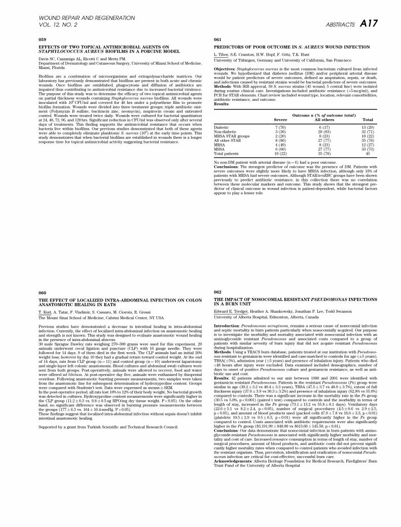
059
EFFECTS OF TWO TOPICAL ANTIMICROBIAL AGENTS ONSTAPHYLOCOCCUS AUREUS BIOFILMS IN A PORCINE MODEL
Davis SC, Cazzaniga AL, Ricotti C and Mertz PM.Department of Dermatology and Cutaneous Surgery, University of Miami School of Medicine,Miami, Florida
Biofilms are a combination of microorganisms and extrapolysaccharide matrices. Ourlaboratory has previously demonstrated that biofilms are present in both acute and chronicwounds. Once biofilms are established, phagocytosis and diffusion of antibiotics areimpaired thus contributing to antimicrobial resistance due to increased bacterial virulence.The purpose of this study was to determine the efficacy of two topical antimicrobial agentson partial thickness wounds containing Staphylococcus aureus biofilms. All wounds wereinoculated with 106 CFU/ml and covered for 48 hrs under a polyurthene film to promotebiofilm formation. Wounds were divided into three treatment groups; triple antibiotic oint-ment (Polymyxin B sulfate, bacitracin zinc, neomycin), mupirocin cream and untreatedcontrol. Wounds were treated twice daily. Wounds were cultured for bacterial quantitationat 24, 48, 72, 96, and 120 hrs. Significant reduction in CFU/ml was observed only after severaldays of treatments. This finding supports the antimicrobial resistance that occurs whenbacteria live within biofilms. Our previous studies demonstrated that both of these agentswere able to completely eliminate planktonic S. aureus (106) at the early time points. Thisstudy demonstrates that when bacterial biofilms are established in wounds there is a longerresponse time for topical antimicrobial activity suggesting bacterial resistance.
060
THE EFFECT OF LOCALIZED INTRA-ABDOMINAL INFECTION ON COLONANASTOMOTIC HEALING IN RATS
T. Kurt, A. Tatar, F. Vladimir, S. Cassaro, M. Cioroiu, R. Grossi
The Mount Sinai School of Medicine, Cabrini Medical Center, NY USA
Previous studies have demonstrated a decrease in intestinal healing in intra-abdominalinfection. Currently, the effect of localized intra-abdominal infection on anastomotic healingand strength is not known. This study was designed to evaluate anastomotic wound healingin the presence of intra-abdominal abscess.30 male Sprague Dawley rats weighing 270–300 grams were used for this experiment. 20animals underwent cecal ligation and puncture (CLP) with 16 gauge needle. They werefollowed for 14 days. 9 of them died in the first week. The CLP animals had an initial 20%weight loss; however by day 10 they had a gradual return toward control weight. At the endof 14 days, rats from CLP group (n¼ 11) and control group (n¼ 10) underwent laparotomyand single-layer left colonic anastomosis. Blood cultures and abdominal swab cultures weresent from both groups. Post-operatively, animals were allowed to recover, food and waterwere offered ad libitum. At post-operative day five, animals were euthanized by thiopentaloverdose. Following anastomotic bursting pressure measurements, two samples were takenfrom the anastomotic line for subsequent determination of hydroxyproline content. Groupswere compared with Students’t test. Data were expressed as means� SEM.In the post-operative period, all rats lost 10% to 12% of their body weight. No bacterial growthwas detected in cultures. Hydroxyproline content measurements were significantly higher inthe CLP group (11.2� 0.3 vs. 9.8� 0.5 ug HPO/mg dry tissue weight. P < 0.05). On the otherhand, no significant difference was observed in bursting pressure measurements betweenthe groups (177� 6.3 vs. 164� 10.4 mmHg. P > 0.05).These findings suggest that localized intra-abdominal infection without sepsis doesn’t inhibitintestinal anastomotic healing.
Supported by a grant from Turkish Scientific and Technical Research Council.
061
PREDICTORS OF POOR OUTCOME IN S. AUREUS WOUND INFECTION
L. Tibor, S.E. Cramton, H.W. Hopf, F. Gotz, T.K. Hunt
University of Tubingen, Germany and University of California, San Francisco
Objectives: Staphylococcus aureus is the most common bacterium cultured from infectedwounds. We hypothesized that diabetes mellitus (DM) and/or peripheral arterial diseasewould be patient predictors of severe outcomes, defined as amputation, sepsis, or death,and infections caused by resistant strains would be bacterial predictors of severe outcomes.Methods: With IRB approval, 50 S. aureus strains (45 wound, 5 central line) were isolatedduring routine clinical care. Investigations included antibiotic resistance (>5 mcg/ml), andPCR for STAR elements. Chart review included wound type, location, relevant comorbidities,antibiotic resistance, and outcome.Results:
Outcome n (% of outcome total)
Severe All others Total
Diabetic 7 (70) 6 (17) 13 (29)Non-diabetic 3 (30) 29 (83) 32 (71)MRSA STAR groups 2 (20) 8 (23) 10 (22)All other STAR 8 (80) 27 (77) 35 (78)MRSA 4 (40) 8 (23) 12 (27)MSSA 6 (60) 27 (77) 33 (73)Total patients 10 (22) 35 (78) 45
No non-DM patient with arterial disease (n¼ 6) had a poor outcome.Conclusions: The strongest predictor of outcome was the presence of DM. Patients withsevere outcomes were slightly more likely to have MRSA infection, although only 33% ofpatients with MRSA had severe outcomes. Although STAR/arsRBC groups have been shownpreviously to predict antibiotic resistance, in this collection there was no correlationbetween these molecular markers and outcome. This study shows that the strongest pre-dictor of clinical outcome in wound infection is patient-dependent, while bacterial factorsappear to play a lesser role.
062
THE IMPACT OF NOSOCOMIAL RESISTANT PSEUDOMONAS INFECTIONSIN A BURN UNIT
Edward E. Tredget, Heather A. Shankowsky, Jonathan P. Lee, Todd Swanson
University of Alberta Hospital, Edmonton, Alberta, Canada
Introduction: Pseudomonas aeruginosa, remains a serious cause of nosocomial infectionand septic mortality in burn patients particularly when nosocomially acquired. Our purposeis to investigate the morbidity and mortality associated with nosocomial infection with anaminoglycoside resistant Pseudomonas and associated costs compared to a group ofpatients with similar severity of burn injury that did not acquire resistant Pseudomonas
during hospitalization.Methods: Using a TRACS burn database, patients treated at our institution with Pseudomo-
nas resistant to gentamicin were identified and case-matched to controls for age (�5 years),TBSA(�5%), admission year (�5 years) and presence of inhalation injury. Patients who died<48 hours after injury were excluded. Data examined included demographics, number ofdays to onset of positive Pseudomonas culture and gentamicin resistance, as well as anti-biotic use and cost.Results: 42 patients admitted to our unit between 1980 and 2001 were identified withgentamicin resistant Pseudomonas. Patients in the resistant Pseudomonas (Ps) group weresimilar in age (39.2� 3.2 vs 40.4� 3.3 years), TBSA (47.5� 3.7 vs 48.9� 3.7%), extent of fullthickness injury (37.0� 3.9 vs 30.3� 3.2%) and presence of inhalation injury (62.8% vs 55.0%)compared to controls. There was a significant increase in the mortality rate in the Ps group(39.5 vs 5.0%, p < 0.001) (paired t test) compared to controls and the morbidity in terms oflength of stay, increased in the Ps group (73.1� 13.2 vs 55.8� 8.1 days). Ventilatory days(22.6� 5.1 vs 8.2� 2.4, p < 0.05), number of surgical procedures (4.5� 0.6 vs 2.9� 2.5,p < 0.05), and amount of blood products used (packed cells 47.9� 7.8 vs 18.6� 3.3, p < 0.01)(platelets 10.5� 2.9 vs 0.5� 0.3, p < 0.01) were all significantly higher in the Ps groupcompared to control. Costs associated with antibiotic requirements were also significantlyhigher in the Ps group ($3,191.90� 848.00 vs $613.60� 145.50, p < 0.01).Conclusions: Our data demonstrate that nosocomial infection in burn patients with amino-glycoside-resistant Pseudomonas is associated with significantly higher morbidity and mor-tality and cost of care. Increased resource consumption in terms of length of stay, number ofsurgical procedures, amount of blood products, and antibiotic costs did not prevent signifi-cantly higher mortality rates when compared to control patients who avoided infection withthe resistant organism. Thus, prevention, identification and eradication of nosocomial Pseudo-
monas infection are critical for cost-effective, successful burn care.Acknowledgements: Alberta Heritage Foundation for Medical Research, Firefighters’ BurnTrust Fund of the University of Alberta Hospital
WOUND REPAIR AND REGENERATIONVOL. 12, NO. 2 ABSTRACTS A17
BIOMATERIALS/BIOENGINEERING
063
THE HUMAN FOREARM BIOPSY MODEL FOR ACUTE WOUND HEALING
T. Serena, V. W. Li, W. W. Li, on behalf of the Wound Healing Cooperative Group
Wound Healing Cooperative Group, The Angiogenesis Foundation, Cambridge, MA USA
Background: The study of wound biology and effects of advanced modalities would benefitfrom a reliable human model for normal healing. Eaglstein et al. previously suggested thatthe forearm biopsy may be useful in acute wound healing studies (J Am Acad Dermatol45:857, 2001). The Wound Healing Cooperative Group (WHCG) here validates this model inhealthy individuals treated with topical growth factor therapy.Methods: In our randomized, double-blinded study design, 20 normal healthy volunteersunderwent four 6 mm biopsies of the flexor surface of both forearms. Biopsy sites wererandomly assigned to a control arm (daily bacitracin) or to one of three treatment arms:i) rhPDGF-BB 0.01% gel (Q.D.), ii) rhPDGF-BB (Q.O.D.), iii) rhPDGF-BB (Q.D.· 7 daysfollowed by bacitracin alone daily). The wounds were examined, measured and photo-graphed daily until complete healing was achieved. Adverse events were monitored. Ratesof healing and time-to-complete closure were measured. Repeat biopsies of a subset ofhealing wounds were performed for gene microarray analysis.Results: The forearm biopsy model allowed direct quantitative and qualitative comparisonsof acute wound healing outcomes achieved by rhPDGF-BB regimens versus standard carealone. There were no infectious complications. Subject compliance was excellent with < 3%of visits missed.Conclusion: The forearm biopsy model has several advantages in the study of acutewounds: it allows for comparison of topical agents in a controlled fashion; studies can beconducted easily with good patient compliance; and the forearm allows for repeat imagingand tissue harvesting for gene profiling during healing.Acknowledgements: Funding from The Angiogenesis Foundation.
064
PROLIFERATION AND GROWTH FACTOR EXRESSION OF HUMANKERATINOCYTES ON MICROCARRIER BEADS
R. Williams1, R.S. Rees2, W.J. Lindblad1
1Dept. of Pharmaceutical Sciences, Wayne State University, Detroit, MI 2Sec. Plastic Surgery,University of Michigan, Ann Arbor, MI
Human keratinocytes have been extensively used for the treatment of large body surfacearea burn injuries. We have studied the potential for treating non-thermal, chronic woundswith keratinocytes grown on microcarrier beads placed into porous retrievable bags. Thecurrent studies were undertaken to examine the growth characteristics of these cells onCytoline-1TM microcarrier beads, and how this growth correlated with the expression ofgrowth factors.Human keratinocytes were expanded in Keratinocyte Basal Medium-2 with standard supple-ments (Clonetics, Inc.). The cells were trypsinized and added to Cytoline-1TM (PharmaciaBiotech) beads at 13.3· 106 cells per mL pre-equilibrated beads. The beads were seeded intoroller bottles and cultured in a humidified 37˚ C incubator for up to two weeks. Cell numberswere monitored by direct cell counting on the beads following hematoxylin staining; and byprotein and DNA content. Aliquots of media were also assayed for levels of PDGF and VEGFby immunoassay (QuantikineR, R&D Systems).Keratinocytes showed a lag in growth with a sustained decrement in cells per bead from 456to 346 cells/bead by day 7. This was followed by a robust growth response to 551 cells/beadby day 11. These values correlated with total cellular protein (994, 738, and 1164 ug); andDNA content. The secretion of PDGF and VEGF by human keratinocyte under these condi-tions, did not directly correlate to the cell number but rather indirectly to the increase in cellnumber after the 7 days of stagnant growth. These results suggest that the vulnerary effectobserved with human keratincytes on microcarrier beads is dependent on acculturation ofthe cells to the microbead environment, after which they begin to express elevated levels ofgrowth factors important for healing.Acknowledgments: Funding was provided by Keracure, Inc. through NIH grant R41GM064064.
065
IMPROVING INTEGRATM
‘‘TAKE’’ WITH NEGATIVELY CHARGEDMETHACRLYLIC ACID BEADS
AA Eckhaus, MD, JS Fish, MD, MSc, FRCS(C), MV Sefton ScD
University of Toronto, Toronto, Ontario, Canada
Introduction: IntegraTM has gained acceptance as a dermal replacement for large burnswith limited donor sites. IntegraTM vascularization takes between 14 and 21 days. Accelerat-ing the vascularization of IntegraTM could improve ‘‘take’’ rates and potentially decrease theobligatory waiting period between application and autografting.Hypothesis: Application of negatively charged methacrylic acid (MAA) beads will enhancevascularization of IntegraTM
Methods Male Wistar rats (n¼ 11) had two 2· 3 cm contra lateral dorsal full thicknesswounds excised and grafted using IntegraTM. The MAA beads were applied under theIntegraTM topically on the wound bed. Three experimental treatments were compared:Group 1- high-dose MAA beads, Group 2- low-dose MAA beads, Group 3- Integra only.Laser Doppler Imaging (LDI) to assess perfusion, as well as digital photography was doneon days 7,10 and 14. Tissue samples for HþE and Factor VIII histological staining werecollected on day 14.Results: The average ‘‘take’’ was 99% (p < 0.05) for high-dose MAA beads, 98% (p < 0.05) forlow-dose MAA beads and 82% in the IntegraTM only group at day 7. At day 7 (p < 0.01) and day10 (p < 0.05), the low-dose MAA group had significantly greater perfusion than the IntegraTM
only group. There were no statistically significant differences at day 14. Microvessel density(MVD) counts revealed a >40% increase in the number of vessels in both the low-dose MAA(p < 0.05) and high-dose MAA (p < 0.05) groups when compared to the IntegraTM only group.There was no difference in LDI perfusion or MVD counts between low and high-dose MAAgroups.Conclusion: Factor VIII staining revealed enhanced angiogenesis in IntegraTM treated withlow and high-dose negatively charged MAA beads. Low-dose negatively charged MAA beadsimproved and accelerated the vascularization of IntegraTM in this rodent model.Acknowledgement: Materials and Manufacturing Ontario
066
FIXATION OF AUTOLOGOUS SKIN GRAFTS WITHOUT STAPLES ORSUTURES – EXPERIMENTAL STUDY
R. Mittermayr1, E. Wassermann1, M. Thurnher1, M. Simunek2, H. Redl1
1LBI for Experimental and Clinical Traumatology, Vienna, Austria 2Baxter AG, Vienna,Austria
The objective of the study was to evaluate the efficacy of skin graft fixation with fibrinsealant (FS) alone by means of spray application with a low setting time (thrombin concen-tration 4–5 IU/ml) in comparison to point suture fixation as a clinical reference method.Furthermore, the influence of two different quantities of FS were tested.Material and Methods: On the dorsum of 6 male pigs four full-thickness wounds (8 · 4 cm)were excised in each animal. Randomly the four defects were divided into 3 groups (FS0.05 ml/cm2; FS 0.15 ml/cm2 (Tisseel, Baxter AG, Vienna, Austria); Suture) and covered withautologous split thickness skin grafts. Outcome measurements included hematoma/seromaformation, graft dislocation, wound contraction, graft take rate and healed wound area.Observational time points were the 5th, 14th and 21st postoperative day.Results: Hematoma formation on day of surgery was more pronounced in the suture group
(FS 0.05 ml/cm2 vs. suture, p < 0.05), similar the situation on the 5th postoperative day butwithout statistical significance. Graft dislocation revealed marked but not significantextended area in the suture group vs. the FS 0.05 ml/cm2 group. The FS 0.05 ml/cm2 grafttake on day 5 was found to be enhanced in comparison to the suture group. Excellent woundhealing was notable on final observation day in the FS 0.05 ml/cm2 group with a healedwound area of 99.7 (3.6/0.3). Corresponding values in the FS 0.15 ml/cm2 was 96.9 (4.7/2.1)and 95.9 (2.7/2.1) for the suture group. Values represents median (Q1/Q3).Conclusion: The application of a thin layer of FS (0.05 ml/cm2) in a low setting rate (4–5 IUthrombin) in split thickness skin transplantation via a spray device shows comparableresults to those yielded with suture point fixation (surrogate for staples). Advantages aree.g. better cosmetic results and initial hemostasis with minimizing the incidence of earlyhematoma formation. Equal properties of manufactured low thrombin (4 IU; Canada) pre-paration vs. 500 IU (US) diluted to a 5 IU thrombin solution was also demonstrated.
WOUND REPAIR AND REGENERATIONA18 ABSTRACTS MARCH–APRIL 2004
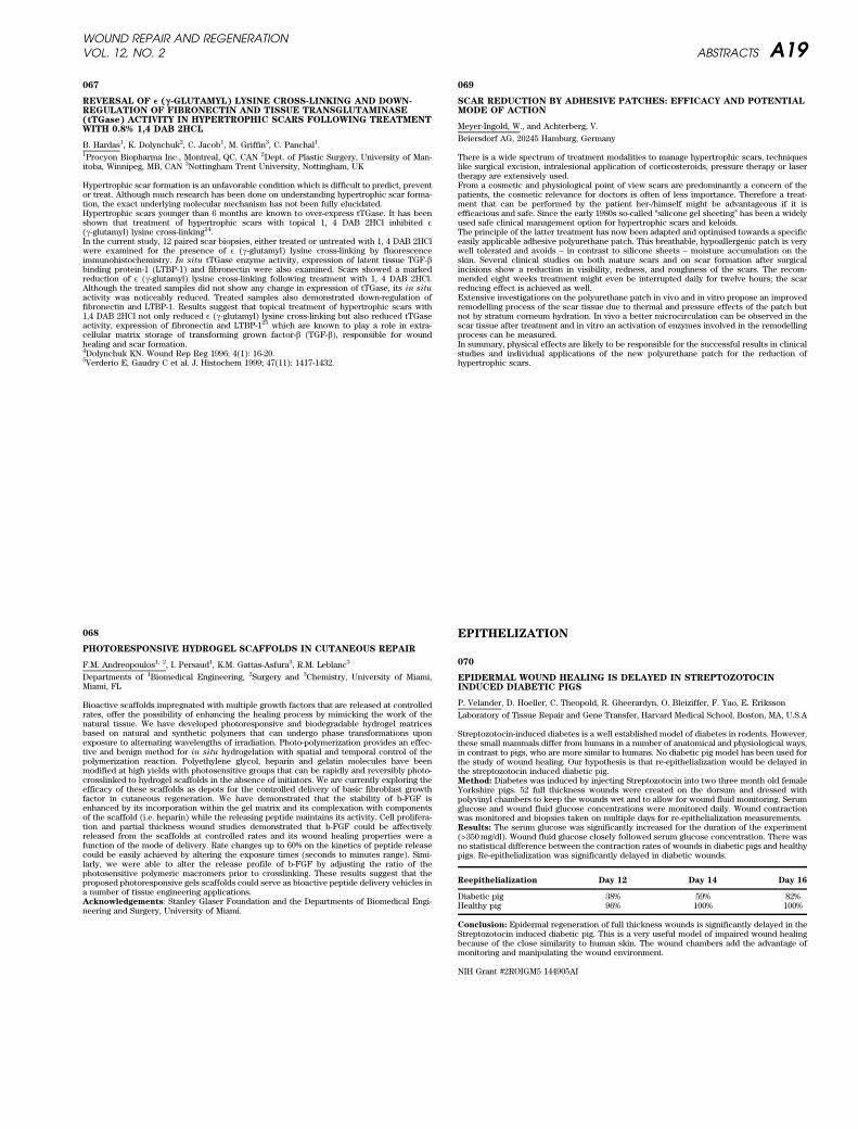
067
REVERSAL OF e (g -GLUTAMYL) LYSINE CROSS-LINKING AND DOWN-REGULATION OF FIBRONECTIN AND TISSUE TRANSGLUTAMINASE(tTGase) ACTIVITY IN HYPERTROPHIC SCARS FOLLOWING TREATMENTWITH 0.8% 1,4 DAB 2HCL
B. Hardas1, K. Dolynchuk2, C. Jacob1, M. Griffin3, C. Panchal1.1Procyon Biopharma Inc., Montreal, QC, CAN 2Dept. of Plastic Surgery, University of Man-itoba, Winnipeg, MB, CAN 3Nottingham Trent University, Nottingham, UK
Hypertrophic scar formation is an unfavorable condition which is difficult to predict, preventor treat. Although much research has been done on understanding hypertrophic scar forma-tion, the exact underlying molecular mechanism has not been fully elucidated.Hypertrophic scars younger than 6 months are known to over-express tTGase. It has beenshown that treatment of hypertrophic scars with topical 1, 4 DAB 2HCl inhibited E(g-glutamyl) lysine cross-linking14.In the current study, 12 paired scar biopsies, either treated or untreated with 1, 4 DAB 2HClwere examined for the presence of E (g-glutamyl) lysine cross-linking by fluorescenceimmunohistochemistry. In situ tTGase enzyme activity, expression of latent tissue TGF-bbinding protein-1 (LTBP-1) and fibronectin were also examined. Scars showed a markedreduction of E (g-glutamyl) lysine cross-linking following treatment with 1, 4 DAB 2HCl.Although the treated samples did not show any change in expression of tTGase, its in situ
activity was noticeably reduced. Treated samples also demonstrated down-regulation offibronectin and LTBP-1. Results suggest that topical treatment of hypertrophic scars with1,4 DAB 2HCl not only reduced E (g-glutamyl) lysine cross-linking but also reduced tTGaseactivity, expression of fibronectin and LTBP-125 which are known to play a role in extra-cellular matrix storage of transforming grown factor-b (TGF-b), responsible for woundhealing and scar formation.4Dolynchuk KN. Wound Rep Reg 1996; 4(1): 16-20.5Verderio E, Gaudry C et al. J. Histochem 1999; 47(11): 1417-1432.
068
PHOTORESPONSIVE HYDROGEL SCAFFOLDS IN CUTANEOUS REPAIR
F.M. Andreopoulos1, 2, I. Persaud1, K.M. Gattas-Asfura3, R.M. Leblanc3
Departments of 1Biomedical Engineering, 2Surgery and 3Chemistry, University of Miami,Miami, FL
Bioactive scaffolds impregnated with multiple growth factors that are released at controlledrates, offer the possibility of enhancing the healing process by mimicking the work of thenatural tissue. We have developed photoresponsive and biodegradable hydrogel matricesbased on natural and synthetic polymers that can undergo phase transformations uponexposure to alternating wavelengths of irradiation. Photo-polymerization provides an effec-tive and benign method for in situ hydrogelation with spatial and temporal control of thepolymerization reaction. Polyethylene glycol, heparin and gelatin molecules have beenmodified at high yields with photosensitive groups that can be rapidly and reversibly photo-crosslinked to hydrogel scaffolds in the absence of initiators. We are currently exploring theefficacy of these scaffolds as depots for the controlled delivery of basic fibroblast growthfactor in cutaneous regeneration. We have demonstrated that the stability of b-FGF isenhanced by its incorporation within the gel matrix and its complexation with componentsof the scaffold (i.e. heparin) while the releasing peptide maintains its activity. Cell prolifera-tion and partial thickness wound studies demonstrated that b-FGF could be affectivelyreleased from the scaffolds at controlled rates and its wound healing properties were afunction of the mode of delivery. Rate changes up to 60% on the kinetics of peptide releasecould be easily achieved by altering the exposure times (seconds to minutes range). Simi-larly, we were able to alter the release profile of b-FGF by adjusting the ratio of thephotosensitive polymeric macromers prior to crosslinking. These results suggest that theproposed photoresponsive gels scaffolds could serve as bioactive peptide delivery vehicles ina number of tissue engineering applications.Acknowledgements: Stanley Glaser Foundation and the Departments of Biomedical Engi-neering and Surgery, University of Miami.
069
SCAR REDUCTION BY ADHESIVE PATCHES: EFFICACY AND POTENTIALMODE OF ACTION
Meyer-Ingold, W., and Achterberg, V.
Beiersdorf AG, 20245 Hamburg, Germany
There is a wide spectrum of treatment modalities to manage hypertrophic scars, techniqueslike surgical excision, intralesional application of corticosteroids, pressure therapy or lasertherapy are extensively used.From a cosmetic and physiological point of view scars are predominantly a concern of thepatients, the cosmetic relevance for doctors is often of less importance. Therefore a treat-ment that can be performed by the patient her-/himself might be advantageous if it isefficacious and safe. Since the early 1980s so-called ‘‘silicone gel sheeting’’ has been a widelyused safe clinical management option for hypertrophic scars and keloids.The principle of the latter treatment has now been adapted and optimised towards a specificeasily applicable adhesive polyurethane patch. This breathable, hypoallergenic patch is verywell tolerated and avoids – in contrast to silicone sheets – moisture accumulation on theskin. Several clinical studies on both mature scars and on scar formation after surgicalincisions show a reduction in visibility, redness, and roughness of the scars. The recom-mended eight weeks treatment might even be interrupted daily for twelve hours; the scarreducing effect is achieved as well.Extensive investigations on the polyurethane patch in vivo and in vitro propose an improvedremodelling process of the scar tissue due to thermal and pressure effects of the patch butnot by stratum corneum hydration. In vivo a better microcirculation can be observed in thescar tissue after treatment and in vitro an activation of enzymes involved in the remodellingprocess can be measured.In summary, physical effects are likely to be responsible for the successful results in clinicalstudies and individual applications of the new polyurethane patch for the reduction ofhypertrophic scars.
EPITHELIZATION
070
EPIDERMAL WOUND HEALING IS DELAYED IN STREPTOZOTOCININDUCED DIABETIC PIGS
P. Velander, D. Hoeller, C. Theopold, R. Gheerardyn, O. Bleiziffer, F. Yao, E. Eriksson
Laboratory of Tissue Repair and Gene Transfer, Harvard Medical School, Boston, MA, U.S.A
Streptozotocin-induced diabetes is a well established model of diabetes in rodents. However,these small mammals differ from humans in a number of anatomical and physiological ways,in contrast to pigs, who are more similar to humans. No diabetic pig model has been used forthe study of wound healing. Our hypothesis is that re-epithelialization would be delayed inthe streptozotocin induced diabetic pig.Method: Diabetes was induced by injecting Streptozotocin into two three month old femaleYorkshire pigs. 52 full thickness wounds were created on the dorsum and dressed withpolyvinyl chambers to keep the wounds wet and to allow for wound fluid monitoring. Serumglucose and wound fluid glucose concentrations were monitored daily. Wound contractionwas monitored and biopsies taken on multiple days for re-epithelialization measurements.Results: The serum glucose was significantly increased for the duration of the experiment(>350 mg/dl). Wound fluid glucose closely followed serum glucose concentration. There wasno statistical difference between the contraction rates of wounds in diabetic pigs and healthypigs. Re-epithelialization was significantly delayed in diabetic wounds.
Reepithelialization Day 12 Day 14 Day 16
Diabetic pig 38% 59% 82%Healthy pig 96% 100% 100%
Conclusion: Epidermal regeneration of full thickness wounds is significantly delayed in theStreptozotocin induced diabetic pig. This is a very useful model of impaired wound healingbecause of the close similarity to human skin. The wound chambers add the advantage ofmonitoring and manipulating the wound environment.
NIH Grant #2ROIGM5 144905AI
WOUND REPAIR AND REGENERATIONVOL. 12, NO. 2 ABSTRACTS A19
071
TGF-ß-MEDIATED PARACRINE REGULATION OF KERATINOCYTE WOUNDCLOSURE
A. Sharif-Afshar, A. E. Gorska, E. G. Neilson, J. M. Davidson, H. L. Moses, N. A. Bhowmick
Vanderbilt University Medical Center, Nashville, TN, USA
The importance of stromal-epithelial interactions in wound healing is well established. Theseinteractions likely involve autocrine and paracrine action of multiple growth factors, includ-ing members of the TGF-ß family. TGF-ß1, ß2 and ß3 isoforms signal by sequentially bindingto the TGF-ß type II and type I receptors, respectively. We address the role of TGF-ßsignaling in dermal fibroblasts using a conditional fibroblastic TGF-ß type II receptor knock-out mouse model (termed FßKO). We found that the loss of TGF-ß signaling in the dermalfibroblasts results in accelerated excision-wound closure compared with similar wounds inwild type mice. The mechanism of the altered rate of re-epitheliaization in the FßKO micewas examined with regard to keratiocyte motility and proliferation. The migration of kera-tinocytes through collagen I coated 8mm pore filters in the presence or absence of fibroblast-conditioned media was tested. These experiments showed increased keratinocyte migrationwhen incubated with FßKO dermal fibroblast conditioned media compared to media condi-tioned in wild type fibroblasts. Immuno-histochemical staining of paraffin embedded intactskin indicated both wild type and FßKO mice had similar low levels of keratinocyte pro-liferation, based on Ki67 staining. In healing wounds, only the distal wound edges of wildtype mice were proliferative. In contrast, the FßKO mice exhibited elevated proliferationacross the length of the wound, including the leading edge of epithelial closure. Together ourresults suggest TGF-ß signaling by the dermal fibroblasts suppresses re-epithelialization ofexcision wounds by regulating keratinocyte motility and proliferation through paracrinemechanisms.
Funding: DOD BC99184 and NIH CA85492.
072
FULL-THICKNESS WOUNDING OF THE MOUSE TAIL AS A MODEL FORDELAYED WOUND HEALING
V. Falanga,a,b,c D. Schrayer,a J. Cha,a Janet Butmarca, P. Carsona, A.B. Roberts,d S-J S. Kim,d;
aRoger Williams Medical Center; bDepartment of Dermatology and cBiochemistry; BostonUniversity; dNational Institutes of Health, Bethesda, MD
Experimentally induced wounds in animal models are useful in gaining a better understand-ing of the cellular and molecular processes of wound healing and in the initial evaluation ofthe safety and effectiveness of potential therapeutic agents. However, studying delayedhealing has proven difficult in animals, whose wounds heal within a few days. In this report,we describe a novel method for establishing mouse wounds that require up to more thanthree weeks for complete closure, and we show the validity of this model in Smad3 null mice,which are known to display accelerated healing. Full-thickness wounds, measuring 0.3 by1.0 cm, were made down to fascia on the dorsal aspect of the mouse tail in Smad3 KO miceand control littermates, approximately 1 cm distal to the body of the animal. The woundswere left to heal by secondary intention and were assessed histologically by computerizedplanimetry for wound closure at various times after wounding. These wounds in wild-typemice displayed delayed healing, with full closure occurring between 14 and 25 days afterwounding. Complete closure of similar wounds in Smad3 null mice healed 30% faster(p < 0.01). By immunostaining with ki67, a marker for proliferation, Smad3 null animalsalso showed increased proliferation of dermal wound cells. Cultured dermal fibroblastsfrom Smad3 null mice showed increased baseline DNA synthesis and, interestingly,enhanced response to TGF-b1. By Western blot analysis, Smad3 null mice fibroblasts showeda compensatory increase in MAPK phosphorylation in response to TGF-b1, suggesting thatMAPK overcompensation together with loss of Smad3 may be involved in the modulation offaster healing. We conclude that this novel tail wounding model can be useful for studyingdelayed or prolonged wound closure.Experimentally induced wounds in animal models can be useful in gaining a better under-standing of the cellular and molecular processes of wound healing. Such models have alsoproven themselves valuable in the initial evaluation of the safety and effectiveness ofpotential therapeutic agents targeted for chronic non-healing wounds. (Gottrup, Agrenet al. 2000). However, no ideal animal model exists which reliably reproduces delayedhealing. In the mouse, a mammal whose genome has been completely cloned and which iseasily manipulated genetically, wounds normally heal within a few days, and with a greatdeal of contraction. (Morris, Wu et al. 1997; Gottrup, Agren et al. 2000) There are modelsutilizing either genetically altered and inbred mice with certain characteristics that causedelayed healing. (Carmeliet 1995) However, it would be useful to have wound healing modelsthat are applicable to most wild type mice used as controls and which would have a largeenough window of observation before healing occurs. In this report, we describe a novelmethod for studying delayed healing in mice. This method utilizes full-thickness wounds madedown to fascia on the dorsal and mostly hairless aspect of the mouse tail. The wounds areleft to heal by secondary intention and assessed histologically by computerized planimetryfor wound closure at various times after wounding. In this first report, the validity of themodel was determined by studying control littermates and Smad3 null mice, which havebeen shown to display accelerated healing. The results shown here suggest that this is auseful model for studying delayed healing in mice.MethodsAnimal protocol
Smad3 null mice were generated as previously described by targeted disruption of the Smad3gene by homologus recombinationWork supported by NIH grants AR42936 and AR46557
073
EXPRESSION OF CASPASES 3, 9, AND 14 IN THE EPIDERMIS OF EARLYWOUNDS
M.K. Kuechle, M.L. Usui, R.A. Underwood.
University of Washington, Seattle, WA USA
Caspases are a family of at least 14 highly conserved aspartate proteases that are the majoreffector arm of apoptosis or programmed cell death. Previous immunohistochemical andbiochemical studies have implicated two of the family members, caspase 3 and caspase 14,as having a role in the process of epidermal terminal differentiation. Reepithelialization ofepidermal defects after wounding requires coordination between signals that initiate growthand migration as well as signals that initiate differentiation. We therefore examined theexpression of caspase 3, caspase 9, and caspase 14 in the epidermis of early (up to 48 hour)human wounds. Two excisional wounds were created on the inner arm using a 3 mm biopsypunch and allowed to heal via secondary intention. Wounds were excised at 24 and 48 hoursusing a 6 mm biopsy punch, placed in OCT, frozen, cryosectioned and immunohistochemi-cally labeled using affinity purified antisera for caspases 3, 9 and 14. At 24 hours, thedistribution of caspase 3 and caspase 9 expression was unchanged from non-woundedskin. Caspase 3 was found in the upper differentiating cells of the granular layer, whilecaspase 9 was expressed in scattered dendritic cells in the epidermis. At 48 hours, caspase 3expression was lost in the epithelial tongue, while caspase 9 expression was found inkeratinocytes at the tip of the migrating tongue. Caspase 14 expression at 24 hours wasnoted at the tip of the migrating epithelial tongue, while at 48 hours, the normal expressionof caspase 14 in the granular layer was lost. These results suggest that in 48 hour wounds,loss of caspase 3 and 14 expression in the migrating epithelium reflects the activated state ofthis tissue. The onset of caspase 9 expression at 48 hours in the leading edge of the woundsuggests a role for caspase 9 in wound remodeling, as caspase 9 is known to participate intissue remodeling during development. Characterization of the caspases involved duringwound healing may lead to future therapies utilizng caspases or caspase inhibitors insituations of poor or aberrant wound healing.
074
WOUND HEALING ENHANCEMENT BY PULSED ELECTROMAGNETICFIELDS
M.J. Callaghan, E.R. Kinnuncan, J. Michaels, B.J. Simon, G.C. Gurtner
The effects of pulsed electromagnetic fields (PEMF) on soft tissue are not well character-ized. This study utilized a diabetic wound model to examine the effects of PEMF effects onsoft tissue healing.Methods: Wounds were created on the dorsum of db/db and wild type C57BL6 mice. Micewere exposed to PEMF (4.5 ms pulse/15 hz) for 8 hrs/day for 14 days. Gross closure wasassessed with digital analysis of area changes over time. Histological examination assessedgranulation and epithelial gap, cell proliferation (BrdU), and endothelial cell density (CD31).Human umbilical vein endothelial cells (HUVECs) were incubated in the presence orabsence of PEMF for 8 hrs and VEGF/FGF2 was measured in culture supernatants by ELISA.Results: Mice exposed to PEMF had accelerated wound closure at day 7 (wound area as %of original, db/db: 60% (PEMF) vs. 78% (control), C57BL6: 15% vs. 42%, p < 0.05) and day 14(db/db: 21% vs. 55%, C57BL6: 8% vs. 28%, p < 0.05), with increased granulation and cellproliferation (db/db day 7: 52� 8 vs. 31� 5 cells/HPF (200x)). Immunohistochemical analysisrevealed significantly higher CD31 density in wounds exposed to PEMF at day 7 (vessels/HPF, db/db: 28� 4 vs. 17� 4, C57BL6: 41� 7 vs. 28� 6) and day 14 (db/db: 32� 6 vs. 21� 5,C57BL6: 48� 5 vs. 40� 5). HUVECs in PEMF exhibited 5-fold higher levels of FGF2 com-pared to controls after 30 min (20.50 pg/ml� 6.75 vs. 4.25 pg/ml� 0.75), with no change inVEGF through 8 hrs.Conclusions: PEMF accelerates closure time and endothelial cell proliferation in woundhealing. Upregulation of FGF2 in HUVECs exposed to PEMF suggests that release ofangiogenic growth factors may explain increased vascular density and accelerated woundclosure. Clinical uses include treatment for diabetic ulcers and other non-healing wounds.
This work is supported in part by the Alumni Fund, Alumni Association of SUNY DownstateCollege of Medicine (MJC).
WOUND REPAIR AND REGENERATIONA20 ABSTRACTS MARCH–APRIL 2004
075
WOUND HEALING IS DELAYED IN WOMEN AND IN THE AGED: APOTENTIAL ROLE FOR THE HPA AXIS
Marucha PT1, Engeland CG1, and Cacioppo JT2.1Dept Periodontics, University of Illinois at Chicago; 2Dept Psychology, University ofChicago
Both senescence and psychological stress have been shown to alter numerous immunecomponents including inflammatory processes. However, whether such effects can be dis-sociated by gender is largely unknown. To determine the effects of gender and age on woundhealing, a 3.5 mm round wound was placed under local anesthesia on the hard palates of 88younger (18–35 years) and 56 older (50þ years) male and female volunteers. Immediatelyprior to wounding and 15, 30 and 60 min afterwards, blood was drawn from which cortisoland ACTH levels were determined, as both are immunosuppressive. Wounds were photo-graphed and measured daily until healed. Preliminary analyses revealed that older individ-uals healed 25.6% slower than younger individuals (P < .001), and women healed 14.0%slower than men (P < .05). These results may have involved alterations in HPA activity, asblood work revealed significant differences in baseline hormone levels: cortisol levels werelower in the older group (P < .05), and ACTH levels were lower in females (P < .01). Further-more, in women only, faster healing rates were related to both increased anxiety at the timeof wounding (P < .05), and higher cortisol levels at all time points (P < .01). Moreover,preliminary results indicated that, in women, higher anxiety was also related to loweredrelease of IL-1b in the first 24 h. In men these relationships were not evident, although higherACTH levels were related to faster healing times (P < .01). Taken together, these findingsindicate that aging delays oral wound healing and provide evidence for a previouslyunreported gender difference in wound healing rates in favor of males. Furthermore, theeffects of anxiety on wound healing seem to be sexually differentiated and transientactivation of the HPA axis, especially in women, appears to be associated with healing rates.
(Supported by NIH P01AG16321)
STEM CELLS
077
HUMAN MESENCHYMAL STEM CELLS INTERACT WITH KERATINOCYTESAND SUCCESFULLY ACCELERATE WOUND HEALING
Sadanori Akita1, Hiroshi Nakagawa1, Masashi Fukui1, Kozo Akino2,
1: Division of Plastic and Reconstructive Surgery2: Division of Anatomy and Neurobiology Nagasaki University, Graduate School of Biomedicaland Sciences, Department of Developmental and Reconstructive Medicine, Nagasaki, Japan
Human mesenchymal stem cells (hMSCs) obtained from a single donor from an iliac crest,were investigated with cutaneous wound healing models using nude rat, eliminating T cell-mediated immune reaction to the grafted stem cell of human origin. Co-culture of the humankeratinocytes and the hMSCs revealed tight basal membranous interaction by electronmicroscopy.1.5· 1.5 cm2 dorsal full-thickness defects including panniculus carnosus of F344/NJCl-rnunude rats were covered by bi-layered porcine-derived collagen sponge artificial skin sub-stitutes, Pelnac�, Johnson & Johnson, Tokyo, Japan, impregnated with 5· 106 hMSCs gratedto along with 0, 1, 10, or 100mg of recombinant human basic fibroblast growth factor (bFGF).The tissues were harvested at 3, 7 42 days after grafting. The defects were remarkablyepithelialized by 7 days after coverage with artificial skin substitutes and 10mg of bFGF,while artificial substitutes with no hMSCs or artificial skin substitutes with hMSCs alone didnot demonstrate such healing. The architectures of the artificial skin substitutes wereintegrated by day 42 in hMSCs-treated groups. The artificial skin substitute at least plays arole in as a template or scaffold of the grafted hMSCs. Up to day 3, the mesenchymal stemcell surface markers such as CD 29 and CD 44 were remained immunopositive in the groupswith hMSCs-treated groups over the reconstructed dermis-like tissue. By 42 days aftergrafting, artificial skin substitutes with hMSCs and 10 mg of bFGF demonstrated the totalepithelization and the keratinocytes by this treatment exhibited the pan-cytokeratin ofhuman origin by immunohistochemical expressions. Thus, the hMSCs with bFGF treatmentusing an artificial skin substitute as a successfully heal. These results suggest that the humanmesenchymal stem cells may be utilized for wound coverage together with bFGF andartificial skin substitutes.
Type acknowledgements here.
078
MECHANICAL STRAIN INDUCES HYPERTROPHIC SCARS IN MICE BYREDUCING STROMAL CELL APOPTOSIS
KA Bhatt, MR Greives, RD Galiano, RL Ashinoff, RG Bonillas, DJ Ceradini, DM Jones,
JP Levine, GC Gurtner.NYU Plastic Surgery.
Intro: Pathophysiology of hypertrophic scar formation remains unclear, potentially invol-ving mechanical strain, burns, or infection. We sought to specifically examine the role ofmechanical strain in hypertrophic scars. To do so, we developed and characterized a novelmurine model which uses mechanical strain to produce hypertrophic scars in mice.Methods: Paired incisions were created on C57BL6 mice (n¼ 20). After closure, mechanicalstrain was applied across one wound using a novel device; the other wound was a control.Starting at day 3, mechanical strain was increased every other day over 4 wks. Wounds wereharvested each week, and examined histologically using Sirius red, DAPI, BrdU for prolifera-tion, and caspase 3 for apoptosis.Results: Strained wounds showed features of hypertrophic scars: raised borders, loss of retepegs and adnexal structures in the epidermis overlying scars, blood vessels that wereperpendicular and fibrillary collagen that was parallel to skin surface. Features persistedbeyond 6 months. Wound collagen deposition in strained wounds increased 6-fold at 1 wkand over 12-fold at 2–4 wks, in parallel to an increase in number of stromal cells within scar(28-fold). This resulted in an increase in number of cells per unit area of collagen (over 42%).In all wounds, there was no significant difference in proliferation. There was a significantreduction in blood vessel (4-fold) and fibroblast (3-fold) apoptosis at 1–4 wks in strainedwounds.Conc: Mechanical strain alone is sufficient to produce hypertrophic scars in this model thatare indistinguishable from human scars. Notably, hypertrophic scars appear to result from amarked reduction in apoptosis, rather than from a significant proliferation and upregulationof cellular collagen deposition, validating previous in vitro studies.
079
MMP 9 MEDIATES ANGIOPOIETIN-1 RECRUITMENT OF ENDOTHELIALPROGENITOR CELLS TO DIABETIC WOUNDS
SG Keswani, JK Parvadia, ED Kozin, A Malik, PW Zoltick, A Radu, D Alaee, AB Katz, and
TM CrombleholmeThe Children’s Hospital of Philadelphia, Philadelphia, PA 19104
Introduction: Adenoviral gene transfer of Angiopoietin-1 (AdAng1) recruits endothelialprogenitor cells (EPCs) and improves diabetic wound healing. A suggested mechanism forEPC mobilization from the bone marrow (BM) is mediated through MMP-9 and stem cellfactor (SCF). We hypothesize that Ang-1 recruits EPCs to diabetic wounds via an MMP-9dependent mechanism.Methods: Lethally irradiated mice were reconstituted with BM from transgenic TIE-2/LacZmice. After engraftment, diabetes was induced with steptozotocin. 8 mm wounds werecreated in BM transplanted (BMT) (n¼ 12) or MMP-9 knockout (KO) (n¼ 12) mice andtreated with 1· 108 PFU of AdAng1, AdGFP or PBS. At 7 days wounds were analyzed forepithelial gap, vessel density, and EPCs. Serum levels of VEGF, proMMP9 and SCF wereassessed. Data are expressed as mean� SEMResults: In diabetic BMT wounds, AdAng1 results in improved reepithelialization (Ang12.3� .2 mm; GFP 3.9� .2; PBS 4.0� .1 p < .0001) neovascularization (Ang1 6.8� .3Caps/Hpf;GFP 3.0� .4; PBS 2.9� .3 p < .0001) and EPC recruitment (Ang1 5.3� .4 EPCs/Hpf; GFP2.1� .3; PBS 2.2� .3 p < .0001). AdAng1 treatment results in increased levels of proMMP-9(Ang1 9.7� .8 ng/ml; GFP 6.3� .9; PBS 6.4� .4 p < .01) and SCF (Ang1 265� 28 pg/ml; GFP119� 16; PBS159� 12 p < .001). In MMP9 KO mice, AdAng1 accelerates reepithelialization(Ang1 3.2� .1 mm; GFP 4.1� .2; PBS 3.8� .2; p<.) but has no significant effect on neovascu-larization (Ang1 4.1� .5 Caps/HPF; GFP 3.9� .4; PBS 3.9� .6), EPC recruitment (Ang1 2� .3EPC/Hpf; GFP 1.5� .3; PBS 1.6� .2) or SCF levels (Ang1 83� 5 pg/ml; GFP 83� 2; PBS88� 6).Conclusions: The effects of Ang-1 on EPC recruitment and neovascularization are depend-ent on MMP-9. Our data support the hypothesis that MMP-9 enables SCF to permit mobil-ization of EPCs.
WOUND REPAIR AND REGENERATIONVOL. 12, NO. 2 ABSTRACTS A21
080
SYNERGISTIC EFFECT OF TGF BETA 1 AND HIF-1A ON THE EXPRESSIONOF A HYPOXIA-RESPONSIVE ELEMENT-CONTAINING PROMOTER
Nakshatra K. Roy ([email protected]), Alan C. Sull, Thomas A. Mustoe
Wound Healing Research Laboratory, Department of Surgery, Northwestern University,Chicago, IL 60611, USA.
Accumulating data strongly suggests that under hypoxic conditions the adaptive physiologicalresponse of the cells involves co-operation between oxygen sensing and growth factorsignals that cause HIF-1 mediated gene expression. The HIF-1 has been identified as acentral and critical molecule in oxygen sensing and possibly a master switch. Of the manycytokines that have been shown to modulate collagen production, TGFb1 appears to becrucial as it can sustain stimulation of collagen production as well as autoinduction of itsown synthesis. Several genes that are known to be hypoxia inducible are also up-regulated byTGFbs, and the promoter of TGFb3, a member of the family, has a hypoxia-response element(HRE). Recent reports indicate that HIF-1a physically interacts with Smad3 and that theTGFb and hypoxia signaling pathways synergize at the transcriptional level to regulate geneexpression. To determine if this interaction upregulates gene expression through the HREelement, we have co-transfected cultured human dermal fibroblasts with the mammalianexpression plasmid for HIF-1a (pCEP1-HIF-1a) with a reporter construct 5HRE-luc, contain-ing a concatemer of five copies of HRE derived from human vascular endothelial growthfactor (VEGF), a minimal cytomegalovirus (CMV) promoter and a reporter gene,luciferase.).When treated with 2 ng/ml of TGFb1 protein, co- transfected hypoxic aged and young humandermal fibroblasts showed significant synergistic upregulation of the reporter gene expres-sion. To further confirm the TGFb-HIF-1a interaction we have created a TGFb1 -RNAiconstruct by subcloning a 19-nucleotide sequence derived from rabbit in a mammalianexpression vector (p-SUPER) that directs the synthesis of small interfering RNAs (siRNAs).. Hypoxic rabbit cultured dermal fibroblasts co-transfected with TGFb1-RNAi and 5HRE-lucshowed down-regulation of reporter gene expression.As a prerequisite to understanding the biology of physiological and pathological ischemictissue repair process it is important to delineate the molecular basis of regulation of collagengene expression in fibroblasts. Each step in the TGFb signaling cascade is a potential targetfor highly specific therapy and information about how hypoxic low-oxygen microenviron-ments affect the TGFb cascade could be useful to develop novel therapeutic strategies thatwill involve agonist and antagonist agents that directly interfere with these steps.
We thank Drs. G. Semenza, Jons Hopkins Univ and J M.Brown, Stanford Univ, for theirgenerous gifts of pCEP1-HIF1a and p5HRE-hCMVmp-luc respectively.
082
CM-1 (CYTOMODULIN), A SYNTHETIC PEPTIDE, PROMOTES COLLAGENTRANSCRIPTION AND WOUND HEALING IN BIOLUMINESCENT MICE
Jayasri Dasgupta*, Rajendra Bhatnagar and Jeffrey M Davidson*.*Department of Pathology, Vanderbilt University and †Research Service, VA Med Ctr.,Nashville, TN, University of California San Francisco, and MediScient, Inc., San Mateo, CA([email protected])
Cytomodulin (CM-1) is a synthetic heptapeptide. Human dermal fibroblasts cultured in the
presence of CM-1 showed increased expression of transforming growth factor–ß (TGF-ß)and collagen I and decreased expression of MMP-1. TGF-b is involved in numerous vitalprocesses in wound healing, and the rate of wound repair is influenced by its overexpressionat the site of injury. To establish the effect of CM-1 in wound healing, we administeredseveral concentrations of CM-1 to incisional wounds and to unwounded skin of collagen-luciferase transgenic mice. Luciferase bioluminescence served as a quantitative reporter ofCO1A2 transcription. Imaging was performed to measure the luciferase activity on thesemice at different time points after administration of 1, 10 and 100 ug CM-1. The highestamount (100mg) showed the highest level of collagen expression both in unwounded andwounded skin. In another experiment, we used a scrambled peptide as a control andadministered the peptides both topically and by injection. CM-1 was effective in stimulatingluciferase expression both by topical and intradermal administration. To determine theeffect of CM-1 on wound strength, we measured the tensile strength in incisional woundsof wild type C57B6 mice at 10 d. Histological analysis was also performed to assess thecollagen content and organization of the tissue. The data showed a statistically significantincrease in tensile strength and collagen content at 10 and 100 mg doses. These resultsillustrate the utility of CM-1 as a novel, low-molecular weight vulnerary agent.
Supported by NIH grant AG06528 and the Department of Veterans Affairs.
083
TRANSFORMING GROWTH FACTOR-BETA LIGAND, RECEPTOR, ANDSMAD EXPRESSION PATTERNS IN FETAL AND ADULT KERATINOCYTES
HP Lorenz, R Faudoa, MT Longaker, TM Krummel
Pediatric Surgical Research Program, Department of Surgery, Stanford University, Stanford,CA USA
Introduction: The transforming growth factor-beta (TGF-ß) cytokine family regulates cel-lular proliferation, differentiation, and migration with important function during both develop-ment and repair. Moreover, the TGF-ßs are growth inhibitory for keratinocytes,proliferative for fibroblasts, and generally profibrotic during repair. In order to better definethe influence of keratinocyte TGF-ß during these processes, we examined the TGF-ß iso-form, receptor, and signal messenger Smad expression in fetal and postnatal keratinocytes.Methods: Sprague-Dawley rat keratinocytes were isolated in primary culture from fetal atE15 and E17 (term¼E22), newborn, and 6 week old adults. Open fetal rat wounds healwithout scar at E17 gestation. Quantitative-polymerase chain reaction was performed forTGF-ß1, -ß2, and -ß3 ligand; TGF-beta receptor1 and -receptor2; Smad3, Smad4, and Smad7expression. All cells were passage 2 or 3.Results: TGF-ß1 expression did not change appreciably from E15 to adult age in keratino-cytes. TGF-ß2 and -ß3 expression increased (8 fold and 2 fold respectively) from E17 tonewborn ages. Overall, the expression of TGF-ß1 was 5–8 fold greater compared to -ß2 and -ß3in both fetal and adult keratinocytes. TGF-ß receptor1 expression increased 2 foldwhereas -receptor2 expression showed little change from E17 to adult age. Smad3 expressionincreased over 50 fold, with Smads 4 and 7 showing a smaller increase from E17 to adult age.Conclusions: The TGF-beta system has differential expression in fetal compared to postnatalkeratinocytes, which suggests function during skin differentiation. TGF-ß1 expression is rela-tively greater than -ß2 and -ß3 in both fetal and adult keratinocytes. However, increases inisoforms -ß2 and -ß3, receptor1 and the Smads occurs at ages associated with scarring, implyingincreased function of the pro-fibrotic TGF-beta response during ages associated with scarring.
Funding: NIH GM-041343–14
POSTERS
084
EFFECTS OF FAT AUTOGRAFTING ON PORCINE SKIN WOUND HEALINGAFTER INJURY
Lijun Fang, Xiaobing Fu, Yuxin Wang, Tongzhu Sun, Biao Cheng.
Wound Healing and Cell Biology Laboratory, Burn institute, 304th Hospital, Trauma Centerof Postgraduate Medical College, Beijing 100037, P.R.China ([email protected];[email protected])
It has been evidenced that the fat may have a potential to secret some growth factors or be asource of stem cells. So, we explore the effects of fat on healing of porcine skin wounds so asto provide a new method for clinical skin wound repair after injury.Forty-eight full-thickness skin wounds were produced on both sides of the back in 6 maleminipigs (8 wounds in each animal). Then these wounds were randomly divided into 4groups, which were saline control group, fat autografting group, basic fibroblast growthfactor (bFGF) treatment group and epidermal growth factor (EGF) treatment group. At day3, 7, 14 and 21 after wounding, the area and the volume of wounds were measured and thehistological examination was performed to evaluate the velocity and quality of woundshealing in different groups.At day 3 and 7, the amount of granulation tissues and vessel density in fat treatment groupwere significantly more than that in other groups. Wound areas and volume in fat treatmentwounds were markedly decreased in compared with those in other groups (P < 0.01). Regene-rated epidermis in fat treatment group was thicker than that in other groups.These results confirmed that the wound healing velocity and quality in wounds treated withfat autografting were enhanced. It indicated that fat has a potential to accelerate velocity andimprove the quality of wound healing after skin injury.
This study was supported in part by the National Natural Science Foundation of China(30170966, 30230370), National Basic Science and Development Programme (973 Pro-gramme, G1999054204)
WOUND REPAIR AND REGENERATIONA22 ABSTRACTS MARCH–APRIL 2004
085
BONE MARROW MESENCHYMAL STEM CELLS AS A POTENTIAL SOURCETO REPAIR THE INJURY SKIN
Xiaobing Fu,1 Lijun Fang,1, 2 Xiaokun Li,1, 3 Biao Cheng,1 Zhiyong Sheng1
1 Wound Healing and Cell Biology Laboratory, Burn Institute, 304th Hospital, Trauma Centerof Postgraduate Medical College, Beijing 100037, P.R.China ([email protected]; fuxiao
[email protected])2 Department of Stomatology of the Second Clinical College, China Medical University,ShenYang 1100033 Pharmaceutical Division, Jinan University, Guangzhou 510632
Adult stem cells are found in various tissues and organs and have potential to differentiateinto different cell lineages, including bone, cartilage, fat, tendon, muscle, epithelial cells ofthe skin and gastrointestinal tract, etc. Here, we report that the expended and purified bonemarrow MSCs might take on phenotypes and characteristics of vascular endothelial cells orepidermal cells after cultured and induced with different lineage-specific culture conditionsin vitro. Also, in vivo grafting experiments confirmed that those labeled MSCs could get thephenotypes of vascular endothelial cells in granulation tissue and sebaceous duct cells andepidermal cells in regenerated skin, which imply that these grafted MSCs might havetransdifferentiated into vascular endothelial cells and sebaceous duct cells and epidermalcells. Our results indicate that locally delivered bone marrow MSCs may generate de novo
intact skin and get perfect skin regeneration after full thickness injury.
This study was supported in part by the National Natural Science Foundation of China(30170966, 30230370), National Basic Science and Development Programme (973 Pro-gramme, G1999054204)
086
SKIN WOUND HEALING IN RED DUROC X YORKSHIRE F1 PIGS: EFFECTOF WOUND DEPTH ON GENE EXPRESSION
CL Gallant, ME Olson, DA Hart
University of Calgary, Calgary, Alberta, CANADA, T2N 4N1
Previous investigations have demonstrated that cutaneous healing in red Duroc pigs (RD)differs from healing in Yorkshire pigs (Y). Specifically, RD animals heal with the formation ofhypercontracted, hyperpigmented scars, features which are not observed in Y animals.Further, this abnormal scarring may be genetically mediated, as demonstrated by the partialtransmission of this phenotype to RD·Y offspring (F1). To further determine the degree oftransmission of this phenotype, this study examined the effect of wound depth upon thehealing response of F1 animals. Ten full thickness (FT) and ten deep dermal (DD, 1.8 mm)skin wounds (2 cm· 2 cm) were created on the backs of juvenile female F1 animals (N¼ 8)using previously published procedures. Biopsies (4 mm) were taken from FT and DD woundson days 14, 28, 42, 56, and 70. Samples were used for histology and molecular analysis usingRT-PCR. There were no detectable differences between the FT and DD wounds on the grossor histologic level. However on a molecular level, there were numerous differences betweenthe FT and DD wounds. Several genes investigated demonstrated increased expression levelsin the DD wounds as compared to the FT wounds. Further, DD wounds demonstrated aunique pattern of expression for several other genes that differed from that observed ineither the F1 FT wounds or in any RD or Y wounds. Given that previous investigations haveshown that FT and DD wounds are nearly identical within each of the RD and Y strains, thisrepresents a novel finding. These results indicate that the healing phenotype of the F1animals may be regulated both by genetics and by the wound size/depth, suggesting thathuman wound healing may be similarly influenced by genetics, wound depth and potentially,environmental factors.
Funding provided by NSERC, CIHR.
087
NORMAL OR PATHOLOGICAL WOUND HEALING: APOPTOSISSENSITIVITY OF MYOFIBROBLASTS
V. Moulin, S. Larochelle, C. Langlois, I. Thibault, C. A. Lopez-Valle, M. Roy
LOEX, Saint-Sacrement Hospital/CHA, surgery department, Laval University, Quebec,Canada.
During wound healing, myofibroblasts play a major role in wound contraction and matrixformation. At the end of healing, myofibroblasts disappear via apoptotic pathways. Hyper-trophic scars are a fibroproliferative disorder that leads to considerable morbidity. Althoughno evidence exists, it has been postulated that a defect in myofibroblast apoptosis could beresponsible for the pathological scar formation. We have isolated and cultured humannormal wound (Wmyo) and hypertrophic scar (Hmyo) myofibroblasts and compared theirbasal apoptotic rates and their sensitivity to serum starvation and Fas antibody-inducedapoptosis to that obtained for dermal fibroblasts (Fb). A higher rate of apoptosis asevidenced by morphological criteria and a propidium iodide assay was observed for Wmyoin comparison to Fb and Hmyo. These results came along with a low level of the anti-apoptotic proteins Bcl-2 and BclxL in Wmyo, whereas there was an increase in the level ofthe pro-apoptotic molecule Bax when compared to the results obtained for Fb and Hmyo.Hmyo showed a higher level of Bcl-2 compared to Fb but no difference in the Bax or BclxL
level. After serum starvation, Wmyo revealed an increased apoptotic rate whereas Hmyo andFb did not show any difference. Anti-Fas treatment did not modify the levels of apoptosis butstrongly increased the cell growth of Hmyo as compared to Wmyo. This is the first studypresenting a broad vision of the apoptotic sensitivity of normal and pathological myofibro-blasts. These results confirmed the hypothesis of defects in apoptosis and growth duringpathological scar formation impeding myofibroblast disappearance at the end of healing.Acknowledgements: This study was supported by the Canadian Institutes of HealthResearch (#MOP-42400) and ‘‘Fondation de l’Hopital du Saint-Sacrement’’. S.L. and V.M.were recipients of scholarships from ‘‘Centre de recherche du CHA’’.
088
TOPICAL SUBSTANCE P MODULATES INFLAMMATORY RESPONSES INHEALING WOUNDS IN NITRIC OXIDE SYNTHASE KNOCKOUT MICE
P Muangman, RN Tamura, LA Muffley, F Frank Isik, NS Gibran
Introduction: Substance P (SP), a proinflammatory neuropeptide released by sensorynerves in response to cutaneous injury, increases nitric oxide (NO) production by targetcells. SP upregulates three nitric oxide synthase (NOS) isoforms: neuronal (nNOS), inducible(iNOS) and endothelial (eNOS). Topical SP application improves wound healing kinetics inNOS KO mice. Since nitric oxide may be a key intermediary mediator for SP inducedresponse to injury, this study was designed to determine whether SP promotes inflammatorycell density in early wounds of NOS KO mice.Methods: Mice of each NOS null strain and appropriate background controls (C57BL/6J – i/eNOS control;B6129SF2/J – nNOS control) were anesthetized with intraperitoneal ketamineand xylazine. After shaving, full-thickness 1.5· 1.5 sq. cm. dorsal excisional wounds werecovered with a semi-occlusive dressing. Mice were randomly assigned to daily topicalinfusion of 10-7 M SP or NS onto wound beds. Wounds were harvested at day 3 and 7 (3mice per group). Cells were isolated by sequential digestion in dispase and hyaluronidase.Cell counts were performed to determine total cell density. Flow cytometry was performedafter staining cells with FITC – labeled antibodies against F4/80 (macrophages), CD45 (panleukocytes), CD31 (endothelial cells) or CD11c (dendritic cells).Results: Total cell number was higher at day 7 post-wounding than at day 3 (P < 0.05) in bothcontrol and NOS null mice. Macrophage, pan-leukocyte and dendritic cell density was greaterin the SP-treated wounds than in NS-treated wounds at both day 3 and 7 post-wounding(P < 0.05) in all NOS KO mice. Whereas SP increased endothelial cell number in nNOS andiNOS mice, SP had no effect on endothelial cell number in the eNOS KO mice (P > 0.05). SPdid not affect cell density in background mice at either time point (P > 0.05).Conclusion: SP upregulated inflammatory cell numbers in all NOS null mice suggestingthat either SP activates NOS isoforms that are not reduced by targeted deletion or that SPinduces inflammatory cell migration by NO-independent pathways. The lack of SP effect onendothelial cell number in eNOS KO wounds suggests that eNOS may mediate SP-inducedwound angiogenesis.
WOUND REPAIR AND REGENERATIONVOL. 12, NO. 2 ABSTRACTS A23
089
TENSILE STRENGTH DEFICIENCY IN SKIN AND WOUNDS OF SUBSTANCEP RECEPTOR (NK1R) KNOCKOUT MICE
C D Carlisle1, J DasGupta1, J Ansel2, J M Davidson1,3
1Department of Pathology, Vanderbilt University, 3Research Service, Department of VeteransAffairs Medical Center, Nashville, TN2; Department of Dermatology, Northwestern Univer-sity, Chicago, IL
Diabetic neuropathy leads to reduced innervation that may in turn diminish the availability oftachykinins such as Substance P (SP). In diabetic mice, exogenous SP has positive effects onwound closure. The role of SP was tested by inactivating the murine SP receptor gene(neurokinin-1 receptor, NK1R). Age-matched, NK1R-/- mice and C57/BL6 control mice wereeach given 2 full thickness 6 mm excisional wounds on their dorsal surface and creation oftwo dead space wounds by the insertion of two 1· 3 mm PVA sponge disks under the skinthrough a 1.5 cm long ventral midline incision. Digital images for wound closure analysiswere collected until day 10 when tissues were harvested for biochemical, histological andtensiometric analysis.
There was a striking reduction (2.5 fold, p < 0.05) in the tensile strength of intact,unwounded skin of the NK1R-/- mice, and tensile strength of 10 day incisions was likewisereduced (1.8 fold, p < 0.0126). Wound closure rates were also reduced in the knockout mice,but not to a significant level. Sponge granulation tissue had equivalent protein and DNAcontent at 10 day post-implantation while collagen content was diminished. Histologicalexamination of excisions, normal skin and sponges showed no readily observable differencein thickness or organization however NK1R-/- mice exhibited visibly less collagen in exci-sional wounds. The principal dermal effect seen in this gene deletion appears to be theaccumulation and organization of connective tissue.
Our preliminary results with a non-diabetic NK1R-/- mouse do not indicate a markeddifference in reepithelization, but instead a significant decrease in biomechanics. The paral-lel diminution of intact skin and wound strength suggests an intrinsic defect in collagen fibermaturation that may be reflected in ultrastructural organization or covalent crosslinking.Acknowledgements: This research was funded by the Department of Veterans Affairs, andNIA grant AG-06528. Our thanks to the Vanderbilt University SDRC Phenotype Core forAnimal Surgical Services and Mouse Pathology Core.
090
EFFECT OF CYTOKINES AND HEAT SHOCK ON HUMAN b-DEFENSIN-2LEVELS IN CULTURED KERATINOCYTES
Satyanarayan Bhat1, Roger J. Bick2, Brian J. Poindexter2, Salil Gulati1, Maximilian Buja2 and
Stephen M. Milner1
1The Institute for Plastic and Reconstructive Surgery, Southern Illinois University School ofMedicine, Springfield, Illinois 62794-96532 The Department of Pathology and LaboratoryMedicine, University of Texas Medical School at Houston, Houston, Texas 77030
Burns have been associated with high levels of circulating pro-inflammatory cytokines whichpromote systemic inflammatory response syndrome (SIRS), immunosuppression and sepsisfor which no effective treatment is currently available. Defensins, a family of cationicnaturally occurring antimicrobial peptides are considered important components of theinnate immune system and enhance adaptive immunity. We have examined the effects ofpro-inflammatory cytokines, interleukin 1b- (IL-1b), gamma-interferon (IFNg) and tumornecrosis factor-a (TNFa) on human b-defensin-2 (HBD-2) levels in cultured keratinocytes.We have also examined the effects of heat shock at 42 ˚C. Our results demonstrate that onlyTNFa shows significant induction of HBD-2 but this induction was not sustained in the longterm. In addition, endogenous levels of defensin were significantly reduced by exposure toheat shock. The keratinocytes also responded to IL-1b by becoming hypertrophic. Theseresults indicate that stress-related, pro-inflammatory cytokines can induce keratinocytes tosynthesize HBD-2, while heat shock appears to reduce its production. Understanding theroles cytokines play in the continued problems associated with severe wounding, and inparticular, major burn injury, is important. This study provides further insight into the role ofnatural anti-microbial peptides under conditions of stress.
091
EXPRESSION OF HUMAN b-DEFENSIN-2 IN THE SKIN APPENDAGES OFBURN WOUNDS
Salil Gulati1, Roger J Bick2, Satyanarayan Bhat1, Brian J. Pondexter2, Maximilian Buja2 and
Stephen M Milner1.1Institute for Plastic Surgery, Southern Illinois School of Medicine, Springfield, Illinois62794 2Department of Pathology and Laboratory Medicine, University of Texas MedicalSchool at Houston, Texas Health Science Center, Houston, Texas 77030
Major burn wounds are often complicated by sepsis which currently accounts for over 54% ofdeaths in these patients. Defensins, a family of cationic naturally occurring antimicrobialpeptides are considered important components of the innate immune system as they play amajor role in body’s defence by inhibiting several bacteria, fungi and enveloped viruses. Theyalso chemoattract immature dendritic cells and T and B-lymphocytes, neutrophils andmacrophages and act as an adjuvant and enhance adaptive immunity. Our prior studiesusing RT-PCR suggest decreased expression of human b defensin-2 (HBD-2) in burn wounds.Furthermore we demonstrated remnants of HBD-2 in the dermis. Our current study, usingfluorescence deconvolution microscopy, has clarified that HBD-2 is present in the survivinghair follicles. The peptide was also located in the eccrine and apocrine sweat gland acini butabsent in the sweat ducts, in representative skin samples of deep partial and full thicknessburns. HBD-2 was also notably absent from the vascular endothelium and fat cells. The resultof these studies will contribute to an understanding of the role of antimicrobial peptides inthe pathophysiology of burn injury, associated immunosuppression and sepsis.
092
ARP2/3 MESSENGER RNA LOCALIZATION AND FIBROBLASTSMIGRATION IN CULTURE AND IN RAT HEALING WOUNDS
Gang Liu, Lisa A. Mingle, Nataly N. Okuhama, Livingston Van De Water and Robert Harmon,
Center for Cell Biology and Cancer Research, Albany Medical College, NY
Directed cell migration plays an important role in wound healing and many other cellularprocesses. To move directionally, a cell must become asymmetric and establish a dominantprotrusion in which localized actin polymerization is a hallmark. We demonstrate that Arp2/3protein complex, an actin polymerization nucleator consisting of seven protein subunits, islocalized to the leading protrusions of cells in culture and in rat healing wounds. Thelocalization of the Arp2/3 protein complex is consistent with its function to promote protru-sion and migration. However, the mechanism that restricts the localization of this complex inmigrating cells remains unknown. We demonstrate that messenger RNAs (mRNAs) for all theseven subunits of the Arp2/3 complex are localized at the leading protrusions of cells inculture and in wounds. This is the first evidence that mRNAs encoding a protein complex areco-localized to a common site of function. Interestingly, mRNA for Dia-I, a formin proteinand another actin nucleator for actin bundles (such as stress fibers), is not localized. Suchdifferential localization of mRNAs for the two actin nucleators suggests that these twofunctions are sequestered by mRNA sorting. We also demonstrate that the Arp2/3 mRNAlocalization is dependent on both the actin and microtubule cytoskeletons because disrupt-ing either system abolishes Arp2/3 mRNA localization. The expression and localization ofArp2/3 mRNAs are dependent on serum, indicating Arp2/3 mRNA expression and sorting areconsequences of a cellular response to the extracellular environment. Current work focuseson identification of the localization signal sequences and the physiological significance ofArp2/3 mRNA sorting in directed fibroblasts migration.
Supported by AMC start up fund (GL) and NIH/GM56442-07 (LVDW).
WOUND REPAIR AND REGENERATIONA24 ABSTRACTS MARCH–APRIL 2004
093
PROSTAGLANDIN E2 INHIBITS FIBROBLAST MIGRATION:IMPLICATIONS FOR WOUND HEALING
V.C. Sandulache, H.S. Li-Korotky, J.E. Dohar and P.A. Hebda
(Children’s Hospital of Pittsburgh, Pittsburgh PA)
Background: Wound healing is a complex process involving multiple cell types, extracel-lular matrix components and soluble mediators. Prostaglandin E2 is an important componentof the inflammatory response to injury. PGE2 can regulate the fibroblast response to injuryvia the EP receptor family. Here, we examine PGE2 regulation of fibroblast migration. Ouranalysis extends to fibroblasts representing a spectrum of wound healing phenotypes.Hypothesis: Prostaglandin E2 mediated inhibition of fibroblast migration is conservedacross multiple fibroblast phenotypes.Methods: Primary cultures of human fetal, adult and keloid fibroblasts were used. Analysisof the EP receptor profile for each fibroblast phenotype was conducted using real-time PCR,Western blot and immunohistochemistry. Fibroblast migration was quantified using a wellestablished in vitro scratch assay.Results: Prostaglandin E2, via EP2/EP4 receptors, inhibits fibroblast migration in all fibro-blast phenotypes. Fetal fibroblasts retain a more robust migratory phenotype when com-pared to normal adult and keloid fibroblasts. Normal adult fibroblasts exhibit a dramaticdestabilization of the actin cytoskeleton which accompanies PGE2 inhibition of cell migra-tion. This effect was not observed in fetal or keloid fibroblasts.Conclusions: Fibroblast activity in the wound bed can be altered by inflammatory media-tors. The effects of prostaglandin E2 appear to be partially conserved across variousfibroblast phenotypes. Variability in the response of these cells, however, indicates thatfibroblasts derived from fetal tissue may retain intrinsic altered response mechanisms toendogenous inflammatory mediators.
We thank Children’s Hospital of Pittsburgh for financial support.
094
FIBROBLAST TRANSMIGRATION FROM COLLAGEN INTO FIBRIN/FIBRONECTIN GELS REQUIRES SYNDECAN-4 PROTEOGLYCAN
Fubao Lin*, Jianqiang An*, Xiangdong Ren#, Greiling Doris#, and Richard A.F. Clark*#
Department of Dermatology#, Medicine and Biomedical Engineering*, School of Medicineand College of Engineering and applied Science, SUNY at Stony Brook, Stony Brook, NY11794-8165
Fibroblast migration from the peri-wound collagenous stroma into the fibrin-laden wound iscritical for granulation tissue formation and subsequent healing. Previously we found thatfibroblast transmigration from a collagen matrix into a fibrin matrix required the presence offibronectin (FN). Several cell surface receptors, namely integrins a4b1, a5b1 and avb3 werealso required for this invasive migration. Here we demonstrated that syndecan-4, a trans-membrane heparan sulfate proteoglycan, known to bind FN at the highly cationic HepIIdomain is also required for fibroblast invasive migration of a fibrin/FN gel. This conclusionwas based on fibroblast migration using two independent means of disrupting syndecan-4:heparinase degradation of heparan sulfate glycosaminoglycans or suppression of syndecan-4core protein with antisense oligodeoxynucleotides. Isolated syndecan-4 from these fibro-blasts bound the Hep II recombinant constructs FN III12–15v > FN III12–15 > FN III12–14 butdid not bind the alternatively spliced IIICs (V) domain. Furthermore, we found that platelet-derived growth factor (PDGF), which is required to stimulate fibroblast migration, markedlyincreased cell levels of syndecan-4 core protein in a time and dose dependent fashion. PDGFalso induced up-regulation of syndecan-4 at transcriptional level as determined by RT-PCR.These results demonstrate that syndecan-4 is essential for fibroblast invasive migration intofibrin clot and that PDGF, the stimulus for migration, induces increased syndecan-4 coreprotein expression.
095
HSP70 IS PROINFLAMMATORY IN VIVO. IMPLICATIONS FOR CHRONICWOUND HEALING.
Perdrizet**, G, Enderle, J. Rewinski, M**, R. Kumar, G. Enderle, M. Berman/ andHightower*, L.Dept of Biomedical Engineering, *Molecular and Cell Biology, University of Connecticut,Storrs, and /Department of Pathology and **Center for Wound Healing and HyperbaricMedicine, University of Connecticut Health Center and Hartford Hospital, Hartford, CT.
Abstract: Failure to downregulate inflammatory responses is often used to explain whychronic wounds fail to heal. The beneficial effects of surgical debridement are thought to bein part due to the transformation of a chronic wound to an acute wound, thereby allowing theinflammatory phase of acute wound healing to be reactivated. We tested the hypothesis thatsubcutaneous injection of the inducible isoform of the 70 kDa mammalian heat shock protein(iHSP70) will cause acute inflammatory swelling in an in vivo murine model. Comparisonsbetween groups were performed using the Student’s t-test and significance was acceptedwhen p < 0.05. Subcutaneous injection of stress proteins iHSP70 was associated with sig-nificantly increased acute swelling in comparison to injections of saline, autologous serum(AS) or denatured iHSP70, *p < 0.05, see figure below. Histological analysis demonstratedincreased inflammatory cell infiltrate in those tissues receiving iHSP70. Stress proteins,within the acute wound environment, are proinflammatory and may set the stage forpersistent inflammation within the chronic wound bed. Stress proteins may be future targetsfor therapeutic manipulation wherever acute inflammation fails to be downregulated.
Exp. Group N Swelling(mm)-24 hrs Swelling(mm)- 48 hrs p vs iHSP70
Saline 9 0.5� 0.25 0.6� 0.15 0.001Serum 8 0.73� 0.18 0.72� 0.17 0.001iHSP70 11 1.67� 0.36 1.55� 0.22 nsDenatured iHSP70 10 1.48� 0.41 1.25� 0.24 0.05
096
BIOCHEMICAL ANALYSIS OF CHRONIC WOUNDS TREATED WITH THEV.A.C.
�SYSTEM
J. K. Stechmiller1 B. B. Childress1, T. D. Blalock2, D. V. Kilpadi3, R. W. Nappo4, G. A. Chin4, D.
W. Mozingo4, & G. S. Schultz2
1Univ of FL College of Nursing, 2Institute of Wound Research, Dept of Obstetrics andGynecology, Univ of FL, Gainesville, FL; 3KCI, San Antonio, TX; 4Dept of Surgery, Univ ofFL, Gainesville, FL
Introduction: Proinflammatory cytokines TNF-a, IL-1b, and matrix metalloproteinases(MMPs) are elevated in chronic wounds and their levels decrease during healing. TNF-aand IL-1b stimulate the production of MMPs which cause degradation of growth factors.Wound fluid from the V.A.C.� is of interest when investigating the mechanisms by whichV.A.C.� therapy aids in wound healing. We hypothesize that the clinical improvement inhealing of chronic wounds by the V.A.C.� system occurs by a shift from a chronic cytotoxicenvironment to a more conducive acute healing wound environment. This study aims atunderstanding the molecular and mechanistic consequences of V.A.C.� therapy on chronicwound healing.Methods: Wound fluid collected from two patients with Stage IV pressure ulcers at baseline,24hours, 72hours, and 7 days following V.A.C.� therapy. Concentrations of TNF-a, IL-1b,MMP-2, -3, -9, TIMP-2, and total protein were determined using ELISA.Results: TNF-a levels in wound fluids decreased from baseline levels in two patientsfollowing 3 days of treatment. Similarly, levels of MMP-3, MMP-9 and TIMP-2 decreasedfrom baseline following 3 days of treatment. There were no consistent trends in IL-1b &MMP-2.Conclusion: The preliminary results from this study suggest that improved wound healingwith V.A.C.� therapy is related to decreased levels of proinflammatory cytokines, MMPs andTIMP-2 in wound fluid.
This study was funded by Kinetics Concepts, Inc., San Antonio, TX.
WOUND REPAIR AND REGENERATIONVOL. 12, NO. 2 ABSTRACTS A25
097
COMPARATIVE ANALYSIS OF HLA-G EXPRESSION BY KERATINOCYTESDERIVED FROM PEMPHIGUS VULGARIS PATIENTS VS. NORMALINDIVIDUALS
F. Yari1, A. Zavaran Hosseini2, M. Nemat-Gorgani3, Parvin Mansouri4, and M.R. Khorramizadeh5
1- PhD student of Immunology, Iran Blood Transfusion Organization, Tehran, Iran.2- Associate Professor, Dept. of Immunology, School of Medical sciences, Tarbiat ModarresUniversity, Tehran, Iran.3- Associate Professor, Institute of Biochemistry and Biophysics, University of Tehran, Iran.4- Professor, Dept. of Dermatology, Tehran University of Medical Sciences, Iran.5- (Correspondence) Assistant Professor, Div. of Biotechnology, Dept. of Pathobiology,School of Public Health, Tehran University of Medical Sciences, P.O.Box 14155-6446, Tehran,Iran, Tel: þ98-21-6462268 Fax: þ98-21-6462267 E-mail: [email protected]
Background: Human leukocyte antigen-G (HLA-G), is a non-classical HLA class I molecule.Over the past few years, HLA-G has been considered as a tolerogenic antigen and a tissueprotective molecule in inflammatory diseases.Objectives: This study was performed to analyze HLA-G expression in pemphigus vulgaris(PV) patients and to assess its possible anti-inflammatory role in skin disorders.Methods: With observing ethical issues, skin biopsies from 4 normal subjects and 3 PVpatients were prepared. The epidermal layer of these biopsies was separated using Dispaseenzyme treatment. After trypsinization, single cell suspensions of keratinocytes were cul-tured in MCDB-153 serum-free medium. Three weeks supernatants were collected andapplied for a sandwich ELISA to detect total soluble isoforms of HLA-G antigen. Two speciesof antibodies used for ELISA were made in our laboratory, i.e. anti-HLA-G polyclonal and5E6H7 monoclonal. A reverse transcriptase polymerase chain reaction (RT-PCR) was devel-oped for pan HLA-G transcript to assess the regulation level of HLA-G expression.Results: The data on ELISA experiments showed significant HLA-G expression by PVkertainocytes (p < 0.001). No traces of HLA-G production were seen in normal cells. RT-PCR revealed, however, variation in HLA-G mRNA expression. These findings indicate thatregulation of HLA-G gene might occur both at transcriptional and translational levels.Conclusions: Collectively, our data show the presence, and hence, advocate the possibleprotective contribution of HLA-G isoforms in skin inflammatory processes.
098
SMOKING, ATHEROSCLEROSIS, AND MCP-1 EXPRESSION
Lina Wong1, 2, Hongwei Yuan1, Manuela Martins-Green1
1Department of Cell Biology and Neuroscience, 2
Graduate Group of Biomedical
Sciences, University of California, Riverside CA 92521
*Both authors contributed equally to this work.
The inflammatory events of atherosclerosis involve monocytes, endothelial cells, smoothmuscle cells, and several cytokines, including chemokines. Atherosclerosis begins withrecruitment of circulating monocytes to sites of blood vessel injury. In the vessel wall,these cells are activated to become macrophages that scavenge toxic material such asoxidized LDL and transition into ‘‘foam cells’’ which will eventually lead to plaque formation.Tobacco smoke is known to exacerbate cardiovascular disease but the mechanisms bywhich this occurs are not known. The chemokine MCP-1 is involved in recruiting monocytesto the injured blood vessel wall and is commonly found in atherosclerotic plaques. Our datashow that both ‘‘first-hand’’ and ‘‘second-hand’’ cigarette smoke stimulate MCP-1 productionin cultured primary human aortic endothelial cells (EC), suggesting that cigarette smoke maycontribute to plaque formation by stimulating aortic EC to produce MCP-1 in blood vesselsafter smoke injury. To test whether MCP-1 links smoking and cardiovascular disease, we arestudying the expression of this molecule in mice that are either transgenic for human ApoBor are null for MCP-1 or its receptor, CCR2, by exposing the mice to smoke in a way thatclosely resembles human smoking. Blood samples from the mice were tested for carboxy-hemoglobin, triglycerides, LDL and HDL, and MCP-1 at successive weeks after initiation ofsmoking and the aortas isolated to analyze for plaque size and MCP-1 expression in thetissue. The carboxyhemoglobin test shows that the mice are exposed to smoke. Plasma MCP-1and HDL levels are reduced in the smoking groups, whereas LDL, triglyceride, and totalcholesterol remain unchanged. On one hand, the decrease in circulating HDL levels suggeststhat the reason smokers develop atherosclerosis may be because the rate of cholesterolclearance is slowed down. On the other hand, a decrease in circulating MCP-1 level mayreflect that this chemokine accumulates in blood vessel walls, conferring stronger ability forattraction of monocytes.
This work is supported by TRDRP and Phillip Morris.
099
‘‘SMART’’ TARGETING OF ANTI-SCARRING AGENTS IN AN IN VITROMODEL OF RETINAL WOUND HEALING
Authors:F E Dhawahir, C Sheridan, D Kent, D Wong, I Grierson, S Roberts
Unit of Ophthalmology, Department of Medicine and Department of Chemistry, University ofLiverpool, UK
Scarring causes the irreversible loss of vision seen in macular degeneration and retinaldetachment. Treatment of both conditions is ineffective. Cells within the retina, retinalpigment epithelial cells (RPE), are pivotal in the formation of these diseases. Modifyingthe behavior of these cells may help in controlling the scarring process. We have shown thatRPE involved in scarring, unlike normal RPE, express a cell surface sugar called theThomsen Friedenreich (TF) antigen. Binding by a naturally occurring substance from thecommon mushroom, called a lectin, can inhibit key RPE functions that are crucial to scarformation. The present study investigates the potential of conjugating this lectin and/or amonoclonal antibody to the TF antigen with known anti-proliferative agents (e.g. Mitomycin)in order to evaluate if the anti-scarring effects of the agents are additive.
Type acknowledgements here
100
AN OPEN, RANDOMISED, EXPLORATIVE, PHASE II, INVESTIGATIONCOMPARING ENAMEL MATRIX DERIVATIVE PROTEINS (EMD) ANDCONTROL IN PATIENTS WITH PRESSURE ULCER STAGE II
Dierick, AM. MD1, Van De Looverbosch, Dirk MD2, Heyman, Hilde RN2, Van De Looverbosch,Dieter MS3
Klina-Hospital, Antwerp1; Extra Mural Department Geriatrics, Antwerp2; University of Gent3
Background: Enamel Matrix Derivative proteins (EMD) have been used as local adjunct toperiodontal surgery in more than 600.000 patients. EMD is approved in Europe, USA andJapan as a medical device class III. There have been no negative reports concerning thesafety of the EMD. This is the first reported controlled investigation in a series of ongoinginvestigations on EMD application in chronic wounds and burns.Aim of the study: The investigation was designed as an open randomised investigation withEMD (30 mg/ml) or vehicle control that consists of propylene glycol alginate (PGA) only. Tenpatients were planned to be included. Male or female patients were >18 years of age and hadhad pressure ulcer present for more than 1 month. Patients with controlled diabetes werealso included in the study. Patients with sacral pressure ulcer were to be excluded. EMD orvehicle control was applied weekly for up to 8 weeks. All patients received a secondarydressing (Mepilex� Border). Ulcer status and area were measured at week 0, 2, 4, 6, and 8.The primary objective was to measure the wound area and the percentage of viable/non-viable tissue. The secondary objectives were to evaluate ulcer tissue in biopsies taken beforeapplication (baseline) and at last visit. Tissue morphology was evaluated on haematoxylin-eosin-stained sections and the presence of EMD, selected growth factors, proliferating cellsand myofibroblasts in tissue sections stained with monoclonal antibodies.Regulatory authority and Ethical committee in Belgium approved the investigation.Results: At the time of the abstract submission, results are not final. The histology andimmunohistochemical evaluations will be performed November/December 2003.Acknowledgement: Molnlycke Health Care AB, Sweden, sponsored the investigation.
WOUND REPAIR AND REGENERATIONA26 ABSTRACTS MARCH–APRIL 2004
101
VAC THERAPY VERSUS CONVENTIONAL THERAPY IN ACUTE ANDCHRONIC WOUNDS
M.C. OBDEIJN1, A. BRAAKENBURG
2, R. FEITZ3, J.H.G. KLINKENBIJL
2
1University Hospital Groningen, 2Rijnstate Hospital Arnhem, 3University Hospital Rotterdam
Introduction: Vacuum therapy is a new concept in wound treatment. Animal studies haveshown good results in chronic and acute wounds. In clinical practice we have used thesystem for many wounds with very promising results. However there are few randomizedclinical trials that prove the efficacy of the system.Patients and methods: In 2002 we have started a randomized prospective clinical trial tocompare the results of Vacuum Assisted Closure versus our wound management protocolwith mainly foam dressings and alginates. In this study we have included acute and chronicwounds. The wounds were assessed 3 times a week by 3 researchers (a medical doctor and 2nurses). We looked at the wound healing, described in terms of color, smell, temperature andaspect of the wound. The size and depth of the wound was recorded and photographs weretaken. Once a week we did a swab of the wound. Besides the effect on wound healing wealso looked at the time that was needed for dressing changes, the benefits for the patient andthe nurses and the costs of the therapy.Results: At this moment we have treated 35 patients. The preliminary results will bepresented at the meeting.With this study as an example we will also illustrate some of the problems that can beencountered when designing a clinical trial in wound healing.
We acknowledge KCI medical for there contribution to this study.
102
EFFECTS OF TYPE II COLLAGEN AND BASIC-FGF ON CARTILAGEWOUND HEALING IN A 3D DEFECT MODEL
C.W. Chen1, 2, Y.H. Tsai1, C. Zuo2, W.F. Lai1, 2
1Taipei Medical University, Taipei, Taiwan, ROC2McLean Hospital/Harvard Medical School, MA, USA
Cartilage preserves limited regenerative capacity and results in insufficient healing after jointinjury. Previous researches showed that biocompatible materials and growth factors enableto facilitate cartilage repair; however, the mechanisms of cartilage regeneration are still notcompletely understood. The purpose of this study was to determine the effects of extra-cellular matrix and growth factor on chondrocyte migration and phenotype during cartilageregeneration. Neocartilage, 35 mm in diameter and 1 mm in thickness, was fabricated byusing rabbit primary chondrocytes embedded in an in vitro three-dimensional collagenmatrix. Five mm diameter defects were made. The effects of collagen matrices and basic-FGF on three-dimensional cartilage wound healing were determined. Results showed thatboth type I and type II collagen matrices (1 mg/ml) facilitated migration of chondrocytesfrom surrounding cartilage into the defect area in the presence of 10 ng/ml basic-FGF. Theaverage of migration speed was 91.5micron/day for type I, and 88.1micron/day for type IIcollagen matrix in the period of 26 days. Incorporation with basic-FGF, chondrocytesincreased approximately 1.5 times motility in the both collagen matrices. However, thecartilaginous phenotype was noted only when embedded in type II collagen matrix. Theresults indicate that type II collagen may trigger the redifferentiation during the migration ofchondrocytes. Extracellular matrix such as type I and type II collagen, in addition tocytokines such as basic-FGF, play an important role in the process of cartilage woundhealing in a three-dimensional defect model.
This work was supported by National Science Council, Taiwan, R.O.C. under project no. NSC91-2314-B-038-033.
103
AUTOLOGOUS THROMBIN: CONTINUING STUDIES
May S. Jacobson, Sherwin V. Kevy
Children’s Hospital, Boston, MA USAHarvard Medical School, Boston, MA USA
Background: As many as 500,000 patients are exposed to bovine thrombin (BT) annually.This is increasing with the use of autologous platelet gel (PG) in surgery and in the treatmentof chronic wounds (diabetic ulcers). The development of anti-phospholipid antibodies afterexposure to bovine thrombin is of concern. We compared the kinetics of growth factorrelease by autologous thrombin (AT) to BT.Methods: Whole blood is incubated with 95% ethanol and then centrifuged in the Smart-PRePTM2 system (Harvest Technologies, Plymouth, MA) simultaneously with the preparationof a platelet concentrate (PC). The supernatant containing thrombin activity is collected.Clotting studies were done. The kinetics of growth factor release was determined bycollecting the supernatant from the PG at 2 hours and daily thereafter for 7 days. The PC’swere prepared in two concentrations by adjusting the volumes to 10 mL and 7 mL. Theactivation of the PC was done at a 10:1 ratio (PC:BT) at 7 mL and 10 mL and 3:1 and 5:1(PC:AT) at 7 mL.Results: Following a 45 minute incubation period the mean clotting time of PC by AT at a 3:1ratio was 14.3 sec and at a 5:1 ratio was 22 sec (n¼ 13). Thrombin activity remained stable for6 hours. Baseline PDGF-AB levels with AT was 50 ng/mL with a maximum of 106 ng/mL. BTusing a 10 mL PC had a baseline level of 100 ng/mL with no increase over time. TGF-B1studies demonstrated an immediate release of 98 ng/mL with a maximum level of 168 ng/mLfor AT compared to BT with an initial level of 107 ng/mL and a maximum of 152 ng/ml.Conclusions: AT produces a sustained release of growth factors during a seven-day periodthat is similar to BT at a 10:1 ratio. AT can be prepared simultaneously with the preparationof a PC.
Harvest Technologies and Children’s Hospital Transfusion Fund.
104
EFFECT OF CONNECTIVE TISSUE GROWTH FACTOR ON PROTEINKINASE EXPRESSION AND ACTIVITY IN HUMAN CORNEAL FIBROBLASTS
S.S. Radhakrishnan1, T.D. Blalock2, G.S. Schultz1
1Institute for Wound Research, University of Florida College of Medicine, Gainesville, FLUSA, 2Schepens Eye Research Institute, Harvard Medical School, Boston, MA USA
Purpose: To evaluate expression patterns of protein kinases and protein kinase activities inhuman corneal fibroblasts treated with connective tissue growth factor (CTGF).Methods: Human corneal fibroblast cultures were grown to confluence and treated withCTGF for 0, 5, and 15 minutes. Cytoplasmic protein extracts were obtained; protein kinaseexpression and activity arrays were performed using KinetworksTM analysis (screening forexpression of 75 different protein kinases and 31 different phosphoproteins). Further studiesusing extended time courses of CTGF exposure in corneal fibroblasts (0, 1, 2, 3, 4, 5, 10, 15,30, and 60 minutes) were performed using immunoblot analysis to detect expression ofprotein kinase A catalytic subunit (PKA-cat), and focal adhesion kinase (FAK). All resultsand were normalized by comparison to beta-actin.Results: After 5 minutes of exposure to CTGF, levels of active proteins increased for 21 ofthe 75 kinases analyzed. Some notable protein kinases that were induced include: death-associated kinase 1, focal adhesion kinase (FAK), G-protein coupled receptor kinase 2,protein kinase A catalytic subunit (PKA-cat), protein kinase B alpha, and protein kinase C(E, m, and z, subunits). Extended time course analysis of PKA-cat and FAK showed statisti-cally significant increases in expression following CTGF stimulation within 15 minutes.Conclusion: CTGF increased the levels of active protein kinases in human corneal fibroblastcultures, including PKA-cat and FAK after 5 minutes of exposure. These results further ourunderstanding of the signal transduction mechanism activated by CTGF in corneal fibro-blasts. This suggests that CTGF mediates the effects of transforming growth factor beta(TGF-b) on protein kinase expression and phosphorylation of second messengers in pro-cesses such as cell proliferation and collagen synthesis.Acknowledgments: Funding source: NEI 05587
WOUND REPAIR AND REGENERATIONVOL. 12, NO. 2 ABSTRACTS A27
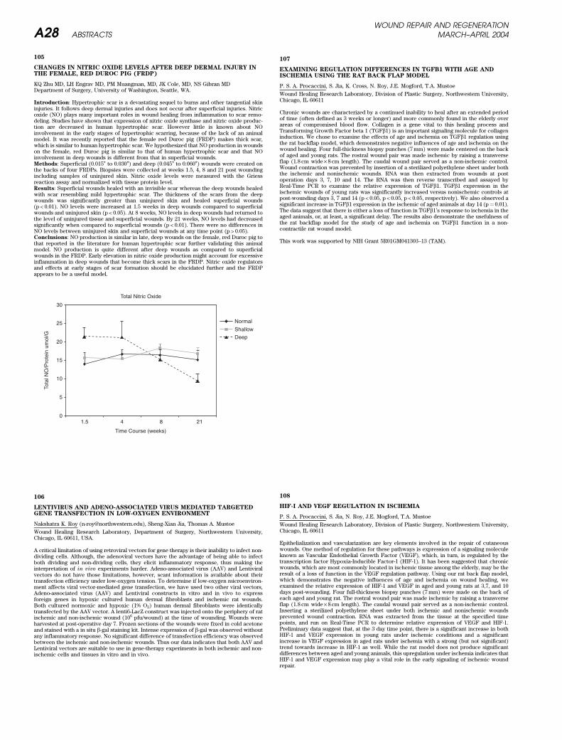
105
CHANGES IN NITRIC OXIDE LEVELS AFTER DEEP DERMAL INJURY INTHE FEMALE, RED DUROC PIG (FRDP)
KQ Zhu MD, LH Engrav MD, PM Muangman, MD, JK Cole, MD, NS Gibran MDDepartment of Surgery, University of Washington, Seattle, WA.
Introduction: Hypertrophic scar is a devastating sequel to burns and other tangential skininjuries. It follows deep dermal injuries and does not occur after superficial injuries. Nitricoxide (NO) plays many important roles in wound healing from inflammation to scar remo-deling. Studies have shown that expression of nitric oxide synthase and nitric oxide produc-tion are decreased in human hypertrophic scar. However little is known about NOinvolvement in the early stages of hypertrophic scarring, because of the lack of an animalmodel. It was recently reported that the female red Duroc pig (FRDP) makes thick scar,which is similar to human hypertrophic scar. We hypothesized that NO production in woundson the female, red Duroc pig is similar to that of human hypertrophic scar and that NOinvolvement in deep wounds is different from that in superficial wounds.Methods: Superficial (0.015’’ to 0.030’’) and deep (0.045’’ to 0.060’’) wounds were created onthe backs of four FRDPs. Biopsies were collected at weeks 1.5, 4, 8 and 21 post woundingincluding samples of uninjured skin. Nitric oxide levels were measured with the Griessreaction assay and normalized with tissue protein level.Results: Superficial wounds healed with an invisible scar whereas the deep wounds healedwith scar resembling mild hypertrophic scar. The thickness of the scars from the deepwounds was significantly greater than uninjured skin and healed superficial wounds(p < 0.01). NO levels were increased at 1.5 weeks in deep wounds compared to superficialwounds and uninjured skin (p < 0.05). At 8 weeks, NO levels in deep wounds had returned tothe level of uninjured tissue and superficial wounds. By 21 weeks, NO levels had decreasedsignificantly when compared to superficial wounds (p < 0.01). There were no differences inNO levels between uninjured skin and superficial wounds at any time point (p > 0.05).Conclusions: NO production is similar in late, deep wounds on the female, red Duroc pig tothat reported in the literature for human hypertrophic scar further validating this animalmodel. NO production is quite different after deep wounds as compared to superficialwounds in the FRDP. Early elevation in nitric oxide production might account for excessiveinflammation in deep wounds that become thick scars in the FRDP. Nitric oxide regulatorsand effects at early stages of scar formation should be elucidated further and the FRDPappears to be a useful model.
106
LENTIVIRUS AND ADENO-ASSOCIATED VIRUS MEDIATED TARGETEDGENE TRANSFECTION IN LOW-OXYGEN ENVIRONMENT
Nakshatra K. Roy ([email protected]), Sheng-Xian Jia, Thomas A. Mustoe
Wound Healing Research Laboratory, Department of Surgery, Northwestern University,Chicago, IL 60611, USA.
A critical limitation of using retroviral vectors for gene therapy is their inability to infect non-dividing cells. Although, the adenoviral vectors have the advantage of being able to infectboth dividing and non-dividing cells, they elicit inflammatory response, thus making theinterpretation of in vivo experiments harder. Adeno-associated virus (AAV) and Lentiviralvectors do not have those limitations, however, scant information is available about theirtransfection efficiency under low-oxygen tension. To determine if low-oxygen microenviron-ment affects viral vector-mediated gene transfection, we have used two other viral vectors,Adeno-associated virus (AAV) and Lentiviral constructs in vitro and in vivo to expressforeign genes in hypoxic cultured human dermal fibroblasts and ischemic rat wounds.Both cultured normoxic and hypoxic (1% O2) human dermal fibroblasts were identicallytransfected by the AAV vector. A lenti6-LacZ construct was injected onto the periphery of ratischemic and non-ischemic wound (106 pfu/wound) at the time of wounding. Wounds wereharvested at post-operative day 7. Frozen sections of the wounds were fixed in cold acetoneand stained with a in situ b-gal staining kit. Intense expression of b-gal was observed withoutany inflammatory response. No significant difference of transfection efficiency was observedbetween the ischemic and non-ischemic wounds. Thus our data indicates that both AAV andLentiviral vectors are suitable to use in gene-therapy experiments in both ischemic and non-ischemic cells and tissues in vitro and in vivo.
107
EXAMINING REGULATION DIFFERENCES IN TGFB1 WITH AGE ANDISCHEMIA USING THE RAT BACK FLAP MODEL
P. S. A. Procaccini, S. Jia, K. Cross, N. Roy, J.E. Mogford, T.A. Mustoe
Wound Healing Research Laboratory, Division of Plastic Surgery, Northwestern University,Chicago, IL 60611
Chronic wounds are characterized by a continued inability to heal after an extended periodof time (often defined as 3 weeks or longer) and more commonly found in the elderly overareas of compromised blood flow. Collagen is a gene vital to this healing process andTransforming Growth Factor beta 1 (TGFb1) is an important signaling molecule for collageninduction. We chose to examine the effects of age and ischemia on TGFb1 regulation usingthe rat backflap model, which demonstrates negative influences of age and ischemia on thewound healing. Four full-thickness biopsy punches (7 mm) were made centered on the backof aged and young rats. The rostral wound pair was made ischemic by raising a transverseflap (1.8 cm wide· 8 cm length). The caudal wound pair served as a non-ischemic control.Wound contraction was prevented by insertion of a sterilized polyethylene sheet under boththe ischemic and nonischemic wounds. RNA was then extracted from wounds at postoperation days 3, 7, 10 and 14. The RNA was then reverse transcribed and assayed byReal-Time PCR to examine the relative expression of TGFb1. TGFb1 expression in theischemic wounds of young rats was significantly increased versus nonischemic controls atpost-wounding days 3, 7 and 14 (p < 0.05, p < 0.05, p < 0.05, respectively). We also observed asignificant increase in TGFb1 expression in the ischemic of aged animals at day 14 (p¼ 0.01).The data suggest that there is either a loss of function in TGFb1’s response to ischemia in theaged animals, or, at least, a significant delay. The results also demonstrate the usefulness ofthe rat backflap model for the study of age and ischemia on TGFb1 function in a non-contractile rat wound model.
This work was supported by NIH Grant 5R01GM041303–13 (TAM).
108
HIF-I AND VEGF REGULATION IN ISCHEMIA
P. S. A. Procaccini, S. Jia, N. Roy, J.E. Mogford, T.A. Mustoe
Wound Healing Research Laboratory, Division of Plastic Surgery, Northwestern University,Chicago, IL 60611
Epithelialization and vascularization are key elements involved in the repair of cutaneouswounds. One method of regulation for these pathways is expression of a signaling moleculeknown as Vascular Endothelial Growth Factor (VEGF), which, in turn, is regulated by thetranscription factor Hypoxia-Inducible Factor-1 (HIF-1). It has been suggested that chronicwounds, which are most commonly located in ischemic tissue among the elderly, may be theresult of a loss of function in the VEGF regulation pathway. Using our rat back flap model,which demonstrates the negative influences of age and ischemia on wound healing, weexamined the relative expression of HIF-1 and VEGF in aged and young rats at 3,7, and 10days post-wounding. Four full-thickness biopsy punches (7 mm) were made on the back ofeach aged and young rat. The rostral wound pair was made ischemic by raising a transverseflap (1.8 cm wide· 8 cm length). The caudal wound pair served as a non-ischemic control.Inserting a sterilized polyethylene sheet under both ischemic and nonischemic woundsprevented wound contraction. RNA was extracted from the tissue at the specified timepoints, and run on Real-Time PCR to determine relative expression of VEGF and HIF-1.Preliminary data suggest that, at the 3 day time point, there is a significant increase in bothHIF-1 and VEGF expression in young rats under ischemic conditions and a significantincrease in VEGF expression in aged rats under ischemia with a strong (but not significant)trend towards increase in HIF-1 as well. While the rat model does not produce significantdifferences between aged and young animals, this upregulation under ischemia indicates thatHIF-1 and VEGF expression may play a vital role in the early signaling of ischemic woundrepair.
30
Total Nitric Oxide
Normal
Shallow
Deep
Time Course (weeks)
25
20
15
Tota
l NO
/Pro
tein
um
ol/G
10
5
01.5 4 8 21
WOUND REPAIR AND REGENERATIONA28 ABSTRACTS MARCH–APRIL 2004
109
USE OF EPR SPECTROSCOPY TO MEASURE TISSUE OXYGEN IN ANISCHEMIC FLAP MODEL
LJ Gould1, J Mushkudiani1, H Hou2, OY Grinberg2, S Grinberg2, HM Swartz2
1University of Texas Medical Branch, Galveston, TX 2EPR Center for Viable BiologicalSystems, Dartmouth Medical School, Hanover, NH
Methods to reliably measure tissue oxygenation in situ are currently lacking. We havedeveloped a vertically oriented, dorsal, bipedicle flap model that is easy to perform, reliablyreproduces tissue ischemia, eliminates craniocaudal variation, and is amenable to studyingtherapeutic modalities. The effect of narrowing this flap on tissue oxygenation measuredwith Licox electrodes has previously been presented. In this study we utilize in situ EPRspectroscopy to demonstrate the oxygen gradient in the flap as a function of flap width andplacement of a silicone sheet directly under the flap. The effect of wound healing over a 2week period is demonstrated.Twenty four, 300 gm male Sprague-Dawley rats underwent creation of the bipedicle flapaccording to the following groups: 2.5 cm flap with silicone, 2.0 cm flap without silicone,2.0 cm flap with silicone. Each group of 6 animals was injected with EMS char at 2 cmintervals along the flap and one injection in the control, non-ischemic tissue. A 4th groupunderwent 2.0 cm flaps with silicone and use of lithium phthalocyanin as the paramagneticmaterial. Wound measurements and EPR spectroscopy were performed on days 3, 7, 10 and14. On day 14, after EPR measurements, the animals were sacrificed and their woundsexcised. One flap and one control wound were preserved for histologic analysis, the otherflap and control wounds were prepared for lactate measurements.EPR spectroscopy demonstrated a gradient of oxygen that was lowest in the center of theflap and greatest at either end. Changes in the oxygen gradient correlated with narrowingand placement of the intervening silicone sheet. This new technology has never been utilizedin an animal model of impaired wound healing. Comparison of recently developed paramag-netic materials for optimal tissue oxygen and free radical measurements will be presented.
110
A NEW ANTIMICROBIAL ALGINATE DRESSING FOR MODERATE TOHEAVILY EXUDING, INFECTED AND CRITICALLY COLONISED WOUNDS
Deborah Addison, Tracy Jane Rennison, Sally-Anne Norris,
Johnson & Johnson Wound Managment
Aim: An antimicrobial dressing that combines high absorbency and potent antimicrobialproperties. The unique composition of the dressing manages exudates in moderate to heavilyexuding, infected or critically colonised wounds, which creates a favourable environment foreffective wound management.Methods: Two test methods were used to evaluate the antimicrobial properties of thedressing: 1) A zone of inhibition sensitivity test that challenges the dressing to a seededagar plate and 2) A log reduction test that exposes a small sample of dressing against a highbacterial loaded broth, measuring the log reduction of the bacteria over a specified timeperiod. The absorbency of the dressing was evaluated using British Pharmacopoeia testmethods.Results: The antimicrobial dressing has been shown to be highly effective against a widerange of microorganisms, including Pseudomonas aeruginosa, Escherichia coli, Streptococcuspyogenes, Staphylococcus aureus, Staphylococcus epidermidis, Klebsiella pneumoniae andCandida albicans and also against resistant organisms MRSE, MRSA and VRE. The productis highly absorbent with absorbency > 16 g/g and 16 g/m2.
111
HB-50: A PRE-CLINICAL STUDY OF A PROPHYLACTIC FOR WOUNDINFECTION
Scott M Harris1, Lijuan Zhang1, Jody Parente1, George T. Rodeheaver2, and Timothy J. Falla1.1Helix BioMedix Inc., Bothell Washington 980212University of Virginia Plastic Surgery Research, Charlottesville, VA 22908
Topical prophylaxis against wound infection by an agent that is active against multi-resistantbacteria does not generate resistance and is rapidly cidal would be of great clinical benefit.Peptides of the innate immune system have long been known to protect a wide range oforganisms from attack by bacterial and fungal pathogens. Helix BioMedix Inc. has developeda short bioactive peptide antimicrobial modeled after these peptides. HB-50 is an amphi-pathic cationic alpha-helical peptide that has broad spectrum activity and is rapidly micro-bicidal. These attributes make HB-50 an ideal candidate for wound infection prophylaxis. Invitro studies have demonstrated HB-50 to be active against both gram-positive and gram-negative bacteria killing 5–7 log orders of bacteria within minutes. In addition, this peptidehas potent activity against Vancomycin and Mupirocin resistant S. aureus. Preliminarytesting of the peptide in a rat abraded skin infection model has shown the peptide’s effec-tiveness in preventing wound infection while not inhibiting wound healing. Additionally, theHB-50 sequence has been specifically developed to be cost effective to manufacture andtherefore is well suited for use as a topical antimicrobial agent.
112
EFFECTS OF HIGH VOLTAGE PULSED CURRENT ON BACTERIALVIABILITY: AN IN VITRO STUDY
Cassie A. Kuykendall, SPT, Trish L. Hutchinson, Ed.D., FACSM, Alan P. Bloedaum, Ph.D.
Angelo State University, San Angelo, Texas
Background and Purpose: High voltage pulsed current (HVPC) has been shown to havepositive effects on wound healing due in part to proposed bacteriocidal effects in woundbeds. The purpose of this study was to examine the effects of electrical stimulation (HVPC)using in vitro Streptococcus A and clinically-accepted wound care guidelines.Methods: Commercially available Streptococcus A was grown overnight in Trypticase SoyBroth. Standard 100 millimeter (mm) Petri dishes of Trypticase Soy Agar with 5% sheepblood were then streaked using sterile cotton swabs. 52 plates were subjected to 45-minutesof HVPC with the following parameters: 120 pps, 200 V, 10-second on/off cycle, and 2-secondramp time. Negative polarity was used as the active electrode. 48 control plates wereprepared identically without the application of the HVPC. Zones of inhibition were measuredthe following day at the positive and negative electrode sites to the nearest millimeter after a24-hour incubation period using the Kirby-Bauer method.Results: Differences were calculated using a two-way ANOVA with SPSS 11.5. Mean inhib-ition at the positive and negative electrodes was 0.00 mm� 0.00 and 10.13� 4.13 mm, respec-tively. Significant between group effects were found for the control and experimental groups(F1,199¼ 289.4, p < 0.001), negative and positive electrodes (F1,199¼ 289.4, p < 0.001), andinteraction of groups by electrodes (F1,199¼ 289, p < 0.001).Conclusion: In vitro, negative polarity HVPC using clinically accepted and feasible para-meters can inhibit Streptococcus A growth around the negative (active) electrode. No growthinhibition was noted around the positive (dispersive) electrode. This could provide analternate means of infection control in assistance with wound healing.
Acknowledgments to Mr. Mark Pape and Dr. Carol B. Diminnie
WOUND REPAIR AND REGENERATIONVOL. 12, NO. 2 ABSTRACTS A29
113
DOES WOUND CARE EDUCATION SPEED HEALING OF INFECTED, ACUTEWOUNDS?
J Sherman, MD, A Lyndon, RNC, MS, CNS, H Hopf, MD,
University of California at San Francisco, Wound Healing Laboratory
Background: Wound infections cause significant morbidity in women after C-Sections.Regular lectures on wound care were instituted for obstetrical residents at our institutionin 1999. We hypothesized that resident education would decrease time to complete closure inpost-C-Section wounds healing by secondary intention.Methods: With IRB approval, a retrospective chart review was performed to obtain thefollowing information in patients treated pre (1996–1999) versus post (2000–2003) educationintervention: age, race, occupation, social support, medical co-morbidities, medications,week of gestation at delivery, duration of 1st stage of labor, length of ruptured membranes,meconium, chorioamnionitis, Group B Strep status, indications for C-Section, antibiotics,wound treatment, wound culture, wound dressing, time to full closure, number of visits forwound care (home, office), and need for repeat procedures.Results: Data are currently being analyzed.Conclusions: If resident education reduces the time to full closure, then we will evaluateexpanding this teaching approach to other departments using a randomized prospectivestudy.
Sponsored by NIH NIGMS 27345.
114
A COMPARISON OF IN VITRO DISC DIFFUSION AND TIME-KILL KINETICASSAYS FOR THE EVALUATION OF ANTIMICROBIAL ACTIVITY OFSILVER CONTAINING WOUND DRESSINGS
Corrie L. Gallant1, Hua Q. Yin2, Rita E. Langford2, Merle E. Olson1, David A. Hart1 and Robert
E. Burrell3
1. Department of Microbiology and Infectious Diseases, University of Calgary, Alberta,Canada2. Nucryst Pharmaceuticals Corporation, Fort Saskatchewan, Alberta, Canada3. Department of Chemical and Materials Engineering, University of Alberta, Edmonton,Alberta, Canada
There is a plethora of new silver containing dressings on the market today. Various manu-facturers attempt to show that their dressings are the most efficacious and therefore shouldbe preferentially employed by health care workers based on the results of their in vitro tests.However, there have been no studies to date that clearly identify which in vitro tests areappropriate for comparison purposes. The available literature suggests that there are pro-blems with disc sensitivity assays in terms of assessing the antimicrobial efficacy of silver.Spadaro (1985) has shown that the diameters of zones of inhibition were not proportional tothe concentration of anodic silver in complex test media. Further, Richards et al (2001) haveshown that zones of inhibition did not appear to correlate to log reduction assay data forsilver containing dressings. Other authors favour log reduction or time kill assays as theybetter compare to clinical data. The purpose of this study was to determine which in vivo
test is most appropriate for evaluating the antimicrobial efficacy of silver-containing dres-sings. This was done by testing 8 different silver containing dressings and 2 non-silver(mafenide acetate or hexamethylene biguanide) containing topical agents against 17 clin-ically relevant microorganisms using both zone of inhibition assays and time-kill kineticassays in complex media. The results for the two assays were then correlated to determine ifthe methods generated similar results. It was determined that the two methods do notcorrelate at all. This is most likely a result of the silver interacting with the media in thezone of inhibition test, thus invalidating the results of this test. We therefore conclude thatzone of inhibition data generated for silver-containing dressings is of little value whenassessing antimicrobial efficacy, and that time-kill assays are of greater use.
Funding provided by Natural Sciences and Engineering Research Council of Canada(NSERC) and Nucryst Pharmaceuticals.
115
CARBOHYDRATE-BASED WOUND DRESSINGS WITH ELASTASE-LOWERING ACTIVITY
Vincent Edwards1, Dorne Yager2, Kelman Cohen2, Steven Goheen3,
(1) Southern Regional Research Center, New Orleans, LA, (2) MCV-VCU, Richmond VA,(3) PNNL, Richland WA,,
The use of carbohydrate-based wound dressings including cotton, xerogels, charcoal cloth,alginates, and hydrogels, for chronic wounds have afforded properties such as absorbency,ease of application and removal, bacterial protection, fluid balance, occlusion, and elasticity.An additional consideration is the use of the dressing to regulate proteolysis in the non-healing wound. Elastase is a serine protease that has been associated with a variety ofinflammatory diseases, and recently has been implicated as a destructive protease thatimpedes wound healing. The presence of elevated levels of elastase in non-healing woundshas been associated with the degradation of important growth factors and fibronectinnecessary for wound healing. Here we consider the design, preparation, and assessment ofcarbohydrate-based wound dressings that either release protease inhibitors or act as seques-tering agents to bind proteases such as elastase and thus reduce their high levels in thechronic wound. Carbohydrates ranging from small monosaccahrides up to large polysacchar-ides are crosslinked onto cotton wound dressings. The resulting carbohydrate conjugates ofcotton cellulose possess unique properties advantageous for chronic wound treatment ran-ging from gelation and elasticity to elastase-sequestering propertiesType in text here making sure it is in a single column format.
Type acknowledgements here.
116
RISK FACTORS FOR PRESSURE ULCERS IN ELDERLY HOSPITALPATIENTS
Mona Baumgarten, D. Margolis, R. Lowe, R. Localio, S. Kagan, J. Holmes, W. Kavesh,
B. Kinosian, S. Abbuhl, A. Ruffin
Elderly hospital patients have ample exposure to situations and procedures that mayincrease their pressure ulcer risk. The aims of this study were to estimate the incidence ofhospital-acquired pressure ulcers in elderly medical patients and to determine whether therisk of pressure ulcers is associated with longer emergency department (ED) stays, lengthyprocedures in the ED, or use of immobilizing restraints, medications, or devices in the ED.The study cohort was made up of 3,233 patients aged 65 or over admitted through the EDs oftwo Philadelphia hospitals. Sixty-five percent of subjects were aged 80 or more, 61% werefemale, and 69% were Black. The incidence of hospital-acquired pressure ulcers on Day 3 ofthe hospital stay, as ascertained by direct skin examination, was 6.2%. There were 266hospital-acquired pressure ulcers among 201 patients (mean per patient 1.3 pressure ulcers)27% of the pressure ulcers were stage 1, 54% were stage 2, and the remainder were stages 3or 4 or could not be staged. The median length of stay in the ED was 6.6 hours. Theassociation between hospital-acquired pressure ulcers and extrinsic risk factors will bepresented. The identification of potentially modifiable risk factors may lead to the develop-ment of effective interventions to prevent hospital-acquired pressure ulcers.
WOUND REPAIR AND REGENERATIONA30 ABSTRACTS MARCH–APRIL 2004
117
CARE OF THE THERMALLY INJURED BREAST
R.E. Ortega, MD, L.H. Riina, MD, T.A. Davenport, MD, L.T. Glickman, MD, R.L. Simpson, MD,
K.M. Burger, PA, D. Hangan, RN
Nassau University Medical Center, 2201 Hempstead Tpk, East Meadow, NY 11554, [email protected]
Introduction: Thermal injury to the female breast is one of the most challenging aspects ofaesthetic burn surgery today. As the ability to provide coverage for large body surfaceinjuries has progressed greatly in recent years; attention can now be directed towardsachieving aesthetically pleasing results. Breast reconstruction in the burn patient can beaccomplished in several ways. In planning the reconstruction, one must account for gender,age, and stage of breast development at the time of injury. The following is the treatmentprotocol utilized at our institution. After determining the level of injury the injured area iscleansed and treated with topical anti-microbial agents such as Silvadene. If the decision ismade to excise and graft the injured area one of two algorithms is followed. The first choiceinvolves excising the burn and placing a split thickness skin graft to the area involved. This isdone by placing a sheet graft and using aerosolized fibrin sealant to affix it to the wound bed.If the burn involves deeper elements of tissue then a second approach is taken whichincludes excision of the burn down to the level of fascia with preservation of the breastmounds and the nipple areola complex (NAR). The (NAR) is spared excision and allowed toheal. Reconstruction of the (NAR) can be deferred for a secondary procedure dependingupon the response to primary healing. A split thickness skin graft is then applied to the areaof injury. Again a sheet graft is preferred and fibrin sealant is utilized to improve graftfixation and contour. We attribute our excellent results to the sheet grafts and fibrin sealantused. It should be noted that the increased vascularity of the breast fat when compared to fatlocated elsewhere in the body allows the grafts to adhere and survive on this generallydifficult to graft surface.Methods: We identified five female patients at our institution over the last 18 months withthermal injuries to the breasts. Each patient was placed into one of the two treatmentalgorithms.Results: The five patients had excellent outcomes. Breast mounds and symmetry werepreserved. Further development of the breast was allowed in each patient. One patienteven underwent a breast augmentation after surviving a 50% TBSA injury. Proper use offibrin sealant and sheet grafts account for the excellent results seen at this institution.Conclusion: Following careful evaluation of the burned female breast cosmetically andfunctionally acceptable results can be attained when following our institution’s protocolfor breast reconstruction in the female burn victim.
118
DERMAGRAFT AS AN ADJUNCT IN THE TREATMENT OF VENOUS LEGULCERS
R.J. Snyder
Medical Director, Wound Healing Center at University, Tamarac, Fl USA
Aim: To document successful healing of a recalcitrant venous ulcer with adjunctive use ofDermagraftMaterials and Methods: A 74-year-old Hispanic male presented with a chronic venous ulcerat the medial aspect of his right ankle. Biopsy failed to reveal malignancy and confirmedstasis changes. Treatment was ongoing including compression therapy, local debridements,and moist wound healing, off-loading, and nutritional support. The wound remained free ofinfection. Due to the continued recalcitrant nature of the lesion multiple applications ofDermagraft were utilized to augment healing.Results: The researcher observed dramatic improvement in wound dimensions, woundcontracture, and granulation tissue.Conclusions: Dermagraft appears to augment healing in patients with venous ulcers andmay be useful as adjunctive therapy.Acknowledgements: Smith & Nephew Wound Management, Largo, Fl.
119
CLINICAL EXPERIENCE WITH THE SELF-ASSEMBLED SKIN SUBSTITUEAS A BIOLOGICAL DRESSING FOR CHRONIC VENOUS LEG ULCERSTREATMENT
N. Dube1, M.-H. Rochon1, C.E. Ospina1, J. Soucy2, L. Germain1, F.A. Auger1
1LOEX, CHAUQ, Hopital du Saint-Sacrement, Universite Laval, Quebec, Canada 2CHAUQ,Hopital de l’Enfant-Jesus, Quebec, Canada
Introduction: Venous ulcers are the most common chronic wounds of the lower limbs. Inour laboratory, the self-assembly method is used to reconstruct autologous skin that can beused as a biological dressing (In Vitro Cell Dev Bio Anim, 1999: 35, 318; J Chem Eng, 2001:79, 663).Goals and Methods: The aim of this study was to establish if our self-assembled skinsubstitute (SASS) allows the closure of nonhealing venous leg ulcers. A case study of a68-year-old woman with a chronic venous leg ulcer present for over one year and measuring135 cm2 is thus herein presented. The autologous cells were isolated from a cutaneous biopsy(1 cm2) and cultivated in vitro (J Chem Eng, 2001: 79, 663). The autologous SASS wasproduced and assembled by the cells thus it does not contain any exogenous or syntheticmaterial (In Vitro Cell Dev Bio Anim, 1999: 35, 318). The wound was debrided, covered withgrafts of SASS and maintained in place with a compressive dressing (Coban, 3M, St-Paul,MN). A new SASS was applied weekly until wound closure. The SASS seemed to lessen thepain caused by the ulcer. Seven applications of the SASS were necessary to lead to a fullwound closure.Conclusion: Venous leg ulcer quickly reepithelialized after application of our SASS Thiscase indicates that use of the SASS can reduce pain and promote closure of nonhealing legulcers Furthermore, we thus hope that our SASS biological dressing maybe clinically usefulto treat difficult-to-heal leg ulcers. This study was approved by the Health Canada’sTherapeutic Products Directorate and by the Ethic Committee of the CHAUQ, Saint-Sacrement Hospital.Acknowledgements: Federation des Pompiers du Quebec pour les Grands Brules.
120
LASER DOPPLER PERFUSION IMAGING FOR THE ASSESSMENT OFHEALING IN PRESSURE ULCERS
Kekan M. and Feldman D.,
Department of Biomedical Engineering University of Alabama at Birmingham, Birmingham,AL, USA
The standard clinical assessment parameter for skin wounds has been time to heal. There isa need for assessments that monitor the healing process and have been validated asindicators of clinical outcome. Although the Laser Doppler Perfusion Imager (LDPI) hasbeen used to assess various wounds there is a need to validate it as a predictor of woundhealing. Since this measure can have a high variability, previous studies were done todetermine the key environmental and operational parameters as well to determine thecorrelation between blood perfusion and tissue oxygen levels. This helped determine theparameters to study as well the utility of using the change in blood perfusion after aprovocation (e.g. heat) rather than the absolute blood perfusion level.The objectives of this study were to further determine operational parameters, to developclinical protocols, and to begin the process of correlating blood perfusion values to clinicaloutcome. The effect of parameters such as light intensity, distance of the laser head from theskin, and skin pigmentation on blood perfusion were examined. Further, the relationshipbetween skin temperature and blood perfusion was studied as well as the best ways todeliver this provocation clinically. Preliminary studies have helped determine the appropri-ate ranges for each parameter to give the most consistent readings. In addition, the best wayto use the heat provocation has also been determined. In ongoing studies similar experi-ments are being carried out on pressure ulcers to assure these protocols work for clinicalwounds. The best clinical protocols will then be used to validate the prognostic capabilitiesof the LDPI in terms of predicting wound healing rates (overall healing rate, epithelializationrate, and contraction rate).Type in text here making sure it is in a single column format.Acknowledgements: Biofisica, LLC and CDC through NCIPC
WOUND REPAIR AND REGENERATIONVOL. 12, NO. 2 ABSTRACTS A31
121
EFFICIENT SEQUESTRATION OF NEUTROPHIL-DERIVED PROTEASES BYA NOVEL POLYMERIC HYDROGEL
D.J. Vachon1, D.R. Yager2
1Aegis BioSciences, LLC, Granada Hills, CA USA 2Virginia Commonwealth University,Richmond, VA USA
There is a growing interest in the development of wound dressings that possess functionalitybeyond providing physical protection and optimal moisture environment. To this end, a noveldressing material based on a sulfonated-hydrocarbon backbone has been developed for thecontrolled release of therapeutic agents into chronic wounds. This versatile polymer pos-sesses an ion-exchange capability that makes it amenable for binding and releasing a varietyof potential agents. Polymer backbone was coated onto polyester fabric and then modifiedby ion exchange to incorporate Naþ, Agþ, or doxycycline onto the polymer. The ability ofseveral formulations of this hydrocarbon backbone product to sequester elastase and neu-trophil collagenase (MMP-8) was then examined.Wound fluids containing elastase and MMP-8 were obtained from pooled wound fluidsobtained from consented patients with pressure ulcers. Equivalent weights of productformulations were incubated for 2–4 hours at 25 ˚C with the wound fluids and then theremaining activities of elastase and MMP-8 determined using specific substrates(methoxysuucinyl-ala-ala-pro-val-p-nitroanilide and collagen type I respectively). Untreatedpolyester fabric served as a control and weight equivalents of gauze and Promogran� werealso included for the purposes of comparison. The average reduction in apparent elastaseactivity in the wound fluid was 4%, 20.4%, 14%, 29%, and 55% for gauze, Promogran�,doxycycline, Agþ, and Naþ formulations respectively. Only Promogran� and the doxycyclineformulation affected a decrease in apparent collagenolytic activity.These preliminary results provide evidence in support of the versatility of these new biomater-ials for the controlled modulation of the chronic wound environment.
122
MICROVASCULAR DRESSING FOR BURN APPLICATIONS
M. Cabodi1, A.D. Stroock1, L. Staiano-Coico2, Roger Yurt2
1Chemical Engineering, Cornell University, Ithaca, NY USA 2Weill Medical College, CornellUniversity, New York, NY USA
Vascular structures are a ubiquitous element of living tissues that allow for efficient masstransfer with the bulk of the tissue. Reasons for the vascularization of tissue include: 1)efficiency of transport over large distances (>1 mm) by convection, 2) spatial control ofdelivery and evacuation of solute from the tissues, and 3) centralization of the influx andefflux from the tissue; this centralization allows for temporal control and monitoring to beperformed. Many of these characteristics would also be valuable in synthetic materialsdesigned for interaction with biological systems. We will present the design and fabricationof a wound dressing that incorporates microvasculature in the form of embedded micro-channels. As in living systems, the basic principle behind our design is that active convectionis used to carry solute over large distances, while diffusion acts over smaller distances.We will describe the microfabrication procedure, based on soft-lithographic techniques, usedto fabricate the microvascular dressing. We will also discuss the design parameters whichare important for the optimization of mass transfer and a model of the system will bepresented. We will report on the physical characterization of the system, with respect tothe mass transport properties. Finally, we will discuss initial considerations about the use ofthese structures as wound dressings.
This material is based upon work supported by the STC Program of the National ScienceFoundation under Agreement No. ECS-9876771.
123
A NEW STUDY DESIGN TO COMPARE WOUND DRESSINGS
Baldur Baldursson MD PhD
Dermatologist University Hospital Reykjavik Iceland
The initial tests of a new ulcer dressing, even if the materials are well known and safe mustinclude comparison with other dressings.In order to test the performance of a new dressing manufactured to treat medium to heavilyexuding wounds we designed a wound model using wounds resulting from harvesting ofpunch grafts for treatment of chronic ulcers. These are usually made by a 6 mm wide biopsypunch and a scalpel and are 7 mm wide round split skin wounds reaching down to thereticular dermis in a number from 2–300.10 consecutive patients with leg ulcers that had a punch graft operation performed at thein-patient Dermatology ward in Reykjavik, Iceland were enrolled. The grafts were harvestedon the antero-lateral aspect of the thigh in a pattern of three groups each with 3–20 punchgraft wounds. A different type of wound dressing was used to treat each group. Dressingchanges were scheduled for one, four, 12 and 18 days after the operation.The nurse graded the appearance of the split skin wounds into infected or non-infected,inflammatory or non-inflammatory. The patient graded the degree of pain at dressingremoval using a visual assessment scale.The dressings were weighed before and after to assess fluid absorption. Detailed digitalphotographs recorded the progress of each group of donor sites.The outcome of the different parameters seemed consistent and repeatable.Using the donor site of punch grafts is a repeatable and consistent way of studying theperformance of new ulcer dressings, comparing them to established dressings.
Ossur hf producing company of Gentleheal founded the study
124
VENOUS ULCER HEALING PREDICTED BY URINE NITRATE ASSAY: ARETROSPECTIVE STUDY
J.V. Boykin1, C. Baylis2, D. Yager3, I.K. Cohen3, V. Sommer1, L. Shawler1, M. Watkins1,
J. Young1, M. Crossland1,1Retreat Hospital Wound Healing Center, 3Div. Plastic Surgery, Virginia CommonwealthUniv., Richmond, VA, 2Dept. of Physiology, West Virginia Univ., Morgantown, WVA
Chronic venous stasis ulcerations (VSU) are associated with significant platelet and leuko-cyte activation with secondary superoxide anion and ROS production that may severelydegrade nitric oxide (NO) production. This study was designed to document the possibleassociation between NO production and the impaired healing of VSU patients. Twenty-foursubjects were used for the study. Group C (n¼ 8) the control group, consisted of healthyadults without history of VSU. Group HU (n¼ 8) the healed ulcer group, consisted of adultswith documented VSU whose ulcers healed with routine wound care and compressiontherapy in 20 weeks or less. Group UU (n¼ 8) the unhealed ulcer group, consisted of adultswith documented VSU without healing within 20 weeks. All groups had normal renal functionand were hospitalized for 24 hours on bed rest with a low arginine and low nitrate diet.Fasting samples were obtained for plasma and urine nitrate (NOx) and plasma arginine,asymmetric dimethylarginine (ADMA) and isoprostane determinations. At 24-hrs, group HUdemonstrated the highest plasma NOx levels (33.25� 11.69 mM) as compared to group C(19.45� 2.63 mM) or group UU (27.17� 3.67 mM); urine NOx excretion of group UU(51.46� 11.77mM) was significantly lower (p < 0.05; Kruskal-Wallis) as compared to groupHU (135� 38.29 mM) or group C (164.3� 38.58 mM). ADMA and arginine were not signifi-cantly different between groups. Ratios of NOx to isoprostane were disproportionatebetween groups. These results suggest, for the first time, a significant relationship betweenimpaired VSU healing and deficient NO production. Furthermore, these findings establish theclinical value of measurements of NO production and bioavailability as effective predictive-biological markers for the healing of VSU.
Supported by a grant from Pfizer Limited
WOUND REPAIR AND REGENERATIONA32 ABSTRACTS MARCH–APRIL 2004
125
‘‘MIST ULTRASOUND THERAPY FOR RECALCITRANT WOUNDS, A NEWAPPROACH’’
William J. Ennis1, Wesley Valdes2, Patricio Meneses3.1Advocate Christ Medical Center, Wound Treatment Program, Medical Director 2Wound CareFellow, Advocate Christ Medical Center, Wound Treatment Program 3Research Director,Advocate Christ Medical Center, Wound Treatment Program
The use of adjunctive peri-wound, ultrasound with either a 1 MgHz or 3 MgHz frequency hasbeen reported on in the literature with variable results. MIST ultrasound therapy is a novelapplication of direct wound bed application of kilohertz ultrasound waves utilizing a normalsaline mist as the coupling mechanism, allowing for no direct patient contact. The mistgenerated by the system is of relative uniform particle size and acts as a conduit of ultra-sound energy. Unlike ultrasonic baths, which dissipate the vibrational energy over a largearea, the transducer horn focuses the energy into a smaller area of application.The MISTTM system consists of an ultrasonic power supply (generator), a transducer (oralternately referred to as the converter), and an applicator coupled to the transducer. Thegenerator converts voltage to high frequency electrical energy. This electrical energy istransmitted to the piezoelectric transducer within the converter, where it is changed tomechanical vibrations. The generator is designed to operate the converter at 40 kHz with adistal displacement of 60 microns. Published reports have described various biologicaleffects of low frequency ultrasound including VEGF and PDGF production, increased proteinsynthesis, improved bone and wound healing. Other authors have published abstractsdemonstrating clinical efficacy of this device on a broad range of patients with recalcitrantwounds. This report describes the clinical results of a prospective, non-randomized, efficacytrial, adding MIST ultrasound therapy to current treatment protocols in recalcitrant wounds.Wound healing trajectories before and after treatment will be evaluated and wound healingwill be assessed with laser Doppler image analysis and trans-cutaneous oximetry.
126
SCROTAL AND PENILE RECONSTRUCTION USING VAC THERAPY
N Snyder1, LJ Gould1
1University of Texas Medical Branch, Galveston, TX
Fournier gangrene is a rapidly progressive necrotizing soft tissue infection that often leavespatients with a large integumentary deficit to their penis and scrotum. Treatment of the skindefect has been described in a multitude of ways including split thickness skin grafting,burying the testes in the thighs, thigh flaps, and a variety of myocutaneous and fasciocutan-eous flaps.Many of the previously described techniques have worked well for smaller defects and forclosure of wounds but fell short of aesthetic reconstruction of sexual organs. We describe atechnique using split-thickness skin grafts with suction dressings or bolster. This wasemployed using the VAC# (Vacuum Assisted Closure Device, KCI#). This technique createsa natural appearing scrotum that holds the testes away from the body in a physiologicmanner and surfaces the penis with a seemingly natural appearance in a functional manner.The technique provides a nearly 100% graft take and greatly decreases hospital length of stay.A case report demonstrates a patient’s full recovery to his professional, social, and sexual lifewithin six weeks of initial reconstructive treatment. Our technique provides a reliabletreatment with good functional and aesthetic outcomes to a difficult problem.
127
THE IMPORTANCE OF ARM DOMINANCE IN ACUTE WOUND HEALING
T. Serena, on behalf of the Wound Healing Cooperative Group
Wound Healing Cooperative Group, The Angiogenesis Foundation, Cambridge, MA USA
Background: Physiological differences between dominant and non-dominant arms may playan important role in wound healing of the upper extremities. The Wound Healing Coopera-tive Group (WHCG) studied this phenomenon in a human forearm model of acute woundhealing.Methods: With informed consent, sixteen normal healthy volunteers underwent four 6 mmbiopsies on the flexor surface of both forearms: 2 biopsies on the dominant arm, and 2 on thenon-dominant arm. The acute wound sites were then treated with i) rhPDGF-BB 0.01% geltopically at various dosing intervals (Q.D. versus Q.O.D.), ii) rhPDGF-BB alone Q.D. · 7 daysfollowed by bacitracin alone, or iii) bacitracin alone. Time-to-complete healing was meas-ured as the primary endpoint.Results: The average time-to-complete healing for the dominant arm was shorter than thetime required to heal the biopsies on the non-dominant arm, irregardless of the treatmentapplied.Conclusion: In the forearm biopsy model of acute wound healing, arm dominance is anindependent variable and must considered in study designs. We postulate that this effect maybe due to variation in microvascular perfusion. These results provoke further investigationinto the physiological mechanisms underlying increased rate of healing in the dominantextremity.Acknowledgements: Funding from The Angiogenesis Foundation.
128
WOUND HEALING MORPHOLOGY IN AN ACUTE WOUND HEALING STUDY
T. Serena, on behalf of the Wound Healing Cooperative Group
Wound Healing Cooperative Group, The Angiogenesis Foundation, Cambridge, MA USA
Background: Physiological healing in acute wounds generally occurs by granulation fromthe wound base and by migration of epithelium from the wound edges toward the center ofthe wound in a uniform, concentric fashion. The Wound Healing Cooperative Group (WHCG)compared this phenomenon in acute wounds treated with standard care versus acutewounds pharmacologically stimulated with growth factor therapy.Methods: With informed consent, 20 normal healthy volunteers underwent four 6 mmbiopsies of the flexor surface of both forearms. The biopsy sites were randomly assignedto a control arm (daily bacitracin) or to one of three treatment arms: i) rhPDGF-BB 0.01% gelQ.D., ii) rhPDGF-BB Q.O.D., iii) daily rhPDGF-BB (Q.D.· 7 days followed by bacitracin alonedaily). Wound morphology was carefully examined and photographed daily until completehealing was achieved.Results: There were distinct differences in the morphological pattern of healing seenbetween control wounds and growth factor-stimulated wounds. Acute wounds treated withbacitracin tended to heal in circumferential fashion, as predicted. Growth factor-stimulatedwounds, by contrast, exhibited accentuated angiogenesis (granulation), with non-uniformepithelial islands streaming into the wound.Conclusion: This pilot study suggests that rhPDGF-BB influences acute wound healing bypromoting accelerated granulation and epithelialization. Accelerated healing is manifest bydifferent healing morphologies. The biological basis for these differences requires furtherhistological and molecular analyses.Acknowledgements: : Funding from The Angiogenesis Foundation.
WOUND REPAIR AND REGENERATIONVOL. 12, NO. 2 ABSTRACTS A33
129
A SUMMARY OF SCIENTIFIC STUDIES AND CLINICAL RESULTS WITH AGLYCERINE BASED DRESSING
E.I. Stout
Southwest Technologies, Inc., N. Kansas City, MO, USA
The importance of controlling the bioburden in wounds can not be over emphasized.Glycerine based gel sheets have been used extensively to show their bacteriostatic/fungi-static properties. Hoekstra studied animal wounds and compared glycerine dressings withwater based dressings and the glycerine showed superior bioburden reduction. Vandeputteshowed similar results when comparing hydrogel and hydrocolloid dressings and looked atthe histology of the wounds to find differences in the quantities of the types of cells present.The reduced scar formation of wounds are thought to be attributed to the influence of theglycerine on the healing process. Hoestra has reported the dramatic reduction in the inflam-mation reaction soon after application of the glycerine gel dressing. Studies by Oliveria-Gandia, Davis, and Mertz showed the glycerine dressings to be more effective than hydrogelor hydrocolloid dressings in reducing bioburden in animal wounds that were inoculated withmicrobes and also reducing biocounts in appropriate growth medium. Vandeputte conducteda diabetic study(no exclusions) that compared the glycerine dressing (n¼ 15) with standardprotocol(n¼ 14) for diabetic foot wounds, that showed the test dressing to be far superior.He along with thousands of other nurses around the world have reported the use of glycerinedressings on superficial burns to reduce pain, reduce the chance for infection, reduce scarformation, and to protect the wound from friction and pressure. J. Baksa extensively usedthe glycerine gel sheets in his burn unit not only for the superficial wounds but also for 3rd
and 4th degree burns on children as well as after surgical removal of hypertrophic and keloidscars to prevent reoccurrence. T.M. Baum and M.J. Busuito also reported the use of theglycerine dressing for scar prevention and treatment. The glycerine dressing has been usedextensively for te treatment and prevention of pressure ulcers in hospitals, nursing homes,athletic fields, as well as, under casts, splints and braces. R. Horchner reported a >95%reduction in pressure ulcers in a direct comparison to the control and to hydrocolloids.
130
DEVELOPMENT AND CHARACTERIZATION OF A SKIN ORGAN CULTURETHAT RESEMBLES HUMAN SKIN
M. Martins-Green, QiJing Li, Min Yao.
Department of Cell Biology and Neuroscience. University of California Riverside, RiversideCA, 92521.
The inability to experiment in humans creates a need to develop culture systems that mimichuman tissues/organs. Skin is arguably the simplest tissue hence provides an excellentprototype for tissue engineering. Moreover, skin is the body’s first line of defense. Currentlyavailable skin replacements have a number of drawbacks. Therefore, there is a need for skinreplacements that: (a) are prepared with pertinent primary human cells but yet can be ready‘‘off’’ the shelf; (b) can be prepared rapidly; (c) contain stable structures, in particularmicrovessels that can rapidly connect with the patient’s vasculature, thereby establishingcirculation in the ‘‘graft’’ and increasing the chances of survival; (d) can be tailored forspecific wound impairments (e) are long lasting. We have developed a generation human‘‘skin’’ that can fulfill these requirements and can potentially be used as a ‘‘living bandage’’.We start with three primary human cell types and a collagen matrix that self-assemble into aconnective tissue containing a network of mature microvessels, is covered with a stratifiedepidermis, expresses biochemical markers, matrix molecules, and cytokines characteristic ofnormal human skin, and matures in 10–15days. Moreover, two additional cell types, pericytesand monocytes, differentiate in situ adjacent to and within microvessels, respectively. Theepidermis expresses keratins typical of mature skin and not characteristically produced inresponse to injury such as keratins 6, 16, 17. This tissue potentially can be developed into askin replacement for patients with impaired healing. In addition, it responds normally tobiological stimuli, providing a powerful vehicle to investigate mechanisms of skin develop-ment and regeneration, understand pathological processes, and test drugs and treatments forskin diseases.
Supported by AHA and TRDRP.
131
LATERAL WICKING, GOOD OR BAD
Baldur Baldursson
University Hospital Reykjavik Iceland, Christopher Collins, Ossur hf Reykjavik.
The ability of polyurethane sponge dressings to absorb and contain wound fluid is ofteninsufficient, resulting in maceration and eczema.The term lateral wicking has been used to describe the fluid film between the peri-ulcer skinand the dressing surface, responsible for the maceration. A ‘‘within dressing’’ lateral wickingis where the wound fluid is absorbed above the ulcer and transported laterally within thedressing without affecting the peri-ulcer skin.We wanted to test this phenomenon in a new type of dressing with a silicon surface towardsthe wound, a polyurethane sponge layer with enclosed absorbent particles and a vapourpermeable outer layer.A pump delivered 10 ml per hr of equine serum to a 1 sq cm surface at the centre of thedressings. The experiment went on for 2.5 hours. Seven dressings of each type were used inthe experiment. The top and bottom aspects of the dressings were photographed at 30 mininterval.In the new ulcer dressing the serum was absorbed and distributed in a ‘‘within dressing’’lateral wicking. In the established dressing an ‘‘under dressing’’ lateral wicking took placeleading to erratic distribution of absorption often at the margin of the dressing instead ofover the centre.ConclusionEnhancing lateral wicking within a chronic ulcer dressing is a realistic way of optimising itsfluid handling characteristics. In this experimental set-up the new ulcer dressing performedas intended while the conventional dressing did not.Improved fluid handling characteristics are needed in today’s advanced dressings however,the importance of this phenomenon in the clinical situation must be tested in clinical trials.
Ossur hf producing company of Gentleheal founded the study
132
DIFFERENTIAL REGULATION OF OSTEOPONTIN AND MATRIXMETALLOPROTEINASES DURING DIABETIC WOUND HEALING
A. Sharma, A. K. Singh, J. Warren, R. K Maheshwari
Uniformed Services University of the Health Sciences, Bethesda, Maryland, USA
Delayed wound healing is a common complication of diabetes resulting in significant clinicalmorbidity. The diabetic wound exhibits impaired cellular infiltration and consequentlyinadequate granulation tissue formation. Additionally, altered patterns of apoptosis. In thisstudy, we have evaluated the differential gene expression pattern in transgenic diabeticfemale mice, 5–6 weeks old, in a full thickness cutaneous punch wound model. We assessedthe role of matrix metalloproteinases (MMPs) and differential gene expression pattern at 4th,7th and 11th day post wounding. Supernatants obtained from diabetic wound tissue homo-genates were subjected to zymogram analysis. The data showed that MMPs were expressedat higher level by 4th day post wounding, whereas expression of MMPs were down regulatedtowards the 11th day post wounding suggesting their role during early phase of woundhealing.The pathway specific gene array data demonstrated differential regulation of several growthfactors, transcription factors and other related genes such as fibroblast growth factors andtheir receptors, ID3 and restin respectively. The cytokine/extracellular matrix protein osteo-pontin (OPN), an important component of cellular immunity and inflammation also showedhigher expression after 4 days post wounding. The expression of OPN remained at higherlevel after 11 days post wounding in diabetic mice, whereas the expression were downregulated to basal level in normal wounded animal suggesting that the expression of OPNwas concomitant with the extent of healing. Other adhesion molecules such as integrin aVand PECAM-1 were also differentially regulated. Though a single gene may not be solelyresponsible for any defect or impairment in healing as it is a very tightly controlled andregulated process, however, a detailed study of these gene(s) may shed some light to thedelayed healing in diabetic mice.Acknowledgements: (These studies are supported by NIH Grant G174KT.)
WOUND REPAIR AND REGENERATIONA34 ABSTRACTS MARCH–APRIL 2004
133
GRANULATION TISSUE INDUCTION BY LASSAR OINTMENT VS.COLLAGEN-POLYVYNILPYRROLIDONE IN VENOUS ULCERS
Edgar Krotzsch1, Arturo Suarez-Colın2, Rosa M. Salgado1, Ana M.Z. Apis3,.1Connective Tissue Laboratory, Division de Investigacion Biomedica, C.M.N. ‘‘20 de Noviem-bre’’, I.S.S.S.T.E. Mexico City, Mexico. 2Plastic and Reconstructive Department, HospitalGeneral de Zona 32, and 3Research Area, Delegacion 3 y 4, I.M.S.S. Mexico City, Mexico.
Venous leg ulcers derived from tissue destruction is the consequence of a chronic inflam-matory process that produces pain and physical disability, diminishing quality of life inpatients. In this work, Lassar ointment and lyophilized collagen-polyvinylpyrrolidone wereadministered separated each on one half in the same ulcer to 9 patients at the beginning andevery 4 days. On day 16, all patients were auto-grafted with partial thickness skin. Granula-tion tissue and graft integration were assessed clinically during 3 months. Inflammatoryinfiltrate, type I and III collagens, elastic fibers, alkaline phosphatase as well as blood vesselswere evaluated histologically or histochemically in biopsies taken at the beginning and16 days after the local treatment.Clinically and morphologically, both treatments demonstrated appropriate granulation tissuepromotion and optimal graft integration since the beginning. Nevertheless, in Lassar oint-ment treated group regionalization of alkaline phosphatase activity was observed, as well asthe presence of granuloma in 2 of the 9 patients. In conclusion, Lassar ointment or lyophi-lized collagen-polyvinylpyrrolidone are two different promoters of granulation tissue invenous leg ulcers, however Lassar ointment has the capability to produce granuloma andan exacerbated immune response; in consequence, ulcer recidivism could be present, may bedue to mineral deposits in the wound.
This work was partially supported by Aspid S.A. de C.V.
134
EVALUATION OF ALTERATIONS IN GENE EXPRESSION IN NORMAL ANDDIABETIC MICE RESULTING FROM DERMAL INJURY
Kathleen E. Rodgers, Ph.D., Dolph D. Ellefson, Ph.D., Theresa Espinoza, M.D., Ya-hsuan Hsu,
M.S., Gere S. diZerega, M.D. Ruty Mehrian-Shai, Ph.D.University of Southern California KECK School of Medicine
C57Bl/6-db/db mice are a model of delayed healing after full thickness dermal excisioninjury. We evaluated the differences in gene expression between C57Bl/6 mice and C57Bl/6-db/db mice in uninjured skin and within a wound 7 days after injury and observed a numberof differences resulting from the diabetic mutation. Cluster analysis indicated differences ingenes involved in all structural organization and metabolism. There were 718 genes that hadgreater than 2 fold difference between these 2 strains of mice in granulation tissue at day 7.In normal and diabetic mice, 884 or 765 genes were altered 2 fold by injury (granulationtissue), respectively. When compared at the 4 fold level, the overlap of genes altered betweennormal and diabetic mice was approximately 68%. In granulation tissue, changes wereobserved in genes associated with inflammation, immune response, cell motility, andresponse to external stimuli. Normal mice had greater immune, stress, inflammatory, andbiotic responses. Wounds of diabetic mice had increased responses in energy metabolismand fatty acid metabolism. These data provide a basis for further understanding of theresponse of skin to injury and the delay that occurs in insulin resistant Type 2 diabetes.
American Diabetes Association.
135
INDUCTION OF GENE EXPRESSION FOR EGF-LIKE LIGANDS IN PORCINEWOUNDS FOLLOWING RECEPTOR OVEREXPESSION
M.I. Okwueze, N.L. Cardwell, A.C. Pollins, L.B. Nanney
Vanderbilt University, Nashville, TN USA
A rationale to explain the role of the large and redundant family of EGF cytokines withmultiple receptor combinations has proven elusive. We examined endogenous gene expres-sion levels during wound repair for four EGF-like cytokines (Transforming growth factor-alpha (TGFa), Heparin-binding epidermal growth factor (HB-EGF), Epiregulin (EPR) andBetacellulin (BC) using a quantitative real-time PCR technique. In addition, cytokine geneexpression was evaluated after overexpression of different of EGF receptor forms (erbB-1,erbB-3 and erbB-4) or a control B-galactosidase gene using an adenoviral transfectionstrategy to introduce either homo or heterodimer receptor combinations. At baseline, endo-genous gene expression levels of TGFa and HB-EGF were 10-fold and 100-fold higher thanEPR and BC, respectively. Expression of TGFa was increased by 3-fold in the receptorcombination of erbB-1/erbB-4 expressing wounds. Recent studies on the EGF-related poly-peptides HB-EGF, EPR and BC have shown preferential binding to erbB-4. Our studysuggests that wounds predominantly expressing erbB-4 showed a 2.5-fold increase in geneexpression of HB-EGF. A synergistic 4-fold increase in gene expression was noted whenerbB-4 overexpression was treated with HB-EGF ligand. In contrast, wounds transfectedwith erbB-4 alone showed no change in EPR gene expression compared to controls. How-ever, wounds treated with TGFa showed 3-fold increase in EPR gene expression and EPRpotentiated itself. Similarly, wounds transfected with erbB-4 and treated with growth factorsTGFa and EGF upregulated the gene expression of BC by 4-fold as compared to controls.Interestingly, wounds overexpressing erbB-3 with topica application of EGF accentuatedexpression of TGFa by 7-fold. In histologic sections, these wounds showed a 2-fold increasein the dermal region compared to control. Thus, the EGF-like family of ligands and its homo-or heterodimer receptor pairs have the potential in vivo to evoke differing profiles orintensities of gene expression patterns as they mediate wound repair.
(This project is funded by the NIH GM40437)
136
CELOSIA ARGENTEA LINN. LEAF EXTRACT IMPROVES WOUNDHEALING IN RAT BURN WOUND MODEL
Kulasekaran S Priya1, 2, Mary Babu2 and Alan Wells1.1Departments of Pathology, University of Pittsburgh, Pittsburgh, PA.USA 2BiomaterialsDivision, Central Leather Research Institute, Chennai, TamilNadu, India.
Celosia argentea is used in traditional medicine for sores, ulcers, and skin eruptions. There-fore, we investigated the healing efficiency of an ointment formulated on a alcohol extract ofCelosia argentea leaves (CA) in a rat burn wound model. Wound closure occurred earlier inthe treated rats (15 days versus 30 in the untreated group; P < 0.05). Collagen and hexso-amine content of the granulation tissue increased at a faster rate in the treated wounds. Toprobe the cell biologic basis of this effect, we found that this extract promoted cell motilityand proliferation of primary dermal fibroblasts but did not alter these responses in primarykeratinocytes. In short, we demonstrate a salutary action of the Celosia argentea extract onwound healing and suggest that this may be due to mitogenic and motogenic promotion ofdermal fibroblasts.
WOUND REPAIR AND REGENERATIONVOL. 12, NO. 2 ABSTRACTS A35
137
THE CUTANEOUS ‘‘UNDERLYING DISORDER’’ IN DIABETIC RATS
S.L. Lu C. Qing T. Xie M.J. Wang Y.W. Niu K. Ge S.W. Jin Z.J. Liao J.X. Shi
Shanghai Burns Institute, Rui Jin Hospital, Shanghai second Medical University, 200025Shanghai, China
Objective To investigate the histological characteristics and pathophysiological changes indiabetic skin.Methods: 12 Sprague-Dawley rats weighing 200–220 g were divided into control and STZ-induced diabetic groups. The shaved skin specimens from the back of rats were collected on8 w post STZ-inducing. The cutaneous histological characteristics were observed. The localcontents of glucose, advanced glycation end products(AGEs) and hydroxyproline, the levelsof aMMP-2 and TIMP-2, and the cell cycles of both keratinocytes and dermal cells weredetermined.Results: The thicknesses of epidermis layer and dermis layer were both reduced obviouslyin diabetic skin, with the morphological characteristics of the obscured multilayer epithe-lium features and the decreased amount of spinous in epidermis; the atrophied, swollen anddegenerated collagen fibers with a focal chronic inflammatory cells infiltration. The resultsalso revealed that contents of glucose, AGEs, the level of aMMP-2 and the ratio of aMMP-2/TIMP-2 in diabetic skin were higher than those in the controls. In diabetic group, thepercentages of S stage and G2/M stages of keratinocytes were obviously decreased, whilethe dermal cells showed the higher percentage of S stage and the normal percentage of G2/Mstages, when compared with the control group.Conclusion: The cutaneous histological and pathophysiological alterations in diabetesmellitus has already been occurred when exogenous damage is not existed, which presentedan ‘‘underlying disorder’’ characteristics in diabetic skin. The cutaneous ‘‘underlying disorder’’in diabetes might be a critical risk factor leading to ulceration and one of the most importantmechanisms in the pathogenesis of impaired wound healing.[Key words]: diabetic skin underlying disorder cell proliferation MMPs collagen
The project is supported by: Major State Basic Research Development Program of China(No. G1999054205).
138
MOLECULAR PATHOGENESIS OF CHRONIC WOUNDS: THE ROLE OF b-CATENIN AND C-MYC
O. Stojadinovic1, H. Brem2, B. Lee1, C. Vouthounis1, H. Entero2 and M. Tomic-Canic1.1Department of Dermatology, NYU School of Medicine, New York; 2Department of Surgery,Mt. Sinai School of Medicine, New York
The lack of understanding of molecular pathogenesis of impaired healing in chronic ulcersleads to a serious health issue that contributes to excessive limb amputations and mortality.Using biopsies from patients with chronic wounds, skin organ culture and primary keratino-cytes in culture we identified that b-catenin and its downstream target, c-myc, play importantrole in development of chronic wounds. In contrast to normal epidermis, we observedsignificant nuclearization of b-catenin and elevated c-myc expression at the non-healingwound edge of patients with chronic ulcers. In vitro studies indicated that activation andstabilization of nuclear b-catenin inhibits wound healing and keratinocyte migration by:blocking EGF response and inducing c-myc. Using Affymetrix large scale microarrays wefound that b-catenin downstream target, c-myc, is induced in skin by an inhibitor of woundhealing (glucocorticoids) and repressed in the initial phase of normal wound healing, 4 to48 hr, whereas it becomes de-repressed at 96 hr post wounding. Therefore, the activation ofb-catenin/c-myc pathway(s) contributes to impaired healing by inhibiting of keratinocytemigration and altering keratinocyte differentiation. The presence of activated b-catenin andc-myc in the epidermis of chronic wounds may serve as molecular markers of impairedhealing and future targets for therapeutic intervention. While b-catenin signaling has beenimplicated in epithelial development and oncogenesis its role in wound healing has neverbeen postulated. This further illustrates the importance of ‘‘tissue context’’ specificity,because b-catenin in the context of malignant tissue promotes invasion whereas in thecontext of a wound environment does the opposite.
Our research is supported by NIH grants AR45974, NR08029, DK59424, DK43093; EPVA andADA.
139
GENE ARRAY PROFILING OF KELOID FIBROBLASTS TO IDENTIFY THETARGET GENES FOR THERAPEUTIC EVALUATIONS
Latha Satish, Jian-Hua Luo, Kulasekaran S Priya and Alan Wells
Department of Pathology, University of Pittsburgh, PA 15261
Keloids are scars that overgrow the original boundaries of the injury, involving aberrantfunctioning of the fibroblasts. We have found that during normal wound healing the ELR-negative CXC chemokines IP-9 (CXCL11) and IP-10 (CXCL10) act to limit fibroblast immi-gration. Our initial goal was to determine if keloid fibroblasts failed to respond to this ‘stop’signal. Immunohistochemical and immunocytochemical studies on keloid fibroblasts showthat these lesions express excessive amounts of IP-9 and CXCR3 receptor. The protein levelsof CXCR3 receptor was also not dissimilar between normal and keloid fibroblasts. Though,the expression of the protein and the receptor seems to be normal, the fibroblasts derivedfrom these abnormal lesions did not respond to IP-9 during EGF induced cell migration. Wehave extended these findings to determine whether other genes are differentially expressedby keloid fibroblasts. The immediate first gene that interest us was the mRNA levels ofCXCR3 receptor (GPR9) wherein, we did not find any significant difference between normaland keloid fibroblasts. Further, we also found interestingly, that genes related to tumorprogression are up regulated in keloid fibroblasts than the normal fibroblasts. The markergenes like calgranulin, BCL-2 associated anthogen 4 (BAG4), and dual specificity phospha-tase 1 (DUSP1). Certain transcription factors like GATA-6 and SpiB are also increased. Apart,the classical growth factor and growth factor receptor related genes which includes, bonemorphogenetic protein BMP-1, heparin binding EGF like growth factor, EGF like repeatsdiscoidin1, IGFBP-5 and 3, EphB1&B2, tumor necrosis factor alpha induced protein, proteintyrosine phosphatase receptor, serine threonine kinase 6 along with chondroitin sulfateproteoglycan brevican and versican and vitronectin are also up regulated. The newl identi-fied genes will open new avenues in targeting keloid lesions by understanding the basicsignaling mechanism of these specific proteins.
This work was supported by grants from NIH/NIGMS and PPRTP.
140
MICROARRAY EXPRESSION ANALYSIS OF FETAL MOUSE SKINDEVELOPMENT: IMPLICATIONS FOR SCARLESS HEALING
R Yun, W Kong, R Faudoa, W Xia, TM Krummel, MT Longaker, and HP Lorenz,
Pediatric Surgical Research Program, Department of Surgery, Stanford University
Introduction: Fetal mouse skin wounds heal without scar before gestational day E17.Because scarless repair is inherent to fetal skin and occurs superimposed on skin differ-entiation, genome-wide gene expression during skin development was tested.Methods: Dorsal skin from BALB/C mice fetuses at E14, E15, E18 and E20 was collected.RNAs from individual fetuses were hybridized to mouse microarrays with 42,000 geneelements. The E14/E18 hybridizations were repeated three times, and the E15/E20 repeatedtwice with different samples. (‘‘x’’¼ fold change).Results: Increased genes on E18 and E20 were clustered into several groups: 1) ECMs: type I(2.8x), III (2.3x), VI, and XIV procollagens; 2) Cell surface: integrin beta1 binding protein2(5.2x), integral membrane protein2A and 2B (avg 3.8x); 3) Proteases: mast cell protease4(15x) and 5 (24x), MMP23 (3x); 4) Growth factor-related: acidic FGF (3.7x), FGF receptor3(2.9x), TGF-alpha (2.3x).Decreased genes on E18 and E20 included: 1) Growth factors: TGF-beta2 (0.42x), PDGF-alpha (0.41x), PDGF receptor-beta (0.37x), NGF receptor associated protein (0.18x); 2)Proteases: MMP 11 (0.27x), 14 (0.39x); 3) ECM: fibromodulin (0.5x), Vcam1 (0.22x), keratin18(0.2x) and 19 (0.46x), collagen XVIII (0.36x).Conclusions: Genes with increased expression at E14, E15 and decreased expression atE18, E20 have possible antifibrotic function. These data identify hundreds of possible anti-fibrotic and pro-fibrotic genes as candidates for further functional analysis as regulators ofrepair.
Supported by NIH DE00463–03
WOUND REPAIR AND REGENERATIONA36 ABSTRACTS MARCH–APRIL 2004
141
HB107: A PRE-CLINICAL THERAPEUTIC PEPTIDE
Scott M Harris1, Lijuan Zhang1, Jody Parente1, Richard L. Gallo2, Phillip H. A. Lee2, and
Timothy J. Falla1.1Helix BioMedix Inc., Bothell Washington 980212University of California, San Diego, 92122
Peptides of the innate immune system play a vital role in the protection and repair of almostall biological systems. Such peptides have been implicated in a range of activities associatedwith prevention of disease and modulation of innate immunity. HB107 is a derivative of onesuch peptide, Cecropin B, that has demonstrated efficacy in enhancing wound healing inboth burn and incision animal models. HB107 has been evaluated for efficacy in a mouseincision model and for safety and efficacy in a pig burn wound model. Topical application ofthe peptide gives RE50 (time needed for 50% re-epithelialization) values of 10.28 days for500 ug/mL and 12.72 days for 100 ug/mL compared to control 16.45 days. Additionally thepeptide was well tolerated in terms of safety both topically and in an IV acute toxicity mousemodel with no adverse effects observed. HB107 not only demonstrated efficacy and safety,but due to being a relatively short synthetic peptide, costs significantly less to manufacturethan the current approved therapies.
142
b-CATENIN AND CARM-1 AS CO-REPRESSORS OF GLUCOCORTICOIDRECEPTOR LEAD TO INHIBITION OF KERATINOCYTE MIGRATION
O. Stojadinovic1, C. Vouthounis1, B. Lee1, M. Stallcup2 and M. Tomic-Canic1.1Department of Dermatology, NYU School of Medicine, New York; 2Department of Pathol-ogy, University of Southern California, Los Angeles
Ability of keratinocytes to migrate relays on appropriate citoskeletal network and is one ofthe key prerequisites of wound healing. Keratins K6 and K16 are the cytoskeletal componentsthat mark migrating keratinocytes. We found K6/K16 to be differentially regulated in micro-array experiments: induced in the wound healing and suppressed by inhibitors of woundhealing, glucocorticoids (GC). We have shown previously that GC suppress K6/K16 expres-sion through a unique molecular mechanism that involves four monomers of glucocorticoidreceptor (GR). Because b-catenin interacts with members of the nuclear receptor family,such as retinoic acid receptors (RAR) and androgen receptor (AR) and participates in bothactivation and repression of transcription and similarly to GC inhibits keratinocytes migra-tion we hypothesized that b-catenin participates in GC-mediated repression of K6/K16. Totest if b-catenin participates in K6/K16 suppression by GC we used co-transfection experi-ments, with primary human keratinocytes. By itself, b-catenin did not affect K6/K16 expres-sion. However, in the presence of GC, b-catenin acted as a GR co-repressor furthersuppressing K6/K16 expression. Moreover, protein arginine methyltransferase CARM-1enhances this co-repression, suggesting that the complex that suppresses K6/K16 promoterscontains GR, b-catenin and CARM-1. Similarly, b-catenin functioned as a co-repressor of GReven if we just incubated keratinocytes with LiCl (stabilized b-catenin) rather than trans-fected its expression plasmid. It has been shown that b-catenin and CARM-1 bind each otherin a co-activator complex with AR, but it was not known if this complex has a co-repressingcapacity. We have shown that by participating in the GC-mdiated repression of K6/K16transcription as a co-repressor with CARM1, b-catenin contributes to the inhibition ofkeratinocyte migration through altering the cytoskeletal network and subsequent inhibitionof wound healing.
Our research is supported by: AR45974, NR08029 and EPVA.
143
HB-107, A NON-ANTIMICROBIAL FRAGMENT OF CECROPIN B,ACCELERATES MURINE WOUND REPAIR
P. H. A. Lee1, S. M. Harris2, L. Zhang2, T. J. Falla2, and R. L. Gallo1
1Dept of Medicine, Div of Dermatology, UCSD, and VA Hospital, San Diego, CA USA;2Helix BioMedix Inc., Bothell, WA USA
Intact skin represents the first line of defense against microbial pathogens. Effectors ofinnate immunity such as antimicrobial peptides are important when this barrier is breacheddue to infection or injury. Antimicrobial peptides can kill microbes directly, however severalinvestigations have suggested they can also modify host cell behaviors to promote woundrepair. It is unclear whether the ability of these molecules to positively influence woundrepair is dependent on their antimicrobial function or on their ability to influence the host.To investigate this, the microbial killing capacity of specific antimicrobial peptide fragmentswas determined and compared with their ability to affect wound repair in a murine model ofaseptic full-thickness excisional injury. HB-107, a peptide fragment derived from Cecropin B,lacks antimicrobial activity yet showed up to 64% improvement in wound closure at day13when compared to either scrambled peptide or vehicle controls. This effect was comparableto that seen after treatment with currently accepted therapy. Histological evaluation ofwounds treated with HB-107 displayed keratinocyte hyperplasia and increased leukocyteinfiltration compared to controls. To explore the mechanism for these findings, we tested theability of HB-107 to stimulate IL-8 secretion. HB-107 peptide was able to stimulate IL-8release from cultured dermal microvascular endothelial cells when compared to scrambledpeptide control or other peptides derived from Cecropin B. Taken together, this data con-firms that antimicrobial peptides can function as important effectors of wound repairindependent of antimicrobial function.Study was funded by a Helix Biomedix, Inc. grant, a VA Merit Award (RG), NIH grants AR-45676 and AI-052453 (RG), and the Generalist Physician-Scientist Training Program NIH-NCI1T32 CA81211 (PL).
144
THE UPREGULATION OF WOUND HEALING RELATED GENES IN VENOUSULCERATIONS TREATED WITH APLIGRAF, A POSSIBLE MECHANISM OFACTION
T.E. Serena, C. Finley
Gannon University; Penn North Centers for Advanced Wound Care, 552 Quaker Hill Rd.Warren, PA 16365 USA
Background: Apligraf, a bioengineered living skin construct, has been shown to acceleratethe healing of chronic venous leg and diabetic foot ulcerations. However, to date themechanism of action of Apligraf in the wound healing process is not well understood.Objective: The primary objective of this study was to determine the levels of expression ofselected wound healing related genes in venous leg ulcers treated with Apligraf in compar-ison to ulcers treated with standard multi-layer compression therapy alone. Gene chiptechnology was employed.Methods: Three patients were randomized into the Apligraf or standard treatment arms. Abaseline 6 mm punch biopsy was obtained prior to the initial application of Apligraf or multi-layer compression therapy. A second biopsy was obtained depending on the randomlychosen biopsy schedule (weeks 1, 2 or 4 following Apligraf application or initiation ofcompression). The biopsy specimens were snap frozen and later analyzed using microarraygene chip technology.Results: The patients treated with Apligraf demonstrated an up-regulation of the genesthought to be important in the wound healing process.Conclusion: The results from this pilot study suggest that Apligraf may function by anup-regulation of the genes involved in wound healing as opposed to compression therapywhich works primarily in a mechanical fashion. Based on these results further study isneeded.Acknowledgements: : Funding from Organogensis Inc.
WOUND REPAIR AND REGENERATIONVOL. 12, NO. 2 ABSTRACTS A37
145
BIOLOGICAL EFFECTS OF CALRETICULIN ON WOUND REPAIR
L.I. Gold, J. Michaels, M.J. Callaghan, R.D. Galiano, M. Rahman, G.C. Gurtner, L.B. Nanney
New York University School of Medicine, New York, NY USAVanderbilt University School ofMedicine, Nashville, TN USA
Calreticulin (CRT) is a major classic Ca-binding chaperone protein of the endoplasmicreticulum. Recently, CRT has been recognized to have widespread extracellular effects aswell. In the current study we show that CRT increases both epithelial migration andgranulation tissue formation in models of porcine and murine wound repair. Partial thick-ness wounds were created on the paravertebral area of pigs (n¼ 4) and 0.1% and 0.5% CRT,and PDGF (positive control) applied for 4 consecutive days. In wounds harvested at 5 days,CRT induced a 28 and 22% greater extent of reepithelialization than PDGF and Tris/Ca buffercontrol, respectively (% healed¼ 56/CRT; 40.5/PDGF; 44/control). In addition, CRT stimu-lated earlier granulation tissue formation in a dose-dependent manner (cumulative dermaldepth, microns: 1615/CRT; 1250/PDGF; 1325/control). A similar granulation tissue inducingeffect of CRT was also observed in a steroid-impaired pig model. As a diabetic model ofwound repair, two 5 mm circular full-thickness wounds were created on the dorsum of db/dbmice; the wounds were splinted open with silicone rings and covered with occlusive dressing(n¼ 24). After 5 days of treatment with 0.1, 0.5, and 5% CRT, a dose-dependent 8-, 4.5-, and2-fold increase in granulation tissue formation was observed (p < 0.05). However, there wasno apparent effect on wound closure. Tissue sections showed a highly cellular dermis in theCRT treated wounds. In addition, CRT (50 pg/ml) stimulated wound closure in a scratch plateassay using fibroblasts by 45%, in 48 hrs, compared to 2% for the control. Therefore, CRT maybe a novel agent for wound healing by acting as a chemoattractant for cells involved inwound remodeling and in epithelial migration.
Suppported by Calretex, LLC. NJ, USA (LIG) and the Alumni Fund, Alumni AssociationDownstate College of Medicine (MJC).
146
ENHANCING THE REPAIR QUALITY OF INJURED SKIN ON PORCINEAFTER AUTOGRAFTING WITH BONE MARROW MESENCHYMAL STEMCELLS
Xiaobing Fu Lijun Fang, Yuxin Wang, Tongzhu Sun, Biao Cheng.
Wound Healing and Cell Biology Laboratory, Burn institute, 304th Hospital, Trauma Centerof Postgraduate Medical College, Beijing 100037, P.R.China ([email protected])
To explore the effects of bone marrow mesenchymal stem cells (MSCs) on the qualityhealing of porcine skin wounds after burn injury so as to provide a new method for clinicalskin repair in the future. Seventy-two deep-partial thickness burn wounds were produced onthe back of 6 minipigs and randomly divided into 6 groups: saline control, MSCs treatment,MSCs plus bFGF treatment, MSCs plus EGF treatment, bFGF treatment or EGF treatmentonly. MSCs were isolated from porcine marrow and cultured in vitro. After labeling withBrdU, MSCs were autografted onto the skin wounds. At 7, 14, 21 and 42 days after injury, thearea of the wounds were measured and the histological examination was performed toevaluate the velocity and quality of wound healing. At 1, 2 and 4 weeks after transplantation,immunohistochemical examinations were carried out to detect the positive staining of BrdU,cytokeratin and S-100 to evaluate the wound healing quality. The area of wounds wasdecreased at day 7 and most of these wounds were healed on day 21 after injury. Therewas no significant difference on the contraction rate among six groups. Histological examin-ation demonstrated that the number of vessel and the expression density of S-100 in MSCsplus bFGF treatment wounds were significantly enhanced than that in other groups. MSCsautografting may benefit to enhance the wound healing quality in porcine skin, which mayopen a new way to reach a ‘‘perfect repair’’ after skin injury.
This study was supported in part by the National Natural Science Foundation of China(30170966, 30230370), National Basic Science and Development Programme (973 Pro-gramme, G1999054204)
147
PERSISTENCE AND EXPANSION OF SEEDED MARROW CELLS INDERMAL SUBSTRATE
PY Liu, XT Wang, C Ponte, Tang, JB, EV BadiavasRoger Williams Medical Center, Providence, RI USABoston University Medical Center
Bone marrow cells have been used to potentiate wound healing in chronic wounds (Badiavasand Falanga, 2003). We postulate that the performance of these autografted cells may beenhanced by seeding them into scaffolding with normal skin architecture; indeed that thedifferentiation of the precursor cells may be determined by the juxtaposition of intactextracellular matrix. This might in turn lead to a prefabricated skin substitute capable ofrecapitulating fully functional skin.We are seeding murine gfpþ bone marrow cells, containing mixed mesenchymal and hema-topoietic precursor cells into Alloderm�, a decellularized preserved skin graft. This seedingis potentiated by meshing the skin and gentle centrifugation of fresh marrow into the dermalaspect of the allograft. Other samples were merely co-cultured with the cells in Dexter’smedia. Duplicate samples were fixed and stained with DAPI at 1, 3, 7, and 14 days. Viewedunder fluorescence, gfpþ cells are bright green.Comparing these images with DAPI-stainingallows viable cells to be identified which are gfpþ. H&E micrographs were used to ascertaincellular morphology.Results: Scant cells were observed in the allografts at the early time points. The cells werepleiomorphic with large nuclei, resembling hematopoietic precursors(Fig 1). Later, at days 7and 14, more abundant cells were apparent, including some in the interior of the dermis. Thissignifies either migration or seeding via centrifugation. The morphology of these cells issignificantly different; the spindle shape suggests a fibroblast (Fig 2). In addition, thepopulation of gfpþ cells has markedly expanded by day 14, attaining near confluence insome sections (Fig 3). No cells were seen in controls (Fig 4).Conclusion: True skin replacement will only be possible when the panoply of skin appen-dages can be recapitulated. The experiments presented here are evidence that pluripotentialprecursor cells can be seeded into dermal matrices providing an optimal environment forregeneration.
Fig. 1.
Fig. 2.
WOUND REPAIR AND REGENERATIONA38 ABSTRACTS MARCH–APRIL 2004
148
2-METHOXYESTRADIOL, AN ENDOGENOUS ESTROGEN METABOLITE,REGULATES APOPTOSIS IN SKIN MICROVASCULAR ENDOTHELIALCELLS
A. Philip, A-M Horvat and M. Chowdhury
Department of Surgery, McGill University, Montreal, Quebec, Canada
Programmed cell death or apoptosis regulates the sequence of cellular events that areinvolved in the wound healing process. Understanding the mechanisms controlling apoptosisduring tissue repair may lead to the development of modalities to improve healing andreduce scarring. 2-Methoxyestradiol (2ME), a naturally occurring metabolite of 17- estradiol,has recently emerged as a promising anti-proliferative and angiostatic agent with minimaltoxicity at pharmacological doses. However, the mechanism by which 2ME induces itsanti-proliferative and apoptotic activities is uncertain, and its physiological function remainsto be determined. In the present study we examined the effects of physiological andpharmacological concentrations 2ME in human skin microvascular endothelial cells.Our findings show that 2ME rapidly activates JNK and p38 MAP kinase pathways at bothphysiological and supraphysiological levels. Our results indicate that the rapid activation ofp38 and JNK are not required for 2ME-induced inhibition of DNA synthesis, caspase-3activation, or cell cycle arrest, and thus apoptosis, in microvascular endothelial cells. Onthe contrary, our results suggest that rapid activation of JNK and p38 MAP kinases likelymediates an early pro-survival response to 2ME. The fine regulation of survival versusapoptotic signals may contribute to the ability of 2ME to elicit its effects in endothelialcells. Since endothelial cells play an essential role during wound healing, identification 2MEas a potent regulator of endothelial cell proliferation and function warrants further studies ofits therapeutic potential as an agent to manipulate wound repair events.
This work was supported by a ‘Canadian Institutes of Health Research’ grant and a ‘Heartand Stroke Foundation Quebec’ grant to AP
149
ANGIOGENESIS INDUCED BY ENRICHED BONE MARROW CELLTRANSPLANTATION IN ISCHEMIC HINDLIMB CANINE MODEL
Anabel S. de la Garza1, Luis Padilla1, Edgar Krotzsch2, Siegfred A. Figueroa1, Gerardo Avila1,
Jose A. Morales3, German Glennie1, Mauricio Di Silvio1.1Experimental Surgery Department, 2Connective Tissue Laboratory, 3Cell Culture Depart-ment, C.M.N. ‘‘20 de Noviembre’’, I.S.S.S.T.E. Mexico City, Mexico.
Bone marrow is an important supply for many progenitor cells, among them endothelialprecursor cells. They have been already considered as potential therapeutic angiogenesis forischemic hindlimb. In this work we performed an unilateral chronic ischemic hindlimbmodel in 20 dogs, we were dissected and ligated the middle sacra artery and external rightiliac artery preserving the internal iliac artery, after one week we removed de femoral arteryperforming a proximal and distal ligation and inserted into the gracilis muscle 3 Silastictubes (0.8 mm diameter) in a middle circle form in order to created fibrocollagenous tunnels.Fifteen days later, we obtained bone marrow (30 mL) and mononuclear cells were separated;at the same time, all tubes were removed and bone marrow cells were transplanted into thefibrocollagenous tunnels, only in the third and fourth groups. During the 8 at 12 days weadministered subcutaneously saline solution to the first and third groups and G-CSF to thesecond and fourth groups. Finally, after 30 days we performed contrasted angiographiesfrom both limbs and was calculated mean angiographic score (MAS) from the number ofvascular intersections, also was taken a biopsy from the central portion of right gracilismuscle for vascular assessment, as well as for vascular proliferating cells by immunohisto-chemistry.Results demonstrated that white bone marrow cell transplantation enrichment by G-CSFadministration stimulates angiogenesis in ischemic hindlimb, twice than controls, and it isbetter than white bone marrow cell transplantation alone. White bone marrow cell is apotential therapy for ischemic hindlimb, because stem cells are growth factor provider, aswell as an important source for endothelial precursors capable to form vessels de novo,mainly when this stem cells are deposited in an appropriate extracellular matrix, such as thefibrocollagenous tunnels in this model.
150
A STUDY ON SCIENTIFIC VALIDATION OF THE INDIAN TRADITIONALWOUND HEALING AGENT
A. Gnanamani1, R. Gopal1, Mary Babu2 and S. Sadulla1
1CHORD, CLRI, Adyar, Chennai, India 2Biomaterials, CLRI, Adyar, Chennai, India
Maththaan thailam, the Datura leaf juice is traditionally used for healing of wounds andrelated ailments in India. However, the scientific validity of the thailam has not beenexplored in the literatures. In the present study, the efficacy of maththaan thailam on healingof three types of wounds namely Open, Burn and Diabetic wound in animal models has beenstudied. Healing pattern and biochemical characterization of granulation tissue wereassessed over the healing periods. Studies were more concentrated on to evaluate the changein the matrix metallo proteinase profile in the three different types of wound with respect tothe applied thailam. Further supports were also expected from the antioxidant levels andhistopathological examination of granulation tissue. It has been observed that among thethree types of wounds, delayed healing was observed with diabetic wound. Complete healingof 2 · 2 cm2 wound size was achieved only after 22 days, whereas burn and open wound tookonly 16 and 9 days for complete closure. Further, MMP’s profile also showed a variation inthe expression of MMP2 and MMP9 with respect to healing property. Biochemical andhistochemical results and the antioxidant levels are highly correlated with the healingproperty irrespective of the wounds
Fig. 3.
Fig. 4.
WOUND REPAIR AND REGENERATIONVOL. 12, NO. 2 ABSTRACTS A39
151
ENGINEERING A HUMAN ‘SKIN’’ WITH ADULT PRIMARY CELLS.
M. Martins-Green, QiJing Li, Min Yao.University of California Riverside, Riverside CA, 92521.
The inability to experiment in humans creates a great need to develop culture systems thatmimic human tissues/organs. Skin is arguably the simplest human tissue and thereforeprovides an excellent prototype for tissue engineering. Moreover, skin is the body’s firstline of defense.Currently available skin replacements have been classified into four categories: (i) those thatare composed completely of epidermal cells; (ii) those consisting of dermal componentsderived from processing of cadaver skin or from collagen and other matrix molecules; (iii)those containing both dermal and epidermal components and (iv) those that contain dermal,epidermal and vascular components. All of these substitutes have drawbacks. Therefore,there is a need for skin replacements that: (a) are prepared with pertinent primary humancells but yet can be ready ‘‘off’’ the shelf; (b) can be prepared rapidly; (c) contain stablestructures in particular microvessels that can rapidly connect with the patient’s vasculaturein this manner establish circulation in the ‘‘graft’’ increasing the chances of survival; (d) canbe tailored for specific wound impairments (e) are long lasting. We have developed a newgeneration human ‘‘skin’’ that can fulfil these requirements and can potentially be used as a‘‘living bandage’’. We start with three primary human cell types and a collagen matrix thatself-assemble into a connective tissue containing a network of mature microvessels, iscovered with a stratified epidermis, expresses biochemical markers, matrix molecules, andcytokines characteristic of normal human skin and matures in 10–15 days. Moreover, twoadditional cell types, pericytes and monocytes, differentiate in situ adjacent to and withinmicrovessels, respectively providing stability to the micriovessels and the epidermisexpresses keratins that are typical of mature skin and not those characteristically producedin response to injury such as keratins 6, 16, 17. This tissue can potentially be developed into askin replacement for patients with impaired healing. In addition, this tissue responds nor-mally to bological stimuli, providing a powerful vehicle to investigate mechanisms of skindevelopment and regeneration, understand pathological processes, and test drugs and treat-ments for skin diseases
152
BLOCKING IL-6 RECEPTORS INCREASES MATRIXMETALLOPROTEINASE-3 PRODUCTION IN HYPERTROPHIC SCARFIBROBLASTS
Mohan R.K.Dasu, Barrow RE, Hawkins HK, McCauley RL, Herndon DN
Shriners Childrens Hospital, Dept. of Surgery, UTMB Galveston, TX.
Introduction: We hypothesize that interleukin-6 receptor (IL-6r) blockade with IL-6r anti-body will increase matrix metalloproteinase-3 (MMP-3) production in hypertrophic scarfibroblasts.To test this hypothesis, MMP-3 protein levels were measured in fibroblasts cultured fromnormal skin and hypertrophic scar with and without IL-6 receptor blockade.Methods: Primary cell cultures of normal skin (NSF) and hypertrophic scar fibroblasts(HTS) were obtained from the same patient (n¼ 5) and seeded into 6 well plates(40,000 cells/cm2) in complete media. Cells were treated with and without IL-6 receptorantibody and IgG (10 ng/ml) for 6 hours. Protein concentrations of MMP-3 are determinedin equal volume of supernatants by ELISA.Statistical analysis was by unpaired t-test (*P < 0.05).Results: Blocking IL-6 receptors with its antibody showed significant increase in MMP-3protein concentration in HTS compared to NSF.
MMP-3(ng/ml) MMP-3(ng/ml)
NSF 1.6� 0.13 HTS 0.7� 0.37NSFþ IL-6r 0.3� 0.15 HTSþ IL-6r 2.3� 0.5*NSFþ IgG 0.26� 0.16 HTSþ IgG 0.75� 0.14
Conclusion: IL-6 receptor mediated signaling is involved in the pathogenesis of hypertrophicscar formation and its blockade may reduce hypertrophic scar formation.
153
KINETIC EVALUATION OF SCAR AFTER FULL THICKNESS INCISIONINJURY IN THE RAT
Kathleen E. Rodgers, Ph.D., Dolph D. Ellefson, Ph.D., Theresa Espinoza, M.D., Norma Roda,
DVM, Sonia Maldonado, Gere S. diZerega, M.D.University of Southern California KECK School of Medicine
Previous studies have shown that angiotensin peptides, NorLeu3-A(1–7) in particular, accel-erate dermal healing and reduce scar formation. In this report, the effect of this peptide onscar formation is more fully delineated. The effect of surgical day, time after injury andobserver on the clinical appearance of the incision was determined. Clinical observations ofincision site included inflammation, dehiscence of the injury and appearance of scar wereconducted by two blinded observers (two observations per time point) twice weekly. Thescores correlated between observers and for the same observer within an observation day.Dehiscence of the incision occurred in 35% to 40% of incisions early (days 4 and 7) afterinjury. Administration of NorLeu3-A(1–7) at the time of injury reduced the incidence ofdehiscence at day 7 to approximately 20%. Further, the length of the wound opening wassignificantly reduced in the peptide-treated incisions at day 7. Starting on day 14 after injury,scar formation was evaluated. Up to 80–90% of control animals had observable scars startingon day 14. Thereafter, the scar remodeled with fewer incisions having visible scar on day 28.With administration of NorLeu3-A(1–7), significantly fewer incisions had observable scarsstarting on day 14 and throughout the study. As few as 20% of the incisions had observablescars on day 28. The histological appearance of the healing wound was also evaluated atweekly intervals starting on day 7 and continuing until day 42. At day 7, the maximal numberof fibroblasts at the wound site was observed. Thereafter, the number gradually reduced,plateauing at day 28. The administration of peptide had no effect on fibroblast number at theincision site. A similar pattern was observed in the thickness of the epidermis with theresolution of te hyperplastic phase at day 21. Administration of the peptide significantlyincreased epidermal height at day 7. Blood vessel formation peaked on day 21 and 28 incontrol wounds and was further enhanced by peptide administration during the neovascular-ization phase. After day 28, blood vessel number was comparable between control andtreated incisions. Collagen deposition and remodeling were are increased by the administra-tion of NorLeu3-A(1–7) at the time of injury. This paper describes the kinetics of scarformation in the rat by clinical and histological observation and further described thebeneficial effect of NorLeu3-A(1–7) on scar formation.Essential Therapeutics, Waltham, MA
WOUND REPAIR AND REGENERATIONA40 ABSTRACTS MARCH–APRIL 2004
Copyright © 2022 FDOKUMEN