A Comparison of Aprotinin and Lysine Analogues in High-Risk Cardiac Surgery
Upload
independentCategory
view
0download
0
FEBS Letters 586 (2012) 905–911
journal homepage: www.FEBSLetters .org
Lysine degradation through the saccharopine pathway in bacteria: LKR and SDHin bacteria and its relationship to the plant and animal enzymes
Guilherme Coutinho de Mello Serrano a,b, Thaís Rezende e Silva Figueira a, Eduardo Kiyota a,Natalia Zanata a, Paulo Arruda a,b,⇑a Centro de Biologia Molecular e Engenharia Genética, Universidade Estadual de Campinas (UNICAMP), 13.083-970 Campinas, SP, Brazilb Departamento de Genética e Evolução, Instituto de Biologia, Universidade Estadual de Campinas (UNICAMP), 13.083-970 Campinas, SP, Brazil
a r t i c l e i n f o a b s t r a c t
Article history:Received 26 January 2012Revised 8 February 2012Accepted 14 February 2012Available online 22 February 2012
Edited by Renee Tsolis
Keywords:Lysine metabolismLysine-ketoglutarate reductaseSaccharopine dehydrogenaseBacteriaMammalPlant
0014-5793/$36.00 � 2012 Federation of European Biodoi:10.1016/j.febslet.2012.02.023
⇑ Corresponding author at: Centro de Biologia MoleUniversidade Estadual de Campinas (UNICAMP), 13.0Fax: +55 19 37881089.
E-mail address: [email protected] (P. Arruda).
Lysine degradation through the saccharopine pathway has been shown only in plants and animals.Here, we show that bacteria possess the genes encoding lysine-ketoglutarate reductase (LKR) andsaccharopine dehydrogenase (SDH). In Silicibacter, the contiguous lkr and sdh genes are inter-spersed, in another frame, by a polypeptide of unknown function. The bacterial enzyme does notcontain the 110-amino-acid interdomain (ID) that intersperses the LKR and SDH domains of theplant enzyme. The ID was found in Cyanobacteria interspersing polypeptides without similaritiesand activities of LKR and SDH. The LKR/SDH bifunctional polypeptide of animals and plants mayhave arisen from a a-proteobacterium with a configuration similar to that of Silicibacter, whereasthe ID in the plant enzyme may have been inherited from Cyanobacteria.� 2012 Federation of European Biochemical Societies. Published by Elsevier B.V. All rights reserved.
1. Introduction
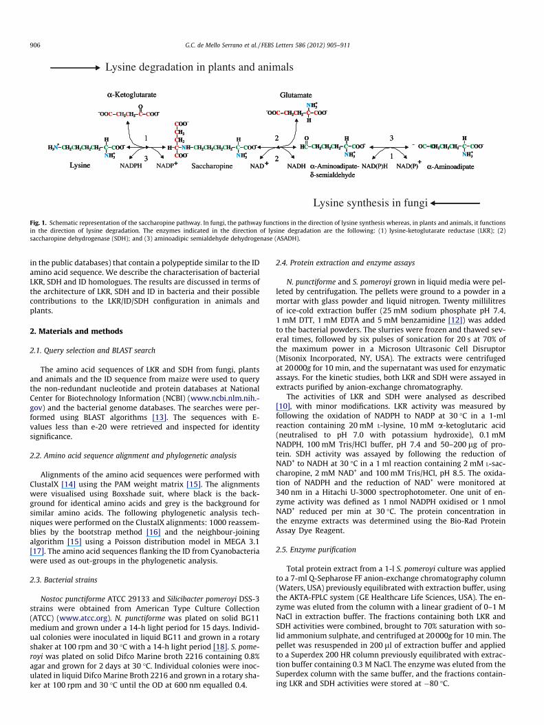
The aspartate-derived amino acid pathway is the main route forlysine synthesis in bacteria and plants [1–3]. In fungi, lysine is syn-thesised through the saccharopine pathway [4–6]. In this pathway,a-aminoadipate is converted to a-aminoadipate-d-semialdehyde,which is then condensed with glutamate to form saccharopine,which is finally hydrolysed to form lysine and a-ketoglutarate[5,6] (Fig. 1). In lysine synthesis, the last two reactions of thesaccharopine pathway are catalysed by saccharopine dehydroge-nase (SDH, EC 1.5.1.9) and lysine-a-ketoglutarate reductase (LKR,EC1.5.1.8) [5,6]. The saccharopine pathway also exists in plantsand animals, but instead of functioning in lysine synthesis, it worksin the reverse reaction, leading to lysine degradation (Fig. 1) [7,8].In the direction of degradation, the first two enzymatic steps canbe viewed as an atypical transamination reaction in which thea-amino group of lysine is transferred to a-ketoglutarate to formglutamic acid (Fig. 1).
What causes the saccharopine pathway to function in a givendirection is still unknown, but one striking characteristic of the
chemical Societies. Published by E
cular e Engenharia Genética,83-970 Campinas, SP, Brazil.
two central enzymes in the pathway is that in fungi, LKR and SDHare monofunctional polypeptides encoded by genes located atseparate loci in the genome [5], whereas in plants and animals,LKR and SDH are encoded by a single gene that, after transcriptionand translation, gives rise to a bifunctional polypeptide that per-forms both activities [9,10]. A major difference between the plantand the animal enzymes is that although in animals the LKR andSDH domains are contiguously linked, in plants they are separatedby a �110-amino-acid peptide referred to as the interdomain (ID)[11]. The role of the ID is unknown, but it is conserved in all plantsand absent in all animals whose genomic information is available inpublic databases.
We previously investigated the role of the bifunctional architec-ture of LKR/SDH and the possible function of the ID in plants. The IDcould be part of the LKR or the SDH domains, and because the IDdoes not exist in animals, it could have additional roles relatedspecifically to plant physiology and metabolism. We observed thatthe SDH domain of the bifunctional maize polypeptide, producedby limited proteolysis, inhibits LKR activity [12]. This inhibitory ef-fect may be associated with the ID linked to the partially hydrolysedSDH domain, as there is no evidence that the SDH domain of animalsinhibits LKR activity [12].
In this work, we searched genomic databases for LKR and SDHbacterial homologues; we then asked if there are other proteinsin any other organism (whose genomic information is available
lsevier B.V. All rights reserved.
C CC CO OO OO
O
H HC C2 2H HC C
2 2C C
NADPH
1
NAD(P)HNADHNADP NAD(P)NADLysine αδ
-Aminoadipate--semialdehyde
α-Aminoadipate
α-Ketoglutarate Glutamate
Saccharopine
O OO O
C C CC CO OO O COOHC 2 HCH HC C2 2H HC C2 2H HC C2 2 HC 2 HC 2 HC 2O OCH H H
HN3+
N
N
N N3
3
3 3H
H
H
H H+
+
+ +
+ ++
C
HC 2
HC 2
COO
COO
C COOHC 2 HC 2 HC 2 HC 2
HHH N
N 3H+
Lysine degradation in plants and animals
32
23 1
C CC CO OO OO
O
HC C2 2H HC C
2 2C C
NADPH
1
NAD(P)HNADHNADP NAD(P)NADLysine αδ
-Aminoadipate--semialdehyde
α-Aminoadipate
α-Ketoglutarate Glutamate
Saccharopine
O OO O
C C CC CO OO O COOHC 2 HCH HC C2 2H HC C2 2H HC C2 2 HC 2 HC 2 HC 2O OCH H H
HN3+
N
N
N N3
3
3 3H
H
H
H H+
+
+ +
+ ++
C
HC 2
HC 2
COO
COO
C COOHC 2 HC 2 HC 2 HC 2
HHH N
N 3H+
32
23 1
C COOO
O
HC2 2 HC2 2C
1
NAD(P)HNADH NAD(P)NADLysine αδ
-Aminoadipate--semialdehyde
α-Aminoadipate
α-Ketoglutarate Glutamate
OO
C C CC CO OO O COOHC 2 HCH HC 2 HC C2 2H HC2 2 HC 2 HC 2 HC 2O OCH H H
HN3+
N
N N3
3
3 3H
H
H
H H+
+
+ +
++
C
HC 2
HC 2
COO
COO
C2 2 2 2HH N
3+
32
Lysine synthesis in fungi
23 1
Fig. 1. Schematic representation of the saccharopine pathway. In fungi, the pathway functions in the direction of lysine synthesis whereas, in plants and animals, it functionsin the direction of lysine degradation. The enzymes indicated in the direction of lysine degradation are the following: (1) lysine-ketoglutarate reductase (LKR); (2)saccharopine dehydrogenase (SDH); and (3) aminoadipic semialdehyde dehydrogenase (ASADH).
906 G.C. de Mello Serrano et al. / FEBS Letters 586 (2012) 905–911
in the public databases) that contain a polypeptide similar to the IDamino acid sequence. We describe the characterisation of bacterialLKR, SDH and ID homologues. The results are discussed in terms ofthe architecture of LKR, SDH and ID in bacteria and their possiblecontributions to the LKR/ID/SDH configuration in animals andplants.
2. Materials and methods
2.1. Query selection and BLAST search
The amino acid sequences of LKR and SDH from fungi, plantsand animals and the ID sequence from maize were used to querythe non-redundant nucleotide and protein databases at NationalCenter for Biotechnology Information (NCBI) (www.ncbi.nlm.nih.-gov) and the bacterial genome databases. The searches were per-formed using BLAST algorithms [13]. The sequences with E-values less than e-20 were retrieved and inspected for identitysignificance.
2.2. Amino acid sequence alignment and phylogenetic analysis
Alignments of the amino acid sequences were performed withClustalX [14] using the PAM weight matrix [15]. The alignmentswere visualised using Boxshade suit, where black is the back-ground for identical amino acids and grey is the background forsimilar amino acids. The following phylogenetic analysis tech-niques were performed on the ClustalX alignments: 1000 reassem-blies by the bootstrap method [16] and the neighbour-joiningalgorithm [15] using a Poisson distribution model in MEGA 3.1[17]. The amino acid sequences flanking the ID from Cyanobacteriawere used as out-groups in the phylogenetic analysis.
2.3. Bacterial strains
Nostoc punctiforme ATCC 29133 and Silicibacter pomeroyi DSS-3strains were obtained from American Type Culture Collection(ATCC) (www.atcc.org). N. punctiforme was plated on solid BG11medium and grown under a 14-h light period for 15 days. Individ-ual colonies were inoculated in liquid BG11 and grown in a rotaryshaker at 100 rpm and 30 �C with a 14-h light period [18]. S. pome-royi was plated on solid Difco Marine broth 2216 containing 0.8%agar and grown for 2 days at 30 �C. Individual colonies were inoc-ulated in liquid Difco Marine Broth 2216 and grown in a rotary sha-ker at 100 rpm and 30 �C until the OD at 600 nm equalled 0.4.
2.4. Protein extraction and enzyme assays
N. punctiforme and S. pomeroyi grown in liquid media were pel-leted by centrifugation. The pellets were ground to a powder in amortar with glass powder and liquid nitrogen. Twenty millilitresof ice-cold extraction buffer (25 mM sodium phosphate pH 7.4,1 mM DTT, 1 mM EDTA and 5 mM benzamidine [12]) was addedto the bacterial powders. The slurries were frozen and thawed sev-eral times, followed by six pulses of sonication for 20 s at 70% ofthe maximum power in a Microson Ultrasonic Cell Disruptor(Misonix Incorporated, NY, USA). The extracts were centrifugedat 20000g for 10 min, and the supernatant was used for enzymaticassays. For the kinetic studies, both LKR and SDH were assayed inextracts purified by anion-exchange chromatography.
The activities of LKR and SDH were analysed as described[10], with minor modifications. LKR activity was measured byfollowing the oxidation of NADPH to NADP at 30 �C in a 1-mlreaction containing 20 mM L-lysine, 10 mM a-ketoglutaric acid(neutralised to pH 7.0 with potassium hydroxide), 0.1 mMNADPH, 100 mM Tris/HCl buffer, pH 7.4 and 50–200 lg of pro-tein. SDH activity was assayed by following the reduction ofNAD+ to NADH at 30 �C in a 1 ml reaction containing 2 mM L-sac-charopine, 2 mM NAD+ and 100 mM Tris/HCl, pH 8.5. The oxida-tion of NADPH and the reduction of NAD+ were monitored at340 nm in a Hitachi U-3000 spectrophotometer. One unit of en-zyme activity was defined as 1 nmol NADPH oxidised or 1 nmolNAD+ reduced per min at 30 �C. The protein concentration inthe enzyme extracts was determined using the Bio-Rad ProteinAssay Dye Reagent.
2.5. Enzyme purification
Total protein extract from a 1-l S. pomeroyi culture was appliedto a 7-ml Q-Sepharose FF anion-exchange chromatography column(Waters, USA) previously equilibrated with extraction buffer, usingthe AKTA-FPLC system (GE Healthcare Life Sciences, USA). The en-zyme was eluted from the column with a linear gradient of 0–1 MNaCl in extraction buffer. The fractions containing both LKR andSDH activities were combined, brought to 70% saturation with so-lid ammonium sulphate, and centrifuged at 20000g for 10 min. Thepellet was resuspended in 200 ll of extraction buffer and appliedto a Superdex 200 HR column previously equilibrated with extrac-tion buffer containing 0.3 M NaCl. The enzyme was eluted from theSuperdex column with the same buffer, and the fractions contain-ing LKR and SDH activities were stored at �80 �C.
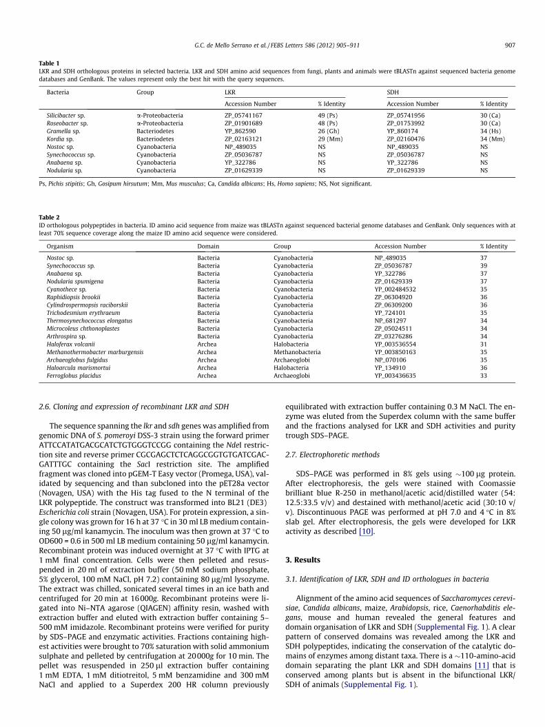
Table 1LKR and SDH orthologous proteins in selected bacteria. LKR and SDH amino acid sequences from fungi, plants and animals were tBLASTn against sequenced bacteria genomedatabases and GenBank. The values represent only the best hit with the query sequences.
Bacteria Group LKR SDH
Accession Number % Identity Accession Number % Identity
Silicibacter sp. a-Proteobacteria ZP_05741167 49 (Ps) ZP_05741956 30 (Ca)Roseobacter sp. a-Proteobacteria ZP_01901689 48 (Ps) ZP_01753992 30 (Ca)Gramella sp. Bacteriodetes YP_862590 26 (Gh) YP_860174 34 (Hs)Kordia sp. Bacteriodetes ZP_02163121 29 (Mm) ZP_02160476 34 (Mm)Nostoc sp. Cyanobacteria NP_489035 NS NP_489035 NSSynechococcus sp. Cyanobacteria ZP_05036787 NS ZP_05036787 NSAnabaena sp. Cyanobacteria YP_322786 NS YP_322786 NSNodularia sp. Cyanobacteria ZP_01629339 NS ZP_01629339 NS
Ps, Pichis stipitis; Gh, Gosipum hirsutum; Mm, Mus musculus; Ca, Candida albicans; Hs, Homo sapiens; NS, Not significant.
Table 2ID orthologous polypeptides in bacteria. ID amino acid sequence from maize was tBLASTn against sequenced bacterial genome databases and GenBank. Only sequences with atleast 70% sequence coverage along the maize ID amino acid sequence were considered.
Organism Domain Group Accession Number % Identity
Nostoc sp. Bacteria Cyanobacteria NP_489035 37Synechococcus sp. Bacteria Cyanobacteria ZP_05036787 39Anabaena sp. Bacteria Cyanobacteria YP_322786 37Nodularia spumigena Bacteria Cyanobacteria ZP_01629339 37Cyanothece sp. Bacteria Cyanobacteria YP_002484532 35Raphidiopsis brookii Bacteria Cyanobacteria ZP_06304920 36Cylindrospermopsis raciborskii Bacteria Cyanobacteria ZP_06309200 36Trichodesmium erythraeum Bacteria Cyanobacteria YP_724101 35Thermosynechococcus elongatus Bacteria Cyanobacteria NP_681297 34Microcoleus chthonoplastes Bacteria Cyanobacteria ZP_05024511 34Arthrospira sp. Bacteria Cyanobacteria ZP_03276286 34Haloferax volcanii Archea Halobacteria YP_003536554 31Methanothermobacter marburgensis Archea Methanobacteria YP_003850163 35Archaeoglobus fulgidus Archea Archaeoglobi NP_070106 35Haloarcula marismortui Archea Halobacteria YP_134910 36Ferroglobus placidus Archea Archaeoglobi YP_003436635 33
G.C. de Mello Serrano et al. / FEBS Letters 586 (2012) 905–911 907
2.6. Cloning and expression of recombinant LKR and SDH
The sequence spanning the lkr and sdh genes was amplified fromgenomic DNA of S. pomeroyi DSS-3 strain using the forward primerATTCCATATGACGCATCTGTGGGTCCGG containing the NdeI restric-tion site and reverse primer CGCGAGCTCTCAGGCGGTGTGATCGAC-GATTTGC containing the SacI restriction site. The amplifiedfragment was cloned into pGEM-T Easy vector (Promega, USA), val-idated by sequencing and than subcloned into the pET28a vector(Novagen, USA) with the His tag fused to the N terminal of theLKR polypeptide. The construct was transformed into BL21 (DE3)Escherichia coli strain (Novagen, USA). For protein expression, a sin-gle colony was grown for 16 h at 37 �C in 30 ml LB medium contain-ing 50 lg/ml kanamycin. The inoculum was then grown at 37 �C toOD600 = 0.6 in 500 ml LB medium containing 50 lg/ml kanamycin.Recombinant protein was induced overnight at 37 �C with IPTG at1 mM final concentration. Cells were then pelleted and resus-pended in 20 ml of extraction buffer (50 mM sodium phosphate,5% glycerol, 100 mM NaCl, pH 7.2) containing 80 lg/ml lysozyme.The extract was chilled, sonicated several times in an ice bath andcentrifuged for 20 min at 16000g. Recombinant proteins were li-gated into Ni–NTA agarose (QIAGEN) affinity resin, washed withextraction buffer and eluted with extraction buffer containing 5–500 mM imidazole. Recombinant proteins were verified for purityby SDS–PAGE and enzymatic activities. Fractions containing high-est activities were brought to 70% saturation with solid ammoniumsulphate and pelleted by centrifugation at 20000g for 10 min. Thepellet was resuspended in 250 ll extraction buffer containing1 mM EDTA, 1 mM ditiotreitol, 5 mM benzamidine and 300 mMNaCl and applied to a Superdex 200 HR column previously
equilibrated with extraction buffer containing 0.3 M NaCl. The en-zyme was eluted from the Superdex column with the same bufferand the fractions analysed for LKR and SDH activities and puritytrough SDS–PAGE.
2.7. Electrophoretic methods
SDS–PAGE was performed in 8% gels using �100 lg protein.After electrophoresis, the gels were stained with Coomassiebrilliant blue R-250 in methanol/acetic acid/distilled water (54:12.5:33.5 v/v) and destained with methanol/acetic acid (30:10 v/v). Discontinuous PAGE was performed at pH 7.0 and 4 �C in 8%slab gel. After electrophoresis, the gels were developed for LKRactivity as described [10].
3. Results
3.1. Identification of LKR, SDH and ID orthologues in bacteria
Alignment of the amino acid sequences of Saccharomyces cerevi-siae, Candida albicans, maize, Arabidopsis, rice, Caenorhabditis ele-gans, mouse and human revealed the general features anddomain organisation of LKR and SDH (Supplemental Fig. 1). A clearpattern of conserved domains was revealed among the LKR andSDH polypeptides, indicating the conservation of the catalytic do-mains of enzymes among distant taxa. There is a �110-amino-aciddomain separating the plant LKR and SDH domains [11] that isconserved among plants but is absent in the bifunctional LKR/SDH of animals (Supplemental Fig. 1).
A.thaliana
G.hirsutum
Z.mays
C.elegans
M.musculus
H.sapiens
Gramella sp
Kordia sp
Silicibacter sp
Roseobacter sp
C.albicans
S.cereviseae
P.stipitis
Synechococcus sp
Nodularia sp
Nostoc sp
Anabaena sp9798
100
100
100
50100
99
100
96 99
98
10065
0.2
C.albicans
P.stipilis
S.cereviseae
Silicibacter sp
Roseobacter sp
C.elegans
M.musculus
H.sapiens
Z.mays
A.thaliana
G.hirsutum
Gramella sp
Kordia sp
Synechococcus sp
Nodularia sp
Nostoc sp
Anabaena sp6596
100
100
100
100
100100
69100
91
78
76
77
0.2
A
B
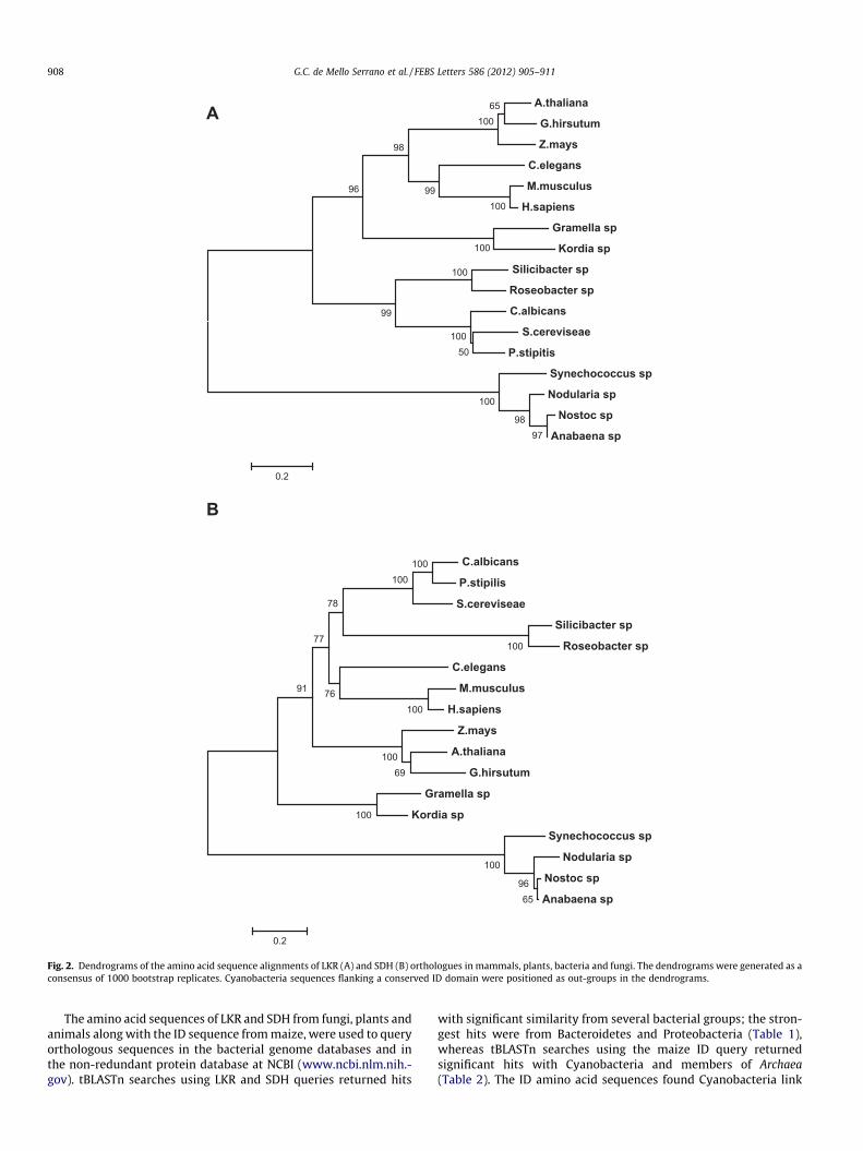
Fig. 2. Dendrograms of the amino acid sequence alignments of LKR (A) and SDH (B) orthologues in mammals, plants, bacteria and fungi. The dendrograms were generated as aconsensus of 1000 bootstrap replicates. Cyanobacteria sequences flanking a conserved ID domain were positioned as out-groups in the dendrograms.
908 G.C. de Mello Serrano et al. / FEBS Letters 586 (2012) 905–911
The amino acid sequences of LKR and SDH from fungi, plants andanimals along with the ID sequence from maize, were used to queryorthologous sequences in the bacterial genome databases and inthe non-redundant protein database at NCBI (www.ncbi.nlm.nih.-gov). tBLASTn searches using LKR and SDH queries returned hits
with significant similarity from several bacterial groups; the stron-gest hits were from Bacteroidetes and Proteobacteria (Table 1),whereas tBLASTn searches using the maize ID query returnedsignificant hits with Cyanobacteria and members of Archaea(Table 2). The ID amino acid sequences found Cyanobacteria link
G.C. de Mello Serrano et al. / FEBS Letters 586 (2012) 905–911 909
polypeptide sequences without significant similarity to LKR or SDH(Table 1). The alignment of the LKR and SDH amino acid sequencesof Silicibacter sp., Roseobacter sp., Gramella sp. and Kordia sp. withthose from plants, animals and fungi confirmed the polypeptideidentity with conserved domains within the amino acid sequences(Supplemental Figs. 2 and 3). The alignment of the amino acid se-quences of the polypeptides flanking the ID of Cyanobacteria waspoor and did not permit us to annotate them as LKR and SDH(Supplemental Figs. 2 and 3). Phylogenetic analysis of the LKR andSDH amino acid sequences from Silicibacter sp., Roseobacter sp.,Gramella sp., Kordia sp., plants, fungi and animals grouped bacteriaand fungi sequences in a cluster, plant and animal sequences in an-other cluster, whereas the sequences of the polypeptides flankingthe ID in Cyanobacteria clustered as out-groups in the dendrograms(Fig. 2). The alignment of the ID amino acid sequences shows thatthis polypeptide is highly conserved among distant groups of bacte-ria and Archea (Supplemental Fig. 4).
3.2. LKR and SDH enzymatic activities in S. pomeroyi
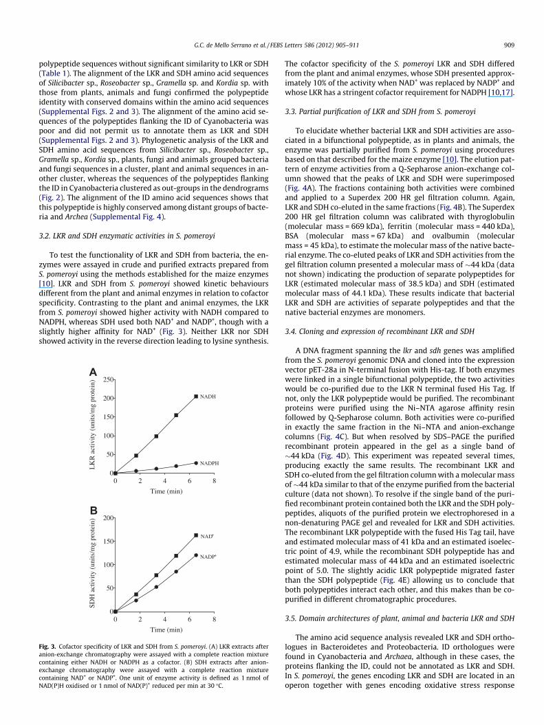
To test the functionality of LKR and SDH from bacteria, the en-zymes were assayed in crude and purified extracts prepared fromS. pomeroyi using the methods established for the maize enzymes[10]. LKR and SDH from S. pomeroyi showed kinetic behavioursdifferent from the plant and animal enzymes in relation to cofactorspecificity. Contrasting to the plant and animal enzymes, the LKRfrom S. pomeroyi showed higher activity with NADH compared toNADPH, whereas SDH used both NAD+ and NADP+, though with aslightly higher affinity for NAD+ (Fig. 3). Neither LKR nor SDHshowed activity in the reverse direction leading to lysine synthesis.
0 2 4 6 80
50
100
150
200
250
NADH
NADPH
Time (min)
LK
R a
ctiv
ity (
units
/mg
prot
ein)
0 2 4 6 80
50
100
150
200
NAD+
NADP+
Time (min)
SDH
act
ivity
(un
its/m
g pr
otei
n)
A
B
Fig. 3. Cofactor specificity of LKR and SDH from S. pomeroyi. (A) LKR extracts afteranion-exchange chromatography were assayed with a complete reaction mixturecontaining either NADH or NADPH as a cofactor. (B) SDH extracts after anion-exchange chromatography were assayed with a complete reaction mixturecontaining NAD+ or NADP+. One unit of enzyme activity is defined as 1 nmol ofNAD(P)H oxidised or 1 nmol of NAD(P)+ reduced per min at 30 �C.
The cofactor specificity of the S. pomeroyi LKR and SDH differedfrom the plant and animal enzymes, whose SDH presented approx-imately 10% of the activity when NAD+ was replaced by NADP+ andwhose LKR has a stringent cofactor requirement for NADPH [10,17].
3.3. Partial purification of LKR and SDH from S. pomeroyi
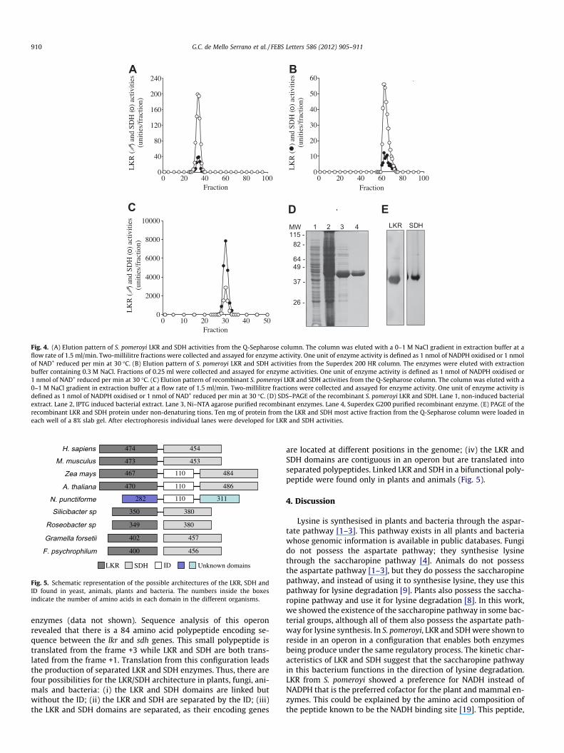
To elucidate whether bacterial LKR and SDH activities are asso-ciated in a bifunctional polypeptide, as in plants and animals, theenzyme was partially purified from S. pomeroyi using proceduresbased on that described for the maize enzyme [10]. The elution pat-tern of enzyme activities from a Q-Sepharose anion-exchange col-umn showed that the peaks of LKR and SDH were superimposed(Fig. 4A). The fractions containing both activities were combinedand applied to a Superdex 200 HR gel filtration column. Again,LKR and SDH co-eluted in the same fractions (Fig. 4B). The Superdex200 HR gel filtration column was calibrated with thyroglobulin(molecular mass = 669 kDa), ferritin (molecular mass = 440 kDa),BSA (molecular mass = 67 kDa) and ovalbumin (molecularmass = 45 kDa), to estimate the molecular mass of the native bacte-rial enzyme. The co-eluted peaks of LKR and SDH activities from thegel filtration column presented a molecular mass of �44 kDa (datanot shown) indicating the production of separate polypeptides forLKR (estimated molecular mass of 38.5 kDa) and SDH (estimatedmolecular mass of 44.1 kDa). These results indicate that bacterialLKR and SDH are activities of separate polypeptides and that thenative bacterial enzymes are monomers.
3.4. Cloning and expression of recombinant LKR and SDH
A DNA fragment spanning the lkr and sdh genes was amplifiedfrom the S. pomeroyi genomic DNA and cloned into the expressionvector pET-28a in N-terminal fusion with His-tag. If both enzymeswere linked in a single bifunctional polypeptide, the two activitieswould be co-purified due to the LKR N terminal fused His Tag. Ifnot, only the LKR polypeptide would be purified. The recombinantproteins were purified using the Ni–NTA agarose affinity resinfollowed by Q-Sepharose column. Both activities were co-purifiedin exactly the same fraction in the Ni–NTA and anion-exchangecolumns (Fig. 4C). But when resolved by SDS–PAGE the purifiedrecombinant protein appeared in the gel as a single band of�44 kDa (Fig. 4D). This experiment was repeated several times,producing exactly the same results. The recombinant LKR andSDH co-eluted from the gel filtration column with a molecular massof�44 kDa similar to that of the enzyme purified from the bacterialculture (data not shown). To resolve if the single band of the puri-fied recombinant protein contained both the LKR and the SDH poly-peptides, aliquots of the purified protein we electrophoresed in anon-denaturing PAGE gel and revealed for LKR and SDH activities.The recombinant LKR polypeptide with the fused His Tag tail, haveand estimated molecular mass of 41 kDa and an estimated isoelec-tric point of 4.9, while the recombinant SDH polypeptide has andestimated molecular mass of 44 kDa and an estimated isoelectricpoint of 5.0. The slightly acidic LKR polypeptide migrated fasterthan the SDH polypeptide (Fig. 4E) allowing us to conclude thatboth polypeptides interact each other, and this makes than be co-purified in different chromatographic procedures.
3.5. Domain architectures of plant, animal and bacteria LKR and SDH
The amino acid sequence analysis revealed LKR and SDH ortho-logues in Bacteroidetes and Proteobacteria. ID orthologues werefound in Cyanobacteria and Archaea, although in these cases, theproteins flanking the ID, could not be annotated as LKR and SDH.In S. pomeroyi, the genes encoding LKR and SDH are located in anoperon together with genes encoding oxidative stress response
0 20 40 60 80 1000
10
20
30
40
50
60
Fraction
LK
R (
) an
d SD
H (o
) ac
tiviti
es(u
nitie
s/fr
actio
n)
BA
DC
0 20 40 60 80 1000
40
80
120
160
200
240
Fraction
LK
R (
) an
d SD
H (o
) ac
tiviti
es(u
nitie
s/fr
actio
n)
0 10 20 30 40 500
2000
4000
6000
8000
10000
Fraction
LK
R (
) an
d SD
H (o
) ac
tiviti
es(u
nitie
s/fr
actio
n)
115 -82 -
64 -49 -
37 -
26 -
MW LKR SDH
E1 2 3 4
Fig. 4. (A) Elution pattern of S. pomeroyi LKR and SDH activities from the Q-Sepharose column. The column was eluted with a 0–1 M NaCl gradient in extraction buffer at aflow rate of 1.5 ml/min. Two-millilitre fractions were collected and assayed for enzyme activity. One unit of enzyme activity is defined as 1 nmol of NADPH oxidised or 1 nmolof NAD+ reduced per min at 30 �C. (B) Elution pattern of S. pomeroyi LKR and SDH activities from the Superdex 200 HR column. The enzymes were eluted with extractionbuffer containing 0.3 M NaCl. Fractions of 0.25 ml were collected and assayed for enzyme activities. One unit of enzyme activity is defined as 1 nmol of NADPH oxidised or1 nmol of NAD+ reduced per min at 30 �C. (C) Elution pattern of recombinant S. pomeroyi LKR and SDH activities from the Q-Sepharose column. The column was eluted with a0–1 M NaCl gradient in extraction buffer at a flow rate of 1.5 ml/min. Two-millilitre fractions were collected and assayed for enzyme activity. One unit of enzyme activity isdefined as 1 nmol of NADPH oxidised or 1 nmol of NAD+ reduced per min at 30 �C. (D) SDS–PAGE of the recombinant S. pomeroyi LKR and SDH. Lane 1, non-induced bacterialextract. Lane 2, IPTG induced bacterial extract. Lane 3, Ni–NTA agarose purified recombinant enzymes. Lane 4, Superdex G200 purified recombinant enzyme. (E) PAGE of therecombinant LKR and SDH protein under non-denaturing tions. Ten mg of protein from the LKR and SDH most active fraction from the Q-Sepharose column were loaded ineach well of a 8% slab gel. After electrophoresis individual lanes were developed for LKR and SDH activities.
467 484110Zea mays
474 454H. sapiens
473 453M. musculus
A. thaliana282 110N. punctiforme 311
470 486110
350 380Silicibacter sp
349 380Roseobacter sp
402 457Gramella forsetii
400 456F. psychrophilum
LKR SDH ID Unknown domains
Fig. 5. Schematic representation of the possible architectures of the LKR, SDH andID found in yeast, animals, plants and bacteria. The numbers inside the boxesindicate the number of amino acids in each domain in the different organisms.
910 G.C. de Mello Serrano et al. / FEBS Letters 586 (2012) 905–911
enzymes (data not shown). Sequence analysis of this operonrevealed that there is a 84 amino acid polypeptide encoding se-quence between the lkr and sdh genes. This small polypeptide istranslated from the frame +3 while LKR and SDH are both trans-lated from the frame +1. Translation from this configuration leadsthe production of separated LKR and SDH enzymes. Thus, there arefour possibilities for the LKR/SDH architecture in plants, fungi, ani-mals and bacteria: (i) the LKR and SDH domains are linked butwithout the ID; (ii) the LKR and SDH are separated by the ID; (iii)the LKR and SDH domains are separated, as their encoding genes
are located at different positions in the genome; (iv) the LKR andSDH domains are contiguous in an operon but are translated intoseparated polypeptides. Linked LKR and SDH in a bifunctional poly-peptide were found only in plants and animals (Fig. 5).
4. Discussion
Lysine is synthesised in plants and bacteria through the aspar-tate pathway [1–3]. This pathway exists in all plants and bacteriawhose genomic information is available in public databases. Fungido not possess the aspartate pathway; they synthesise lysinethrough the saccharopine pathway [4]. Animals do not possessthe aspartate pathway [1–3], but they do possess the saccharopinepathway, and instead of using it to synthesise lysine, they use thispathway for lysine degradation [9]. Plants also possess the saccha-ropine pathway and use it for lysine degradation [8]. In this work,we showed the existence of the saccharopine pathway in some bac-terial groups, although all of them also possess the aspartate path-way for lysine synthesis. In S. pomeroyi, LKR and SDH were shown toreside in an operon in a configuration that enables both enzymesbeing produce under the same regulatory process. The kinetic char-acteristics of LKR and SDH suggest that the saccharopine pathwayin this bacterium functions in the direction of lysine degradation.LKR from S. pomeroyi showed a preference for NADH instead ofNADPH that is the preferred cofactor for the plant and mammal en-zymes. This could be explained by the amino acid composition ofthe peptide known to be the NADH binding site [19]. This peptide,
G.C. de Mello Serrano et al. / FEBS Letters 586 (2012) 905–911 911
located in C. albicans between residues 210 and 225 is identical inPichia stipitis (Supplemental Fig. 2). The peptide is almost fully con-served in S. pomeroyi and Roseobacter sp. that differs from that of C.albicans by a change of leucine to isoleucine at position 1, and a cys-teine to valine at position 9. This peptide in plant and animal isremarkably different conserving only a valine at position 2, a gly-cine at position 7 and glycine and valine at positions 12 and 13(Supplemental Fig. 2).
A major difference among the primary structure of LKR/SDH fromthe diverse organisms we analysed is the presence of an ID polypep-tide in plants, Cyanobacteria, Eubacteria and Archaea (Table 2). Theoccurrence of ID orthologues with highly conserved amino acid se-quences in bacteria of different genera suggests that this peptidecould be involved in an important regulatory mechanism. Most sur-prising was the observation that, in Cyanobacteria, the ID ortho-logues apparently do not link LKR and SDH. It is reasonable topostulate that if the ID is conserved in bacteria and plants, thenthe plants inherited the ID from a common ancestor that they sharewith Cyanobacteria. However, the question of how the ID from bothbacteria and plants evolved to link different proteins is intriguing. Inthe case of the Cyanobacteria, it is possible that the polypeptidesflanking the ID could be reminiscent of orthologues of LKR andSDH that diverged in their amino acid sequences and lost both enzy-matic activities. The loss of these enzymes during evolution could bepossible, as we found an orthologue of LKR in Cyanothece sp. butcould not find an orthologue of SDH (data not shown).
Some CFB (Bacteroidetes/Chlorobi group) bacteria also possessLKR and SDH orthologues, but the polypeptides are encoded bygenes located at different positions in the bacterial genome. Thus,it is possible that an operon configuration such as the one found inS. pomeroyi could be the ancestor of the animal and plant LKR/SDH. Yet, this leaves unresolved questions about the origin of theplant ID. One possible scenario is that a member of the a-proteobac-teria group with a gene/protein configuration similar to that foundin S. pomeroyi underwent the endosymbiosis process that gave riseto mitochondria [20,21]. The lkr and sdh genes present in this bacte-rium-originated mitochondria were later transferred to the nucleargenome, as was most of the primitive mitochondrial genome [21].Similarly, the chloroplast, which originated from an endosymbioticprocess involving Cyanobacteria, incorporated the ID into the chlo-roplast genome, and this was later transferred to the plant nucleargenome [22]. During plant evolution, the ancestral CyanobacteriaID-encoding sequence was inserted between the LKR and SDH do-mains of the nuclear-incorporated mitochondrial gene.
The presence of LKR and SDH along with genes encoding en-zymes involved in oxidative stress response suggests that theselysine-degrading enzymes may interact, in a novel mechanism ofoxidative stress response.
Acknowledgements
G.C. Mello Serrano and N. Zanata received fellowships fromFapesp, TR Silva Figueira received a fellowship from the Coorde-nação de Aperfeiçoamento de professores do Ensino Superior
(CAPES). PA is a recipient of a CNPq productivity fellowship. Thiswork was supported by FAPESP grant 10/50114-4.
Appendix A. Supplementary data
Supplementary data associated with this article can be found, inthe online version, at doi:10.1016/j.febslet.2012.02.023.
References
[1] Azevedo, R.A., Arruda, P., Turner, W.L. and Lea, P.J. (1997) The biosynthesis andmetabolism of the aspartate derived amino acids in higher plants.Phytochemistry 46, 395–419.
[2] Azevedo, R.A. (2002) Analysis of the aspartic acid metabolic pathway usingmutant genes. Amino Acids 22, 217–230.
[3] Azevedo, R.A., Lancien, M. and Lea, P.J. (2006) The aspartic acid metabolicpathway, an exciting and essential pathway in plants. Amino Acids 30, 143–162.
[4] Trupin, J.S. and Broquist, H.P. (1963) Lysine biosynthesis in Neurospora crassa.Fed. Proc. 22, 243–250.
[5] Bhattacharjee, J.K. (1985) Alpha-aminoadipate pathway for the biosynthesis oflysine in lower eukaryotes. Crit. Rev. Microbiol. 12, 131–151.
[6] Broquist, H.P. and Trupin, J.S. (1966) Amino acid metabolism. Ann. Rev.Biochem. 35, 231–274.
[7] Arruda, P., Sodek, L. and da Silva, W.J. (1982) Lysine-ketoglutarate reductaseactivity in developing maize endosperm. Plant Physiol. 69, 988–989.
[8] Arruda, P., Kemper, E.L., Papes, F. and Leite, A. (2000) Regulation of lysinecatabolism in higher plants. Trends Plant Sci. 5, 324–330.
[9] Markovitz, P.J. and Chuang, D.T. (1987) The bifunctional aminoadipicsemialdehyde synthase in lysine degradation. J. Biol. Chem. 262, 9353–9358.
[10] Gonçalves-Butruille, M., Szajner, P., Torigoi, E., Leite, A. and Arruda, P. (1996)Purification and characterization of the bifunctional enzyme lysine-ketoglutarate reductase-saccharopine dehydrogenase from maize. PlantPhysiol. 11, 765–771.
[11] Kemper, E.L., Cord Neto, G., Papes, F., Moraes, K.C.M., Leite, A. and Arruda, P.(1999) The role of Opaque2 in the control of lysine-degrading activities indeveloping maize endosperm. Plant Cell 11, 1981–1993.
[12] Kemper, E.L., Cord-Neto, G., Capella, A.N., Goncalves-Butruile, M., Azevedo,R.A. and Arruda, P. (1998) Structure and regulation of the bifunctional enzymelysine-oxoglutarate reductase-saccharopine dehydrogenase in maize. Eur. J.Biochem. 253, 720–729.
[13] Altschul, S.F., Madden, T.L., Schaffer, A.A., Zhang, J.H., Zhang, Z., Miller, W. andLipman, D.J. (1997) Gapped BLAST and PSI-BLAST: a new generation of proteindatabase search programs. Nucleic Acids Res. 25, 3389–3402.
[14] Thompson, J.D., Gibson, T.J., Plewniak, F., Jeanmougin, F. and Higgins, D.G.(1997) The CLUSTALX windows interface. Flexible strategies for multiplesequence alignment aided by quality analysis tools. Nucleic Acids Res. 25,4876–4882.
[15] Saitou, N. and Nei, M. (1987) The neighbor-joining method: a new method forreconstructing phylogenetic trees. Mol. Biol. Evol. 4 (406), 425.
[16] Felsenstein, J. (1985) Confidence limits on phylogenies: an approach using thebootstrap. Evolution 39, 783–791.
[17] Kumar, S., Tamura, K. and Nei, M. (2004) MEGA3: Integrated software formolecular evolutionary genetic analysis and sequence alignment. Brief. Bioinf.5, 150–163.
[18] Porchia, A.C., Curatti, L. and Salerno, G.L. (1999) Sucrose metabolism incyanobacteria: sucrose synthase from Anabaena sp. Strain PCC 7119 isremarkably different from the plant enzymes with respect to substrateaffinity and amino-terminal sequence. Planta 210, 34–40.
[19] Garrad, R., Schmidt, T.M. and Bhattacharjee, J.K. (1994) Molecular andfunctional analysis of the LYS1 gene of Candida albicans. Infec. Immun. 62,5027–5031.
[20] Emelyanov, V.V. (2001) Rickettsiaceae, rickettsia-like endosymbionts, and theorigin of mitochondria. Biosci. Rep. 21, 1–17.
[21] Searcy, D.G. (2003) Metabolic integration during the evolutionary origin ofmitochondria. Cell Res. 13, 229–238.
[22] McFadden, G.I. (2001) Chloroplast origin and integration. Plant Physiol. 125,50–53.
Copyright © 2022 FDOKUMEN


![Regulation of Lysine Catabolism through Lysine[mdash]Ketoglutarate Reductase and Saccharopine Dehydrogenase in Arabidopsis](https://static.fdokumen.com/doc/165x107/631cc83693f371de19019c93/regulation-of-lysine-catabolism-through-lysinemdashketoglutarate-reductase-and.jpg)